Embed Size (px)
Citation preview
West Indian Med J 2014; 63 (6): 667
Large Cell Neuroendocrine Carcinoma of the Ovary and Its Skin MetastasesA Case Report and Review of the Literature
S Cokmert1, L Demir2, L Doganay1, N Demir1, K Kocacelebi3, IT Unek4, E Gezer1, K Kilic1, M Alakavuklar1
ABSTRACT
Large cell neuroendocrine carcinoma in the gynaecological organs affects the uterine cervix and ovary.Large cell neuroendocrine carcinoma of the ovary is extremely rare, and prognosis is quite poor evenwhen diagnosed at an early stage. These tumours respond poorly to standard chemotherapy regimens.The clinical observation of skin metastasis in patients with epithelial ovarian cancer is relativelyuncommon, occurring in only 3.5% of patients. These lesions are observed mostly in skin of theabdominal wall adjacent to the primary ovarian tumours. Metastatic skin lesions on extremities aremuch more rare; it is reported that only 12% of epithelial ovarian carcinoma skin metastases occur onthe limbs. Skin metastasis due to large cell neuroendocrine carcinoma of the ovary has not beenpreviously reported. We report the case of a large cell neuroendocrine tumour of the ovary with skinmetastases on extremities appearing two months after surgery in a 68-year old woman.
Keywords: Large cell, neuroendocrine carcinoma, ovary, skin metastasis
El Carcinoma Neuroendocrino de Células Grandes de Ovario y Sus Metástasis Cutáneas: Reporte de un Caso y Revisión de la Literatura
S Cokmert1, L Demir2, L Doganay1, N Demir1, K Kocacelebi3, IT Unek4, E Gezer1, K Kilic1, M Alakavuklar1
RESUMEN
El carcinoma neuroendocrino de células grandes en los órganos ginecológicos afecta el cuello del úteroy el ovario. El carcinoma neuroendocrino de células grandes del ovario es extremadamente raro, y elpronóstico es muy pobre, incluso cuando se diagnostica en una etapa temprana. Estos tumoresresponden pobremente a los regímenes de quimioterapia estándar. La observación clínica demetástasis de la piel en pacientes con cáncer epitelial de ovario es relativamente poco frecuente,ocurriendo sólo en el 3.5% de los pacientes. Estas lesiones se observan principalmente en la piel de lapared abdominal adyacente a los tumores ováricos primarios. Las lesiones cutáneas metastásicas enlas extremidades son mucho más raras. Se reporta que sólo el 12% de las metástasis cutáneas decarcinoma ovárico epitelial ocurre en las extremidades. La metástasis cutánea a causa del carcinomaneuroendocrino ovárico de células grandes no ha sido reportada con anterioridad. Reportamos el casode una mujeres de 68 años con tumor neuroendocrino ovárico de células grandes, que presentómetástasis cutáneas en las extremidades dos meses después de la cirugía.
Palabras claves: Células grandes, carcinoma neuroendocrino, ovario, metástasis cutánea
West Indian Med J 2014; 63 (6): 667
From: 1Department of Medical Oncology, Kent Hospital, Izmir, Turkey,2Department of Medical Oncology, Aydin State Hospital, Aydin, Turkey,3Department of Nuclear Medicine, EgeRad Imaging Center, Izmir, Turkeyand 4Department of Medical Oncology, Izmir Tepecik Training and ResearchHospital, Izmir, Turkey.
Correspondence: Dr S Cokmert, Department of Medical Oncology, KentHospital, 8229/1, No. 56, Cigli 35630, Izmir, Turkey; and Department ofBasic Oncology, Institute of Oncology, Dokuz Eylul University, 35340Inciralti, Izmir, Turkey. E-mail: [email protected]
DOI: 10.7727/wimj.2014.010
668
INTRODUCTIONLarge cell neuroendocrine carcinoma was first described inthe lung (1). Ovarian large cell neuroendocrine carcinoma(LCNEC) is an exceedingly rare tumour (2). To date,approximately 48 cases have been reported (3−25), and theprognosis is very poor (2−25). Generally, in epithelialovarian cancers, the most common sites of distant metastasisare the pleura, liver, lung and lymph nodes (26). The clinical
observation of skin metastasis in ovarian cancer cases isuncommon, occurring in only 3.5% of patients (26, 27). Theincidence of skin metastasis from large cell neuroendocrineovarian tumours is unknown, because none of the 45 reportedcases of LCNEC of the ovary had skin metastases (Table).We report a rare case of LCNEC with skin metastasis for thefirst time.
Table: Pathological and clinical features of large cell neuroendocrine carcinoma cases in the literature
Reference Age Epithelial stromal component Recurrence Outcomes(years) sites
(3) 40 Mucinous carcinoma None NED (8 months)
(4) 46 Pure large cell neuroendocrine N/A Died in 4 monthscarcinoma of ovary
(5) 44 Mucinous intraepithelial carcinoma None NED (6 months)(6) 68 Serous carcinoma Abdomen DOD (7 months)(7) 39 Mucinous adenocarcinoma Unclear AWD (8 months)(7) 55 Mucinous LMP with intraepithelial carcinoma None NED (68 months)(7) 42 Benign cyst and teratoma in contralateral ovary Unclear DOD (20 months)(7) 53 Endometrioid adenocarcinoma None NED (37 months)(7) 47 Adenocarcinoma, NOS and teratoma None NED (11 months)(7) 25 Mature cystic teratoma Unclear DOD (36 months)(7) 55 Mucinous LMP Unclear DOD (2 months)(7) 54 Mucinous carcinoma, endometrioid None NED (66 months)
adenocarcinoma(7) 63 Endometrioid adenocarcinoma Unclear DOD (9 months)(7) 59 High-grade adenocarcinoma, NOS None NED (28 months)(7) 22 Endometrioid adenocarcinoma N/A N/A(8) 73 Microinvasive mucinous adenocarcinoma Retroperitoneal space, DOD (8 months)
bone, liver(8) 44 Mucinous intraepithelial carcinoma Retroperitoneal space DOD (4 months)(9) 76 Pure large cell neuroendocrine carcinoma of ovary N/A Died(10) 71 Serous carcinoma None NED (8 months)(11) 65 Mucinous cystadenoma Liver, abdomen DOD (10 months)(12) 27 Pure large cell neuroendocrine carcinoma of ovary None NED (10 months)(13) 77 Endometrioid adenocarcinoma Abdomen, bone, lung DOD (19 months)(13) 36 Mucinous adenoma Recent(13) 45 Mucinous cystadenoma and partial intraepithelial Unclear DOD (36 months)
carcinoma(13) 68 Mucinous adenocarcinoma Unclear LTF(13) 58 Mucinous cystadenoma and partial intraepithelial Unclear DOD (8 months)
carcinoma(14) 34 Mucinous cystadenoma and mucinous Liver, brain, PAN, DOD (8 months)
adenocarcinoma pelvis(15) 22 Mucinous cystadenoma and mucinous Liver DOD (3 months)
adenocarcinoma(16) 33 Endometrioid adenocarcinoma PAN DOD (4 months)(17) 56 Mucinous adenocarcinoma and teratoma Unclear DOD (10 months)(17) 35 Mucinous adenoma None NED (10 years)(18) 31 Mucinous adenoma Unclear Unclear(19) 64 Pure large cell neuroendocrine carcinoma of ovary None NED (9 months)(20) 73 Pure large cell neuroendocrine carcinoma of ovary Brain NED (12 months)
Ovarian Large Cell Neuroendocrine Carcinoma
669
CASE REPORTA 68-year old, nulligravida, postmenopausal woman pre-sented with progressive abdominal distension and pain overa period of six months. Physical examination revealed a firmirregular mass in the lower abdomen. Computed tomographyconfirmed a 20 cm abdominal tumour. Extensive pelvic andparaaortic lymphadenopathies were also identified. A solidleft ovarian mass with tumour deposits on the capsule wasobserved by the surgeon during the operation. A total abdo-minal hysterectomy with bilateral salpingo-oophorectomy,omentectomy, and retroperitoneal lymph node biopsy wereperformed. The tumour involved the myometrium, endo-metrium, serosa of the bladder, sigmoid colon and omentum.
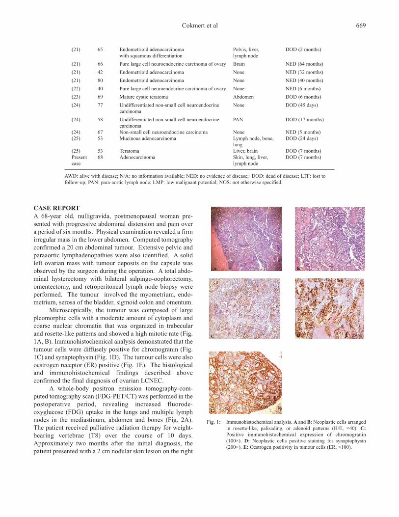
Microscopically, the tumour was composed of largepleomorphic cells with a moderate amount of cytoplasm andcoarse nuclear chromatin that was organized in trabecularand rosette-like patterns and showed a high mitotic rate (Fig.1A, B). Immunohistochemical analysis demonstrated that thetumour cells were diffusely positive for chromogranin (Fig.1C) and synaptophysin (Fig. 1D). The tumour cells were alsooestrogen receptor (ER) positive (Fig. 1E). The histologicaland immunohistochemical findings described aboveconfirmed the final diagnosis of ovarian LCNEC.
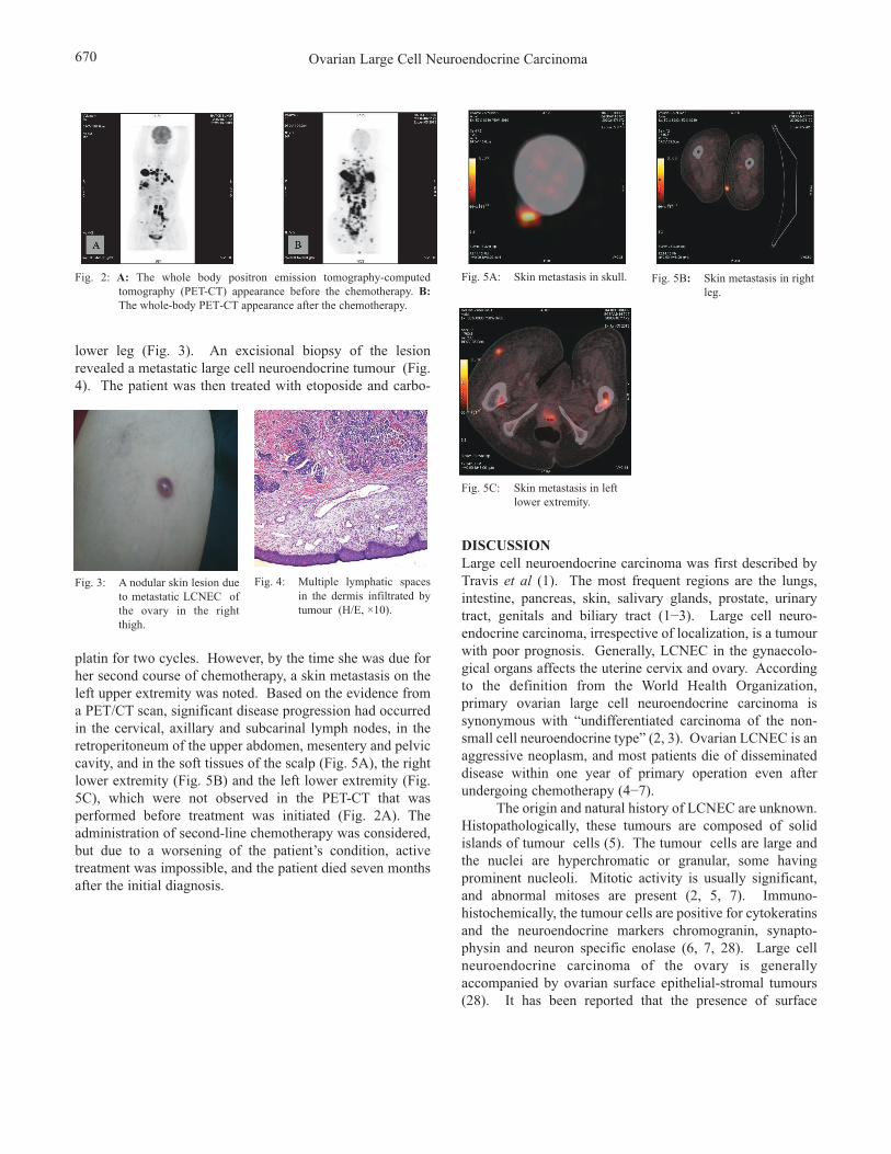
A whole-body positron emission tomography-com-puted tomography scan (FDG-PET/CT) was performed in thepostoperative period, revealing increased fluorode-oxyglucose (FDG) uptake in the lungs and multiple lymphnodes in the mediastinum, abdomen and bones (Fig. 2A).The patient received palliative radiation therapy for weight-bearing vertebrae (T8) over the course of 10 days.Approximately two months after the initial diagnosis, thepatient presented with a 2 cm nodular skin lesion on the right
(21) 65 Endometrioid adenocarcinoma Pelvis, liver, DOD (2 months)with squamous differentiation lymph node
(21) 66 Pure large cell neuroendocrine carcinoma of ovary Brain NED (64 months)(21) 42 Endometrioid adenocarcinoma None NED (32 months)(21) 80 Endometrioid adenocarcinoma None NED (40 months)(22) 40 Pure large cell neuroendocrine carcinoma of ovary None NED (6 months)(23) 69 Mature cystic teratoma Abdomen DOD (6 months)(24) 77 Undifferentiated non-small cell neuroendocrine None DOD (45 days)
carcinoma(24) 58 Undifferentiated non-small cell neuroendocrine PAN DOD (17 months)
carcinoma(24) 67 Non-small cell neuroendocrine carcinoma None NED (5 months)(25) 53 Mucinous adenocarcinoma Lymph node, bone, DOD (24 days)
lung(25) 53 Teratoma Liver, brain DOD (7 months)Present 68 Adenocarcinoma Skin, lung, liver, DOD (7 months)case lymph node
AWD: alive with disease; N/A: no information available; NED: no evidence of disease; DOD: dead of disease; LTF: lost tofollow-up; PAN: para-aortic lymph node; LMP: low malignant potential; NOS: not otherwise specified.
Cokmert et al
Fig. 1: Immunohistochemical analysis. A and B: Neoplastic cells arrangedin rosette-like, palisading, or adenoid patterns (H/E, ×40). C:Positive immunohistochemical expression of chromogranin(100×). D: Neoplastic cells positive staining for synaptophysin(200×). E: Oestrogen positivity in tumour cells (ER, ×100).
670
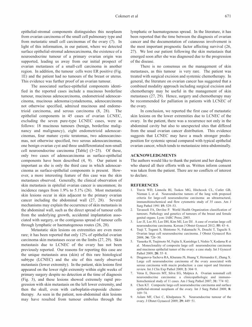
lower leg (Fig. 3). An excisional biopsy of the lesionrevealed a metastatic large cell neuroendocrine tumour (Fig.4). The patient was then treated with etoposide and carbo-
Fig. 2: A: The whole body positron emission tomography-computedtomography (PET-CT) appearance before the chemotherapy. B:The whole-body PET-CT appearance after the chemotherapy.
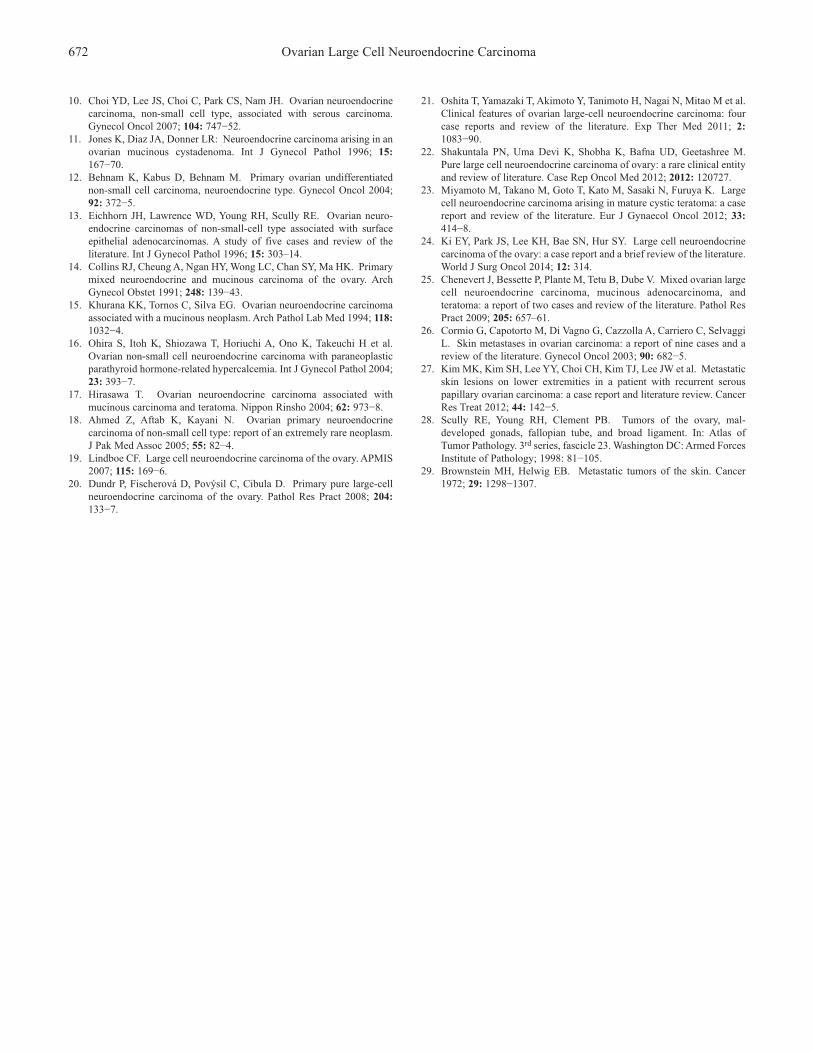
platin for two cycles. However, by the time she was due forher second course of chemotherapy, a skin metastasis on theleft upper extremity was noted. Based on the evidence froma PET/CT scan, significant disease progression had occurredin the cervical, axillary and subcarinal lymph nodes, in theretroperitoneum of the upper abdomen, mesentery and pelviccavity, and in the soft tissues of the scalp (Fig. 5A), the rightlower extremity (Fig. 5B) and the left lower extremity (Fig.5C), which were not observed in the PET-CT that wasperformed before treatment was initiated (Fig. 2A). Theadministration of second-line chemotherapy was considered,but due to a worsening of the patient’s condition, activetreatment was impossible, and the patient died seven monthsafter the initial diagnosis.
Fig. 3: A nodular skin lesion dueto metastatic LCNEC ofthe ovary in the rightthigh.
Fig. 4: Multiple lymphatic spacesin the dermis infiltrated by tumour (H/E, ×10).
Ovarian Large Cell Neuroendocrine Carcinoma
Fig. 5A: Skin metastasis in skull. Fig. 5B: Skin metastasis in rightleg.
Fig. 5C: Skin metastasis in leftlower extremity.
DISCUSSIONLarge cell neuroendocrine carcinoma was first described byTravis et al (1). The most frequent regions are the lungs,intestine, pancreas, skin, salivary glands, prostate, urinarytract, genitals and biliary tract (1−3). Large cell neuro-endocrine carcinoma, irrespective of localization, is a tumourwith poor prognosis. Generally, LCNEC in the gynaecolo-gical organs affects the uterine cervix and ovary. Accordingto the definition from the World Health Organization,primary ovarian large cell neuroendocrine carcinoma issynonymous with “undifferentiated carcinoma of the non-small cell neuroendocrine type” (2, 3). Ovarian LCNEC is anaggressive neoplasm, and most patients die of disseminateddisease within one year of primary operation even afterundergoing chemotherapy (4−7).
The origin and natural history of LCNEC are unknown.Histopathologically, these tumours are composed of solidislands of tumour cells (5). The tumour cells are large andthe nuclei are hyperchromatic or granular, some havingprominent nucleoli. Mitotic activity is usually significant,and abnormal mitoses are present (2, 5, 7). Immuno-histochemically, the tumour cells are positive for cytokeratinsand the neuroendocrine markers chromogranin, synapto-physin and neuron specific enolase (6, 7, 28). Large cellneuroendocrine carcinoma of the ovary is generallyaccompanied by ovarian surface epithelial-stromal tumours(28). It has been reported that the presence of surface
671
epithelial-stromal components distinguishes this neoplasmfrom ovarian carcinoma of the small cell pulmonary type andfrom metastatic small cell carcinomas of the ovary (7). Inlight of this information, in our patient, where we detectedsurface epithelial-stromal adenocarcinoma, the existence of aneuroendocrine tumour of a primary ovarian origin wassupported, leading us away from our initial prospect ofovarian metastases of a small-cell carcinoma in anotherregion. In addition, the tumour cells were ER positive (Fig.1E) and the patient had no tumours of the breast or uterus.This evidence was further proof of an ovarian tumour.
The associated surface-epithelial components identi-fied in the reported cases include a mucinous borderlinetumour, mucinous adenocarcinoma, endometrioid adenocar-cinoma, mucinous adenoma/cystadenoma, adenocarcinomanot otherwise specified, admixed mucinous and endome-trioid carcinoma, and serous carcinoma (8, 28). Theepithelial components in 45 cases of ovarian LCNEC,excluding the seven pure-type LCNEC cases, were asfollows: 18 mucinous tumours (benign, borderline malig-nancy and malignancy), eight endometrioid adenocar-cinomas, four mature cystic teratomas, two adenocarcino-mas, not otherwise specified, two serous adenocarcinomas,one benign ovarian cyst and three undifferentiated non-smallcell neuroendocrine carcinoma [Table] (3−25). Of these,only two cases of adenocarcinoma as surface-epithelialcomponents have been described (4, 9). Our patient isimportant as she is only the third case in which adenocar-cinoma as surface-epithelial components is present. How-ever, a more interesting feature of this case was the skinmetastases observed. Generally, the clinical observation ofskin metastasis in epitelial ovarian cancer is uncommon; itsincidence ranges from 1.9% to 5.1% (26). Most metastaticskin lesions occur in skin adjacent to the primary ovariancancer including the abdominal wall (27, 28). Severalmechanisms may explain the occurrence of skin metastasis inthe abdominal wall, such as the direct spread of tumour cellsfrom the underlying growth, accidental implantation asso-ciated with surgery, or the contiguous spread of tumour cellsthrough lymphatic or haematogenous routes (26, 28).
Metastatic skin lesions on extremities are even morerare; it has been reported that only 12% of epithelial ovariancarcinoma skin metastases occur on the limbs (27, 29). Skinmetastasis due to LCNEC of the ovary has not beenpreviously reported. Our reasons for reporting this case arethe unique metastasis area (skin) of this rare histologicalsubtype (LCNEC) and the site of this rarely observedmetastasis (lower extremity). In the patient, skin lesions firstappeared on the lower right extremity within eight weeks ofprimary surgery despite no detection at the time of diagnosis(Fig. 3), and these lesions showed extremely rapid pro-gression with skin metastasis on the left lower extremity, andthen the skull, even with carboplatin-etoposide chemo-therapy. As seen in the patient, non-abdominal skin lesionsmay have resulted from tumour embolus through the
lymphatic or haematogenous spread. In the literature, it hasbeen reported that the time between the diagnosis of ovariancancer and the documentation of cutaneous involvement isthe most important prognostic factor affecting survival (26,27). We lost our patient following the skin metastasis thatemerged soon after she was diagnosed due to the progressionof the disease.
There is no consensus on the management of skinmetastases, as this tumour is very rare. The patient wastreated with surgical excision and systemic chemotherapy. Ingeneral, the literature on ovarian cancer has suggested that acombined modality approach including surgical excision andchemotherapy may be useful in the management of skinmetastases (27, 29). Hence, surgery and chemotherapy maybe recommended for palliation in patients with LCNEC ofthe ovary.
In conclusion, we reported the first case of metastaticskin lesions on the lower extremities due to LCNEC of theovary. In the patient, there was a recurrence not only in theabdominal cavity but also to unexpected sites that differedfrom the usual ovarian cancer distribution. This evidencesuggests that LCNEC may have a much stronger predis-position for systemic spread compared with typical epithelialovarian cancer, which tends to metastasize intra-abdominally.
ACKNOWLEDGMENTSThe authors would like to thank the patient and her daughterswho shared all their efforts with us. Written inform consentwas taken from the patient. There are no conflicts of interestto declare.
REFERENCES1. Travis WD, Linnoila RI, Tsokos MG, Hitchcock CL, Cutler GB,
Nieman L et al. Neuroendocrine tumors of the lung with proposedcriteria for large-cell neuroendocrine carcinoma: an ultrastructural,immunohistochemical and flow cytometric study of 35 cases. Am JSurg Pathol 1991 15: 529–53.
2. Tavassoli FA, Devilee P. World Health Organization classification oftumours. Pathology and genetics of tumours of the breast and femalegenital organs. Lyon: IARC Press; 2003.
3. Lee JJ, Lee JO, Lee DH, Kim MJ, Lee DJ. A case of ovarian large cellneuroendocrine carcinoma. Korean J Obstet Gynecol 2012; 55: 43−6.
4. Tsuji T, Togami S, Shintomo N, Fukamachi N, Douchi T, Taguchi S.Ovarian large cell neuroendocrine carcinoma. J Obstet Gynaecol Res2008; 34: 726−30.
5. Yasuoka H, Tsujimoto M, Fujita S, Kunishige I, Nishio Y, Kodama R etal. Monoclonality of composite large cell neuroendocrine carcinomaand mucinous epithelial tumor of the ovary: a case study. Int J GynecolPathol 2009; 28: 55−8.
6. Draganova-Tacheva RA, Khurana JS, Huang Y, Hernandez E, Zhang X.Large cell neuroendocrine carcinoma of the ovary associated withserous carcinoma with mucin production: a case report and literaturereview. Int J Clin Exp Pathol 2009; 2: 304−9.
7. Veras E, Deavers MT, Silva EG, Malpica A. Ovarian nonsmall cellneuroendocrine carcinoma: a clinicopathologic and immuno-histochemical study of 11 cases. Am J Surg Pathol 2007; 31: 774−82.
8. Chen KT. Composite large-cell neuroendocrine carcinoma and surfaceepithelial-stromal neoplasm of the ovary. Int J Surg Pathol 2008; 8:169−74.
9. Aslam MF, Choi C, Khulpateea N. Neuroendocrine tumour of theovary. J Obstet Gynaecol 2009; 29: 449−51.
Cokmert et al
672
10. Choi YD, Lee JS, Choi C, Park CS, Nam JH. Ovarian neuroendocrinecarcinoma, non-small cell type, associated with serous carcinoma.Gynecol Oncol 2007; 104: 747−52.
11. Jones K, Diaz JA, Donner LR: Neuroendocrine carcinoma arising in anovarian mucinous cystadenoma. Int J Gynecol Pathol 1996; 15:167−70.
12. Behnam K, Kabus D, Behnam M. Primary ovarian undifferentiatednon-small cell carcinoma, neuroendocrine type. Gynecol Oncol 2004;92: 372−5.
13. Eichhorn JH, Lawrence WD, Young RH, Scully RE. Ovarian neuro-endocrine carcinomas of non-small-cell type associated with surfaceepithelial adenocarcinomas. A study of five cases and review of theliterature. Int J Gynecol Pathol 1996; 15: 303–14.
14. Collins RJ, Cheung A, Ngan HY, Wong LC, Chan SY, Ma HK. Primarymixed neuroendocrine and mucinous carcinoma of the ovary. ArchGynecol Obstet 1991; 248: 139−43.
15. Khurana KK, Tornos C, Silva EG. Ovarian neuroendocrine carcinomaassociated with a mucinous neoplasm. Arch Pathol Lab Med 1994; 118:1032−4.
16. Ohira S, Itoh K, Shiozawa T, Horiuchi A, Ono K, Takeuchi H et al.Ovarian non-small cell neuroendocrine carcinoma with paraneoplasticparathyroid hormone-related hypercalcemia. Int J Gynecol Pathol 2004;23: 393−7.
17. Hirasawa T. Ovarian neuroendocrine carcinoma associated withmucinous carcinoma and teratoma. Nippon Rinsho 2004; 62: 973−8.
18. Ahmed Z, Aftab K, Kayani N. Ovarian primary neuroendocrinecarcinoma of non-small cell type: report of an extremely rare neoplasm.J Pak Med Assoc 2005; 55: 82−4.
19. Lindboe CF. Large cell neuroendocrine carcinoma of the ovary. APMIS2007; 115: 169−6.
20. Dundr P, Fischerová D, Povýsil C, Cibula D. Primary pure large-cellneuroendocrine carcinoma of the ovary. Pathol Res Pract 2008; 204:133−7.
21. Oshita T, Yamazaki T, Akimoto Y, Tanimoto H, Nagai N, Mitao M et al.Clinical features of ovarian large-cell neuroendocrine carcinoma: fourcase reports and review of the literature. Exp Ther Med 2011; 2:1083−90.
22. Shakuntala PN, Uma Devi K, Shobha K, Bafna UD, Geetashree M.Pure large cell neuroendocrine carcinoma of ovary: a rare clinical entityand review of literature. Case Rep Oncol Med 2012; 2012: 120727.
23. Miyamoto M, Takano M, Goto T, Kato M, Sasaki N, Furuya K. Largecell neuroendocrine carcinoma arising in mature cystic teratoma: a casereport and review of the literature. Eur J Gynaecol Oncol 2012; 33:414−8.
24. Ki EY, Park JS, Lee KH, Bae SN, Hur SY. Large cell neuroendocrinecarcinoma of the ovary: a case report and a brief review of the literature.World J Surg Oncol 2014; 12: 314.
25. Chenevert J, Bessette P, Plante M, Tetu B, Dube V. Mixed ovarian largecell neuroendocrine carcinoma, mucinous adenocarcinoma, andteratoma: a report of two cases and review of the literature. Pathol ResPract 2009; 205: 657–61.
26. Cormio G, Capotorto M, Di Vagno G, Cazzolla A, Carriero C, SelvaggiL. Skin metastases in ovarian carcinoma: a report of nine cases and areview of the literature. Gynecol Oncol 2003; 90: 682−5.
27. Kim MK, Kim SH, Lee YY, Choi CH, Kim TJ, Lee JW et al. Metastaticskin lesions on lower extremities in a patient with recurrent serouspapillary ovarian carcinoma: a case report and literature review. CancerRes Treat 2012; 44: 142−5.
28. Scully RE, Young RH, Clement PB. Tumors of the ovary, mal-developed gonads, fallopian tube, and broad ligament. In: Atlas ofTumor Pathology. 3rd series, fascicle 23. Washington DC: Armed ForcesInstitute of Pathology; 1998: 81−105.
29. Brownstein MH, Helwig EB. Metastatic tumors of the skin. Cancer1972; 29: 1298−1307.
Ovarian Large Cell Neuroendocrine Carcinoma