Embed Size (px)
Citation preview
Exercise 11
The Appendicular Skeleton
The Appendicular Skeleton
• The appendicular skeleton contains 126 bones.
• Consists of the upper and lower limbs, the pectoral girdles, and the pelvic girdles.
• Consists of the upper and lower limbs, the pectoral girdles, and the pelvic girdles.
• The pectoral girdles attach the upper limbs to the axial skeleton.
• The pelvic girdles attach the lower limbs to the axial skeleton.
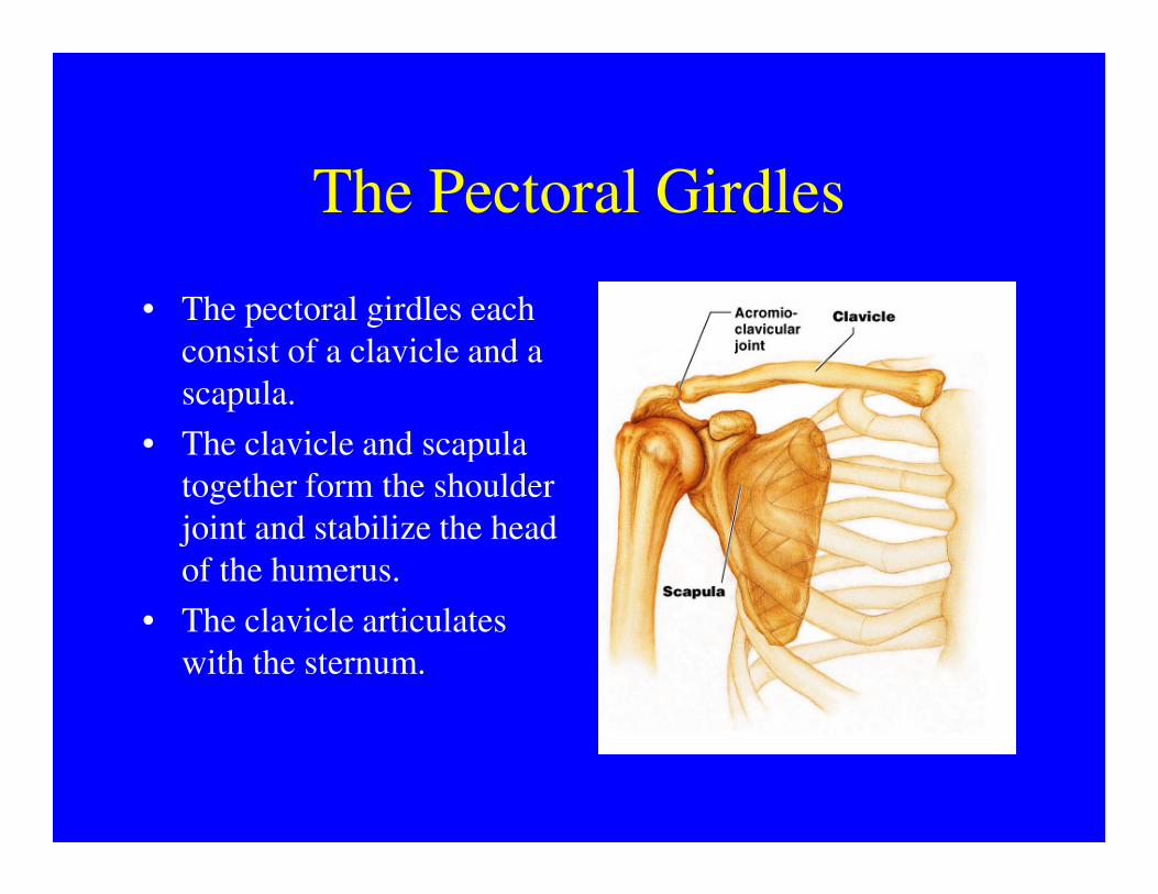
The Pectoral Girdles
• The pectoral girdles each
consist of a clavicle and a
scapula.
• The clavicle and scapula • The clavicle and scapula
together form the shoulder
joint and stabilize the head
of the humerus.
• The clavicle articulates
with the sternum.
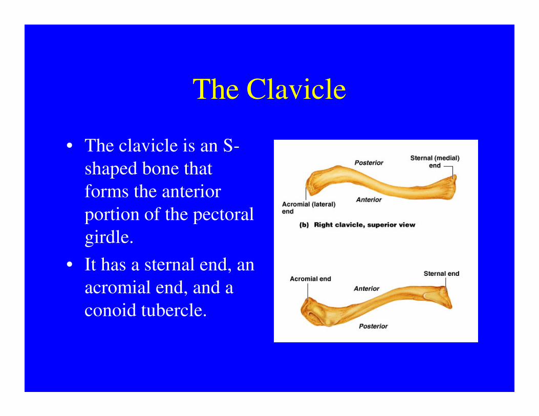
The Clavicle
• The clavicle is an S-
shaped bone that
forms the anterior
portion of the pectoral portion of the pectoral
girdle.
• It has a sternal end, an
acromial end, and a
conoid tubercle.
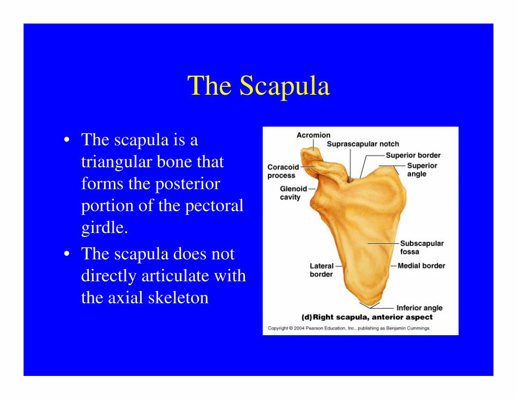
The Scapula
• The scapula is a
triangular bone that
forms the posterior
portion of the pectoral portion of the pectoral
girdle.
• The scapula does not
directly articulate with
the axial skeleton
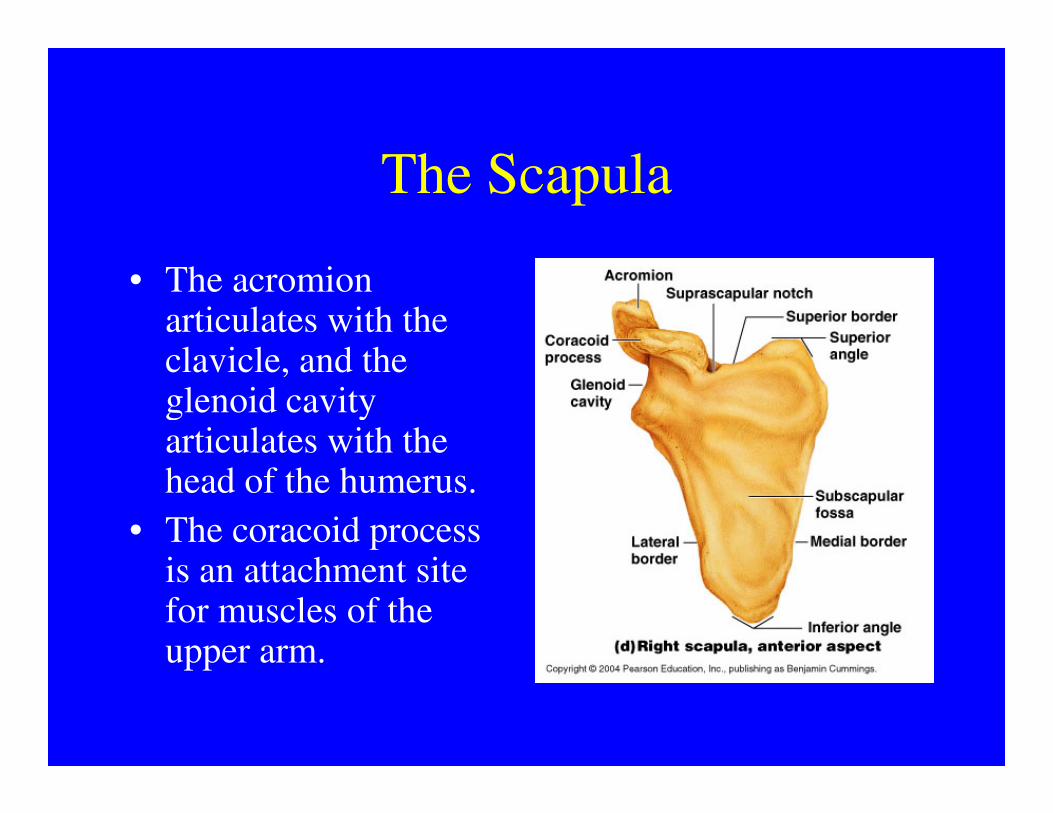
The Scapula
• The acromion articulates with the clavicle, and the glenoid cavity glenoid cavity articulates with the head of the humerus.
• The coracoid process is an attachment site for muscles of the upper arm.
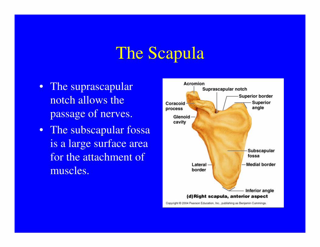
The Scapula
• The suprascapular
notch allows the
passage of nerves.
• The subscapular fossa • The subscapular fossa
is a large surface area
for the attachment of
muscles.
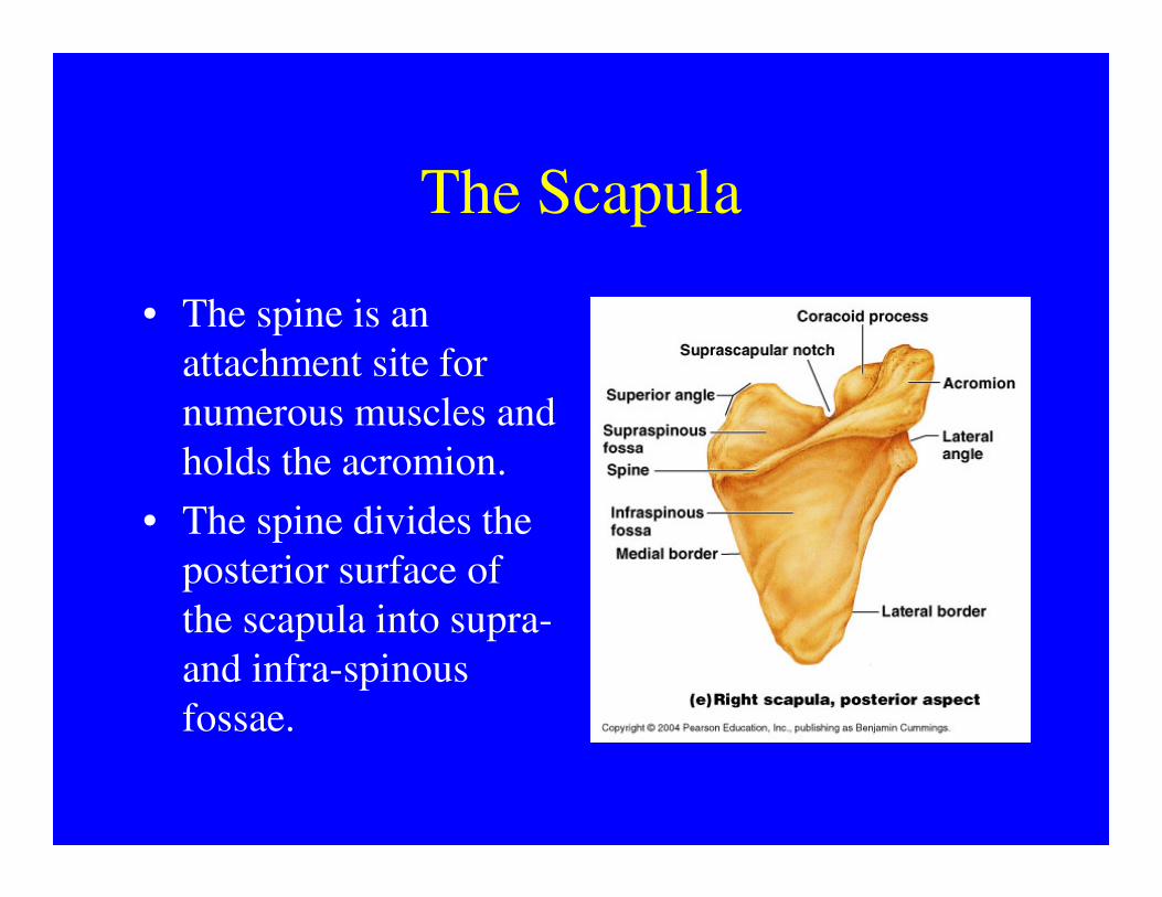
The Scapula
• The spine is an
attachment site for
numerous muscles and
holds the acromion.holds the acromion.
• The spine divides the
posterior surface of
the scapula into supra-
and infra-spinous
fossae.
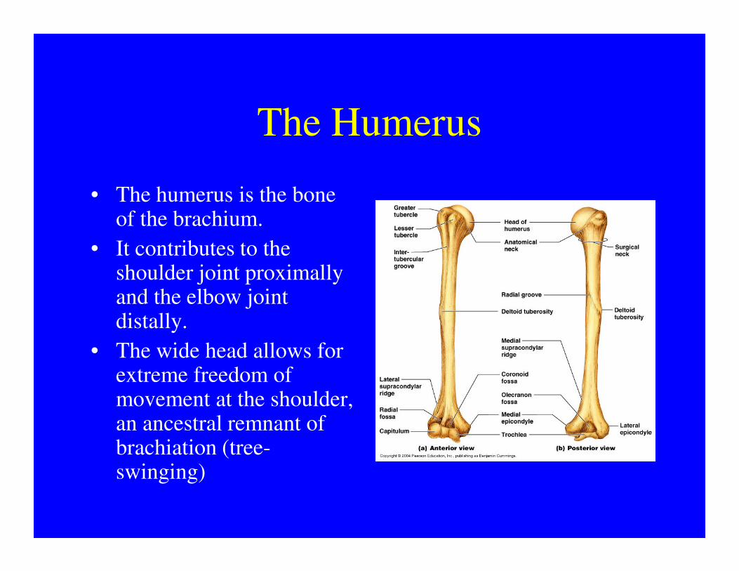
The Humerus
• The humerus is the bone of the brachium.
• It contributes to the shoulder joint proximally and the elbow joint and the elbow joint distally.
• The wide head allows for extreme freedom of movement at the shoulder, an ancestral remnant of brachiation (tree-swinging)
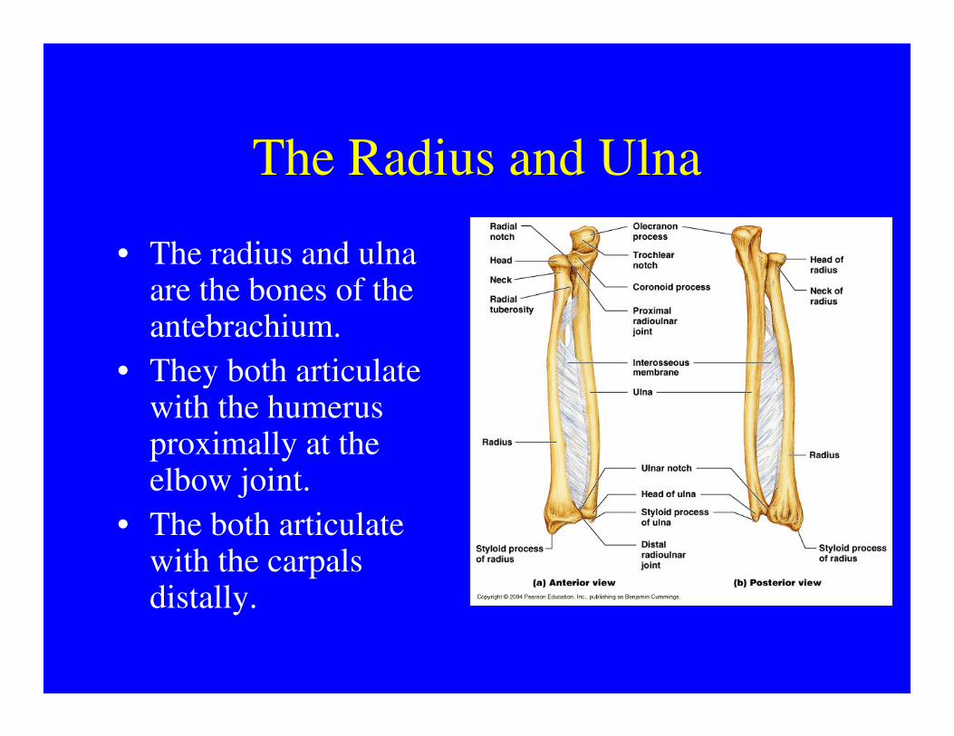
The Radius and Ulna
• The radius and ulna are the bones of the antebrachium.
• They both articulate • They both articulate with the humerus proximally at the elbow joint.
• The both articulate with the carpals distally.
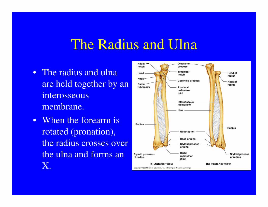
The Radius and Ulna
• The radius and ulna
are held together by an
interosseous
membrane.membrane.
• When the forearm is
rotated (pronation),
the radius crosses over
the ulna and forms an
X.
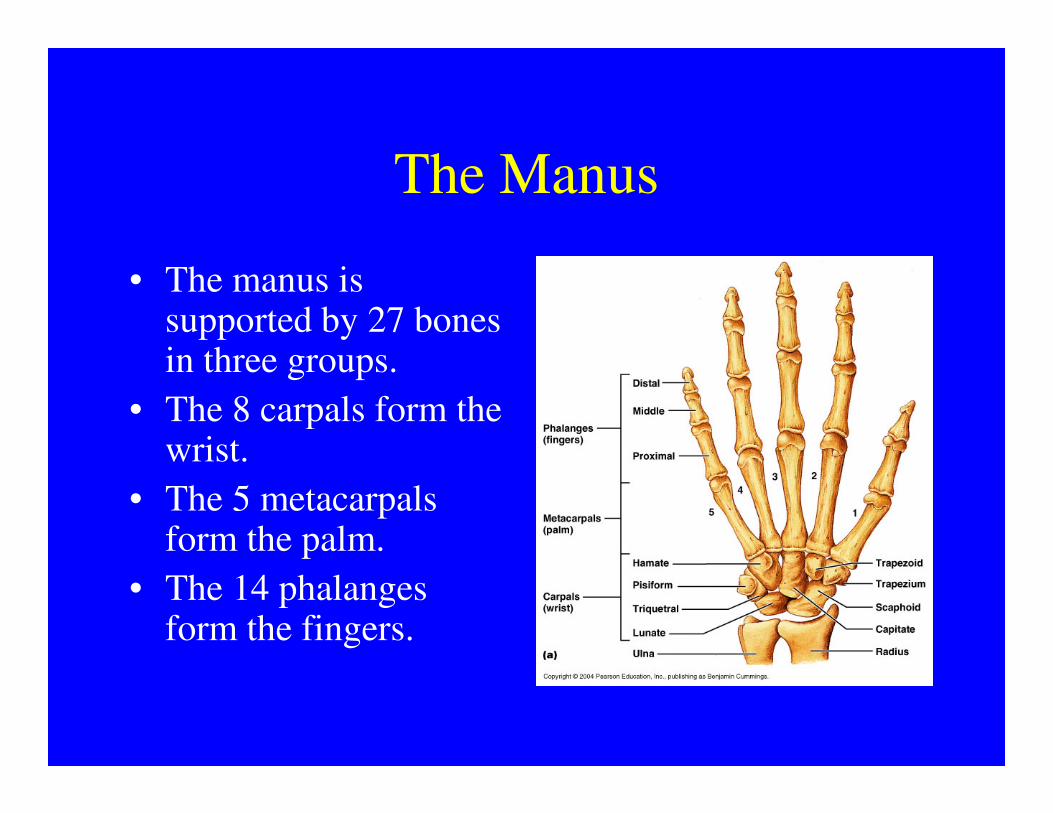
The Manus
• The manus is supported by 27 bones in three groups.
• The 8 carpals form the • The 8 carpals form the wrist.
• The 5 metacarpals form the palm.
• The 14 phalanges form the fingers.
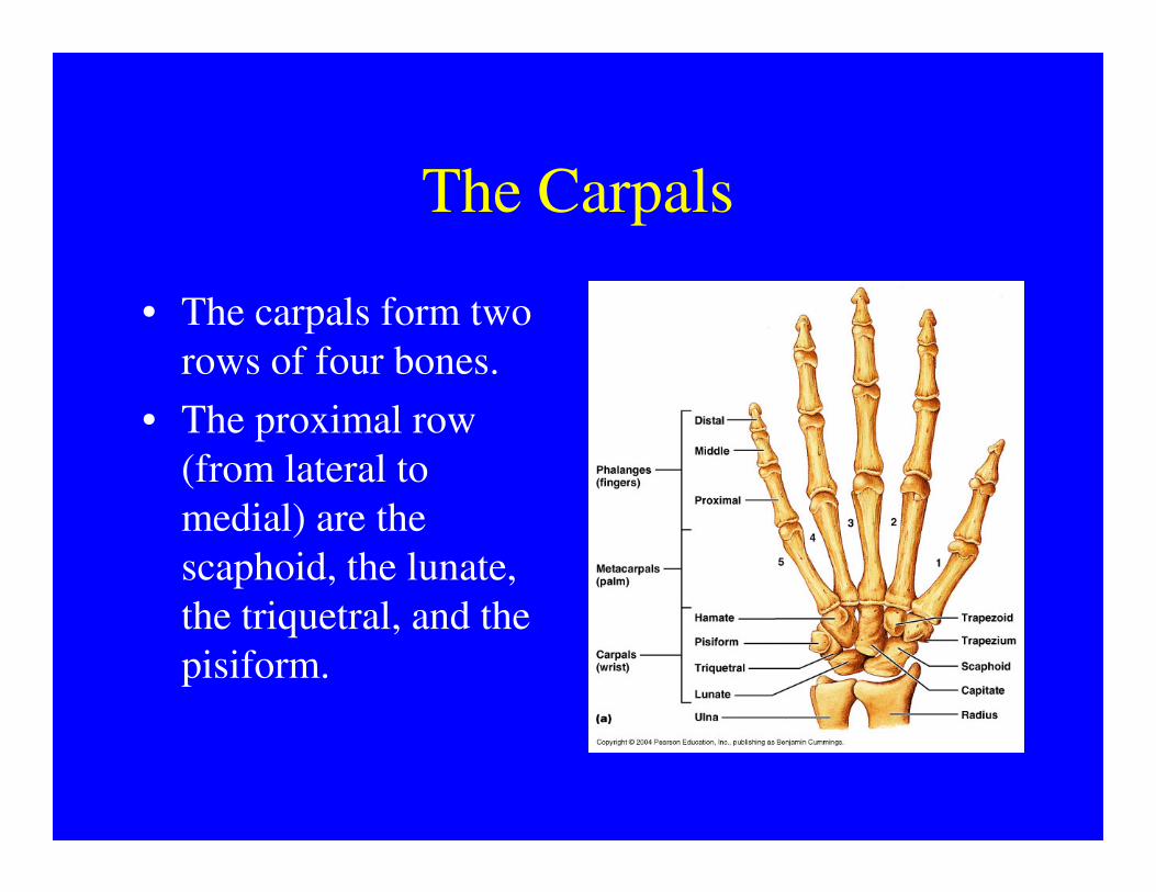
The Carpals
• The carpals form two
rows of four bones.
• The proximal row
(from lateral to (from lateral to
medial) are the
scaphoid, the lunate,
the triquetral, and the
pisiform.
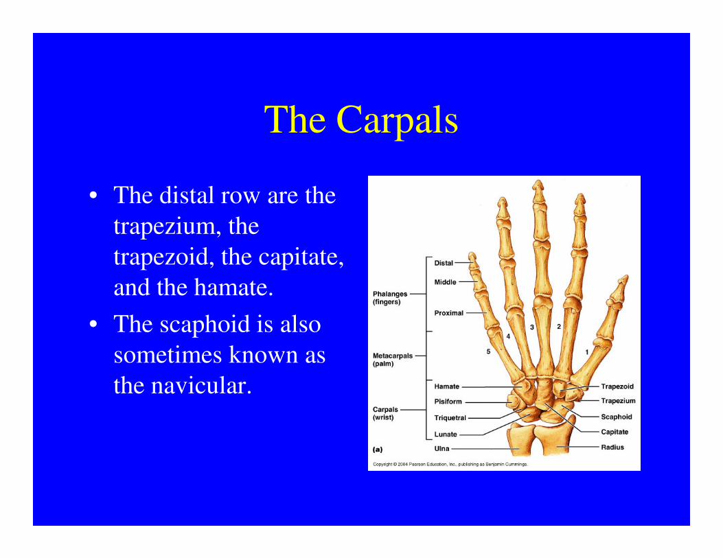
The Carpals
• The distal row are the
trapezium, the
trapezoid, the capitate,
and the hamate.and the hamate.
• The scaphoid is also
sometimes known as
the navicular.

The Metacarpals
• The five metacarpals are numbered from one to five, starting with the lateral with the lateral (thumb) side.
• The metacarpals articulate with the distal row of carpals and with the proximal phalanges.

The Phalanges
• The fingers are numbered the same way as the metacarpals, with the metacarpals, with the thumb being number one.
• Each finger has three phalanges, except for the thumb, which has two.

The Phalanges
• Each finger has a proximal, a middle, and a distal phalanx, except for the thumb, except for the thumb, which has only a proximal and distal phalanx.
• All of the phalanges and metacarpals are long bones.
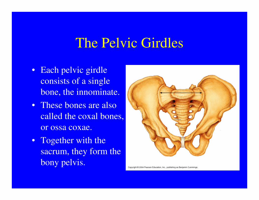
The Pelvic Girdles
• Each pelvic girdle
consists of a single
bone, the innominate.
• These bones are also • These bones are also
called the coxal bones,
or ossa coxae.
• Together with the
sacrum, they form the
bony pelvis.
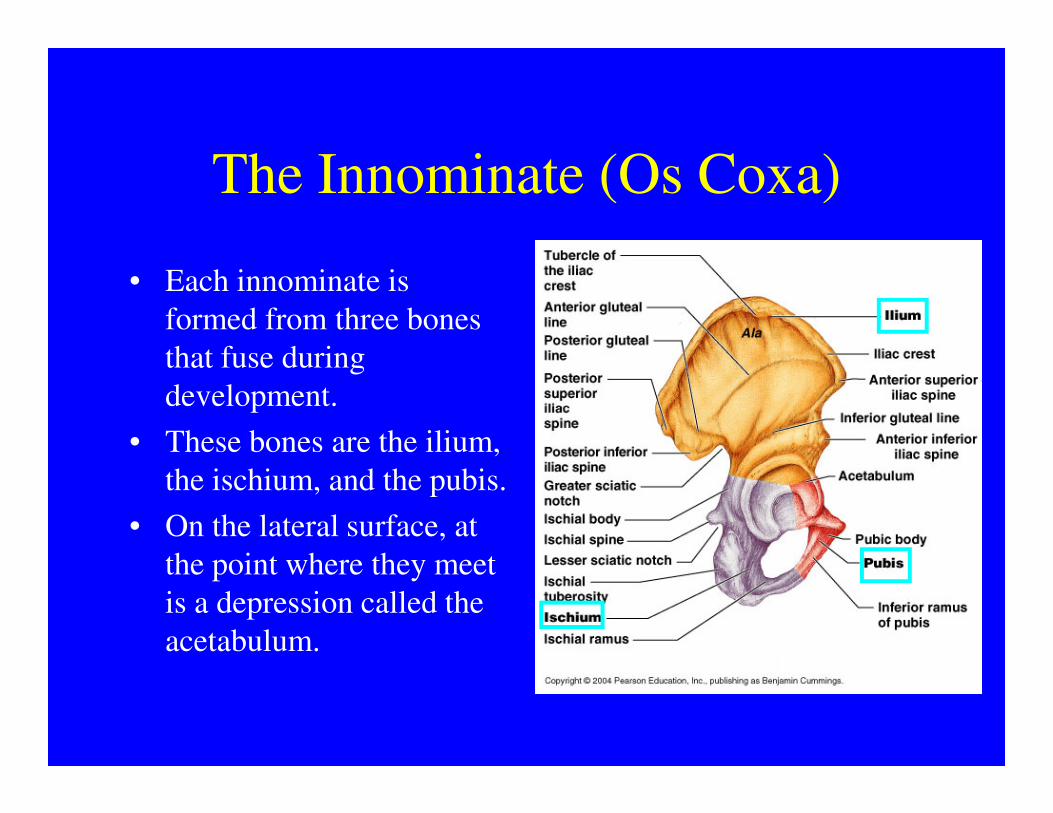
The Innominate (Os Coxa)
• Each innominate is
formed from three bones
that fuse during
development.development.
• These bones are the ilium,
the ischium, and the pubis.
• On the lateral surface, at
the point where they meet
is a depression called the
acetabulum.
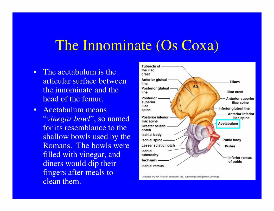
The Innominate (Os Coxa)
• The acetabulum is the articular surface between the innominate and the head of the femur.
• Acetabulum means • Acetabulum means “vinegar bowl”, so named for its resemblance to the shallow bowls used by the Romans. The bowls were filled with vinegar, and diners would dip their fingers after meals to clean them.
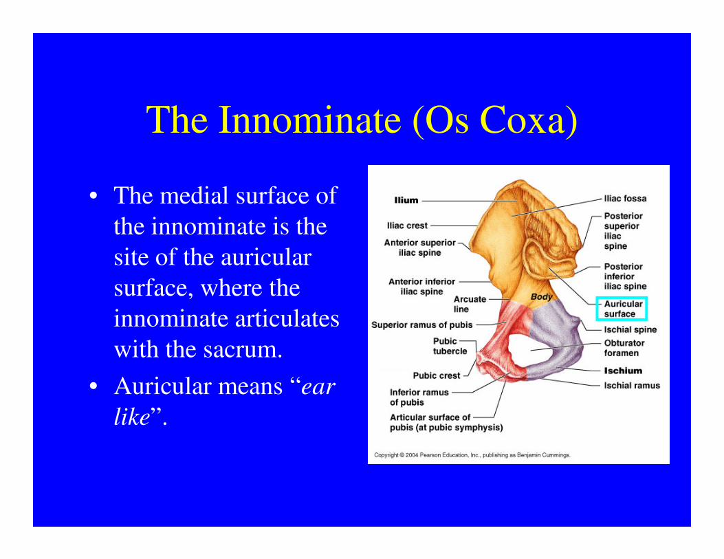
The Innominate (Os Coxa)
• The medial surface of
the innominate is the
site of the auricular
surface, where the surface, where the
innominate articulates
with the sacrum.
• Auricular means “ear
like”.
The Femur
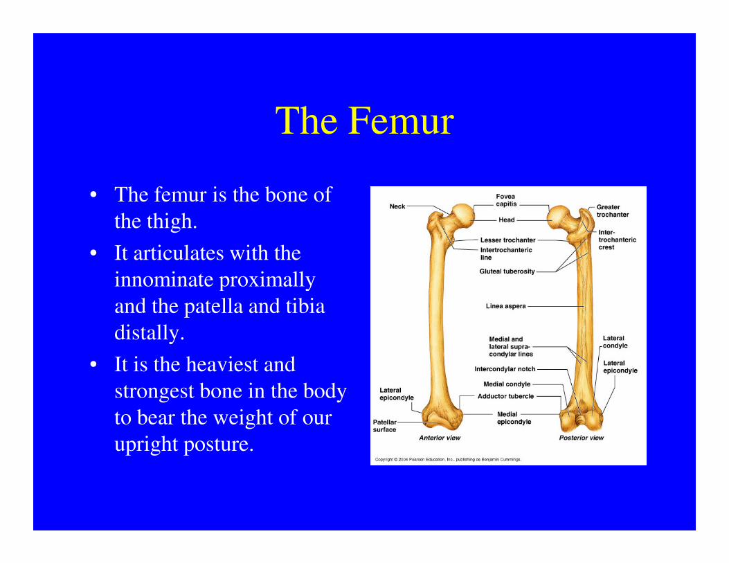
• The femur is the bone of
the thigh.
• It articulates with the
innominate proximally innominate proximally
and the patella and tibia
distally.
• It is the heaviest and
strongest bone in the body
to bear the weight of our
upright posture.
The Femur
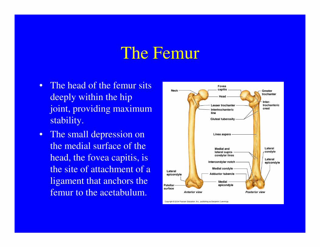
• The head of the femur sits
deeply within the hip
joint, providing maximum
stability.stability.
• The small depression on
the medial surface of the
head, the fovea capitis, is
the site of attachment of a
ligament that anchors the
femur to the acetabulum.
The Femur
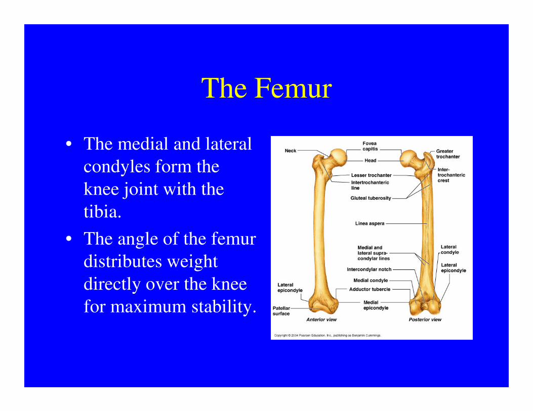
• The medial and lateral
condyles form the
knee joint with the
tibia.tibia.
• The angle of the femur
distributes weight
directly over the knee
for maximum stability.
The Patella
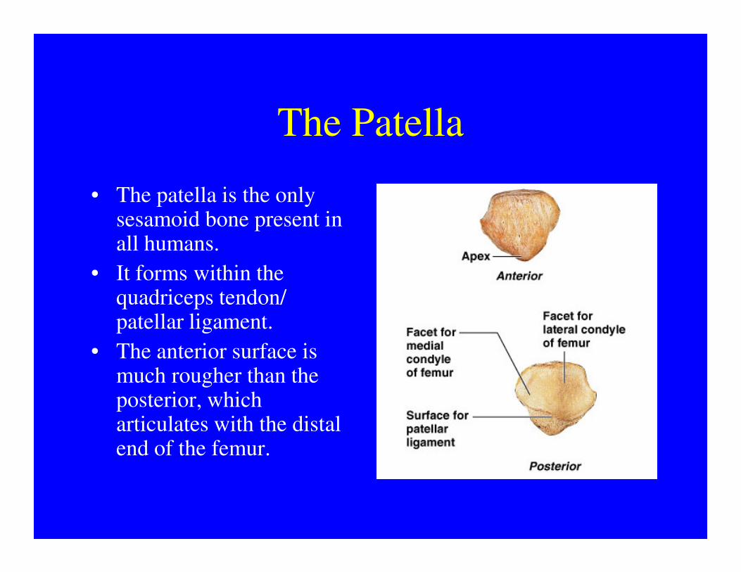
• The patella is the only sesamoid bone present in all humans.
• It forms within the quadriceps tendon/ quadriceps tendon/ patellar ligament.
• The anterior surface is much rougher than the posterior, which articulates with the distal end of the femur.
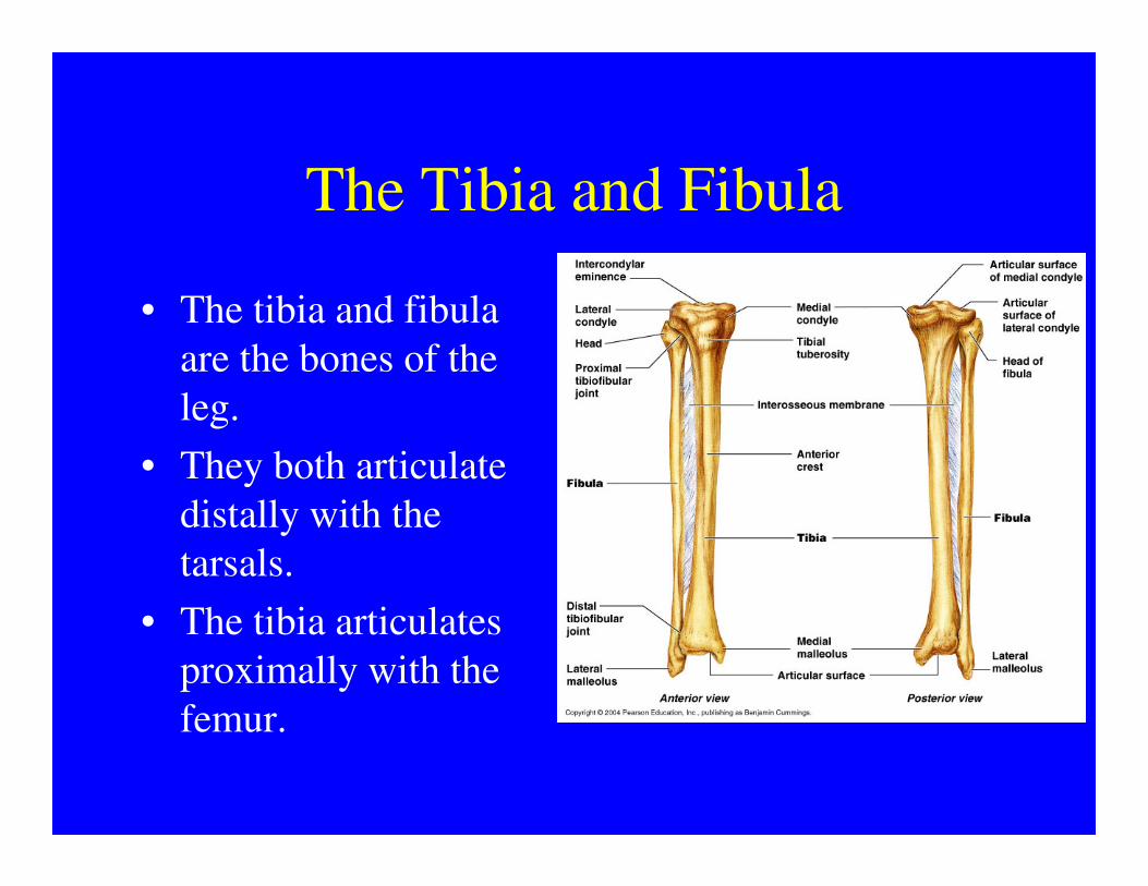
The Tibia and Fibula
• The tibia and fibula
are the bones of the
leg.
• They both articulate • They both articulate
distally with the
tarsals.
• The tibia articulates
proximally with the
femur.
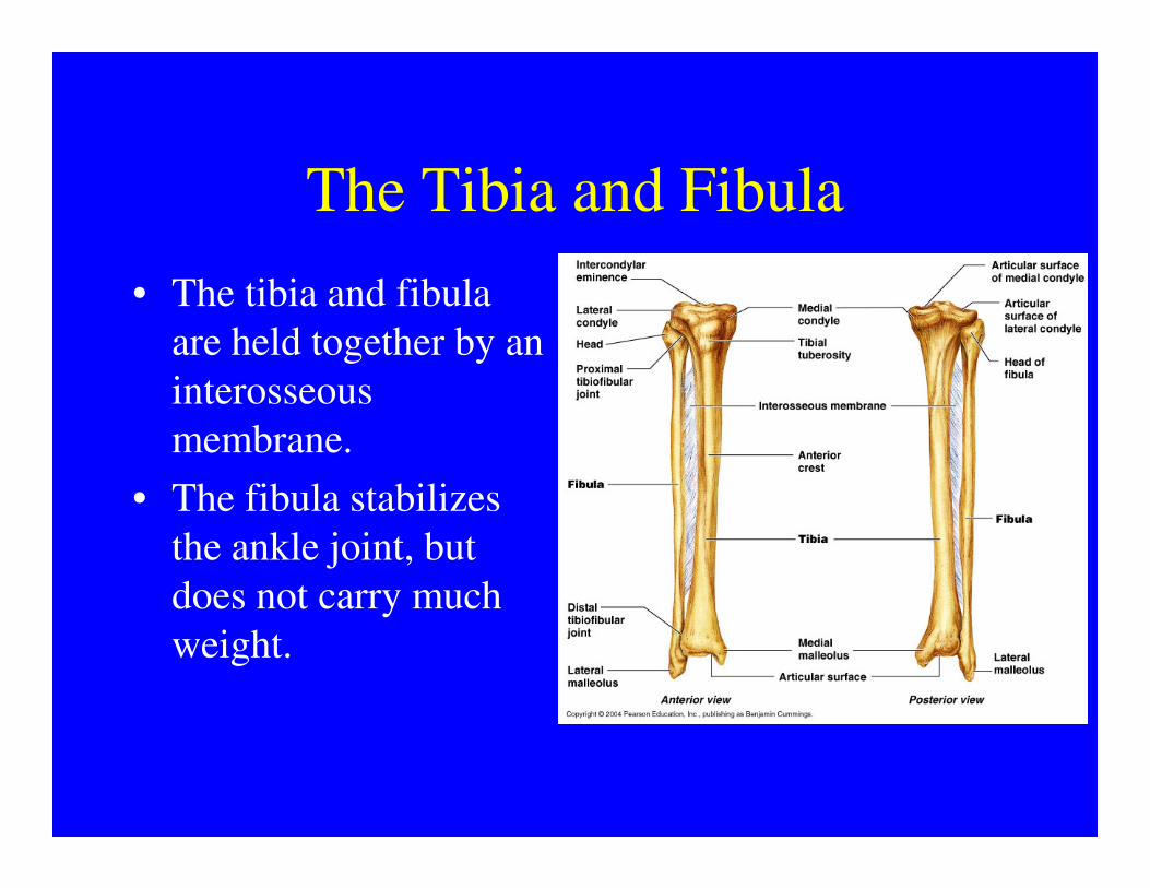
The Tibia and Fibula
• The tibia and fibula
are held together by an
interosseous
membrane.membrane.
• The fibula stabilizes
the ankle joint, but
does not carry much
weight.
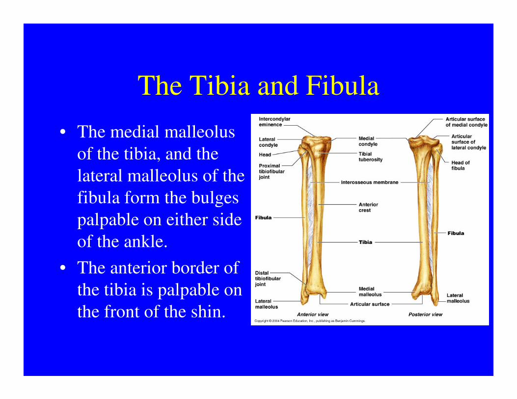
The Tibia and Fibula
• The medial malleolus
of the tibia, and the
lateral malleolus of the
fibula form the bulges fibula form the bulges
palpable on either side
of the ankle.
• The anterior border of
the tibia is palpable on
the front of the shin.

The Pes
• The pes is supported by 26 bones in three groups.
• 7 tarsal bones form the • 7 tarsal bones form the ankle.
• 5 metatarsal bones form the foot.
• 14 phalanges form the toes.

The Tarsals
• The tarsals are
arranged in two rows.
• The proximal row
(from medial to (from medial to
lateral) are the
navicular, the talus,
and the calcaneus.
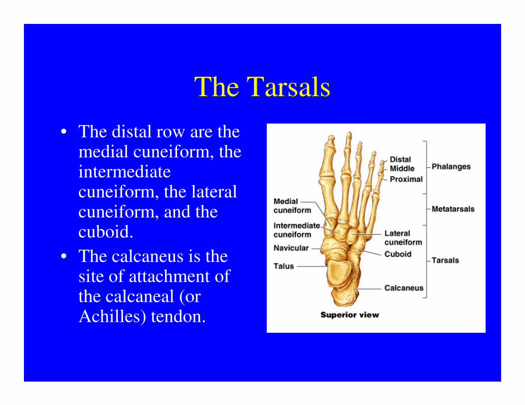
The Tarsals
• The distal row are the medial cuneiform, the intermediate cuneiform, the lateral cuneiform, and the cuneiform, and the cuboid.
• The calcaneus is the site of attachment of the calcaneal (or Achilles) tendon.

The Metatarsals
• The five metatarsals are numbered from one to five starting with the medial (great toe) side.
1
5toe) side.
• They articulate with the distal row or tarsals proximally and the proximal phalanges distally.
5
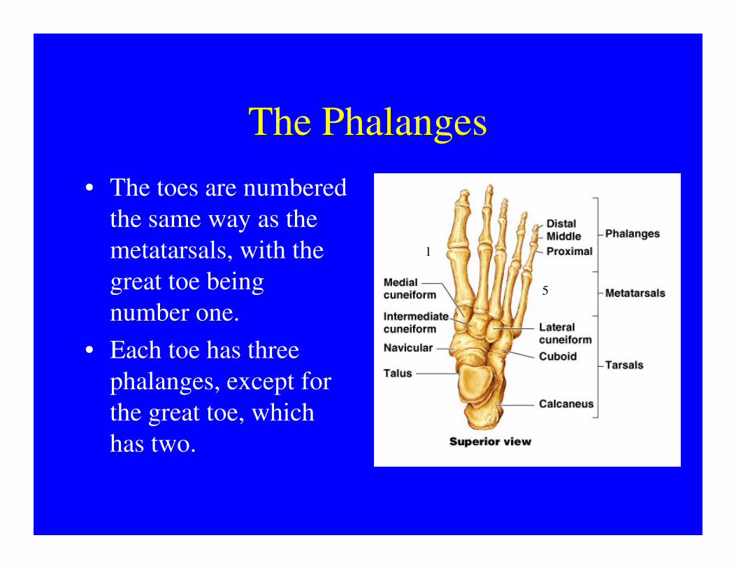
The Phalanges
• The toes are numbered
the same way as the
metatarsals, with the
great toe being
1
5great toe being
number one.
• Each toe has three
phalanges, except for
the great toe, which
has two.
5
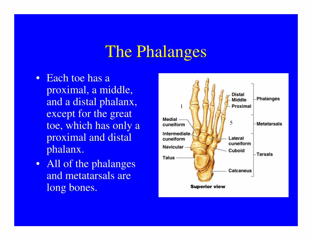
The Phalanges
• Each toe has a proximal, a middle, and a distal phalanx, except for the great toe, which has only a
1
5toe, which has only a proximal and distal phalanx.
• All of the phalanges and metatarsals are long bones.
5
General Considerations
• Study the bones and bone markings listed on your structure list.
• Use not only the diagrams in your manual, but also the bones in class.
• Use not only the diagrams in your manual, but also the bones in class.
• You will be required to identify left from right bones for the appendicular skeleton.
• Learning left from right will be much easier if you study the 3D models.
General Considerations
• On the exam, make sure that you provide
the full name for a bone or bone marking,
especially if there is more than one of a especially if there is more than one of a
particular structure.
• For example, styloid process (or just
styloid) is not enough. You must specify
styloid process of the radius (or ulna, or
temporal bone).
General Considerations
• Finally, do not point to the bones with the tip of a pen or pencil. Use a mall probe.
• It is too easy to inadvertently mark a bone, and the marks are extremely difficult to
• It is too easy to inadvertently mark a bone, and the marks are extremely difficult to clean off.
• Anyone caught deliberately marking on a bone will have their highest quiz grade converted to a zero.