Embed Size (px)
Citation preview
822
Septic Saddle Embolus Causing Basilar Artery Rupture without Mycotic Aneurysm Douglas H. Yock , Jr.'
Intracranial hemorrhage is a well known complication of mycotic aneurysms in patients with infective endocarditis. Focal arterial wall destruction without aneurysm formation is a less well recognized consequence of septic emboli . We report a case of non aneurysmal rupture of the basilar artery after septic saddle embolization, with radiographic and pathologic correlation.
Case Report
A 27-year-old man had been in excellent health until 2 months earlier, when he was admitted to another hospital with a 3-week history of fever , night sweats , and rapid, pounding pulse. Examination showed aortic regurgitation , and blood cultures grew viridans streptococci. A diagnosis of subacute bacterial endocarditis was made, and the patient was treated with penicillin and gentamicin intravenously for 3 weeks.
Two weeks after discharge, he was readmitted with symptoms of congestive heart failure. Severe aortic regurgitation was documented, elective valve replacement was scheduled, and the patient was discharged again after 10 days of hospitalization. He remained afebrile during this admission, and no additional antibiotics were administered. The next evening, while resting quietly , he experienced acute vertical diplopia, dysequilibrium, and dysarthria. These symptoms waxed and waned for several hours. Imbalance, diplopia, and slurred speech recurred the next morning, and he came to the emergency room at Metropolitan Medical Center.
Neurologic examination on admission was unremarkable. A computed tomographic (CT) scan showed a small density at the basilar artery tip, believed to represent an embolus. Lumbar puncture demonstrated 18 white blood cells (11 mononuclear, 7 polymorphonuclear), 18 red blood cells, protein of 31 mg/dl , and glucose of 51 mg/ dl. Echocardiogram showed a bicuspid aortic valve. A large vegetation was seen on the noncoronary cusp , which prolapsed into the left ventricle . Intravenous penicillin was begun.
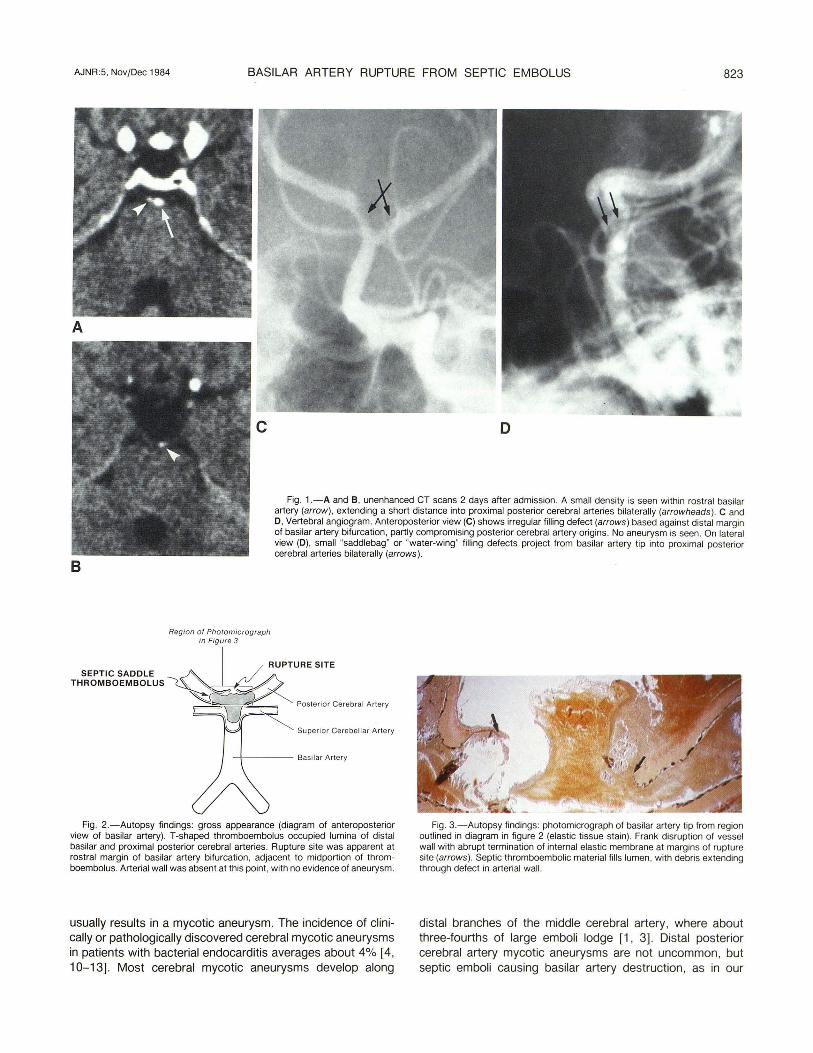
The patient's symptoms continued to fluctuate, and he developed a new left facial weakness and left arm drift. A repeat CT scan 2 days after admission (figs. 1 A and 1 B) again showed the small density at the basilar artery tip, extending into the proximal segments of both posterior cerebral arteries. In addition, an area compatible with recent infarction had developed in the right thalamus. Cerebral angiography (figs. 1 C and 10) demonstrated a filling defect at the basilar artery bifurcation, extending into the posterior cerebral arteries bilaterally. There was no evidence of aneurysm at this site or elsewhere. The
Received June 2, 1983; accepted July 3, 1983.
carotid studies showed mild spasm of both supraclinoid internal carotid arteries and an equivocal embolus in a small branch of the left middle cerebral artery.
A decision was made to proceed with aortic valve replacement. The operation was performed on hospital day 4, with intraoperative anticoagulation consisting of 20,000 U of heparin reversed with protamine at the end of the procedure. A bicuspid aortic valve with friable debris on the perforated non coronary cusp was found. No organisms were seen on Gram stain of the vegetation. The patient made an excellent immediate postoperative recovery, with no new neurologic deficit. His blood pressure after surgery averaged 125/80 mm Hg, as compared with 130/50 mm Hg before valve replacement. Intravenous heparin was begun at 400 U/hr.
Thirty-six hours after surgery, the patient complained of headache and rapidly lost consciousness, developing dilated pupils and decorticate posturing. An emergency CT scan demonstated diffuse subarachnoid hemorrhage, with densest clot surrounding the rostral brainstem. The patient's condition continued to deteriorate, and he died 3 days later.
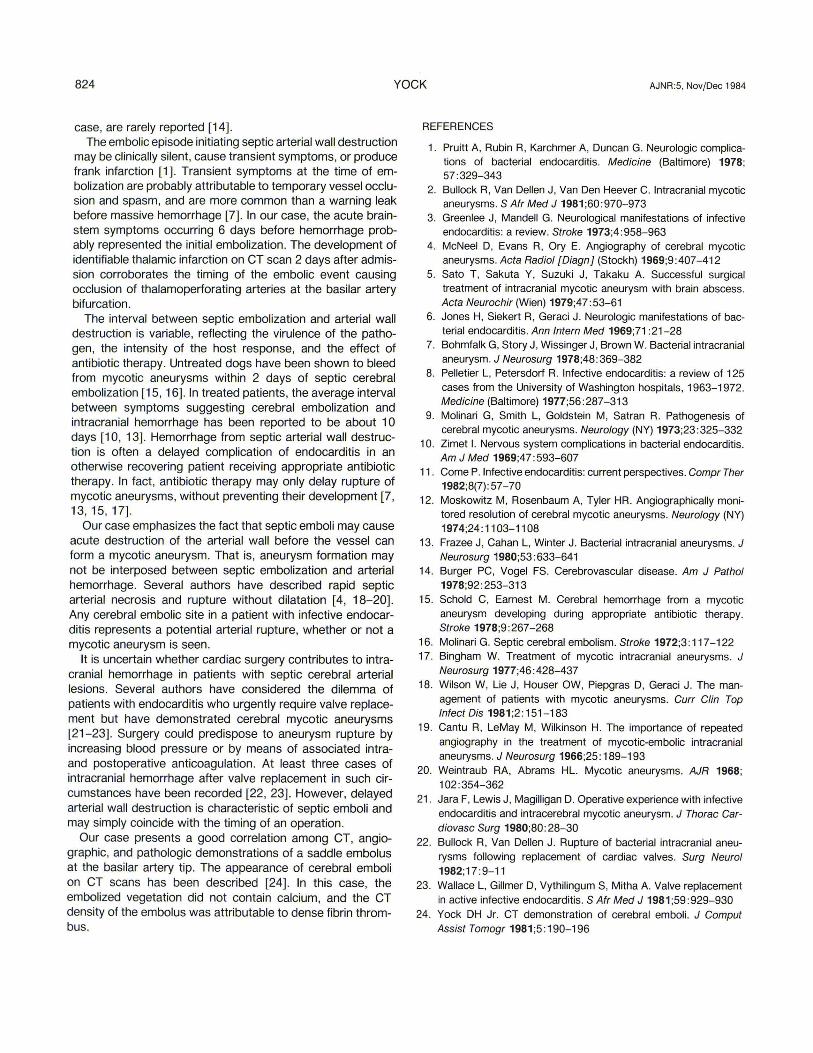
Autopsy documented dense basal subarachnoid hemorrhage. A 2-mm defect was present at the distal margin of the basilar artery tip (figs. 2 and 3). Fluid injected into the artery spurted through the defect. No true or false aneurysm was present at this site. A septic saddle embolus was lodged within the lumina of the distal basilar and proximal posterior cerebral arteries, containing polymorphonuclear leukocytes and a few gram-positive cocci.
Discussion
Septic embolization is a common and serious complication of infective endocarditis. The brain receives a large share of these emboli , and potential sequelae include infarction, leptomeningitis , cerebritis, abscess, mycotic aneurysm, and intracerebral or subarachnoid hemorrhage. Of patients with bacterial endocarditis, 20%-40% have neurologic signs and symptoms; 5%-15% present with a neurologic chief complaint [1-6] . The overall mortality of 20%-40% in infective endocarditis rises to 50%-60% in the presence of neurologic symptoms and 80%-90% with rupture of a mycotic aneurysm [1 , 3, 6-8]. Unfortunately, the incidence and morbidity of cerebral complications in endocarditis have changed very little in the antibiotic era [3, 9] .
Arterial wall destruction secondary to septic embolization
, Department of Radiology, Metropolitan Medical Center, 900 S. 8th St. , Minneapolis, MN 55404.
AJNR 5:822-824, November/December 1984 0195-6108/84/0506-0822 $00.00 © American Roentgen Ray Society
AJNR :5, Nov/Dec 1984 BASILAR ARTERY RUPTURE FROM SEPTIC EMBOLUS 823
o
Fig. 1.-A and B, unenhanced CT scans 2 days after admission. A small density is seen within rostral basilar artery (arrow) , extending a short distance into proximal posterior cerebral arteries bilaterally (arrowheads ). C and 0 , Vertebral angiogram. Anteroposterior view (C) shows irregular filling defect (arrows ) based against distal margin of basilar artery bifurcation, partly compromising posterior cerebral artery origins. No aneurysm is seen. On lateral view (D) , small "saddlebag" or "water-wing" filling defects project from basilar artery tip into proximal posterior cerebral arteries bilaterally (arrows) .
B
Region of Photomicrograph in Figure 3
~ RUPTURE SITE
SEPTIC SADDLE ----,A THROMBOEMBOLUS ~
~ V ~ u~ ,~",'o, c,,,,,,, M,O
Su perior Ce rebell ar Artery
~---- Basilar Artery
Fig. 2.-Autopsy findings : gross appearance (diagram of anteroposterior view of basilar artery). T-shaped thromboembolus occupied lumina of distal basilar and proximal posterior cerebral arteries. Rupture site was apparent at rostral margin of basilar artery bifurcation, adjacent to midportion of thromboembolus. Arterial wall was absent at this point , with no evidence of aneurysm.
usually results in a mycotic aneurysm. The incidence of clinically or pathologically discovered cerebral mycotic aneurysms in patients with bacterial endocarditis averages about 4% [4, 10-13]. Most cerebral mycotic aneurysms develop along
Fig . 3.-Autopsy findings: photomicrograph of basilar artery tip from region outlined in diagram in figure 2 (elastic tissue stain) . Frank disruption of vessel wall with abrupt termination of internal elastic membrane at margins of rupture site (arrows). Septic thromboembolic material fills lumen , with debris extending through defect in arterial wall.
distal branches of the middle cerebral artery, where about three-fourths of large emboli lodge [1, 3] . Distal posterior cerebral artery mycotic aneurysms are not uncommon, but septic emboli causing basilar artery destruction , as in our
824 YOCK AJNR:5, Nov/Dec 1984
case, are rarely reported [14] . The embolic episode initiating septic arterial wall destruction
may be clinically silent, cause transient symptoms, or produce frank infarction [1] . Transient symptoms at the time of embolization are probably attributable to temporary vessel occlusion and spasm, and are more common than a warning leak before massive hemorrhage [7]. In our case, the acute brainstem symptoms occurring 6 days before hemorrhage probably represented the initial embolization. The development of identifiable thalamic infarction on CT scan 2 days after admission corroborates the timing of the embolic event causing occlusion of thalamoperforating arteries at the basilar artery bifurcation .
The interval between septic embolization and arterial wall destruction is variable, reflecting the virulence of the pathogen, the intensity of the host response, and the effect of antibiotic therapy. Untreated dogs have been shown to bleed from mycotic aneurysms within 2 days of septic cerebral embolization [15, 16]. In treated patients, the average interval between symptoms suggesting cerebral embolization and intracranial hemorrhage has been reported to be about 10 days [10, 13]. Hemorrhage from septic arterial wall destruction is often a delayed complication of endocarditis in an otherwise recovering patient receiving appropriate antibiotic therapy. In fact, antibiotic therapy may only delay rupture of mycotic aneurysms, without preventing their development [7, 13, 15, 17].
Our case emphasizes the fact that septic emboli may cause acute destruction of the arterial wall before the vessel can form a mycotic aneurysm. That is, aneurysm formation may not be interposed between septic embolization and arterial hemorrhage. Several authors have described rapid septic arterial necrosis and rupture without dilatation [4, 18-20]. Any cerebral embolic site in a patient with infective endocarditis represents a potential arterial rupture, whether or not a mycotic aneurysm is seen.
It is uncertain whether cardiac surgery contributes to intracranial hemorrhage in patients with septic cerebral arterial lesions. Several authors have considered the dilemma of patients with endocarditis who urgently require valve replacement but have demonstrated cerebral mycotic aneurysms [21-23] . Surgery could predispose to aneurysm rupture by increasing blood pressure or by means of associated intraand postoperative anticoagulation . At least three cases of intracranial hemorrhage after valve replacement in such circumstances have been recorded [22, 23]. However, delayed arterial wall destruction is characteristic of septic emboli and may simply coincide with the timing of an operation .
Our case presents a good correlation among CT, angiographic, and pathologic demonstrations of a saddle embolus at the basilar artery tip. The appearance of cerebral emboli on CT scans has been described [24]. In this case, the embolized vegetation did not contain calcium, and the CT density of the embolus was attributable to dense fibrin thrombus.
REFERENCES
1. Pruitt A, Rubin R, Karchmer A, Duncan G. Neurologic complications of bacterial endocarditis. Medicine (Baltimore) 1978; 57:329-343
2. Bullock R, Van Dellen J, Van Den Heever C. Intracranial mycotic aneurysms. S Afr Med J 1981 ;60 :970-973
3. Greenlee J, Mandell G. Neurological manifestations of infective endocarditis: a review. Stroke 1973;4 :958-963
4. McNeel D, Evans R, Ory E. Angiography of cerebral mycotic aneurysms. Acta Radial [DiagnJ (Stockh) 1969;9 :407-412
5. Sato T, Sakuta Y, Suzuki J, Takaku A. Successful surgical treatment of intracranial mycotic aneurysm with brain abscess. Acta Neurochir (Wien) 1979;47:53-61
6. Jones H, Siekert R, Geraci J. Neurologic manifestations of bacterial endocarditis. Ann Intern Med 1969;71 :21-28
7. Bohmfalk G, Story J, Wissinger J, Brown W. Bacterial intracranial aneurysm. J Neurosurg 1978;48:369-382
8. Pelletier L, Petersdorf R. Infective endocarditis: a review of 125 cases from the University of Washington hospitals, 1963-1972. Medicine (Baltimore) 1977;56:287-313
9. Molinari G, Smith L, Goldstein M, Satran R. Pathogenesis of cerebral mycotic aneurysms. Neurology (NY) 1973;23 :325-332
10. Zimet I. Nervous system complications in bacterial endocarditis. Am J Med 1969;47 :593-607
11. Come P. lnfective endocarditis: current perspectives. Compr Ther 1982;8(7) :57-70
12. Moskowitz M, Rosenbaum A, Tyler HR. Angiographically monitored resolution of cerebral mycotic aneurysms. Neurology (NY) 1974;24: 11 03-11 08
13. Frazee J, Cahan L, Winter J. Bacterial intracranial aneurysms. J Neurosurg 1980;53: 633-641
14. Burger PC, Vogel FS. Cerebrovascular disease. Am J Pathol 1978;92 : 253-313
15. Schold C, Earnest M. Cerebral hemorrhage from a mycotic aneurysm developing during appropriate antibiotic therapy. Stroke 1978;9: 267 -268
16. Molinari G. Septic cerebral embolism. Stroke 1972;3 : 117-122 17. Bingham W. Treatment of mycotic intracranial aneurysms. J
Neurosurg 1977;46 :428-437 18. Wilson W, Lie J, Houser OW, Piepgras D, Geraci J. The man
agement of patients with mycotic aneurysms. Curr Clin Top Infect Dis 1981 ;2 : 151-183
19. Cantu R, LeMay M, Wilkinson H. The importance of repeated angiography in the treatment of mycotic-embolic intracranial aneurysms. J Neurosurg 1966;25 : 189-193
20. Weintraub RA, Abrams HL. Mycotic aneurysms. AJR 1968; 102 :354-362
21 . Jara F, Lewis J, Magilligan D. Operative experience with infective endocarditis and intracerebral mycotic aneurysm. J Thorac Cardiovasc Surg 1980;80:28-30
22. Bullock R, Van Dellen J. Rupture of bacterial intracranial aneurysms following replacement of cardiac valves. Surg Neurol 1982;17 : 9-11
23 . Wallace L, Gillmer D, Vythilingum S, Mitha A. Valve replacement in active infective endocarditis. S Afr Med J 1981 ;59 :929-930
24. Yock DH Jr. CT demonstration of cerebral emboli. J Comput Assist Tomogr 1981 ;5 :190-196