Embed Size (px)
Citation preview
Volume 15 · Number 3 · September 2013 137
Endovascular Mechanical Thrombectomy in Basilar Artery Occlusion: Initial Experience
Bum-Soo Park, Chang-Woo Kang, Hyon-Jo Kwon, Seung-Won Choi, Seon-Hwan Kim, Hyeon-Song Koh,
Jin-Young Youm, Shi-Hun SongDepartment of Neurosurgery, School of Medicine, Chungnam National University, Daejeon, Korea
Objective : This study was conducted to assess the efficacy and safety of endovascular mechanical thrombectomy (EMT) for patients diagnosed with basilar artery (BA) occlusion.
Materials and Methods : We retrospectively analyzed clinical and imaging data of 16 patients diagnosed with BA occlusion who were treated with endovascular intervention from July 2012 to February 2013. Direct suc-tion using the Penumbra system and thrombus retrieval by the Solitaire stent were the main endovascular techniques used to restore BA flow. The outcomes were evaluated based on rate of angiographic recanaliza-tion, rate of improvement of National Institutes of Health Stroke Scale (NIHSS) score, rate of modified Rankin Scale (mRS) at discharge and after 3 months, and rate of cerebral hemorrhagic complications. Successful re-canalization was defined as achieving Thrombolysis In Cerebral Infarction (TICI) of II or III.
Results : Sixteen patients received thrombectomy. The mean age was 67.8 ± 11 years and the mean NIHSS score was 12.3 ± 8.2. Eight patients treated within 6 hours of symptom onset were grouped as A and the other 8 patients treated beyond 6 hours (range, 6-120) were grouped as B. Successful recanalization was met in six patients (75%) for group A and 7 (87.5%) for group B. Favorable outcome occurred in 4 patients (50%) for group A and 5 (62.5%) for group B.
Conclusion : Our study supports the effectiveness and safety of endovas-cular mechanical thrombectomy in treating BA occlusion even 6 hours af-ter symptom onset.
J Cerebrovasc Endovasc Neurosurg. 2013 September;15(3):137-144Received : 14 June 2013Revised : 11 July 2013Accepted : 1 August 2013
Correspondence to Chang-Woo KangDepartment of Neurosurgery, School of Medicine, Chungnam National University, 282 Munhwa-ro, Jung-gu, Daejeon301-721, Korea
Tel : 82-42-280-7361Fax : 82-42-280-7363E-mail : [email protected]
This is an Open Access article distributed under the terms of the Creative Commons Attribution Non- Commercial License (http://creativecommons.org/li-censes/by-nc/3.0) which permits unrestricted non- commercial use, distribution, and reproduction in any medium, provided the original work is properly cited.
Keywords Basilar artery occlusion, Endovascular mechanical thrombectomy, Therapeutic time window, Recanalization
Journal of Cerebrovascular and Endovascular NeurosurgeryISSN 2234-8565, EISSN 2287-3139, http://dx.doi.org/10.7461/jcen.2013.15.3.137 Clinical Article
INTRODUCTION
The basilar artery (BA) supplies the brain stem, cer-
ebellum, thalami, occipital lobes and medial temporal
lobes. If the BA is occluded, symptoms can include
decreased consciousness, quadriparesis, pupillary and
oculomotor abnormalities, dysarthria, dysphagia or
even sudden death.10) BA occlusion accounts for about
20% of ischemic strokes.13) Acute occlusion of this ar-
tery produces high morbidity and mortality, with a
rate of 85-95%, if left untreated.6)13)21) Without recanal-
ization, the likelihood of good outcome is about 2%.6)
The chief goal in treating acute ischemic stroke is to
restore cerebral blood flow as rapidly and safely as
ENDOVASCULAR MECHANICAL THROMBECTOMY IN BA OCCLUSION
138 J Cerebrovasc Endovasc Neurosurg
A B
C
D
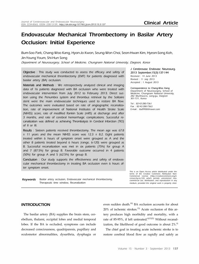
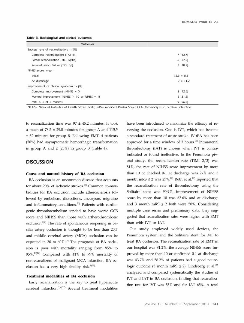
Fig. 1. (A) The angiogram shows occlusion of the basilar artery. (B) After placing the Penumbra system at the occlusion site, suc-tion was performed and partial recanalization is shown. (C) Endovascular mechanical thrombectomy using the Penumbra system was carried out again and follow up angiogram shows complete recanalization. (D) Thrombus is pulled out by the Penumbra system.
possible.1)4)17) Intravenous tissue plasminogen activator
(IV-tPA) or endovascular techniques are widely used
to produce vascular patency and to improve clinical
outcomes. It has become a fairly standard practice in the
anterior circulation to set a time restriction to the first 6
hours of symptom onset for endovascular mechanical
thrombectomy (EMT) to limit hemorrhagic trans-
formation of infarctions and because of its poor con-
tribution to favorable clinical outcome.15) However, the
effective therapeutic time window has not been estab-
lished for the posterior circulation.16) Therefore, we con-
ducted a study to verify the efficacy and safety of endo-
vascular mechanical thrombectomy for BA occluded pa-
tients, even beyond 6 hours of symptom onset.
MATERIALS AND METHODS
Medical records of 16 consecutive patients who
were treated for symptomatic acute BA occlusion
from July 2012 to Feb 2013 were reviewed. To rule
out hemorrhagic stroke or old infarction, all patients
underwent brain-computed tomography (CT) on
arrival. Magnetic resonance images (MRI), such as dif-
fusion-weighted image (DWI), MR angiography and
perfusion-weighted image (PWI) were then tracked to
detect the occluded vessels, DWI-PWI mismatch, and
measure infarct territories. Intravenous thrombolysis
(IVT) was started with 0.9 mg/kg IV-tPA when the
diagnosis was confirmed within 3 hours of symptom
onset. If refractory to IVT or contraindicated or the
patients arrived more than 3 hours after symptom on-
set, EMT was performed. Indications for EMT treat-
ment were a National Institutes of Health Stroke Scale
(NIHSS) score of 4 or more, age more than 18 years
and arrival at the stroke center beyond 3 hours of
symptom onset. Considering these uncertainties and
high mortality rate of untreated BA occluded patients,
our stroke center adopted a protocol to treat not only
patients who came within 6 hours of symptom onset
but also who came beyond 6 hours with symptom ag-
gravation, mismatch between DWI and PWI.
All procedures were performed under local anes-
thesia by an endovascular neurosurgeon. The occlu-
sion status was confirmed by taking angiography af-
ter locating the guiding catheter (Guider XF SoftipTM,
Boston Scientific, Plymouth, MN, USA) to the prox-
imal vertebral artery. To prevent procedural throm-
boembolic events, a mixture of 2,000 IU of heparin
and 0.9% normal saline 1,000 ml was administered
continuously through the guiding catheter. This was
applied even to patients who received IV-tPA. The
main endovascular techniques used to restore BA
flow were direct thrombectomy using the Penumbra
thromboaspiration catheter (Penumbra Inc., Alameda,
CA, USA) (Fig. 1) and thrombus retrieval by the
Solitaire stent (ev3 Inc., Irvine, CA, USA) (Fig. 2).
The Penumbra system with microcatheter (Prowler
SELECTTM PLUS, Codman Neurovascular, Raynham,
BUM-SOO PARK ET AL
Volume 15 · Number 3 · September 2013 139
A B
C
D
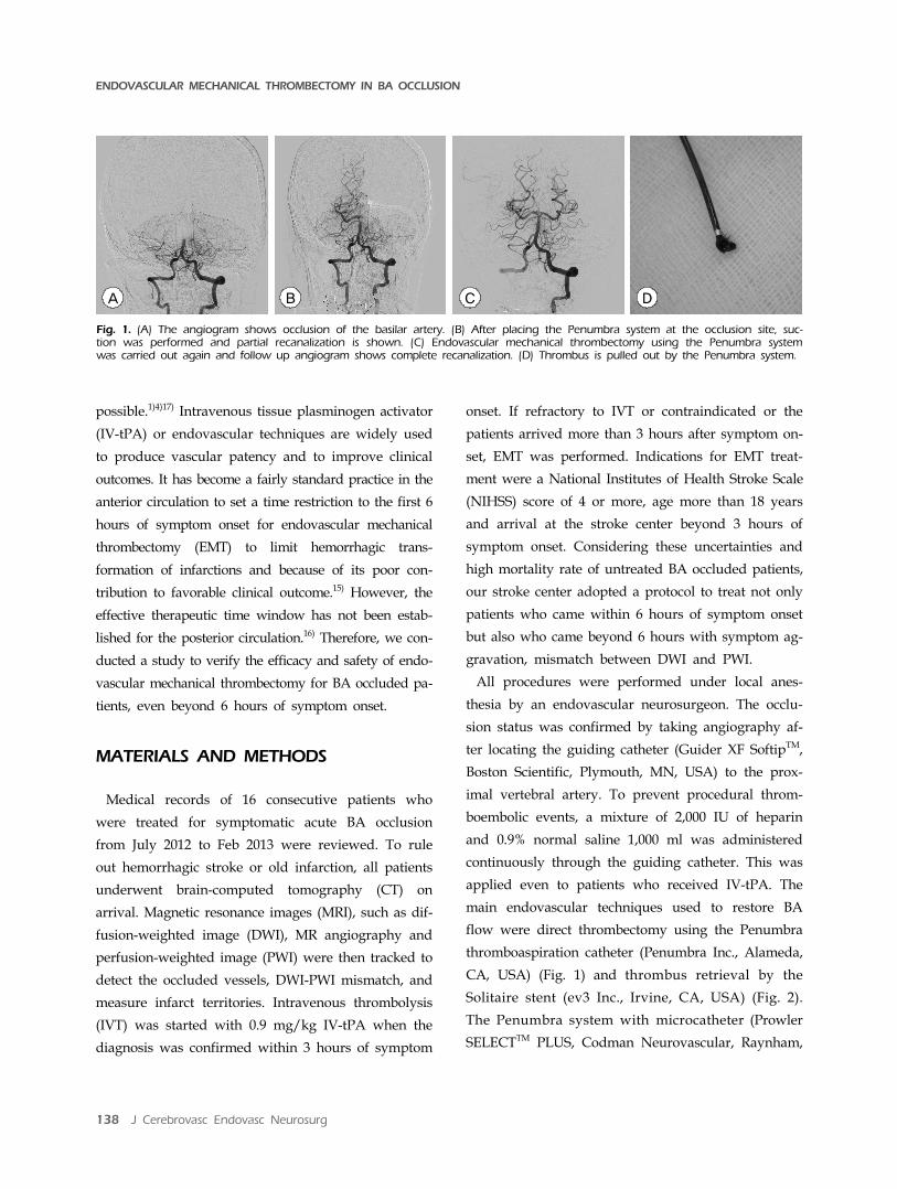
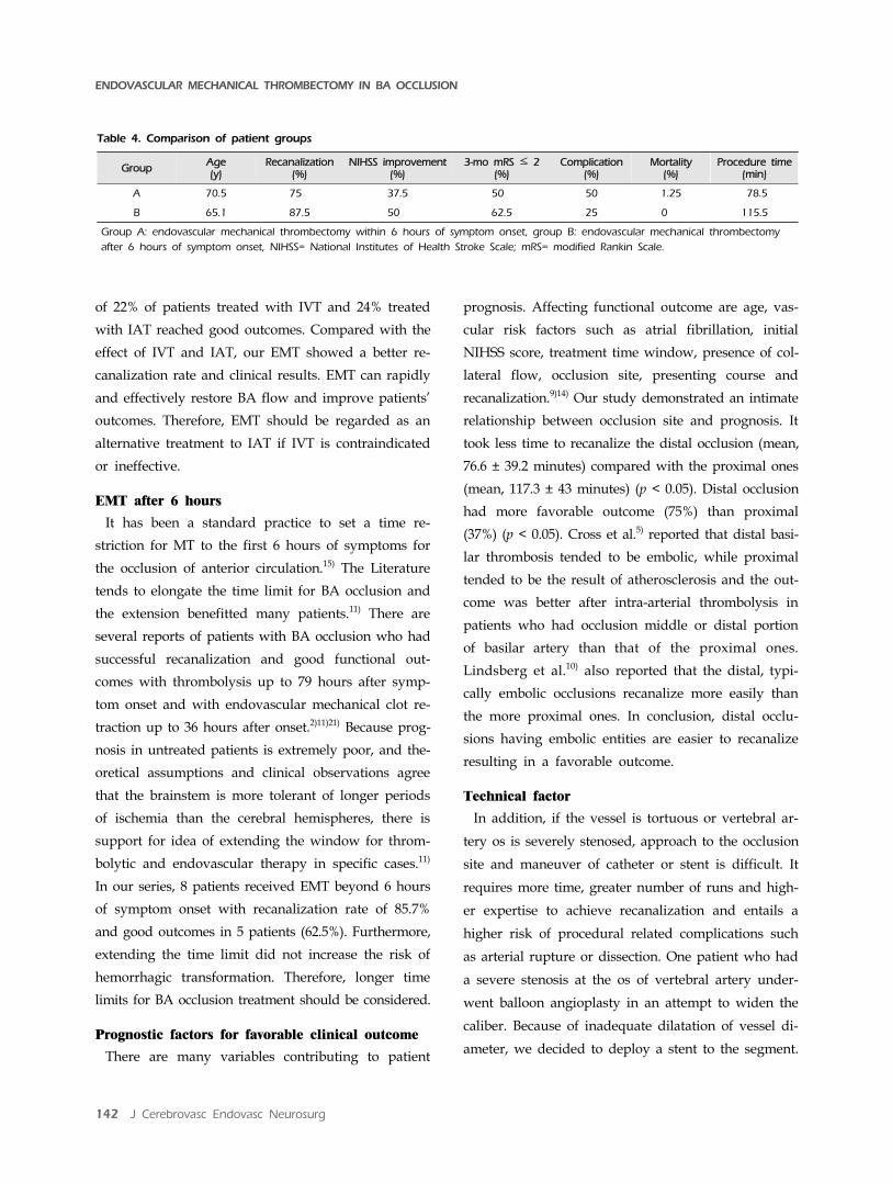
Fig. 2. (A) Initial angiogram in the anteroposterior plane shows basilar artery occlusion (thrombolysis in cerebral infarction grade 0). (B) Cerebral angiogram after temporary deployment of the Solitaire stent shows sufficient coverage and transient flow restoration of the occluded segment. (C) After retrieval of the stent, complete recanalization is shown. (D) Thrombus is trapped by the stent.
MA, USA) and microwire (SynchroTM-14, Stryker
Neurovascular, Fremont, CA, USA) were brought into
the target vessel and placed at the distal portion of
the occlusion site after penetrating the thrombus
segment. Using a 50 cc syringe, negative pressure was
applied through the Penumbra system. If recanaliza-
tion failed, the Solitaire stent was used for mechanical
thrombectomy. The Solitaire stent was deployed
where it could completely cover the occluded seg-
ment and left in place for at least 2 minutes before
retrieval. The microcatheter and the stent were gently
withdrawn together with the guiding catheter while a
50 cc syringe maintained negative pressure through
the guiding catheter to aspirate the thrombus and to
prevent migration of the emboli. If stenosis of verte-
bral artery was too severe for catheter entry, balloon
angioplasty and stent deployment was performed at
the segment and post-balloon was carried out if vas-
cular patency was unsatisfactory. After EMT, the
brain was scanned routinely by CT within 1 hour to
check for any procedural related intracerebral hemor-
rhage and by MRI after 24 hours to see any changes
in the infarct region.
Neurologic status was recorded based on NIHSS
score. The outcome data were collected by: (1) rate of
angiographic recanalization; (2) rate of improvement
of NIHSS score and mRS at discharge and after 3
months; and (3) rate of cerebral hemorrhagic compli-
cations such as symptomatic hemorrhages or paren-
chymal hematomas, measured to evaluate the safety
of EMT. Successful recanalization was defined as ach-
ieving TICI II (partial recanalization) or III (complete
recanalization).18) Assessment of the modified Rankin
Scale (mRS) was performed 90 days after treatment. A
mRS of 0 to 2 was defined as a good neurological
outcome. To assess the efficacy and safety of EMT
performed after 6 hours, patients were assigned to 2
groups. Group A was defined as patients treated
within 6 hours of symptom onset and group B as
those treated beyond 6 hours. The data from these
studies were statistically analyzed using the SPSS ver-
sion 20. (IBM Corp., Armonk, NY, USA). Statistical
procedures included Student’s t-test and Pearson’s
chi-square as appropriate for continuous or categorical
variables. Null hypotheses of no difference were re-
jected if p values were less than 0.05.
RESULTS
Of the 16 consecutive patients with BA occlusion
treated mainly with EMT from July 2012 to Feb 2013,
12 were men and 4 were women, with a mean age of
67.8 ± 11 years. The median symptom-to-puncture
time was 5.8 hours (range, 3-120). In initial conven-
tional angiography, proximal thrombosis was present
in 50% and middle and distal basilar thrombosis in
the remaining 50%. Four patients (case number 1, 2,
6, 14) who came to our stroke center within 3 hours
ENDOVASCULAR MECHANICAL THROMBECTOMY IN BA OCCLUSION
140 J Cerebrovasc Endovasc Neurosurg
Case Age SexVascular
risk factors
Initial NIHSS
Occlusion site
IV-tPA dose(mg)
Onset topuncture
time (min)
NIHSS(D/C)
mRS(D/C)
3mo-mRS TOAST PcoA
existence TICI Hemorrhagic infarction
Procedure time(min)
1 83 F 2 10 Proximal 50 190 7 5 5 LAA No I Yes 109
2 76 M 4 16 Distal 67.5 192 36 6 6 LAA Yes I No 120
3 76 F 4 6 Proximal n/a 219 17 5 5 CE Yes IIa Yes 61
4 52 M 2 24 Distal n/a 220 2 1 1 LAA No IIa No 78
5 82 M 4 15 Distal n/a 255 5 3 2 CE Yes III Yes 56
6 84 M 1 11 Proximal 70 277 4 3 1 LAA No III Yes 91
7 56 F 2 9 Distal n/a 287 0 0 0 LAA Yes III No 28
8 55 M 1 12 Proximal n/a 335 10 5 5 LAA Yes III No 85
9 71 M 3 8 Distal n/a 363 3 1 1 CE No III No 65
10 74 M 0 13 Proximal n/a 365 13 5 5 LAA Yes IIa Yes 137
11 60 M 2 7 Distal n/a 653 0 0 0 LAA Yes IIa No 147
12 61 M 1 19 Distal n/a 682 6 4 3 LAA No III No 74
13 54 M 3 34 Proximal n/a 1088 34 5 5 LAA Yes 0 No 132
14 75 M 2 6 Proximal 72 1235 5 4 2 LAA Yes III Yes 205
15 63 M 2 1 Proximal n/a 4691 1 1 0 LAA No IIa No 119
16 63 F 1 5 Distal n/a 6933 1 1 0 LAA No IIa No 45
NIHSS= National Institutes of Health Stroke Scale; IV-tPA= intravenous tissue plasminogen activator; mRS= modified Rankin Scale; D/C= discharge; TOAST= Trial of Org 10172 in Acute Stroke Treatment; PcoA= posterior communicating artery; TICI= thrombolysis in cerebral infarction; n/a= not available; LAA= large-artery atherosclerosis; CE= cardioembolism.
Table 1. Demographic characteristics
Characteristic
Number of patients 16
Demographic data
Mean age, (Range) 67.8 (52-84)
Male : Female 12 : 4
Risk factors, n, (%)
Hypertension 12 (75)
Type 2 diabetes 7 (43.8)
Previous stroke 4 (25)
Atrial fibrillation 3 (18.8)
Site of occlusion, n, (%)
Proximal 8 (50)
Middle or distal 8 (50)
Table 2. Patient characteristics
of symptom onset were treated with IVT. They pre-
sented with gradual aggravation of symptoms and re-
ceived EMT. The other 12 patients depended on EMT
as a first-line treatment (Table 1). The most commonly
diagnosed cardiovascular co-morbidities were hyper-
tension (75%), type 2 diabetes (43.8%), previous stroke
(25%) and atrial fibrillation (18.8%). The major etio-
logic risk factors were large artery atherosclerosis
(81.2%) followed by cardioembolism (18.8%) (Table 2).
Three patients (18.8%) were unconscious and need-
ed respiratory support to maintain adequate ven-
tilation, while 1 patient died, a mortality rate was
6.2%. Of the 16 patients, revascularization was ach-
ieved in 13 (81.3%) (TICI grade IIa/IIB and III). The
mean initial NIHSS score was 12.3 ± 8.2 and mean
NIHSS score at discharge was 9 ± 11.2. In 7 patients
(43.7%), NIHSS scores improved more than 10 or
were 0-1 at the time of discharge. At 3 months, 56.3%
(9 of 16) of patients had a good neurologic outcome
(mRS 0-2) (Table 3). Eight patients who were treated
within 6 hours of symptom onset were grouped as A.
Of those patients recanalization (TICI II or III) was
achieved in 6 patients (75%) and 4 (50%) had good
outcomes. The other 8 patients were treated with
EMT after 6 hours and were grouped as B. Successful
recanalization was achieved in 7 patients (87.5%) and
5 (62.5%) had favorable outcomes. The mean puncture
BUM-SOO PARK ET AL
Volume 15 · Number 3 · September 2013 141
Outcomes
Success rate of recanalization, n (%)
Complete recanalization (TICI III) 7 (43.7)
Partial recanalization (TICI IIa/IIb) 6 (37.5)
Recanalization failure (TICI 0/I) 3 (18.7)
NIHSS score, mean
Initial 12.3 ± 8.2
At discharge 9 ± 11.2
Improvement of clinical symptom, n (%)
Complete improvement (NIHSS = 0) 2 (12.5)
Marked improvement (NIHSS ≥ 10 or NIHSS = 1) 5 (31.2)
mRS ≤ 2 at 3 months 9 (56.3)
NIHSS= National Institutes of Health Stroke Scale; mRS= modified Rankin Scale; TICI= thrombolysis in cerebral infarction.
Table 3. Radiological and clinical outcomes
to recanalization time was 97 ± 45.2 minutes. It took
a mean of 78.5 ± 29.8 minutes for group A and 115.5
± 52 minutes for group B. Following EMT, 4 patients
(50%) had asymptomatic hemorrhagic transformation
in group A and 2 (25%) in group B (Table 4).
DISCUSSION
Cause and natural history of BA occlusion
BA occlusion is an uncommon disease that accounts
for about 20% of ischemic strokes.13) Common co-mor-
bidities for BA occlusion include atherosclerosis fol-
lowed by embolism, dissections, aneurysm, migraine
and inflammatory conditions.20) Patients with cardio-
genic thromboembolism tended to have worse GCS
score and NIHSS than those with artherothrombotic
occlusion.3)7) The rate of spontaneous reopening in ba-
silar artery occlusion is thought to be less than 20%
and middle cerebral artery (MCA) occlusion can be
expected in 30 to 60%.17) The prognosis of BA occlu-
sion is poor with mortality ranging from 85% to
95%.13)17) Compared with 41% to 79% mortality of
nonrecanalizers of malignant MCA infarction, BA oc-
clusion has a very high fatality risk.8)19)
Treatment modalities of BA occlusion
Early recanalization is the key to treat hyperacute
cerebral infarction.1)4)17) Several treatment modalities
have been introduced to maximize the efficacy of re-
versing the occlusion. One is IVT, which has become
a standard treatment of acute stroke. IV-tPA has been
approved for a time window of 3 hours.15) Intraarterial
thrombectomy (IAT) is chosen when IVT is contra-
indicated or found ineffective. In the Penumbra piv-
otal study, the recanalization rate (TIMI 2/3) was
81%, the rate of NIHSS score improvement by more
than 10 or checked 0-1 at discharge was 27% and 3
month mRS ≤ 2 was 25%.12) Roth et al.17) reported that
the recanalization rate of thrombectomy using the
Solitaire stent was 90.9%, improvement of NIHSS
score by more than 10 was 63.6% and at discharge
and 3 month mRS ≤ 2 both were 50%. Considering
multiple case series and preliminary data, they sug-
gested that recanalization rates were higher with EMT
than with IVT or IAT.
Our study employed widely used devices, the
Penumbra system and the Solitaire stent for MT to
treat BA occlusion. The recanalization rate of EMT in
our hospital was 81.2%, the average NIHSS score im-
proved by more than 10 or confirmed 0-1 at discharge
was 43.7% and 56.2% of patients had a good neuro-
logic outcome (3 month mRS ≤ 2). Lindsberg et al.10)
analyzed and compared systematically the studies of
IVT and IAT in BA occlusion, finding that recanaliza-
tion rate for IVT was 53% and for IAT 65%. A total
ENDOVASCULAR MECHANICAL THROMBECTOMY IN BA OCCLUSION
142 J Cerebrovasc Endovasc Neurosurg
Group Age(y)
Recanalization (%)
NIHSS improvement(%)
3-mo mRS ≤ 2(%)
Complication (%)
Mortality (%)
Procedure time(min)
A 70.5 75 37.5 50 50 1.25 78.5
B 65.1 87.5 50 62.5 25 0 115.5
Group A: endovascular mechanical thrombectomy within 6 hours of symptom onset, group B: endovascular mechanical thrombectomyafter 6 hours of symptom onset, NIHSS= National Institutes of Health Stroke Scale; mRS= modified Rankin Scale.
Table 4. Comparison of patient groups
of 22% of patients treated with IVT and 24% treated
with IAT reached good outcomes. Compared with the
effect of IVT and IAT, our EMT showed a better re-
canalization rate and clinical results. EMT can rapidly
and effectively restore BA flow and improve patients’
outcomes. Therefore, EMT should be regarded as an
alternative treatment to IAT if IVT is contraindicated
or ineffective.
EMT after 6 hours
It has been a standard practice to set a time re-
striction for MT to the first 6 hours of symptoms for
the occlusion of anterior circulation.15) The Literature
tends to elongate the time limit for BA occlusion and
the extension benefitted many patients.11) There are
several reports of patients with BA occlusion who had
successful recanalization and good functional out-
comes with thrombolysis up to 79 hours after symp-
tom onset and with endovascular mechanical clot re-
traction up to 36 hours after onset.2)11)21) Because prog-
nosis in untreated patients is extremely poor, and the-
oretical assumptions and clinical observations agree
that the brainstem is more tolerant of longer periods
of ischemia than the cerebral hemispheres, there is
support for idea of extending the window for throm-
bolytic and endovascular therapy in specific cases.11)
In our series, 8 patients received EMT beyond 6 hours
of symptom onset with recanalization rate of 85.7%
and good outcomes in 5 patients (62.5%). Furthermore,
extending the time limit did not increase the risk of
hemorrhagic transformation. Therefore, longer time
limits for BA occlusion treatment should be considered.
Prognostic factors for favorable clinical outcome
There are many variables contributing to patient
prognosis. Affecting functional outcome are age, vas-
cular risk factors such as atrial fibrillation, initial
NIHSS score, treatment time window, presence of col-
lateral flow, occlusion site, presenting course and
recanalization.9)14) Our study demonstrated an intimate
relationship between occlusion site and prognosis. It
took less time to recanalize the distal occlusion (mean,
76.6 ± 39.2 minutes) compared with the proximal ones
(mean, 117.3 ± 43 minutes) (p < 0.05). Distal occlusion
had more favorable outcome (75%) than proximal
(37%) (p < 0.05). Cross et al.5) reported that distal basi-
lar thrombosis tended to be embolic, while proximal
tended to be the result of atherosclerosis and the out-
come was better after intra-arterial thrombolysis in
patients who had occlusion middle or distal portion
of basilar artery than that of the proximal ones.
Lindsberg et al.10) also reported that the distal, typi-
cally embolic occlusions recanalize more easily than
the more proximal ones. In conclusion, distal occlu-
sions having embolic entities are easier to recanalize
resulting in a favorable outcome.
Technical factor
In addition, if the vessel is tortuous or vertebral ar-
tery os is severely stenosed, approach to the occlusion
site and maneuver of catheter or stent is difficult. It
requires more time, greater number of runs and high-
er expertise to achieve recanalization and entails a
higher risk of procedural related complications such
as arterial rupture or dissection. One patient who had
a severe stenosis at the os of vertebral artery under-
went balloon angioplasty in an attempt to widen the
caliber. Because of inadequate dilatation of vessel di-
ameter, we decided to deploy a stent to the segment.
BUM-SOO PARK ET AL
Volume 15 · Number 3 · September 2013 143
Due to an unstable guiding catheter, we failed to de-
ploy the stent to the lesion, and the narrow and tor-
tuous vessel disturbed our further catheter maneuver
and hindered our reaching the occlusion site of basi-
lar artery. Another case who had a tortuous vessel
took a long time for the procedure. Newly formed
thrombi kept appearing in the angiography. Although
partial recanalization succeeded, recurrent obstruction
by the thrombi deterred our treatment. There are
many variables influencing the recanalization rate of
EMT so further study is needed to gain a better un-
derstanding and to improve the efficacy of EMT for a
better outcome.
CONCLUSION
If a basilar artery occlusion is left untreated, the
prognosis is extremely poor. Therefore, it is important
to reverse the occluded artery for a better outcome.
Our study suggests that endovascular mechanical
thrombectomy has advantage of prompt flow restora-
tion with the potential for a good outcome, even be-
yond 6 hours of symptom onset. Further studies with
larger sample size and a longer duration are needed
to support this idea.
REFERENCES
1. Andersson T, Kuntze Soderqvist A, Soderman M, Holmin S, Wahlgren N, et al. Mechanical thrombectomy as the primary treatment for acute basilar artery occlu-sion: Experience from 5 years of practice. J Neurointerv Surg. 2013 May;5(3):221-5.
2. Bergui M, Stura G, Daniele D, Cerrato P, Berardino M, Bradac GB. Mechanical thrombolysis in ischemic stroke attributable to basilar artery occlusion as first-line treatment. Stroke. 2006 Jan;37(1):145-50.
3. Chandra RV, Law CP, Yan B, Dowling RJ, Mitchell PJ. Glasgow coma scale does not predict outcome post-in-tra-arterial treatment for basilar artery thrombosis. AJNR Am J Neuroradiol. 2011 Mar;32(3):576-80.
4. Clarencon F, Blanc R, Gallas S, Hosseini H, Gaston A. Thrombectomy for acute basilar artery occlusion by us-ing double Merci retriever devices and bilateral tempo-rary vertebral artery flow reversal. Technical note. J Neurosurg. 2009 Jul;111(1):53-6.
5. Cross DT 3rd, Moran CJ, Akins PT, Angtuaco EE, Derdeyn CP, Diringer MN. Collateral circulation and outcome after basilar artery thrombolysis. AJNR Am J Neuroradiol. 1998 Sep;19(8):1557-63.
6. Grigoriadis S, Gomori JM, Grigoriadis N, Cohen JE. Clinically successful late recanalization of basilar artery occlusion in childhood: What are the odds? Case report and review of the literature. J Neurol Sci. 2007 Sep;260(1-2):256-60.
7. Kashiwagi J, Kiyosue H, Hori Y, Okahara M, Tanoue S, Sagara Y, et al. Endovascular recanalization of acute intra-cranial vertebrobasilar artery occlusion using local fibrinolysis and additional balloon angioplasty. Neuroradiology. 2010 May;52(5):361-70.
8. Lee CY, Ryu CW, Koh JS, Kim GK. Late spontaneous recanalization of chronic middle cerebral artery occlusion. Neurointervention. 2012 Sep;7(2):113-6.
9. Lindsberg PJ, Mattle HP. Therapy of basilar artery occlu-sion: A systematic analysis comparing intra-arterial and intravenous thrombolysis. Stroke. 2006 Mar;37(3):922-8.
10. Mattle HP, Arnold M, Lindsberg PJ, Schonewille WJ, Schroth G. Basilar artery occlusion. Lancet Neurol. 2011 Nov;10(11):1002-14.
11. Noufal M, Schmidley JW, Erdem E, Keyrouz SG. Basilar artery occlusion treated with mechanical thrombectomy be-yond eight hours with successful recanalization and good functional outcomes. Cerebrovasc Dis. 2009;27(6):614-5.
12. Penumbra Pivotal Stroke Trial Investigators. The pe-numbra pivotal stroke trial: Safety and effectiveness of a new generation of mechanical devices for clot removal in intracranial large vessel occlusive disease. Stroke. 2009 Aug;40(8):2761-8.
13. Lindsberg PJ, Soinne L, Tatlisumak T, Roine RO, Kallela M, Happola O, et al. Long-term outcome after intra-venous thrombolysis of basilar artery occlusion. JAMA. 2004 Oct;292(15):1862-6.
14. Pfefferkorn T, Holtmannspotter M, Schmidt C, Bender A, Pfister HW, Straube A, et al. Drip, ship, and re-trieve: Cooperative recanalization therapy in acute basi-lar artery occlusion. Stroke. 2010 Apr;41(4):722-6.
15. Pfefferkorn T, Mayer TE, Opherk C, Peters N, Straube A, Pfister HW, et al. Staged escalation therapy in acute basilar artery occlusion: Intravenous thrombolysis and on-demand consecutive endovascular mechanical throm-bectomy: Preliminary experience in 16 patients. Stroke. 2008 May;39(5):1496-500.
16. Roth C, Mielke A, Siekmann R, Ferbert A. First experi-ences with a new device for mechanical thrombectomy in acute basilar artery occlusion. Cerebrovasc Dis. 2011;32(1):28-34.
17. Smith WS. Intra-arterial thrombolytic therapy for acute basilar occlusion: Pro. Stroke. 2007 Feb;38(2 Suppl):701-3.
18. Smith WS, Sung G, Saver J, Budzik R, Duckwiler G, Liebeskind DS, et al. Mechanical thrombectomy for acute ischemic stroke: Final results of the Multi MERCI trial. Stroke. 2008 Apr;39(4):1205-12.
19. Vahedi K, Hofmeijer J, Juettler E, Vicaut E, George B, Algra A, et al. Early decompressive surgery in malig-nant infarction of the middle cerebral artery: A pooled

ENDOVASCULAR MECHANICAL THROMBECTOMY IN BA OCCLUSION
144 J Cerebrovasc Endovasc Neurosurg
analysis of three randomised controlled trials. Lancet Neurol. 2007 Mar;6(3):215-22.
20. Voetsch B, DeWitt LD, Pessin MS, Caplan LR. Basilar artery occlusive disease in the New England Medical
Center Posterior Circulation Registry. Arch Neurol. 2004 Apr;61(4):496-504.
21. Yu W, Kostanian V, Fisher M. Endovascular recanaliza-tion of basilar artery occlusion 80 days after symptom onset. Stroke. 2007 Apr;38(4):1387-9.

![Endovascular aspiration thrombectomy in acute ischemic ......Stroke is a global health problem and a leading cause of disability [1]. On average, every 40 s someone in the USA has](https://img.pdfslide.us/doc/110x75/60df00d282ae9228ff7b8495/endovascular-aspiration-thrombectomy-in-acute-ischemic-stroke-is-a-global.jpg)