Embed Size (px)
Citation preview
Accepted Manuscript
Basilar Invagination: Case Report and Literature Review
Nauman S. Chaudhry, MD, Alp Ozpinar, BS, Wenya Linda Bi, MD, PhD, VamsidharChavakula, MD, John H. Chi, MD, MPH, Ian F. Dunn, MD
PII: S1878-8750(15)00084-4
DOI: 10.1016/j.wneu.2015.02.007
Reference: WNEU 2716
To appear in: World Neurosurgery
Please cite this article as: Chaudhry NS, Ozpinar A, Bi WL, Chavakula V, Chi JH, Dunn IF, BasilarInvagination: Case Report and Literature Review, World Neurosurgery (2015), doi: 10.1016/j.wneu.2015.02.007.
This is a PDF file of an unedited manuscript that has been accepted for publication. As a service toour customers we are providing this early version of the manuscript. The manuscript will undergocopyediting, typesetting, and review of the resulting proof before it is published in its final form. Pleasenote that during the production process errors may be discovered which could affect the content, and alllegal disclaimers that apply to the journal pertain.
MANUSCRIP
T
ACCEPTED
ACCEPTED MANUSCRIPT
Basilar Invagination: Case Report and Literature Review
Nauman S. Chaudhry, MD1*, Alp Ozpinar, BS2*, Wenya Linda Bi, MD, PhD1, Vamsidhar Chavakula, MD1, John H. Chi, MD, MPH1, Ian F. Dunn, MD1
1Department of Neurosurgery, Brigham and Women’s Hospital, Harvard Medical School, Boston, MA
2Department of Neurological Surgery, Oregon Health Sciences University, Portland, OR
*These authors contributed equally Please address all correspondence to: Ian F. Dunn, M.D. Department of Neurosurgery Brigham and Women’s Hospital 15 Francis Street, PBB-3 Boston, MA 02115 Phone: 617-525-8371 Email: [email protected] Running Title: Anterior vs. Posterior Decompression for Basilar Invagination Key Words: Anterior odontoid decompression, posterior odontoid decompression, basilar invagination, cervicomedullary junction compression, endoscopic endonasal odontoidectomy
MANUSCRIP
T
ACCEPTED
ACCEPTED MANUSCRIPT
Abstract Background: Basilar invagination (BI) is a rare clinical condition characterized by upward protrusion of the odontoid process into the intracranial space, leading to bulbomedullary compression. It is often encountered in adults with rheumatoid arthritis (RA). Transoral microscopic or endonasal endoscopic decompression may be pursued, with or without posterior fixation. We present a case of basilar invagination with C1-C2 autofusion and discuss an algorithm for choice of anterior versus posterior approaches. Case: A 47-year-old woman with RA presented with severe occipital and cervical pain, dysphagia, hoarseness, and arm paresthesias. MRI revealed moderate cranial settling with the odontoid indenting the ventral medulla, with no posterior compression. CT demonstrated bony fusion at C1-C2 without lateral sag. Given autofusion of C1-C2 in proper occipito-cervical alignment and the absence of posterior compression, the patient underwent endoscopic endonasal odontoidectomy without further posterior fusion, with satisfactory resolution of symptoms. Conclusion: Endoscopic endonasal odontoidectomy offers a safe and effective method for anterior decompression of basilar invagination. Pre-operative assessment for existing posterior fusion, absence of posterior compression, and preservation of the anterior C1 ring during operative decompression help stratify the need for lone anterior approach versus the need for combined anterior and posterior treatment.
MANUSCRIP
T
ACCEPTED
ACCEPTED MANUSCRIPT
Introduction Symptomatic anomalies of the craniovertebral junction (CVJ) present a unique challenge
for neurosurgical management with regard to surgical approach selection and planning and the accompanying technical difficulty. Of the CVJ anomalies, basilar invagination (BI) confers a particular therapeutic challenge. BI often coexists with or is a result of other medical conditions (e.g. Chiari malformation, Down syndrome, Klippel Feil, osteogenesis imperfecta, and rheumatoid arthritis (RA)), which complicate achieving atlanto-axial stability and increases peri-operative complications (23). In patients with BI, the ventral aspect of the brainstem or the craniocervical junction is often compressed leading to neurological symptoms necessitating decompression. Because of the mechanical and anatomical complexities, surgical treatment continues to center on varying combinations of anterior or posterior decompression with or without traction or fusion. Common routes for decompression include endonasal and transoral access ventrally or an occipital-cervical posterior decompression and fixation (15). We present a RA case with odontoid basilar compression and autofusion of C1-C2, in whom an endonasal decompression approach was pursued, without posterior fusion. We further discuss a systematic algorithm for selection of operative approaches to treatment of basilar invagination based on our experience and literature published
Case
A 47-year-old woman with long-standing RA presented with 6 months of increasing neck
and occipital pain, occasional dysphagia, dysphonia, and difficulty breathing. Her MRI and CT revealed basilar invagination and moderate cranial settling (Figure 1). Review of previous cervical spine films revealed mild subluxation of C1 and C2 since 1998. Operative intervention was indicated given her symptomatic and radiographic progression of brainstem compression. Observation of autofusion at the C1-C2 joint in proper occipito-cervical alignment prompted an endoscopic endonasal approach for odontoidectomy and decompression, deferring an accompanying posterior fusion. The odontoid was exposed following a small inferior posterior septectomy and lateralization of the buccopharyngeal fascia from the level of the Eustachian tubes to C2. The inferior aspect of the clivus was drilled to expose foramen magnum and the odontoid peg was resected in the setting of pannus and hypertrophied ligamentous tissue. Adequate decompression was achieved with preservation of the arch of C1. The patient tolerated the procedure well and reported near-resolution of occipital and cervical pain, with only intermittent mild dysphagia, on follow-up. Discussion
Currently, no defined guidelines exist for management of patients with basilar invagination. Each patient is different and complex due to frequently accompanying concomitant diseases. Decisions are based on imaging findings, often complicated by abnormalities such as cranial settling, autofusion of cervical vertebrae, platybasia, and syringomyelia. Management should account for both compression of the brain stem and cervical cord as well as for craniospinal instability. There is no clear consensus as to treatment approaches for this condition, with options ranging from cervical traction, lone anterior decompression posterior sagittal realignment and fusion with or without bony decompression, as well as combined anterior/posterior approaches (9,13-15,22,26). Preoperative considerations
MANUSCRIP
T
ACCEPTED
ACCEPTED MANUSCRIPT
Preoperative imaging of the connective tissue, musculature, and osseous alignment
should be closely evaluated. In cases where there is evidence of ventral tissue contraction, posterior reduction may be limited, thus necessitating an anterior approach. Identification of the relationship of the vertebral arteries to the C1-C2 lateral masses, posterior arch, and entry at the occiput is essential. The course of the vertebral arteries is also known to course irregularly along the paraesophageal tract and ventral body of C2, affecting the operative approach (20,21). Considerations for posterior approaches
In cases of BI without cranial nerve palsies, suggesting low-level ventral compression, posterior realignment and fusion has shown to be successful when a mobile and reducible C1-C2 complex is present (1). This realignment can be achieved with preoperative traction, a Halo-vest, distraction of C1-C2 by intra-articular spacers, or by intraoperative traction, followed by subsequent posterior instrumentation and fusion (1,9,28). Considerations for anterior approaches
In cases of moderate to severe ventral compression, initial criteria to consider are presence of cranial nerve palsies, in which case, an anterior approach is often necessary independent of posterior complex stability (17,26). When C1-C2 is fused, unsafe to distract, or if distraction fails to alleviate the ventral compression, anterior approaches have been shown to be successful and necessary (8,11,26,32). Autofusion of nearby bodies such as C2-C3 is insufficient if the C1 integrity is not preserved (12). In select cases, anterior decompression can be safely pursued without causing significant instability, thereby avoiding a posterior fusion (3,8,11,27,32) (Table 1). Deteriorating neurological function any time following a basilar decompression without posterior fusion would need to be further investigated, as a posterior fusion may eventually be required.
If there is evidence of auto-fusion of the C1-C2 lateral masses, either transoral or endonasal odontoidectomy is effective. The approach is best decided in accordance to the patient’s anatomy and surgeon’s intuition regarding the exposure needed to successfully treat the patient. The endonasal approach is favored when the lesion is located 2 cm above the palate (6). The nasopalatine line can assist with determining the most inferior point of dissection possible (2,4). Morbidities related to the endosnasal approach involve disruption of the nasal anatomy (i.e. middle turbinates, ethmoids, and posterior septum). Disruption of the nasal mucosa can results in tissue atrophy and ongoing nasal complications, some of which can be avoided if an additional corridor is used through the oral cavity to avert damage (4,6).
The majority of complications related to the trans-oral approach involve the oral cavity. There is risk of damage to the tongue, teeth, phonation, and speech due to velopharyngeal incompetence (12). Risk of infection is also high with pharyngeal incisions due to constant contamination from oral flora (16). As such, these patients often require prolonged extubation times and nasogastric tube feedings (12,18,32). Consideration for a combined anterior and posterior approach
A caveat of the anterior approach is that odontoidectomy may cause cervical instability, especially if the C1-C2 lateral masses have ligamentous laxity. In cases where there is clear evidence of C1-C2 instability, such as demonstrated lateral expulsion of the C1 lateral mass, due to transection of the C1 anterior arch, posterior instrumentation and fusion is necessary (8,24,25). The location and orientation of the odontoid is important. If the odontoid is 7-8 mm above the
MANUSCRIP
T
ACCEPTED
ACCEPTED MANUSCRIPT
Chamberlain line, has a posteriorly oriented tip, or there is severe medullary compression anterior approach and maximal decompression will be necessary. Of note, in elderly and frail patients without evidence of C1-C2 instability that may not be able to tolerate the posterior approach, anterior decompression through an odontoidectomy with maintenance of C1 anterior arch may be preferred (5,17,31).
The combined procedure may be staged or done in one setting. Reasons for staging include minimizing operating room time, anesthesia related risks, and blood loss; however, there is potential for complications from instability as great as subluxation, which has known to occur even in the OR during positioning (21). Although there are many reports of anterior decompressions followed by posterior fusion in patients with a high concern of spinal instability, posterior cervical fusion may be performed first in order to better stabilize the cervical spine with the skull base (2,10). The age of the patient may also influence the choice for a one- versus two-stage procedure. Algorithm for operative approaches to basilar invagination
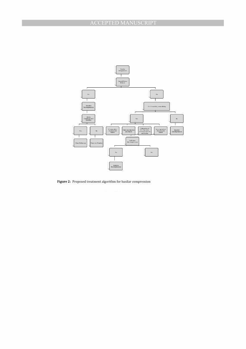
In our patient, the particular anatomic orientation of cranial settling and basilar impression allowed for adequate decompression through an isolated endonasal approach. Specifically, the coronal alignment of the occipital condyle with the C1 lateral mass and C2 facet joints suggested C1-C2 stability. Foregoing posterior fusion in patients with these characteristics is a viable option, but requires close post-operative follow-up. Improvement in neurologic symptoms and absence of neck pain calls for continued clinical observation. On the other hand, deteriorating neurological function or progressively worsening neck pain may be a harbinger of cervical instability and posterior stabilization should be considered. Intraoperative CT following a resection may provide valuable utility in determining decompression was adequate (7). We highlight our proposed algorithm in Figure 2.
Limitations
This is a single case report and our algorithm may not be applicable to other surgeons.
Based on the literature reviewed and our experience with this case, we believe our algorithm offers a way to stratify surgical approaches to basilar invagination. Surgeon preferences and experience may dictate the most appropriate approach to treatment of BI. Conclusions Basilar invagination is a CVJ anomaly that poses considerable management challenges. No well-established studies clearly delineate a treatment algorithm for this condition. From review of the literature and our experience, we propose that an anterior approach should be considered first in patients with high cervicomedullary compression and lack of C1-C2 mobility. During this procedure, effort should be made to preserve the anterior ring of C1 and the lateral masses of C1-C2 to avoid fusion (8). In cases without cranial nerve palsies and presence of C1-C2 mobility, a posterior approach with fusion is preferred. If the posterior decompression fails to relieve the symptoms or there is evidence of a high-level compression, then a combination of anterior and posterior approaches is necessary. Throughout the management triage process, careful preoperative planning and thorough discussion of peri-operative risks should be carried out with the patient.

MANUSCRIP
T
ACCEPTED
ACCEPTED MANUSCRIPT
Acknowledgements: We express our sincere gratitude to Dr. Vincent Traynelis from the Department of Neurosurgery at the Rush University Medical Center for his critical appraisal and advice on this manuscript.
MANUSCRIP
T
ACCEPTED
ACCEPTED MANUSCRIPT
References 1. Bollo RJ, Riva-Cambrin J, Brockmeyer MM, Brockmeyer DL: Complex Chiari
malformations in children: an analysis of preoperative risk factors for occipitocervical fusion. J Neurosurg Pediatr 10:134-141, 2012
2. Choudhri O, Mindea SA, Feroze A, Soudry E, Chang SD, Nayak JV: Experience with intraoperative navigation and imaging during endoscopic transnasal spinal approaches to the foramen magnum and odontoid. Neurosurg Focus 36:E4, 2014
3. Dasenbrock HH, Clarke MJ, Bydon A, Sciubba DM, Witham TF, Gokaslan ZL, et al: Endoscopic image-guided transcervical odontoidectomy: outcomes of 15 patients with basilar invagination. Neurosurgery 70:351-359; discussion 359-360, 2012
4. de Almeida JR, Zanation AM, Snyderman CH, Carrau RL, Prevedello DM, Gardner PA, et al: Defining the nasopalatine line: the limit for endonasal surgery of the spine. Laryngoscope 119:239-244, 2009
5. Duntze J, Eap C, Kleiber JC, Theret E, Dufour H, Fuentes S, et al: Advantages and limitations of endoscopic endonasal odontoidectomy. A series of nine cases. Orthop Traumatol Surg Res, 2014
6. El-Sayed IH, Wu JC, Dhillon N, Ames CP, Mummaneni P: The importance of platybasia and the palatine line in patient selection for endonasal surgery of the craniocervical junction: a radiographic study of 12 patients. World Neurosurg 76:183-188; discussion 174-188, 2011
7. Gande A, Tormenti MJ, Koutourousiou M, Paluzzi A, Fernendez-Miranda JC, Snydermnan CH, et al: Intraoperative computed tomography guidance to confirm decompression following endoscopic endonasal approach for cervicomedullary compression. J Neurol Surg B Skull Base 74:44-49, 2013
8. Gladi M, Iacoangeli M, Specchia N, Re M, Dobran M, Alvaro L, et al: Endoscopic transnasal odontoid resection to decompress the bulbo-medullary junction: a reliable anterior minimally invasive technique without posterior fusion. Eur Spine J 21 Suppl 1:S55-60, 2012
9. Goel A: Treatment of basilar invagination by atlantoaxial joint distraction and direct lateral mass fixation. J Neurosurg Spine 1:281-286, 2004
10. Goel A, Bhatjiwale M, Desai K: Basilar invagination: a study based on 190 surgically treated patients. J Neurosurg 88:962-968, 1998
11. Grammatica A, Bonali M, Ruscitti F, Marchioni D, Pinna G, Cunsolo EM, et al: Transnasal endoscopic removal of malformation of the odontoid process in a patient with type I Arnold-Chiari malformation: a case report. Acta Otorhinolaryngol Ital 31:248-252, 2011
12. Hankinson TC, Grunstein E, Gardner P, Spinks TJ, Anderson RC: Transnasal odontoid resection followed by posterior decompression and occipitocervical fusion in children with Chiari malformation Type I and ventral brainstem compression. J Neurosurg Pediatr 5:549-553, 2010
13. Hwang SW, Heilman CB, Riesenburger RI, Kryzanski J: C1-C2 arthrodesis after transoral odontoidectomy and suboccipital craniectomy for ventral brain stem compression in Chiari I patients. Eur Spine J 17:1211-1217, 2008
14. Jian FZ, Chen Z, Wrede KH, Samii M, Ling F: Direct posterior reduction and fixation for the treatment of basilar invagination with atlantoaxial dislocation. Neurosurgery 66:678-687; discussion 687, 2010
15. Joseph V, Rajshekhar V: Resolution of syringomyelia and basilar invagination after traction. Case illustration. J Neurosurg 98:298, 2003
MANUSCRIP
T
ACCEPTED
ACCEPTED MANUSCRIPT
16. Kassam AB, Snyderman C, Gardner P, Carrau R, Spiro R: The expanded endonasal approach: a fully endoscopic transnasal approach and resection of the odontoid process: technical case report. Neurosurgery 57:E213; discussion E213, 2005
17. Klekamp J: Treatment of basilar invagination. Eur Spine J 23:1656-1665, 2014 18. Laufer I, Greenfield JP, Anand VK, Hartl R, Schwartz TH: Endonasal endoscopic
resection of the odontoid process in a nonachondroplastic dwarf with juvenile rheumatoid arthritis: feasibility of the approach and utility of the intraoperative Iso-C three-dimensional navigation. Case report. J Neurosurg Spine 8:376-380, 2008
19. Leng LZ, Anand VK, Hartl R, Schwartz TH: Endonasal endoscopic resection of an os odontoideum to decompress the cervicomedullary junction: a minimal access surgical technique. Spine (Phila Pa 1976) 34:E139-143, 2009
20. Mazzatenta D, Zoli M, Mascari C, Pasquini E, Frank G: Endoscopic endonasal odontoidectomy: clinical series. Spine (Phila Pa 1976) 39:846-853, 2014
21. McGirt MJ, Attenello FJ, Sciubba DM, Gokaslan ZL, Wolinsky JP: Endoscopic transcervical odontoidectomy for pediatric basilar invagination and cranial settling. Report of 4 cases. J Neurosurg Pediatr 1:337-342, 2008
22. Menezes AH: Craniovertebral junction abnormalities with hindbrain herniation and syringomyelia: regression of syringomyelia after removal of ventral craniovertebral junction compression. J Neurosurg 116:301-309, 2012
23. Menezes AH, VanGilder JC, Graf CJ, McDonnell DE: Craniocervical abnormalities. A comprehensive surgical approach. J Neurosurg 53:444-455, 1980
24. Mouchaty H, Perrini P, Conti R, Di Lorenzo N: Craniovertebral junction lesions: our experience with the transoral surgical approach. Eur Spine J 18 Suppl 1:13-19, 2009
25. Naderi S, Pamir MN: Further cranial settling of the upper cervical spine following odontoidectomy. Report of two cases. J Neurosurg 95:246-249, 2001
26. Salunke P, Behari S, Kirankumar MV, Sharma MS, Jaiswal AK, Jain VK: Pediatric congenital atlantoaxial dislocation: differences between the irreducible and reducible varieties. J Neurosurg 104:115-122, 2006
27. Scholtes F, Signorelli F, McLaughlin N, Lavigne F, Bojanowski MW: Endoscopic endonasal resection of the odontoid process as a standalone decompressive procedure for basilar invagination in Chiari type I malformation. Minim Invasive Neurosurg 54:179-182, 2011
28. Simsek S, Yigitkanli K, Belen D, Bavbek M: Halo traction in basilar invagination: technical case report. Surg Neurol 66:311-314; discussion 314, 2006
29. Singh H, Grobelny BT, Harrop J, Rosen M, Lober RM, Evans J: Endonasal access to the upper cervical spine, part one: radiographic morphometric analysis. J Neurol Surg B Skull Base 74:176-184, 2013
30. Wu JC, Huang WC, Cheng H, Liang ML, Ho CY, Wong TT, et al: Endoscopic transnasal transclival odontoidectomy: a new approach to decompression: technical case report. Neurosurgery 63:ONSE92-94; discussion ONSE94, 2008
31. Yu Y, Hu F, Zhang X, Ge J, Sun C: Endoscopic transnasal odontoidectomy combined with posterior reduction to treat basilar invagination: technical note. J Neurosurg Spine 19:637-643, 2013
32. Yu Y, Wang X, Zhang X, Hu F, Gu Y, Xie T, et al: Endoscopic transnasal odontoidectomy to treat basilar invagination with congenital osseous malformations. Eur Spine J 22:1127-1136, 2013
MANUSCRIP
T
ACCEPTED
ACCEPTED MANUSCRIPT
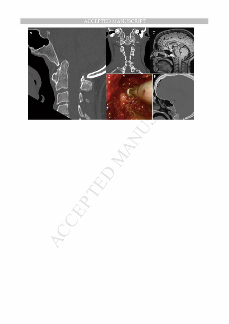
Figure Legend Figure 1. Basilar invagination pre-operative and post-operative imaging. Pre-operative cervical spine CT demonstrating (A) basilar compression of the medulla in mid-sagittal view, and (B) cranial C1-C2 settling on coronal view. (C) Pre-operative sagittal view of the cervicomedullary compression on T2-weighted FLAIR MRI. (D) Intra-operative drilling of odontoid peg. (E) Post-operative CT illustrating successful anterior decompression following endoscopic endonasal odontoidectomy. Figure 2. Management algorithm for anterior and posterior approaches to basilar invagination.
MANUSCRIP
T
ACCEPTED
ACCEPTED MANUSCRIPT
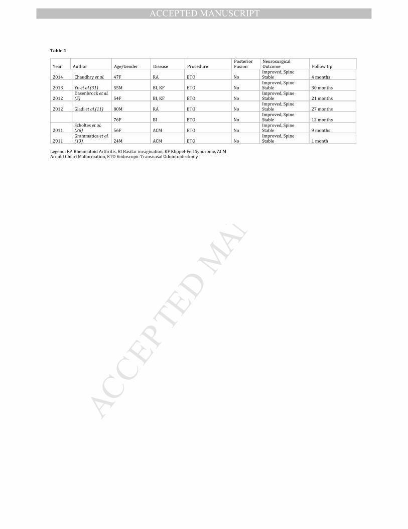
Table Legend Table 1. Reported cases of successful basilar decompression via an endoscopic endonasal approach without posterior fusion.
MANUSCRIP
T
ACCEPTED
ACCEPTED MANUSCRIPT
Table 1
Year Author Age/Gender Disease ProcedurePosterior Fusion
Neurosurgical Outcome Follow Up
2014 Chaudhry et al. 47F RA ETO NoImproved, Spine Stable 4 months
2013 Yu et al.(31) 55M BI, KF ETO NoImproved, Spine Stable 30 months
2012Dasenbrock et al.(5) 54F BI, KF ETO No
Improved, Spine Stable 21 months
2012 Gladi et al.(11) 80M RA ETO NoImproved, Spine Stable 27 months
76F BI ETO NoImproved, Spine Stable 12 months
2011Scholtes et al.(26) 56F ACM ETO No
Improved, Spine Stable 9 months
2011Grammatica et al.(13) 24M ACM ETO No
Improved, Spine Stable 1 month
Legend: RA Rheumatoid Arthritis, BI Basilar invagination, KF Klippel-‐Feil Syndrome, ACM Arnold Chiari Malformation, ETO Endoscopic Transnasal Odointoidectomy
MANUSCRIP
T
ACCEPTED
ACCEPTED MANUSCRIPT
MANUSCRIP
T
ACCEPTED
ACCEPTED MANUSCRIPT
MANUSCRIP
T
ACCEPTED
ACCEPTED MANUSCRIPT
Highlights: • We here present a case with odontoid basilar compression that was decompressed
anteriorly without posterior fusion. • Evidence of autofusion of C1-C2 should be investigated in pre-operative imaging. • Basilar compression with cranial nerve palsies often necessitates anterior
approach when distraction is not possible. • We propose a systematic algorithm for selection of operative approaches. • Intraoperative CT following a resection may provide valuable utility in
determining decompression was adequate
MANUSCRIP
T
ACCEPTED
ACCEPTED MANUSCRIPT
Abbreviations:
Basilar invagination (BI), Rheumatoid Arthritis (RA), Craniovertebral Junction (CVJ),