Embed Size (px)
Citation preview
Volume 18 · Number 4 · December 2016 391
Spontaneous Absorption of Cerebral Air Embolus Developed Accidentally during an Intra-arterial Procedure
Tae Ki YangDepartment of Neurosurgery, School of Medicine, Jeju National University, Jeju, Korea
Cerebral arterial air embolism (CAAE), although infrequent, is a complica-tion that can occur at any time during an invasive medical procedure. We experienced two cases of CAAE during cerebral angiography accidentally. The author reports the two cases of CAAE wherein air em-boli dissolved spontaneously and immediately under normal atmospheric pressure, not under therapeutic hyperbaric environment. One of the cases shows entire dissolution of the air embolus on the moving image. This report shows that arterial air embolus can be absorbed spontaneously, and air embolus size is one of the factors that influence air embolus dis-solution besides hyperbaric oxygen condition.
J Cerebrovasc Endovasc Neurosurg. 2016 December;18(4):391-395Received : 26 April 2016Revised : 1 September 2016Accepted : 19 September 2016
Correspondence to Tae Ki YangDepartment of Neurosurgery, School of Medicine, Jeju National University, 15 Aran 13-gil, Jeju 63241, Korea
Tel : 82-64-754-5104Fax : 82-64-717-1630E-mail : [email protected] : http://orcid.org/0000-0003-2473-5880
This is an Open Access article distributed under the terms of the Creative Commons Attribution Non- Commercial License (http://creativecommons.org/li-censes/by-nc/3.0) which permits unrestricted non- commercial use, distribution, and reproduction in any medium, provided the original work is properly cited.Keywords Air embolism, Cerebral angiography, Spontaneous dissolution
Journal of Cerebrovascular and Endovascular NeurosurgerypISSN 2234-8565, eISSN 2287-3139, http://dx.doi.org/10.7461/jcen.2016.18.4.391 Case Report
INTRODUCTION
Cerebral air embolism is known to be a harmful event
affecting deep-sea divers. It can also occur iatrogeni-
cally during invasive medical procedures. Although
infrequent, it has been steadily reported. Most of the
reported cerebral arterial air embolisms (CAAE) are
paradoxical transvenous embolism, but trans-arterial
CAAE has also been reported occasionally.6)10)11)14) The
mainstay treatment of CAAE is considered hyperbaric
oxygen (HBO) therapy.2) However, HBO therapy does
not always mean improvement. This report introduces
two cases of CAAE that occurred during cerebral an-
giography, which improved without HBO therapy,
and discusses the factors that influence air embolus
absorption with the literature review.
CASE REPORT
Case 1
A 65-year-old man presented with vertigo. The cere-
bral magnetic resonance angiography revealed an un-
ruptured aneurysm in the left middle cerebral artery
bifurcation. Coil embolization was performed under
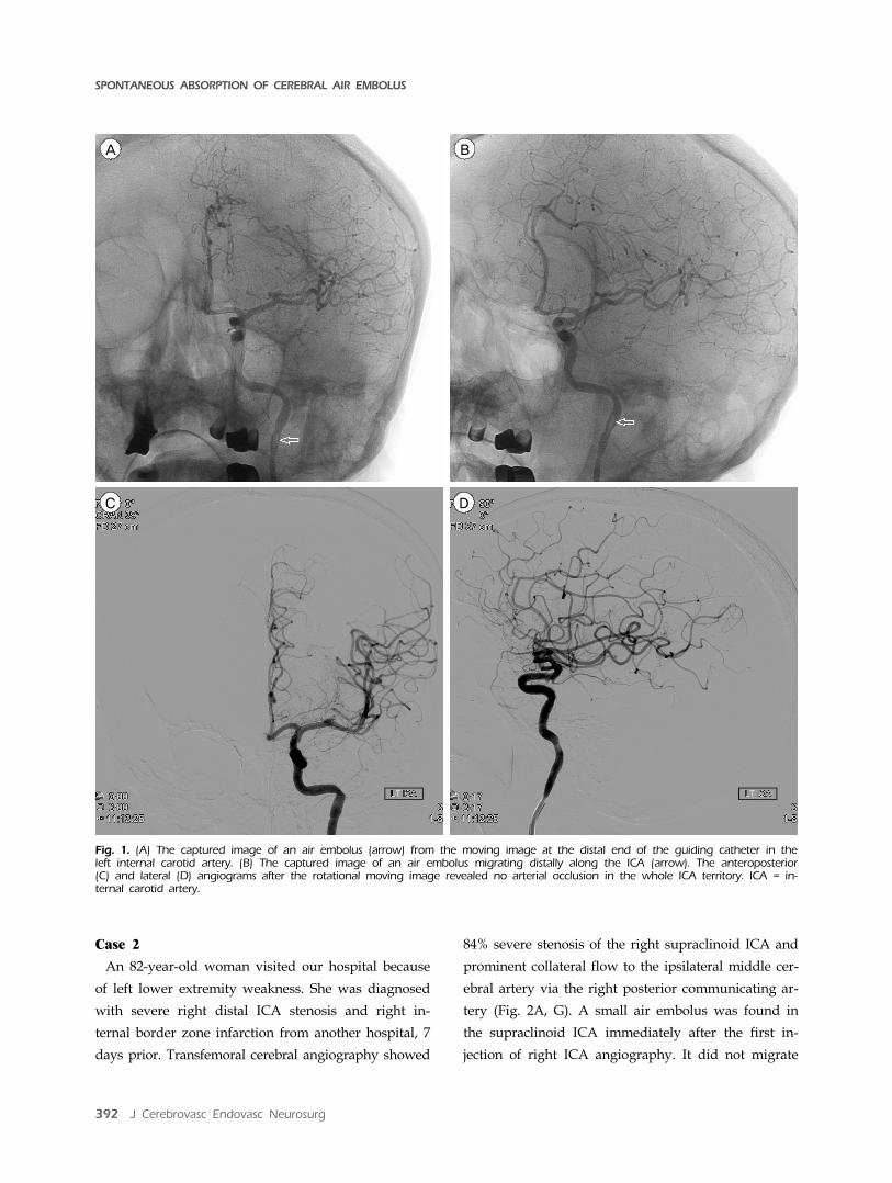
general anesthesia. With the first rotational image ac-
quired during the operation, we detected an air em-
bolus at the distal end of the guiding catheter in the
left internal carotid artery (ICA) (Fig. 1A). The air em-
bolus migrated distally along the middle cerebral ar-
tery (Fig. 1B). During the migration, the embolus de-
creased in size and can be traced to the distal M2 seg-
ment of the middle cerebral artery. We detected no
arterial occlusion from the angiograms taken immedi-
ately after the event (Fig. 1C, D). This air embolus
disappeared spontaneously in a very short time. The
embolization of the aneurysm was performed success-
fully, and the patient recovered from the anesthesia
without any subsequent neurological complications.
SPONTANEOUS ABSORPTION OF CEREBRAL AIR EMBOLUS
392 J Cerebrovasc Endovasc Neurosurg
A
B
C
D
Fig. 1. (A) The captured image of an air embolus (arrow) from the moving image at the distal end of the guiding catheter in the left internal carotid artery. (B) The captured image of an air embolus migrating distally along the ICA (arrow). The anteroposterior (C) and lateral (D) angiograms after the rotational moving image revealed no arterial occlusion in the whole ICA territory. ICA = in-ternal carotid artery.
Case 2
An 82-year-old woman visited our hospital because
of left lower extremity weakness. She was diagnosed
with severe right distal ICA stenosis and right in-
ternal border zone infarction from another hospital, 7
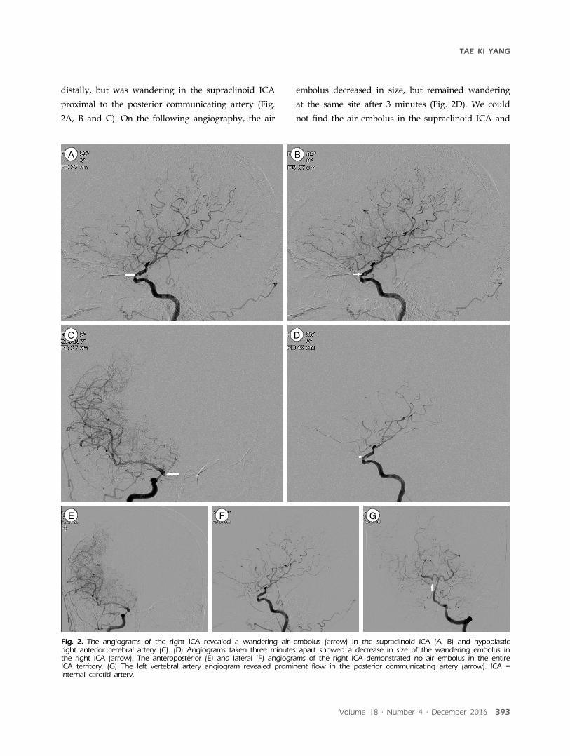
days prior. Transfemoral cerebral angiography showed
84% severe stenosis of the right supraclinoid ICA and
prominent collateral flow to the ipsilateral middle cer-
ebral artery via the right posterior communicating ar-
tery (Fig. 2A, G). A small air embolus was found in
the supraclinoid ICA immediately after the first in-
jection of right ICA angiography. It did not migrate
TAE KI YANG
Volume 18 · Number 4 · December 2016 393
A
B
C
D
E
F
G
Fig. 2. The angiograms of the right ICA revealed a wandering air embolus (arrow) in the supraclinoid ICA (A, B) and hypoplastic right anterior cerebral artery (C). (D) Angiograms taken three minutes apart showed a decrease in size of the wandering embolus in the right ICA (arrow). The anteroposterior (E) and lateral (F) angiograms of the right ICA demonstrated no air embolus in the entire ICA territory. (G) The left vertebral artery angiogram revealed prominent flow in the posterior communicating artery (arrow). ICA = internal carotid artery.
distally, but was wandering in the supraclinoid ICA
proximal to the posterior communicating artery (Fig.
2A, B and C). On the following angiography, the air
embolus decreased in size, but remained wandering
at the same site after 3 minutes (Fig. 2D). We could
not find the air embolus in the supraclinoid ICA and
SPONTANEOUS ABSORPTION OF CEREBRAL AIR EMBOLUS
394 J Cerebrovasc Endovasc Neurosurg
the distal cerebral vasculature 27 minutes after the
event on the following angiography (Fig. 2E, F). The
patient showed no neurological abnormality after the
angiography.
DISCUSSION
Based on reported literatures, the CAAE has occurred
iatrogenically during various clinical procedures. Most
of the CAAEs occur paradoxically, with air entering
the vein during different medical procedures and
moving into the arterial circulation through various
arteriovenous fistulas.3)8)13) The trans-arterial CAAE
during the cerebral angiography has been reported
steadily, like in these cases.6)12)14)
Although the treatment of CAAE has not been es-
tablished, HBO therapy is considered the primary
treatment.2) However, HBO therapy has not always
improved patients with CAAE; some patients treated
with HBO therapy have neurological sequelae or even
died. In contrast, some patients improved without
HBO therapy.6)11-13) Two patients without HBO ther-
apy also had no neurological abnormality in this report.
According to Henry's law, the amount of dissolved
gas, such as oxygen and nitrogen, is influenced by its
partial pressure and temperature. The fact that HBO
therapy does not always coincide with a patient's results
means that other factors affect air embolus absorption
besides the partial pressure of gas, when the body
temperatures are assumed not significantly different.
Many factors are postulated in the literatures such as
the volume of air in the artery, air delivery rates, en-
try route of air and HBO therapy onset.1)3)11)15)
The two CAAE cases in this report occurred during
angiography, and the measured emboli diameters
were 1.75 × 5.4 mm (52 μL) and 1.74 mm (22 μL). The
first case showed the entire process of air embolus
disappearance while migrating distally in the blood
stream. The embolus in the second case was wander-
ing in the supraclinoid internal carotid artery, gradu-
ally decreased in size, and was finally absorbed. The
emboli sizes in our cases were relatively smaller than
those reported in the literature.4)5)7)9) The small embo-
li can be absorbed in the atmospheric condition, not
under hyperbaric environment, like in the present
cases. As long as the embolus is smaller than the arte-
rial lumen diameter, as seen in our two cases, it will
gradually dissolve while migrating or wandering
through the blood stream.
Case 1 showed the completed process in real time,
wherein the air embolus from the guide catheter
gradually decreased in size and disappeared from the
middle cerebral artery M3 segment while migrating
distally. This may be the first case that shows the en-
tire absorption process of an air embolus on the mov-
ing image.
CONCLUSION
Some of the CAAE can be improved spontaneously
without HBO therapy like in these cases; hence, we
can expect that other factors such as embolus size be-
sides pressure and temperature might affect improve-
ment (of CAAE). Our cases demonstrating the dis-
solution of an air embolus in the blood stream pro-
vide a clue on factors affecting the dissolution.
Disclosure
The authors report no conflict of interest concerning
the materials or methods in this study or the fundings
specified in this paper.
REFERENCES
1. Annane D, Troche G, Delisle F, Devauchelle P, Hassine D, Paraire F, et al. Kinetics of elimination and acute consequences of cerebral air embolism. J Neuroimaging. 1995 Jul;5(3):183-9.
2. Bauerle J, Fischer A, Hornig T, Egger K, Wengenmayer T, Bardutzky J. Therapeutic hypothermia in cerebral air embolism: a case report. Springerplus. 2013 Aug;2:411.
3. Benson J, Adkinson C, Collier R. Hyperbaric oxygen therapy of iatrogenic cerebral arterial gas embolism. Undersea Hyperb Med. 2003 Summer;30(2):117-26.
4. Dexter F, Hindman BJ. Recommendations for hyperbaric
TAE KI YANG
Volume 18 · Number 4 · December 2016 395
oxygen therapy of cerebral air embolism based on a mathematical model of bubble absorption. Anesth Analg. 1997 Jun;84(6):1203-7.
5. Fritz H, Hossmann KA. Arterial air embolism in the cat brain. Stroke. 1979 Sep-Oct;10(5):581-9.
6. Gupta R, Vora N, Thomas A, Crammond D, Roth R, Jovin T, et al. Symptomatic cerebral air embolism dur-ing neuro-angiographic procedures: incidence and prob-lem avoidance. Neurocrit Care. 2007;7(3):241-6.
7. McDermott JJ, Dutka AJ, Koller WA, Pearson RR, Flynn ET. Comparison of two recompression profiles in treat-ing experimental cerebral air embolism. Undersea Biomed Res. 1992 May;19(3):171-85.
8. Muth CM, Shank ES. Gas embolism. N Engl J Med. 2000 Feb;342(7):476-82.
9. Reasoner DK, Dexter F, Hindman BJ, Subieta A, Todd MM. Somatosensory evoked potentials correlate with neurological outcome in rabbits undergoing cerebral air embolism. Stroke. 1996 Oct;27(10):1859-64.
10. Sayama T, Mitani M, Inamura T, Yagi H, Fukui M. Normal diffusion-weighted imaging in cerebral air em-
bolism complicating angiography. Neuroradiology. 2000 Mar;42(3):192-4.
11. Surve RM, Reddy KR, Bansal S, Ramalingaiah A. Massive cerebral air embolism during stent-assisted coiling of in-ternal carotid artery aneurysm. Neurol India. 2013 Jan-Feb; 61(1):95-7.
12. Tan LA, Keigher KM, Lopes DK. Symptomatic cerebral air embolism during stent-assisted coiling of an unruptured middle cerebral artery aneurysm: intraoperative diagnosis and management of a rare complication. J Cerebrovasc Endovasc Neurosurg. 2014 Jun;16(2):93-7.
13. Tsetsou S, Eeckhout E, Qanadli SD, Lachenal Y, Vingerhoets F, Michel P. Nonaccidental arterial cerebral air embo-lism: a ten-year stroke center experience. Cerebrovasc Dis. 2013;35(4):392-5.
14. Voorhies RM, Fraser RA. Cerebral air embolism occur-ring at angiography and diagnosed by computerized tomography. Case report. J Neurosurg. 1984 Jan;60(1):177-8.
15. Yesilaras M, Atilla OD, Aksay E, Kilic TY. Retrograde cerebral air embolism. Am J Emerg Med. 2014 Dec;32(12): 1562.e1-2.


![MAPK and pro-inflammatory mediators in the walls of brain ... · or permanent occlusion of a cerebral artery most often by a thrombus or an embolus [10, 11]. When an ischemic stroke](https://img.pdfslide.us/doc/110x75/5f886caff20b9d69481daf8f/mapk-and-pro-inflammatory-mediators-in-the-walls-of-brain-or-permanent-occlusion.jpg)