Embed Size (px)
Citation preview

CHAPTER 5 – PART 1
The Skeletal System

Two Divisions of the Skeleton:
1. Axial Skeleton – The bones that form the longitudinal axis of the body.
2. Appendicular Skeleton – The bones of the limbs and girdles.

The Skeletal System Includes:
1. Bones2. Cartilages3. Joints4. Ligaments
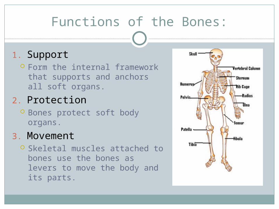
Functions of the Bones:
1. Support Form the internal framework
that supports and anchors all soft organs.
2. Protection Bones protect soft body
organs.
3. Movement Skeletal muscles attached to
bones use the bones as levers to move the body and its parts.
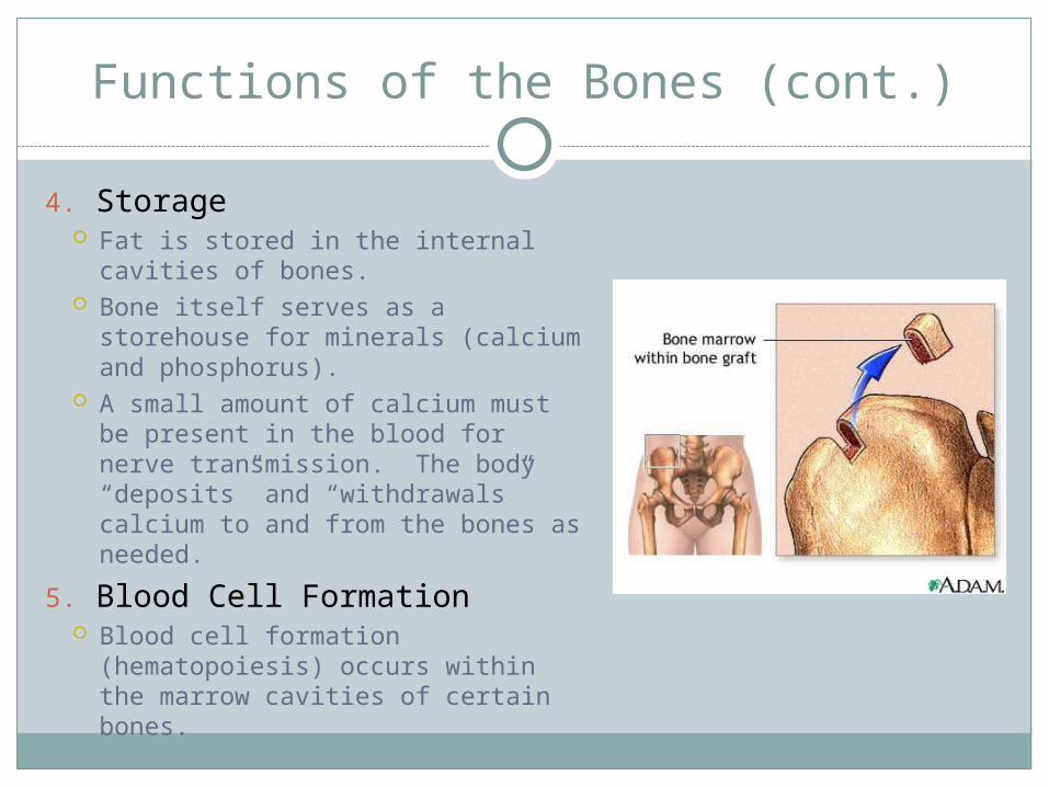
Functions of the Bones (cont.)
4. Storage Fat is stored in the internal
cavities of bones. Bone itself serves as a storehouse
for minerals (calcium and phosphorus).
A small amount of calcium must be present in the blood for nerve transmission. The body “deposits” and “withdrawals” calcium to and from the bones as needed.
5. Blood Cell Formation Blood cell formation
(hematopoiesis) occurs within the marrow cavities of certain bones.

Classification of Bones: Types
The adult skeleton is composed of 206 bones.
There are two basic types of bone tissue:
1. Compact Bone – Dense and looks smooth and homogenous.
2. Spongy Bone – Composed of small needlelike pieces of bone and lots of open space.

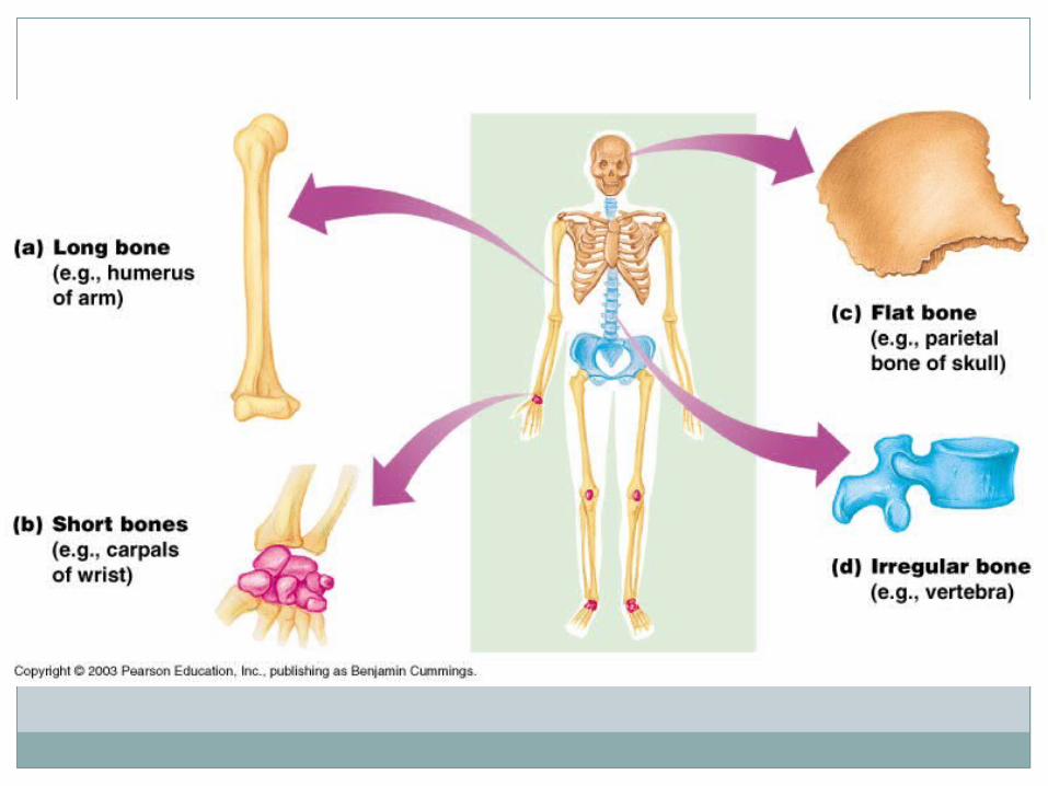
Classification of Bones: Shapes
Bones are classified according to shape:
1. Long Bones – Bones that are typically longer than they are wide; Made mostly of compact bone.
As a rule they have a shaft with heads at both ends.
Examples: All of the bones of the limbs (except the wrist and ankle bones)
2. Short Bones – Generally cube-shaped; Contain mostly spongy bone.
Examples: ankle and wrist bones, patella (kneecap), sesamoid bones (form within tendons)

Classification of Bones: Shapes
Flat Bones – Bones that are thin, flattened, and usually curved; Contain two thin layers of compact bone sandwiched between a layer of spongy bone. Examples: Skull, ribs, sternum
(breastbone). Irregular Bones – Bones that do
not fit one of the preceding categories. Examples: Vertebrae and the hip
bones.

Structure of a Long Bone
Diaphysis – Shaft; Makes up most of the bone’s length and is composed of compact bone. Periosteum – A fibrous CT membrane that covers and
protects the diaphysis.

Structure of a Long Bone
Epiphyses – Ends of the long bones. Each end is composed of a thin layer of compact bone
enclosing an area filled with spongy bone. Articular Cartilage – Covers the external surface of
the epiphyses. Provides a smooth, slippery surface that decreases
friction at joint surfaces.

Structure of a Long Bone
Epiphyseal Line – Thin line of bony tissue spanning the epiphysis that looks a bit different from the rest of the bone in that area.
Epiphyseal Plate – A flat plate of hyaline cartilage seen in young, growing bone. Cause the lengthwise growth of a long bone. By the end of puberty, when hormones stop long bone
growth, epiphyseal plates have been completely replaced by bone, leaving only the epipyseal lines to mark their previous location.


Structure of a Long Bone
Yellow Marrow Cavity (Medullary Cavity) – Cavity of a shaft; Primarily a storage area for adipose (fat) tissue.
Red Bone Marrow – Confined to the cavities of spongy bone of flat bones and the epiphyses of some long bones; Where red blood cells are produced.
Structure of a Long Bone
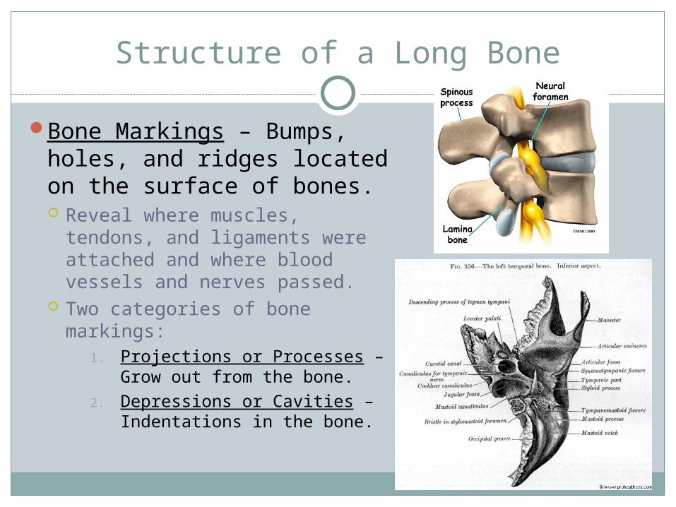
Bone Markings – Bumps, holes, and ridges located on the surface of bones. Reveal where muscles,
tendons, and ligaments were attached and where blood vessels and nerves passed.
Two categories of bone markings:
1. Projections or Processes – Grow out from the bone.
2. Depressions or Cavities – Indentations in the bone.


Bone Cells
Osteocytes – Mature bone cells.Osteoblasts – Bone building cells.Osteoclasts – Bone destroying cells.

Compact Bone Anatomy
Lacunae – Tiny cavities in bones or cartilage where osteocytes are found.
Lamellae – Concentric circles in which the lacunae are arranged.
Haversian Canals and Systems
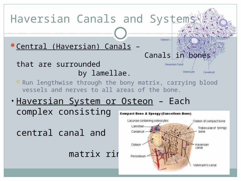
Central (Haversian) Canals – Canals in bones that are surrounded by lamellae. Run lengthwise through the bony matrix, carrying blood
vessels and nerves to all areas of the bone.
• Haversian System or Osteon – Each complex consisting of central canal and matrix rings.

Canaliculi
Canaliculi – Tiny bone canals Radiate outward from the central canals to all
lacunae. Form a transportation system that connects all the
bone cells to the nutrient supply through the hard bone matrix.

Perforating (Volkmann’s) Canals
Perforating (Volkmann’s) Canals – The communication pathway from the outside of the bone to its interior (and the central canals). Run into the compact bone at right angles to the
shaft.


Characteristics of Bone
Because of the elaborate network of canals, bone cells are well nourished in spite of the hardness of the matrix. Bone injuries heal quickly and well.
One of the hardest materials in the body. Although relatively light in weight, it has a
remarkable ability to resist tension and other forces acting on it.

Characteristics of Bone
Extremely strong supporting system, without giving up mobility. Hardness = Calcium salts
deposited in the matrix. Bone’s Flexibility and Strength
= Organic parts such as the collagen fibers.

Bones: From Embryo to Adults
In embryos, the skeleton is primarily made of hyaline cartilage.
In a young child, most of the cartilage has been replaced by bone.
In adults, cartilage remains only in isolated areas such as the bridge of the nose, parts of the ribs, and the joints.