Embed Size (px)
Citation preview
The Skeleton
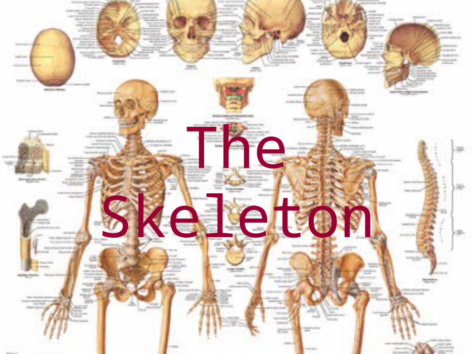
Two Divisions
• Axial
• Appendicular
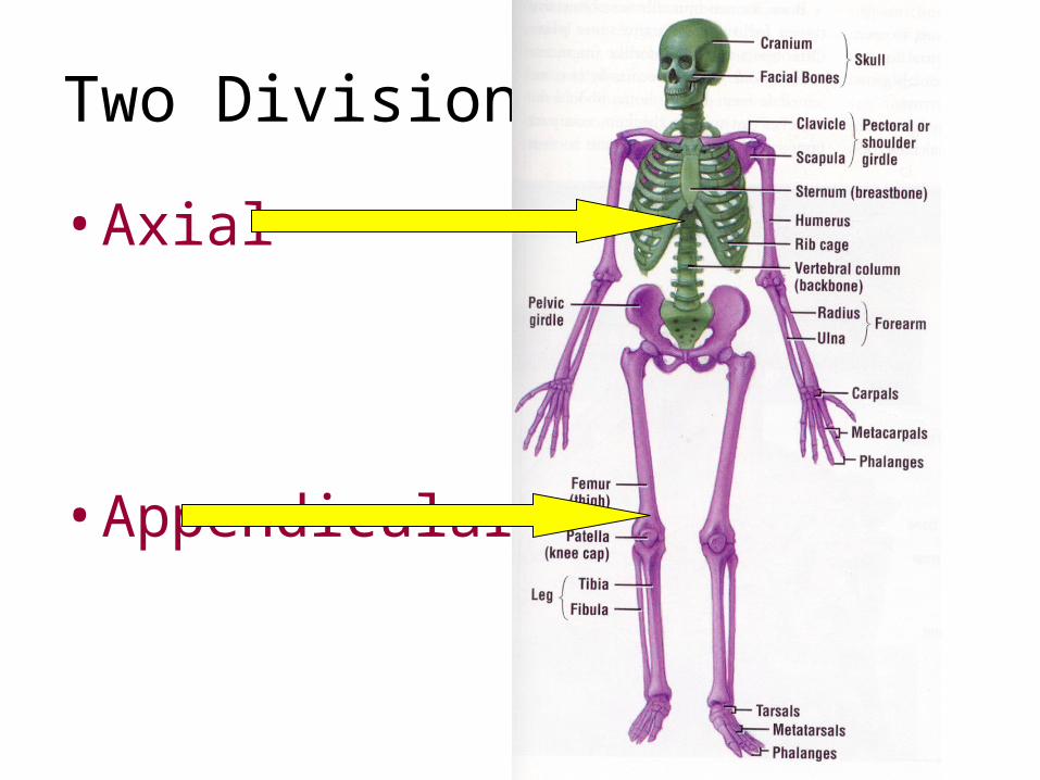
Axial Skeleton
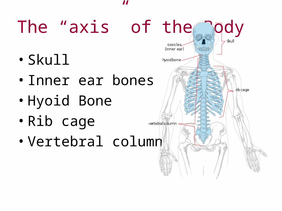
The “axis” of the Body
• Skull
• Inner ear bones
• Hyoid Bone
• Rib cage
• Vertebral column
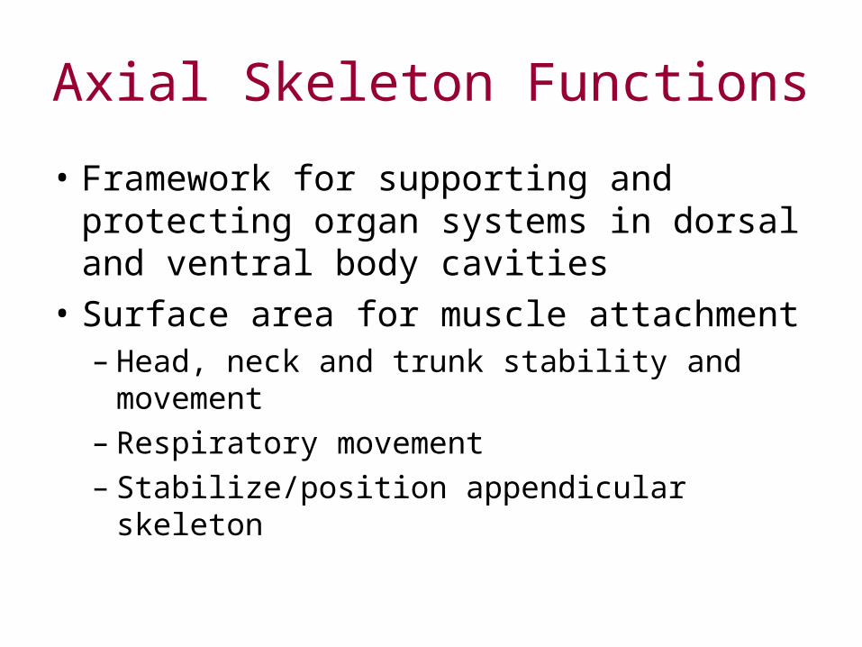
Axial Skeleton Functions
• Framework for supporting and protecting organ systems in dorsal and ventral body cavities
• Surface area for muscle attachment– Head, neck and trunk stability and movement– Respiratory movement– Stabilize/position appendicular skeleton
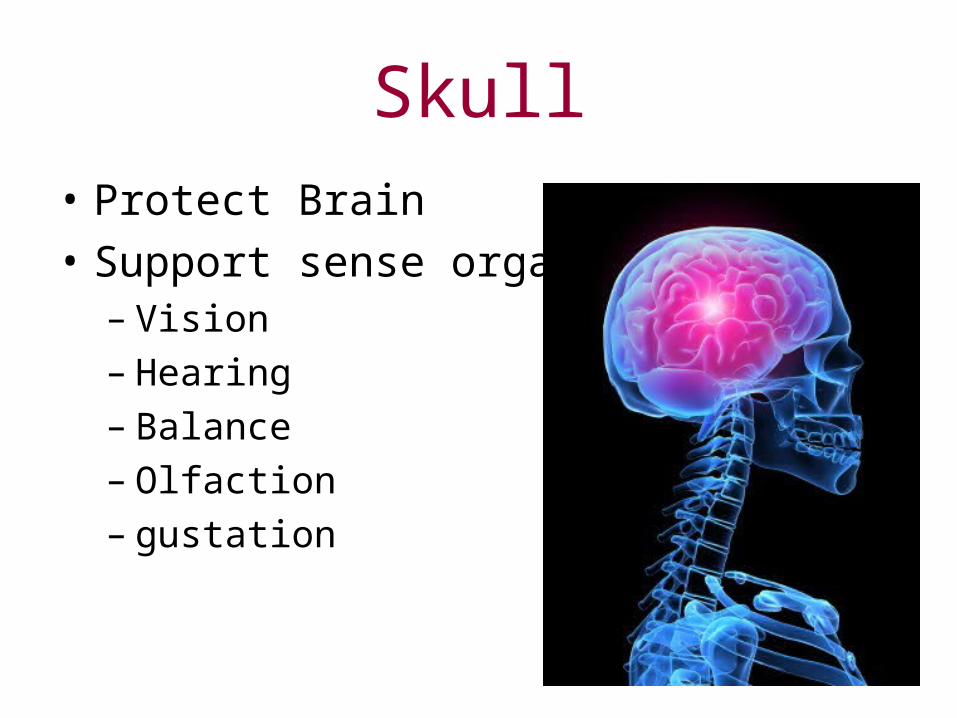
Skull• Protect Brain
• Support sense organs– Vision– Hearing– Balance– Olfaction– gustation
Skull
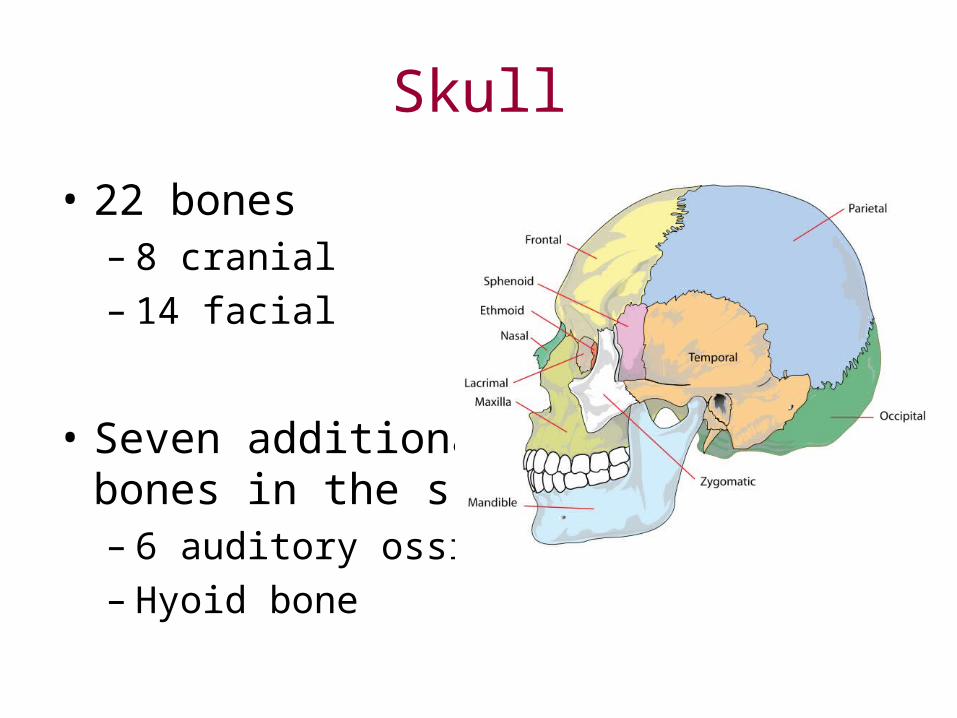
• 22 bones– 8 cranial– 14 facial
• Seven additional bones in the skull– 6 auditory ossicles– Hyoid bone

Hyoid Bone
• Suspended below the skull by ligaments
• Muscle base for the larynx (voice box)
• Supports and positions the larynx
Vertebral Column
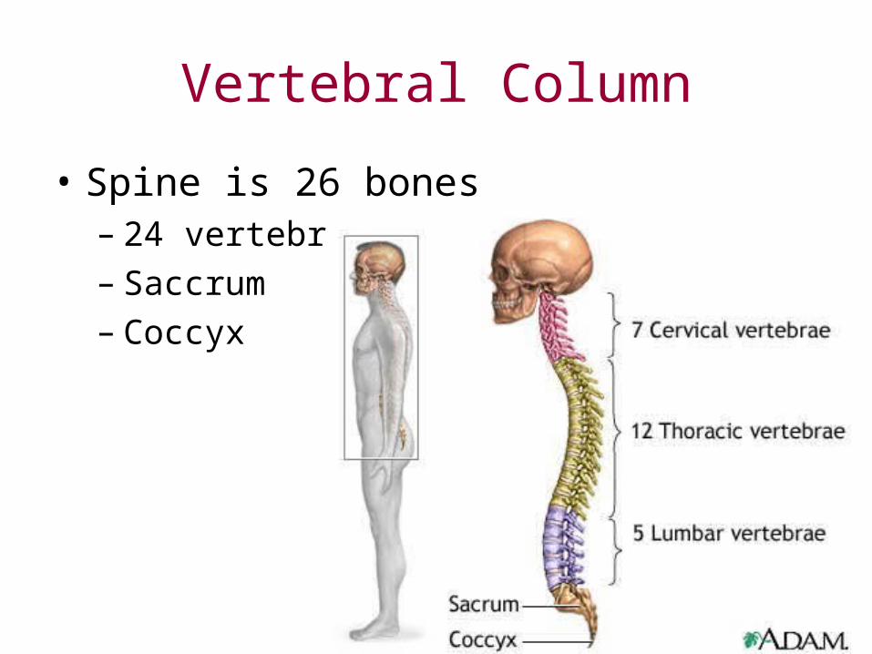
• Spine is 26 bones– 24 vertebrae– Saccrum– Coccyx
Vertebral Column
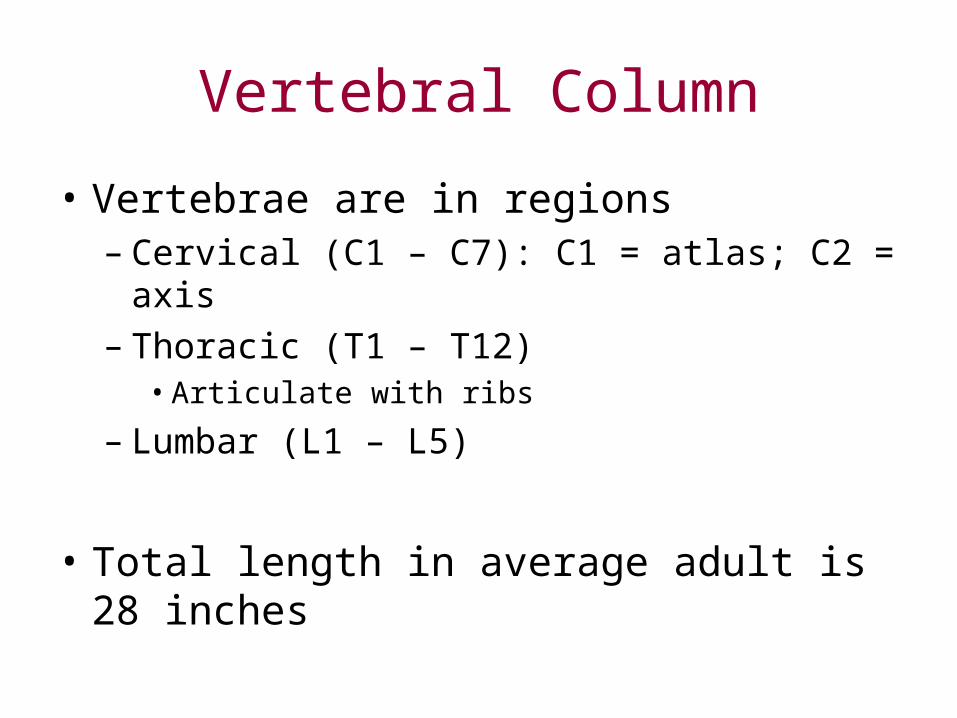
• Vertebrae are in regions– Cervical (C1 – C7): C1 = atlas; C2 = axis– Thoracic (T1 – T12)
• Articulate with ribs
– Lumbar (L1 – L5)
• Total length in average adult is 28 inches
Intervertebral Disc
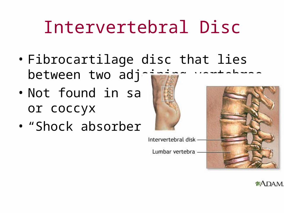
• Fibrocartilage disc that lies between two adjoining vertebrae
• Not found in sacrum or coccyx
• “Shock absorbers”
• Act as ligaments that hold the vertebrae of the spine together and as cartilaginous joints that allow for slight mobility in the spine.
• Allow for movement at the waist as they act as a pivot point and allow the lumbar spine to bend, rotate, and twist
Vertebrae Anatomy
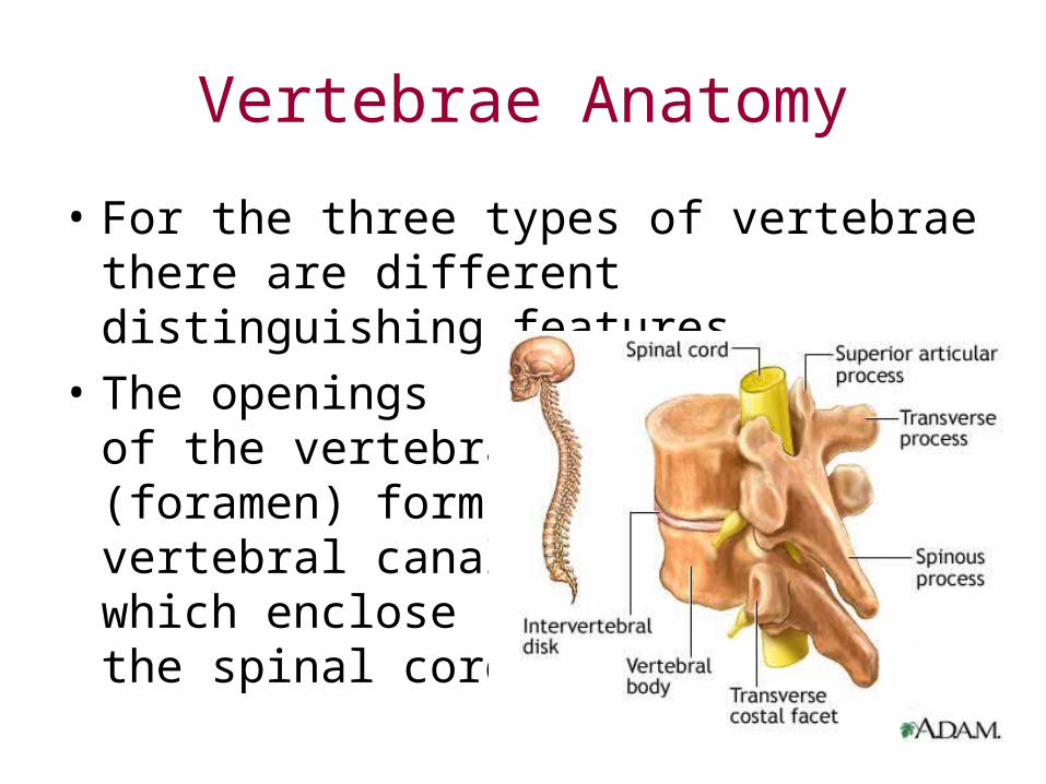
• For the three types of vertebrae there are different distinguishing features
• The openingsof the vertebrae(foramen) form thevertebral canalwhich enclosethe spinal cord
Vertebrae Anatomy
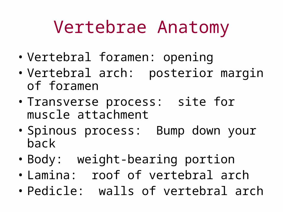
• Vertebral foramen: opening• Vertebral arch: posterior margin of
foramen• Transverse process: site for muscle
attachment• Spinous process: Bump down your back• Body: weight-bearing portion• Lamina: roof of vertebral arch• Pedicle: walls of vertebral arch
Cervical Vertebrae
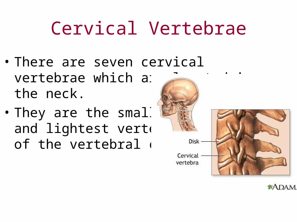
• There are seven cervical vertebrae which are located in the neck.
• They are the smallest, and lightest vertebrae of the vertebral column.
Cervical Vertebrae Anatomy
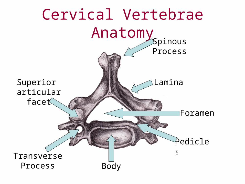
Body
Lamina
Transverse Process
Superior articular facet
Foramen
Spinous Process
Pedicle
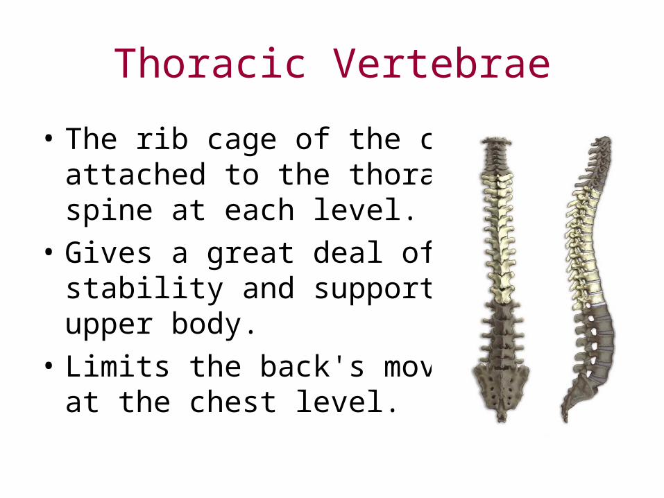
Thoracic Vertebrae
• The rib cage of the chest is attached to the thoracic spine at each level.
• Gives a great deal of stability and support to the upper body.
• Limits the back's movement at the chest level.
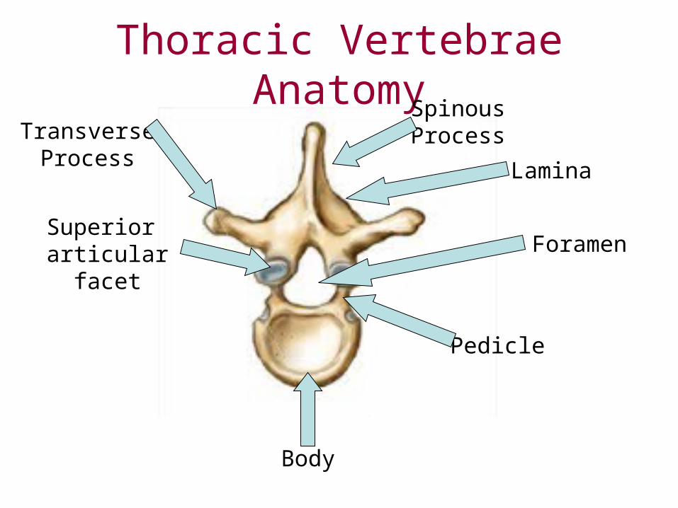
Thoracic Vertebrae Anatomy
Body
Lamina
Transverse Process
Superior articular facet Foramen
Spinous Process
Pedicle
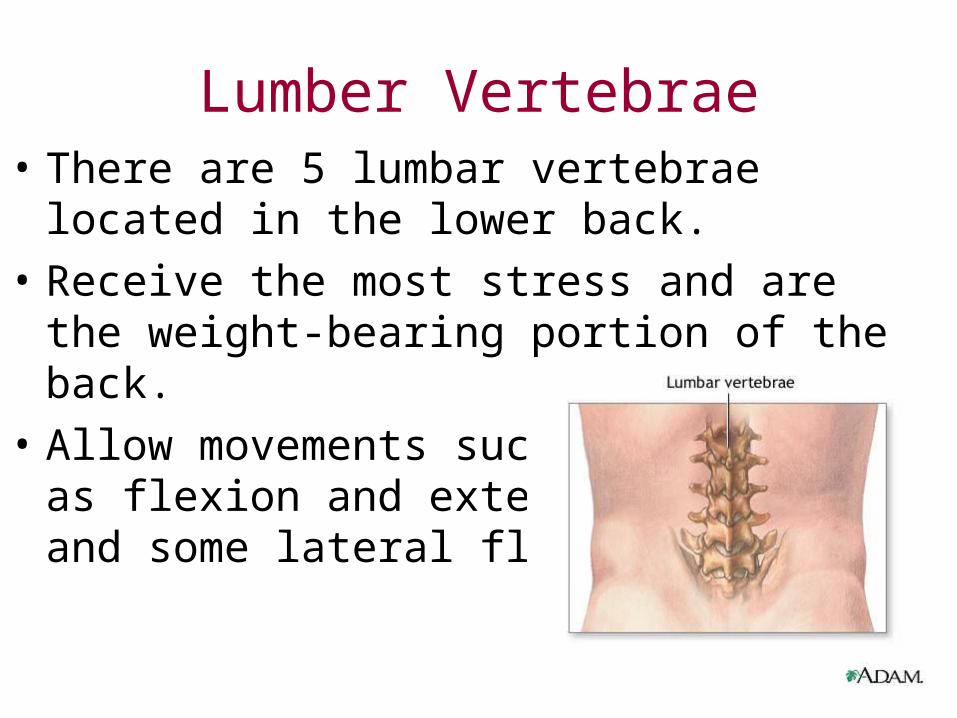
Lumber Vertebrae• There are 5 lumbar vertebrae located in the
lower back.
• Receive the most stress and are the weight-bearing portion of the back.
• Allow movements such as flexion and extensionand some lateral flexion.
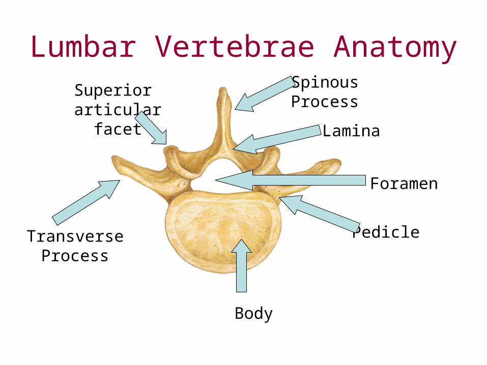
Lumbar Vertebrae Anatomy
Body
Lamina
Transverse Process
Superior articular facet
Foramen
Spinous Process
Pedicle
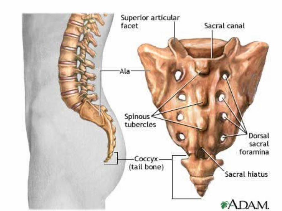
Sacrum and Coccyx
• Sacrum: five fused vertebrae– Protects reproductive and digestive organs– Attaches axial to appendicular skeleton– Extensive muscle attachment
• Coccyx: 3-5 fused vertebrae– Attachment site for muscle that closes anal
opening
Spinal Curves
• Curved to allow for weight distribution• 2 primary curves: appear in late fetal
development– Thoracic– Sacral
• 2 secondary curves: occur months after birth– Cervical– lumbar
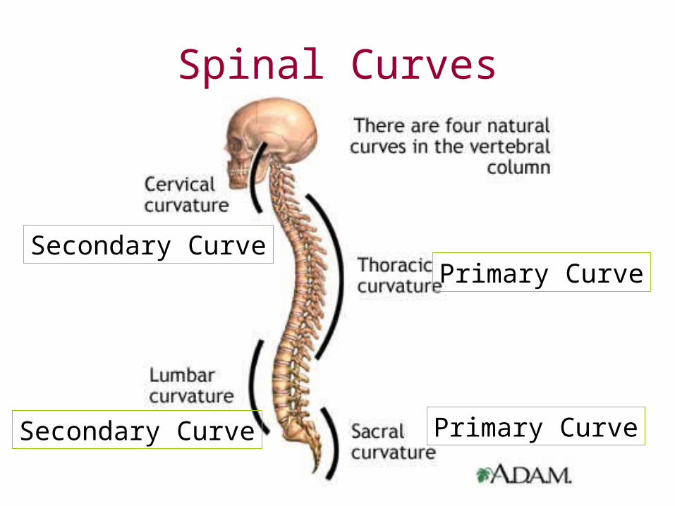
Spinal Curves
Primary CurveSecondary Curve
Primary CurveSecondary Curve
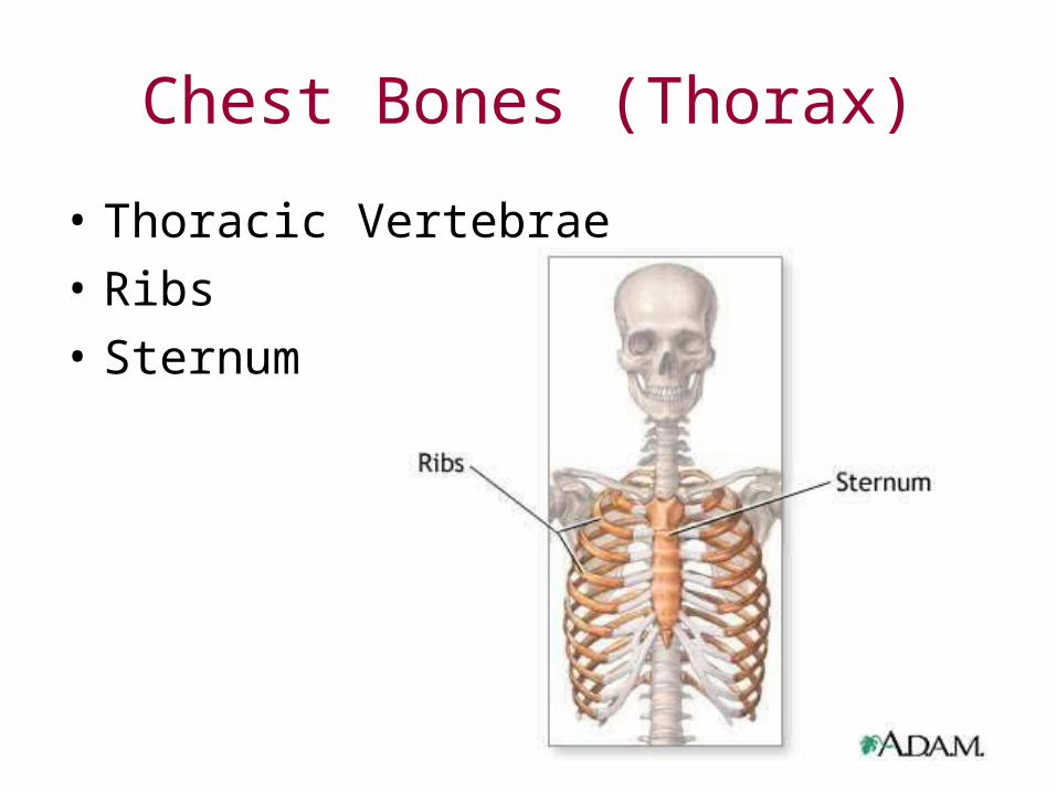
Chest Bones (Thorax)
• Thoracic Vertebrae
• Ribs
• Sternum
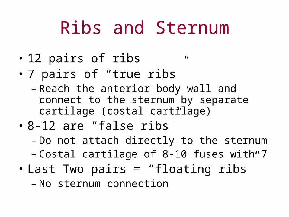
Ribs and Sternum
• 12 pairs of ribs• 7 pairs of “true ribs”
– Reach the anterior body wall and connect to the sternum by separate cartilage (costal cartilage)
• 8-12 are “false ribs”– Do not attach directly to the sternum– Costal cartilage of 8-10 fuses with 7
• Last Two pairs = “floating ribs”– No sternum connection
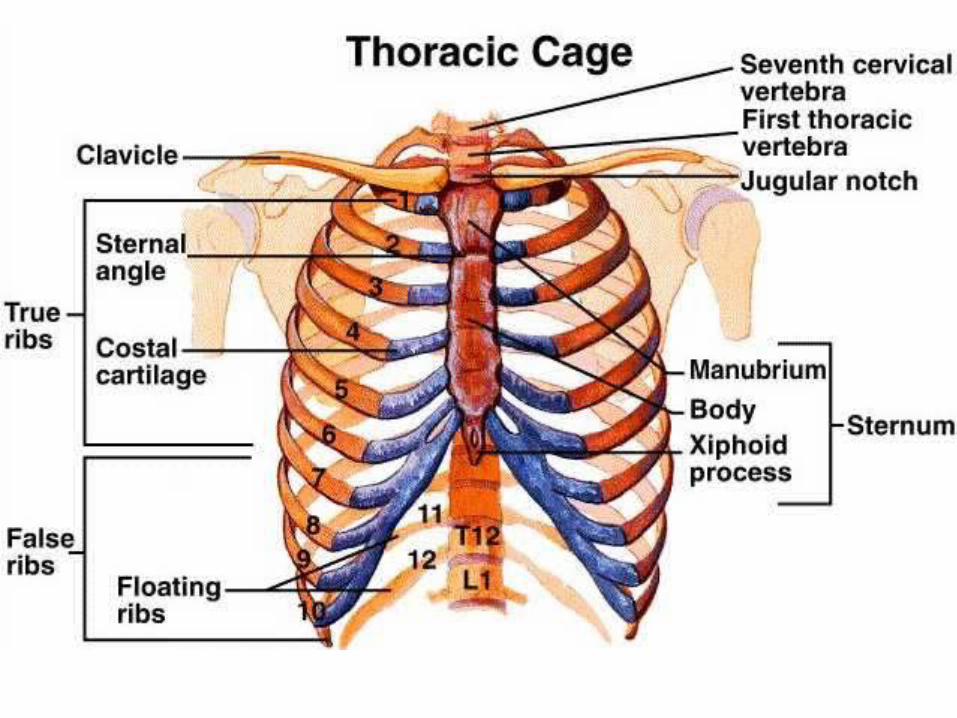
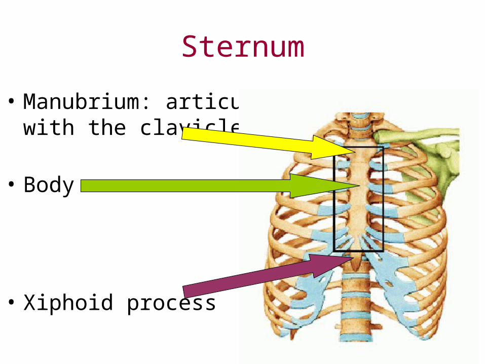
Sternum
• Manubrium: articulateswith the clavicle
• Body
• Xiphoid process
intervertebral disc x ray
• http://www.chirogeek.com/000_disc_anatomy.htm
• http://spanky.thehawkeye.com/features/surgery/index.html