Embed Size (px)
Citation preview
2072 | Phys. Chem. Chem. Phys., 2014, 16, 2072--2084 This journal is© the Owner Societies 2014
Cite this:Phys.Chem.Chem.Phys.,
2014, 16, 2072
Exploring non-covalent interactions in guanine-and xanthine-based model DNA quadruplexstructures: a comprehensive quantum chemicalapproach†
Yevgen P. Yurenko,a Jan Novotny,ab Vladimir Sklenarabc and Radek Marek*abc
The study aimed to cast light on the structure and internal energetics of guanine- and xanthine-based
model DNA quadruplexes and the physico-chemical nature of the non-covalent interactions involved.
Several independent approaches were used for this purpose: DFT-D3 calculations, Quantum Theory of
Atoms in Molecules, Natural Bond Orbital Analysis, Energy Decomposition Analysis, Compliance
Constant Theory, and Non-Covalent Interaction Analysis. The results point to an excellent degree of
structural and energetic compatibility between the two types of model quadruplexes. This fact stems
from both the structural features (close values of van der Waals volumes, pore radii, geometrical
parameters of the H-bonds) and the energetic characteristics (comparable values of the energies of
formation). It was established that hydrogen bonding makes the greatest (B50%) contribution to the
internal stability of the DNA quadruplexes, whereas the aromatic base stacking and ion coordination
terms are commensurable and account for the rest. Energy decomposition analysis performed for
guanine (Gua) and xanthine (Xan) quartets B4 and higher-order structures consisting of two or three
stacked quartets indicates that whereas Gua structures benefit from a high degree of H-bond
cooperativity, Xan models are characterized by a more favorable and cooperative p–p stacking.
The results of electron density topological analysis show that Na+/K+ ion coordination deeply affects the
network of non-covalent interactions in Gua models due to the change in the twist angle between the
stacked tetrads. For Xan models, ion coordination makes tetrads in stacks more planar without changing
the twist angle. Therefore, the presence of the ion seems to be essential for the formation of planar
stacks in Xan-based DNA quadruplexes. Detailed study of the nature of ion-base coordination suggests
that this interaction has a partially covalent character and cannot be considered as purely electrostatic.
Investigation of the H-bond and ion-base coordination strengths by various independent approaches
agrees well with the results of QTAIM analysis.
1. Introduction
Non-covalent interactions, including ion–ion, dipole–dipole,ion–dipole, and dispersion interactions, hydrogen (H) bonding,steric clashes (SC), are ubiquitous forces that play a paramountrole in all areas of biology and nanoscience.1–3 The most
striking evidence of the importance of non-covalent inter-actions is their significance in governing spatial structures anddriving folding processes in nucleic acids, proteins, and theirassemblies. Moreover, an intricate interplay of various non-covalent interactions determines, to a great extent, the courseof all biochemical processes inside a living cell,2 where themutual recognition of biomolecules via non-covalent contactsoccurs. In particular, a comprehensive study of non-covalentinteractions would be beneficial for understanding the poly-morphism and heterogeneity of DNA, since non-covalent forcesthat govern the different forms of DNA are not completelyunderstood. Although experimental studies provide valuableinsights into the internal characteristics of non-covalent forces,they are usually hindered by complications arising from environ-mental effects, as well as competing non-covalent interactions.Therefore, quantum chemistry investigations of model systems
a CEITEC – Central European Institute of Technology, Masaryk University,
Kamenice 5/A4, CZ – 62500 Brno, Czech Republic. E-mail: [email protected] National Center for Biomolecular Research, Faculty of Science,
Masaryk University, Brno, Czech Republicc Department of Chemistry, Faculty of Science, Masaryk University, Brno,
Czech Republic
† Electronic supplementary information (ESI) available: Figures with spatialstructures of two and three stacked tetrads as well as tables with the values ofthe van der Waals volumes and more detailed characteristics of individual non-covalent interactions. See DOI: 10.1039/c3cp53875c
Received 12th September 2013,Accepted 14th November 2013
DOI: 10.1039/c3cp53875c
www.rsc.org/pccp
PCCP
PAPER
Ope
n A
cces
s A
rtic
le. P
ublis
hed
on 1
5 N
ovem
ber
2013
. Dow
nloa
ded
on 1
1/30
/202
1 5:
59:2
5 PM
. T
his
artic
le is
lice
nsed
und
er a
Cre
ativ
e C
omm
ons
Attr
ibut
ion-
Non
Com
mer
cial
3.0
Unp
orte
d L
icen
ce.
View Article OnlineView Journal | View Issue
This journal is© the Owner Societies 2014 Phys. Chem. Chem. Phys., 2014, 16, 2072--2084 | 2073
can be considered as a unique approach that paves way forunderstanding the role of the main non-covalent forces arisingin the forms of DNA and the transition states between them.
Among the possible DNA structures, guanine quadruplexes(G-quadruplexes) deserve special attention. They are formed inguanine-rich sequences that occur frequently in eukaryoticgenomes.4 It should be noted that direct evidence for theformation of G-quadruplexes in vivo was absent for a long time.Only very recently, have Biffi et al. engineered an antibody thatbinds specifically to G-quadruplex structures and enables thedirect visualization of G-tetraplex sites in chromosomes.5
In addition, the indirect data also prove convincingly the supremebiological importance of G-quadruplexes. For instance, proteinsthat can bind to G-quadruplexes have been observed,6 as well ashelicases7 and nucleases8 that act specifically on these structures.However, the most striking fact is that quadruplexes are mostlikely present in the telomeres9 of eukaryotic cells. A link betweenimmortality and telomere maintenance in stem and germ linecells has been demonstrated.10 The proliferating cancerous cellsare immortal due to the high activity of the telomerase enzymemaintaining the length of the telomeres. Whereas single-strandedDNA is a telomerase substrate, G-quadruplex DNA is not. In thisrespect, numerous strategies for the inhibition of telomerase,based on folding single-stranded DNA to G-quadruplexes, havebeen proposed.11,12
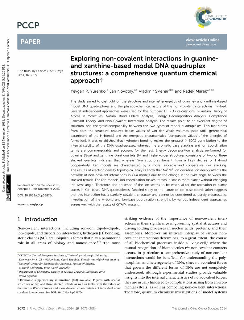
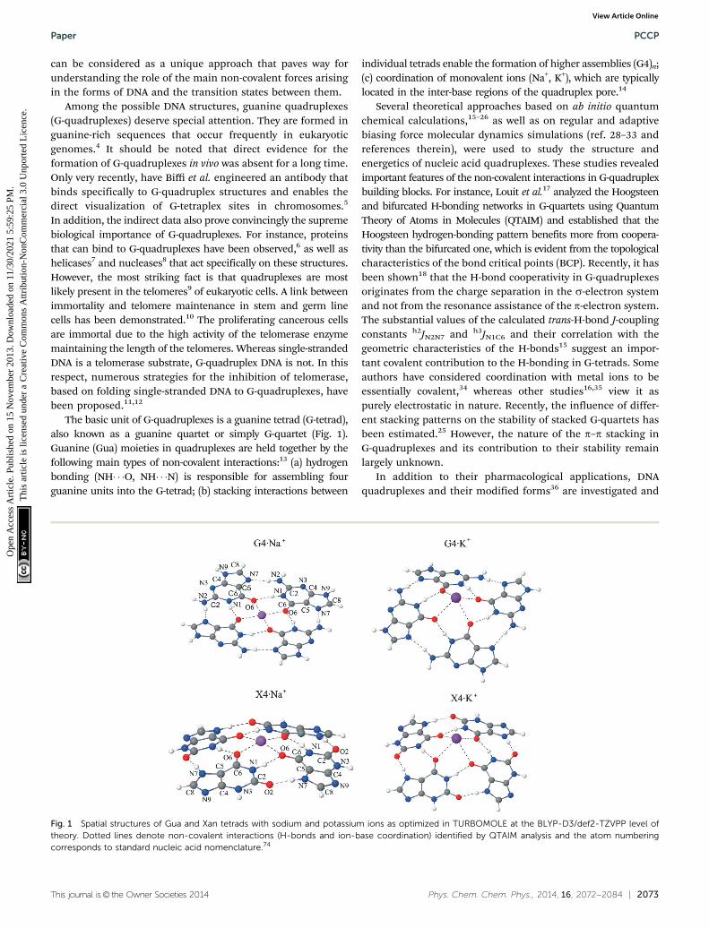
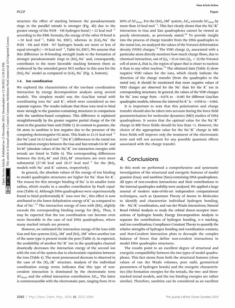
The basic unit of G-quadruplexes is a guanine tetrad (G-tetrad),also known as a guanine quartet or simply G-quartet (Fig. 1).Guanine (Gua) moieties in quadruplexes are held together by thefollowing main types of non-covalent interactions:13 (a) hydrogenbonding (NH� � �O, NH� � �N) is responsible for assembling fourguanine units into the G-tetrad; (b) stacking interactions between
individual tetrads enable the formation of higher assemblies (G4)n;(c) coordination of monovalent ions (Na+, K+), which are typicallylocated in the inter-base regions of the quadruplex pore.14
Several theoretical approaches based on ab initio quantumchemical calculations,15–26 as well as on regular and adaptivebiasing force molecular dynamics simulations (ref. 28–33 andreferences therein), were used to study the structure andenergetics of nucleic acid quadruplexes. These studies revealedimportant features of the non-covalent interactions in G-quadruplexbuilding blocks. For instance, Louit et al.17 analyzed the Hoogsteenand bifurcated H-bonding networks in G-quartets using QuantumTheory of Atoms in Molecules (QTAIM) and established that theHoogsteen hydrogen-bonding pattern benefits more from coopera-tivity than the bifurcated one, which is evident from the topologicalcharacteristics of the bond critical points (BCP). Recently, it hasbeen shown18 that the H-bond cooperativity in G-quadruplexesoriginates from the charge separation in the s-electron systemand not from the resonance assistance of the p-electron system.The substantial values of the calculated trans-H-bond J-couplingconstants h2JN2N7 and h3JN1C6 and their correlation with thegeometric characteristics of the H-bonds15 suggest an impor-tant covalent contribution to the H-bonding in G-tetrads. Someauthors have considered coordination with metal ions to beessentially covalent,34 whereas other studies16,35 view it aspurely electrostatic in nature. Recently, the influence of differ-ent stacking patterns on the stability of stacked G-quartets hasbeen estimated.25 However, the nature of the p–p stacking inG-quadruplexes and its contribution to their stability remainlargely unknown.
In addition to their pharmacological applications, DNAquadruplexes and their modified forms36 are investigated and
Fig. 1 Spatial structures of Gua and Xan tetrads with sodium and potassium ions as optimized in TURBOMOLE at the BLYP-D3/def2-TZVPP level oftheory. Dotted lines denote non-covalent interactions (H-bonds and ion-base coordination) identified by QTAIM analysis and the atom numberingcorresponds to standard nucleic acid nomenclature.74
Paper PCCP
Ope
n A
cces
s A
rtic
le. P
ublis
hed
on 1
5 N
ovem
ber
2013
. Dow
nloa
ded
on 1
1/30
/202
1 5:
59:2
5 PM
. T
his
artic
le is
lice
nsed
und
er a
Cre
ativ
e C
omm
ons
Attr
ibut
ion-
Non
Com
mer
cial
3.0
Unp
orte
d L
icen
ce.
View Article Online
2074 | Phys. Chem. Chem. Phys., 2014, 16, 2072--2084 This journal is© the Owner Societies 2014
designed as building blocks for materials science, biosensors,and nanotechnology.14 It should be mentioned that naturalG-quadruplexes can be used in the above-mentioned applications.However, chemical modifications of the nucleobases and/or asugar–phosphate backbone are needed to improve or enhancespecifically desired quadruplex properties, such as conductivity orthe ability to form supramolecular structures. Xanthine26 and its3-substituted derivatives,27 as suggested recently, can be consid-ered as promising candidates for quadruplex formation since theyare able to form stable base tetrads, as shown by nano-ESI massspectra, NMR spectroscopy, and quantum chemical computations.Despite the fact that xanthine and its derivatives27 are able to formquartets, the latter are different from the corresponding guanineassociates in various ways. For instance, the H-bonding network inxanthine quartets shows little or no cooperativity as compared toG-tetrads.18 In our recent work,37 the idea of a xanthine scaffoldwas extended to the construction of artificial N3-xanthosine-modified DNA quadruplexes. The results of molecular dynamicssimulations indicate several differences between guanine-basedand N3-xanthine-modified DNA quadruplexes, such as preferredconformational states of the sugar–phosphate backbone and ion-transporting barriers and mechanisms. In spite of these differ-ences, our simulations37 demonstrate the considerable stability ofxanthine quadruplexes and imply a good structural compatibilityof the xanthine and guanine tetrads in the DNA quadruplexes.
In this paper, we aim to shed light on the electronic natureand intrinsic characteristics of the non-covalent interactions inguanine- and xanthine-based quadruplexes (H-bonding, aromaticp–p stacking, and metal ion-base coordination). Using simplemolecular models, we apply a large arsenal of current state-of-the-art quantum chemical techniques including Bader’s QTAIMtheory,38 Natural Bond Orbital (NBO) analysis,39 Energy Decom-position Analysis (EDA),40 Non-Covalent Interaction (NCI) plots,41,42
Grunenberg’s compliance constants theory,43,44 and chargeanalyses. We aim to answer the following questions: (i) to whatdegree guanine- and xanthine-based model quadruplexes arestructurally complementary; (ii) how the H-bonding, stacking,and ion-base coordination influence the formation energies ofmodel structures; (iii) what are the fundamental physico-chemical forces that determine the sophisticated network ofnon-covalent interactions in quadruplex structures; and (iv) whatis the electronic nature of the non-covalent interactions in thestudied systems and how do these interactions influence eachother. For the first time, we identify and characterize individualvan der Waals contacts arising in model guanine- and xanthine-quadruplexes and evaluate their role in maintaining the quad-ruplex structures. We also discuss possible trends for introducingmodifications into the xanthine base in such a way that thenon-covalent interactions in new structures would lead toquadruplexes with improved properties.
2. Computational methodology
We used the following molecular models for our investigation:H-bonded base tetrads B4 (B = Gua, Xan); base tetrads with
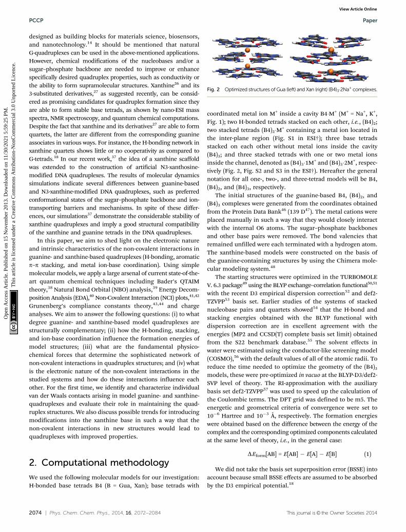
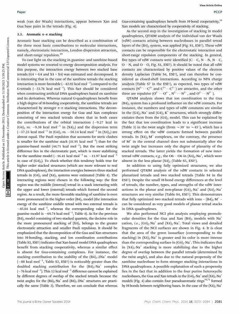
coordinated metal ion M+ inside a cavity B4�M+ (M+ = Na+, K+,Fig. 1); two H-bonded tetrads stacked on each other, i.e., (B4)2;two stacked tetrads (B4)2�M+ containing a metal ion located inthe inter-plane region (Fig. S1 in ESI†); three base tetradsstacked on each other without metal ions inside the cavity(B4)3; and three stacked tetrads with one or two metal ionsinside the channel, denoted as (B4)3�1M+ and (B4)3�2M+, respec-tively (Fig. 2, Fig. S2 and S3 in the ESI†). Hereafter the generalnotation for all one-, two-, and three-tetrad models will be B4,(B4)2, and (B4)3, respectively.
The initial structures of the guanine-based B4, (B4)2, and(B4)3 complexes were generated from the coordinates obtainedfrom the Protein Data Bank46 (139 D47). The metal cations wereplaced manually in such a way that they would closely interactwith the internal O6 atoms. The sugar–phosphate backbonesand other base pairs were removed. The bond valencies thatremained unfilled were each terminated with a hydrogen atom.The xanthine-based models were constructed on the basis ofthe guanine-containing structures by using the Chimera mole-cular modeling system.48
The starting structures were optimized in the TURBOMOLEV. 6.3 package49 using the BLYP exchange–correlation functional50,51
with the recent D3 empirical dispersion correction52 and def2-TZVPP53 basis set. Earlier studies of the systems of stackednucleobase pairs and quartets showed54 that the H-bond andstacking energies obtained with the BLYP functional withdispersion correction are in excellent agreement with theenergies (MP2 and CCSD(T) complete basis set limit) obtainedfrom the S22 benchmark database.55 The solvent effects inwater were estimated using the conductor-like screening model(COSMO),56 with the default values of all of the atomic radii. Toreduce the time needed to optimize the geometry of the (B4)3
models, these were pre-optimized in vacuo at the BLYP-D3/def2-SVP level of theory. The RI-approximation with the auxiliarybasis set def2-TZVPP57 was used to speed up the calculation ofthe Coulombic terms. The DFT grid was defined to be m5. Theenergetic and geometrical criteria of convergence were set to10�6 Hartree and 10�3 Å, respectively. The formation energieswere obtained based on the difference between the energy of thecomplex and the corresponding optimized components calculatedat the same level of theory, i.e., in the general case:
DEform[AB] = E[AB] � E[A] � E[B] (1)
We did not take the basis set superposition error (BSSE) intoaccount because small BSSE effects are assumed to be absorbedby the D3 empirical potential.18
Fig. 2 Optimized structures of Gua (left) and Xan (right) (B4)3�2Na+ complexes.
PCCP Paper
Ope
n A
cces
s A
rtic
le. P
ublis
hed
on 1
5 N
ovem
ber
2013
. Dow
nloa
ded
on 1
1/30
/202
1 5:
59:2
5 PM
. T
his
artic
le is
lice
nsed
und
er a
Cre
ativ
e C
omm
ons
Attr
ibut
ion-
Non
Com
mer
cial
3.0
Unp
orte
d L
icen
ce.
View Article Online
This journal is© the Owner Societies 2014 Phys. Chem. Chem. Phys., 2014, 16, 2072--2084 | 2075
Because some of the optimized structures appeared to benon-planar, which is not relevant to real quadruplex systemswhere stacked tetrads are almost planar, we performed partialoptimization of some planar structures to find the energydifferences between the non-planar and planar structures, aswell as any distinctions in the structural characteristics. Theplanar forms of tetrad assemblies were prepared from opti-mized structures by orienting them axially in the z-directionand assigning an identical average z-coordinate to all of theatoms within a tetrad. To obtain planar structures duringoptimization, we set a constrained symmetry group Cs for thecase of the tetrads B4. For higher order structures, i.e., (B4)2
and (B4)3, an additional dihedral angle y = C8–N1–N1–C8 wasfrozen at 1801 in each stacked tetrad.
Additionally, all model structures were re-optimized bymeans of the ADF program,58 using the same method (BLYP-D3)in combination with an uncontracted polarized triple-z basisset of Slater type orbitals (TZP). The resulting optimized struc-tures were subjected to energy decomposition analysis (EDA) atthe same level of theory into electrostatic interaction DVelstat,Pauli-repulsive orbital interactions DEPauli, attractive orbitalinteractions DEoi, and dispersion energy DEdisp terms:
DEint = DVelstat + DEPauli + DEoi + DEdisp (2)
In eqn (2), the DEoi term accounts for charge transfer(i.e., donor–acceptor interactions between occupied orbitalson one fragment and vacant orbitals of the other) and polariza-tion (mixing of vacant and occupied orbitals in one region dueto the presence of another available region). It should be notedhowever that molecular orbital-based energy decompositionanalysis may overestimate the polarization and charge transfercomponents.59 In the course of energy decomposition we chosemolecular regions in a way that allowed separating and evaluatingthe contribution of a particular type of non-covalent interaction(H-bonding, p–p stacking, or ion coordination) to the stability ofthe B4, (B4)2, and (B4)3 complexes.
Non-covalent interactions (H-bonds, van der Waals contacts,and ion-base coordination contacts) were initially identified bymeans of QTAIM38 methodology using the AIMAll program.60
The presence of a critical point (3, �1) (the so-called BondCritical Point – BCP), a gradient path between two interactingatoms, and a positive value of the Laplacian of electron density,Dr, were considered as indicators of closed-shell non-covalentinteractions (van der Waals contacts, H-bonds, and metalion coordination). The wavefunctions for QTAIM analysiswere calculated in the Gaussian 09 suite of programs61 forTURBOMOLE BLYP-D3/def2-TZVPP geometries at the samelevel of theory (using the BLYP functional and def2-TZVPP basisset imported from the EMSL Basis Set Library).62,63
To study the charge transfer property in the interactingorbitals of H-bonds, we resorted to the NBO analysis39 imple-mented in Gaussian,61 which interprets the electronic wavefunction in terms of a set of occupied Lewis and a set of vacantnon-Lewis localized orbitals. A second-order Fock matrix analysiswas carried out to evaluate the interaction between the donor (i)and acceptor (j) orbitals. The result of such interaction is a
migration of the electron density from the idealized Lewisstructure into a vacant non-Lewis orbital s*. For each donor(i)–acceptor (j) pair, the stabilization energy is calculated asfollows:
E2 ¼ DEij ¼ qiFði; jÞ2
ej � ei(3)
where qi is the donor orbital occupancy, ej and ei are diagonalelements, and F(i,j) is the off-diagonal element of the NBO Fockmatrix.
The H-bond energies were evaluated in several ways. Themost straightforward method was the use of the Espinosa–Molins–Lecomte (EML)64 formula based on the electron densitydistribution at the (3, �1) BCPs of the H-bonds:
EHB = 0.5V(r) (4)
where V(r) is the value of the local potential energy density(virial field) at the (3, �1) BCPs. Because the formula (4) withthe coefficient 0.5 provides only a rough evaluation of theH-bond energy (sometimes smaller values of the coefficientare required to obtain the best fit)65 we also applied therelationship suggested by Nikolaienko et al.66 to estimateformation enthalpies (in kcal mol�1) for the NH� � �O hydrogenbonds: ENH� � �O = �2.03 + 225rBCP, where rBCP is the electrondensity at BCP (in atomic units). This relationship was derivedby comparing the QTAIM and vibrational characteristics ofB2901 conventional hydrogen bonds in 4424 conformers ofDNA-related biomolecules. We could not use this alternativemethodology to estimate the energies of the NH� � �N hydrogenbonds in Gua-containing structures because, to the best of ourknowledge, there are no reliable literature data relating the BCPcharacteristics of NH� � �N bonds and their enthalpies that havebeen tested on a large number of H-bonds of this type.
The relative strengths of the H-bonds and ion coordinationinteractions in the guanine and xanthine tetrads were addi-tionally estimated by using Grunenberg’s compliance constantsformalism.43–45 In contrast to force constants, the numericalvalues of compliance constants do not depend on the coordi-nate system. The physical meaning of compliance constants isdeduced from their definition as the partial second derivativeof the potential energy due to an external force:
Cij ¼@2E
@fi@fj(5)
The H� � �O/N and M+� � �O distances (where O/N are theH-bond acceptor and the oxygen coordinating with the metalion M+) were used as internal coordinates for the calculationof the Cij constants. In other words, compliance constantsmeasure the displacement of an internal coordinate resultingfrom a unit force acting on it. As follows from this definition,a lower numerical value of the compliance constant representsa stronger bond. The compliance constants were calculated byusing the Compliance 3.0.2 program.43,44 It should be notedthat we were unable to apply this methodology to evaluate thestrengths of the van der Waals contacts between stacked bases
Paper PCCP
Ope
n A
cces
s A
rtic
le. P
ublis
hed
on 1
5 N
ovem
ber
2013
. Dow
nloa
ded
on 1
1/30
/202
1 5:
59:2
5 PM
. T
his
artic
le is
lice
nsed
und
er a
Cre
ativ
e C
omm
ons
Attr
ibut
ion-
Non
Com
mer
cial
3.0
Unp
orte
d L
icen
ce.
View Article Online

2076 | Phys. Chem. Chem. Phys., 2014, 16, 2072--2084 This journal is© the Owner Societies 2014
in the (B4)2 and (B4)3 models. This complication stems fromthe fact that this methodology requires the calculation of theHessian for these large structures, which was computationallyinfeasible in our case.
Finally, the strength and location of the non-covalent inter-actions in model quadruplex structures was studied using NCIanalysis,41,42 which is based on the dependence between thereduced electron density gradient s(r) and the sign(l2)r, wherel2 is the second eigenvalue of the electron density Hessian andr is the electron density. This method allowed us to identify theregions where the non-covalent interactions arise and allowedus to decide if the interactions are stabilizing or not.
3. Results and discussion3.1. Energetic preferences and structural characteristics ofmodel quadruplex structures
It is important to note that any nucleobase B that can poten-tially form artificial G-compatible quadruplex structures shouldsatisfy several requirements: (i) steric compatibility with Gua;(ii) the ability to form stable H-bonded quartets; (iii) feasibilityof ion coordination inside the central pore of a quadruplexwithout major hindrances; (iv) the formation of helical stacksconnected by a relaxed sugar–phosphate backbone; (v) thequadruplex formation should be energetically favorable, i.e.,the formation energies of quadruplex structures with base Bshould be comparable with energies for the correspondingGua-containing structures. Taking into account these simplecriteria, we will examine some geometrical and energetic pre-ferences of xanthine-based model quadruplex structures andcompare their properties with guanine-containing models.
We start with a brief comparison of some specific propertiesof Gua and Xan bases. Both bases have two H-bond acceptingand H-bond donating sites. They are also able to form stableHoogsteen base pairs through two H-bonds, which is importantfor the stability of base quartets. The partial atomic charges forGua and Xan monomeric units optimized at the BLYP-D3/def2-TZVPP level of theory were estimated by two different methods(Table 1). The charge analysis allowed the following conclu-sions: (i) the O6 charge values in Gua and Xan bases are ratherclose, which points out that the O6 of Xan can be a goodH-bond acceptor. The slightly lower O6 charge of Xan indicatesthat this base has an intrinsic tendency to form somewhatweaker coordination bonds with Na+/K+ ions; (ii) the O2 atom ofXan has a greater negative charge relative to O6 and thus can bea stronger H-bond acceptor. However, the H-bonding capability
of both oxygens can vary, being influenced by the environment(presence of ion(s), solvent, stacking); (iii) the charges of theH1 atoms are similar in both monomeric units, which indi-cates a similar degree of polarity of the H-bond donatingmoieties N1H.
To characterize the steric compatibility of the Gua andXan bases, we calculated their van der Waals volumes foroptimized geometries (Table S1 in the ESI†) by using the VegaZZ program.67 The close values of these volumes (114.9 Å3 forXan and 116.7 Å3 for Gua) imply good steric compatibility of theGua- and Xan-based quadruplexes. We checked this statementby calculating the van der Waals volumes for higher orderGua- and Xan-containing structures, tetrads B4 and two stackedtetrads (B4)2 with/without ions (Fig. 1 and 2, Fig. S1–S3 in theESI†). It turned out that even for two stacked tetrads withNa+/K+ the maximal difference in the volumes of Gua- andXan-based structures does not exceed 5% (Table S1, ESI†).However, it should be noted that the steric complementarity inreal Gua- and Xan-containing quadruplexes is also determinedby the conformational preferences of their sugar–phosphatebackbone, which are not included in our simple models.
To evaluate the energetic preferences of formation of theGua- and Xan-based structures, we calculated the formationenergies (Table S2 in the ESI†) for the optimized B4, (B4)2, and(B4)3 structures with or without Na+/K+ ion(s). The influence ofthe environment was taken into account by using the COSMOmodel.56 The obtained results (Table S2, ESI†) indicate thatformation energies for the hollow Gua and Xan B4, (B4)2, and(B4)3 models (�33 kcal mol�1,�89 kcal mol�1,�139 kcal mol�1
for Gua and �30 kcal mol�1, �87 kcal mol�1, �145 kcal mol�1
for Xan B4, (B4)2, and (B4)3 complexes, respectively) are similar.These values are close to those reported in ref. 18. It isinteresting that whereas the formation of Gua quartets andtwo stacked Gua quartets is slightly more favorable, the systemof three stacked Xan quartets (X4)3 appears to be more stablethan the (G4)3. This fact can be rationalized by the morefavorable stacking between parallel Xan tetrads. It becomesmore pronounced as the number of stacked tetrads increases(see below). However, the addition of Na+/K+ ions has substan-tially more stabilizing influence on Gua-containing structuresvs. Xan-based models, which can be partially explained by thehigher negative charge of the O6 atom in Gua (Table 1).
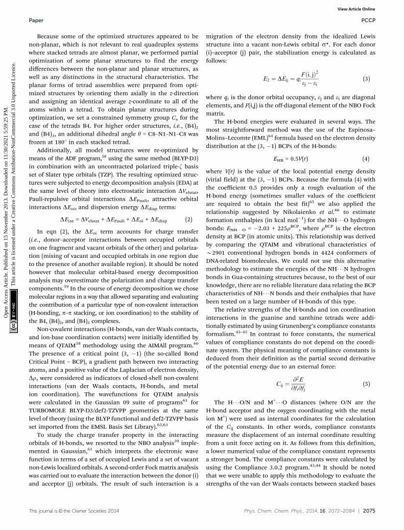
H-bonding, p–p stacking, and ion-base coordination areconsidered to be the most important non-covalent interactionsthat determine the structure of quadruplexes. In this regard,it is important to know to what extent each type of non-covalentinteraction contributes to the energetic stability of the DNAquadruplexes. To tackle this question, we tried to estimate therole of the individual contributions for our models (Gua andXan tetrads, two stacked tetrads, and three stacked tetrads)by means of energy decomposition performed in ADF.58 Theobtained results (Fig. 3, Table S3, ESI†) for (B4)2 suggest thatin the case of two stacked tetrads with ions, the H-bondingplays a dominant role in stabilizing the model quadruplexstructure (its contribution is around 50%) whereas stackingand ion coordination make up 10–14% and 34–41% of the total
Table 1 Table of atomic charges calculated for Gua and Xan monomericunits (ESP-Kolmann and NBO charges)
O6 H1 O2 NH2 N7/H7
G ESP �0.6387 0.4153 0.4019/0.4060 �0.7031NBO �0.6294 0.4211 0.4052/0.4122 �0.4470
X ESP �0.6195 0.4038 �0.6618 0.4039NBO �0.6043 0.4296 �0.6246 0.4447
PCCP Paper
Ope
n A
cces
s A
rtic
le. P
ublis
hed
on 1
5 N
ovem
ber
2013
. Dow
nloa
ded
on 1
1/30
/202
1 5:
59:2
5 PM
. T
his
artic
le is
lice
nsed
und
er a
Cre
ativ
e C
omm
ons
Attr
ibut
ion-
Non
Com
mer
cial
3.0
Unp
orte
d L
icen
ce.
View Article Online
This journal is© the Owner Societies 2014 Phys. Chem. Chem. Phys., 2014, 16, 2072--2084 | 2077
interaction energy, respectively. In this context, it is crucial tofind out whether these ratios remain the same in the case ofreal DNA quadruplexes, where more stacked tetrads are pre-sent. Energy decomposition analysis of the interaction energiesin (B4)3 models with two Na+/K+ ions (Fig. 3, Table S3, ESI†)suggests that in larger stacks with several ions the H-bondingcontribution remains approximately the same (B50%), whilethe balance between coordination and stacking shifts slightlytowards the latter due to the repulsion between the cations insidethe central channel. For instance, in the (X4)3�2K+ structure theH-bonding, stacking, and coordination contributions are 55%,21%, and 24%, respectively. The results of energy decompositionanalysis for these simple models (two stacked tetrads and threestacked tetrads with one and two cations, respectively) can, inprinciple, be extrapolated to larger DNA quadruplex stackssince every tetrad in DNA quadruplexes interacts with 2 (forinternal tetrads) or 1 (external) neighbor(s), and up to 1 cationcan be sandwiched between tetrad layers. Therefore, H-bondingis estimated to contribute approximately 50% to the internalstability of the DNA quadruplex in the absence of the sugar-phosphate backbone (Fig. 3), whereas the stacking and coordinationterms are commensurable and correspond to the remaining B50%of the internal stability, neglecting any equilibria in the solvent.
As far as geometrical characteristics are concerned, someoptimized structures exhibit a noticeable non-planarity. Sincebase tetrads in real quadruplexes are mainly planar, we tried toevaluate any possible impact of non-planarity on the topologiesand energetics of Gua- and Xan-based quadruplex models. Firstof all, planar structures were built from optimized geometriesusing the procedure described in the Computational method-ology section. Some of the resulting planar structures, namelyG4, X4, (G4)2, (X4)2, (G4)2�Na+, and (X4)2�Na+, were partiallyoptimized with the dihedral angle y constrained to 1801
(for a definition, see Computational methodology). Then theroot-mean-square deviations (RMSD) were calculated for indi-vidual base quartets in the fully optimized and planar com-plexes. The latter included six of the above-mentioned partiallyoptimized structures, as well as several selected planarized(B4)3 structures, which, however, were not energy minimized inview of the computational limitations. The results (Table 2) showthat the xanthine tetrad X4 and xanthine quartets in (X4)3�1Na+
and (X4)3�1K+ structures manifest the greatest deviations fromthe planar form. In contrast, guanine-containing structures adoptan almost planar conformation with the exception of the lowertetrad in the (G4)2 model. The energy differences DE between theplanar and non-planar configurations appeared to be very small.The maximum DE (1.8 kcal mol�1) is observed for the xanthinecomplex (X4)2. Therefore, reasonable RMSD values and smallenergy differences (DE) confirm that fully optimized structurescan be considered as very good models of tetrad stacks in DNAquadruplexes.
In addition, twist angles, corresponding to the angularrotation that is needed to get from one base tetrad to thenext, were calculated for the optimized geometries (Table 3).In the case of two stacked Gua and Xan tetrads with a hollowcavity – (G4)2 and (X4)2 – the values of the twist angles are rather
Fig. 3 Relative contributions (in %) of hydrogen bonding, stacking, and coordination to the internal stability of (B4)2�M+ (left) and (B4)3�2M+ (right) models.The interaction energies DEint were obtained by energy decomposition analysis in the ADF58 suite of programs at the BLYP-D3/TZP level of theory in vacuo(for a definition of DEint, see formula (2)). The geometries were optimized at the same level of theory using the COSMO solvent model of water.
Table 2 Characterization of the tetrad planarity
Structure RMSD DE Structure RMSD DE
G4 0.07 0.1 X4 0.49 1.1(G4)2 0.21/0.10 0.3 (X4)2 0.23/0.28 1.8(G4)2�Na+ 0.08/0.09 0.1 (X4)2�Na+ 0.09/0.18 1.2(G4)3�Na+ 0.05/0.08/0.08 (X4)3�Na+ 0.24/0.22/0.11(G4)3�2Na+ 0.12/0.01/0.07 (X4)3�2Na+ 0.23/0.15/0.07(G4)3�K+ 0.11/0.05/0.05 (X4)3�K+ 0.25/0.22/0.24(G4)3�2K+ 0.12/0.02/0.09 (X4)3�2K+ 0.22/0.22/0.22
Paper PCCP
Ope
n A
cces
s A
rtic
le. P
ublis
hed
on 1
5 N
ovem
ber
2013
. Dow
nloa
ded
on 1
1/30
/202
1 5:
59:2
5 PM
. T
his
artic
le is
lice
nsed
und
er a
Cre
ativ
e C
omm
ons
Attr
ibut
ion-
Non
Com
mer
cial
3.0
Unp
orte
d L
icen
ce.
View Article Online
2078 | Phys. Chem. Chem. Phys., 2014, 16, 2072--2084 This journal is© the Owner Societies 2014
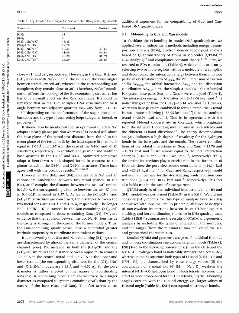
close – 311 and 291, respectively. However, in the Gua (B4)2 and(B4)3 models with Na+/K+ ion(s) the values of the twist anglesbetween tetrads exceed 401, whereas in the corresponding Xancomplexes they remain close to 301. Therefore, Na+/K+ coordi-nation affects the topology of the Gua-containing structures buthas only a small effect on the Xan complexes. It should beremarked that in real G-quadruplex DNA structures the twistangle between two adjacent quartets may vary from B101 toB501 depending on the conformation of the sugar–phosphatebackbone and the type of connecting loops (diagonal, lateral, orpropeller).68
It should also be mentioned that in optimized quartets Na+
adopts a nearly planar position whereas K+ is located well abovethe base plane of the tetrad (the distance from the K+ to themean plane of the tetrad built by the least square fit method isequal to 2.01 Å and 1.97 Å in the case of the G4�K+ and X4�K+
structures, respectively). In addition, the guanine and xanthinebase quartets in the G4�K+ and X4�K+ optimized complexesadopt a bent-down saddle-shaped form, in contrast to theplanar tetrads in the G4�Na+ and X4�Na+ structures. These factsagree well with the previous results.15,18,20,27
However, in the (B4)2 and (B4)3 models both Na+ and K+
cations are sandwiched between two tetrad planes. In the(G4)3�2Na+ complex the distance between the two Na+ cationsis 3.50 Å, the corresponding distance between the two K+ ionsin (G4)3�2K+ is equal to 3.57 Å. As far as the (X4)3�2Na+ and(X4)3�2K+ structures are concerned, the distances between thetwo metal ions are 4.04 Å and 3.76 Å, respectively. The longerNa+� � �Na+/K+� � �K+ distances in the Xan-containing (X4)3�2M+
models as compared to those containing Gua, (G4)3�2M+, areevidence that the repulsion between the two Na+/K+ ions insidethe cavity is stronger in the case of the former models. Thus,the Gua-containing quadruplexes have a somewhat greaterintrinsic propensity to coordinate monovalent cations.
It is noteworthy that Gua and Xan-containing (B4)3 systemsare characterized by almost the same diameter of the centralchannel (pore). For instance, in both the (G4)3�2K+ and the(X4)3�2K+ structures the distance between opposite O6 atoms isB4.60 Å in the central tetrad and B4.78 Å in the upper andlower tetrads (the corresponding distances for the (G4)3�2Na+
and (X4)3�2Na+ models are 4.43 Å and B4.52 Å). So, the porediameter is rather affected by the nature of coordinatingions (e.g., K+-containing models are characterized by a largerdiameter as compared to systems containing Na+) than by thenature of the base (Gua and Xan). This fact serves as an
additional argument for the compatibility of Gua- and Xan-based DNA quadruplexes.
3.2. H-bonding in Gua and Xan models
To elucidate the H-bonding in model DNA quadruplexes, weapplied several independent methods including energy decom-position analysis (EDA), electron density topological analysisbased on Quantum Theory of Atoms in Molecules (QTAIM),38
NBO analysis,39 and compliance constant theory.43–45 First, weresorted to EDA calculations (Table 4), which enable arbitrarilyselecting two or more regions within a molecule or a complex,and decomposed the interaction energy between them into fourparts: an electrostatic term DVelstat, the Pauli repulsion of electronshells DEPauli, the orbital interaction DEoi, and the dispersioncontribution DEdisp. First, the simplest models – the H-bondedHoogsteen base pairs Gua2 and Xan2 – were analyzed (Table 4).The interaction energy for the latter pair (�19.06 kcal mol�1) isnoticeably greater than for Gua2 (�16.14 kcal mol�1). However,when two base pairs are combined to form a tetrad, the G-tetradis much more stabilizing (�55.95 kcal mol�1) than the xanthinetetrad (�38.94 kcal mol�1). This is in agreement with thereported H-bond cooperativity in G-tetrads, which originatesfrom the different H-bonding mechanisms in both tetrads andthe different H-bond directions.18 The energy decompositionanalysis indicates a high degree of covalency for the hydrogenbonds in the base pairs and the tetrads. The relative contribu-tions of the orbital interactions in Gua2 and Xan2 (�15.76 and�18.93 kcal mol�1) are almost equal to the total interactionenergies (�16.14 and �19.06 kcal mol�1, respectively). Thus,the orbital interactions play a crucial role in the formation ofH-bonds since the pure electrostatic contributions (�24.34 kcaland �25.93 kcal mol�1 for Gua2 and Xan2, respectively) wouldnot even compensate for the destabilizing Pauli repulsion con-tributions (28.64 and 30.17 kcal mol�1, respectively). This factalso holds true in the case of base quartets.
QTAIM analysis of the individual interactions in all B4 and(B4)2 models was performed (Table S4 in the ESI†). We did notconsider (B4)3 models for this type of analysis because (B4)2
complexes with ions include, in principle, all three basic typesof non-covalent interactions between bases (H-bonding, p–pstacking, and ion coordination) that arise in DNA quadruplexes.Table S4 (ESI†) summarizes the results of QTAIM and geometricanalyses by including the types of interaction, the numbers,and the ranges (from the minimal to maximal value) for BCPand geometrical characteristics.
Detailed QTAIM and geometric analyses of individual H-bondsand ion-base coordination interactions in tetrad models (Table S4,ESI†) lead to the following observations: (i) in the G4 tetrad theN1H� � �O6 hydrogen bond is noticeably stronger than N2H� � �N7,whereas in the X4 structure both types of H-bond (N1H� � �O6 andN7H� � �O2) are characterized by close energy values; (ii) thecoordination of a metal ion M+ (M+ = Na+, K+) weakens theinternal N1H� � �O6 hydrogen bond in both tetrads, however, thiseffect is more pronounced for the Gua tetrads; (iii) the H-bondingangles correlate with the H-bond energy, i.e., larger values ofH-bond angle (Table S4, ESI†) correspond to stronger bonds.
Table 3 Equilibrated twist angle for Gua and Xan (B4)2 and (B4)3 models
Structure Top twist Bottom twist
(G4)2 31(X4)2 29(G4)2�1Na+/1K+ 46/45(X4)2�1Na+/1K+ 29/28(G4)3�1Na+/1K+ 40/35 43/44(G4)3�2Na+/2K+ 41/46 44/44(X4)3�1Na+/1K+ 28/28 31/29(X4)3�2Na+/2K+ 28/29 28/28
PCCP Paper
Ope
n A
cces
s A
rtic
le. P
ublis
hed
on 1
5 N
ovem
ber
2013
. Dow
nloa
ded
on 1
1/30
/202
1 5:
59:2
5 PM
. T
his
artic
le is
lice
nsed
und
er a
Cre
ativ
e C
omm
ons
Attr
ibut
ion-
Non
Com
mer
cial
3.0
Unp
orte
d L
icen
ce.
View Article Online

This journal is© the Owner Societies 2014 Phys. Chem. Chem. Phys., 2014, 16, 2072--2084 | 2079
To take into account the influence of aromatic p–p stackingon H-bonding in Gua and Xan quartets, the H-bonding inhollow (G4)2 and (X4)2 structures was characterized using theQTAIM methodology. In the cases of both Gua and Xan modelswe expected that the energies of the individual H-bonds wouldincrease under the presence of stacking. However, it turned outthat for the (G4)2 structure the energies of the N1H� � �O6 andN2H� � �N7 hydrogen bonds within two stacked tetrads do notsubstantially change compared to the optimized G4 quartet(Table S4, ESI†). This can be due to the fact that the individualtetrads in the hollow (G4)2 complex exhibit an appreciabledegree of non-planarity (Table 2), which weakens the H-bondsand counterbalances the possibly favorable impact of aromaticp–p stacking on the H-bond strengths.
To tackle this problem, we compared the H-bond energies inthe planarized G4 and (G4)2 structures (Table S4 in the ESI†).It was found that the sum of the H-bond energies in the(G4)2 planar structure (151.96 kcal mol�1 according to EMLformula (4)) exceeds double the total H-bond energy in G4(71.69 kcal mol�1), which documents that p–p stackingenhances the H-bonding in Gua quadruplex structures. As faras the corresponding xanthine models are concerned, thestrengths of both types of H-bond (N1H� � �O6 and N7H� � �O2)in the upper and lower tetrads is increased in the (X4)2 complexrelative to the X4 quartet. Therefore, stacking between paralleltetrad layers has a similar effect on the H-bonding in both the(G4)2 and (X4)2 models.
In contrast to the X4�Na+ and G4�Na+ quartets, the ioncoordination in the central cavity of the (G4)2�Na+ and (X4)2�Na+
models does not substantially influence the total energy of theH-bonds. This can be explained by the fact that metal ioncoordination further increases the tetrad planarity in two-stackcomplexes, which gives rise to stronger H-bonds and compen-sates for the weakening of the N1H� � �O6 hydrogen bonds dueto the polarization of electron density around O6. However, inthe case of (G4)2�K+ and (X4)2�K+, the situation is totallydifferent: the O6� � �K+ contacts are stronger than the O6� � �Na+
(due to the steric requirements of K+, see BCP characteristics),which weakens both types of H-bond (N1H� � �O6 and N7H� � �O2)and subsequently leads to a decrease in the total H-bond energy.Thus, QTAIM analysis of the local M+� � �O6 interactions showsthat K+ tends to bind a bit more strongly to O6 atoms ascompared with Na+ cation, although the global effect (the totalinteraction energies of the ions with the whole system) is alsogoverned by other factors, such as Pauli repulsion and cationinteraction with other parts of the system. It should be notedthat the presence of p–p stacking and ion coordination in the(B4)2 models affects the strength of the individual H-bonds ascompared to tetrads B4. For instance, whereas in the G4 tetradthe N1H� � �O6 hydrogen bond is stronger than the N2H� � �N7bond, in (G4)2�Na+ these two H-bonds are characterized byapproximately the same strength (Table S4, ESI†). The situationis different for the corresponding xanthine-containing models:both of the H-bonds in X4 (N1H� � �O6 and N7H� � �O2) are
Table 4 Energy decomposition analysis (EDA) of interaction energies DEin between different regions of Gua and Xan-based quadruplex models. Modelregions were chosen in a way that allows separating the contributions of the different types of non-covalent interactions. The optimization of thegeometry and subsequent EDA were performed at the BLYP-D3/TZP level of theory. The energy unit is kcal mol�1
Base Type of non-covalent interaction Model/optimized structurea DEoi DEPauli DVelstat DEdisp DEint = SDEi VDDb (ion)
G H-bonding B + B/B2 �15.76 28.64 �24.34 �4.69 �16.14 —B2 + B2/B4 �42.47 59.57 �62.41 �10.64 �55.95 —
Stacking B4 + B4/(B4)2 �9.27 44.71 �11.97 �57.23 �33.76 —B4 (int) + 2*B4 (ext)/(B4)3 �19.50 95.94 �25.41 �120.77 �69.74 —
Ion coordination B4 + Na+/B4�Na+ �38.11 23.40 �90.72 �5.60 �111.04 �0.085(B4)2 + Na+/(B4)2�Na+ �41.61 9.54 �102.64 �13.92 �148.63 �0.078(B4)3 + Na+/(B4)3�1Na+ �44.64 13.30 �111.17 �14.41 �156.92 �0.076(B4)3�Na+ + Na+/(B4)3�2Na+ �44.47 11.40 �56.64 �13.82 �103.53 �0.082B4 + K+/B4�K+ �29.26 23.10 �70.65 �7.33 �84.13 �0.063(B4)2 + K+/(B4)2�K+ �40.55 28.29 �98.86 �15.04 �126.16 �0.058(B4)3 + K+/(B4)3�1K+ �44.07 32.09 �104.45 �14.96 �131.39 �0.059(B4)3�K+ + K+/(B4)3�2K+ �42.74 32.13 �52.76 �16.36 �79.73 �0.063
X H-bonding B + B/B2 �18.93 30.17 �25.93 �4.37 �19.06 —B2 + B2/B4 �38.34 60.40 �51.87 �9.13 �38.94 —
Stacking B4 + B4/(B4)2 �9.38 41.91 �16.41 �58.14 �42.02 —B4 (int) + 2*B4 (ext)/(B4)3 �19.27 87.19 �33.22 �119.74 �85.04 —
Ion coordination B4 + Na+/B4�Na+ �37.76 24.79 �71.05 �5.51 �89.53 �0.080(B4)2 + Na+/(B4)2�Na+ �42.53 12.51 �77.43 �13.20 �120.65 �0.078(B4)3 + Na+/(B4)3�1Na+ �43.42 13.36 �81.75 �13.84 �125.65 �0.078(B4)3�Na+ + Na+/(B4)3�2Na+ �43.87 16.04 �30.85 �14.44 �73.12 �0.080B4 + K+/B4�K+ �28.74 25.11 �55.30 �7.08 �66.01 �0.061(B4)2 + K+/(B4)2�K+ �41.10 33.89 �75.96 �14.43 �97.59 �0.060(B4)3 + K+/(B4)3�1K+ �42.60 34.78 �79.74 �14.76 �102.32 �0.060(B4)3�K+ + K+/(B4)3�2K+ �41.47 34.85 �24.85 �16.37 �47.85 �0.064
a B + B – formation of Hoogsteen base pair from two bases, B2 + B2 – formation of tetrad from two H-bonded Hoogsteen base pairs, B4 + B4 –formation of a hollow (B4)2 system from two tetrads, B4 (int) + 2*B4 (ext) – interaction of an internal tetrad with two external tetrads in a three-stacked system, B4 + Na+ and B4 + K+ – formation of tetrads with a coordinated ion by adding sodium or potassium cations, respectively, (B4)2 + Na+
and (B4)2 + K+ – formation of (B4)2 models with a coordinated ion by adding sodium or potassium cations, (B4)3 + Na+ and (B4)3 + K+ – interactionof a stacked three-tetrad system with a coordinated Na+/K+ cation, (B4)3�Na+/K+ + Na+/K+– interaction of a stacked three-tetrad system with Na+/K+
when the other ion is located inside the channel. b Voronoi deformation density of the Na+/K+ cation in a complex.
Paper PCCP
Ope
n A
cces
s A
rtic
le. P
ublis
hed
on 1
5 N
ovem
ber
2013
. Dow
nloa
ded
on 1
1/30
/202
1 5:
59:2
5 PM
. T
his
artic
le is
lice
nsed
und
er a
Cre
ativ
e C
omm
ons
Attr
ibut
ion-
Non
Com
mer
cial
3.0
Unp
orte
d L
icen
ce.
View Article Online
2080 | Phys. Chem. Chem. Phys., 2014, 16, 2072--2084 This journal is© the Owner Societies 2014
characterized by close energy values, but in (X4)2�Na+ one ofthem (N1H� � �O6) is stronger (Table S4, ESI†).
All of the H-bonds in Gua and Xan quadruplex modelstructures manifest a highly directional character, with AH� � �Bangles close to 1801 (Table S4, ESI†). As far as the distancesdH� � �B between the hydrogen (H) and the acceptor B (B = O, N)atoms are concerned, they correlate perfectly with the BCPcharacteristics: smaller dH� � �B distances correspond to greatervalues of electron density r, the Laplacian of electron densityDr, and the H-bond energy calculated by using the EMLformula. Since the EML formula (4) is known to overestimateH-bond strengths,65,66 we resorted to an alternative way ofevaluating the NH� � �O hydrogen-bond energies by using theempirical relationship from ref. 66 (for details, see Computa-tional methodology and Table S4 footnote, ESI†). It turned outthat the use of this approach yields much smaller H-bondenergy values compared to the EML formula. However, thequantitative trends are well preserved. Therefore, an H-bondenergy calculated by the EML formula can be considered as anupper limit of the real H-bond energy.
To further investigate the H-bonding interactions in modelDNA quadruplexes, we performed Natural Bond Orbital (NBO)analysis of the H-bonding interactions in model Gua- and Xan-based DNA quadruplexes (Table S5 in the ESI†). The resultsshow a substantial charge transfer LP - BD* from one (in thecase of nitrogen) or several (in the case of oxygen) Lewiselectron lone pairs (LP) of the H-bond acceptor atom to theanti-bonding orbital (BD*) corresponding to H-bond donorgroup NH. The amount of the charge transfer can be expressedin terms of the BD* orbital populations, which range from0.045 to 0.064. Thus, our data unambiguously confirm theimportant contribution of the covalent component to theH-bonding reported recently.18 As far as stabilization energiesE2 are concerned, they are higher than the EHB energiescalculated by formula (4). However, E2 and EHB agree qualita-tively since they both reflect the order of strength for theH-bonds. The correlation coefficient between E2 and EHB isequal to 0.83 (Fig. S4 in the ESI†).
To check the dependence of the H-bond characteristics onthe geometry/density functional/basis set type, we obtainedoptimized geometries of the (G4)�Na+, (G4)�K+, (X4)�Na+, and(X4)�K+ tetrads in Gaussian 09 at the B3LYP-D/6-31++G(d,p)level of theory (Table S6 in the ESI†). As a result, the geometriesof all of the tetrads, except (G4)�K+, appeared to be similar toTURBOMOLE structures. As far as (G4)�K+ is concerned, thedifference between the Gaussian and TURBOMOLE structureslies in the fact that the latter exhibits a much greater degree ofnon-planarity, with the K+ ion located well above the tetrad.In contrast, the Gaussian structure is much more planar andthe ion takes a position closer to the O6 atoms. Comparison ofthe H-bond energies calculated by the EML formula and the E2
NBO stabilization energies of the LP - BD* donor–acceptorinteractions, shows that despite some differences in the absolutevalues, the qualitative and even quantitative trends are preserved(Tables S4 and S6, ESI†). The weaker H-bonds in the (G4)�K+
Gaussian structure compared to the related TURBOMOLE
complex are explained by the above-mentioned differences inthe geometries, especially the smaller distance separating theK+ and O6 atoms, which enfeebles the N1H� � �O6 hydrogenbond strength.
In addition, we estimated the relative strengths of theH-bonds by means of Grunenberg’s compliance constantanalysis.43–45 As a result, the compliance constants for theH-bonds fall into the 2.681 to 7.500 Å per mDyn range (highervalues of compliance constants correspond to weaker inter-actions, Table S6 in the ESI†). Therefore, the complianceconstants reproduce well the energetic order of the H-bonds,which makes them suitable for evaluating the strengths of thenon-covalent interactions in DNA quadruplexes.
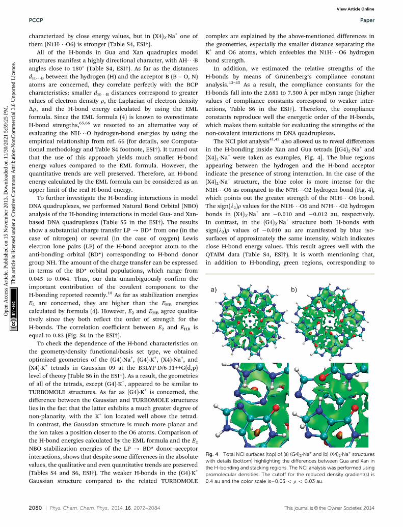
The NCI plot analysis41,42 also allowed us to reveal differencesin the H-bonding inside Xan and Gua tetrads [(G4)2�Na+ and(X4)2�Na+ were taken as examples, Fig. 4]. The blue regionsappearing between the hydrogen and the H-bond acceptorindicate the presence of strong interaction. In the case of the(X4)2�Na+ structure, the blue color is more intense for theN1H� � �O6 as compared to the N7H� � �O2 hydrogen bond (Fig. 4),which points out the greater strength of the N1H� � �O6 bond.The sign(l2)r values for the N1H� � �O6 and N7H� � �O2 hydrogenbonds in (X4)2�Na+ are �0.010 and �0.012 au, respectively.In contrast, in the (G4)2�Na+ structure both H-bonds withsign(l2)r values of �0.010 au are manifested by blue iso-surfaces of approximately the same intensity, which indicatesclose H-bond energy values. This result agrees well with theQTAIM data (Table S4, ESI†). It is worth mentioning that,in addition to H-bonding, green regions, corresponding to
Fig. 4 Total NCI surfaces (top) of (a) (G4)2�Na+ and (b) (X4)2�Na+ structureswith details (bottom) highlighting the differences between Gua and Xan inthe H-bonding and stacking regions. The NCI analysis was performed usingpromolecular densities. The cutoff for the reduced density gradient(s) is0.4 au and the color scale is�0.03 o r o 0.03 au.
PCCP Paper
Ope
n A
cces
s A
rtic
le. P
ublis
hed
on 1
5 N
ovem
ber
2013
. Dow
nloa
ded
on 1
1/30
/202
1 5:
59:2
5 PM
. T
his
artic
le is
lice
nsed
und
er a
Cre
ativ
e C
omm
ons
Attr
ibut
ion-
Non
Com
mer
cial
3.0
Unp
orte
d L
icen
ce.
View Article Online
This journal is© the Owner Societies 2014 Phys. Chem. Chem. Phys., 2014, 16, 2072--2084 | 2081
weak (van der Waals) interactions, appear between Xan andGua base pairs in the tetrads (Fig. 4).
3.3. Aromatic p–p stacking
Aromatic base stacking can be described as a combination ofthe three most basic contributions to molecular interactions,namely, electrostatic interaction, London dispersion attraction,and short-range repulsion.69
To cast light on the stacking in guanine- and xanthine-basedmodel systems we resorted to energy decomposition analysis. Forthis purpose, the interaction energy between two stacked basetetrads (G4 + G4 and X4 + X4) was estimated and decomposed. Itis interesting that in the case of the xanthine tetrads the stackinginteraction is more favorable (�42.02 kcal mol�1) compared to theG-tetrads (�33.76 kcal mol�1). This fact should be consideredwhen constructing artificial DNA quadruplexes based on xanthineand its derivatives. Whereas natural G-quadruplexes benefit froma high degree of H-bonding cooperativity, the xanthine tetrads arecharacterized by stronger p–p stacking interactions. The decom-position of the interaction energy for Gua and Xan complexesconsisting of two stacked tetrads shows that in both casesthe contributions of the orbital interaction (�9.27 kcal in(G4)2 vs. �9.38 kcal mol�1 in (X4)2) and the dispersion term(�57.23 kcal mol�1 in (G4)2 vs. �58.14 kcal mol�1 in (X4)2) arealmost equal. The Pauli repulsion that accounts for steric clashesis smaller for the xanthine stack (41.91 kcal mol�1) than for theguanine-based model (44.71 kcal mol�1). But the most strikingdifference lies in the electrostatic part, which is more stabilizingfor the xanthine model (�16.41 kcal mol�1 vs. �11.97 kcal mol�1
in case of (G4)2). To check whether this tendency holds true forhigher order stacked structures (which are more relevant to realDNA quadruplexes), the interaction energies between three stackedtetrads in (G4)3 and (X4)3 systems were estimated (Table 4). Theinteracting regions were chosen in the following way: the firstregion was the middle (internal) tetrad in a stack interacting withthe upper and lower (external) tetrads which formed the secondregion. As a result, the more favorable stacking of xanthine is evenmore pronounced in the higher order (B4)3 model (the interactionenergy of the xanthine middle tetrad with two external tetrads is�85.04 kcal mol�1, whereas the corresponding value for theguanine model is �69.74 kcal mol�1, Table 4). As for the previous(B4)2 model consisting of two stacked quartets, the decisive role inthe more pronounced stacking of (X4)3 belongs to the greaterelectrostatic attraction and smaller Pauli repulsion. It should beemphasized that the decomposition of the Gua and Xan structuresinto H-bonding, stacking, and ion coordination contributions(Table S3, ESI†) indicates that Xan-based model DNA quadruplexesbenefit from stacking cooperativity, whereas a similar effectis absent for Gua-containing complexes. For instance, thestacking contribution to the stability of the (B4)3�2Na+ model(�88 kcal mol�1, Table S3, ESI†) is noticeably greater than thedoubled stacking contribution for the (B4)2�Na+ complex(�76 kcal mol�1). This 12 kcal mol�1 difference cannot be explainedby different degrees of overlap of the stacked tetrads because thetwist angles for the (B4)2�Na+ and (B4)3�2Na+ structures are practi-cally the same (Table 3). Therefore, we can conclude that whereas
Gua-containing quadruplexes benefit from H-bond cooperativity,18
Xan models are characterized by cooperativity of stacking.As the second step in the investigation of stacking in model
quadruplexes, QTAIM analysis of the individual van der Waals(vdW) contacts arising between nucleobases in parallel tetradlayers of the (B4)2 system, was applied (Fig. S1, ESI†). These vdWcontacts can be responsible for the electrostatic interaction andshort-range repulsion components of the stacking. In general,five types of vdW contacts were identified (C� � �C, N� � �N, N� � �C,O� � �N, and O� � �O, Fig. S1, ESI†). It should be noted that all vdWcontacts are characterized by positive values of the electrondensity Laplacian (Table S4, ESI†), and can therefore be con-sidered as closed-shell interactions. According to NPA chargeanalysis (Table S7 in the ESI†), as expected, two types of vdWcontacts (Nd�� � �Cd+ and Cd+� � �Cd�) are attractive, and the otherthree are repulsive (Od�� � �Od�, Nd�� � �Nd�, and Od�� � �Nd�).
QTAIM analysis shows that ion coordination to the Gua(B4)2 system has a profound influence on the vdW contacts. Forinstance, the numbers and types of vdW constants are similarin the (G4)2�Na+ and (G4)2�K+ structures, which strongly differ-entiates them from the (G4)2 model. This can be explained bythe fact that ion coordination leads to a significant increase(Table 3) in the twist angle (from B301 to B451), which has astrong effect on the vdW contacts formed between paralleltetrads. In (X4)2�M+ complexes, on the contrary, the presenceof M+ in the central channel does not substantially alter thetwist angle but increases only the degree of planarity of thestacked tetrads. The latter yields the formation of new inter-tetrad vdW contacts, e.g., the O6� � �O6 in (X4)2�Na+, which wereabsent in the less planar (X4)2 (Table S5, ESI†).
In addition to using fully optimized structures, we alsoperformed QTAIM analysis of the vdW contacts in selectedplanarized tetrads and two stacked tetrads (Table S4 in theESI†). Despite the small H-bond energy differences at the levelof tetrads, the number, types, and strengths of the vdW inter-actions in the planar and non-planar (G4)2�Na+ and (X4)2�Na+
structures are very similar (Table S4, ESI†). This demonstratesthat fully optimized two stacked tetrads with ions – (B4)2�M+ –can be considered as very good models of planar tetrad stacksin DNA quadruplexes.
We also performed NCI plot analysis employing promole-cular densities for the Gua and Xan (B4)2 models with Na+
cation, i.e., (G4)2�Na+ and (X4)2�Na+. Total views and detailedfragments of the NCI surfaces are shown in Fig. 4. It is clearthat the area of the green isosurface (corresponding to thestacking) in (X4)2�Na+ is greater and its color is more intensethan the corresponding surface in (G4)2�Na+. This indicates thatin (X4)2�Na+ stacking is more stabilizing due to the higherdegree of overlap between the parallel tetrads (determined bythe twist angle), and also due to the natural propensity of thexanthine nucleobase to form stronger stacking interactions inDNA quadruplexes. A possible explanation of such a propensitylies in the fact that in addition to the four purine heterocyclicnucleobases, the Gua and Xan tetrads in the (G4)2�Na+ and (X4)2�Na+
models (Fig. 4) also contain four pseudoaromatic rings70,71 formedby H-bonds between neighboring bases. In the case of the (X4)2�Na+
Paper PCCP
Ope
n A
cces
s A
rtic
le. P
ublis
hed
on 1
5 N
ovem
ber
2013
. Dow
nloa
ded
on 1
1/30
/202
1 5:
59:2
5 PM
. T
his
artic
le is
lice
nsed
und
er a
Cre
ativ
e C
omm
ons
Attr
ibut
ion-
Non
Com
mer
cial
3.0
Unp
orte
d L
icen
ce.
View Article Online
2082 | Phys. Chem. Chem. Phys., 2014, 16, 2072--2084 This journal is© the Owner Societies 2014
structure the effect of stacking between the pseudoaromaticrings in the parallel tetrads is stronger (Fig. 4b) due to thegreater energy of the N1H� � �O6 hydrogen bond (B12 kcal mol�1
according to the EML formula; the energy of the other H-bond isB10 kcal mol�1, Table S4, ESI†), whereas in (G4)2�Na+ theN1H� � �O6 and N2H� � �N7 hydrogen bonds are more or less ofequal strength (B10 kcal mol�1, Table S4, ESI†). We assume thatthis difference in H-bonding strength leads to the formation ofstronger pseudoaromatic rings in (X4)2�Na+ and, consequently,contributes to the more favorable stacking between them asevidenced by the extended green NCI surface in this area for the(X4)2�Na+ model as compared to (G4)2�Na+ (Fig. 4, bottom).
3.4. Ion coordination
We explored the characteristics of the ion-base coordinationinteraction by energy decomposition analysis using severalmodels. The simplest model was the Gua/Xan tetrad withcoordinating ions Na+ and K+, which were considered as twoseparate regions. The results indicate that these ions tend to bindmore strongly to the guanine-containing structures in comparisonwith the xanthine-based complexes. This difference is explainedstraightforwardly by the greater negative partial charge of the O6atom in the guanine base unit (Table 1). In contrast to guanine, theO6 atom in xanthine is less negative due to the presence of thecompeting electronegative O2 atom. This leads to 21.51 kcal mol�1
(for Na+) and 18.12 kcal mol�1 (for K+) differences in the metal ioncoordination energies between the Gua and Xan tetrads G4�M+ andX4�M+ (absolute values of the Na+/K+ ion interaction energies withtetrads are listed in Table 4). The corresponding differencesbetween the (G4)2�M+ and (X4)2�M+ structures are even moresubstantial (27.98 kcal and 28.57 kcal mol�1 for the (B4)2
models with Na+ and K+ cations, respectively).In general, the absolute values of the energy of ion binding
to model quadruplex structures are higher for Na+ than for K+.The reason for this stronger binding of Na+ is its smaller ionicradius, which results in a smaller contribution by Pauli repul-sion (Table 4). Although DNA quadruplexes were experimentallyfound to bind preferentially K+ ions over Na+, this effect is nowattributed to the lower dehydration energy of K+ as compared tothat of Na+.72 The interaction energy of ions with (B4)3 slightlyexceeds the corresponding binding energy for (B4)2. Thus, itmay be expected that the ion coordination can become evenmore favorable in the case of real DNA quadruplexes, wheremany stacked tetrads are present.
However, we estimated the interaction energy of the ions withGua and Xan systems (G4)3�2M+ and (X4)3�2M+ when another ionof the same type is present inside the pore (Table 4). As expected,the availability of another Na+/K+ ion in the quadruplex channeldrastically decreases the interaction energy of the second ionwith the rest of the system due to electrostatic repulsion betweenthe ions (Table 4). The most pronounced decrease is observed inthe case of the (X)3�2K+ structure. Analysis of the individualcoordination energy terms indicates that this type of non-covalent interaction is dominated by the electrostatic termDVelstat and the orbital interaction contribution DEoi. The latteris commensurable with the electrostatic part, ranging from 30 to
80% of DVelstat. For the (X4)3�2M+ system, DEoi exceeds DVelstat bymore than 10 kcal mol�1. This fact clearly shows that the Na+/K+
interaction in Gua and Xan quadruplexes cannot be viewed aspurely electrostatic, as previously stated.16 To provide insightinto the process of charge transfer from the DNA quadruplex tothe metal ion, we analyzed the values of the Voronoi deformationdensity (VDD) charges.73 The VDD charge QA associated with aparticular atom directly monitors how much charge flows, due tochemical interaction, out of (QA > 0) or into (QA o 0) the Voronoicell of atom A, that is, the region of space that is closer to nucleusA than to any other nucleus.73 The obtained data (Table 4) shownegative VDD values for the ions, which clearly indicate thedirection of the charge transfer (from the quadruplex to themetal ion). It should be mentioned that more negative values ofVDD charges are observed for the Na+ than for the K+ ion incorresponding structures. In general, the values of the VDD chargesfor Na+ ions range from �0.076 to �0.085 for different Gua/Xanquadruplex models, whereas the interval for K+ is�0.058 to�0.064.
It is important to note that this polarization and chargetransfer should also be taken into account during the force fieldparameterization for molecular dynamics (MD) studies of DNAquadruplexes. It seems that the optimal value for the Na+/K+
charge in MD force fields should be less than +1. However, thechoice of the appropriate value for the Na+/K+ charge in MDforce fields will improve only the treatment of the electrostaticterm and will not account for any possible quantum effectsassociated with the charge transfer.
4. Conclusions
In this work we performed a comprehensive and systematicinvestigation of the structural and energetic features of modelguanine (Gua)- and xanthine (Xan)-containing DNA quadruplexes.All types of non-covalent interactions and their contributions tothe internal quadruplex stability were analyzed. We applied a largearsenal of modern state-of-the-art independent computationaltechniques, such as Quantum Theory of Atoms in Moleculesto identify and characterize individual hydrogen bonding,O6� � �Na+/K+ coordination, and van der Waals interactions; NaturalBond Orbital Analysis to study the orbital donor–acceptor inter-actions of hydrogen bonds; Energy Decomposition Analysis toseparate the contributions of hydrogen bonding, p–p stacking,and ion coordination; Compliance Constant Theory to quantify therelative strengths of hydrogen bonding and coordination contacts;and Non-Covalent Interaction plots to decouple the complexbalance of forces that define non-covalent interactions inmodel DNA quadruplex structures.
The results point to an excellent degree of structural andenergetic compatibility between the two types of model quadru-plexes. This fact stems from both the structural features (closevalues of van der Waals volumes, pore radii, geometricalparameters of hydrogen bonds) and the energetic characteris-tics (the formation energies for the tetrads, the two- and three-stacked tetrad models, and the ion binding energies are rathersimilar). Therefore, xanthine can be considered as an excellent
PCCP Paper
Ope
n A
cces
s A
rtic
le. P
ublis
hed
on 1
5 N
ovem
ber
2013
. Dow
nloa
ded
on 1
1/30
/202
1 5:
59:2
5 PM
. T
his
artic
le is
lice
nsed
und
er a
Cre
ativ
e C
omm
ons
Attr
ibut
ion-
Non
Com
mer
cial
3.0
Unp
orte
d L
icen
ce.
View Article Online
This journal is© the Owner Societies 2014 Phys. Chem. Chem. Phys., 2014, 16, 2072--2084 | 2083
candidate for constructing artificial DNA quadruplexes that canbe used in different areas of bio- and nanotechnology.
Energetic analysis indicates that hydrogen bonding makesthe greatest (B50%) contribution to the stability of model DNAquadruplexes, whereas the aromatic base stacking and ioncoordination terms are commensurable and account for theremaining B50%.
From the point of view of non-covalent interactions, Gua- andXan-based models are somewhat different. Whereas Gua quadru-plexes strongly benefit from H-bond cooperativity,18 Xan struc-tures are characterized by cooperativity of the stacking. This stemsfrom the fact that, unlike the corresponding Gua complexes, thestacking energies in Xan three-stacked tetrad models, i.e., (B4)3,are noticeably greater than the doubled stacking energies in theXan (B4)2 models consisting of two stacked parallel tetrads. It canthus be expected that in real Xan-based DNA quadruplexes thestabilizing effect of stacking can be even more pronounced ascompared to our simple models and can partially compensate forthe lack of H-bond cooperativity.
Detailed investigation of the Na+/K+ coordination suggeststhat despite the dominating electrostatic contribution, thisinteraction has some degree of covalency and therefore cannotbe considered as purely electrostatic.
QTAIM analysis of the non-covalent interactions reveals thatthe presence of stacking enhances hydrogen bonding in bothGua and Xan structures. Ion coordination deeply affects thenon-covalent interactions in Gua models by weakening thehydrogen bonds and altering the twist angle, which changesthe character of the van der Waals contacts between parallelstacked tetrads. In contrast, in the Xan models the coordina-tion does not alter the twist angle but modulates the hydrogenbonds and the van der Waals contacts by planarizing thestacked tetrads and enfeebling the internal hydrogen bonds.From this fact it can be inferred that the presence of an ioninside the channel is essential for the formation of planarstacked Xan-based DNA quadruplex structures.
To sum up, xanthine and its derivatives represent verypromising candidates for the design of artificial quadruplexesor tetrad-based quadruplex-binding ligands. The moleculardesign of xanthine derivatives with even more favorable hydrogenbonding, stacking, and ion coordinating properties comparedto xanthine and which preserve the steric compatibility withG-quadruplexes should result in improved stability and ion-conducting properties of xanthine-derived DNA quadruplexesand quadruplex-binding ligands. This research is currentlyunderway in our laboratory.
Acknowledgements
Y.P.Y. is deeply grateful to Dr Tymofii Nikolaienko (Kiev NationalUniversity) for fruitful discussions regarding technical aspects ofthe calculations. This work was carried out at CEITEC – CentralEuropean Institute of Technology with research infrastructuresupported by the project CZ.1.05/1.1.00/02.0068 financed fromthe European Regional Development Fund and in project INBIOR
(CZ.1.07/2.3.00/20.0042) from the European Social Fund and thestate budget of the Czech Republic. The access to the CERIT-SCcomputing and storage facilities provided under the programCenter CERIT Scientific Cloud, part of the Operational ProgramResearch and Development for Innovations (CZ.1.05/3.2.00/08.0144),is highly appreciated.
References
1 P. Hobza and K. Muller-Dethlefs, Non-covalent interactions:Theory and Experiment, RSC Theoretical and ComputationalChemistry Series No. 2, RSC Publishing, Cambridge, 2010.
2 E. Frieden, J. Chem. Educ., 1975, 52, 754.3 A. Warshel, A. Papazyan and P. A. Kollman, Science, 1995,
269, 102.4 J. L. Huppert and S. Balasubramanian, Nucleic Acids Res.,
2007, 35, 406.5 G. Biffi, D. Tannahill, J. McCafferty and S. Balasubramanian,
Nat. Chem., 2013, 5, 182.6 J. D. Wen, C. W. Gray and D. M. Gray, Biochemistry, 2001,
40, 9300.7 P. Mohaghegh, J. K. Karow, R. M. Brosh, V. A. Bohr and
I. D. Hickson, Nucleic Acids Res., 2001, 29, 2843.8 H. Sun, A. Yabuki and N. Maizels, Proc. Natl. Acad. Sci. U. S. A.,
2001, 98, 12444.9 E. H. Blackburn, Nature, 1991, 350, 569.
10 L. L. Mantell and C. W. Greider, EMBO J., 1994, 13, 3211.11 R. D. Gray and J. B. Chaires, Nucleic Acids Res., 2008, 36, 4191.12 M. Kumar and S. Maiti, Nucleic Acids Res., 2005, 33, 6723.13 K. E. Riley and P. Hobza, Wiley Interdiscip. Rev.: Comput.
Mol. Sci., 2011, 1, 3.14 J. T. Davis, Angew. Chem., Int. Ed., 2004, 43, 668.15 T. van Mourik and A. J. Dingley, Chem.–Eur. J., 2005,
11, 6064.16 G. Louit, A. Hocquet, M. Ghomi, M. Meyer and J. Suhnel,
PhysChemComm, 2003, 6, 1.17 G. Louit, A. Hocquet, M. Ghomi, M. Meyer and J. Suhnel,
PhysChemComm, 2002, 5, 94.18 C. Fonseca Guerra, H. Zijlstra, G. Paragi and M. Bickelhaupt,
Chem.–Eur. J., 2011, 17, 12612.19 T. van der Wijst, C. Fonseca Guerra, M. Swart, M. Bickelhaupt
and B. Lippert, Angew. Chem., Int. Ed., 2009, 48, 3285.20 A. K. Jissy, U. P. M. Ashik and A. Datta, J. Phys. Chem. C,
2011, 115, 12530.21 J. Gu and J. Leszczynski, J. Phys. Chem. A, 2000, 104, 6308.22 M. Meyer, M. Brandl and J. Suhnel, J. Phys. Chem. A, 2001,
105, 8223.23 M. Meyer and J. Suhnel, J. Biomol. Struct. Dyn., 2003, 20, 507.24 E. H. Clay and I. R. Gould, J. Mol. Graphics Modell., 2005,
24, 138.25 C. J. Lech, B. Heddi and A. T. Phan, Nucleic Acids Res., 2013,
41, 2034.26 G. Paragi, L. Kovacs, Z. Kupihar, J. Szolomajer, B. Penke,
C. Fonseca Guerra and F. M. Bickelhaupt, New J. Chem.,2011, 35, 119.
Paper PCCP
Ope
n A
cces
s A
rtic
le. P
ublis
hed
on 1
5 N
ovem
ber
2013
. Dow
nloa
ded
on 1
1/30
/202
1 5:
59:2
5 PM
. T
his
artic
le is
lice
nsed
und
er a
Cre
ativ
e C
omm
ons
Attr
ibut
ion-
Non
Com
mer
cial
3.0
Unp
orte
d L
icen
ce.
View Article Online
2084 | Phys. Chem. Chem. Phys., 2014, 16, 2072--2084 This journal is© the Owner Societies 2014
27 J. Szolomajer, G. Paragi, G. Batta, C. Fonseca Guerra,F. M. Bickelhaupt, Z. Kele, P. Padar, Z. Kupihar andL. Kovacs, New J. Chem., 2011, 35, 476.
28 P. Stadlbauer, M. Krepl, T. E. Cheatham III, J. Koca andJ. Sponer, Nucleic Acids Res., 2013, 41, 7128.
29 J. Sponer, X. Cang and T. E. Cheatham III, Methods, 2012,57, 25.
30 P. Akhshi, A. Gregory and G. Wu, J. Phys. Chem. B, 2012,116, 9363.
31 S. Agrawal, R. P. Ojha and S. Maiti, J. Phys. Chem. B, 2008,112, 6828.
32 X. Cang, J. Sponer and T. E. Cheatham III, J. Am. Chem. Soc.,2011, 133, 14270.
33 M. Cavallari, A. Calzolari, A. Garbesi and R. Di Felice, J. Phys.Chem. B, 2006, 110, 26337.
34 W. S. Ross and C. C. Hardin, J. Am. Chem. Soc., 1994,116, 6070.
35 J. Gu and J. Leszczynski, Chem. Phys. Lett., 1999, 311, 209.36 J. Sagi, J. Biomol. Struct. Dyn., 2013, DOI: 10.1080/07391102.
2013.775074.37 J. Novotny, P. Kulhanek and R. Marek, J. Phys. Chem. Lett.,
2012, 3, 1788.38 R. F. W. Bader, Atoms in Molecules: A Quantum theory, Oxford
University Press, New York, US, 1990.39 F. Weinhold and C. R. Landis, Valency and Bonding. A Natural
Bond Orbital Donor-Acceptor Perspective, Cambridge UniversityPress, UK, 2005.
40 G. te Velde, F. M. Bickelhaupt, E. J. Baerends, C. FonsecaGuerra, S. J. A. van Gisbergen, J. G. Snijders and T. Ziegler,J. Comput. Chem., 2001, 22, 931.
41 E. R. Johnson, S. Keinan, P. Mori-Sanchez, J. Contreras-Garcia,A. J. Cohen and W. Yang, J. Am. Chem. Soc., 2010, 132, 6498.
42 J. Contreras-Garcia, E. R. Johnson, S. Keinan, R. Chaudret,J.-P. Piquemal, D. N. Beratan and W. Wang, J. Chem. TheoryComput., 2011, 7, 625.
43 K. Brandhorst and J. Grunenberg, Chem. Soc. Rev., 2008,37, 1558.
44 K. Brandhorst and J. Grunenberg, J. Chem. Phys., 2010,132, 184101.
45 J. Grunenberg and G. Barone, RSC Adv., 2013, 3, 4757.46 http://www.pdb.org/pdb/home/home.do.47 Y. Wang and D. J. Patel, J. Mol. Biol., 1993, 234, 1171.48 E. F. Pettersen, T. D. Goddard, C. C. Huang, G. S. Couch,
D. M. Greenblatt, E. C. Meng and T. E. Ferrin, J. Comput.Chem., 2004, 25, 1605.
49 TURBOMOLE V6.3 2011, a development of Universityof Karlsruhe and Forschungzentrum Karlsruhe GmbH,1989–2007, TURBOMOLE GmbH, since 2007; available fromhttp://www.turbomole.com.
50 A. D. Becke, Phys. Rev., 1988, 38, 3098.51 C. Lee, W. Yang and R. G. Parr, Phys. Rev. B: Condens. Matter
Mater. Phys., 1988, 37, 785.52 S. Grimme, J. Antony, S. Ehrlich and H. Krieg, J. Chem. Phys.,
2010, 132, 154104.53 F. Weigend and R. Ahlrichs, Phys. Chem. Chem. Phys., 2005,
7, 3297.
54 T. van der Wijst, C. Fonseca Guerra, M. Swart, F. M. Bickelhauptand B. Lippert, Angew. Chem., Int. Ed., 2009, 48, 3285.
55 P. Jurecka, J. Sponer, J. Cerny and P. Hobza, Phys. Chem.Chem. Phys., 2006, 8, 1985.
56 A. Klamt and G. Schuurmann, J. Chem. Soc., Perkin Trans. 2,1993, 799.
57 F. Weigend, Phys. Chem. Chem. Phys., 2006, 8, 1057.58 ADF2012.01, SCM, Theoretical Chemistry, Vrije Universiteit,
Amsterdam, The Netherlands, http://www.scm.com.59 Z. Lu, N. Zhou, Q. Wu and Y. Zhang, J. Chem. Theory
Comput., 2011, 7, 4038.60 T. A. Keith, AIMAll (Version 12.09.23, Professional), 2012,
Retrieved from http://www.aim.tkgristmill.com.61 M. J. Frisch, G. W. Trucks, H. B. Schlegel, G. E. Scuseria, M. A.
Robb, J. R. Cheeseman, G. Scalmani, V. Barone, B. Mennucci,G. A. Petersson, H. Nakatsuji, M. Caricato, X. Li, H. P. Hratchian,A. F. Izmaylov, J. Bloino, G. Zheng, J. L. Sonnenberg, M. Hada,M. Ehara, K. Toyota, R. Fukuda, J. Hasegawa, M. Ishida,T. Nakajima, Y. Honda, O. Kitao, H. Nakai, T. Vreven, J. A.Montgomery Jr, J. E. Peralta, F. Ogliaro, M. Bearpark, J. J. Heyd,E. Brothers, K. N. Kudin, V. N. Staroverov, T. Keith, R. Kobayashi,J. Normand, K. Raghavachari, A. Rendell, J. C. Burant, S. S.Iyengar, J. Tomasi, M. Cossi, N. Rega, J. M. Millam, M. Klene,J. E. Knox, J. B. Cross, V. Bakken, C. Adamo, J. Jaramillo,R. Gomperts, R. E. Stratmann, O. Yazyev, A. J. Austin,R. Cammi, C. Pomelli, J. W. Ochterski, R. L. Martin,K. Morokuma, V. G. Zakrzewski, G. A. Voth, P. Salvador,J. J. Dannenberg, S. Dapprich, A. D. Daniels, O. Farkas,J. B. Foresman, J. V. Ortiz, J. Cioslowski and D. J. Fox,Gaussian, Inc., Wallingford CT, 2010.
62 D. Feller, J. Comput. Chem., 1996, 17, 1571–1586.63 K. L. Schuchardt, B. T. Didier, T. Elsethagen, L. Sun,
V. Gurumoorthi, J. Chase, J. Li and T. L. Windus, J. Chem.Inf. Model., 2007, 47, 1045.
64 E. Espinosa, E. Molins and C. Lecomte, Chem. Phys. Lett.,1998, 285, 170.
65 I. Mata, I. Alkorta, E. Espinosa and E. Molins, Chem. Phys.Lett., 2011, 507, 185.
66 T. Y. Nikolaienko, L. A. Bulavin and D. M. Hovorun, Phys.Chem. Chem. Phys., 2012, 14, 7441.
67 A. Pedretti, L. Villa and G. Vistoli, J. Mol. Graphics Modell.,2002, 21, 47.
68 R. V. Reshetnikov, A. M. Kopylov and A. V. Golovin, ActaNat., 2010, 2, 72.
69 J. Sponer, J. E. Sponer, A. Mladek, P. Jurecka, P. Banas andM. Otyepka, Biopolymers, 2013, 99, 978.
70 J. J. Dannenberg and R. Rios, J. Phys. Chem., 1994, 98, 6714.71 A. Dutta, A. D. Jana, S. Gangopadhyay, K. K. Das, J. Marek,
R. Marek, J. Brus and M. Ali, Phys. Chem. Chem. Phys., 2011,13, 15845.
72 N. Hud, F. Smith, F. Anet and J. Feigon, Biochemistry, 1996,35, 15383.
73 C. Fonseca Guerra, J. W. Handgraaf, E. J. Baerends andF. M. Bickelhaupt, J. Comput. Chem., 2004, 25, 189.
74 S. Neidle, Principles of nucleic acid structure, Elsevier,Amsterdam, 2008.
PCCP Paper
Ope
n A
cces
s A
rtic
le. P
ublis
hed
on 1
5 N
ovem
ber
2013
. Dow
nloa
ded
on 1
1/30
/202
1 5:
59:2
5 PM
. T
his
artic
le is
lice
nsed
und
er a
Cre
ativ
e C
omm
ons
Attr
ibut
ion-
Non
Com
mer
cial
3.0
Unp
orte
d L
icen
ce.
View Article Online