Embed Size (px)
Citation preview

8/3/2019 Luka Bakar Book
http://slidepdf.com/reader/full/luka-bakar-book 1/157

8/3/2019 Luka Bakar Book
http://slidepdf.com/reader/full/luka-bakar-book 2/157
OOOOOOOOOOOOOOOOOOOOOOOOOOOOOOOOOOOOOOOOOOOOOOOOOOOOOOOOOOOOOOOOOOOOOOOOOOOOOOOOOOOOOOOOOOOOOOOOOOOOOOOOOOOOOOOOOOOOOOOOOOO
Burns Regenerative Medicine and Therapy

8/3/2019 Luka Bakar Book
http://slidepdf.com/reader/full/luka-bakar-book 3/157
OOOOOOOOOOOOOOOOOOOOOOOOOOOOOOOOOOOOOOOOOOOOOOOOOOOOOOOOOOOOOOOOOOOOOOOOOOOOOOOOOOOOOOOOOOOOOOOOOOOOOOOOOOOOOOOOOOOOOOOOOOO
Rong Xiang Xu
BurnsRegenerative
Medicineand
TherapyEditor
Co-Editor
Collaboration of
Xia Sun
Bradford S. Weeks
Mo Xiao W Xiangqing Zhang W Junxiang Zhao W
Chengqun Luo W Zenglu Xu W Ruiqing Zhao W
Guangshun Wang W Hongsheng Wang W Dongcai Hu
69 figures, 39 in colour and 68 tables, 2004
Basel W Freiburg W Paris W London W New York W
Bangalore W Bangkok W Singapore W Tokyo W SydneyABC

8/3/2019 Luka Bakar Book
http://slidepdf.com/reader/full/luka-bakar-book 4/157
OOOOOOOOOOOOOOOOOOOOOOOOOOOOOOOOOOOOOOOOOOOOOOOOOOOOOOOOOOOOOOOOOOOOOOOOOOOOOOOOOOOOOOOOOOOOOOOOOOOOOOOOOOOOOOOOOOOOOOOOOOO
Burns Regenerative Medicine and Therapy
Library of Congress Cataloging-in-Publication Data
Xu, Rong Xiang.
Burns regenerative medicine and therapy / Rong Xiang Xu; editor, Xia Sun; co-editor, Bradford S. Weeks;
collaboration of Mo Xiao ... [et al.].
p. ; cm.
Includes bibliographical references and index.ISBN 3-8055-7661-7 (hardcover)
1. Burns and scalds. 2. Burns and scalds--Treatment. 3. Wound healing. 4. Wounds and injuries. I. Sun, Xia.
II. Weeks, Bradford S. III. Title.
[DNLM: 1. Burns--therapy. 2. Wound Healing. 3. Complementary Therapies. 4. Ointments. 5. Sitosterols.
WO 704 X86b 2004]
RD96.4.X8 2004
617.1)106--dc22
2003069164
All opinions, conclusions, or regimens are those of the author, and do not necessarily reflect the views
of the publisher.
Bibliographic Indices. This publication is listed in bibliographic
services, including Current Contents® and Index Medicus.
Drug Dosage. The authors and the publisher have exerted every
effort to ensure that drug selection and dosage set forth in this text are
in accord with current recommendations and practice at the time of
publication. However, in view of ongoing research, changes in govern-
ment regulations, and the constant flow of information relating to drug
therapy and drug reactions, the reader is urged to check the package
insert for each drug for any change in indications and dosage and for
added warnings and precautions. This is particularly important when
the recommended agent is a new and/or infrequently employed drug.
All rights reserved. No part of this publication may be translated
into other languages, reproduced or utilized in any form or by any
means, electronic or mechanical, including photocopying, recording,
microcopying, or by any information storage and retrieval system,
without permission in writing from the publisher.
© Copyright 2004 by S. Karger AG,
P.O. Box, CH– 4009 Basel (Switzerland)
www.karger.com
Printed in Switzerland on acid-free paper by
Reinhardt Druck, Basel
ISBN 3–8055–7661–7

8/3/2019 Luka Bakar Book
http://slidepdf.com/reader/full/luka-bakar-book 5/157
OOOOOOOOOOOOOOOOOOOOOOOOOOOOOOOOOOOOOOOOOOOOOOOOOOOOOOOOOOOOOOOOOOOOOOOOOOOOOOOOOOOOOOOOOOOOOOOOOOOOOOOOOOOOOOOOOOOOOOOOOOO
Contents
VII Preface
1 Brief Introduction to the History of Burns Medical Science
5 Introduction
5 Consideration of Scientific Paradigms and Research Reasoning from the Viewpoint of
Foundation and Development of Medical Science Systems
7 Research Status of Stem Cell and Regenerative Medicine and Therapy from a Holistic
Philosophy
8 Discussion of the Future of Regenerative Medicine and Therapy Based on the Results of
Multi-Organ Regeneration Research
13 Rationale Foci of Local Treatment of Burns Medicine and Therapy
13 Pathogenesis Focus of Burns Wounds
14 Pathological Focus of Burns Wounds16 Therapeutics Focus
19 Evaluation and Classification of Burn Severity
19 Clinical Assessment of Burn Area
20 Clinical Evaluation on Depth of the Burns Wound
23 Clinical Classification of Burns Severity
27 Clinical Principles of Burns Regenerative Medicine and Therapy
27 Standardized Local Treatment of the Burns Wound
27 Background Information of Standardized Local Treatment and Sources
28 Standardized Local Treatment of Burns Wounds
34Indications and Diagnostic Principles of Burns Regenerative Medicine and Therapy
34 Diagnostic Principles of Burns Medical Therapy
34 Burns Regenerative Medicine and Therapy (BRT with MEBT/MEBO)
35 Burns Surgical Therapy with Excision Followed by Skin Grafting or Cultured Composite
Autografting Technique
36 Intensive Description of Burns Regenerative Therapy with MEBT/MEBO
36 Concept and Principle of BRT with MEBT/MEBO
37 Therapeutic Effects of Moist-Exposed Burns Ointment (MEBO)
37 Clinical Application of BRT with MEBT/MEBO
37 Clinical Treatment
40 Systemic Comprehensive Treatment with BRT with MEBT/MEBO
V

8/3/2019 Luka Bakar Book
http://slidepdf.com/reader/full/luka-bakar-book 6/157
45 Experimental and Clinical Study on Burns Regenerative Medicine
and Therapy with MEBT/MEBO
47 Systemic Antishock Effect of Local Treatment with BRT with MEBT/MEBO
47 A Comparative Study on the Antishock Effect between BRT with MEBT/MEBO and
Conventional, Dry-Exposed Burn Therapy Using a Rabbit Model
50 Experimental Study on Maintaining Physiological Moist Effect of BRT with
MEBT/MEBO on Treating Burns Wounds
53 Clinical Study on Invisible Water Loss of Burns Wounds Treated with BRT withMEBT/MEBO
55 Experimental Study of Moist-Exposed Burn Ointment on Improving Wound
Microcirculation of the Zone of Stasis in the Early Stages after Burns
57 Clinical Study of Moist-Exposed Burns Ointment on Improving Microcirculation of
Burns Wounds
60 Experimental Study of the Effect of BRT with MEBT/MEBO on Hematological
Parameters in the Treatment of Burned Rabbits
63 Studies on the Anti-Infection Effect of BRT with MEBT/MEBO
63 Effect of BRT with MEBT/MEBO on the Immunity of Burns Patients
68 Study on the Bacterial Count of Viable Tissue of Burns Wounds Treated with BRT with
MEBT/MEBO
70 Comparative Study of the Effects of Moist-Exposed Burn Ointment, Silver Sulfadiazine
and Hot Dry-Exposed Therapy on Controlling Burn Wound Infection with
Pseudomonas aeruginosa74 Experimental Research on the Mechanism of the Anti-Infection Effect of BRT with
MEBT/MEBO
77 Primary Exploration on the Mechanism of the Anti-Infection Effect of BRT with
MEBT/MEBO
82 Experimental Research on the Anti-Anaerobic and Anti-Fungal Effect of MEBO
88 Studies on the Effects of BRT with MEBT/MEBO on Regeneration and Healing of Burns Wounds
88 A Comparative Study of Fibronectin and Moist-Exposed Burns Ointment (MEBO) in
the Treatment of Experimental Corneal Alkali Burns in Rabbits
89 A Comparative Study of the Effects of Moist-Exposed Burns Ointment (MEBO) and
Other Drugs on the Healing Rate of Corneal Epithelial Defect in Rabbits
92 Exploration of Pathological Changes and Mechanism of Experimentally Burned Rabbits
after Treatment with Moist-Exposed Burns Ointment
96 Electron-Microscopic Observation of One Case of Skin Burns Wounds Treated with MEBO
99 Pathomorphological Changes of Deep Burns Wounds Treated with MEBO104 Observation of Microcirculation in Nail Folds at the Recovery Stage of Burns Wounds
Treated with BRT with MEBT/MEBO
106 Physiological Healing Procedure and Histological Observation on Deep Second-Degree
Burns Treated with BRT with MEBT/MEBO
111 Clinical Procedure and Histological Observation of Full-Thickness Burns Treated with
BRT with MEBT/MEBO: A Case Report
114 Effect of BRT with MEBT/MEBO on the Expression and Regeneration of Epidermal
Regenerative Stem Cells
119 Clinical Reports of Burns Regenerative Medicine and Therapy (MEBT/MEBO)
119 Clinical Trial Report of Burns Regenerative Medicine and Therapy (MEBT/MEBO):
Multicenter Study
129
Clinical Demonstrations of Burns Regenerative Medicine andTherapy (MEBT/MEBO) on Successful Treatment of Extensive Burns
130 Extensive Burns Cases with Most Wounds of Superficial Partial-Thickness
131 Extensive Burns Cases with Most Wounds of Deep Partial-Thickness
134 Extensive Burns Cases with Most Wounds of Full-Thickness
141 Clinical Results of Surgical Excision and Skin Grafting Therapy in
the Treatment of Extensive Burns Patients
145 A Commentary on Burns Medical and Regenerative Therapy
149 Conclusion
VI Contents

8/3/2019 Luka Bakar Book
http://slidepdf.com/reader/full/luka-bakar-book 7/157
OOOOOOOOOOOOOOOOOOOOOOOOOOOOOOOOOOOOOOOOOOOOOOOOOOOOOOOOOOOOOOOOOOOOOOOOOOOOOOOOOOOOOOOOOOOOOOOOOOOOOOOOOOOOOOOOOOOOOOOOOOO
Preface
This book, which you now hold in your hands, will
change how medicine is practiced around the world. It is
an extraordinary book written by an extraordinary medi-
cal doctor who is also a pioneering scientist in the best
sense of the word. Prof. Rong Xiang Xu has a very rare
spirit, for he is a man with a compassionate heart who
observed the terrible suffering of his burns patients and
rather than simply accepting conventional treatments
(which do little to correct the burns trauma), this doctor
created, with much diligence and hard work, the new stan-
dard of care for burns treatment.I first learned of Dr. Xu’s work through reading the
burns literature and learning of his research efforts in Chi-
na. After analyzing his published research in the late
1980s, I determined to meet and question this man whose
research was so daring and innovative. In 1991, I brought
a group of American doctors to China to study Dr. Xu’s
MEBT/MEBO protocols. What I saw in Dr. Xu’s burns
clinics astounded me.
I trained at major American teaching hospitals such as
Harvard’s Massachusetts General Hospital, University of
Vermont Medical Center and Dartmouth Hitchcock
Medical Center, each of which offered what we believed
to be the best burns treatments in the world. We were con-
fident in the 1980s that no one took better care of burns
patients than we did. Our burns patients were treated in
technologically endowed surgical suites, given potent dou-
ble antibiotic intravenous protocols along with topical
silver-impregnated cold cream, all this administered un-
der utterly sterile conditions in isolation suites and, of
course, costing enormous sums of money. Our goals were,
in retrospect, quite humble: keep the patients alive,
reduce their pain, control their infection, and perform any
surgery necessary to maximize their cosmetic and func-tional recovery. Typically, the majority of our patients left
our burns units horribly scarred yet appreciative of our
efforts.
Today, I know that the burns treatment protocols
offered in the best American hospitals are obsolete and
despite our best intentions, scientifically irresponsible.
We must not be satisfied with clinical results which leave
our patients so disabled and in such pain. That is a provo-
cative statement and I offer it with the earnest hope that
you, dear reader, will determine for yourself whether it is
a valid statement. The book you hold in your hand with
its many references describes a new way of treating burns
patients and, while you may question its scientific ratio-
nale, you must, at the end of the day, behold its superior
clinical results. Dr. Xu offers intriguing opinions about
regenerative medicine and therapy which may or may not
be validated in the future. He raises, once again, the
ancient dichotomy between Vitalism and Materialism
which we, in our infatuation with quantitative scientific
methodology, have turned away from as we split atoms
into leptons, quarks and neutrinos. Today as we wade into
genetic analysis, we are not inclined to step back and see
the vital context within which the genetic process oper-ates. We see the trees but not the forest. But again, as clini-
cians who have taken the oath to serve our patients, I sug-
gest that once you have done your due diligence and
investigated Dr. Xu’s clinical results, then you will no lon-
ger be able to practice conventional dry burns therapy
again. Therefore, like all revolutionary books, this one is
somewhat disconcerting. My sympathies are with you!
It is my honor to add a few preface words and I see my
challenge as helping introduce the reader to these innova-
tive ideas in a manner most conducive to enhancing colle-
gial and collaborative discussion. Therefore, I want to
address our human need for certainty and our aversion to
new ideas in general. Without intending to evoke defen-
siveness in the reader, I am reminded of a story of a wom-
an who traveled far and wide to find the right doctor for
her problem. Finally, she selected a very famous and tal-
ented doctor and during their first consultation, she
exclaimed, ‘Oh doctor, I am so pleased that you will care
for me. I do hope that you can treat what is wrong with
me!’ whereupon the doctor responded: ‘My dear lady, it is
my hope that you have what I treat!’ We doctors tend to be
better practitioners than students of science and we are all
guilty at times of being slow to learn new approaches tofamiliar problems. Innovation is not an easy path for a
doctor to follow as lives are at stake and somehow we are
encouraged to ‘let someone else do the research.’ In the
old days, the doctor always observed his patient and con-
sidered various factors that impacted the progress of the
treatment. The doctor was always an innovator and al-
ways felt responsible for doing his part in pushing back
the frontiers of knowledge. Today, however, things have
changed for most doctors and very few of us continue
scientific work after beginning to practice. That does not
have to be so, but to innovate as a doctor is not without
peril.
VII

8/3/2019 Luka Bakar Book
http://slidepdf.com/reader/full/luka-bakar-book 8/157
There is a saying in America that you can determine
which is the pioneer in a crowd of men by looking at their
backs, for the pioneer is the one with the most knives in
his back. All people, scientists and doctors included, are
uncomfortable with change and the innovator is often
unfairly criticized as he tends to ‘rock the boat’. It is part
of human nature to be wary of change, especially if some-one tries to improve what we ourselves are offering to our
patients. In medicine, where unscientific practices can kill
people, we all should be cautious before embracing new
ideas. I know from experience that most of the medical
practitioners are well-intended and we do our heart-felt
best to advance science for the benefit of our trusting and
long-suffering patients. So why do we resist change? Why
are innovations met with distrust and resistance? Consid-
er what a professor might feel if he were to learn that what
he taught other doctors and what he published as recom-
mended treatment protocols now no longer were the opti-
mum protocol. That would feel very uncomfortable. Thatmight be, depending upon the character of the professor,
almost unbearable, for to the degree we offer out-dated
treatments, to that same degree we are exacerbating rather
than ameliorate the suffering of our patients.
Therefore, despite ourselves, doctors are slow to study
innoative ideas, choosing instead to focus our effort on im-
proving only that which we currently practice, not learning
something new and different. The scientists among us know
that economics and politics interfere too often in the scien-
tific world and so I urge you, dear reader, to put aside pre-
judices and comfortable paradigms and to remember the
last time you listened to a dressing being changed for a
burns patient. Listen in your mind’s memory to the screams
of pain as the dried scabs are pulled away from living tissue
beneath in order to cleanse the burns wound. Remember
the look of anguish on the faces of both patient and nurse as
the blood flows anew before a new layer of Silvadene© is
applied. In my clinical experience, no nursing task is more
heart-breaking than the dressing change of a burns patient.
Now, remember if you will, the last time you shook hands
with a ‘successfully treated’ burns patient upon discharge
from the hospital as she returned home, scarred almost
beyond recognition and still suffering from restrictedmovement due surgical procedures and consequent deep-
tissue scarring. You know you did your best as her doctor,
but what a horrible outcome. She remains scarred for life.
Now, comes the ‘what if’? What if, dear reader, a burns
treatment protocol exists that takes away severe pain, that
requires no horrendous dressing changes, that features a
self-cleaning circulation within the wound that removes
dead cells and bacterial debris and delivers regenerative
nutrients to the living tissue at the base of the burns
wound? What if this burns treatment protocol works in
accordance with the natural laws of tissue regeneration so
that minimal antibiotic use is required and so that burns
wounds heal faster and with practically no scarring com-
pared to the burns treatments offered today in the finest
hospitals around the world? ‘What if’ indeed!
As you read ahead, please remember two things:
First, please remember that Dr. Xu is offering his
scientific experience to anyone interested in learning
about his innovative burns treatment protocol. He hasfounded research institutions, sponsored international
symposia, published scientific journals and been recog-
nized by his government as the inventor of one of the
most significant technologies in China today. Dr. Xu is
seeking colleagues to continue this research and writes
this book now as an invitation for other dedicated scien-
tist to investigate this new paradigm. Dr. Xu has done his
research and has published his findings on burns regener-
ative therapy. Now it is our turn. As his medical col-
leagues worldwide, it is up to us now to accept the respon-
sibility to determine for ourselves whether there is merit
in his claims. He now welcomes medical colleagues fromaround the world to come and learn what he has to teach.
The world can no longer ignore his gift. These medical
claims, though they sound fantastic to western ears, are
indeed supported by rigorous and controlled scientific
studies – both in vitro and in vivo.
Secondly, remember if you will, that I myself took time
off from my practice and went to China on my own
expense to determine whether Dr. Xu really was able to
treat burns patients with MEBO/MEBT so that his pa-
tients were in minimal pain and upon discharge, walked
away happy to look in a mirror – not scarred in any signif-
icant way. What I saw in Dr. Xu’s burns hospital beds and
through his microscopes at his research centers has in-
spired me to treat my burns patients with MEBO/MEBT.
He has also inspired me to renew my commitment to
practice, first and foremost, scientific medicine so as to
always be open to learning innovative ways of offering the
best care possible for my patients. He himself is an excel-
lent example of this work ethic.
Burns regenerative therapy with moist-exposed burns
ointment is the new standard of care for burns treatment.
In the pages ahead, you will learn how Dr. Xu, in coopera-
tion with natural laws inherent in living tissue, foundedthe new science of regenerative medicine for the benefit of
burns patients in particular, and all mankind in general.
Let us work together to silence forever the screams of pain
during burns dressing changes which haunt too many of
us in the field of burns treatment. Great suffering can
serve to inspire heroic efforts. Today we can begin a his-
toric collaboration together in the field of regenerative
medicine and therapy, thanks to the pioneering effort of
Prof. Rong Xiang Xu.
Bradford S. Weeks, MDThe Weeks Clinic Recipient:
International Orthomolecular Physician of the Year, 2003
VIII Preface

8/3/2019 Luka Bakar Book
http://slidepdf.com/reader/full/luka-bakar-book 9/157
OOOOOOOOOOOOOOOOOOOOOOOOOOOOOOOOOOOOOOOOOOOOOOOOOOOOOOOOOOOOOOOOOOOOOOOOOOOOOOOOOOOOOOOOOOOOOOOOOOOOOOOOOOOOOOOOOOOOOOOOOOO
Brief Introduction to the History ofBurns Medical Science
1
Fire was, perhaps, man’s first double-edged sword, for,
throughout history, it has both served and destroyed man-
kind. While fire served to keep wild animals at bay in thenight and warm people chilled by the winter air, it also
turned on its master. From time to unfortunate time, fire
leapt out at man and caused what remains today one of
the most painful of human experiences, the burn.
Burns injuries were first described in the Ebers papy-
rus (1500 B.C.) which tells the reader that a delicate mix-
ture of cattle dung and black mud was ‘just what the doc-
tor ordered’ for a burn. Through centuries that followed
any physician worthy of note had a favorite remedy for
the relief of burns pain and suffering. Dupuytren, the
famous 19th century French surgeon who first described
the contracture that bears his name wrote: ‘Burns had
been the object of one of the most bizarre treatment meth-
ods’. Fabricius Hildanus, a 15th century German physi-
cian, was the first to classify burns into three degrees and
debates raged well into the 20th century about how best to
treat the burns – to cool or to not cool, to moisten and
drain or to dry and seal for sterility. Finally, consensus
was reached after the First World War that the best treat-
ment for burns was surgical skin transplantation with sub-
sequent scar reduction and pain control medications as
needed. In the early 1950s, spurred on by thermal injuries
during the Korean War, the US government establishedthe original Surgical Research Unit (The US Army Burn
Center) at Brooke Army Hospital in San Antonio, Tex.,
USA where skin grafting became the preferred treatment
for 30% total body surface area (TBSA) burns. Survival
was now the expected prognosis and one counted oneself
lucky to survive.
Since the 1950s and 1960s, many medical experts from
other countries threw themselves into the research work
of burns medical science and contributed a great amount
of experimental data which advanced the field of burns
treatment. By now, patients with more than 90% TBSA
burns can expect a fighting chance for survival when of-
fered treatment from a protocol involving surgical burns
therapy consisting of localized treatment and systemic
medical management. Once established in academicteaching centers, this two-pronged approach was quickly
practiced around the world. The localized treatment of
the 1960s was typified by a drying of the burned skin
which enabled a crust (deep, partial-thickness) or eschar
(full-thickness) to develop over the burned tissue. This
crusting was accompanied by surgical excision of necrotic
skin tissues and of viable dermis (tangential excision of
crust). In addition, whole subcutaneous tissue (fascial
debridement of eschar) was also an all too frequent aspect
of the treatment. After this debridement was achieved,
autografts or cultured epithelial autografts were placed on
top of the lesion to close the wound from exogenous infec-
tious agents. In the case of small, deep burns, initial exci-
sion and immediate autografts were recommended in the
early stage after an injury. The systemic treatment, based
upon what was then known about burns pathophysiology,
was practiced in accordance with conventional surgical
wounds management. This combined therapy consisted
of medical management to avoid shock syndrome as well
as to avoid infection while at the same time offering local
and systemic nutrition support for tissue and whole body
physiology, respectively. A great many protocol formulas
were championed by leading scientists and doctors andthese were offered with qualified success worldwide. This
treatment became the ‘standard of care’ and became
known collectively as ‘conventional surgical burns thera-
py’ or ‘surgical excision and skin grafting burns therapy’.
Its theories and treatment measures were compiled in
medical textbooks worldwide prior to being introduced
into China in the late 1950s. A recent improvement of this
conventional surgical therapy was the innovation by
American doctors who successfully treated patients with
extensive, deep burns by using cultured composite auto-
grafts. This represented an important advance in the auto-
graft technique.

8/3/2019 Luka Bakar Book
http://slidepdf.com/reader/full/luka-bakar-book 10/157
2 Burns Regenerative Medicine and Therapy
In the 1980s, burns specialists began to look deep-
er into the physiology of traumatic burns wounds re-
sponding to conventional therapies. To their chagrin,
these burns specialists discovered that these ‘state-of-the-
art’ clinical treatment protocols, while representing a life-
saving improvement compared to the primitive pre-
1940s protocols, nonetheless remained a merely destruc-tive therapy as far as the localized tissue was concerned.
These burns specialists noted that conventional therapies
neither rehabilitate the burned tissue itself, nor do they
cooperate with the natural physiological repair mecha-
nisms of burned tissue. Therefore, the feasibility and rea-
sonableness of conventional surgical therapy, character-
ized as it is by dryness, excision and grafting, was evaluat-
ed and found lacking both in theory and methodology.
Although Western researchers conducted massive experi-
mental studies that addressed concerns of desiccation,
excision and skin grafting, little progress was attained and
ultimately the clinician was left with a suboptimal medi-cal result – the disfiguring scar. This arena of painful
dressing changes, rampant infection, devitalized tissue
and residual scarring was the frustrating stage upon which
the burns therapist pleaded for innovation but upon
which no champions advanced until recently.
During World War II, an alert and observant Army
surgeon, Joseph E. Murray (born April 1, 1919), had
noted that skin grafts were only compatible between iden-
tical twins. From this observation, Murray then postu-
lated that transplantation of internal organs might also be
fraught with rejection and he began the experimentation,
initially with canine and later with human kidneys, which
ultimately resulted in his sharing the 1990 Nobel Prize for
Physiology or Medicine with E. Donnall Thomas. Mur-
ray’s work in organ- and tissue-transplant techniques set
the tone for burns therapies for the rest of the 20th centu-
ry. Consistent with the reductionistic genius of the Ameri-
can mind, an ill patient was seen as a collection of parts –
some functioning better than others. In the case of the
burns patient, the therapeutic goal became to surgically
remove the burned parts before transplanting thereupon
some unburned parts. It was no surprise that, prior to
Murray and Thomas, the host system rejected the grafttissue since a living being is far more than the sum of its
parts. Today, potent immunosuppressive pharmaceutical
agents are required for successful transplantation proto-
cols in burns. Though life-saving, these drugs, true to their
name, hobble the native host immune system of the sur-
viving burns patient. Frequently, the doctor is chagrined
at the trade-off whereby his patient survives – but at the
expense of his immune system. As in most areas of medi-
cine and surgery, burns specialists suffered along with
their patients for they knew that there must be a better
way to help those burned patients.
Nonetheless, despite the frustrating situation where
the best the burns specialist could offer would be a life
hobbled by chronic pain and disfiguring and motion-
restriction scarring topped by systemic immunosuppres-
sion, no one was ‘thinking outside of the box’. Beneath
this consensus that transplantation surgery was the treat-
ment of choice, we can now discover another unspokenconsensus, i.e. that burns are a disease of the skin and
therefore ought to be treated dermatologically rather than
systemically or holistically. Everyone saw that the burned
part was the problem and that it should be replaced.
In the 1970s, in China, Professor Xu Rong Xiang alone
was thinking outside of the box where he boldly estab-
lished an entirely new theory of burns physiology upon
which he then built a dramatically effective burns treat-
ment which he called ‘Burns Regenerative Therapy’
(BRT). This innovation, which integrates moist-exposed
burns treatment (MEBT) and moist-exposed burns oint-
ment (MEBO), was a balm to the struggling burns therapyindustry. The therapeutic essence of MEBT/MEBO is to
maintain the burns wound in an optimum physiologically
moist environment through the use of a specially designed
ointment – MEBO. Rather than surgically excising the
burned tissue and its underlying dermis, the goal became
to heal the burned tissue and stack the cards in favor of
tissue regeneration – an unimagined goal. MEBO, the pat-
ented topical remedy, is composed of natural plant ex-
tracts dissolved in a sterile and refined sesame-oil base
with beeswax as a preservative. When applied topically,
MEBO promotes burns tissue repair in an astonishingly
effective manner. Initially, MEBO cleans the burned tis-
sue by stimulating the discharge and removal of debris
(liquefaction of necrotic tissues). As a complementary
healing benefit, MEBO also enhances the regeneration
and repair of the residual viable tissue at the base and
periphery of the burn in order to anchor vitality within
the wound-healing process. Coincident with the applica-
tion of MEBO, a systemic comprehensive treatment is ini-
tiated based on the natural pathophysiology of burned tis-
sue. Accordingly, BRT and MEBT/MEBO is distin-
guished from conventional surgical therapy in that dry-
ness, excision, skin grafting and scarring as well as theexcruciating pain associated with dressing changes is no
longer a necessary component of burns care.
The history of MEBT/MEBO is quite auspicious and
parallels the ascendancy of China in the marketplace of
modern times. Today, the West embraces China as one of
the three countries in the history of mankind which were
able to safely send a man into space. Equally so, Western
doctors who have observed the miracle regenerative cures
of MEBT/MEBO embrace Dr. Xu and his team as pio-
neers in burns therapies. The West first learned about
MEBT/MEBO on August 16, 1988 via a Chinese press
release that declared the clinical success of this newly dis-
8/3/2019 Luka Bakar Book
http://slidepdf.com/reader/full/luka-bakar-book 11/157
Brief Introduction to the History of Burns Medical Science 3
covered burns treatment theory and its uniquely effica-
cious therapy. Bolstered not only by clinical success (both
in China and abroad) but also supported by copious scien-
tific research, MEBT/MEBO immediately altered the di-
rection of academic research in burns treatment world-
wide.
Dr. Xu is one of the bright lights in the firmament of scientists alive today. Yet he too stands above the shoul-
ders of scientists who came before him. The treatment
philosophies of traditional Chinese medicine urge the
pursuit of regeneration as opposed to replacement of
burned or diseased tissues as have a precious few Western
doctors who sought to apply agents to improve and accel-
erate the wound-healing process. Ambroise Pare (1510–
1590) postulated that a surgeon’s goal in wound manage-
ment was to create an environment where the healing pro-
cess could proceed in an optimal fashion. Pare demon-
strated the beneficial effect of the application of hot oil to
fresh open wounds. Since then and over the centuriesmany publications have pointed out that a moist environ-
ment enhances epithelialization in the wound-healing
process. Controlled experimental and clinical data have
in recent times supported the suggestion that a moist envi-
ronment enhances wound healing in the form of an occlu-
sive dressing compared with a dry environment. Xu has
developed MEBT – a therapeutic procedure based on the
moist environment of the wound, using an ointment that
enhances epithelial repair, and in particular that of par-
tial-thickness burns wounds. MEBO consists only of natu-
ral ingredients including – apart from honey and sesame
oil –17 amino acids, 14 fatty acids, and 4 polysaccharides.
The ointment’s main active substance is considered to be
ß-sitosterol at a concentration of 0.25%. Clinical and
experimental investigations by Chuanji, Yunying and Xu
have indicated that MEBO has the following therapeutic
effects:
1 Analgesic: MEBO reduces pain in partial-thickness
burns wounds.
2 Anti-shock: MEBO reduces evaporation of water from
the burns wound surface and improves microcircula-
tion by decreasing peripheral and systemic capillary
exudation.3 Anti-bacterial: MEBO changes the biological behavior
of bacteria, inducing a decrease in bacterial toxicity
and invasive capacity, as well as sensitivity to antibiot-
ics; it also increases the wound’s local and systemic
immunity.
4 MEBO promotes epithelial repair; it also reduces heal-
ing time in partial-thickness burns.
5 MEBO improves and reduces scar formation and con-
tributes to the formation of a smooth, thin, and aes-
thetically acceptable scar, thus preventing the forma-
tion of hypertrophic scars.
In 1989, Americans finally learned that the paradigmhad shifted in burn care when Newsweek published a
report subtitled: ‘Could a new medication from China
change the world’s approach to treating burn injuries?’
This caught many US doctors unawares and even today,
14 years later, 90% of US burns specialists are unaware
that this BRT and MEBT/MEBO has been validated in
hundreds of experimental studies and clinical practices
around the world. These results substantiate the claim
that the theory and practice of BRT and MEBT/MEBO
comprise a successful revolution in burns care by offering
a patently superior methodology of burns treatment when
compared to the desiccation, excision and grafting re-quired by conventional therapy. In addition, BRT and
MEBT/MEBO also offered the first sophisticated and
accurate characterization of natural burns pathogenesis,
allowing scientists around the world to finally understand
the principles of effective therapeutic burns treatment.
MEBT/MEBO therefore attained the rarified status of a
truly revolutionary and beneficial clinical success story.
With this new therapy, which heralds an advancement
into a new field of burns medical science, patients sustain-
ing partial-thickness or full-thickness dermis burns can-
not only survive what once were life-threatening burns
injuries, but can now do so without inordinate pain,
immune-depleting surgical excision or the disfiguring
scars from the now obsolete surgical technique of skin
grafting. Today, the history of burns therapy has ad-
vanced into a bright and promising future. Professor Xu is
teaching the world to work with the regenerative forces of
nature. In the pages that follow, Professor Xu welcomes
collaboration as we surge forward together committed to
reducing the pain, disfigurement and suffering of burns
patients the world over. Let us strive together for this
noble and finally attainable goal.
Bradford S. Weeks, MD

8/3/2019 Luka Bakar Book
http://slidepdf.com/reader/full/luka-bakar-book 12/157
8/3/2019 Luka Bakar Book
http://slidepdf.com/reader/full/luka-bakar-book 13/157
OOOOOOOOOOOOOOOOOOOOOOOOOOOOOOOOOOOOOOOOOOOOOOOOOOOOOOOOOOOOOOOOOOOOOOOOOOOOOOOOOOOOOOOOOOOOOOOOOOOOOOOOOOOOOOOOOOOOOOOOOOO
Introduction
5
Regenerative medicine and therapy is an innovative
concept described through a new research field and repre-
sents a unique approach towards the goal of regeneratingfunctional tissues and organs. On the occasion of the pub-
lishing of Burns Regenerative Medicine and Therapy, I
would like to share with readers the insights into the gene-
sis, current research status and exciting advances in this
critically important realm of health sciences – regenera-
tive medicine.
Consideration of Scientific Paradigms and
Research Reasoning from the Viewpoint of
Foundation and Development of MedicalScience Systems
Medical historians today are fortunate to be able to
scan, across thousands of years, the extensive research
focusing on human health problems and related therapies
which have evolved today into the modern disciplines of
life science and medicine.
During the development of these modern disciplines,
certain questions have consistently arisen in the minds of
generations of researchers including: ‘What are the advan-
tages and disadvantages of a current medical system?’,‘What medical practice will be adopted in the future that
is most advantageous for human physiology and health?’,
and ‘Is it possible for the average human being to attain
one hundred years of age and still be in good health?’ The
question as to what the future of medicine will reveal has
always teased men and women in the health sciences. As
early as 2,000 years ago, both eastern and western medi-
cine originally arose from an apprenticeship with nature
and natural phenomena. Everyone attempted to harness
nature’s secrets to solve the health problems of their time.
The first written documentation on traditional Chinese
medicine is the Huang-Di Nei-Jing or Yellow Emperor’s
Cannon of Internal Medicine (http://www.hungkuen.net/
tcm-history.htm) that was finished during the Spring and
Autumn Warring States Period (between 800 and 200BC). This documentation represents the development of
medicine away from sorcery and en route to being used as
the foundation of Chinese medicine. Shen Nong (3493
BC), hailed as the ‘Divine Cultivator’, tested myriad
herbs and in so doing gave birth to the art of medicine.
Hua Tuo (110–207 AD) was the most famous doctor in
ancient China who developed the use of Mafei San (surgi-
cal anesthesia) a good 1,600–1,700 years before western
doctors learned about ether and other chemical or phar-
macological anesthetic agents. These and other great
achievements supported the foundation of Chinese medi-
cine with its comprehensive and systematic gifts which
include modern day’s internal medicine and surgery.
Ancient Greece and Rome dominated the empiricism
of the ancient west. At around 6 BC, Alcaemon (http://
emuseum.mankato.msus.edu/prehistory/aegean/culture/
greekmedicine.html), from ancient Greece, performed
human autopsies and concluded that the brain was the
organ of thought and sense. By the 5th century BC, Hip-
pocrates, father of modern western medicine, after
studying the conditions of dying patients (http://www.
cpus.gov.cn/kxrw/index.asp?rw=419&jiang=0), articulat-
ed the elaborate general doctrine that all of the Four Hu-mors, phlegm, blood, yellow bile and black bile, had to
be in correct proportion to one another for good health to
result (http://www.med.virginia.edu/hs-library/historical/
antiqua/textn.htm). At almost the same time, Aristotle
(http://www-groups.dcs.st-and.ac.uk/Fhistory/Mathema-
ticians/Aristotle.html), the student of Plato, pushed back
the frontiers of knowledge and superceded his teacher
by proposing that the earth was composed of the four
elements: earth, water, air and fire (http://galileo.imss.
firenze.it/museo/b/earisto.html). With about 2,500 years
of development, there came into being two academic sys-
tems: eastern and western medicine. Eastern medicine,

8/3/2019 Luka Bakar Book
http://slidepdf.com/reader/full/luka-bakar-book 14/157
6 Burns Regenerative Medicine and Therapy
which originated from ancient Chinese medicine, has
brought tons of benefits and contributions to human
health by providing treatments based on plain philosophy
and holism, while western medicine experienced two peri-
ods: one during the warring period of ancient Egypt and
ancient Rome when the massive wounded were treated,
which brought morphologic research from anatomy toapplied surgery, and the other during the Renaissance
when medicinal chemistry was developed based on alche-
my, thereby resulting in the rudiments of modern western
medicine and surgery.
Historically, both eastern and western medicine have
continuously integrated modern scientific discoveries into
their medical treatments and thus continued to develop.
However, historians might also question what kind of sig-
nificant benefits, whether in Chinese or western medicine,
these discoveries have played in promoting human health
and in effectively treating diseases. Let me share with you
an image that concerns me. Imagine a modern, well-edu-cated medical doctor holding a knife in his left hand and a
pharmaceutical drug, a cellular poison, in his right. Now he
suggests to the patient: ‘I will use the knife to excise your
injured organ to cure disease and save your life and then I
will use the ‘‘poison’’ to cure the disease. Is that OK?’ You
see, combating poison with poison, is the paradigm which
we were taught by the older generations of doctors. And
because no one offered a more reasonable option, western
drugs today are made primarily of chemical toxins which
are incompatible with life and which, not surprisingly,
when applied to diseased human beings, inevitably have
deleterious side effects on health. Therefore, it is not an
unjust comparison to liken western drugs to poison when
seen in the context of the rule of life or vitality.
For many centuries, medical professionals the world
over have sought to reduce drug toxicity as much as possi-
ble while many governments have set up national drug-
control administrations to ensure drug safety for humans.
However, no substantial and meaningful changes have
been made to the traditional medical system due to the
inflexible concept of ‘poison’ and, until now, due to the
lack of effective nontoxic options for the treatment of dis-
ease. Where is the new medical system that conforms tothe principles of human vitality? In which direction
should the practice of human medicine go? Herein, I
would like to share with devoted readers the exciting story
of the establishment of regenerative medicine and therapy
as well as our compelling research which supports this
new paradigm shift towards a medicine which is in accor-
dance with the laws of human health and wellness.
We inaugurated the research into the secrets of regen-
erative medicine and therapy in early 1980. Although
many difficult challenges fell before us since 1987 (the
year we established out Research Center), our pub-
lished research results demonstrate that we are presently
amongst the leaders in this field. Back in 1989, I pub-
lished research demonstrating the heretofore unthinkable
result of scar-free healing of burns through the application
of regenerative cells. The clinical results were impressive
and the pictures demonstrating irrefutable clinical effects
(no scars) are available for the interested reader in The
Chinese Journal of Burns, Wounds and Surface Ulcers.Subsequently, the work done by Dr. James A. Thom-
son and his colleagues from Wisconsin University in 1989
revealed that when cells were isolated directly from the
inner cell mass of human embryos at the blastocyst stage
and then cultured in vitro to produce a pluripotent stem
cell line, they would then transform into many types of
cells. Thomson’s group believe that any cell from a fertil-
ized egg, termed as ‘totipotent stem cells’, if placed into a
woman’s uterus, has the potential to develop into a fetus
and then to form an entire viable organism. Meanwhile,
Dr. John Gearhart and his colleagues isolated pluripotent
stem cells from fetal tissue of terminated pregnancies andconfirmed Dr. Thomson’s results. Their work was pub-
lished in Science and saluted as ‘the first breakthrough out
of the ten big achievements in 1999’.
This technological achievement triggered a burst of
stem cell research and a whirlwind of ethical debate fol-
lowed immediately by a drive for commercialization,
some of which was quite unscrupulous. For example, a
certain laboratory announced that they had created a
human ear on the dorsum of rats. More stir! Not surpris-
ingly though, on closer inspection, we learned that their
statement was not actually true. In fact, the scientists in
that laboratory did something different though not entire-
ly insignificant. They managed to first make a human ear
model scaffold using polyglycolic acid (macromolecule
chemical material) and then, after placing this structure
beneath the rat subcutis, cartilage cells cultured and pro-
liferated within the said scaffold creating something that
looked like an ear but was not one at all. Like a shadow
perpetually attached to its master, commercialization is
never far from the frontiers of science.
Imagination, while an important component of sci-
ence, is only a distraction unless the rigor of the scientific
method is also employed. No trickery is allowed. Unfortu-nately, such tricky performances – such as human ears on
the backs of mice – disturb the current field of stem cell
research. Traditionally, Chinese scientists and doctors
prefer to investigate principles from experimental results
and holistic concepts in order to discover tri-dimensional
development modes en route to comprehensive conclu-
sions. In contrast, westerners are adept at imaging from
scantling phenomenon, then designing several research
directions for further exploration before finally attaining
an answer. The Western mode of research necessarily
requires adequate funding which seems to not be in short
supply. For example, a result that might require ten thou-

8/3/2019 Luka Bakar Book
http://slidepdf.com/reader/full/luka-bakar-book 15/157
Introduction 7
sand dollars in China might require, in the West, a price
of ten million dollars. Nonetheless, despite funding dis-
crepancies, we are pleased to reveal that, though relatively
underfunded, Chinese researchers have accomplished the
clinical application of regenerative medicine while West-
ern researchers are still formulating strategies. This differ-
ence in degree of clinical success validates the eastern wayof thinking about research, which produces empirically
superior clinical results in an expeditious manner.
Our focus in this book will be to reveal that the clinical
results springing from the research on burns wherein data
suggest that most dry wounds heal with scar formation
whereas most moist wounds heal with less scarring. While
probing the mechanism of this superior healing over
many years, we discovered one type of unknown cell that
has a regenerative capacity which may play a significant
role in this process. After years of basic research and clini-
cal study, we found that the cells with regenerative poten-
tial turned out to be keratin 19 positive expressed epider-mis stem cells which appear to be the primitive cells at the
start of human embryonal development. Coincidently,
this understanding shed a great light on the mystery of
optimal physiological healing of deep burns by regenera-
tion. Using wound repair as a model, we dynamically
demonstrated that the process of skin regeneration and
development can resemble embryonic tissue develop-
ment. Based on the discovered skin regenerative law, we
conducted experimental studies on the regeneration and
repair of tissues and organs of mammals by creating a
vital environment. I am now pleased to report that up to
the present, we have had consistent success in repairing
and regenerating 55 types of tissues and organs.
Research Status of Stem Cell and
Regenerative Medicine and Therapy from a
Holistic Philosophy
The ‘healing’ process can be observed to result in one
or the other of two major sequelae – scarred and scar-free
healing. Healing with scar formation is the result of aber-rant and suboptimal physiological processes while scar-
free healing is the result of healthy and appropriate physi-
ological processes working in conjunction with the forces
of regeneration. Mankind has always known this to be
true but until now has failed to discover the dynamics
behind the variable healing results. Certainly, if one could
comprehend and reveal this mystery in order to apply it to
medical fields, then the health of people the world over
would be astonishingly enhanced. Such a goal is worthy
and, accordingly, that has been my focus and aspiration
since I pioneered the science of regenerative medicine and
therapy many years ago.
Let’s begin with definitions. The term ‘regeneration’
implies that the human body can be stimulated to regener-
ate by itself through the use of its own potential but this
stimulation requires both an appropriate trigger or promo-
tion factor as well as an appropriate physiological environ-
ment. In fact, each tissue or organ, including epidermis,
epithelium mucosa, vascular endotheliocyte as well asblood cell in human body is engaged in exactly this process
all the time. Disease, therefore, can be understood to occur
when the speed of repair is slower than the speed of injury.
Until the present, a lot of pathological and physiological
mechanisms remain obscure to those using the conven-
tional paradigm. Therefore, in order to uncover the mys-
tery of regeneration in human body, we must avoid the
thoughts of traditional medical thought and instead utilize
a new body of thought which we can apply to the observa-
tion and study of human physiology. This new body of
science has led us to the field of regenerative medicine.
Our whole framework of regenerative medicine hasepoch-making significance – diseases will be cured and the
people’s health will be improved by the potentials whirling
unharnessed within each human cell, tissue and organ.
In 1989, I announced the embryonic form of regenera-
tive medicine. Today, 13 years later, American scientists
are offering similar concepts, which they call ‘treatment of
future regeneration’. Although they use the crude trans-
plantation approach to accomplish the renaissance of
organs, nonetheless, they do make use of the human
body’s regenerative potential. Our schematic thoughts of
regenerative medicine focused on the in vivo and in situ
organ regeneration, it’s the life regeneration combined
with human physiological activities. While already bear-
ing clinical fruit, I believe our system of regenerative med-
icine will continue to develop and mature as we complete
our research. Until now, our ideas are the most advanced
and, to our knowledge, are the only ones whose efficacy is
confirmed by clinic practice. Because of this, we submit
our proof of regenerative medicine as a scientific conclu-
sion, not a hypothesis.
On February 26th 2002, we attended the Stem Cells
Regenerative Medicine Conference held in New Jersey.
Participants had intense debates focusing on areas of stemcell research which we had already finished and where we
had a lot of great achievements.
Though some scientists announced their success in
reconstituting ‘bone’ or ‘heart’, experts and investors alike
declared that they only wanted to see some real results.
This is in accordance with the principles of science where
results are what counts. Results are more important than
theories. Accomplished research which springs from the
solid foundation of truthful thought is the path to progress
and innovation.
Physiological tissue repair and functional organ regen-
eration through cultivation in deep burns management

8/3/2019 Luka Bakar Book
http://slidepdf.com/reader/full/luka-bakar-book 16/157
8 Burns Regenerative Medicine and Therapy
has been demonstrated in our research results. The repair
and promotion of mucosal tissue regeneration in the gas-
trointestinal tract is of interest but will not be detailed at
this time. Stem cell research, which is widely known to the
public, mainly refers to conventional hematopoietic stem
cells. However, great debates are continuing over whether
hematopooietic stem cells are the appropriate ones to usebecause these cells are immature. What is a stem cell? A
stem cell is an undifferentiated or partly differentiated
cell with the capacity of transforming into ‘mediate cells’
with the structure and function of tissue and organs. Stem
cells are similar to tumor cells as regards their prolif-
erative capacity, but the former constitute normal tissue
and organs ultimately, while the later form tumors. The
unique characteristic of stem cells is that they can develop
into fully functional organs.
In the February conference, I presented our research
results. Comparisons were made to current American
advances in this field. Though we found that histiocytes of each tissue and organ have the potential to regenerate, the
challenging problem to doctors and researchers is how to
maintain and induce the regeneration of these cells. In our
burns treatment, we have worked out a great success. We
use moist-exposed burns ointment (MEBO) to treat deep
second-degree burns and by creating a physiological envi-
ronment and adding life-regenerative substances, we fa-
cilitate healing without scar formation. Information about
regenerating skin subsequent to second- and third-degree
burns wounds will be discussed later. This innovative
burns medical therapy (MEBT) is not only applicable to
treating burns injuries, but also to the replacement and
reformation of human skin – an innovation from which
everyone may benefit.
Entering into the 21st century, almost every doctor
may question which innovative therapy is most promising
for modern medicine. Many life scientists and physicians
have turned their attention to stem cell research. There
are various research approaches to the study of stem cell
potential. Foremost of these is embryonic stem cells,
hematopoietic stem cells and adult stem cells. No matter
which kind of stem cell, the dream of renewing the human
body’s physiological function lies in stem cell researchboth in vivo and in situ. The law of in situ regeneration is
the only one with any value for medical application.
Discussion of the Future of Regenerative
Medicine and Therapy Based on the Results of
Multi-Organ Regeneration Research
Despite continuous progresses in science and technolo-
gy, few attempts have been made to successfully develop
functional tissue or organs from human cells. The excep-
tion is our embryology study and our work on the adult
stem cells in vivo and in situ. Almost one decade ago,
American researchers tried to establish a new life science
system using various approaches and electronic technol-
ogies, but ended up only describing an ideal blueprint for
the human genome. However, without sufficient under-
standing about cells, the genomic research that only fo-cuses on life substance within the cell is of little applicable
value since genes play their roles under the assistance of
the function of cells. It is true that genomic research is
very important in the life sciences, but such research will
accomplish nothing if it is removed from cellular bio-
chemistry and cytology. While an important approach to
life science research, gene technology proves inadequate
to solve any health problem or to cure any disease unless
combined with the appropriate use of cytology focused on
harnessing the function of the cell, life’s smallest unit.
Stem cell research and its application is another hot
topic in life science apart from genomics. According tocurrent reports from over the world, the most advanced
stem cell research is the isolation and culture of stem cell
in vitro before transplanting ‘tissue’ which has been engi-
neered (e.g. epithelium tissues and cartilage transplanted
into the patients). However, a challenging problem that
remains unsolved is how to maintain continuous prolifer-
ation of stem cells in vitro. It is well known that the envi-
ronment in vitro does not completely meet the actual
physiological requirements as that in vivo and in situ. The
inadequate transmission of information and suboptimal
regulation between histiocytes results in an inadequate
physiological linkage and constitution. This failing is
magnified when the scale increases to commensurate with
the macro-physiological function of tissue or human or-
gans. Our research focused on the adult stem cells* in vivo
and in situ and revealed that the damaged tissues and
organs are able to repair themselves only if the adult cells
can be transformed into stem cells with the potential of
reconstituting tissue and organs. Until now, we have
accomplished physiological tissue repair and functional
organ regeneration in situ by cultivating skin stem cells in
deep burns management. The following is the briefing of
our current research status and achievements.
* Adult stem cell: Now we named these special cells ‘potential regenerative
cells’ (PRCs), which means that the special differentiated histiocyte has the
potential ability to regenerate to a functional tissue similar to a stem cell but
normally exists in tissue as a histiocyte. It can also be called the special differen-
tiated histiocyte in all types of organs in the organism coming from proliferat-
ing cells during different development stages.
The major difference between PRC and adult stem cell (SC) is: PRC is a
mature differentiated tissue cell, while SC normally refers to the undifferen-
tiated cell. Some SCs can be identified by some special markers and, in skin
regeneration, SCs are the proliferating form of PRCs. SCs can repair injured
and defective skin by restructuring and regenerating new skin according to the
original skin physiological structure.

8/3/2019 Luka Bakar Book
http://slidepdf.com/reader/full/luka-bakar-book 17/157
Introduction 9
Gastrointestinal Mucosa Regeneration
One paper published in Science in the December 7,
2001 issue evoked great responses in the field of cell and
tissue research. The authors collected small intestine tis-
sues from embryonic mice and identified the types of cells
by a special staining approach. The tissue slices from 17-day-old mouse embryo showed that the intestinal epithe-
lium derived from four principal cell types. The report is
an experimental study describing in detail that intestinal
mucosa villas are composed of many types of cells.
We herein compared their reports to our results in
cloning villas of small intestine with cells. We cultured
gastric and intestinal wall tissue from mouse embryos in
vitro, using a tissue culture composition called GIC that
can promote the proliferation of stem cells*. The results
showed that in the culture of gastric tissues, GIC stimulat-
ed the cells cluster beneath the gastric wall mucosa to per-
sist in division and to form new tissue by proliferation. Inthe culture of intestinal tissues, GIC initiated the cells
adjacent to the intestinal wall mucosa to become stem
cells with the potential of proliferation. They ultimately
differentiated into brush-border muscosa with absorptive
function, or into endocrine cells in intestinal tract that
proliferated until forming new intestinal tissues. The
intestinal tissue section worked upon by American re-
searchers is identical to our cultured intestinal tissue sec-
tion. As a thought for a further step forward, we have reli-
able results in many functional assays. The cloning pro-
cess of our gastrointestinal tissues in vitro can be visible
during the months of culture but this itself is only attain-
able through the development of stem cells.
This is the first time in the history of the life sciences
that tissue or organs can develop in vitro. To ensure the
novelty of our achievement, we have conducted a world-
wide search of the published literature on this subject.
The search by a subsidiary of the National Science and
Technology Ministry did not reveal any report of similar
results. The website of www.stemcellresearchnews.com in
the United States covered our results as the headline news
on the issue of December 23, 2001.
These results offer proof that we have successfullycloned two different types of organ, stomach and intes-
tine, in vitro. GIC, as the necessary substance for cells,
serves as the nutritive culture medium and protector. It is
regarded as the only agent currently available for initiat-
ing cells to proliferate in order to repair tissue. The re-
search of the role on GIC in promoting the growth of
mucosal stem cells in the gastrointestinal tract has great
clinical value. In the treatment of gastric diseases, GIC
can protect the gastric wall and also repair ulcerative tis-
sues. GIC can repair injured intestinal mucosa, and
ensure the intestinal mucosal cells’ ability to absorb nutri-
ments. Using a mouse model featuring acute mucosal
ulcers, we found that a 3-day treatment with GIC repaired
the ulcers without scarring and resulted in recovery of full
function. GIC is suggested as the first priority before sur-
gery for any gastrointestinal disease.
Nerve Regeneration
Sciatic nerves from white mice were sampled, cut in
two and cultured in two different culture media in vitro
with one containing GIC and the other with normal tissue
culture medium without GIC. The results showed new
nerve which had regenerated from the residual nerve cul-
tured in GIC. Of note, the nerve in the control group
shrank. Thus, we demonstrate that regenerative technolo-
gy makes it possible to physiologically regenerate the
defective nerve, thus advancing the tissue and organ
regeneration from cytology to histology.
Kidney Regeneration
Failure of renal function is a very tough issue in medi-
cal practice. Because of pathological changes to the glo-
merulus and the renal tubules once deprived of filtering
and reabsorption, a lot of patients need dialysis therapy.
Our studies suggest a hope of regenerating glomerulus and
renal tubules using regenerative technology. Cortical cells
were taken from kidney and transformed to stem cells in
culture. Glomerulus and renal tubules were formed by the
cloning and constitution of stem cells. Regeneration in
situ results are the same as the in vitro results, which begin
when a regenerative substance is injected into a kidney
with function failure. Animal experiments are now in pro-
cess.
Marrow Regeneration
In this study, we took progenitor cells from marrow
and cultured them in specific regenerative substances in
vitro to form new marrow. Marrow transplantation is
known as the best way to treat colony growth factors and
the best method for promoting the formation of marrow
progenitor cells. In our research, the regenerative poten-tial of the progenitor was activated. One progenitor can
develop into marrow consisting of various hematopoietic
stem cells. The regeneration of human marrow tissue,
once achieved in vivo and in situ, may lead to the possible
cure of various blood disease.
Pancreas Regeneration
In histology and cytology, the function of the pancreas
is as follows: The intestinal mucosa is stimulated by the
food such as sugar or starch, then the intestinal mucosa
sends the signal to the acinar cells to release amylopsim.

8/3/2019 Luka Bakar Book
http://slidepdf.com/reader/full/luka-bakar-book 18/157
10 Burns Regenerative Medicine and Therapy
After amylopsim enters into the intestine, the starch, after
turning into glucose, is absorbed. Meanwhile, the acinar
cell also informs its neighbor, the islet cell, to release insu-
lin. At this point, the glucose is converted into energy by
insulin after entering into blood. This whole process is
controlled by endocrine and nerve functions. The two
types of cell in the pancreas coexist and are codependent,each of them having its own secretory role.
Diabetes is the result of a disorder of growth and func-
tion of the acinar and islet cells of the pancreas. The disor-
der may result in excessive hyperplasia of the acinar cells
(type II diabetes) or atrophy of the islet cells (type I dia-
betes). There is no physiologically effective therapy avail-
able to treat diabetes until now. It is necessary to under-
stand how acinar cells grow and coexist with islet cells in
terms of histological and cytological regulation. Some
researchers only isolated and cultured islet cells from
embryonic pancreas tissue in vitro, which destroyed the
integrity of the pancreas. On the other hand, traditionalChinese medicine, working in conjunction with the laws
of balance, suggests that both strengthening body resis-
tance and consolidating the constitution are equally im-
portant therapeutic goals.
In the experiment, we found that all pancreas cells
died after culturing in media only containing regular
MEM media for 8 days. In contrast, in the other group,
after coexisting for 65 days, acinar and islet cells estab-
lished a harmonious proliferation when cultured in
MEM medium containing additional ‘life substance’. On
day 80, acinar and islet cells showed the tightest linkage
until forming a new pancreas on day 92. Function exami-
nation on the nascent pancreas showed that before tissue
necrosis in the control group, the amylopsin levels were
remarkably different in the two groups. In the control
group, it was several times higher than normal; but it was
normal in the experimental group. Also, the pH value in
the experimental group was normal while that in the con-
trol group was much higher. Determination of insulin
showed that both the nascent and the mature pancreas is
capable of producing abundant insulin while no insulin
was produced in the control group because of the death
of islet cells. These results indicated that normal pancreastissue has been successfully cultured in vitro. Within 1 or
2 years, such results will be commercialized for thera-
peutic purposes and diabetic patients will be greatly
relieved.
Skin Regeneration
Skin is the largest organ of the human body. The com-
monly observed skin regeneration occurs as regeneration
of epidermis, which is easily achieved as long as basal cells
are available. In fact, skin regeneration is not as simple as
the regeneration of cells, but involves the physiological
adhesion, assembly and regeneration of multiple cells and
multiple tissues with the final formation of functional full-
thickness skin as a result. Full-thickness skin should in-
clude the combination of three germinal layers, physiolog-
ical conjunction with subcutaneous tissues and coexis-
tence with the host body. Therefore, it is inappropriate to
define skin regeneration as the regeneration of any indi-vidual tissue or cell. Last year, in an international confer-
ence on stem cell research held in Singapore, French
scientists, claiming to be ‘Fathers of Skin’, announced
that they fulfilled skin regeneration in vitro. I questioned
the French scientists whether the ‘skin’ that they cultured
was composed of epidermis, dermis and appendages, and
whether the dermis further involved blood vessel, lymph,
nerve, sebaceous gland, follicle and sweat gland. Their
faces turned red. Therefore, a quotation mark should be
added to their cultured ‘skin’ as they, in fact, only cultured
epidermis.
Skin histiocytes are derived from three germinal layers:ectoderm, mesoderm and endoderm. Skin regeneration
requires the regeneration of all skin tissue, such as muscle
in the endoderm, connective tissue in the mesoderm and
epithelia in the ectoderm. Currently, we alone in the
world of scientists have been able to accomplish the regen-
eration of skin. This book will cover our techniques in
detail and demonstrate how these techniques have been
widely used in clinics as the dominant modality of burns
therapy.
Surgical therapy has been the dominant approach in
burns therapy all over the world for decades. However,
almost all surgeons admit that they adopted surgical skin
grafting not because it is the best therapy, but because
quite simply it was the only choice. Surgery treats burns
wounds by excising the burned skin and converts burns
wounds into surgical wounds in preparation for skin graft-
ing. This technique only treats complications of burns,
instead of curing burns tissue. I was a surgeon for many
years and I still remember when, as a student in medical
school, teachers had such an expression that nobody
would be willing to perform surgery as a burns treatment
if skin regeneration were possible. Another instance, as
textbooks indicate, second-degree burns healed below thescab by epithelial growth and covering the wound along
the area below the scab, which indeed is the surgical way
to heal the wound. Therefore, it is important to distin-
guish between the two different medical conceptions.
As early as before 1989, we have matured burns skin
regeneration therapy that was derived from successful
burns treatment in clinic practice. Subsequent to burns,
the human body has an instinct to initiate the regenera-
tive potential of stem cells in vivo and in situ. However,
the typical use of disinfectants and antibacterial agents on
burns wounds makes it impossible to create a physiologi-
cal environment sufficient to initiate and activate stem

8/3/2019 Luka Bakar Book
http://slidepdf.com/reader/full/luka-bakar-book 19/157
Introduction 11
cell activation in burns wounds. The goal was to maintain
and promote stem cells in order that they might prolifer-
ate and differentiate to further repair and clone organs.
In the 1980s, I put forward an innovative conception on
burns management, keeping the burn physiologically
moist in order to promote repair and regeneration. This
innovation finally led to the establishment of Burns Regen-erative Medicine and Therapy (Moist-Exposed Burns
Therapy, ‘MEBT’) and the discovery of Moist-Exposed
Burns Ointment (MEBO), a topical drug used for main-
taining a physiological environment for burns wounds.
MEBO should be used under the technical criteria of burns
regenerative therapy (BRT) in order to fulfill the thera-
peutic potential. Years of clinical practice have testified
that this treatment can heal deep second-degree burns Fig. 1. Schematic illustrations of burns regenerative therapy.
Fig. 2. Procedure of organ cultivation by
stem cell in vivo and in situ.
Fig. 3. Histological expression process of
regeneration and duplication of human skin
tissue and organs in vivo and in situ by adult
stem cells after burns.

8/3/2019 Luka Bakar Book
http://slidepdf.com/reader/full/luka-bakar-book 20/157
12 Burns Regenerative Medicine and Therapy
without scarring and also to spontaneously heal superfi-
cial third-degree burns. Numerous successes of clinical
practice encouraged me to further explore the mecha-
nisms of wound repair. Eight years of basic research dis-
closed that the mystery of physiological regeneration of
burned skin lay in tissue stem cells. Based on this discov-
ery, burns skin regenerative medicine was established andthrough physiologically repairing and regenerating skin,
we were able to culture stem cells in vivo and in situ.
The principal part of BRT is MEBT and MEBO that
consist of two procedures and eight techniques. Two pro-
cedures refer to liquefaction and discharge of necrotic tis-
sues without causing secondary injuries, and maximum
regeneration of skin tissue over the basal layer of viable
tissue on wounds. Eight technologies include: initiation
and regulation of stem cells; culture of stem cells in vivo
and in situ; discharging necrotic tissues by liquefaction
without causing further injury; exogenous tissue culture
medium (MEBO) for skin regeneration; physiologicallycontrolling bacteria and toxin infection by non-bacteri-
cidal mode; creation of a physiologically moist environ-
ment for skin regeneration; micro-isolation of skin wound
for regeneration, as well as supply of oxygen and nutri-
ments required for skin regeneration (fig. 1).
BRT is the only technology currently available to suc-
cessfully repair and clone organs by the culture of stem
cells in vivo and in situ. The cloning process of other
organs will soon be identified subsequent to the success of
cloning skin. On May 28th, 2002, we disclosed one of
our research results ‘Mapping process of regenerating
and cloning human tissues and organs’ which has been
submitted for patent application. The website www.
stemcellresearchnews.com in the United States made a
full coverage on this significant event. The mapping ob-
jectively demonstrated that evolvement of cells in repair-
ing injured tissue is indeed a process of differentiation
and integration. Firstly, when the body is injured, the via-
ble cells in situ are initiated and transformed into adult
stem cells. Secondly, adult stem cells are further induced
and directionally differentiated into various tissue stem
cells that will commit to tissue repair in the late stage.
Thirdly, the nascent tissues come into being and the newlyregenerated tissue stem cells further link with the nascent
tissue. Finally, various nascent tissues integrate into the
injured organ to form new functional tissue and organs
and therefore fulfill the repair and regeneration of tar-
geted tissues and organs in vivo and in situ (fig. 2).
This research result is a milestone in the human life
sciences. It confirmed the assumption of the following:
(1) injured tissues have potentials to repair with full re-
covery of function in vivo and in situ; (2) activity of cells
plays the principal role in this repairing process, and (3) it
is within our ability to initiate the stem cells, regulate the
directional differentiation, repair tissues and regenerate
organs in vivo and in situ.
The mapping process of regeneration and duplicationof human tissues and organs in vivo and in situ represents
a general and typical process. While each different tissue
or organ has its own mapping, we will publish them short-
ly. Figure 3 shows the histological expression process of
regeneration and duplication of human skin tissue and
organs in vivo and in situ by adult stem cells after burns.
Burns Regenerative Medicine and Therapy presents the
basic research and clinical results in repairing burns
wounds by the culture of epidermal stem cells in vivo and
in situ, which is only a small proportion of our scientific
research results. Achievements on the repair and regener-
ation of other organs including stomach, intestine, mar-row, pancreas, liver, kidney, heart as well as nerve will be
published in separate volumes of Regenerative Medicine
and Therapy. Regenerative medicine, while a dream in
the west, is happily a clinical reality in China. We are
pleased to be publishing volumes of Regenerative Medi-
cine and Therapy in order to contribute to the knowledge
base of scientists and doctors the world over who will be
challenged and themselves stimulated by our advances in
stem cell research.
Above, we present the basic concept of regenerative
medicine. Future research in this field requires delibera-
tive and cooperative efforts between scientists and doc-
tors from every country. Embryonic stem cell research is
one of the approaches, but it has little value of clinical
application as the tissue and organs of humans are more
appropriately and advantageously considered as a whole
system. Available successful clinical results from tradi-
tional Chinese medicine should be considered as the basis
of regenerative medicine research in ‘total’ practice. Tra-
ditional Chinese medicine offers a valuable philosophy
that should be further expanded through the incorpora-
tion of concrete scientific methodology, innovative re-
search approaches, well-established scientific thoughts,rational analysis, and rigorous conclusion. This is our
common course as we establish a systemic academic col-
lege of world-class scientists. We believe that significant
achievements in regenerative research will be ultimately
obtained through our collaborative efforts and we wel-
come all who in a like manner will apply themselves to
this noble cause.

8/3/2019 Luka Bakar Book
http://slidepdf.com/reader/full/luka-bakar-book 21/157
OOOOOOOOOOOOOOOOOOOOOOOOOOOOOOOOOOOOOOOOOOOOOOOOOOOOOOOOOOOOOOOOOOOOOOOOOOOOOOOOOOOOOOOOOOOOOOOOOOOOOOOOOOOOOOOOOOOOOOOOOOO
Rationale Foci of Local Treatment ofBurns Medicine and Therapy
13
Burns are systemic complex injuries following skin
exposure to thermal energy. In this chapter, we focus on
the local pathogenesis of burns when and after thermalinjuries occur to disclose the pathogenic basis and ratio-
nale of local therapeutics.
Pathogenesis Focus of Burns Wounds
Following thermal injury, skin undergoes three injury
phases in pathogenic order: physical injury, biochemical
injury, and rejection response of necrotic tissue.
Physical Injury Phase
This includes direct and indirect physical injury. Im-
mediately following skin surface exposure to thermal
source, the resultant direct injury leads to necrosis of
interface skin, which is called ‘direct physical thermal
injury’. Although the thermal source causing the direct
injury is removed, the heat does not dissipate from the
skin immediately. The residual heat continues to produce
a cumulative thermal effect which causes secondary ther-
mal injury to the skin. This secondary trauma usually per-
sists for 6–12 h. This is called the ‘indirect physical injuryphase’.
Biochemical Injury Phase
Local biochemical injury begins within 1 h of the ther-
mal insult and lasts for up to approximately 72 h post-
burn. This persists through the thermal biochemical reac-
tion phase and the biochemical inflammatory reaction
phase on the time order. At 1–2 h postburn, there is a
significant increase in capillary permeability occurring in
the injured, though still viable tissues, contiguous with the
necrosis caused by direct thermal injury. This results in
exudation of intravascular fluid toward the wound surface
and interstitial space while tissue ischemia is occurring.
Simultaneously, the injured but viable tissues and cells inthe area of lesion develop edema due to metabolic disor-
der. At this time, the permeable capillaries release plenty
of chemical substances which not only locally aggravate
the injury itself and damage the peripheral uninjured
areas, but also may subsequently result in systemic injury.
Although it is not quite clear what these chemical sub-
stances are, they appear to include histamine, 5-HT,
hydrogen ion, kinin and bradykinin, etc. This phase is
called the ‘thermal biochemical reaction phase’. About 2 h
later, the thermal biochemical reaction continues to affect
the viable tissues in the injured area to cause a series of
inflammatory reactions. The initiation of such an inflam-
matory pathological reaction in the injured area may
result in the full spectrum of pathological injuries. For
instance, inflammatory reaction activates the blood coag-
ulation system to induce progressive thrombosis of the
microcirculation, which may cause necrosis of the injured
but viable tissues and may also result in ischemic and
anoxic necrosis of the surrounding uninjured tissues. This
process may last for 72 h postburn and is called the ‘bio-
chemical inflammatory reaction phase’.
Reject Reaction of Necrotic Tissues
At 72 h postburn, the wound tissue comes into a phase
of rejection reaction, which is a response of the viable tis-
sues due to disintegration of necrotic tissue and cells in
the interface of the lesion area. Usually mixed and exten-
sive, this reaction process primarily includes three patho-
geneses: (1) the disintegration of necrotic histiocytes in
the injury interface; (2) the regeneration of viable histio-
cytes in the interface of the lesion area; (3) microbial in-
fection in the injury interface. Besides an inflammatory
reaction, disintegration of necrotic histiocytes may induce
cell liquefaction in the injury interface and, more impor-

8/3/2019 Luka Bakar Book
http://slidepdf.com/reader/full/luka-bakar-book 22/157
14 Burns Regenerative Medicine and Therapy
Fig. 4. Illustration of the pathomorphological characteristic of burns
wounds.
tantly, the accumulation of cell liquefied products contin-
ues the aggravation of the injured tissue. Meanwhile, the
residual viable tissues in the injury interface begin instinc-
tive regeneration when disintegrated tissues develop intoa destructive substance unfavorable to the environment
of cell regeneration, thereby inducing serious inflamma-
tion. The combination of the two above pathogeneses dis-
turbs flora residing in the skin and causes the destruction
of microbes in the injured area, both of which further
aggravate damage and may result in systemic injury at
any time. This process is called ‘rejection injury of necrot-
ic tissues’, and is the last primary injury of a burns
wound.
Pathological Focus of Burns Wounds
Pathological changes after burns consist of injury pa-
thology, repair pathology and physiology according to the
changes of local wound. The injury pathological focus
mainly refers to pathomorphological changes following
thermal injury of skin while repair pathology and physiol-
ogy refers to auto-repairing pathological and physiological
changes of injured skin.
Characteristic of Morphological Changes of Injury
Pathology
The injury area of burns skin is divided into necrotic
and reaction layers from superficial to interior. The for-
mer results from physical injury, the latter from chemical
reactions following thermal injury. In accordance with
pathogenesis characteristic of burns, the tissue in the ther-
mal chemical reaction layer is gradually transformed to
the progressive necrosis and inflammatory reaction lay-
ers, thus the unique morphological appearance of local
pathology following burns injury is formed. There are
three concentric zones of thermal injury from superficial
to internal which exist in burns wounds (excepting first-
degree burns) (see fig. 4). The central ‘zone of necrosis’ is
directly injured by the heat source, causing immediate cell
death. Outside this zone is the ‘zone of stasis’ which is due
to indirect thermal injury and chemical injury resulting
from the circulatory stasis and tissue degeneration caused
by progressive microcirculatory thrombosis. The outer-most zone is the ‘zone of hyperemia’ where skin tissue
experiences an inflammatory reaction caused by local
thermal and chemical injury. This zone is characterized
by a series of fully reversible pathomorphological changes
including tissue edema, hyperemia, anoxia and exuda-
tion.
The pathological injury changes within the three zones
reveal the most complicated biodynamics of all traumatic
wounds. Apart from the natural changes among the three
zones, their changes are also closely related to the admin-
istration of different clinical therapies. The application of
a therapy which causes further injury to local wound mayworsen the viability of all three zones. If no secondary
injury is caused, the three zones may resolve in a natural
process. However, if one uses a therapy which is protec-
tive and therapeutically effective to the tissue beneath the
necrotic tissues, the progressive injury of the tissue in the
zone of stasis may be prevented or reversed. Though the
necrotic layer of the burns wound surface is impossible to
rescue, the management of necrotic tissue of burns
wounds affects viable tissue in the deep layer. If the
necrotic layer is left alone, a nonphysiological pressure
exerted upon the underlying tissue results due to tissue
dehydration and lack of normal skin elasticity. The pres-
sure and increased microcirculatory blood concentration
may lead to pressure ischemia with consequent anoxia
thus aggravating the progressive necrosis of the underly-
ing tissue. Application of therapy characterized by dry,
coagulation, formation of crust or eschar will cause lethal
injury to stasis and hyperemia tissues, and thereby cause
extension of the depth of the burn wound even to full-
thickness necrosis. However, if measures for losing the
necrotic layer or preventing pressure to the underlying tis-
sue are adopted, this full-thickness necrosis can be pre-
vented and reversible changes of underlying stasis andhyperemia tissue may be attained.
Besides the aforementioned indirect factors, treatment
of the zone of stasis is also affected by various direct fac-
tors. For example, the application method of crust/eschar
formation characterized by drying, dehydration and pro-
tein coagulation, or maceration method may speed up the
microcirculatory progressive thrombosis. Alternatively,
options exist for protecting the deep tissue which opti-
mize the recovery of the tissue.
Repeated observation has taught the astute observer
that the zone of hyperemia may recover naturally if no
further injury occurs to the stasis tissues. Unfortunately,

8/3/2019 Luka Bakar Book
http://slidepdf.com/reader/full/luka-bakar-book 23/157
Rationale Foci of Local Treatment of Burns Medicine and Therapy 15
most typical burns treatments inadvertently allow pro-
gression of burn to necrotic tissue due to serious ischemia,
anoxia and cell death.
Characteristic of Repairing Pathological and
Physiological Changes
A revolutionary concept for the thorough repair of the
aforementioned pathomorphological changes is put for-
ward by the author after years of study of skin regenera-
tion. The data derived from previous studies worldwide is
marginally useful as it involved tissues treated by the stan-
dard treatment model of conventional burns surgery and
burns care. Of note is that this treatment itself prevented
people from understanding the natural repair mecha-
nisms of burns wounds healing. A case in point is Dr.
Jeckson who stated that he had never had a chance to
observe how burns wounds heal in spite of his several
decades of experience in the research and treatment of burns. What he had observed, admittedly, was either the
burns wound covered by crust/eschar and thick dressing,
reactive granulation tissue, or the absence of burn tissue
due to surgical excision. His admission suggests that con-
ventional burns therapy worldwide is limited to surgical
excision and skin grafting therapy. Confirmation of that
unfortunate fact is offered by the famous burn surgeon
and chairman of the American Burn Association Dr.
Deitch who stated in 1988: ‘Burn surgeons only know how
to excise and graft skin instead of how to regenerate skin.’
These remarks pinpoint the importance of evaluating
innovations in burns regenerative medicine and therapy.
Following the separation, rejection or discharge of
necrotic tissues, the residual viable skin tissue or informa-
tion tissue (isogenous tissues and cells residing in subcuta-
neous tissue related to dermis and epidermis) remains in
the injured area. The pathological change of natural burns
repairing begins as follows:
1 Superficial second-degree burns involve only the epi-
dermis, so the repairing takes place in the epidermis
tissue. The wound itself heals spontaneously without
leaving a trace of scar whatever therapy is used since
epidermis is formed by the layer-by-layer changes of basal cell layers.
2 Deep second-degree burns involve part or most of the
necrotic dermis. The pathology of repairing varies
when different therapeutic techniques are applied.
When treatment of dry and crust formation is applied,
necrotic tissues are promoted to form a crust that is
rejected from the underlying viable tissues along with
the zone of leukocyte infiltration. If no infection and
suppurative pathological change occur in the sub-crust,
then the epithelial cells in residual dermis may grow
along the zone of sub-crustal leukocyte infiltration.
This then covers the wound under which dermis col-
lagenous fibers and blood vessels proliferate in a disor-
derly manner. The wound closes pathologically via this
epithelization and scar formation follows the shedding
of crust. If subcrustal infection and suppurative patho-
logical changes occur, the wound may be further in-
jured and deep second-degree burns may progress into
third-degree trauma followed by a full-thickness necro-sis resulting in granulation of the wound. The wound
resolves with permanent pathological healing even if it
had a chance to close by skin grafting. However, sup-
pose the necrotic tissues were to be discharged from the
wound without causing any injury to the wound. Sup-
pose also that the residual viable tissues were retained
to the degree that a physiological environment is estab-
lished sufficient to promote spontaneous residual tis-
sue repair. In this case, we would witness wound heal-
ing without scar formation. By managing environment
and local substances to optimize endogenous repair
and regeneration, we facilitate healing of deep second-degree burns resulting in scar-free healing and recovery
to normal tissue anatomy and physiology.
3 Third-degree burns are equivalent to full-thickness
burns and involve tissue beneath the dermis. They are
defined according to the concept of skin burns. In
terms of anatomy or histology or cytology, the skin
consists of two layers: the epidermis derived from
ectoderm, and dermis (corium) derived from meso-
blast. Full-thickness refers to the combination of epi-
dermis and dermis. As the conjunction area between
the underlayer of dermis and subcutaneous tissue is an
area like a rugged highland instead of a plane, full-
thickness projects deep into the surface layer of subcu-
taneous tissue. In other words, full-thickness or third-
degree burns involve tissue as deep as the surface layer
of the subcutaneous tissue. Burn injuries involving
most of the subcutaneous tissue and muscle layer
extend beyond and should be excluded from the con-
ception of skin burns. Diagnosis should be made in
accordance with the injured tissue. For example, burns
involving partial or major subcutaneous tissue should
be termed subcutaneous tissue burns, burns involving
full subcutaneous tissue and muscle layer should betermed muscle burns, burns involving full muscle layer
and bone should be termed bone burns. It is same with
the diagnosis of electric injury: burns caused by elec-
tricity are the ordinary skin burns while burns caused
by electric current involve skin, subcutaneous, muscle,
bone as well as other tissues which electric current pen-
etrates. For a better and simpler understanding, the
author has tried to classify third-degree burns into
third superficial and third deep burns, of which the lat-
ter refers to non-skin burns involving the tissue under
the subcutaneous layer. Thus, we might differentiate
between skin and non-skin burns.

8/3/2019 Luka Bakar Book
http://slidepdf.com/reader/full/luka-bakar-book 24/157
16 Burns Regenerative Medicine and Therapy
The pathological repairing of third-degree burns is
characterized by the repairing of granulation tissue. There
is no epithelial cell in subcutaneous tissue for closing the
wound due to the full-thickness necrosis. It is convention-
ally recognized that a wound with a diameter of around
2 cm may close by migration of epithelial cells from the
wound margin and heal spontaneously, while the larg-er wound should only be closed and healed by surgical
skin grafting. Remarkably, despite this conventional wis-
dom, the author’s studies proved that third-degree burns
wounds therapy is possible through direct pathological or
physiological healing without surgical intervention. The
results of these studies indicated that: (1) Subsequent to
burns, the adult tissue cells in residual viable subcuta-
neous and/or fat layer may be converted into adult skin
stem cells. (2) Adult stem cells have the potential to regen-
erate and duplicate the organ of full-thickness skin.
(3) The aforementioned regeneration and duplication was
accomplished by the collaborative efforts of endogenoushuman regenerative potentials and control of localized
tissue environmental conditions. (See relevant informa-
tion below.)
Therapeutics Focus
Burns regenerative medicine and therapy refers to the
medical management up to the complex pathogenesis of
burns. Emphasis in this volume is made on the therapeu-
tics focus of local burns wounds, an especially conclusive
description. Considering the management of the burns
wound environment, two techniques are currently avail-
able worldwide for local burns treatment. One option is
based upon the perceived benefit of maintaining the
wound in a dry and dehydrated state while the other
strives to maintain the wound in a physiologically moist
state. Research clearly demonstrates that the former com-
promises while the latter encourages tissue regeneration.
Simply stated, one is pathological and the other physiolog-
ical as regards tissue repair. In clinical treatment, careful
consideration is needed for choosing the appropriate burntherapy according to the depth of the burns wound. For
superficial burns, as long as pain is relieved and further
injury is prevented, any burns therapy may achieve suc-
cessful results. For deep second-degree and/or third-
degree burns, the choice of therapy is more critical since
pathological healing may result in disability and lifelong
distress for the patient.
Due to differences of cultures and academic ideologies
in the medical circles, two categories of burns therapy pre-
dominate in treating deep burns wounds. These are:
(1) ‘surgical excision and skin grafting therapy’, and
(2) ‘conservative repairing therapy (burns regenerative
medicine and therapy)’. The former is symbolized by the
therapy established in the 1930s, with the characteristic of
excision and skin grafting (a variety of autografts) for
wound closure. As the main stream in the western medical
circles, this therapy has been adopted in hospitals all over
the world. The latter, burns regenerative medicine and
therapy, involves two modalities: moist-exposed burnstreatment (MEBT) and moist-exposed burns ointment
(MEBO). This innovative and impressive modality was
established by Dr. Rongxiang Xu in the late 20th century.
It features the discharge of necrotic tissue by liquefaction
in a manner that does not cause further secondary injury
and also supports the establishment of a physiological
environment sufficient to repair residual viable tissues
while regenerating skin tissue. This therapy has been suc-
cessfully exported to 48 countries and enjoys wide clinical
application while attaining the predominant status for
burns care in eastern medical circles. Herein to follow are
the main points of the two categories of the burn thera-pies.
Therapeutics Focus of Surgical Excision and Skin
Grafting Therapy
Surgical excision and skin grafting therapy is estab-
lished upon the premise that no effective method is avail-
able for treating a series of postburn illness. It is consid-
ered that the tissue in the zone of stasis of deep second-
degree burns is doomed to a complicated and dangerous
progressive necrosis. Additionally, it is assumed that
wound with necrosis of full-thickness dermis is unlikely to
heal spontaneously. During the procedure of conservative
repairing treatment for deep burns wounds, infection,
inflammation and other serious complications may devel-
op and become life-threatening, and the treatment result
will be pathological. Based upon the above consideration,
a therapy was established: First transform the burns
wound to a traumatic wound via surgical intervention
and then perform the conventional burns treatment in an
attempt to increase survival rate. In the clinic setting, the
whole burned necrotic tissue together with some viable
dermis or subcutaneous tissue are removed, creating asurgical wound of muscle layer over which a variety of
autografts are placed to close the wound. Admittedly, this
therapeutic option anticipates a compromised and subop-
timal result while striving mostly to save the patient’s life.
This therapy is a purely surgical technique and functions
with disregard to burns physiology. As a treatment, it
resembles the treatment of a gastric ulcer by surgical inter-
vention – subtotal gastrectomy. Therefore, this therapy
does not treat burns tissue itself but constitutes simply a
surgical therapy for treating muscle or deeper burns rather
than skin burns.
8/3/2019 Luka Bakar Book
http://slidepdf.com/reader/full/luka-bakar-book 25/157
Rationale Foci of Local Treatment of Burns Medicine and Therapy 17
Therapeutics Focus of Burns Regenerative Medicine
and Therapy (MEBT/MEBO)
MEBT was invented on the basis of a series of burns
natural pathogeneses, appreciating each aspects of burns
tissue’s physiological response including physical, chemi-
cal and biochemical reactions. Additionally, it incorpo-rates an understanding of necrotic tissue rejection as well
as principles of physiological repair and regeneration. The
main therapeutic focus is manifested in the following
aspects: (1) alleviation of wound pain by microprotection
of injured nerve ending and by relief of hair arrectores
pilorum spasm; (2) prevention or resolution of continuous
physical thermal injury by the application of an ointment
which draws away the residual heat from the wound
through a specially designed frame structure dosage;
(3) discharge of necrotic tissues by liquefaction without
causing further secondary injury while allowing the resid-
ual viable tissues to continue an endogenous process of regeneration; (4) creation of a physiologically moist envi-
ronment to ensure the physiological repair of residual skin
tissues; (5) realization of skin regeneration in compliance
with the principles of endogenous histological and cyto-
logical regeneration; (6) control of microbial concentra-
tion and toxicity at the wound site so as to prevent and
control pathogenic infection through continuous active
drainage of the wound as well as by other mechanisms;
(7) regulation of the physiological repair of burns wounds
with the comprehensive active ingredients of the MEBO
ointment.
Burns regenerative medicine and therapy (MEBT/
MEBO) was established in the context of a worldwide
consensus that surgical burns therapy comprised a subop-timal therapy. It arose in a therapeutic vacuum where no
substantial innovations had been offered for modern
burns treatment. MEBT/MEBO has basically realized the
treatment of burns tissue itself, and become the main-
stream medical therapy for skin burns. However, even
MEBT/MEBO has its limitations, for presently it also is
not suitable for treating burns involving muscle or deeper
layers. Unfortunately, current research has made no pro-
gress in regenerating new skin from muscle tissue. For
burns with a diameter less than 20 cm involving the mus-
cle layer, the wound may heal with MEBT/MEBO by the
migrating of epithelial cells from the wound margin trans-versely to regenerate skin and then close the wound. With
the assistance of a surgical technique, electric burns and
local burns involving bones may be treated with satisfac-
tory results (data attached below). Happily, burns replace-
ment therapy offers a breakthrough therapeutic benefit in
that it may enable larger muscle layer burns to heal spon-
taneously.

8/3/2019 Luka Bakar Book
http://slidepdf.com/reader/full/luka-bakar-book 26/157

8/3/2019 Luka Bakar Book
http://slidepdf.com/reader/full/luka-bakar-book 27/157
OOOOOOOOOOOOOOOOOOOOOOOOOOOOOOOOOOOOOOOOOOOOOOOOOOOOOOOOOOOOOOOOOOOOOOOOOOOOOOOOOOOOOOOOOOOOOOOOOOOOOOOOOOOOOOOOOOOOOOOOOOO
Evaluation and Classification ofBurn Severity
19
OOOOOOOOOOOOOOOOOOOOOOOOOOOOOOOOOOOOOOOOOOOOOOOOOOOOOOOOOOOOOOOOOOOOOOOOOOOOOOOOOOOOOOOOOOOOOOOOOOOOOOOOOOOOOOOOOOOOOOOOOOO
Clinical Assessment of Burn Area
In 1961, Wellace advanced the generally accepted ‘rule
of nines’ which, while simple and practical, is not very
accurate. The ‘hand method’, which uses the patient’s
hand as a standard for measurement (i.e. the surface area
covered by the patient’s hand with fingers closed being
roughly 1% of the whole body surface) is very useful for
measurement of small or multiple scattered areas.
The head and neck is 9% of the area of the whole body,
two upper limbs is 18% (2! 9), two lower limbs (includ-
ing buttock) is 46% (5! 9 + 1), the front and the back of
the trunk (including perineum) is 27% (3 ! 9). This pro-
portion may be different according to the patient’s age
and gender. Therefore, some more accurate methods have
been put forward. In 1970, ‘Chinese rule of nines’ was
named, this is based on the actual proportion of body sur-
face area of Chinese people, and is now popularly applied
(table 1). In China, there is another rule named the ‘rule of
tens’ which considers the head and the neck area as 10%,
upper limbs 2 ! 10%, trunk (including perineum and
buttock) 3! 10% and lower limbs 4! 10%. This method
is also very simple and easy to apply.
Table 1. The ‘Chinese rule of nines’ for
determination of burn area Position % of adult body
surface area
% of child body
surface area
Head and neck hair 3
face 3 (9 in total) 9+ (aged below 12)
neck 3
Both upper limbs upper arms 7
forearms 6 (2! 9 in total) 2! 9
hands 5
Trunk front 13
back 13 (3! 9 in total) 3! 9
perineum 1
Both lower limbs buttocks 5*
thighs 21(5! 9 + 1 in total) 5! 9 + 1– (aged below 12)
shanks 13
feet 7*
* For adult females, buttocks and feet account for 6%.

8/3/2019 Luka Bakar Book
http://slidepdf.com/reader/full/luka-bakar-book 28/157
20 Burns Regenerative Medicine and Therapy
OOOOOOOOOOOOOOOOOOOOOOOOOOOOOOOOOOOOOOOOOOOOOOOOOOOOOOOOOOOOOOOOOOOOOOOOOOOOOOOOOOOOOOOOOOOOOOOOOOOOOOOOOOOOOOOOOOOOOOOOOOO
Clinical Evaluation on Depth of the Burns Wound
Since burns wounds are conventionally treated with
dry-exposed therapy or bandaging method, the resultant
obscuring of the wound by scar or dressing preventsinspection of the wound the next day. People can only see
the crust and the dressings, thus it becomes very difficult
to estimate wound depth accurately. Though some instru-
ments have been invented for diagnosis of burn depth,
their accuracy typically proves to be less than visual esti-
mates by experienced doctors. One advantage of burns
regenerative medicine and therapy (MEBT/MEBO) is
that burns wounds can be inspected in the whole treat-
ment process by the naked eye. This allows doctors to
accurately observe the changes in the wound over time,
thus allowing for simple determination of wound depth.
At this point, thanks to accurate observation of the patho-logical changes in the burn area, the clinical characteris-
tics of burns wounds and the clinical diagnosis of burn
depth can be summarized as follows.
First-Degree Burns
Diagnosis of first-degree burns is the same as the con-
ventional surgical standard. The burns wound is ery-
thematous in appearance, has no blister, may be painful
and have slight swelling. These wounds typically heal
spontaneously by 3 days postburn with varying degrees of
epithelial exfoliation. The injured area should not be
included when estimating the total extent of the burn
since, pathologically speaking, the skin was only thermally
irritated and not burned, as there was no structure injury
to epidermal basal layer and no formation of zone of sta-
sis. MEBO can accelerate the recovery of first-degree
burns with resolution of erythematous skin within 24 h
and immediate cessation of pain. The injured epithelia in
the superficial layer of epidermis can be exfoliated and
removed together with the MEBO and no systemic reac-
tion occurs.
Second-Degree Burns
Worldwide, the incidence of second-degree burns is
clinically the highest. The pathological changes of second-
degree burns are very complicated and, until now, quite
difficult to manage. Second-degree burn is often painful
and sensitive to pin-prick. The microcirculation in the
injured tissue is damaged. Congestion and exudation
occur, and a zone of stasis may exist in the dermis. When
progressive necrosis of the dermal tissue occurs due to
microthrombosis formation, this worsens the clinical pic-
ture. Second-degree burns destroy the skin barrier and
result in a serial systemic reaction and infection. Without
application of BRT, as epithelial tissue is seriously in-
jured, the wounds healed with disfiguring and painfulscar formation, dooming the patient to a lifetime of suf-
fering.
The diagnosis of second-degree burns is not difficult.
However, with conventional burns treatments, it is diffi-
cult to differentiate betwen superficial and deep second-
degree burns because the wounds cannot be inspected
directly and clearly. Therefore, the diagnosis is based only
on the doctor’s conception of the process and not on direct
experience. However, when burns regenerative medicine
and therapy (MEBT) is applied, there is direct and ade-
quate evidence for establishing the correct diagnosis.
Superficial Second-Degree Burns
Scald Wound. Within 2 h post-scald, obvious color
change occurs in the dermis and blisters of different size
appear. After blister exfoliation, the skin tissue looks
moist, slightly red with exudation and has good elasticity.
Fine hair stands straight and is sensitive to pain when
pulled out. The wound is smooth. Two hours later, wound
exudate increases with bright red color, and wound swell-
ing (especially wounds in the face) can be observed. 48 h
later, swelling subsides and exudate decreases gradually.
Four days later, epidermis of the wound thickens and the
wound heals in about 6 days.
Burns Wound. The wounds may have different appear-
ances according to different causes, e.g. epidermal layer of
the wound burned by a gas fire flame has a dark color.
Compared with scald wounds, the wound surface caused
by flame is drier and after removal of the blisters, the
wound looks dark red, because flame results in seri-
ous dehydration. Pathological changes in burns wounds
caused by flame are the same as in scald wounds.
Deep Second-Degree Burns
Deep second-degree burns may be further differen-tiated into two subtypes: (1) superficial subtype of deep
second-degree burns with thermal injuries reaching the
dermal papilla layer; (2) deep subtype of deep second-
degree burns with injuries reaching the dermal reticular
layer. The key points to distinguishing the deep second-
degree burn from the superficial second-degree burn at
the early stage are that in the former, the necrotic layer
and the zone of stasis do exist in the dermis of the
wound, while in the latter (superficial second-degree
burn), there is no formation of either the necrotic layer or
the zone of stasis. Therefore, in the treating procedure,
superficial second-degree burns wounds do not have

8/3/2019 Luka Bakar Book
http://slidepdf.com/reader/full/luka-bakar-book 29/157
Evaluation and Classification of Burn Severity 21
liquefied products of necrotic tissue, while deep second-
degree burns wounds produce copious amounts of lique-
fied products from necrotic tissue before the wound heal-
ing.
The Superficial Subtype of Deep Second-Degree Burns
Wounds. The appearance of this type of wound is similar
to superficial second-degree burns wounds. Scald woundshave blisters of different size and after blister exfoliation,
the dermal tissue looks light red or pale with much exu-
date. The wound has good elasticity. 24 h later, numerous
red pin-spots appear in the pale wound while the superfi-
cial layer of the dermis becomes semi-transparent, sensi-
tive to pain, and gives a positive reaction when the hair is
pulled out. After the exudation stage, the wound looks
red, superficial layer of the dermis begins to liquefy, the
base of the wound is in a position lower than skin surface.
In about 10 days, liquefaction products decrease, the
wound grows up to the skin surface. Healing occurs within
about 15 days without scar formation. Hyperpigmenta-tion may appear temporarily. After 1 month, the skin col-
or recovers to normal. The burns wounds appear dark or
brown in the dermis at the early stage and blisters of dif-
ferent size may exist. After removal of the blisters, the
dermis looks red alternating with white. Exudate occurs
but fine hair disappears. The wound is sensitive to pain,
gives positive reaction to needle stab test. After the exuda-
tion stage, the wound looks red much as the scald wound
does.
The Deep Subtype of Deep Second-Degree Burns
Wounds. At the early stage postburn, no blister appears,
or small blisters may appear after a while. Scald wounds
with no blister have epidermis separated and adhered to
the dermis. Flame burns wound is dry with black carbon-
ized substances on the epidermis. The wound is obtuse to
pain. Some patients even have no pain sensation. If
treated inappropriately, loss of pain sensation will con-
tinue, which is an unnecessary and unfortunate conse-
quence. After removal of dead epidermis, the wound
looks pale and leathery. In the deep region, dark red spots
may be seen. The wound surface is dry with less exudate.
After the exudation stage, the necrotic layer begins to
liquefy. At this point, the wound base is obviouslyrecessed relative to the skin surface. The wound heals to
the skin surface in about 25–28 days. Generally, scar for-
mation is not obvious. Often the final skin color is not
uniform as some patients retain a disfiguring hypopig-
mentation.
Third-Degree Burns
Diagnosis of third-degree burns is easier than second-
degree burns. The scald third-degree wound is pale with
no blood supply and almost no exudate. Pain sensation is
lost or reduced. Flame burns wound has a carbonized epi-
dermis which can be adherent to the dermis. The wound
surface is lower than surrounding normal skin surface,
there is a loss of pain sensation, and no exudate appears at
the exudation stage. After treatment with MEBT/MEBO,
sparse exudate can be seen. After the exudation stage, the
necrotic tissue begins to liquefy, but this occurs much
slower than that of deep second-degree burns wounds.Liquefaction completes in 15–20 days and granulation
tissue appears in the wound. The superficial third-degree
burn with thermal injury reaching the subcutaneous layer
may heal by skin regeneration. A small area of deep third-
degree burns wound (diameter !18 cm) with thermal
injury reaching the muscle layer may heal without skin
grafting but with scar formation. Large deep third-degree
burns wound needs skin grafting. Pediatric third-degree
burns wound is usually dark and red in color. When con-
ventional surgical burns therapy is applied, it is difficult
to estimate burn depth accurately, especially to distin-
guish deep second-degree and third-degree burns.
Pathological Basis of ‘Three Degree Four Division
Method’ for Diagnosis of Burns Depth
First-Degree Burns
Only the epidermal layer is injured without blister. The
blood vessels are dilatated and the microcirculation is
congested. The injured area is red shortly postburn. This
area is sensitive to pain, because the sensory nerve end-
ings are still there and endogenous chemicals such as ki-
nin are released from thermal injured tissues, irritating
the nerves. First-degree burns wounds heal without scar
formation. When treated with conventional therapy, hy-
perpigmentation may be caused temporarily, but this typ-
ically vanishes in a few months. The skin recovers nor-
mally. When treated with MEBO, the skin recovers to
normal appearance in about half a month.
Second-Degree Burns
Thermal injury reaches epidermal and dermal layers.
The injured area is moist, with blister formation and epi-
dermis exfoliation. Congestion is obvious. If injury
reaches the dermal layer, swelling in the dermis and deeptissues may press the blood capillaries and obstruct the
blood flow which causes the wound to look pale. The
injured area is very sensitive to pain as is the first-degree
burn. Second-degree burns can be classified as superficial
second-degree burns or as deep second-degree burns.
Superficial Second-Degree Burns Wounds. Some epi-
dermis extended to papilla, so they are not injured. The
basal layer of the epidermis, i.e. regenerative layer, sur-
vives. The basal cells can divide and regenerate to cover
the wound, thus the wound could be completely epidermi-
dalized.

8/3/2019 Luka Bakar Book
http://slidepdf.com/reader/full/luka-bakar-book 30/157
22 Burns Regenerative Medicine and Therapy
Fig. 5. Diagram of skin structure illustrating burns depth.
Deep Second-Degree Burns Wounds. Epidermis is com-
pletely destroyed, only parts of the skin appendages sur-
vive, i.e. basal cells of follicles and sweat glands which are
located in the deep part of the dermis. The wound can also
be covered and healed by division and regeneration of
these surviving cells in the appendages. When conven-
tional surgical therapy is applied, in most cases the wound
results in infection and worsens, ultimately healing with
epithelialization. Conversely, when MEBT/MEBO is ap-
plied, epidermidalization can occur, the full thickness of
skin can be completely recovered. Changes in skin pig-
mentation may occur to different extent.
Third-Degree Burns
Progressive stasis zone necrosis and coagulation necro-
sis of epidermis occur, dermis and deep layer tissues of
dermis are completely destroyed by thermal injury. No
blood flow can be found in the necrotic area, the wound is
pale. When treated with dry-exposed therapy, a black
crust may form and even carbonization may occur. In
children, wounds are red and black, because children havemore water in the tissue and the skin is thinner. In the
third-degree burns area, one can see some blood vessels in
a thrombotic state. The lesion is not sensitive to pain,
because sensory nerve endings are destroyed. The inter-
face of the basal part of the injured area and normal tissue
has reduced pain sensation. Small third-degree burns
wounds can be covered by wound edge surviving epithe-
lial cell proliferation and regeneration. But for large third-
degree burns wounds, skin grafting or epithelial cell trans-
plantation is required. Recently, it was found that sweat
gland epithelia in the subcutaneous fatty layer can regen-
erate to cover the wounds. Accordingly, third-degree
wound injury reaching the fatty layer is capable of sponta-
neous healing.
Burns due to different causes may have different
depths, so for diagnosis of burn depth, the doctor needs to
clearly establish the cause of burn and understand the var-
ied characteristics. For example, burns wounds with car-
bonized surface may not necessarily be third-degreeburns. A moist scald wound with blisters must be a sec-
ond-degree scald. An acid burns wound with crust is
much shallower than a non-crust alkali burns wound.
When applying MEBT/MEBO, the wounds are ex-
posed to the naked eye’s inspection in the whole course,
which provides an advantageous condition for clinical
diagnosis.
‘Three Degree Six Division Method’
People usually classify burn depth according to the
penetration of anatomical structure. i.e. superficial, par-tial and full-thickness burns. Burns are also classified into
three degrees by numbers: first-degree burns, only superfi-
cial cell layer is involved; second-degree burns involve
basal layer of the epidermis and superficial layer of the
dermis, while third-degree burns involve subcutaneous
tissue. According to wound manifestation and healing,
deep burns wounds can be divided in an even more de-
tailed manner. Second-degree burns can be divided into
three grades: Superficial second-degree burns refers to
burn injury reaching the basal layer of epidermis with
some of the basal layer still surviving. Superficial deep
second-degree burns refers to burn injury reaching the
dermal papillary layer while deep second-degree deep
burns refers to burn injury reaching the dermal reticu-
lar layer. The theoretical basis is that in superficial sec-
ond-degree burns wounds, the skin microcirculation has
been injured but there is no stasis zone and most of the
skin structure is retained. In deep second-degree burns
wounds, the stasis zone occurs in the dermal papillary
layer but epidermal structure is lost while most of the skin
appendages are still retained. In deep second-degree deep
burns wounds, a microcirculation stasis zone is formed in
the dermal reticular layer and only a small part of theappendages is retained.
Third-degree burns can be divided into two grades.
Superficial third-degree burns refers to necrosis of the der-
mis, but subcutaneous tissues still have vitality and a
small part of the epithelial tissue of the sweat glands in
deep subcutaneous areas survives. Deep third-degree
burns refers to muscular layer thermal injury. Bone burns
refers to bone injury (fig. 5).
In clinical practice, people often find that some burns
wounds contain an obvious granulation while, around the
lesion, there are a few scattered skin island tissues. This is
called mixed-degree burn.

8/3/2019 Luka Bakar Book
http://slidepdf.com/reader/full/luka-bakar-book 31/157
Evaluation and Classification of Burn Severity 23
OOOOOOOOOOOOOOOOOOOOOOOOOOOOOOOOOOOOOOOOOOOOOOOOOOOOOOOOOOOOOOOOOOOOOOOOOOOOOOOOOOOOOOOOOOOOOOOOOOOOOOOOOOOOOOOOOOOOOOOOOOO
Clinical Classification of Burns Severity
The classification of burns patients according to severi-
ty is very helpful as it aids in triage (the organization of
rescue and transfer of the patients to medical facilities andthe rational allocation of manpower and material re-
sources). With clear classification, care can be delivered in
an orderly manner according to relative importance and
urgency.
Classification Criteria Applied in China
Slight Burns
Second-degree burns with a total area below 10% body
surface area (BSA).
Moderate BurnsTotal burn area 11–30% BSA, or third-degree burn less
than 10% BSA.
Severe Burns
Total burn area 31–50% BSA of third-degree burns
area 11–20% BSA; and total burn area less than 31% BSA,
but having one of the following conditions: (1) severe sys-
temic condition or shock; (2) compound injury or compli-
cated injury; (3) moderate or severe inhalation injury.
Extraordinarily Severe Burns
Total burn area above 51% BSA or third-degree burns
area above 21% BSA.
Attention Points in Estimation of Burns Severity
1 To estimate the severity of burns, in addition to burn
area and depth, consideration should include patient’sage and health condition, complications, intoxication,
etc.
2 The purpose of classification is to help organization of
rescue, transfer of patients and allocation of manpower
and material resources, but not for grading the treat-
ment level. Patients with slight burns may deteriorate
into a serious situation so that they also need careful
and high level treatment.
3 Classification should be done in a proper way and
requires confirmation.
4 Accurate and detailed notation of the estimation and
classification decisions should be recorded to assure
quality control.

8/3/2019 Luka Bakar Book
http://slidepdf.com/reader/full/luka-bakar-book 32/157

8/3/2019 Luka Bakar Book
http://slidepdf.com/reader/full/luka-bakar-book 33/157
OOOOOOOOOOOOOOOOOOOOOOOOOOOOOOOOOOOOOOOOOOOOOOOOOOOOOOOOOOOOOOOOOOOOOOOOOOOOOOOOOOOOOOOOOOOOOOOOOOOOOOOOOOOOOOOOOOOOOOOOOOO
Clinical Principles of BurnsRegenerative Medicine and Therapy
25
8/3/2019 Luka Bakar Book
http://slidepdf.com/reader/full/luka-bakar-book 34/157
OOOOOOOOOOOOOOOOOOOOOOOOOOOOOOOOOOOOOOOOOOOOOOOOOOOOOOOOOOOOOOOOOOOOOOOOOOOOOOOOOOOOOOOOOOOOOOOOOOOOOOOOOOOOOOOOOOOOOOOOOOO
Clinical Principles of BurnsRegenerative Medicine and Therapy
27
OOOOOOOOOOOOOOOOOOOOOOOOOOOOOOOOOOOOOOOOOOOOOOOOOOOOOOOOOOOOOOOOOOOOOOOOOOOOOOOOOOOOOOOOOOOOOOOOOOOOOOOOOOOOOOOOOOOOOOOOOOO
Standardized Local Treatment of the Burns Wound
Background Information of Standardized Local
Treatment and Sources
In clinical burns treatment, as in all areas of medicine,
there is a ‘voltage drop’ between the rarified academic
environment and the trenches of clinical practice. The cli-
nician often cannot keep abreast of academic advances in
treatment techniques. Many experienced doctors may
disregard innovations preferring to stay with the ‘tried
and true’. In some cases, fidelity to past protocols and
maintenance of their dignity and reputation is more
important than the actual therapeutic results experienced
by their patients. Thus we see in medicine, as in all arenas
of human commerce, an unfortunate phenomenon where-
by the innovator must promote an improvement in the
status quo to a temperamentally unresponsive profession-
al audience. Rather than being accepted on their own
merits, innovations are typically greeted with a cold
shoulder and an unfortunate degree of suffering is visited
upon patients until the paradigm shift is accomplished.
Rare is the doctor who seeks out and consults an inventor
about proposed improvements in clinical protocols. Evenin today’s information age where theories and practices
can be easily investigated, many doctors remain unable or
unwilling to consider proposed improvements to conven-
tional and outdated treatment techniques.
In order to meet this challenge and to demonstrate to
medical professionals and the public the benefits of an
innovation in burns treatments, this chapter will present a
comparison of two groups of clinical pictures of burns
patients treated either by the contemporary methods or
by the burns regenerative medicine and therapy (BRT)
protocols (MEBO/MEBT). These pictures compel the
viewer to rise above petty loyalties to different schools of
thought and to rely instead upon the desire to offer the
best possible care to those suffering from burn injury.
These pages invite burns doctors around the world to join
the collaborative effort and further this exciting area of
research and clinical care.
The author has restrained himself from commenting
on the relative therapeutic effects pictured below, choos-
ing instead for the reader to experience their merit for
him/herself.
Sources of Representative Cases
Case of extensive deep burns treated by conventional
surgical dry therapy (excision and skin grafting, abbr. dry
therapy): A case of 71% third-degree burns, source from a
burns center standing for the international level of burns
surgery. Another case with 81% third-degree burns
treated with cultured composite autograft (CCA) technol-
ogy, and the data from the international journal Burns
[vol. 25, No. 8, 1999].
Extensive deep burns treated by BRT (MEBT/MEBO):
A case of 85% third-degree extensive burns treated by a
burns team led by Professor Rongxiang Xu who is theinventor of this therapy, data from The Chinese Journal of
Surface Burns, Wounds and Ulcers, No. 3, 1997.
Severity of Burns of Three Cases
In accordance with the international classifications
and standards of burn severity, 3 cases were significantly
comparable. Though there are remarkable differences in
medical conditions, the results revealed many more dif-
ferences in therapeutic effects (table 2).

8/3/2019 Luka Bakar Book
http://slidepdf.com/reader/full/luka-bakar-book 35/157
28 Burns Regenerative Medicine and Therapy
Table 2. Comparison of severity of burns and medical conditions among three cases
Sex Age Sign on
admission
Cause
of burn
Third-degree
BSA
Inhalation
injury
Hospital
level
Complicated
injury
Ward
condition
Dry therapy
M 23 shock flame 71% tracheotomy first class no sterilization
and isolation
Moist therapy
M 35 shock flame and
hot cement
85 % tracheotomy secondary
class
open multiple
metatarso-
phalangeal fractures
ordinary
ward
Composite autograft therapy
F 12 shock flame 81% tracheotomy advanced
hospital in USA
open left tibia
and fibula fractures
sterilization
and isolation
Compared Parts and Burn Depths
To accurately and objectively demonstrate the clinical
treatment, anterior chest and face with comparable third-
degree burns wounds in each case were selected for com-
parison. The case of composite autograft therapy serves as
a reference to show the common ground and contempora-
ry development of surgical excision and skin grafting ther-
apy. Autografts are widely used in the standard surgical
burns management and cultured composite autografts
(CCA) have recently been used in the United States for
skin grafting.
Standardized Local Treatment of Burns Wounds
To help facilitate the understanding of a variety of
burn treatment techniques, 3 cases were compared at the
following three clinical procedures: treatment of burnt
skin, healing and closure of wound, and need for recon-
struction after wound healing.
Case 1: Surgical Excision and Skin Grafting Burns
Therapy
Background Information
A 23-year-old male was burned when fire burned his
cotton clothes ignited by steel residue at his workplace.
Immediate antishock management was administrated at
the factory clinic. At 4 h postburn, the patient was trans-
ferred and arrived at the hospital 7 h 20 min later. Upon
arrival, initial assessment revealed that the patient suf-
fered severe burns, including face and both auricles, ante-
rior neck, both hands, chest, abdomen, left thigh and both
legs; wound showing leather-like; dendritic vascular em-
bolism. His vital signs included: T: 35.9 °C, P: 44/min, R:
32/min and BP: unmeasurably low. The patient devel-
oped hypovolemic shock postburn which was compli-
cated by inhalation injury.
On admission, rapid fluid resuscitation was started to
correct shock and tracheotomy was performed to improve
ventilation. Escharectomy was then performed on the
third-degree wounds of the left forearm and both legs to
relieve pressure and improve blood circulation at the
extremities. Superior vena cava puncturing and right car-
diac floating catheterization were performed to monitor
heart function. On day 2 postburn, surgical eschar exci-
sion to the underlying fascia and micro-particle autograft-
ing was performed on the extremities. On day 6, the
patient received eschar excisions on the chest and abdo-
men, on which evenly holed allograft sheets were applied.
Four days after the operation (day 10 postburn) small
pieces of split-thickness autografts were placed on these
wounds through the openings of the allograft. The patientdeveloped severe Pseudomonas septicemia, and became
comatose with low body temperature for 1 week. Septi-
cemia was well controlled after intensive care. After that,
repeated skin grafting was performed 9 times and most of
the wounds healed. On day 43 postburn, corneal ulcer in
the left eye occurred and was treated with eye drops and
retrobulbar injection. Corresponding measures were tak-
en to prevent stress ulcer, control infection and prevent
pulmonary complications. The length of hospitalization
was 70 days.

8/3/2019 Luka Bakar Book
http://slidepdf.com/reader/full/luka-bakar-book 36/157
Clinical Principles of Burns Regenerative Medicine and Therapy 29
Fig. 6. a Before treatment. b Exposure and dryness of burned skin.
c Adopting various methods to enable dryness, dehydration and
eschar formation of burned skin. d Excision with electric knife and
removal of dead burned skin, subcutaneous tissue together with via-
ble fat layer down to the underlying fascia. e Muscle layer covered by
viable deep fascia appeared after excision.
Procedure and Results (fig. 6, 7)
First Step: Treatment of Burned Skin
Dryness and debridement of eschar replaced the burn
wound by a surgically induced traumatic wound with nei-
ther burnt tissue nor skin tissue.
Second Step: Healing and Closure of Wound
Third Step: Reconstruction after Wound Healing
During a period of 14 months, nine surgical recon-
structive operations were performed. However, disable-
ment and disfigurement still presented.
Fig. 7. a Punching holes evenly on prepared allograft sheet. b Stretch-
ing the graft as mesh and covering the wound. Four days later, small
pieces of split-thickness autografts were placed on the wound through
the allograft openings. c Bandaged with adequate dressings. d After
20 days, the allograft was rejected. Autografts survived partially.
e Re-autografting of areas where the previous grafting failed. f Even
monkey skin was grafted (on day 47 postburn). g Gradual wound
healing after multiple grafts. h On the 74th day after injury, the
wound was healed but the patient was disabled.
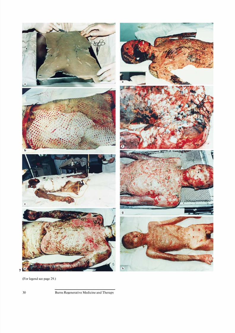
8/3/2019 Luka Bakar Book
http://slidepdf.com/reader/full/luka-bakar-book 37/157
30 Burns Regenerative Medicine and Therapy
7
(For legend see page 29.)
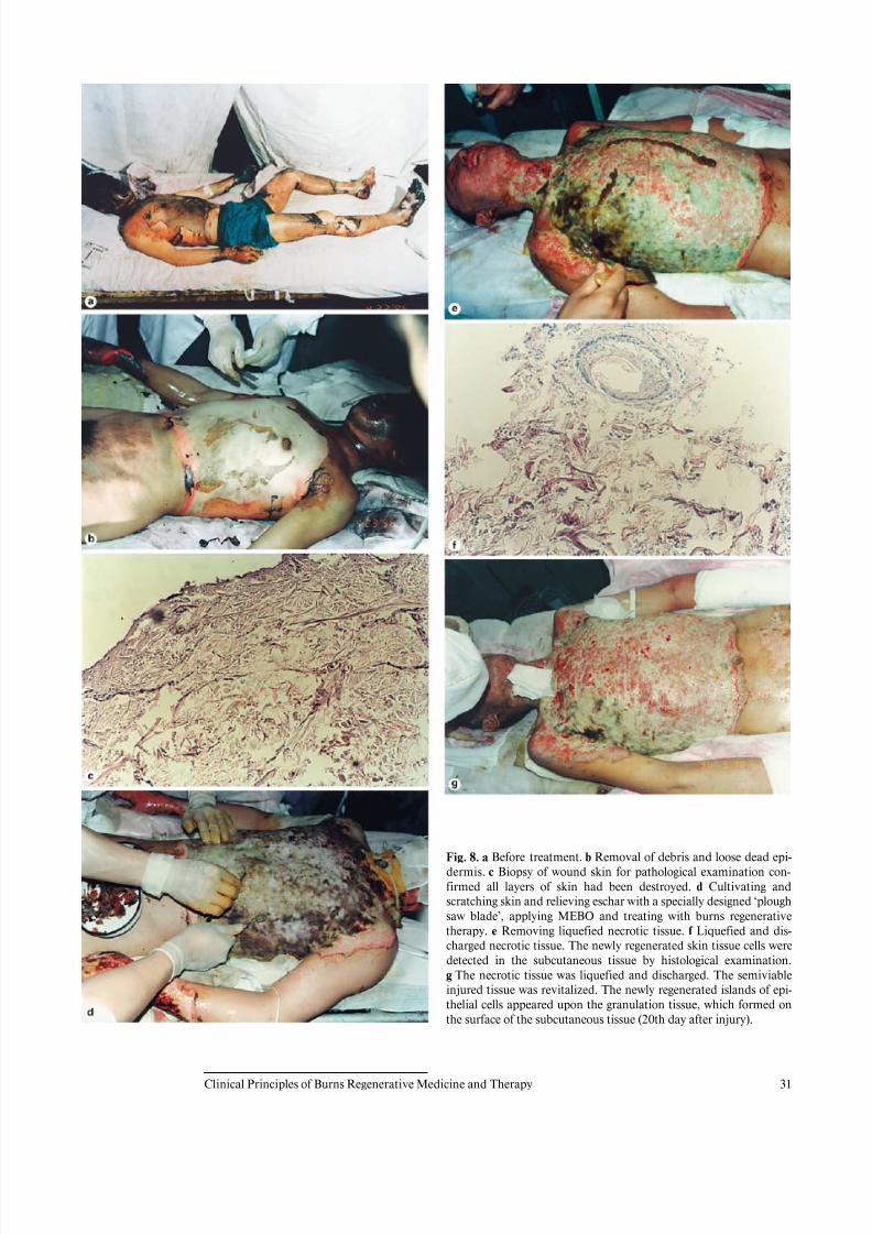
8/3/2019 Luka Bakar Book
http://slidepdf.com/reader/full/luka-bakar-book 38/157
Clinical Principles of Burns Regenerative Medicine and Therapy 31
Fig. 8. a Before treatment. b Removal of debris and loose dead epi-
dermis. c Biopsy of wound skin for pathological examination con-
firmed all layers of skin had been destroyed. d Cultivating and
scratching skin and relieving eschar with a specially designed ‘plough
saw blade’, applying MEBO and treating with burns regenerative
therapy. e Removing liquefied necrotic tissue. f Liquefied and dis-
charged necrotic tissue. The newly regenerated skin tissue cells were
detected in the subcutaneous tissue by histological examination.
g The necrotic tissue was liquefied and discharged. The semiviable
injured tissue was revitalized. The newly regenerated islands of epi-
thelial cells appeared upon the granulation tissue, which formed on
the surface of the subcutaneous tissue (20th day after injury).

8/3/2019 Luka Bakar Book
http://slidepdf.com/reader/full/luka-bakar-book 39/157
32 Burns Regenerative Medicine and Therapy
9

8/3/2019 Luka Bakar Book
http://slidepdf.com/reader/full/luka-bakar-book 40/157
Clinical Principles of Burns Regenerative Medicine and Therapy 33
Case 2: Burns Regenerative Medicine and Therapy
(BRT with MEBT/MEBO)
Background Information
A 35-year-old male sustained scalds by 1,000 °C hot
cement and flame burns secondary to a cement kiln col-lapse accident at 8:30 p.m. on April 12, 1996. He was
admitted to the hospital 4.5 h after injury. Initial assess-
ment showed: (1) burn-blast combined injury; (2) exten-
sive deep burns (85% TBSA); (3) severe inhalation injury;
(4) shock; (5) open multiple fractures on both feet.
On admission, the patient was in a critical state and in
shock. The extensive deep burns wounds were covered by
cement powder. He had inhaled cement and his nasal
hairs were singed. He suffered from respiratory abnormal-
ities and hoarseness. Tracheotomy was performed imme-
diately. BRT with MEBT/MEBO treatment and cultivat-
ing technique was started on the wound and systemic
comprehensive management begun. Histological exami-
nation of the wound skin showed third-degree burns. On
day 30 postburn, liquefaction and discharge of wound
necrotic tissues were finished. On day 49 postburn, newly
regenerated skin was present on the wounds. Ten days lat-
er, large sections of regenerated skin appeared on the
wounds and all wounds had healed completely on day 72
postburn. One year later, follow-up showed the patient
free of disablement, capable of independent viability and
no need for reconstruction.
Fig. 9. a Continuous treatment with BRT with MEBT/MEBO and
protection of the wound with the ointment (MEBO). Pathological
examination showed regeneration of skin tissue, and there were someislands of epithelial cells distributed on the surface of the granulation
tissue. b Histological examination revealed that those islands were
masses of regenerating skin tissue from subcutaneous tissue com-
posed of capillaries, collagen, epithelial cells, etc. c The epithelial
islands expanded gradually and started to integrate. d The regener-
ated skin islands connected to form a larger piece. e On the 49th day
after injury, histological examination confirmed that the newly
regenerated skin was of physiological full-thickness. f On some parts
of already integrating skin, there was a physiological anatomic struc-
ture of large blood vessels in the subcutaneous tissue. g All areas of
the wound were covered by regenerating skin, either closing the
wound, or developing new skin of a similar structure, appearance and
function to that of normal skin. h 72nd day; appearance of healed
wound with regeneration of full-thickness skin.
Procedure and Results (fig. 8, 9)
First Step: Treatment of Burned Skin
Second Step: Healing and Closure of the Wound
Third Step: Reconstruction after Wound Healing
No need.
Ed. note: In the spirit of brevity, the author has offered
photographic documentation of 2 cases only. However,
the author has documented hundreds of similar cases and
for those who would appreciate reviewing that extensive
photographic library, we refer you either to the literature
or to www.mebo.com.
Case 3: Surgical Excision and Cultured Composite
Autograft Therapy
Background Information
Cultured epithelial autografts (CEA) have been used as
an adjunct in the surgical management of extensive ther-
mal burns. Unfortunately, the lack of a dermal matrix
makes CEA susceptible to infection, shearing forces and
limits their incorporation into the burn wound. A cul-
tured composite autograft (CCA) has been developed
recently in which autologous keratinocytes and fibroblasts
are surgically harvested from the burns patient’s normal
skin. These components are proliferated and then com-
bined to form an epidermal and dermal matrix which
grows into confluence and is then applied to the lesion.
Standard wound coverage techniques as well as CCA
techology were utilized for successful wound closure in a
12-year-old female with an 81% third-degree burn. After
fascial excision and allograft coverage, autografts were
placed on her posterior burns and then 7,500 cm2 of CCA
was placed onto her anterior thorax, abdomen and lower
extremities. Sixty percent of the burns was covered with
CCA resulting in a success rate of 40%. No evidence of
infection was noted, even in areas where CCA failed,
although in those areas random epithelialization ap-peared to occur which then seemed to facilitate autograft
placement. Early debridement and allografting followed
by conventional autografts and CCA placement may pro-
vide an effective skin coverage strategy in patients with
extensive deep burns.
Procedure and Results
Disablement and disfigurement. Reconstruction was
required. Pictures of the treatment procedure are not
available here as copyright is concerned. See Burns
1999;25:771–779 for details.

8/3/2019 Luka Bakar Book
http://slidepdf.com/reader/full/luka-bakar-book 41/157
34 Burns Regenerative Medicine and Therapy
OOOOOOOOOOOOOOOOOOOOOOOOOOOOOOOOOOOOOOOOOOOOOOOOOOOOOOOOOOOOOOOOOOOOOOOOOOOOOOOOOOOOOOOOOOOOOOOOOOOOOOOOOOOOOOOOOOOOOOOOOOO
Indications and Diagnostic Principles of Burns
Regenerative Medicine and Therapy
It is concluded from a comparison of the descriptions in
the previous section that burn injuries involving skin onlyshould be treated with BRT with MEBT/MEBO rather
than with surgical excision and skin grafting therapy. The
latter is only appropriate in the treatment of burns wounds
with full-thickness necrosis of subcutaneous tissue togeth-
er with muscle or deeper burns. To facilitate the clinical
performance, the diagnostic principles and clinical indica-
tions of various therapies are standardized below.
Diagnostic Principles of Burns Medical Therapy
Many textbooks describe the method of diagnosis of
burn depth. It is based on naked eye observation and the
doctor’s own experience; therefore, it is often difficult to
differentiate between full-thickness burns and deep par-
tial-thickness injury. Understandably, therefore, wounds
should not be excised since the result is the removal of all
skin tissues and superficial fascia. After surgical excision,
we see that the prognosis is worse and the mortality and
disablement rates are elevated.
In order to standardize the diagnosis of burn depth, the
following principles should be followed.
Principle of Clinical Diagnosis
First of all, it is necessary to determine whether the
burn wound requires surgical excision or not. If the
wound demonstrates surviving skin tissue in the deep
layer with appearance of exudate within 6 h after injury,
then the subcutaneous tissues are viable with functional
microcirculation and surgical excision is not required.
After treatment with this BRT, white exudates will appear
on the wound surface. One notices that the more the exu-
date, the more superficial the wound. If the wound has noexudate 3 days postburn, surgical operation should be
considered. If the wound reveals no hemorrhage of subcu-
taneous tissue after the fasciotomy, it can be excised.
However, this does not apply to the wound where the exu-
date disappeared after treatment with dry therapy. If such
cases occur, there are mistakes in the treatment.
Pathological Diagnosis
Pathological diagnosis is used to diagnose the depth of
burns wounds without exudate and to determine whether
the wounds need to be excised. Wounds with exudate do
not need pathological diagnosis. Pathological diagnosis is
easy and painless. If there is misdiagnosis of one biopsysample of a small piece of skin including subcutaneous
tissue from the wound, histological examination of the
section is performed. If most of the subcutaneous tissue is
necrotic, the wound can be excised and treated with skin
grafting. If the subcutaneous tissue is still structurally
vital, then the wound should not be excised and BRT
(MEBT/MEBO) should be applied. Accurate pathological
diagnosis based upon scientific investigation is feasible
and, when performed correctly, can afford the patient cor-
rect diagnosis and optimum prognosis. It is no longer
acceptable for the physician to rely upon the naked eye as
too many treatment errors could result.
Burns Regenerative Medicine and Therapy
(BRT with MEBT/MEBO)
Indications
BRT with MEBT/MEBO is an independent method:
1 For treating superficial second-degree and deep sec-
ond-degree burns and scald wounds of various causes
and in different areas.2 In coordination with cultivating and relieving tech-
niques, BRT can be used for treating full-thickness der-
mis burns and scald wounds, provided viable subcuta-
neous tissue of various causes and different areas are
present.
3 For treating burns wounds deep in the muscular layer
with diameters of less than 20 cm.
4 For treating wounds at the skin donor site.
5 For treating granulation wounds deep in the muscular
layer, for promoting regeneration of granulation tissue
in burned bone after debridement, and to create a
physiological environment at the receiving site for skingrafting.
6 For treating all kinds of surface wounds.
7 For treating other skin lesions including hemorrhoids,
leg ulcers, bedsores, chronic ulcers, infected wounds,
chilblains, etc.
Clinical Application
Direct application of MEBO – a specially developed
topical drug for BRT with MEBT/MEBO – onto the
wound surface to a thickness of 0.5–1.0 mm every 4–6 h.
Detailed clinical treatment is recommended as follows:

8/3/2019 Luka Bakar Book
http://slidepdf.com/reader/full/luka-bakar-book 42/157
Clinical Principles of Burns Regenerative Medicine and Therapy 35
1 For first aid at home (especially in the kitchen): Imme-
diately apply MEBO on the wound to relieve pain, stop
bleeding, alleviate injuries and prevent infection in
cases of scalds and burns by hot oil, boiling water, or
friction burns. The sooner, the better. The consequent
treatment should be conducted according to the follow-
ing specific cases.2 Treatment for first-degree burns or scalds: Directly
smear MEBO onto the wound 2–3 times daily.
3 Treatment for superficial second-degree burns or
scalds: Directly smear MEBO onto the wound to a
thickness of 0.5–1.0 mm. Renew the ointment every
4–6 h; before doing so gently wipe off any residual oint-
ment and exudates. It usually takes 6–7 days to heal.
Blisters, if present, should be punctured and dis-
charged while blister skin should be kept intact in the
early stage. No disinfectant, saline or water is required
or in fact even allowed except in the case where exoge-
nous toxins remain at the site such as might be the casewith chemical burns or other dirty wounds. Patients
sustaining moderate or extensive burns should be sent
to hospital or a clinic experienced with the BRT treat-
ment protocols.
4 Treatment for deep second-degree burns: Treatment in
the early stage is the same as that for superficial second-
degree burns. Remove the blister skin on day 5–6 after
injury. As the dermis tissues are damaged and white in
color, the application of MEBO should be continued on
the wound to a thickness of 0.5–1.0 mm every 4 h.
White metabolic products resulting from liquefaction of
necrotic tissue by the ointment will appear on the
wounds (do not misdiagnose this cleansing process as
infection). Be sure that the residual ointment and white
liquefied products are wiped off gently (do not irritate
or debride the tissue) before reapplying MEBO. Allow
another 6–7 days for the necrotic tissue to be liquefied
and discharged completely, then continue the above
treatment using less dosage of MEBO until the wound
heals. In the event that the wound is still not healed after
25 days postburn, the diagnosis should be changed to
full-thickness degree. In brief, the venerable medical
principle of ‘primum non nocere’ (first do no harm) andof ‘no secondary injuries’ should be honored during the
whole treatment procedure. We accomplish that by:
(1) protection of the treated wound in the early stage
from further injuries (avoid any measures which may
irritate, debride or exacerbate wounds); (2) liquefaction
and removal of the necrotic tissue without causing sec-
ondary injuries; (3) regeneration and skin repair with-
out causing secondary injuries (any method which may
irritate or damage the wounds is not allowed). Patients
sustaining moderate and extensive burns should be sent
to hospital or a clinic with experience of BRT and
MEBT for appropriate treatment.
5 Treatment for second-degree burns: For the small-area
burn wound, we recommend cultivating tissue and
then preparing the lesion for application of MEBO
through gentle loosening of necrotic tissues by scratch-
ing with a specially designed device – ‘plough saw
blade’ is the appropriate treatment for the deep sec-
ond-degree burns wounds. For larger burns wounds,the aforementioned method is adopted if the patient’s
systemic condition is stable. The principle of ‘no sec-
ondary injuries’ should be followed strictly during the
treatment. Patient sustaining third-degree burns must
be hospitalized at clinics offering care from clinicians
experienced in BRT with MEBT/MEBO.
6 In the treatment of small burns wounds occurring in
inconveniently exposed body parts, bandaging is rec-
ommended. However, dressing changes and renewal of
MEBO ointment at a thickness of 2–3 mm every 12 h is
recommended. Contrary to the typical dressing change
protocol, however, rather than debride the woundbeneath the bandage, we recommend that the bandage
be gently removed leaving the residual ointment and
metabolic products to continue their cleansing activi-
ty.
7 Treatment for other superficial trauma wounds includ-
ing abrasion, friction burns, skin cracking, and stasis
ulcers: Treat the ulcer wounds according to the instruc-
tions for either superficial or deep second-degree
burns, or dress the wounds with MEBO in accordance
with the surgical methods. However, any disinfectant,
antiseptic or saline is contraindicated as they are both
unnecessary and deleterious to wound health.
8 Treatment for hemorrhoids: Directly apply MEBO
onto the affected area every morning and evening, or
smear MEBO onto the postoperative wound to relieve
pain and promote healing.
Burns Surgical Therapy with Excision Followed
by Skin Grafting or Cultured Composite
Autografting Technique
Indications and Application
1 Full-thickness degree burns wounds reaching the lower
layer of the subcutaneous tissue of different areas and
of different causes.
2 Skin grafting technique is used for treating granulation
tissue wounds without epithelial regeneration and for
plastic surgery.
3 The hospitals should be qualified to conduct surgery
and the operation should be conducted by surgeons
specialized in BRT with MEBT/MEBO and/or burns
surgery.

8/3/2019 Luka Bakar Book
http://slidepdf.com/reader/full/luka-bakar-book 43/157
36 Burns Regenerative Medicine and Therapy
OOOOOOOOOOOOOOOOOOOOOOOOOOOOOOOOOOOOOOOOOOOOOOOOOOOOOOOOOOOOOOOOOOOOOOOOOOOOOOOOOOOOOOOOOOOOOOOOOOOOOOOOOOOOOOOOOOOOOOOOOOO
Intensive Description of Burns Regenerative Therapy with
MEBT/MEBO
BRT with MEBT/MEBO is an entirely new burns
treatment technique that operates in compliance with thelaw of life. BRT was invented on the basis of academic
thoughts according to the pathogenesis of burns. This new
therapy comprises a complete set of theories and tech-
niques for the local and systemic treatment of burns. The
profile of this therapy is that through liquefaction and
removal of the necrotic tissue, culture and regeneration of
residual viable skin, and through repair and replication,
burns wounds are finally healed. Wounds are not kept in a
dry environment as required in conventional surgical
burns therapy, but in a physiological moist environment.
For local treatment, BRT with MEBT/MEBO is associ-
ated with MEBO. For systemic treatment, it forms an inde-pendent system in compliance with the law of life, includ-
ing its theory, methodology and therapeutic results. In
local treatment, BRT resolved the problems of wound pain
and complete regenerative healing of deep second-degree
wounds. Through reducing bacterial toxicity by varia-
tion, and promoting local resistance to infections, BRT
with MEBT/MEBO effectively prevented and controlled
wound infections. Through creating a physiologically
moist environment and good nutrition supply, BRT with
MEBT/MEBO promoted the culture and differentiation of
stem cells from the epithelia and relevant tissues in the
residual fat layer and finally healed the wounds to full-
thickness. Furthermore, exposed bone wounds from burns
can also be healed by drilling on the bone in combination
with MEBO application, culturing granulation tissue to
cover bone and heal the wounds. In systemic treatment,
measures of strengthening cardiac function and removing
the obstacles in blood supply of renal parenchymal blood
vessels are adopted, and then blood volume replacement
and comprehensive antishock measures are taken. Accord-
ing to the severity of the burn case, effective broad-spec-
trum antibiotics are applied to control infections at an ear-
ly stage, but the antibiotics are stopped in order to protectthe function of the organs at days 7–10 postburn.
Due to the remarkable effect of this therapy, it has
been introduced in Syria, the United Arab Emirates,
Thailand, the Republic of Korea, Singapore, etc., and has
achieved great clinical success in these countries. Now
BRT with MEBT/MEBO is spreading its academic
thought as well as its technology to the United States and
European countries. At the Congress of the Pan-Arab
Association for Burns and Plastic Surgery held in the
United Arab Emirates on February 22, 2000, specialists
from dozens of countries gave presentations on their
research on BRT with MEBT/MEBO.
Concept and Principle of BRT with MEBT/MEBO
Concept
BRT with MEBT/MEBO is a medical treatment which
promotes the cell regeneration of residual skin tissue and
wound healing by keeping burns wounds in a three-
dimensional physiologically moist environment and facil-
itating the liquefaction and discharge of necrotic skin tis-
sue.
Principle
MEBO, specially invented for BRT with MEBT/
MEBO, is an ointment whose base includes a frame struc-
ture composed of beeswax which musters a refined botan-
ical oil containing active ingredients.
When applied onto the wounds, the oil sequestered in
the beeswax frame is warmed up in contact with body
temperature and thus penetrates into the wounds. Four
biochemical reactions between MEBO’s active constitu-
ents and the burn tissue take place while the nutritional
substances are supplied continuously onto the surviving
cells in the wounds. The oil then loses its lipophilic nature,
seeps out of the drug layer and is discharged from thewound. Fresh MEBO continues to penetrate into the tis-
sue, so that liquefied necrotic skin is removed from the
superficial to the interior planes without causing further
injury. Simultaneously, this mechanism of action also fea-
tures a replenishment of the drug in continuous cycles.
These cycles ensure active drainage of metabolic waste
products including pathogenic bacteria from the injured
viable tissue, hence the caveat to not apply topical steriliz-
ing agents. Tissue fluid in the wound supplies the requi-
site physiologically moist environment. The cycle also
ensures continuous supply of indispensable nutritional
substrates and enzymes to the recuperating wound tissue.
Furthermore, the frame structure of MEBO base effec-
tively insulates and isolates the vulnerable wound from
the external environment by forming a viscous dressing.
A remarkable characteristic of this ‘dressing’ is that it pre-
vents foreign external contaminants from penetrating
while allowing for the exodus of metabolic debris prod-
ucts resulting from the regeneration of the wounded tis-
sue. In a metabolic sense, the MEBO allows the wound to
‘breathe’ in a manner very similar to that of normal skin.

8/3/2019 Luka Bakar Book
http://slidepdf.com/reader/full/luka-bakar-book 44/157
Clinical Principles of Burns Regenerative Medicine and Therapy 37
Therapeutic Effects of Moist-Exposed Burns
Ointment (MEBO)
Under the direction of qualified BRT with MEBT/
MEBO therapists, MEBO has the following therapeutic
effects:1 Variation of pathogenic microorganism and reduction
in bacterial toxicity.
2 Effectively killing pain by protecting nerve endings
and relaxing pilorum arrectors.
3 Anti-inflammatory by the effects of ß-sitosterol and
other ingredients.
4 Made of nutrient food, MEBO may protect cells by
increasing the tension in the cell membrane and help
dying cells convert into vigorous normal ones.
5 With the co-ordination of BRT with MEBT/MEBO,
MEBO develops a physiologically moist environment,
favorable to the regeneration and repair of tissue struc-tures. Thus, it is effective for reducing scar formation,
enhancing the power of self-repairing of wounds and
promoting the regeneration and differentiation of stem
cells from residual epithelial tissue, vascular plexus
and fibrous tissue in the fat layer to regenerate skin.
Clinical Application of BRT with MEBT/MEBO
Treatment Conditions
Strictly sterilized conditions are not emphasized. De-
bridement using any disinfectant, saline or water is for-
bidden. Small burns can be dealt with at home with
MEBO. Moderate and minor burns encountered in the
battlefield can also be treated with BRT with MEBT/
MEBO. For treating large burns, the room should be kept
at a temperature of 30–34°C and first-aid apparatuses or
devices should be equipped with it.
General Application
Directly smear MEBO onto the wounds with a thick-
ness of 1 mm. At the beginning, no debridement is re-
quired except for chemical burns or dirty wounds. Renew
MEBO every 3–4 h, before which wiping off the residual
ointment and liquefaction products with gauze or tissue
paper (gentle and careful renewal is demanded to avoid
pains and bleeding). For wounds with blisters, be sure to
preserve the blister skin, directly apply MEBO until the
blister skin is removed 5 days later. For deep second-
degree burns, after applying MEBO, dermal tissue in the
necrotic layer begins to liquefy on day 7 postburn. Renew
MEBO and wipe off the liquefaction product timely. After
the complete discharge of necrotic tissue, apply less
MEBO and renew every 4–6 h till the wounds heal. For
third-degree burns, treatment with a special debridement
technique can be applied coordinately.
Special Application
For treating not easily exposed small burns wounds,
apply MEBO with a thickness of 2–3 mm, then apply a
decompression bandage using dry gauze. Before changing
the dressing every 12 h, gently remove the drug sediment
and liquefied necrotic tissue. For treating traumatic, ul-
cerative and operative wounds, 1–2 layers of gauze im-
pregnated with MEBO also could be used.
Principle of Systemic Treatment
Burn is a systemic disease caused by thermal injury.
The changes in topical treatment directly affect the sys-temic pathophysiological status. BRT with MEBT/MEBO
systemic treatment is essentially different from conven-
tional surgical dry therapy. While using BRT with MEBT/
MEBO, the protocol of surgical dry therapy is forbidden.
Two principles should be followed in this treatment on
extensive deep burns: (1) In the early stage, a comprehen-
sive antishock treatment principle is applied, which in-
volves enhancing cardiac function, protecting renal func-
tion and supplementing effective blood volume according
to the vital signs. (2) In the middle and later stages, expec-
tant treatment is applied, with a protocol of keeping a
water-electrolyte balance, nutritional support and main-
taining a comprehensive balance. In the anti-infection
treatment, a large dose of strong and powerful broad-spec-
trum antibiotics should be used in the early stage for 7–10
days and then withdrawn immediately, in order to protect
and enhance the anti-infective function of the internal
organs. For nutritional supporting treatment, a protocol
of oral administration is desired. Others are dealt with
according to the case.
Clinical Treatment
Treatment for First-Degree Burns
The clinical signs of first-degree burns include skin red-
ness, slight swelling and pain. Immediate application of
MEBO may relieve the pain. The erythema gradually
diminishes as MEBO is warmed in situ and absorbed
through the skin. At 12 h postburn, the skin may return to
normal. For burns with edema, the epidermis is partially
destroyed, the pain may be relieved more slowly and the
wounds heal in 2–4 days when the superficial stratum cor-
neum exfoliates.

8/3/2019 Luka Bakar Book
http://slidepdf.com/reader/full/luka-bakar-book 45/157
38 Burns Regenerative Medicine and Therapy
Treatment for Superficial Second-Degree Burns
According to the pathogenic process of superficial sec-
ond-degree burns, the treatment can be carried out in two
stages.
First Stage. Treatment in the early stage – the period
from emergency treatment postburn to the end of shock
period (within 3–4 days after injury). The clinical signsin this stage include pain, edema, blisters, and a great
amount of blood plasma exudated from the site where
blister skin exfoliates. According to the principle of BRT
with MEBT/MEBO, apply MEBO directly all over the
wound, puncture the blister (if present) on the lower part
to discharge liquid. Do not remove the blister skin, direct-
ly apply MEBO on the blister skin 3–4 times daily. With
the application of MEBO, a layer of thin soft membrane
forms upon the wound free of blister skin, the membrane
still allows the exudates to ooze through, and then it grad-
ually thickens. Do not remove the soft membrane, since it
can substitute the skin role of fulfilling breathing and pro-
tection. Continue MEBO application directed by BRT.
Second Stage. It is the wound-repairing period when
the shock stage ends and the residual viable epidermis tis-
sue begins to regenerate and recover, usually lasting 3–4
days. In this period, the basal cells in the epidermis recov-
er to form a granular layer and thus promote wound heal-
ing. In clinical treatment of BRT with MEBT/MEBO,
after the edema period, the blister skin loosens and exfo-
liates, and the thin soft membrane formed on the wounds
also loosens and exfoliates. Simply cleaning away the blis-
ter skin and soft membrane is first desired, then continuethe application of MEBO to protect the regenerated
wounds till healing. During the whole treatment, neither
pain nor further injury to wounds is allowed. The correct
application method helps the wounds heal without caus-
ing any infection, pain, scar formation or hyperpigmenta-
tion. Generally, superficial second-degree wounds treated
with BRT with MEBT/MEBO heal within 6–8 days and
the skin recovers completely to its normal physiological
status within 3 months.
Treatment for Deep Second-Degree Burns
According to pathological and clinical manifestations,
deep second-degree burns can be divided into injury on
the dermal papillary layer and injury on the reticular
layer, or simply referred to as deep second-degree superfi-
cial (DIIS) and deep second-degree deep (DIID) burns.
DIIS
Clinical signs include wound pain, extensive blisters,
wound without blister skin becoming red and white, the
superficial dermal tissue is necrotic and turns white, the
surviving deep dermis tissue is red, while under pressure
it turns white and soon returns to red after release of pres-
sure (DIID burns wounds respond more slowly). The exu-
dates of the wounds are only less than those of superficial
second-degree burns.
BRT with MEBT/MEBO treatment and clinical mani-
festation: It is a four-period process: firstly, treatment in
the early stage (shock period), same as that for superficialsecond-degree burns; secondly, liquefaction and rejection
period of necrotic dermal tissue (rejection period); third-
ly, regeneration and recovery period, and, fourthly, reha-
bilitation period of skin physiological function after
wound healing.
First Period. The treatment of the first period is the
same as that for superficial second-degree burns, empha-
sizing on wound protection. A thin layer of soft mem-
brane may appear on the wound free of putrid or blistered
skin. The next treatment period starts on day 4–5 post-
burn when the wound edema diminishes gradually.
Second Period. Clean away the putrid skin, blisteredskin or thin soft membrane in the same way as dealing
with superficial second-degree burns. Continue the appli-
cation of MEBO. Gradually, the necrotic layer of wound
surface begins to liquefy from the superficies to the interi-
or and produces white liquefied products floating over the
wound surface. Usually at 3–4 h after application of
MEBO, the wound is totally covered with whitish lique-
fied products, indicating that MEBO is consumed com-
pletely. The liquefaction products must be cleaned away
before the renewal of MEBO. Another 3–4 h later, the re-
newed MEBO is consumed again when the liquefaction
products float over the wound. Clean the liquefaction
products, renew MEBO again and keep the clean-renew-
clean process going until the necrotic tissues are entirely
liquefied and discharged. This process generally occurs 5–
15 days postburn. Patients with large-area burns are urged
to turn over during drug renewal.
Third Period. After the second period treatment, the
chestnut-like residual dermis tissues, millet-sized, are ex-
posed on the basal layer of the wound. Continue MEBO
with less thickness directed under BRT with MEBT/
MEBO and renew every 4–5 h (every 6–8 h at night). Cov-
ered and protected by MEBO, the residual dermal tissuereconstructs and regenerates. Once dermis tissue regener-
ates to smooth skin, less irritation to the wound is
allowed. Again reduce dosage and renewing times as long
as the wound is not dry, but crust formation is forbidden,
i.e. not only prevent wounds from being macerated by
MEBO, but also avoid the wounds becoming dry and get-
ting covered by a crust. Duly keep the normal skin around
the wound clean. For large-area burn patients, do as in the
second period by helping them turn over regularly on the
basis of drug change intervals. The pressured parts of the
body still need MEBO protection till the wounds heal.
This period occurs 15–20 days postburn.

8/3/2019 Luka Bakar Book
http://slidepdf.com/reader/full/luka-bakar-book 46/157
Clinical Principles of Burns Regenerative Medicine and Therapy 39
Fourth Period. Although the wound heals after the
third stage, the functions of the newly healed skin still
need to rehabilitate completely. The epidermis requires
further physiological adjustment and metabolism; the
sebaceous glands need compensatory metabolism; the ex-
cretory duct of the sweat gland is not yet clear; the func-
tions of pigment cells are unable to meet the requirementsof normal skin. Under these circumstances, MEBO is still
necessary to be used as a skin-care oil for another 10–15
days. Or apply newly developed MEBO series products –
MEBO Cleansing Cream to promote quicker recovery of
skin function, or apply MEBO Itch Relieving Cream to
stop itching.
DIID
The clinical signs of deep second-degree burns are sim-
ilar to those of deep second-degree superficial burns,
except that DIID has more serious injuries and more seri-
ous response during the liquefying period and thereforethe reconstruction and regeneration of wounds become
more complicated. The clinical treatment for deep sec-
ond-degree burns also can be divided into four periods.
First Period. Clinical signs – no extensive blisters, epi-
dermis entirely destroyed and adherent to injured dermis,
the wounds free of putrid skin are no longer sensitive to
pain, the wounds are white, with little exudate. Some of
the wounds may be red alternating with white, but the
color-changing response to pressure is very slow. This
period begins from the first day of burn through the 7th
day postburn. During this period, simply apply MEBO to
protect the wound.
Second Period. From day 7 to day 20 postburn, clear
away all adhering substances to expose necrotic dermis
and apply BRT with MEBT/MEBO immediately. For
small wounds, simply use this treatment to liquefy and
discharge the necrotic layer. For large burns, simple de-
bridement should be used coordinately. The necrotic
layer is so deep that the wound liquefying may be incom-
plete and cause lumps exfoliation of necrotic tissues,
which need to be cut with surgical scissors and removed.
Attention should be paid to keep a certain distance
between the surviving viable tissue and the cutting. Anyfurther damage to the surviving viable tissue such as
bleeding (which may cause infection) should be absolutely
avoided. Simple debridement can be adopted according
to the condition of the wounds. When the necrotic layer is
almost completely liquefied, clean away the liquefaction
products in time to ensure that the non-smooth survival
tissue is kept in a MEBO environment, but not in an envi-
ronment filled with liquefaction products.
Third Period. The period of reconstruction and regen-
eration of residual dermis tissue. As little residual dermis
tissue is left and the dermis frame is fundamentally
destroyed, correct BRT with MEBT/MEBO is quite vital
in this period. The reconstruction of dermis tissue in-
volves three parts: (a) the reconstruction of vascular tree;
(b) the reconstruction of fibrous tissue dependent on vas-
cular tree; (c) the regeneration of skin appendages, gland
tissue, formation of excretory ducts, and formation of
skin tissue. Any careless injuries and pressure to the
wound are forbidden. This period usually happens ondays 20–28 postburn.
Fourth Period. Aiming at helping the healed skin re-
turn to normal, the treatment in this period varies ac-
cording to burns severity and skill in the treatment. The
severe injuries to skin and the factors affecting the skin
during reconstruction make the newly healed skin quite
different from normal skin in structure, appearance and
function. So the rehabilitation is very important, includ-
ing two aspects: ‘protective therapy for healed skin’ and
‘functional exercise’. The former is accomplished by ad-
justing the structure of newly regeneration skin tissue with
the application of MEBO Scar Lotion, by adjusting thefunction with MEBO Cleansing Cream, and by killing itch
with MEBO Itching Reliever just after the wounds heal.
Treatment for Third-Degree Burns
Third-degree burns are also an indication for BRT
with MEBT/MEBO. As the epidermis and dermis of
third-degree burn wound are totally destroyed, it is quite
difficult to cure third-degree burns. The conventional
medical science for burns is convinced that third-degree
wounds cannot heal spontaneously, and the only solution
is to use surgical skin grafting to close them. The clinical
study and administration of BRT with MEBT/MEBO for
curing third-degree burns wounds will be described in
detail thereinafter. The following is just a brief descrip-
tion of the principle and method of this treatment.
Principle. Decompression of the deep tissues to relieve
any pressure caused by the necrotic layer is of critical
importance; protect the necrotic full-thickness skin; pro-
mote stem cells containing the skin information in subcuta-
neous tissue to regenerate and differentiate to form a skin
island; culture the newly regenerated skin island while
liquefying and discharging the necrotic dermis; promotethe skin island to spread and cover subcutaneous tissue to
form new skin; and help third-degree burns wounds heal
spontaneously. For third-degree wounds injured down to
the muscle layer, excise most of the necrotic tissue by surgi-
cal operation, liquefy and discharge the rest of the necrotic
layer with BRT with MEBT/MEBO, upon which culture
granulation tissue, then plant skin cells till the wound closes
and heals. For wounds with bone exposed, clean away the
exposed outer soft tissue, drill holes on the bone surface
with a bone drill at intervals of 0.5–1 cm, deep into medul-
lary cavity of bone until bleeding. Apply MEBO to cover
the wounds, and culture and support the growth of granula-

8/3/2019 Luka Bakar Book
http://slidepdf.com/reader/full/luka-bakar-book 47/157
40 Burns Regenerative Medicine and Therapy
tion tissue from the holes. When the granulation tissue
spreads to cover the bone surface, skin grafting can be per-
formed to close the wound, or the wound heals by migra-
tion of epithelial cells from the wound edges.
The necrotic tissue of third-degree burns should be
decompressed by cutting both horizontally and vertically
at a 1 mm tooth distance and depth with a speciallydesigned method: ‘plough saw blade’. Then apply MEBO
for protection and clean away the exfoliated or liquefied
tissue.
Systemic Comprehensive Treatment with BRT
with MEBT/MEBO
Principles of Initial Treatment
Principles of First Aid 1 Keep the patient in a horizontal supine position with
slight elevation of the head. Turning over is contraindi-
cated. Expectant administration.
2 Application of any topical drug that may be harmful or
irritative to burns wounds is contraindicated. Adopt
measures to relieve pain and protect burns wounds as
soon as possible.
3 Avoid changes of patient body temperature which con-
sume vital energy striving instead to keep the patient
warm as much as possible.
4 If appropriate and well-tolerated, cardiotonic and sed-
ative medications may be given through intramuscular
injection or intravenous infusion.
5 If the wound is deemed appropriate for treatment with
BRT with MEBT/MEBO, smear MEBO directly onto
the wound. Once the wound is thus protected, cover
the wound with adequate dressing and transfer the
patient immediately to the nearest hospital for further
treatment.
Principles of Emergent Treatment and Nursing
1 Treatment condition: a clean or sterile (which is not
absolutely necessary) environment is required. Tem-perature around the wound surface should be kept at
34–38°C.
2 Early wound care: any feculency and dirt should be
cleared away. Do not use any method or topical drug
that may cause further injury to the wound or promote
tissue hydrolysis.
3 Principle of initial nursing: do not turn the patient
over. Alternately lie on one side or alternately change
pressure at various body parts.
4 If BRT with MEBT/MEBO treatment is adopted,
wound debridement with MEBO and topical applica-
tion of MEBO should be performed for wound care.
Wound Debridement with MEBO. Cover the wound
with MEBO immediately after injury regardless of the
presence of dirt or chemicals. Two or three hours later,
gently clear away the feculency and dirt together with
residual MEBO before the renewal of MEBO. This meth-
od is applicable for first-aid treatment as well as wound
debridement after hospitalization when daily cleansing isappropriate.
Topical Application of MEBO. Smear MEBO onto the
wound at a thickness of 0.5–1 mm immediately after
wound cleansing. Gently wipe off liquefied products be-
fore renewing MEBO every 4–6 h. Renewing intervals
could be increased to every 6–8 h during the wound repair
period.
Antishock Therapy
The author considers that in the antishock therapy
postburn, it is more important to protect and recover thefunctions and structures of internal organs than to supple-
ment blood volume only. The principles of the treatment
are as follows:
Protection and Enhancement of Cardiac Function
We propose that a lot of protein degradation products
released from burned skin tissue could be absorbed into
the blood circulation, and could further inhibit and de-
crease cardiac function, thus inducing cardiogenic shock.
Therefore, severely burned patients (TBSA 150% and/or
third-degree 110%) should be routinely injected intrave-
nously with cedilanid (lanatoside C) 0.2 mg in 25–50%
GS 50 ml q.d. after injury or admission. Then, the
amount and frequency of cedilanid should be regulated
according to the changes in heart rate and peripheral cir-
culation. 48 h postinjury, the administration of cedilanid
should be stopped unless the patient is still suffering
from abnormal cardiac function, in which case cedilanid
should be applied until the symptoms disappear. If symp-
toms of heart failure arise during the course of treatment,
the patient should be treated with 0.2–0.4 mg cedilanid
immediately. One treatment is frequently sufficient.
Protection of Renal Function
After massive burns, one of the main complications in
the shock stage is renal dysfunction that is caused firstly
by microvascular spasm of the renal parenchyma and
renal ischemia. It is also the major etiology of renal fail-
ure. Therefore, treatment of renal function is the crux of
antishock and comprehensive treatment to relieve the
microvessels in the renal parenchyma. This needs to be
addressed as early as possible. The principles of renal
treatment are follows: After injury or immediately up-
on admission, severely burned patients routinely require
an intravenous drip with 1% procaine 100 ml, caffeine

8/3/2019 Luka Bakar Book
http://slidepdf.com/reader/full/luka-bakar-book 48/157
Clinical Principles of Burns Regenerative Medicine and Therapy 41
sodiobenzoate 0.5 g, vitamin C 1.0 g, 25% GS 100–
200 ml, q.d. or b.i.d.–t.i.d. depending upon the degree of
shock and the amount that urine production is reduced.
This intravenous drip should be continued in patients
with anuria until urination is recovered. The routine
treatment plan may be maintained until wound healing.
Supplement Blood Volume
After massive burns, a great deal of intravascular fluid
exudes toward the wound surface and tissue space, which
leads to the reduction in effective blood volume resulting
in hypovolumic shock. Therefore, during the above treat-
ment course, the blood volume should be monitored and
replenished as needed. In particular, attention must be
paid to avoid massive intravenous infusion blindly with-
out precise attention being paid to cardiac and renal func-
tions, as well as other excretory functions. The principle is
as follows:
Compositions of Fluid Infusion. The ratio of crystalloidsolution (normal saline or 5% GNS) to colloid solution
should be 1:1. The colloid solution should be composed of
3/4 parts of plasma and 1/4 part of whole blood when the
condition allows, otherwise 1/2 part of plasma and 1/2
part of plasma substitute can be used.
Amount of Fluid Infusion. According to the basic prin-
ciples of surgery, the amount of fluid infusion should be
equal to the amount of body deficiency. In the shock stage
of massive burns patients (during 48–72 h after injury),
we offer a more detailed formula:
Total amount of fluid infusion (ml/day) =
physiological water needs (5% GS 2,000–2,500 ml) +
[1 (ml/kg)! TBSA% (2nd to 3rd degree)
! body weight (kg)! 100%]
hourly urine volume (ml)/body weight (kg)! 1 (ml/kg)
Speed of Fluid Infusion. After extensive burns, the trau-
ma stresses the heart, kidney and brain tissue, making their
functions vulnerable. During the first 24 h postburn, 1/2 of
total fluid amount should be infused in the first 8 h, an-
other 1/2 should be infused over the next 16 h evenly again,
with regard to cardiac and renal functions. During the sec-
ond 24 h postburn, all of the fluid should be infused at auniform speed. During the third 24 h after injury, the
amount and speed of fluid infusion must be determined
strictly in the light of the symptoms of shock and the
amount of urine. When the symptoms of shock are im-
proved markedly or disappeared and the amount of urine
is 11 ml/h Wkg, the speed of fluid infusion should be
decreased and the fluid amount should be reduced by 1/3.
Nursing Care in Shock Stage
After severe burns, the onset of shock would be related
to thermal injury as well as adequate nursing care. The
burns patient can hardly withstand any further stress due
to the already severely compromised condition of all
internal organs. Thus, nursing care constitutes a critical
service in supporting as stress-free a recovery period as
possible. Nurses should:
a Directly apply MEBO on the wound surface immediate-
ly, isolate the wound from contacting with air, relieve
wound pain, protect the wound from any irritative dam-age, resist the tendency to debride the wound.
b Apply air conditioner or bedstead and sheeting to
maintain room temperature at 30–34°C, and prevent
fluctuation in room temperature.
c Smooth out the bed sheet and dressing, protect the
wound from any compression, change dressing and
MEBO every 12 h gently, while keeping the patient in
the horizontal supine position. Again, turning the pa-
tient over is contraindicated.
d Control the speed of fluid infusion such that it flows at
a constant rate remembering that rapid fluctuation of
infusion speed is forbidden.
Anti-Infection Therapy
We have observed that there are two pathogenic types
of postburn infection. One has an endogenous pathogene-
sis, while the other is exogenous. The endogenous infec-
tion is similar, but different from the primary infection
typically noted by surgical burns therapists. This consists
of subclinical infection in that the possibility of postburn
infection always remains a potential reaction to burns.
The latter infection consists of postburn infection caused
by all exogenous sources and factors including iatrogenic
burns.
Principles of Anti-Infection Treatment
Principled Scheme of Routine Treatment. Burned pa-
tients with TBSA !30% generally do not need to be
treated with systemic antibiotics. All the burned patients
with TBSA 130% (TBSA 110% in children) must be
treated with systemic anti-infection drugs routinely
whether infection occurred or not. The principle is: (1) To
apply one or more powerful broad-spectrum i.v. or i.m.
antibiotic as early as possible after injury until the 5th to7th day for massive deep second-degree burns and the 7th
to 10th day for massive third-degree burns. (2) The more
extensive TBSA and deeper the wounds, the more power-
ful and broad-spectrum antibiotics are required. (3) Re-
gardless of the patient’s condition, stop applying all anti-
biotics at the aforementioned time.
Principle of Expectant Anti-Infection Treatment. In
order to prevent and treat secondary and routine infec-
tion, a prophylactic antibiotic protocol should be admin-
istrated. However, it is very important to rule out inflam-
matory and noninfectious etiologies initially as antibiotics
would be contraindicated if infection is not problematic.

8/3/2019 Luka Bakar Book
http://slidepdf.com/reader/full/luka-bakar-book 49/157
42 Burns Regenerative Medicine and Therapy
Remember that some patients with fever and increased
heart rate may not be infected. After postburn routine
treatment, the burned body struggles to repair and re-
equilibrate a myriad of physiological functions and un-
necessary antibiotic administration at this time would
interfere with native resistance functions. Indications for
antibiotic use: Three clinical manifestations must occursimultaneously: body temperature 139.5°C or !36.0°C;
heart rate 1140/min; toxic granules in neutrophil leuko-
cytes. Clinical vigilance is required.
Profile of Expectant Anti-Infection Treatment
One single dose of one or more powerful broad-spec-
trum renal-sparing antibiotics should be applied. It may
be repeated until examination reveals that the neutro-
philic toxic granules are no longer present. The patients
should be examined for secondary infection sites and
treated appropriately. The failure to control infections is
commonly due to occult foci of infection within internalorgans or under the wound surface. The patient recover-
ing from general asthenia should be offered fresh blood
infusions to aid in the regulation of internal balance.
Abuse of antibiotics without indications of infection is
contraindicated.
Balance-Regulating Treatment at the Stage of Wound
Liquefaction
After shock stage is addressed, the burn wound enters
the rejection stage. For deep second-degree burns wounds,
injured and necrotic tissue starts to be rejected from resid-
ual viable tissue at approximately the 5th day postburn.
This rejection reaction continues until all the necrotic tis-
sue is discharged. The role of the physician and nursing
staff at this time is to not interfere with this natural cleans-
ing and regenerative process. During this stage, internal
organs and many physiologic systems which had been
stressed are particularly vulnerable to rejection which can
then lead to single or multiple organ failure. Therefore, this
is the most critical stage of the BRT (MEBT/MEBO) and
requires extreme vigilance. Based on our clinical experi-
ence of many years, we consider that the key to treatmentin this stage is to enhance and restore the systemic vitality
and comprehensive balance, without which any monother-
apy would only have a suboptimal chance of success. This
therapeutic measure is termed ‘balance-regulating treat-
ment’, and its protocol includes the following.
Wound Drainage
During the stage of wound liquefaction, as necrotic
skin tissue is liquefied from the superficial to the deeper
layer (under the effect of MEBO), it is very important to
clean up liquefied materials prior to successive applica-
tions. Remember, there are differences between clearance
of MEBT/MEBO liquefied materials and surgical de-
bridement. After treatment with MEBO, the changes in
burns wounds should be supervised continuously. When
MEBO on the wound surface completely changes into a
whitish liquefied material, this layer should be wiped off
or cleaned with a soft dry absorbent gauze or tissue paper
at once. When the necrotic skin tissue separates intopieces without liquefying completely, it should be cut
away gently from the wound (not debridement) and then
MEBO ointment should be reapplied immediately. Un-
like debridement, the RBT patient typically feels no pain
during the cleaning process. If he does, it is an indication
that the cleaning is too aggressive. Any harmful tissue
stimulation should be strictly prohibited. In order to en-
sure for correct clinical practice, there are six operative
rules for this treatment, i.e. the burn wound must not
hurt, there should be no fresh bleeding, no maceration, no
desiccation, no liquefied materials, and no lack of applied
MEBO.
Treatment of Body Fluid Equilibrium
After extensive burns, large amounts of body fluid
exude toward the wound surface and evaporate. We know
that this body fluid plays a vital role in systemic reactions
that result from traumtic stress. Therefore, it is an impor-
tant procedure of the comprehensive treatment to main-
tain body fluid equilibrium. The principles of this treat-
ment are as follows: The amount of fluid infusion for the
burns patients suffering from TBSA 150% should be ini-
tially b.i.d. in response to physiologic demand. Subse-
quently, the amount of fluid infusion should be modified
in response to changes of urine volume and shock symp-
toms. The amount of peroral fluid should be calculated
together with intake volume per day. The range of in-
crease or decrease in fluid infusion should not be greater
than 10% of total volume. The compositions of fluid infu-
sion and the regulations of water-electrolyte balance con-
form with the basic principles of surgical treatment. In
this treatment stage, the fluid amount of nutritional sup-
port treatment should be included into the total fluid vol-
ume. Note that after fluid infusion, the quantitative and
qualitative changes of urine should be carefully moni-tored and treated prophylactically.
Regulation of Body Temperature
During the stage of wound liquefaction, the basal
metabolism rate upregulates as an adaptive mechanism
including an increase in catabolism to supply energy for
regenerative needs. At the same time, the burns patients
frequently show hyperpyrexia because of an interference
in feedback regulation of burned skin to the thermoregu-
latory center. The clinical treatment is as follows: firstly,
make the diagnosis of adaptive hyperthermia clear and,
secondly, do not misdiagnose high fever as infection.

8/3/2019 Luka Bakar Book
http://slidepdf.com/reader/full/luka-bakar-book 50/157
Clinical Principles of Burns Regenerative Medicine and Therapy 43
Treat accordingly. The diagnostic indexes of this reg-
ulative imbalance of body temperature are as follows:
(1) body temperature139.5°C and which fluctuates irreg-
ularly suggests no indication of infection; (2) no relation-
ship between symptoms and high fever (body temperature
is high, but the patient feels as ‘usual’) suggests no infec-
tion; (3) no abnormal signs in the wound suggests no local-ized infection. Rather than inappropriately relying upon
the antipyretic effect of antibiotics, the physician should
avail himself of simple physical cooling (for example, fan-
ning the patient and the wound surface), as well as clear-
ing away the liquefied materials, thereby facilitating heat
release from the wound. If physical cooling produces little
effect, especially in pediatric burns, a small dose of gluco-
corticoid should be applied, being cautious to prevent
hemorrhage of digestive tract ulceration.
Trilogy Syndrome of Heart Rate, Respiration and
Body Temperature After a massive burn and during the stage of wound
liquefaction, we see an adaptive increase in heart rate of
1120/min, respiratory rate of 130/min, and body tem-
perature of 139.5°C. The symptoms are similar to sepsis
in many ways, e.g. shortness of breath, confusion, marked
hypoxia, and a murky gray or brown discoloration of the
wound. This trilogy syndrome of heart rate, respiration
and body temperature is often due to tiredness, mental
stress and insomnia. Most of the patients have a history of
the syndrome and are in a calm state of before the onset of
the syndrome. It is considered preliminarily that the
mechanism of this syndrome is myocardial strain, and
that the reaction of heart failure resulted from serious
insomnia and mental fatigue. The principle of treatment
is immediate enhancement of cardiac function and intra-
venous injection of lanatoside C (0.2–0.4 mg in 25–50%
GS 50–100 ml). If the trilogy is accurately identified, the
symptoms should disappear immediately upon treat-
ment. The possibility of concurrent infection should be
entertained if the above-mentioned treatment was not
very effective. In clinical practice, many patients suffering
from this trilogy syndrome are misdiagnosed with sepsis,
and treated inappropriately with massive antibiotic intra-venous infusion. This is unfortunate and contraindicated
as the window of opportunity for optimizing regeneration
has been lost, and these patients die of cardiac failure
though the cause of death would be mistakenly attributed
to sepsis.
Protective Treatment of Multiple Organs’ Function
In the stage of wound liquefaction, heart, lungs, kid-
neys, liver, brain, gastrointestinal tract and other organs
are experiencing posttraumatic stress, global hypofunc-
tion and setting the stage for their individualized restora-
tion. Any treatment increasing the metabolic burden on
these organs constitutes an additional stress. Therefore, it
is necessary to create a favorable physiological environ-
ment for the organ’s recovery. The methods for creating
this environment are exactly the principles of protective
treatment of multiple organs’ functions: (1) The conse-
quences of all treatment protocols on internal organs in
the shock stage should be re-examined. (2) Stop applyingany drug that is harmful to the healthy function of heart,
lungs, kidneys, liver, digestive tract and other organs.
(3) Stop applying any drug that is detrimental to the syn-
thesis of protein. (4) To ensure an adequate energy supple-
ment, reduce all factors predisposing to catabolism. (5) To
apply some drugs temporarily which can protect the func-
tions of liver, kidneys, digestive tract and other organs.
Nutritional Support Treatment
Extensive burns patients must be treated continually
with nutritional support from the transition from shockstage until full rehabilitation. The principles of nutritional
support treatment with BRT (MEBT/MEBO) are basical-
ly the same as the principle of supporting treatment of
traumatic surgery. However, the supplementary amounts
of total energy and protein for the former are significantly
higher and of longer duration as compared with that of
general traumatic patients. In clinical practice, we recom-
mend nutritional support from the 4th to the 8th day after
injury since protein is constantly required for repair and
rehabilitation. After the shock stage, it is optimal to take
food by mouth as soon as possible as nutrition supply
through the digestive tract is encouraged. The principles
of treatment are as follows:
1 Daily caloric requirement of burned patient (kcal) =
24 (kcal/kg) ! body weight (kg) + 40 (kcal) !
TBSA%.
2 Glucose should provide between 55 and 60% of the
total calories, fat between 20 and 30%, and protein
between 15 and 20%.
3 The ratio of nitrogen to calorie should be 1:150–200.
In addition to the above supplementation, burned
patients should take protein-rich foods and vegetables as
liberally as possible.
Comprehensive Expectant Therapy
For extensive burns patients, the comprehensive treat-
ment should include attention to multiple sites, e.g. cardio-
vascular system, respiratory system, digestive system, uro-
genital system, nervous system, endocrine system, and not
only attend to the healing of local wounds. In clinical prac-
tice, there are no fixed models and schemes of comprehen-
sive expectant treatment. The doctors must observe and
analyze the changes of burned patients’ conditions careful-
ly and work out a medical scheme individually.

8/3/2019 Luka Bakar Book
http://slidepdf.com/reader/full/luka-bakar-book 51/157

8/3/2019 Luka Bakar Book
http://slidepdf.com/reader/full/luka-bakar-book 52/157
OOOOOOOOOOOOOOOOOOOOOOOOOOOOOOOOOOOOOOOOOOOOOOOOOOOOOOOOOOOOOOOOOOOOOOOOOOOOOOOOOOOOOOOOOOOOOOOOOOOOOOOOOOOOOOOOOOOOOOOOOOO
Experimental and Clinical Study onBurns Regenerative Medicine and
Therapy with MEBT/MEBO
45

8/3/2019 Luka Bakar Book
http://slidepdf.com/reader/full/luka-bakar-book 53/157
OOOOOOOOOOOOOOOOOOOOOOOOOOOOOOOOOOOOOOOOOOOOOOOOOOOOOOOOOOOOOOOOOOOOOOOOOOOOOOOOOOOOOOOOOOOOOOOOOOOOOOOOOOOOOOOOOOOOOOOOOOO
Experimental and Clinical Study onBurns Regenerative Medicine and
Therapy with MEBT/MEBO
OOOOOOOOOOOOOOOOOOOOOOOOOOOOOOOOOOOOOOOOOOOOOOOOOOOOOOOOOOOOOOOOOOOOOOOOOOOOOOOOOOOOOOOOOOOOOOOOOOOOOOOOOOOOOOOOOOOOOOOOOOO
Systemic Antishock Effect of Local Treatment with BRT
with MEBT/MEBO
47
A Comparative Study on the Antishock
Effect between BRT with MEBT/MEBO and
Conventional, Dry-Exposed Burn Therapy
Using a Rabbit Model
Introduction
Many clinical reports have shown that treatment with
BRT with MEBT/MEBO reduces the volume of fluid loss,
thereby minimizing the shock effect in the treatment of extensive burns. Using an 8-hour postscald rabbit model,
the authors designed a comparative study between this
therapy and conventional dry-exposed burns therapy in
order to evaluate the antishock effect of BRT with MEBT/
MEBO.
Materials and Methods
18 healthy adult male and female rabbits weighing 2.0–2.6 kg
were divided randomly into group A (treatment with MEBO, n = 9)
and group B (dry exposure, n = 9). The animals were anesthetized
with 3% pentobarbital sodium 30 mg/kg intravenously via ear vein,intubated and then catheterized (left common carotid artery). The
cannulae were connected with a multi-channel physiological record-
er so that the respiratory rate (RR), heart rate (HR), arterial blood
pressure (ABP) and electrocardiogram (ECG) were able to be moni-
tored continuously. At 30 min after operation, when the above-
noted vital signs were stable, the anesthetized animals’ backs were
scalded with 75°C water for 20 s in order to produce consistent
deep second-degree burns of 40% BSA. The degree of tissue burn
trauma was then verified by pathological examination. Next, the
wounds of both groups were put under a 100 W incandescent light
bulb at a distance of 50 cm. The wounds of group A were treated
with MEBO applied at a thickness of 1 mm, and renewed every 2 h.
The wounds of group B were washed with 0.1% bromogeramine and
normal saline. Additionally, both groups received the following total
amount of fluid infusion in the first 24 h after injury (ml) = 40! body
weight (kg) ! 2 (ml) plasma substitute +40 ! body weight (kg) !
2 (ml) balanced salt solution +400 ml 5% glucose solution. Half of the
total amount was infused at a corrective rate during the first 8 h after
injury.
The RR, HR, ABP and ECG were measured at 5 min and at the
end of each hour until the 8th hour after injury. Through the carotid
artery catheter, blood samples were taken from 6 rabbits in each
group at 5 min and 8 h, respectively. A type NXE-1 viscometer was
used to measure blood viscosity at different shear rates. The wound
tissues were sampled for pathological examination at the end of the
experiment. The statistical analysis was derived from auto-controlt test.
Results
Before injury and at 5 min after injury, RR, HR and
ABP were not significantly different between the two
groups. In group B, RR and HR increased, ABP decreased
progressively postinjury and blood viscosity was raised
markedly after treatment compared with that before
treatment. In group A, the MEBO-treated group, the RR,
HR and ABP of all animals gradually recovered to the
preinjury levels after treatment while blood viscositydecreased significantly (tables 3, 4).
By contrast, 6 animals in group B developed ventricu-
lar premature beat at 4 h postinjury. One of them ap-
peared apneic, required the rescue effort of artificial respi-
ration and closed cardiac massage, but died 1 h later
despite this. Another animal died at 8 h postinjury. These
two deaths were the only fatalities in the study and both
were from the non-MEBO-treated group. In group A, 2
animals developed occasional ventricular premature
beats, but no death resulted. Upon gross inspection,
wounds in group A were moist and supple with a pale red
color and normal microcirculation. In group B, wounds

8/3/2019 Luka Bakar Book
http://slidepdf.com/reader/full/luka-bakar-book 54/157
48 Burns Regenerative Medicine and Therapy
Table 3. Changes in respiratory rate, heart rate and arterial blood
pressure (meanB SE)
Time Group A Group B
Respiratory rate, times/min
Before injury 48.125B6.578 47.625B6.391
0 h postburn 77.375B9.999*** 79.750B12.033***
1 h postburn 58.500B10.337* 71.750B12.256**
2 h postburn 52.125B6.289 79.375B11.999***
3 h postburn 50.500B6.302 89.500B12.047***
4 h postburn 52.500B8.502 93.750B9.823***
5 h postburn 44.250B10.250 95.750B7.959***
6 h postburn 44.500B10.610 97.250B7.778***
7 h postburn 49.125B15.179 97.250B7.790**
Heart rate, times/min
Before injury 146.25B5.1755 145.00B10.6905
0 h postburn 163.50B8.9921** 166.88B15.7973**
1 h postburn 141.25B11.2599 173.75B9.1613***
2 h postburn 131.75B13.5831 178.75B8.3452***
3 h postburn 133.75B13.0247 187.50B13.8873***
4 h postburn 134.64B20.7571 195.50B28.4076***5 h postburn 126.62B17.4105 210.00B24.6740**
6 h postburn 138.75B14.3487 218.00B24.9280***
7 h postburn 137.45B15.2993 223.00B33.7004***
Arterial blood pressure, mm Hg
Before injury 104.5838B13.31 112.2075B11.94
0 h postburn 77.7925B19.89** 79.9530B19.24**
1 h postburn 88.2500B11.20* 72.2075B21.64**
2 h postburn 90.5012B8.59* 73.2088B20.27**
3 h postburn 89.7913B9.69 69.3775B19.58***
4 h postburn 92.9163B9.50 56.0838B11.57***
5 h postburn 94.3750B14.55 56.8750B12.16***
6 h postburn 93.8324B12.15 48.4587B15.70***
7 h postburn 94.9579B
13.22 41.3750B
15.76***
Compared with before injury: * p ! 0.05, ** p ! 0.01, *** p !
0.001.
Table 4. Change of whole blood viscosity
(mPa) (meanB SE)Shear rate
s–1
Group A
before treatment 8 h after treatment
Group B
before treatment 8 h after treatment
5.75 8.87B3.82 6.56B3.28** 8.28B2.75 16.02B2.31**
3.60 6.94B3.24 5.24B2.77* 6.57B2.16 9.04B2.07**15.30 5.78B2.85 4.13B2.35** 5.80B1.87 8.74B1.08**
30.72 4.82B2.15 3.32B1.93** 5.59B1.56 7.76B1.25*
38.40 4.55B2.08 3.08B1.78* 5.38B1.32 7.08B1.16*
115.0 3.47B1.30 2.74B1.12* 4.75B0.69 6.02B1.68*
230.0 3.31B1.14 2.03B0.99* 4.50B0.62 5.62B0.86*
307.2 2.86B1.05 2.09B0.95* 4.34B0.61 5.07B0.69*
Compared with before treatment: * p ! 0.05, ** p ! 0.01.
appeared wizened with a layer of eschar at the surface,
fluid congestion under the crust membrane and compro-
mise in the microcirculation as seen by stasis and dark red
flaking lesions. Pathomorphological examination showed
epidermis defects and residual degenerative necrotic tis-
sues in dermis surface only in group B. Furthermore, dra-
matic congestion and edema of the intradermis and a sig-nificant tortuosity and thrombosis of the capillaries were
also observed. By contrast, pathomorphological examina-
tion of group A showed swelling of the dermis, mild con-
gestion with telangiectasia, infiltration of the small lym-
phocytes and a total absence of thrombosis.
Conclusion
Burns regenerative medicine and therapy (BRT) has
antishock effects by the proposed mechanism of reduc-
tion of water evaporation from the wound surface, main-
tenance of a sufficient blood volume, decreasing bloodviscosity, improving the local and systemic microcircula-
tion, and reducing the absorption of burn toxins.
Discussion
MEBO Is Superior to Dry-Exposed Burns Therapy in
Antishock Effect
The authors used a rabbit model with the same area
of second-degree scalds to be treated with the same
antishock fluid resuscitation but different wound care
management, i.e. MEBT/MEBO in group A and con-ventional dry-exposed therapy for group B. Measure-
ment of HR, RR and ABP were made and the same
statistic analyses indicated that animals in group A
8/3/2019 Luka Bakar Book
http://slidepdf.com/reader/full/luka-bakar-book 55/157
Experimental and Clinical Study on Burns Regenerative Medicine and Therapy with MEBT/MEBO 49
experienced a smoother shock period than those in
group B. The ECG showed that 2 rabbits in group A
developed ventricular premature beat (22.2%) and the
death rate was 0, compared to 6 rabbits with ventricu-
lar premature beat (66.6%) and two deaths with a mor-
tality of 77% in group B. The results showed that treat-
ment in group A was superior to that in group B withrespect to antishock effect. In the management of exten-
sive burns, BRT with MEBT/MEBO has proven itself
to be the treatment of choice though questions remain
regarding the precise protocols of fluid infusion in order
to afford subjects the optimal volume for protection
against shock.
Mechanism of Antishock Effect of BRT with
MEBT/MEBO
Maintenance of Effective Blood Volume. For extensive
burns, the permeability of local capillaries increases sig-
nificantly with the majority of exudation of plasma-likefluid occurring in the first 2 h postinjury. Another in-
crease in exudation then occurs during the 6–8 h post-
injury. Furthermore, the sympathetic upregulation due to
pain and nervousness may increase the water loss from
evaporation through the respiratory tract. All of the above
factors conspire to increase blood concentration, reduce
effective circulating volume, and dispose the physiology
toward hypovolemic shock. The base of MEBO includes
sesame oil and beeswax. Sesame oil has a minimal molec-
ular weight and high lipophilic tendency that utilizes cer-
tain properties of its co-ingredient, beeswax, to resist ran-
cidification and saponification. Therapeutic aspects of
this sesame/beeswax base conspire to prevent evaporation
of water from the wound site while isolating wound tissue
from the outside environment. In addition, the applica-
tion of MEBO to the wound has an immediate and much
welcomed analgesic effect. Remembering that pain-in-
duced stress or anxiety and since many clinic reports have
verified the effectiveness of MEBO as a potent analgesic,
it is reasonable to assume that MEBO also reduces water
evaporation from the respiratory tract. This study can be
interpreted as confirmatory in that rabbits in group A
received BRT with MEBT/MEBO treatment within 8 hpostinjury and suffered less water evaporation than those
in group B, indicating that an effective blood volume was
maintained, thereby protecting them from hypovolemic
shock.
To Improve the Microcirculation. Shock is a clinical
syndrome mainly characterized by acute dysfunction of
the microcirculation. In the clinical setting, symptoms
include microvascular contraction, enhanced capillary
permeability, endothelial cells injury and functional/mor-
phological changes of blood cells, which may lead to
thrombosis. Efforts should be made to reverse microcircu-
lation dysfunction in the period 1–4 h postinjury through
aggressive antishock therapy. Failure to do so can prove
lethal. In this study, gross and micropathological exami-
nations revealed unfortunate dryness of the wound, flak-
ing of ecchymosis under the eschar, and tortuosity with
thrombosis of capillaries in group B.
The presence of thrombosis postburn was associated
with relatively sluggish blood flow, varied vascular struc-ture, altered blood concentration and aggregation of blood
cells. Excessive water evaporation from the wound sites
magnified blood concentration and deteriorated microcir-
culation stasis, finally leading to dry necrosis. This cas-
cade contributed to the obvious shock in group B animals.
In contrast, wounds in group A appeared rubicund and
moist with an absence of capillary microthrombi, and a
normal blood circulation without coagulopathy. Accord-
ingly, there were only slight symptoms of shock in group
A. These differences may be attributable to the effect of
BRT with MEBT/MEBO in improving microcirculation.
By preventing water from excessive evaporation, thisBRT method maintained an effective blood volume. This
relatively fast blood flow prevented the microcirculation
from developing stasis and enforced effective perfusion.
Additionally, as wound tissue remained moist, cell dehy-
dration was minimized and the physiology resisted ten-
dencies toward shock.
To Reduce Whole Blood Viscosity. Viscosity refers to
the degree of friction of blood elements during fluid flow.
Viscosity of the blood may be increased by the sympexis
resulting from slow blood flow and/or aggregation of
erythrocytes, and by increases of macromolecule proteins
such as fibrinogen. Trappeiner was the first to propose
that burns could cause a concentration of blood. Baxter
also reported an increased viscosity of the blood in burns
patients. It was observed that following a burn injury,
plasma exudation and high concentration of blood might
result in the above changes and therefore increase blood
viscosity, as burn shock is aggravated by an excessive
blood viscosity. The results of this study showed a signifi-
cantly decreased viscosity of the blood at 8 h postinjury in
group A compared to group B. This suggests that BRT
with MEBT/MEBO reduces plasma exudation, mini-
mizes aggregation of erythrocytes, improves blood viscos-ity and flow, and reduces sympexis, all of which signifi-
cantly minimizes the risk of shock as compared to dry-
exposed burns therapy.
To Reduce the Resorption of Burn Toxins. When skin is
burned, many new compounds called burn toxins are pro-
duced and absorbed into the circulation. Hunter and
Rosenthal isolated a 12,000–14,000-dalton protein from
burn exudates which contained peptide, polynucleotide,
hexose and pentose. This protein inhibited cardiac muscle
function and so they named it myocardial depressant fac-
tor (MDF). Mitsuyoshi found an increase of serum lipid
peroxide (LPO) in burns patients which he proposed

8/3/2019 Luka Bakar Book
http://slidepdf.com/reader/full/luka-bakar-book 56/157
50 Burns Regenerative Medicine and Therapy
could cross-link with lipoprotein in serum to produce tox-
ins. Mombnhko intravenously administered the rat puri-
fied burn toxin extracted via immunoadsorption. At 2 h
postinjection, arterial blood pressure decreased from 125
B 7 to 74 B 8 mm Hg, suggesting that burn toxins
reduced blood pressure to aggravate shock by inhibiting
cardiac function. The results of this study showed anobvious decrease in blood pressure, 66.6% of rabbits
developing ventricular premature beats and 2 deaths in
group B, which suggested a remarkable shock resulting
from burn toxins. In group A, shock was minor with less
burn toxins. One of three possible solutions put forward
to counter burn toxins was the early excision and removal
of burned tissue so as to prevent burn toxins from absorp-
tion, and therefore alleviate shock.
MEBO was applied to animals in group A at a standard
dosage at room temperature. Two layers appeared on the
wound surface subsequent to application, an ointment
layer and a liquid layer. Warmed by the body temperatureof the wound, the ointment layer becomes less viscous and
attaining a relatively liquid form it mobilizes the necrotic
tissue with the wound. MEBO then mixes with exudates,
liquefies necrotic tissue and metabolic products to pro-
duce a conglomerate of lipophobic, whitish material
which is propelled away from the wound. Thus, by con-
verting the lipophilic necrotic debris into a lipophobic
MEBO complex, the wound is cleansed via an automatic
drainage process while neither irritating nor distressing
the viable wound tissues. This innovative process is supe-
rior to the excision of necrotic tissues and results in the
advantage of a reduced absorption of burn toxins. In this
way, MEBO reduces shock potential as it causes no de-
crease in blood pressure, no inhibition of cardiac muscle
and no damage to the microcirculation. In contrast, con-
ventional therapy involving dry-exposed burns therapy
results in the drying of necrotic tissue, and an unhealthy
absorption of burn toxins which increases the odds of
shock and even death.
Experimental Study on MaintainingPhysiological Moist Effect of BRT with
MEBT/MEBO on Treating Burns Wounds
Introduction
The essential therapeutic principle of BRT with
MEBT/MEBO is to keep the burn wound in a physiologi-
cal (moist) environment in order to promote the graceful
discharge and removal of necrotic tissue as well as the
regeneration of healthy tissue from the wound. Only BRT
with MEBT/MEBO offers this therapeutic advantage. No
other burns protocol offers what is unique to BRT – the
ability of maintaining a moist physiologic wound environ-
ment while at the same time optimizing efficient drain-
age.
Materials and Methods
Forty-two healthy adult rabbits of both sexes (including nonpreg-nant females) weighing 2.1–3.0 kg were used in this study. Animals
had their backs depilated via application of 8% sodium sulfide solu-
tion. Anesthesia was achieved by intraperitoneal injection of pento-
barbital sodium (40 mg/kg). On each side of the back, one deep sec-
ond-degree burn wound was formed by igniting a heated cake of cop-
per, 3 cm in diameter copper plate at 100 °C for 4 s. The depth and
tissue characteristics of the wound were verified by pathological
examination.
Vaseline was produced by Yangzizhou Pharmaceutical Factory,
Nanchang City, PR China. Model EPIC evaporometer (Servomed)
was used for measuring wound evaporation.
Experiment 1
BRT with MEBT/MEBO treatment with auto-control: Ten rab-bits were used in this experiment. The water evaporation value of
normal skin was measured before injury. Then the animal was
burned on both sides of the back. The wound on one side was treated
with MEBO, while the wound on the other side was allowed to dry
expose spontaneously as auto-control. MEBO was applied at a thick-
ness of 0.5–1 mm and reapplied at an interval of 6–8 h. Measurement
of wound water evaporation was done at 0.5, 6, 24, 48, 72 and 168 h
postburn. The healing time of each wound was recorded.
Experiment 2
BRT with MEBT/MEBO treatment with dry-exposed therapy
control: Twenty-four rabbits were randomly divided into 2 groups,
MEBO and dry-exposed therapy groups, 12 in each. The treatment
method and observations were done as described in experiment 1.Pathological examinations were conducted at the same time.
Experiment 3
Vaseline treatment: Eight rabbits with deep second-degree burns
wounds on both sides of the back were treated with Vaseline. The
treatment method and observations were done as described in exper-
iment 1. The results were compared with the MEBO-treated group.
The ambient temperature was 25–28 °C and humidity 50B 5%.
The data were presented as mean valueB SD and analyzed statisti-
cally using the t test.
Results
Wound Water Evaporation
Compared with normal skin, the water evaporation
values of burns wounds treated with dry-exposed therapy
in both experiments 1 and 2 increased dramatically and
reached their peaks at 6 h postinjury, and then tended to
decrease slowly. Conversely, the water evaporation of
wounds treated with MEBO was much lower (p ! 0.01;
table 5a, b). The water evaporation of wounds in the Vase-
line-treated group was always kept at a low level, signifi-
cantly different from that of normal skin as well as MEBO
treated wounds (p ! 0.01) (table 5c).
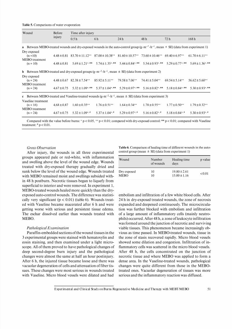
8/3/2019 Luka Bakar Book
http://slidepdf.com/reader/full/luka-bakar-book 57/157
Experimental and Clinical Study on Burns Regenerative Medicine and Therapy with MEBT/MEBO 51
Table 5. Comparisons of water evaporation
Wound Before
injury
Time after injury
0.5 h 6 h 24 h 48 h 72 h 168 h
a Between MEBO-treated wounds and dry-exposed wounds in the auto-control group (g Wm–2Wh–1, meanB SE) (data from experiment 1)
Dry exposed
(n =10) 4.48B0.81 83.70B11.12++ 87.00B10.38++ 81.60B10.57++ 73.60B10.46++ 69.40B6.97++ 61.70B6.11++
MEBO treatment
(n = 10) 4.48B0.81 5.69B1.21+,** 5.74B1.35+,** 5.44B0.84+, ** 5.54B0.93+, ** 5.29B0.77+, ** 5.69B1.36+, **
b Between MEBO-treated and dry-exposed groups (g Wm–2Wh–1, meanB SE) (data from experiment 2)
Dry exposed
(n = 24) 4.48B0.67 82.38B7.54++ 85.92B5.11++ 79.58B7.06++ 74.41B5.04++ 69.54B5.14++ 56.62B5.60++
MEBO treatment
(n = 24) 4.67B0.75 5.32B1.09+,** 5.37B1.04+,** 5.29B0.97+, ** 5.16B0.82+, ** 5.18B0.84+, ** 5.30B0.93+, **
c Between MEBO-treated and Vaseline-treated wounds (g Wm–2Wh–1, meanB SE) (data from experiment 3)
Vaseline treatment
(n = 16) 4.68B0.87 1.60B0.35++ 1.76B0.51++ 1.64B0.34++ 1.78B0.55++ 1.77B0.50++ 1.79B0.32++
MEBO treatment(n = 24) 4.67B0.75 5.32B1.09+, # 5.37B1.04+, # 5.29B0.97+, # 5.16B0.82+, # 5.18B0.84+, # 5.30B0.93+, #
Compared with the value before burns: + p ! 0.05, ++ p ! 0.01; compared with dry-exposed control: ** p ! 0.01; compared with Vaseline
treatment: # p ! 0.01.
Gross Observation
After injury, the wounds in all three experimental
groups appeared pale or red-white, with inflammation
and swelling above the level of the wound edge. Wounds
treated with dry-exposed therapy gradually dried and
sunk below the level of the wound edge. Wounds treated
with MEBO remained moist and swellings subsided with-
in 48 h postburn. Necrotic tissues began to liquefy from
superficial to interior and were removed. In experiment 1,
MEBO-treated wounds healed more quickly than the dry-
exposed auto-control wounds. The difference was statisti-
cally very significant (p ! 0.01) (table 6). Wounds treat-
ed with Vaseline became macerated after 6 h and were
getting worse with serious and persistent tissue edema.
The eschar dissolved earlier than wounds treated with
MEBO.
Pathological Examination
Paraffin-embedded sections of the wound tissues in the
3 experimental groups were stained with hematoxylin and
eosin staining, and then examined under a light micro-
scope. All of them proved to have pathological changes of
deep second-degree burn injury and the pathological
changes were almost the same at half an hour postinjury.
After 6 h, the injured tissue became loose and there was
vacuolar degeneration of cells and attenuation of fiber tis-
sues. These changes were most serious in wounds treated
with Vaseline. Micro blood vessels were dilated and had
Table 6. Comparison of healing time of different wounds in the auto-
control group (meanB SE) (data from experiment 1)
Wound Number
of wounds
Healing time
days
p value
Dry exposed 10 19.80B2.61!0.01
MEBO 10 15.00B1.16
embolism and infiltration of a few white blood cells. After
24 h in dry-exposed treated wounds, the zone of necrosis
expanded and deepened continuously. The microcircula-
tion was further blocked with embolism and infiltration
of a large amount of inflammatory cells (mainly neutro-
phils) occurred. After 48 h, a zone of leukocyte infiltrationwas formed around the junction of necrotic and surviving
viable tissues. This phenomenon became increasingly ob-
vious as time passed. In MEBO-treated wounds, tissue in
the zone of stasis recovered rapidly. Micro blood vessels
showed some dilation and congestion. Infiltration of in-
flammatory cells was scattered in the micro blood vessels.
After 48 h, the cells concentrated on the junction of
necrotic tissue and where MEBO was applied to form a
dense area. In the Vaseline-treated wounds, pathological
changes were quite different from those in the MEBO-
treated ones. Vacuolar degeneration of tissues was more
serious and the inflammatory reaction was diffused.

8/3/2019 Luka Bakar Book
http://slidepdf.com/reader/full/luka-bakar-book 58/157
52 Burns Regenerative Medicine and Therapy
Conclusion
This research revealed that burns wounds treated with
MEBO had less water evaporation than those treated with
conventional dry-exposed therapy. The rate of evapora-
tion was approximate to that of normal skin. Unlike Vase-
line, MEBO had excellent permeability allowing active
drainage of excreta. MEBO demonstrated that its moistphysiological environment is favorable to wound healing.
Discussion
Burn injury destroys the body surface barrier. Injured
skin loses its ability to prevent evaporation of water from
the body. This results in an excessive loss of body fluid
from the lesion area which itself is a stress against healing
since a burn wound that is allowed to dehydrate will be
further injured by dehydration and necrosis. For deep sec-
ond-degree burns wounds, the injured tissue in the zone of stasis can be rescued, but excessive drying will cause irre-
versible damage. This tendency to dehydration is a main
disadvantage of conventional dry-exposed therapy. Stud-
ies of pathophysiology teach that a moist environment is
favorable for the regeneration and repair of injured tissue.
The specially designed MEBO ointment alone succeeds in
creating an environment in which burns wounds can re-
main exposed while avoiding the ravages of dehydration
and maceration.
The key to the maintaining of water in burns wounds is
to prevent evaporation. The results of this study revealed
that burns wounds treated with MEBO had significantly
less evaporation than those given dry-exposed therapy.
MEBO is different from other medical lubricants or oint-
ments such as Vaseline in that MEBO retains wound
moisture while keeping the wound surface permeability.
For example, excessive water can permeate through the
MEBO ointment layer and be removed easily. The differ-
ence between water evaporation of MEBO-treated and
Vaseline-treated wounds was very significant (p ! 0.01).
This was proved by the test of skin sweat excretion [1].
What attracts our great attention was that although the
water evaporation of burns wounds treated with MEBOwas significantly different from that of normal skin (p !
0.05), the difference between the mean values was less
impressive. MEBO’s water retention abilities have been
compared to sebum. MEBO maintains wound moisture
without compromising drainage, therefore comprising a
moist environment that is optimal for wound repair and
healing [2].
It has been reported that there is a zone of thermal-
injured tissue with viability in the deep part and the sur-
roundings of coagulated tissue postburn. This is called the
zone of stasis [3]. Blood flow of micro blood vessels in this
zone diminishes progressively after burns, and remains
very susceptible to further injury. Dehydration or me-
chanical injury will tip the scale toward tissue necrosis in
this zone of stasis. Therefore, it is really difficult to distin-
guish this zone from the initially coagulated tissue that
presents after 5 days postburn. This tendency to allow the
zone of stasis (a recussible wound state) to deteriorate into
a non-recussible death state is consistent with convention-al dry-exposed therapy. By comparison, MEBO inhibited
the deterioration of this process by avoiding secondary
injuries such as dehydration. Therefore, MEBO promotes
the recovery of tissues within the zone of stasis and thus
proves favorable to wound tissue regeneration and repair.
Burns wounds treated with MEBO healed more quickly
than those treated with dry-exposed therapy (p ! 0.01).
This result could not be attributed to the moisture main-
taining effect of MEBO alone, but without a moist envi-
ronment, it is hard for any medical treatment to achieve
[2]. Vaseline treatment, though it also kept the wound
moist but macerated, resulted in suboptimal changes inwound tissues as opposed to the healthy tissue resulting
from MEBO treatment. Based on our results, MEBO pro-
vided a physiologically moist environment uniquely fa-
vorable to wound tissue regeneration and repair.
The unique dosage form and pharmacokinetic features
of MEBO are responsible for its moisture-maintaining
effect. The following three aspects may account for the
mechanism of its action:
1 MEBO has its unique dosage form with frame struc-
ture, which, when applied on the wound, warms and
morphs into an equilibrium of two phases, liquid and
semisolid. It isolates the wound from external irrita-
tion and prevents excessive water evaporation from
the wound surface.
2 MEBO has an active drainage effect whereby excessive
water, liquefaction products and excreta are passively
and automatically removed through the drug layer [2,
4].
3 MEBO base contains hydrophilic and lipophilic groups
and has high surface activity. It has higher affinity to
the wound tissue than water, which allows for the for-
mation of a strong adsorptive film on the wound which
protects it from either maceration or dehydration.
References
1 Xu RX: Moist Exposed Burn Therapy (MEBT): Textbook for Technologi-
cal Training. Beijing, Beijing Guangming Chinese Medicine Institute for
Burns, Wounds and Ulcers, 1989, pp 160–168.
2 Xu RX: The medical science of burns and ulcers: A general introduction.
Chin J Burns Wounds Surface Ulcers 1989;1:11–21.
3 Research Group for Burns of the Third Military Medical University: Burn
Therapy. Beijing, People’s Health Publishing House, 1977, p 125.
4 Du HE: The micro-mechanism of moist exposed burn ointment. Chin J
Burns Wounds Surface Ulcers 1993;5:9–10.

8/3/2019 Luka Bakar Book
http://slidepdf.com/reader/full/luka-bakar-book 59/157
Experimental and Clinical Study on Burns Regenerative Medicine and Therapy with MEBT/MEBO 53
Clinical Study on Invisible Water Loss of
Burns Wounds Treated with BRT with
MEBT/MEBO
Introduction
It has been confirmed that treatment with BRT withMEBT/MEBO offers a unique ability to inhibit excessive
loss of water evaporation through the burn wound surface
[1–4]. However, there has not been adequate quantitative
clinical research at this time. In this study, the authors
observed minimal dehydration of burns wounds treated
with BRT with MEBT/MEBO in 25 burn patients with
different area and different depths compared to the water
evaporation value of wound to that of normal skin.
Materials and Methods
Twenty-five acute burns patients, 21 males and 4 females, were
hospitalized within 6 h after injury. Burn area ranged from 4% total
body surface area (TBSA) to 95% TBSA. After simple debridement,
all patients were treated with BRT with MEBT/MEBO [5]. Observa-
tion at 6, 24, 48 and 72 h postburn measured the water evaporation
capacity of superficial second-degree, deep second- and third-degree
wounds, compared to normal skin as a control. In 4 cases with burn
area over 50% TBSA, their daily amounts of water evaporation were
determined based on wound evaporation and body surface area.
During the liquefaction period, 12 cases with deep second- and third-
degree wounds received semi-exposed MEBO treatment, and their
wound water evaporation was compared with those receiving ex-
posed MEBO treatment.
A Model EPIC evaporometer (Servomed) was used to measurewound water evaporation [6]. Measurement was done at 2 h after
application of MEBO, in a room sheltered from the wind. Room
temperature was kept at 32–36°C with a relative humidity of 45–
62%.
Results
Normal skin water evaporation of the 25 burns pa-
tients had an average value of 18.48 B 5.02 g Wm–2Wh–1.
Wounds were classified according to their depths and one
typical wound from each degree was selected randomly.
Blister skin (necrotic epidermis) of every selected typical
wound was divided in half with one half remaining intactand the other half having its protective skin removed.
Then the evaporation of both halves of the wound was
measured, respectively. The results revealed that water
evaporation in wounds with preserved epidermis had a
very significantly lower evaporation capacity than those
with removed epidermis (p ! 0.01). Water evaporation
reached its peak at 6 h postburn and then decreased, but
still remained markedly higher than that of the normal
skin (p ! 0.05). For very deep wounds, water evaporation
of wounds with removed epidermis tended to decrease
(p ! 0.05), while evaporation in wounds with preserved
epidermis did not decrease significantly (p 1 0.05) (ta-
ble 7).
In 4 extensive burns patients with burn area over 50%
TBSA, their total body surface and burns areas were mea-
sured. Daily water evaporations of the wounds with dif-
ferent depths were calculated, respectively. Then invisible
dehydration per hour per 1% of second- and third-de-
gree wounds can be obtained. The average value was
0.71 mlWh–1W % TBSA–1 (table 8).
During the liquefaction period, deep wounds were
treated with exposed or semi-exposed method, the two
methods did not result in a significant difference inwound water evaporation (p 1 0.05) (table 9).
Table 7. Evaporation of wounds with different depth at different intervals (g Wm–2Wh–1, meanBSE)
Depth of wound Blister
skin
Number
of
wounds
Time after injury
6 h 24 h 48 h 72 h
Superficial second-degree removed 22 57.45B6.40** 43.86B5.88** 35.73B4.23** 32.68B3.70**
preserved 22 28.68B7.58** 24.23B4.62** 23.09B4.56** 22.86B2.88**
Deep second-degree removed 25 44.84B4.66** 37.40B4.94** 32.36B4.53** 30.56B3.62**
preserved 25 26.40B4.80** 24.68B3.44** 23.24B4.05** 22.20B4.80**
Third-degree removed 15 42.53B5.30** 35.93B4.06** 32.07B3.84** 29.07B40.53**
preserved 15 25.13B2.13** 23.67B2.58** 22.33B4.25* 21.93B2.85*
Compared with normal skin: * p ! 0.05, ** p ! 0.01.
Comparison between blister skin removed and preserved, all the differences were very significant: p ! 0.01.

8/3/2019 Luka Bakar Book
http://slidepdf.com/reader/full/luka-bakar-book 60/157
54 Burns Regenerative Medicine and Therapy
Table 8. Invisible dehydration of 4 extensive burns patients
Case
No.
Burn area of different depth, % TBSA
TBSA superficial
second-degree
deep
second-degree
third
degree
Wound evaporation, g W m–2W h–1
superficial
second-degree
deep
second-degree
third
degree
Daily invisible
dehydration
ml
Invisible
dehydration
ml Wh–1W
% TBSA–1
1 53 23 30 61 44 1,097 0.86
2 73 6 21 46 49 45 26 888 0.503 94 40 54 53 39 1,598 0.71
4 95 3 67 25 60 42 36 1,746 0.77
Table 9. Comparison of water evaporation between the exposed and
semi-exposed wounds (meanB SE)
Wound Number
of wounds
Evaporation
g Wm–2Wh–1
p value
Exposed 13 42.58B7.8910.05
Semi-exposed 12 43.92B8.43
Conclusion
1 BRT with MEBT/MEBO effectively inhibited water
evaporation from wounds and decreased total body
fluid loss. Additionally, preservation of necrotic epi-
dermis, including blister skin, also helped to decrease
water loss.
2 The fluid injection of burns patients in the early
stage postburn follows the formula: 0.4–1.2 ml/(h !
% BSA), with adjustment according to depth of the
wound and exfoliation of the epidermis.
3 During the liquefaction period, MEBO could be ap-
plied in either the exposed or the semi-exposed mode.
Discussion
Skin is the surface barrier of the human body. As such,
it plays an important role in maintaining body fluid sta-
tus. Burn injury causes damage of this barrier and in-creases vascular permeability, causing a significant rate of
increase of dehydration. Loss of body fluid through burns
wounds is proportional to the burn area and depth as well
as being related to burn intervals postburn [7]. The results
of this study showed that water loss through the wound in
the first few days postburn was much greater than through
normal skin (p ! 0.05) and reached its peak at 6 h post-
burn, then tended to decline and gradually decrease along
with the deepening of wound. Preservation of necrotic
epidermis, including blistered skin, also helped to de-
crease water loss. Previous studies gave similar results [8,
9]. However, our degree of local wound water loss was
much less than that of those previous reports due to the
use of the BRT protocol (MEBT/MEBO) to treat the
wounds. We conclude that BRT reduced wound exuda-
tion and inhibited evaporation.
Increased capillary permeability causes exudation of
body fluid into the interstitial space, thus forming ‘burnedema’. Another pathway of water loss is invisible dehy-
dration through the burn wound. In this study with 4
cases having burn area over 50% TBSA, the average water
loss per hour per 1% of second-degree and third-degree
wound water loss was 0.71 ml. It would be helpful to eval-
uate the volume of fluid resuscitation during the shock
stage postburn. Based on this study, the fluid injection of
burns patients in the early stage postburn can follow the
formula: 0.4–1.2 ml/(h ! % BSA), also adjust according
to the depth of the wound and the exfoliation of the epi-
dermis.
MEBT suggests application of MEBO ointment with
the exposed method, but in some cases, the semi-exposed
method may be also advisable. The efficacy of the semi-
exposed method was comparable to that of the exposed
method. Our limited experiment proved that there was no
significant difference in wound water evaporation be-
tween the two methods during the liquefaction period (p 1
0.05), indicating that application of MEBO with the
exposed or semi-exposed methods had a similar efficacy
on inhibiting water loss from the wound surface. It is
interesting to note that wound water evaporation mea-
sured at the liquefaction period was greater than duringthe edema resorption period. This might be due to the
requirement of more water for the liquefied reaction
between MEBO and necrotic tissues as well as the produc-
tion of liquefaction products.
References
1 Xu RX: Moist Exposed Burn Therapy (MEBT): Textbook for Technologi-
cal Training. Beijing, Beijing Guangming Chinese Medicine Institute for
Burns, Wounds and Ulcers, 1989, pp 160–168.
2 Wang CC, Pu ZB, Liu HB, et al: Experimental study on maintaining physi-
ological moist effect of moist exposed burn therapy/moist exposed burn
ointment on treating burn wound. Chin J Burns Wounds Surface Ulcers
1998;10:18–20.
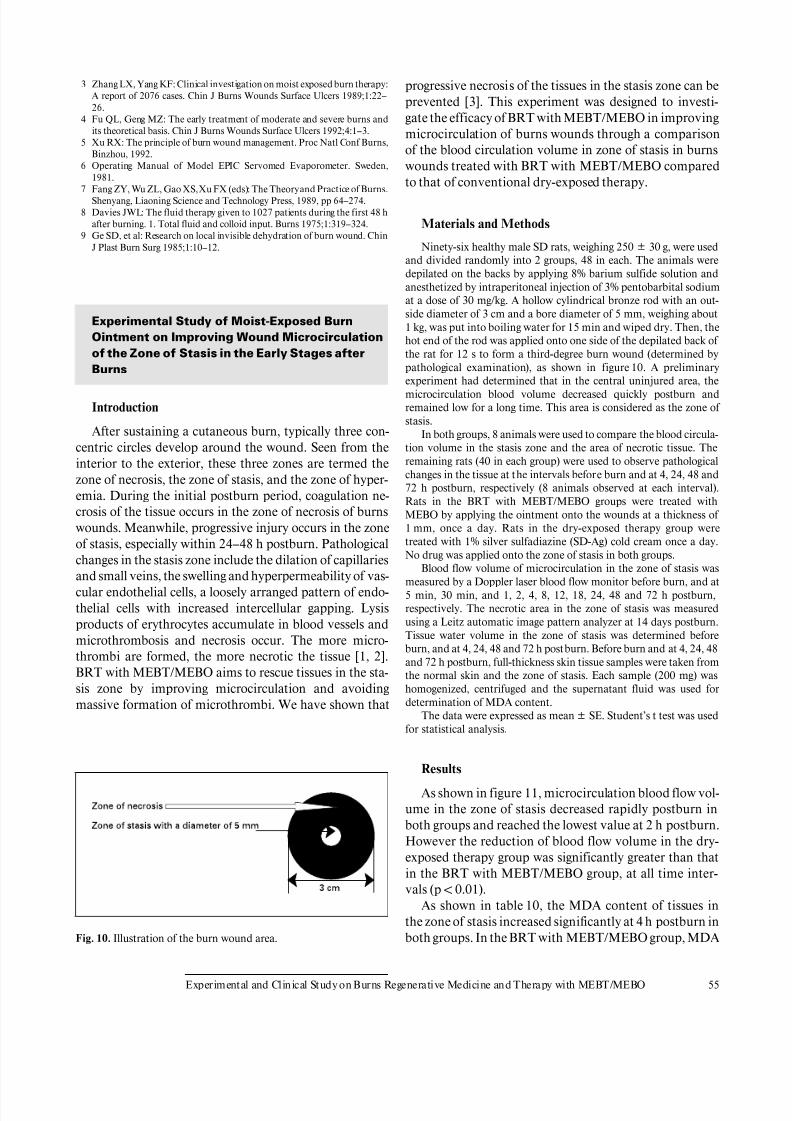
8/3/2019 Luka Bakar Book
http://slidepdf.com/reader/full/luka-bakar-book 61/157
Experimental and Clinical Study on Burns Regenerative Medicine and Therapy with MEBT/MEBO 55
3 Zhang LX, Yang KF: Clinical investigation on moist exposed burn therapy:
A report of 2076 cases. Chin J Burns Wounds Surface Ulcers 1989;1:22–
26.
4 Fu QL, Geng MZ: The early treatment of moderate and severe burns and
its theoretical basis. Chin J Burns Wounds Surface Ulcers 1992;4:1–3.
5 Xu RX: The principle of burn wound management. Proc Natl Conf Burns,
Binzhou, 1992.
6 Operating Manual of Model EPIC Servomed Evaporometer. Sweden,
1981.
7 Fang ZY, Wu ZL, Gao XS, Xu FX (eds): The Theory and Practice of Burns.Shenyang, Liaoning Science and Technology Press, 1989, pp 64–274.
8 Davies JWL: The fluid therapy given to 1027 patients during the first 48 h
after burning. 1. Total fluid and colloid input. Burns 1975;1:319–324.
9 Ge SD, et al: Research on local invisible dehydration of burn wound. Chin
J Plast Burn Surg 1985;1:10–12.
Experimental Study of Moist-Exposed Burn
Ointment on Improving Wound Microcirculation
of the Zone of Stasis in the Early Stages after
Burns
Introduction
After sustaining a cutaneous burn, typically three con-
centric circles develop around the wound. Seen from the
interior to the exterior, these three zones are termed the
zone of necrosis, the zone of stasis, and the zone of hyper-
emia. During the initial postburn period, coagulation ne-
crosis of the tissue occurs in the zone of necrosis of burns
wounds. Meanwhile, progressive injury occurs in the zone
of stasis, especially within 24–48 h postburn. Pathological
changes in the stasis zone include the dilation of capillariesand small veins, the swelling and hyperpermeability of vas-
cular endothelial cells, a loosely arranged pattern of endo-
thelial cells with increased intercellular gapping. Lysis
products of erythrocytes accumulate in blood vessels and
microthrombosis and necrosis occur. The more micro-
thrombi are formed, the more necrotic the tissue [1, 2].
BRT with MEBT/MEBO aims to rescue tissues in the sta-
sis zone by improving microcirculation and avoiding
massive formation of microthrombi. We have shown that
Fig. 10. Illustration of the burn wound area.
progressive necrosis of the tissues in the stasis zone can be
prevented [3]. This experiment was designed to investi-
gate the efficacy of BRT with MEBT/MEBO in improving
microcirculation of burns wounds through a comparison
of the blood circulation volume in zone of stasis in burns
wounds treated with BRT with MEBT/MEBO compared
to that of conventional dry-exposed therapy.
Materials and Methods
Ninety-six healthy male SD rats, weighing 250B 30 g, were used
and divided randomly into 2 groups, 48 in each. The animals were
depilated on the backs by applying 8% barium sulfide solution and
anesthetized by intraperitoneal injection of 3% pentobarbital sodium
at a dose of 30 mg/kg. A hollow cylindrical bronze rod with an out-
side diameter of 3 cm and a bore diameter of 5 mm, weighing about
1 kg, was put into boiling water for 15 min and wiped dry. Then, the
hot end of the rod was applied onto one side of the depilated back of
the rat for 12 s to form a third-degree burn wound (determined by
pathological examination), as shown in figure 10. A preliminary
experiment had determined that in the central uninjured area, the
microcirculation blood volume decreased quickly postburn and
remained low for a long time. This area is considered as the zone of
stasis.
In both groups, 8 animals were used to compare the blood circula-
tion volume in the stasis zone and the area of necrotic tissue. The
remaining rats (40 in each group) were used to observe pathological
changes in the tissue at the intervals before burn and at 4, 24, 48 and
72 h postburn, respectively (8 animals observed at each interval).
Rats in the BRT with MEBT/MEBO groups were treated with
MEBO by applying the ointment onto the wounds at a thickness of
1 mm, once a day. Rats in the dry-exposed therapy group were
treated with 1% silver sulfadiazine (SD-Ag) cold cream once a day.
No drug was applied onto the zone of stasis in both groups.
Blood flow volume of microcirculation in the zone of stasis was
measured by a Doppler laser blood flow monitor before burn, and at
5 min, 30 min, and 1, 2, 4, 8, 12, 18, 24, 48 and 72 h postburn,
respectively. The necrotic area in the zone of stasis was measured
using a Leitz automatic image pattern analyzer at 14 days postburn.
Tissue water volume in the zone of stasis was determined before
burn, and at 4, 24, 48 and 72 h postburn. Before burn and at 4, 24, 48
and 72 h postburn, full-thickness skin tissue samples were taken from
the normal skin and the zone of stasis. Each sample (200 mg) was
homogenized, centrifuged and the supernatant fluid was used for
determination of MDA content.
The data were expressed as meanB SE. Student’s t test was used
for statistical analysis.
Results
As shown in figure 11, microcirculation blood flow vol-
ume in the zone of stasis decreased rapidly postburn in
both groups and reached the lowest value at 2 h postburn.
However the reduction of blood flow volume in the dry-
exposed therapy group was significantly greater than that
in the BRT with MEBT/MEBO group, at all time inter-
vals (p ! 0.01).
As shown in table 10, the MDA content of tissues in
the zone of stasis increased significantly at 4 h postburn in
both groups. In the BRT with MEBT/MEBO group, MDA

8/3/2019 Luka Bakar Book
http://slidepdf.com/reader/full/luka-bakar-book 62/157
56 Burns Regenerative Medicine and Therapy
content turned to the level of pre-injury at 24 h postburn,
while in the dry-exposed therapy group, the MDA level
rose continuously and was significantly higher than that
in the BRT with MEBT/MEBO group, at different time
intervals (p ! 0.01).
As shown in table 11, water volume of tissues in the
zone of stasis increased at 4 h postburn in both groups. In
Fig. 11. Dynamic changes of microcirculation blood flow volume in
the zone of stasis postburn.
the dry-exposed therapy group, water volume increased
significantly at 4 and 48 h postburn and returned to the
normal level at 72 h postburn, while in BRT with MEBT/
MEBO group, water volume turned to the normal level at
48 h postburn (p ! 0.05).
In the dry-exposed therapy group, necrotic tissue ar-
ea in the zone of stasis at 14 h postburn was 20.96B
3.51 mm2, it was significantly larger than 8.38 B
1.78 mm2 in the MEBT/MEBO group (p ! 0.01).
Conclusion
Treatment with MEBT/MEBO in the early stages after
burns could improve the wound microcirculation in the
zone of stasis, lessen further injuries to the support tissues
of the burn wound, then increase the recovery of injured
tissue at the zone of stasis.
Discussion
Microthrombosis and vascular hyperpermeability are
major changes in the wound microcirculation system after
burns. In the zone of stasis, microthrombosis is often
obvious [1, 2]. BRT involves covering the burn wound
with MEBO because this specially designed ointment is
effective for improving wound microcirculation and pre-
Table 10. MDA levels in the zone of stasis between the two groups (nmol/mg protein, meanB SE)
Group Before injury Time after injury
4 h 24 h 48 h 72 h
MEBT/MEBO 0.065B0.07 0.147B0.047* 0.078B0.024* 0.069B0.019 0.063B0.022
Dry-exposed 0.065B0.021 0.240B0.078*, + 0.303B0.097*, + 0.363B0.099*, + 0.378B0.114*, +
Compared with before injury: * p ! 0.01; compared with group 1: + p ! 0.01.
Table 11. Comparison of water volume in the zone of stasis between the two groups (%, mean B SE)
Group Before injury Time after injury
4 h 24 h 48 h 72 h
MEBT/MEBO 66.2B2.5 77.3B3.1* 76.5B2.64* 67.1B2.7 65.9B2.4
Dry-exposed 66.2B2.5 89.9B4.3**, + 86.4B3.9**, + 77.2B4.0* 68.9B3.3
Compared with before injury: * p ! 0.05, ** p ! 0.01; compared with MEBT/MEBO group: + p ! 0.05.
8/3/2019 Luka Bakar Book
http://slidepdf.com/reader/full/luka-bakar-book 63/157
Experimental and Clinical Study on Burns Regenerative Medicine and Therapy with MEBT/MEBO 57
venting progressive necrosis of tissues in the zone of stasis
[3]. The zone of stasis is developing progressively at the
early stages postburn, so its area is difficult to measure.
Our experiment provided a unique model which helped
to solve this problem.
In the dry-exposed therapy group, the necrotic area of
tissue in the zone of stasis was 20.96B
3.51 mm2
at 14days postburn. This value was significantly higher than
the 8.38 B 1.78 mm2 of the MEBT/MEBO group (p !
0.01). This proved that MEBO is effective for improving
microcirculation and preventing further coagulation ne-
crosis of the injured tissue with viability in the zone of
stasis. According to the results of this study, we concluded
that BRT with MEBT/MEBO has the following advan-
tages:
1 Improving microcirculation in the zone of stasis, pre-
venting massive microthrombosis, increasing blood
flow volume, lessening the degree and shortening the
duration of both ischemia and hypoxia relative to tis-sues in the zone of stasis, and thus protecting the vitali-
ty of the viable tissues.
2 Reducing capillary permeability, preventing massive
exudation through burns wounds and tissue edema.
Exudation and edema will cause ischemia and necrosis
in the surrounding tissues of burns wounds or even
hypovolemic shock.
3 Reducing the production of oxygen free radicals and
inhibiting injury due to lipid peroxidation. MDA is a
product of lipid peroxidation, its content reflects the
degree of the reaction. In this study, MEBO quickly
decreased the MDA content of tissues in the zone of
stasis, while in the dry-exposed therapy group, the
MDA level rose continuously.
References
1 Bonaldi LA, Frank DH: Pathophysiology of the burn wound; in Achauer
BM (ed): Management of the Burned Patient. Norwalk, Appleton & Lange,
1987, pp 21–47.
2 Li A (Ngao) (ed): Burn Therapy, ed 2. Beijing, People’s Health Publishing
House, 1995, pp 94–96.
3 Xu RX (ed): New Advances of Modern Surgery. Beijing, Chinese Medical
Science & Technology Press, 1998, pp 18–48.
Clinical Study of Moist-Exposed Burns
Ointment on Improving Microcirculation of
Burns Wounds
Introduction
The typical changes in microcirculation immediately
following burn trauma include cellular and gross anatomi-
cal changes. At the cellular level, we see an increase of
vascular permeability and an increased risk of micro-
thrombus. At the macro level, we see three concentric cir-
cles developing around the wound after cutaneous burns,
ranging from interior to exterior and termed the zone of
necrosis, the zone of stasis and the zone of hyperemia [1].
The microcirculation in the zone of stasis is characterized
by the formation of progressive microthrombus. The loca-tion of the zone of stasis varies according to the depth of
the burns. Burns affecting the dermis are termed second-
degree burns while third-degree burns involve the full-
thickness of skin as well as subcutaneous tissues. The ulti-
mate burn depth is determined by the development of the
stasis zone, which may extend full-thickness, arresting
blood circulation and causing tissue necrosis resulting in a
third-degree burn. However, if there is some improve-
ment in the microcirculation in the zone of stasis, deep
second-degree burns will rarely deteriorate into third-
degree burns. BRT was designed to deal with the zone of
stasis by improving the microcirculation, avoiding theformation of microthrombus and thereby arresting pro-
gressive necrosis.
Recent research suggests that significant release of vas-
cular active factors in plasma, endothelin (ET) and nitric
oxide (NO), has a close correlation with the deterioration
of vulnerable tissue within the zone of stasis during the
early postburn stage [2–4]. ET and NO released from the
burn wound become the primary source of ET and NO in
plasma. The studies during the previous decade have paid
great attention to ET and NO known as ‘star molecules’
because they are the most powerful vascular contraction
and dilation factors known to man. Under normal non-
traumatic situations, vascular endothelial cells maintain a
relatively stable ratio of ET/NO to ensure the normal
capacity for vasoconstriction and dilation within the mi-
crocirculation of the skin. However, certain pathological
conditions such as stress, ischemia, hypoxia and acidosis
may stimulate the massive increase in the synthesis and
release of ET and NO. These challenges upset the original
balance of ET and NO, causing a cascade of reactive
pathological damages. This study investigates the relative
impact on plasma ET/NO ratio by BRT (MEBT) and con-
ventional therapies and gives a further investigation onthe mechanism of MEBO for improving the microcircula-
tion of burns wounds.
Materials and Methods
The study involved 34 adult patients who suffered 30–40% TBSA
burns with more than 20% TBSA of deep second-degree wounds, all
of whom were admitted to the hospital at 4–5 h after injury. All sub-
jects were randomized to receive either BRT with MEBT/MEBO or
conventional dry-exposed therapy. The MEBO group included 18
patients (11 male, 7 female) with a mean age of 26B 10.9 years while
the dry-exposed group comprised 16 patients (10 male, 6 female)
with a mean age of 28B 12.0 years.
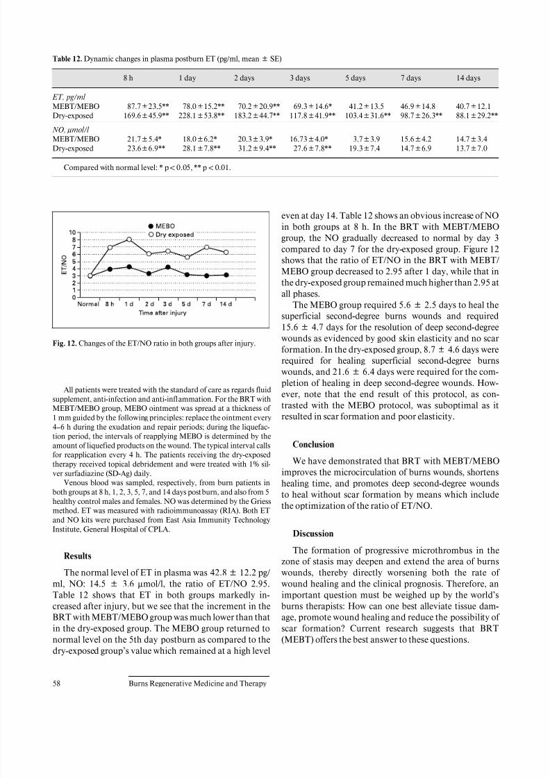
8/3/2019 Luka Bakar Book
http://slidepdf.com/reader/full/luka-bakar-book 64/157
58 Burns Regenerative Medicine and Therapy
Table 12. Dynamic changes in plasma postburn ET (pg/ml, meanB SE)
8 h 1 day 2 days 3 days 5 days 7 days 14 days
ET, pg/ml
MEBT/MEBO 87.7B23.5** 78.0B15.2** 70.2B20.9** 69.3B14.6* 41.2B13.5 46.9B14.8 40.7B12.1
Dry-exposed 169.6B45.9** 228.1B53.8** 183.2B44.7** 117.8B41.9** 103.4B31.6** 98.7B26.3** 88.1B29.2**
NO, Ìmol/l MEBT/MEBO 21.7B5.4* 18.0B6.2* 20.3B3.9* 16.73B4.0* 3.7B3.9 15.6B4.2 14.7B3.4
Dry-exposed 23.6B6.9** 28.1B7.8** 31.2B9.4** 27.6B7.8** 19.3B7.4 14.7B6.9 13.7B7.0
Compared with normal level: * p ! 0.05, ** p ! 0.01.
Fig. 12. Changes of the ET/NO ratio in both groups after injury.
All patients were treated with the standard of care as regards fluidsupplement, anti-infection and anti-inflammation. For the BRT with
MEBT/MEBO group, MEBO ointment was spread at a thickness of
1 mm guided by the following principles: replace the ointment every
4–6 h during the exudation and repair periods; during the liquefac-
tion period, the intervals of reapplying MEBO is determined by the
amount of liquefied products on the wound. The typical interval calls
for reapplication every 4 h. The patients receiving the dry-exposed
therapy received topical debridement and were treated with 1% sil-
ver surfadiazine (SD-Ag) daily.
Venous blood was sampled, respectively, from burn patients in
both groups at 8 h, 1, 2, 3, 5, 7, and 14 days postburn, and also from 5
healthy control males and females. NO was determined by the Griess
method. ET was measured with radioimmunoassay (RIA). Both ET
and NO kits were purchased from East Asia Immunity TechnologyInstitute, General Hospital of CPLA.
Results
The normal level of ET in plasma was 42.8B 12.2 pg/
ml, NO: 14.5 B 3.6 Ìmol/l, the ratio of ET/NO 2.95.
Table 12 shows that ET in both groups markedly in-
creased after injury, but we see that the increment in the
BRT with MEBT/MEBO group was much lower than that
in the dry-exposed group. The MEBO group returned to
normal level on the 5th day postburn as compared to the
dry-exposed group’s value which remained at a high level
even at day 14. Table 12 shows an obvious increase of NO
in both groups at 8 h. In the BRT with MEBT/MEBO
group, the NO gradually decreased to normal by day 3
compared to day 7 for the dry-exposed group. Figure 12
shows that the ratio of ET/NO in the BRT with MEBT/MEBO group decreased to 2.95 after 1 day, while that in
the dry-exposed group remained much higher than 2.95 at
all phases.
The MEBO group required 5.6B 2.5 days to heal the
superficial second-degree burns wounds and required
15.6B 4.7 days for the resolution of deep second-degree
wounds as evidenced by good skin elasticity and no scar
formation. In the dry-exposed group, 8.7B 4.6 days were
required for healing superficial second-degree burns
wounds, and 21.6B 6.4 days were required for the com-
pletion of healing in deep second-degree wounds. How-
ever, note that the end result of this protocol, as con-
trasted with the MEBO protocol, was suboptimal as it
resulted in scar formation and poor elasticity.
Conclusion
We have demonstrated that BRT with MEBT/MEBO
improves the microcirculation of burns wounds, shortens
healing time, and promotes deep second-degree wounds
to heal without scar formation by means which include
the optimization of the ratio of ET/NO.
Discussion
The formation of progressive microthrombus in the
zone of stasis may deepen and extend the area of burns
wounds, thereby directly worsening both the rate of
wound healing and the clinical prognosis. Therefore, an
important question must be weighed up by the world’s
burns therapists: How can one best alleviate tissue dam-
age, promote wound healing and reduce the possibility of
scar formation? Current research suggests that BRT
(MEBT) offers the best answer to these questions.
8/3/2019 Luka Bakar Book
http://slidepdf.com/reader/full/luka-bakar-book 65/157
Experimental and Clinical Study on Burns Regenerative Medicine and Therapy with MEBT/MEBO 59
This study was designed to investigate the effect of
MEBO on plasma ET/NO so as to further discover the
mechanisms of MEBT therapy. ET, separated from super-
natant fluid of cultured pig aorta endothelial cell by Ya-
nagisawa in 1988, is mainly synthesized in the vascular
endothelial cell and is known to be the strongest vasocon-
strictive peptide in addition to other biological actions.However, the excessive release of ET may cause micro
blood vessels to contract and spasm for an extended peri-
od of time, resulting in microthrombus formation. NO is
a potent gaseous free radical and is synthesized in the
cytoplasm of vascular endothelial cells, vascular smooth
muscle cells, macrophages, platelets, etc. This reaction
occurs when nitrogen monoxide synthetase (NOS) cata-
lyzes the guanidin-end nitrogen of L-Arg to combine with
oxygen. There are two types of NOS: constitutional NOS
(cNOS) and inducible NOS (iNOS). NO has a very short
half-life as it may react with circulating oxyhemoglobin,
deoxyhemoglobin, superoxide anion or free oxygen tocreate stable products such as nitrite and nitrate. These
are metabolized mainly through the kidney.
NO expresses its effects in two ways. On the one hand,
NO is antagonistic with ET in dilating micro blood ves-
sels, forming a protective layer in the tunica intima, pre-
venting platelets and neutrophils from adhering to the
vessel wall, and inhibiting the formation of microthrom-
bus. On the other hand, the excessive release of NO may
result in severe wound exudation and edema in the early
stages postburn, thus increasing damage to tissue and
cells. Under the normal situation, NO and ET maintain a
certain dynamic equilibrium as an increase of ET pro-
motes an elevation of NO synthesis while NO inhibits the
synthesis of ET. A physiologic ratio of ET/NO represents
a healthy balance.
According to the studies in recent years, the iNOS and
ET mRNA genes located in burns wounds, heart, lung,
kidney, liver and gastrointestinal tract express an increas-
ing activity postburn, which leads to a massive release of
ET and NO, especially from the wound. The significant
increase of ET and NO in the circulation and the imbal-
ance of the ET/NO ratio have a close correlation with
shock, acute renal failure, acute respiratory failure, stressulcer and cerebral edema after severe burns. Great care
must be paid to the role of ET/NO if one is to prevent
progressive damage of the wound tissues in the zone of
stasis at the early stages postburn [2]:
1 NO and ET, known as the most potent vasoconstric-
tion and vasodilatory factors, should maintain a
healthy balance during the critical postburn period.
2 The increment of NO and ET in blood plasma post-
burn has a positive relationship with the increase of the
capillary permeability, leading to massive exudation
and hyperedema [3, 4].
3 The increase of ET in plasma, the contraction and
spasm of the micro blood vessels, the thrombus forma-
tion around the wound and underlying tissue, and ede-
ma pressure may cause secondary ischemia and necro-
sis to the adjacent tissues.
4 The exudates, being rich in protein, provide a good cul-
ture medium for bacteria growth.It was found that the application of a non-selective ET
receptor antagonist, AK-044, may alleviate the secondary
damage to burns wounds resulting from the increase of
ET [5]. In this study, the increment of ET and NO in the
BRT with MEBT/MEBO group was obviously lower than
that in the dry-exposed group. In the former group, the
ET/NO ratio in plasma decreased after 1 day toward a
normal level of 2.95, representing a timely improvement
in the tissue microcirculation status particularly in the
zone of stasis. This prevented the occurrence of progres-
sive microthrombosis and prevented the progressive ne-
crosis of tissue by optimizing the wound environment. Inthe dry-exposed group, ET and NO increased significantly
at all phases and the ET/NO ratio in plasma remained
much higher than normal (2.95), usually at around 6. ET
might play a dominant role in this change by keeping the
micro blood vessel contracted and spasming for a long
time resulting in microthrombosis, low vitality of wound
tissues, and poorer healing process compared to that in
the BRT with MEBT/MEBO group. The results suggest
that the BRT with MEBT/MEBO group is superior to the
dry-exposed group in wound healing time and outcome.
However, further study is yet to be conducted for the spe-
cific mechanism on how MEBO controls the adequate
release of ET and NO from the burns wounds, and on how
to promote the return of the ET/NO ratio to normal as
rapidly as possible.
References
1 Bonaldi LA, Frank DH: Pathophysiology of the burn wound; in Achauer
BM (ed): Management of the Burned Patient. Norwalk, Appleton & Lange,
1987, pp 21–47.
2 Battal MN, Hata Y, Matsuka K, et al: Reduction of progressive burn injury
by using a new nonselective endothelin-A and endothelin-B receptor antag-
onist, Tak-044: An experimental study in rats. Plast Reconstr Surg 1997;
99:1610–1619.3 Kowal VA, Walenga JM, Sharp PM, et al: Postburn edema and related
changes in interleukin-2, leukocytes, platelet activation, endothelin-1, and
C1 esterase inhibitor. J Burn Care Rehab 1997;18:99–103.
4 Sozumi T: The role of nitric oxide in vascular permeability after a thermal
injury. Ann Plast Surg 1997;39:272–277.
5 Jia XM, Zhu ZM, Zeng Q, et al: The dynamic changes of endothelin-1 in
rat plasma, wound and organs at the early stages postburn. Med J PLA
1997;22:40–42.

8/3/2019 Luka Bakar Book
http://slidepdf.com/reader/full/luka-bakar-book 66/157
60 Burns Regenerative Medicine and Therapy
Experimental Study of the Effect of BRT with
MEBT/MEBO on Hematological Parameters in
the Treatment of Burned Rabbits
Introduction
Many researchers found that treatment with BRT with
MEBT/MEBO improved the microcirculation in burns
wounds. However, the effect of BRT with MEBT/MEBO
on systemic postburn situations has not been established.
The authors used a rabbit model for the following experi-
mental study on this subject.
Materials and Methods
Seventy-two healthy adult rabbits of either sex eating a uniform
diet and weighing 2.0 B 0.5 kg were divided randomly into three
groups: group 1 (normal control, n = 12), group 2 (treated withMEBO, n = 30), and group 3 (dry exposure, n = 30). Without anesthe-
sia, all animals were shaved on the waist with barium sulfide. Ani-
mals in group 1 received no treatment and cardiac blood was sam-
pled for measuring normal hematological parameters. Animals in
groups 2 and 3 sustained deep second-degree burns of 10% BSA via
100°C water applied for 5 s (confirmed by pathological examina-
tion). Animals in group 2 were then treated with BRT with MEBT/
MEBO renewed every 6 h. Animals in group 3 were offered no treat-
ment with wounds remaining dry and exposed. No fluid infusion was
administered. Blood samples were taken from the left ventricle of the
heart for determining hematological values at 4, 24, 48, 72 h and 6
days postinjury. The parameters tested included apparent blood vis-
cosity at different shear rates, plasma viscosity, hematocrit (HCT),
erythrocyte agglutinative index (EAI) and erythrocyte transforma-tion kinetics (ETK). To standardize the experimental conditions and
instruments, blood sampling was performed in accordance with
requirements for determination of blood flow viscosity established
by the International Council for Standardization in Haematology
(ICSH) in 1988. These laboratory determinations were made by a
specially assigned technologist at room temperature using a Type N
E-1 viscometer (manufactured by Chengdu Instruments, China) [1].
Apparent blood viscosity at the shear rates of 230, 115, 46, 11.5,
and 5.75 s–1 were determined, respectively, using 1.3 ml blood. Plas-
ma viscosity is the viscosity at the shear rate of 115 s –1. Hematocrit
(HCT) was determined by a Wintrope tube, type LXJ-64-01 centri-
fuge, at 3,000 rpm for 30 min [2]. Erythrocyte agglutinative index
(EAI) was the ratio of apparent blood viscosity obtained at low and
high shear rates (5.75 and 230 s–1). Erythrocyte transformation kinet-
ics (TK) was calculated according to the formula given in Chen [3].
Results
Owing to the minor difference of sex and strain in the
hematological parameters of the rabbits, it was feasible to
work out a uniform range [1–4]. Randomized division
kept animals in a normal control group, allowing the
MEBO-treated and the dry-exposed groups to live under
the same conditions. Therefore, no repeated blood sam-
pling was allowed and the data were significantly valu-
able. Apparent blood viscosity and plasma viscosity at
different shear rates in group 3 were higher than those in
group 2. Table 13 shows that the parameters in group 3
began to increase at 4 h post-injury, peaked at 24 h, and
remained higher at 48 h, 72 h and 6 days than those in
group 2. There were significant statistical differences (ta-
ble 14; p ! 0.01). Compared to the normal group, parame-
ters including apparent blood viscosity, plasma viscosity
and others in the MEBO-treated group began to increase
at 4 h postinjury, peaked at 24 h, decreased at 48 h, fur-
ther decreased at 72 h and nearly returned to normal at 6
days (table 15). But statistical analysis showed that hema-tological parameters did not change significantly (p 1
0.05), except at 24 h postinjury when the blood viscosity
at shear rates of 11.5 s–1 (t = 2.4696, p ! 0.05) and 5.75 s–1
(t = 2.700, p ! 0.05) increased markedly (table 16).
Table 13. Changes of hemorrheological parameters in group 3 (dry group) (mean B SE)
Parameters 4 hpostinjury
24 hpostinjury
48 hpostinjury
72 hpostinjury
6 dayspostinjury
Blood viscosity
230 s–1 5.45B0.54 8.66B0.59 6.24B0.188 4.57B0.588 5.61B0.43
115 s–1 5.92B0.66 8.86B1.37 6.55B0.28 5.27B1.13 6.57B0.46
46 s–1 6.49B0.71 10.87B1.42 7.99B0.48 5.98B0.44 7.14B0.41
23 s–1 7.26B0.43 12.71B1.43 9.32B0.33 8.03B0.46 8.84B1.59
11.5 s–1 12.89B1.32 21.13B2.65 13.12B0.34 10.74B1.52 10.88B0.73
5.75 s–1 18.12B3.42 25.22B0.96 19.99B4.32 14.39B1.74 12.39B0.12
Plasma viscosity (115s–1) 2.71B0.08 2.64B0.32 2.33B0.123 2.15B0.116 2.30B0.095
HCT 0.42B0.051 0.43B0 0.4B0.026 0.36B0.04 0.036B0.015
EAI 3.29B0.27 2.86B0.12 3.17B0.68 3.14B0.023 2.21B0.255
TK 0.64B0.086 0.96B0.098 0.84B0.093 0.82B0.21 0.95B0.144

8/3/2019 Luka Bakar Book
http://slidepdf.com/reader/full/luka-bakar-book 67/157
Experimental and Clinical Study on Burns Regenerative Medicine and Therapy with MEBT/MEBO 61
Table 14. Comparison of hemorrheological parameters between groups 2 and 3
Parameters 4 h postinjury
t p
24 h postinjury
t p
48 h postinjury
t p
72 h postinjury
t p
6 days postinjury
t p
Blood viscosity
230 s–1 4.888 !0.001 10.74 !0.001 5.716 !0.001 1.412 10.05 3.558 !0.01
115 s–1 3.776 !0.01 8.212 !0.001 3.364 !0.01 2.765 !0.05 3.289 !0.0146 s–1 1.838 10.05 8.031 !0.001 4.528 !0.001 4.125 !0.01 2.691 !0.05
23 s–1 0.861 10.05 7.014 !0.001 4.435 !0.01 3.502 !0.01 3.909 !0.01
11.5 s–1 4.611 !0.001 116.15 !0.001 6.572 !0.001 4.985 !0.001 3.261 !0.01
5.75 s–1 4.564 !0.001 10.47 !0.001 23.12 !0.001 6.031 !0.001 2.043 10.05
Plasma viscosity 0.634 10.05 0.125 10.01 1.188 10.05 1.426 10.01 0.254 10.05
HCT 1.866 10.05 1.237 10.05 0.563 10.05 0.574 10.05 0.563 10.05
EAI 2.038 10.05 0.121 10.05 1.075 10.05 9.306 !0.001 0.547 10.05
TK 2.648 !0.05 1.748 10.05 6.106 !0.001 3.276 !0.01 5.048 10.001
Table 15. Hemorrheological parameters in group 1 and changes in group 2 (meanB SD)
Parameters Group 1
(normal
control)
Group 2
4 h
postinjury
24 h
postinjury
48 h
postinjury
72 h
postinjury
6 days
postinjury
Blood viscosity
230 s–1 4.20B0.05 4.13B0.52 4.48B0.7 4.01B0.65 4.00B0.72 4.12B0.72
115s–1 0.69B0.71 4.54B0.68 5.67B0.91 4.58B0.98 4.27B0.8 4.42B1.10
46 s–1 5.53B0.99 5.44B1.00 6.97B1.03 5.12B1.07 4.88B0.86 5.11B1.26
23 s–1 6.79B1.32 6.59B1.30 8.49B1.20 6.00B1.18 5.59B1.17 6.04B1.40
11.5s–1 7.61B1.69 8.5B1.69 9.55B1.26 7.34B1.46 6.69B1.52 7.41B1.81
5.75 s–1 9.33B2.58 12.21B2.7 13.45B1.9 9.92B1.86 8.33B1.88 9.57B2.28Plasma-viscosity (115 s–1) 2.05B0.39 2.48B0.6 2.60B0.55 2.60B0.38 2.28B0.16 2.27B0.20
HCT 0.36B0.05 0.37B0.05 0.40B0.04 0.38B0.06 0.34B0.06 0.34B0.06
EAI 2.19B0.58 2.73B0.47 0.82B0.55 2.65B0.86 2.07B0.19 2.32B0.34
TK 0.73B0.08 0.75B0.08 0.82B0.34 0.75B0.05 0.64B0.04 0.72B0.09
Table 16. Comparison of hemorrheological parameters between groups 1 and 2
Parameters 4 h postinjury
t p
24 h postinjury
t p
48 h postinjury
t p
72 h postinjury
t p
6 days postinjury
t p
Blood viscosity
230 s–1 0.1490 10.05 0.6514 10.05 0.4635 10.05 0.1826 10.05 0.1437 10.05
115s–1 0.3052 10.05 1.6984 10.05 0.1818 10.05 0.2855 10.05 0.4152 10.05
46 s–1 0.0128 10.05 2.0160 10.05 0.5630 10.05 0.9918 10.05 0.5243 10.05
23s–1 0.3239 10.05 1.8600 10.05 0.3276 10.05 0.7940 10.05 0.2600 10.05
11.5 s–1 0.7449 10.05 2.4696 !0.05 0.2430 10.05 0.8100 10.05 0.1632 10.05
5.75 s–1 1.5470 10.05 2.7000 !0.05 0.3907 10.05 0.6635 10.05 0.1456 10.05
Plasma viscosity 1.2020 10.05 1.6320 10.05 2.0210 10.05 1.0440 10.05 1.0040 10.05
HCT 0.2916 10.05 1.2964 10.05 0.5249 10.05 0.5249 10.05 0.5249 10.05
EAI 1.4471 10.05 1.5768 10.05 0.8872 10.05 0.3239 10.05 0.4260 10.05
TK 0.3536 10.05 1.7166 10.05 2.4241 10.05 2.0142 10.05 0.1661 10.05

8/3/2019 Luka Bakar Book
http://slidepdf.com/reader/full/luka-bakar-book 68/157
62 Burns Regenerative Medicine and Therapy
Conclusion
Treatment with MEBO after burns could ameliorate
total body stress reaction, reduce water evaporation from
the wound surface, lessen local and systemic capillary
exudation and thus improve the hemorrheological charac-
teristics of microcirculation. It also suggested that when
the minor or moderately burned patient was treated withMEBO at an early stage, fluid infusion and/or blood trans-
fusion would not be necessary, whereas for severely
burned patients, the amount of fluid infusion could be
reduced as tolerated.
Discussion
Hemorrhagic change is considered to be one of the
pathophysiological changes following burns and serves as
a basis of microcirculation disorder. Subsequent to exten-
sive burns, microvascular permeability increases and co-
pious intravascular plasma exudes toward the wound sur-
face and tissue space leading to localized hemoconcentra-
tion, reduction of effective blood volume, decreased plas-
ticity of red blood cells and increased blood viscosity.
These hemorrhagic changes comprise the pathophysiolog-
ical basis of burn shock and contribute to the deleterious
stress reaction immediately following the trauma of
burns. For instance, adrenaline, 5-HT, and prostaglandin
may all increase the activation of platelets to erythrocytes,
thereby changing the localized electrical potential. The
increased secretion of catecholamines due to stress reac-
tion directly promotes platelet adhesiveness. The injuredsub-microstructure of the vascular wall elaborates an
adhesion protein on platelets and erythrocytes causing
significant intravascular platelet aggregation and contrib-
uting to adhesion and aggregation of platelets and erythro-
cytes. This, of course, precipitates thrombotic events [5].
In this study, we compared the blood viscosity of a
MEBO-treated group with controls and demonstrate that
the viscosity of the MEBO group approximated that of the
normal controls. As animals in the MEBO-treated group
were treated only with MEBO (they received neither fluid
replacement nor special feeding), such changes verified
that MEBO alleviated body stress reaction following
burns and ensured body recovery.
Despite becoming a systemic disease, burn injuries
begin with a wound on one region of the body surface. It
has been reported that BRT with MEBT/MEBO could
have both local and systemic therapeutic effects on burn
management [6]. We are pleased to report that rabbits inthe MEBO-treated group were as active as normal rabbits
and fed freely. Blood viscosity did not change significant-
ly (p 1 0.05), except at shear rates of 11.5and 5.75 s–1 24 h
postinjury.
The blood viscosity value of normal rabbits in this
study was slightly lower than the mean values as indicated
in many domestic and international reports. This differ-
ence is probably explained by the fact that venous blood
from rabbits (weighing 2.5 kg) was sampled in those
reports instead of cardiac blood from rabbits (weighing
2.0 B 0.5 kg) which we used in our study. Different
dietary factors as well as geographical differences maycontribute as well. Blood viscosity is a comprehensive
marker as it indicates aggregation, deformability and the
rheological properties of platelets, RBCs and WBCs. In
this study, blood viscosity and plasma viscosity of rabbits
were compared to and found to be lower than those of
human beings. It remains to be further discussed whether
this lower viscosity is associated with low HCT, difficulty
of RBC aggregation, or whether it is simply some biologi-
cal characteristic associated with ‘herbivores’.
References
1 Du ZY, Jiang XQ: Analysis on hemorrheology of healthy rabbits. Trans
Taishan Med Coll 1991;12:134–136.
2 Zhang YM, Jiang XQ, et al: Investigation and analysis on hemorrheological
parameters of human health. Chin J Hemat 1992;13:312–313.
3 Chen WJ (ed): Hemorrheology. Tianjin, Tianjin Science & Technology
Press, 1987, pp 50–53.
4 Guo ZR, et al: Experimental study on the effects of different resuscitation
regimens on hemorrheology during burn shock period. Chin J Plastic Surg
Burns 1988;4:130–132.
5 Xu RX: The medicine of burn and ulcer: A general introduction. Part 1.
Chin J Burns Wounds Surface Ulcers 1989;1:11–21.
6 Xu RX: A summary report of the first session of the editorial board of the
journal and to the national symposium on the moist exposed burn therapy.
Chin J Burns Wounds Surface Ulcers 1992;4:2–5.

8/3/2019 Luka Bakar Book
http://slidepdf.com/reader/full/luka-bakar-book 69/157
Experimental and Clinical Study on Burns Regenerative Medicine and Therapy with MEBT/MEBO 63
OOOOOOOOOOOOOOOOOOOOOOOOOOOOOOOOOOOOOOOOOOOOOOOOOOOOOOOOOOOOOOOOOOOOOOOOOOOOOOOOOOOOOOOOOOOOOOOOOOOOOOOOOOOOOOOOOOOOOOOOOOO
Studies on the Anti-Infection Effect of BRT with
MEBT/MEBO
Effect of BRT with MEBT/MEBO on the
Immunity of Burns Patients
Introduction
The antibiotic and wound-healing properties of MEBO
have been proven in clinical practice. There are different
opinions about the mechanism of this antibiotic effect, so
research on this subject is very important. During the
period from January 1993 through December 1995, we
conducted clinical observations on the effect of MEBO onburns patients’ immunity and demonstrated that MEBO
enhanced patients’ immunity as part of its antibiotic and
wound-healing effects.
Materials and Methods
Clinical Data
One hundred and twenty burns patients were divided randomly
into two groups. Sixty patients in the MEBO group, including 40
males and 20 females, aged 6–65 (35.5B 14.8) years. Course of dis-
ease: 1–36 h (18.5B 8.8 h) before administration of MEBO. Cause of
burns: direct flame, 30 cases; scald, 16 cases, and chemical burn, 14
cases. Burn position: craniofacial, 17 cases; neck, 17 cases; trunk, 18cases, and limbs, 23 cases. Burns depth: superficial second-degree
burns, 26 cases; deep second-degree burns, 24 cases; third-degree
burns, 10 cases. Burn area: 1–25% (13B 6%) total body surface area
(TBSA). By contrast, there were 60 cases in the control group includ-
ing 41 males and 19 females, aged from 7 to 65 years (36 B 14.5
years). Course of disease: 1–35 h (18B 8.5 h) before administration.
Cause of burns: direct flame, 29 cases; scald, 17 cases, and chemical
burns, 14 cases. Burn position: craniofacial, 13 cases; neck, 7 cases;
trunk, 18 cases, and limbs, 22 cases. Burn depth: superficial second-
degree burns, 24 cases; deep second-degree burns, 26 cases, and
third-degree burns, 10 cases. Burn area: 1–26% (13.5B 6.3%) TBSA.
The data of the two groups were similar and comparable (p 1 0.05).
Treatment and ExaminationAll patients were subjected to debridement before collecting sam-
ples of skin tissue. Patients in the MEBO group were treated with
burns regenerative therapy (MEBT/MEBO) [1, 2], and skin tissues
were taken twice from the original sites before the treatment began
and after the wounds healed, respectively. Patients in the control
group were treated with another traditional Chinese burns oint-
ment – Jing Wan Hong –, and skin tissue was taken at the same time
phase as in the MEBO group. Patients in both groups were observed
closely, and their wound healing time and incidence of wound infec-
tion were compared.
Five pieces of skin tissues taken from the edge of the burns
wounds using a pair of biopsy forceps were immersed in 10% forma-
lin and kept in separate ice bottles, respectively. Four pieces of skin
tissue were also taken from the normal (non-burned) skin of the same
patient and treated identically as the other tissue. In addition, normal
skin tissues from 60 surgical cases were taken during subdermal cystoperations and treated identically to both burns tissues. The levels of
IgA-, IgG-, and IgM-producing cells and C3 were determined using
the frozen section immunohistochemical method. The first antibody
was supplied from Vector and the second from DAKO. Venous
blood samples of burns patients and healthy persons were taken in
the morning before breakfast. Peripheral blood immunoglobulin (Ig)
levels were determined using the agar diffusion method.
Classification Standard of Antibody Producing Cell and C 3The classification standard was as follows: – = no positive cells
or particles were found or only occasionally found in the whole
slide; + = positive cells accounted for less than 30% of the total
number of interstitial cells in the lamina propria; ++ = positive cells
accounted for 31–70% of the total number of interstitial cells in thelamina propria; +++ = positive cells accounted for more than 71%
of the total number of interstitial cells in the lamina propria. Histo-
logical diagnosis was based on the criteria stipulated at the China
National Pathology Research Group Conference held in Zhengzhou
in 1978.
Results
Clinical Efficacy Assessment
In the MEBO group, burns wounds with different
depths had shorter healing time than those in the control
group (p ! 0.01; table 17). Only one case (1.7%) in the
MEBO group had wound infection, compared to 9 cases
(15%) in the control group. The difference between the
Table 17. Comparison of healing time of wounds with different depth in two groups (day, mean B SE)
Group Superficial
second-degree
Deep
second-degree
Third-degree Average
healing time
MEBO 9.5B2.3 (5–14) 25.5B2.3 (21–30) 36.5B2.8 (31–43) 23.5B9.3 (5–42)
Control 13B3 (7–19) 29.5B2.8 (24–35) 42B3.5 (35–49) 28B10.5 (7–49)**
t value 4.65 5.49 3.88 2.49
Compared with MEBO group: ** p ! 0.01.

8/3/2019 Luka Bakar Book
http://slidepdf.com/reader/full/luka-bakar-book 70/157
64 Burns Regenerative Medicine and Therapy
Table 18. Comparison of staining intensity of immune factors in burns and non-burns areas of the patients (%)
Area Cases IgA
^ (+) 6 (++)
IgG
^ (+) 6 (++)
IgM
^ (+) 6 (++)
C3
^ (+) 6 (++)
Burns area 120 60 (50.0) 60 (50.0) 59 (49.2) 61 (50.8) 57 (47.5) 63 (52.3) 58 (48.3) 62 (51.7)
Non-burns area 120 80 (66.7) 40 (33.3) 79 (65.8) 41 (34.2) 77 (64.2) 43 (35.8) 82 (68.3) 38 (31.7)
Normal person 60 44 (73.0) 16 (28.0) 42 (70.0) 18 (30.0) 39 (65.0) 21 (35.0) 42 (70.0) 18 (30.0)
Table 19. Staining intensity of local immune factors and depth of burns wound (%)
Wound Cases IgA
^ (+) 6 (++)
IgG
^ (+) 6 (++)
IgM
^ (+) 6 (++)
C3
^ (+) 6 (++)
Superficial second-degree 50 28 (56.0) 22 (44.0) 29 (58.0) 21 (42.0) 27 (54.0) 23 (46.0) 28 (56.0) 22 (44.0)
Deep second-degree 50 26 (52.0) 24 (48.0) 24 (48.0) 26 (52.0) 25 (50.0) 25 (50.0) 24 (48.0) 26 (52.0)
Third-degree 50 6 (30.0) 14 (70.0) 6 (30.0) 14 (70.0) 5 (25.0) 15 (75.0) 6 (30.0) 14 (70.0)
Table 20. Staining intensity of local immune factors and burn area (%)
TBSA Cases IgA
^ (+) 6 (++)
IgG
^ (+) 6 (++)
IgM
^ (+) 6 (++)
C3
^ (+) 6 (++)
^5% 52 29 (55.8) 23 (44.2) 29 (55.8) 23 (44.2) 30 (55.7) 22 (42.3) 30 (57.2) 22 (42.3)
6–15% 50 26 (52.0) 24 (48.0) 25 (50.0) 25 (50.0) 22 (44.0) 28 (56.0) 23 (46.0) 37 (54.0)
616% 20 5 (27.8) 13 (72.2) 5 (27.8) 13 (72.2) 5 (72.8) 13 (72.2) 5 (72.8) 13 (72.2)
two groups was significant (¯2 = 5.35, p ! 0.05). MEBO
was proven to have infection-controlling and healing-pro-
moting effects.
Experimental Results
IgA-, IgG-, and IgM-producing cells and C3 in the
burns area had higher staining intensity compared to
those of the non-burns area and to those of normal per-
sons (p ! 0.01). The immunity of the local area changedpostburn. Immunologic function and reaction were en-
hanced. In the non-burns area, the immunity was similar
to that of normal persons. The difference was not marked
(p 1 0.05; table 18).
Local immunity was closely related to burn depth.
IgA-, IgG-, and IgM-producing cells and C3 in the local
area of third-degree burns wounds had stronger staining
intensity than those in superficial second-degree burns
wounds (p! 0.05). The deeper the wound, the stronger the
staining intensity (table 19).
Burn area was positively related to local immune factor
staining intensity. IgA-, IgG-, and IgM-producing cells
and C3 of patients with burn area616% TBSA had stron-
ger staining intensity than patients with burn area ^5%
TBSA (p ! 0.05). The larger the burn area of TBSA, the
stronger the staining intensity of the immune factors (ta-
ble 20).
In burns wounds after treatment with MEBO, the local
IgA-, IgG-, and IgM-producing cells and C3 had stronger
staining intensity than before treatment and than those in
the control group (p ! 0.05). MEBO significantly en-hanced the staining intensity of local IgA-, IgG-, and IgM-
producing cells and C3, while the Jing Wan Hong oint-
ment treatment group did not show a significant effect
(p 1 0.05; table 21).
There was no significant correlation between staining
intensity of local IgA-, IgG-, and IgM-producing cells and
C3, and the duration of MEBO treatment (p 1 0.05). The
immunity of the patients was enhanced, irrespective of
the duration of the treatment (table 22).
There was no positive correlation between the Jing Wan
Hong ointment treatment and the staining intensity of local
IgA-, IgG-, and IgM-producing cells and C3 (p 1 0.05).

8/3/2019 Luka Bakar Book
http://slidepdf.com/reader/full/luka-bakar-book 71/157
Experimental and Clinical Study on Burns Regenerative Medicine and Therapy with MEBT/MEBO 65
Table 21. Staining intensity of local immune factors of patients in the two groups, before and after treatment (%)
Group Cases IgA
^ (+) 6 (++)
IgG
^ (+) 6 (++)
IgM
^ (+) 6 (++)
C3
^ (+) 6 (++)
MEBO treatment
Before 60 31 (51.7) 29 (48.3) 29 (48.3) 31 (51.7) 28 (46.7) 32 (53.3) 30 (50.0) 30 (50.0)
After 60 18 (30.0) 42 (70.0) 17 (28.3) 43 (71.7) 16 (30.0) 44 (70.0) 17 (28.4) 43 (71.7)
Control treatment
Before 60 29 (48.3) 31 (51.7) 30 (50.0) 30 (50.0) 29 (48.3) 31 (51.7) 28 (46.7) 32 (53.3)
After 60 29 (48.3) 31 (51.7) 28 (46.7) 32 (53.3) 28 (46.7) 32 (53.2) 28 (46.7) 32 (53.3)
Table 22. Staining intensity of local immune factors and the course of MEBO treatment (%)
Course
days
Cases IgA
^ (+) 6 (++)
IgG
^ (+) 6 (++)
IgM
^ (+) 6 (++)
C3
^ (+) 6 (++)
^10 19 6 (31.6) 13 (68.4) 6 (31.6) 13 (68.4) 5 (26.3) 14 (73.7) 6 (31.6) 13 (68.4)
10–20 10 3 (30.0) 7 (70.0) 3 (30.0) 7 (70.0) 3 (30.0) 7 (70.0) 3 (30.0) 7 (70.0)
21–30 13 4 (30.8) 9 (69.2) 3 (23.1) 10 (76.9) 4 (30.8) 9 (69.2) 4 (30.8) 9 (69.2)
631 18 5 (27.8) 13 (72.2) 5 (27.8) 13 (72.2) 4 (22.2) 14 (77.8) 4 (22.2) 14 (77.8)
Table 23. Staining intensity of local immune factors and the course of Jing Wan Hong ointment treatment (%)
Course
days
Cases IgA
^ (+) 6 (++)
IgG
^ (+) 6 (++)
IgM
^ (+) 6 (++)
C3
^ (+) 6 (++)
^10 18 9 (50.0) 9 (50.0) 8 (44.4) 10 (55.6) 8 (44.4) 10 (55.6) 9 (50.0) 9 (50.0)
10–20 11 6 (54.5) 5 (45.5) 5 (45.5) 6 (54.5) 5 (45.5) 6 (54.5) 4 (36.4) 7 (63.6)
21–30 12 6 (50.0) 6 (50.0) 6 (50.0) 6 (50.0) 6 (50.0) 6 (50.0) 5 (41.7) 7 (58.3)
631 19 8 (42.1) 11 (57.9) 9 (47.4) 10 (52.6) 9 (47.4) 10 (52.6) 10 (52.6) 9 (47.4)
Table 24. Staining intensity of local immune factors and depth of burns wounds treated with MEBO (%)
Wound Cases IgA
^ (+) 6 (++)
IgG
^ (+) 6 (++)
IgM
^ (+) 6 (++)
C3
^ (+) 6 (++)
Superficial second-degree 26 9 (34.6) 17 (65.4) 8 (30.8) 18 (69.2) 8 (30.8) 18 (69.2) 7 (26.9) 19 (73.1)
Deep second-degree 24 7 (29.2) 17 (71.8) 7 (29.2) 17 (71.8) 6 (25.0) 18 (75.0) 7 (29.2) 17 (71.8)
Third-degree 16 2 (20.0) 8 (80.0) 2 (20.0) 8 (80.0) 2 (20.0) 8 (80.0) 3 (30.0) 7 (70.0)
Changes in staining intensity of immune factors were not
attributed to Jing Wan Hong ointment (table 23).
After treatment with MEBO, the deeper the burn
wound, the stronger the staining intensity of local IgA-,
IgG-, and IgM-producing cells and C3, but no statistical
difference (p 1 0.05). MEBO enhanced the local immuni-
ty of different depths of burns wounds (table 24).
After treatment with MEBO, the larger the burn
wound, the stronger the staining intensity of local IgA-,
IgG-, and IgM-producing cells and C3, but no statistical

8/3/2019 Luka Bakar Book
http://slidepdf.com/reader/full/luka-bakar-book 72/157
66 Burns Regenerative Medicine and Therapy
Table 25. Staining intensity of local immune factors and area of burns wounds treated with MEBO (%)
Area
(TBSA)
Cases IgA
^ (+) 6 (++)
IgG
^ (+) 6 (++)
IgM
^ (+) 6 (++)
C3
^ (+) 6 (++)
^5% 24 9 (37.5) 15 (62.5) 8 (33.3) 16 (66.7) 8 (33.3) 16 (66.7) 8 (33.3) 16 (66.7)
6–15% 25 7 (28.0) 18 (72.0) 6 (24.0) 19 (76.0) 6 (24.0) 19 (76.0) 7 (28.0) 18 (72.0)
616% 11 2 (18.2) 9 (81.8) 3 (27.0) 8 (73.0) 2 (18.2) 9 (81.8) 2 (18.2) 9 (81.8)
Table 26. Changes in peripheral blood and
serum immunoglobulin level of patients
in two groups before and after treatment
(g/l, meanB SE)
Group Cases Blood IgA Blood IgG Blood IgM Serum IgA
MEBO treatment
Before 60 2.43B0.71 12.50B1.51 1.56B0.36 2.68B0.64
After 60 2.72B0.72 13.42B1.55 1.88B0.38 2.93B0.65
Control treatment
Before 60 2.41B0.70 12.48B1.50 1.64B0.35 2.68B0.63
After 60 2.42B0.72 12.51B1.27 1.66B0.35 2.67B0.62
Normal persons 60 2.41B0.70 12.50B1.50 1.65B0.25 2.65B0.65
Table 27. Peripheral blood and serum
immunoglobulin level and depth of
burns wounds treated with MEBO
(g/l, meanB SE)
Wound Cases Blood IgA Blood IgG Blood IgM Serum IgA
Superficial second-degree 26 2.57B0.70 12.76B1.48 1.76B0.35 2.82B0.62
Deep second-degree 24 2.67B0.72 13.49B1.56 1.84B0.40 2.89B0.65
Third-degree 10 2.92B0.74 14.01B1.58 2.04B0.40 3.08B0.68
Table 28. Peripheral blood and serum
immunoglobulin level and area of
burns wounds treated with MEBO
(g/l, meanB SE)
Area Cases Blood IgA Blood IgG Blood IgM Serum IgA
^5% 24 2.57B0.70 12.78B1.51 1.78B0.34 2.79B0.63
6–15% 25 2.69B0.71 13.50B1.56 1.86B0.37 2.80B0.64
616% 11 2.90B0.75 13.98B1.58 2.00B0.42 3.20B0.68
difference (p 1 0.05). MEBO enhanced the local immuni-
ty of burns wounds with different areas (table 25).
There was no significant difference in peripheral blood
IgA, IgG, and IgM and serum IgA levels between burns
patients and normal persons (p 1 0.05). After treatment
with MEBO, peripheral blood IgA, IgG, and IgM and
serum IgA levels were significantly higher than those
before treatment and those in the control group and nor-
mal persons (p ! 0.05), while in the control group, the
before and after treatment difference was not significant
(p 1 0.05). MEBO significantly raised the levels of periph-
eral blood and serum immunoglobulin (table 26).
The deeper the burns wounds treated with MEBO, the
higher the level of peripheral blood and serum Ig, but no
statistical difference (p 1 0.05). MEBO enhanced the
immunity of peripheral blood and serum of patients with
different burn depths (table 27).
The larger the burns wounds treated with MEBO, the
higher the level of peripheral blood and serum Ig, but no
statistical difference (p 1 0.05). MEBO enhanced the
immunity of peripheral blood and serum of patients with
different burns area (table 28). Statistical analysis is
shown in tables 29 and 30.
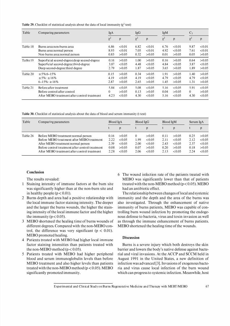
8/3/2019 Luka Bakar Book
http://slidepdf.com/reader/full/luka-bakar-book 73/157
Experimental and Clinical Study on Burns Regenerative Medicine and Therapy with MEBT/MEBO 67
Table 29. Checklist of statistical analysis about the data of local immunity (¯2 test)
Table Comparing parameters IgA
¯2 p
IgG
¯2 p
IgM
¯2 p
C3
¯2 p
Table 18 Burns area:non-burns area 6.86 !0.01 6.82 !0.01 6.76 !0.01 9.87 !0.01
Burns area:normal person 8.93 !0.01 7.05 !0.01 4.92 !0.05 7.61 !0.01
Non-burns area:normal person 0.83 10.05 0.32 10.05 0.01 10.05 0.05 10.05
Table 19 Superficial second-degree:deep second-degree 0.16 10.05 1.00 10.05 0.16 10.05 0.64 10.05
Superficial second-degree:third-degree 3.87 !0.05 4.48 !0.05 4.84 !0.05 3.87 !0.05
Deep second-degree:third degree 2.79 10.05 1.87 10.05 3.65 10.05 1.89 10.05
Table 20 ^5%:6–15% 0.15 10.05 0.34 10.05 1.91 10.05 1.40 10.05
^5%:^16% 4.19 !0.05 4.19 !0.05 4.79 !0.05 4.79 !0.05
6–15%:616% 2.87 10.05 2.65 10.05 1.45 10.05 1.31 10.05
Table 21 Before:after treatment 5.84 !0.05 5.08 !0.05 5.16 !0.05 5.91 !0.05
Before control:after control 0 10.05 0.13 10.05 0.04 10.05 0 10.05
After MEBO treatment:after control treatment 4.23 !0.05 4.30 !0.05 5.16 !0.05 4.30 !0.05
Table 30. Checklist of statistical analysis about the data of blood and serum immunity (t test)
Table Comparing parameters Blood IgA
t p
Blood IgG
t p
Blood IgM
t p
Serum IgA
t p
Table 26 Before MEBO treatment:normal person 0.16 10.05 0 10.05 0.11 10.05 0.25 10.05
Before MEBO treatment:after MEBO treatment 2.22 !0.05 1.99 !0.05 2.11 !0.05 2.12 !0.05
After MEBO treatment:normal person 2.39 !0.05 2.00 !0.05 2.43 !0.05 2.37 !0.05
Before control treatment:after control treatment 0.08 10.05 0.07 10.05 0.20 10.05 0.18 10.05
After MEBO treatment:after control treatment 2.28 !0.05 2.06 !0.05 2.13 !0.05 2.24 !0.05
Conclusion
The results revealed:
1 Staining intensity of immune factors at the burn site
was significantly higher than at the non-burn site and
in healthy people (p ! 0.01).
2 Burns depth and area had a positive relationship with
the local immune factor staining intensity. The deeper
and the larger the burns wounds, the higher the stain-
ing intensity of the local immune factor and the higher
the immunity (p ! 0.05).3 MEBO shortened the healing time of burns wounds of
different degrees. Compared with the non-MEBO con-
trol, the difference was very significant (p ! 0.01).
MEBO promoted healing.
4 Patients treated with MEBO had higher local immune
factor staining intensities than patients treated with
the non-MEBO method (p ! 0.05).
5 Patients treated with MEBO had higher peripheral
blood and serum immunoglobulin levels than before
MEBO treatment and also higher levels than patients
treated with the non-MEBO method (p ! 0.05). MEBO
significantly promoted immunity.
6 The wound infection rate of the patients treated with
MEBO was significantly lower than that of patients
treated with the non-MEBO method (p ! 0.05). MEBO
had an antibiotic effect.
The relationship between changes of local and systemic
immunity and the depth and the area of the burns was
also investigated. Through the enhancement of native
immunity of burns patients, MEBO was capable of con-
trolling burn wound infection by promoting the endoge-
nous defense to bacteria, virus and toxin invasion as wellas through the immune enhancement of burns patients.
MEBO shortened the healing time of the wounds.
Discussion
Burns is a severe injury which both destroys the skin
barrier and lowers the body’s native defense against bacte-
rial and viral invasions. At the ACCP and SCCM held in
August 1991 in the United States, a new definition of
infection was advanced [3]. Invasions of exogenous bacte-
ria and virus cause local infection of the burn wound
which can progress to systemic infection. Meanwhile, host

8/3/2019 Luka Bakar Book
http://slidepdf.com/reader/full/luka-bakar-book 74/157
68 Burns Regenerative Medicine and Therapy
defenses also induce an inflammatory immunologic reac-
tion. Thus, systemic inflammatory reaction syndrome
(SIRS) may result [4]. MEBO itself does not have a direct
bactericidal effect in vitro. Some researchers considered
that MEBO serves as an immunologic barrier in burn
wound surface thereby protecting the injured skin. MEBO
may create an environment (temperature, humidity, nu-trition, oxygen supply, metabolism, etc.) suitable for re-
sidual skin tissue repair. In effect, it creates an ideal iso-
lated ‘aseptic ward’. In addition, MEBO is capable of
altering the toxic potential of bacteria and virus in burns
wounds therefore lowering the infection rate [5].
This study investigated the effect of MEBO on the local
and systemic immunity of burns patients, and proved that
MEBO, through regulating human immunity, protected
burns wounds from infection at the same time as it pro-
moted wound healing. After MEBO treatment, the inci-
dence of wound infection was reduced to 1.7%, signifi-
cantly lower than that in the control group (p ! 0.05). Theaverage wound healing time in the MEBO treatment
group was 23.5 B 9.3 days, representing a significant
course of treatment as compared with the control group
(p ! 0.01). A vast amount of clinical data proved that
MEBO significantly lowered the infection rate of burns
wounds when compared with other methods [1]. MEBO is
applied directly onto the wound surface and is therefore
easily absorbed into the local tissue fluid circulation en
route to participation in systemic metabolism. MEBO
stimulates the immune system via enhancement of the
immunoglobulin level and strengthens body resistance
against infections. This paper reports the results of local
and peripheral blood and serum immunity of patients
with different depths and different areas of burns. It
proved that MEBO enhanced local and systemic immu-
nologic function as well as enhancing resistance to infec-
tion for burns patients.
Determination of local and peripheral blood and se-
rum immunoglobulin level of burns patients is an impor-
tant criterion for evaluating the effect of MEBO on hu-
man immunologic function. IgA, IgG, and IgM are im-
portant proteins with anti-bacterial, anti-viral and anti-
toxin activities. They also activate complement C3 toachieve bacteriolysis, phagocytosis and neutralization of
toxins. C3 takes part in nonspecific and specific immune
reactions and is a factor of body defense. It helps produce
immunoglobulin IgA, IgG and IgM. With appropriate reg-
ulation of the neurohumoral system, the human body can
enjoy enhanced resistance to infectious factors. MEBO
contains polysaccharides, lipids and proteins, which in
combination and when applied to burns wounds can bind
with bacteria and toxins to form protein complexes.
These complexes, in turn, stimulate the human immune
system, and induce a variation of the bacteria which
reduces their toxicity. Therefore, MEBO should not be
used together with other topical drugs which may lessen
its efficacy. When applying MEBO to the burn wound, the
thickness of the ointment should be appropriate since the
combination of MEBO with the proteins will be hindered
if MEBO is smeared too thickly or too thinly. For the
same reason, the time interval of MEBO application
should be appropriate.Before treatment, it was found that the local immunity
of the burns patients was higher than in the non-burn area
and normal persons. Burns patients with large area and
deep wounds had higher local immunity than small area
and superficial burns patients. However, the systemic
immunity of these burns patients was almost the same as
compared with that of normal persons. After MEBO treat-
ment, local and systemic immunity of the burns patients
increased significantly more than before treatment and in
controls. Therefore, we see that MEBO enhanced local
and systemic immunologic function of patients suffering
with burns of different depths, different areas and withdifferent courses of treatment. Our results may provide a
basis for further research and clinical application of
MEBO.
References
1 Xu RX: General introduction to medicine of burns, wounds and ulcers.
Chin J Burns Wounds Surface Ulcers 1989;1:11–21.
2 Xu RX: The principles of the treatment of burn wounds. Chin J Burns
Wounds Surface Ulcers 1992;4:7–21.
3 Bone RC, Balk RA, Cerra FB, Dellinger RP, Fein AM, Knaus WA, Schein
RM, Sibbald WJ: Definitions for sepsis and organ failure and guidelines for
the use of innovative therapies in sepsis. The ACCP/SCCM ConsensusConference Committee. American College of Chest Physicians/Society of
Critical Care Medicine. Chest 1992;101:1644–1655.
4 Sinclair S, Singer M: Intensive care. Postgrad Med J 1993;69:340–358.
5 Qu YY, Qiu SC, Wang YP, et al: Experimental research on the mechanism
of the anti-infection effect of MEBO. Industr Med J 1995;8:1–3.
Study on the Bacterial Count of Viable
Tissue of Burns Wounds Treated with BRT
with MEBT/MEBO
Introduction
In order to verify the ability of BRT with MEBT/
MEBO to inhibit localized infections, we conducted a
study on the bacterial number on viable tissue of burns
wounds. The results showed that MEBO therapy effec-
tively controlled bacterial number to less than 104 per
gram viable tissue during the whole treatment procedure.
This result suggests a strong capacity for the prevention of
wound invasive infection.

8/3/2019 Luka Bakar Book
http://slidepdf.com/reader/full/luka-bakar-book 75/157
Experimental and Clinical Study on Burns Regenerative Medicine and Therapy with MEBT/MEBO 69
Materials and Methods
The backs of 28 adult healthy guinea pigs of either sex were depi-
lated and scalded on both sides by a hot test tube to obtain full-thick-
ness necrotic wounds with a diameter of 3 cm. One wound on each
side of every animal randomly served as a blank exposed group (con-
trol group) while the other side served as the BRT with MEBT/
MEBO treatment group (test group). Wounds in the control group
were kept clean and allowed to heal spontaneously, while wounds in
the test group received BRT in the typical manner. Twenty-eight ani-
mals were harvested seven at a time at four different time intervals
(days 3, 6, 10 and 20 postinjury). Viable tissue underlying the
wounds was sampled for bacterial count and compared with normal
subcutaneous tissue.
Viable Tissue Sampling and Bacterial Counting
The wound surface was sterilized with iodine and alcohol after
each animal was sacrificed. Sterilized tissue scissors were used to cut
tissues from each wound without touching the deep fascia and mus-
cle tissue. The sampled wound tissues were spread flat in sterilized
cloth with the subcutaneous tissue exposed. Sterilized ophthalmic
scissors were then used to excise viable subcutaneous tissues (exclud-
ing necrotic tissues). The sampled viable tissues were weighed, tritu-
rated and diluted. Diluents were inoculated into agar culture me-
dium at a specific concentration and cultured for 48 h (at a constant
temperature of 37°C). Bacterial colony count was performed from
which total bacterial count and bacteria per gram of viable tissue
were obtained.
Results
The bacterial number in viable tissues of burns wounds
was less than 104/g in both groups, indicating there was no
wound invasive infection during the whole test period.
However, the bacterial number in wound viable tissue of the test group was significantly less than that in the con-
trol group at each observation time (table 31).
Discussion
There are several general clinical methods for de-
termining wound bacterial determination, including
(1) swab culture of wound surface; (2) quantifying bacte-
rial number of full-thickness burns wound; (3) bacterial
counting of sub-eschar viable tissues. The first method is
easy to perform, but fails to provide appropriate proof for
the existence of invasive infection on burns wounds. Clin-ical application and lack of comparability limit the second
method. The third method is usually adopted in clinics to
predict the possible success or failure of skin grafting and
is regarded as an ideal method for detecting the existence
of wound invasive infection. Many authors used this
method for early diagnosis of wound sepsis.
In 1983, Bharadulj reported that more than 105 bacte-
ria per gram sub-eschar viable tissue is diagnostic for
wound sepsis. He also found that patients who died from
systemic infection bore more than 108 bacteria per gram
viable tissue. Other authors agree [Robson et al.]. There-
fore, we too adopted this method for the purposes of this
Table 31. Bacterial number per gram of viable tissue
Groups Test group Control group p
Day 3 2.61B1.14!103 4.43B2.09!103!0.05
Day 6 3.81B0.27!103 3.24B0.73!104 !0.001
Day 10 3.57B0.64!103 1.08B0.10!105 !0.001
Day 20 2.82B1.16!103 7.02B0.43!103!0.05
Normal skin 2.10B0.52!103 2.2B0.63!103 10.05
study. The results demonstrate that in different stages
postburn, no more than 104/g was detected in sub-eschar
viable tissues taken from the BRT with MEBT/MEBO
group. We also noted that there was no obvious difference
throughout all stages although the peak occurred during
the period of wound rejection reaction. In the control
group, although no infection symptoms were observed onwounds, bacterial number per gram sub-eschar viable tis-
sue increased progressively until a peak of 105/g during
the wound-rejecting reaction period. A significant differ-
ence occurred between the two groups. Based upon bacte-
rial number, we see that MEBO had a unique effect in
controlling wound infection compared to normal tissue.
There are four dominant sources for burn wound infec-
tion: (1) parasitic bacteria in underlying wound viable
tissue; (2) burned tissues; (3) external contamination;
(4) hematogenous infection. It has been believed for a
long while that a dry, clean or sterilized wound environ-
ment may lead to a lower incidence of infection than
would a damp environment. Some experiments have
been conducted while maintaining the wound moist, but
the results suggested that a dry wound status was pref-
erable. Therefore, a therapeutic principle of drying the
wound was established to prevent infection and damp-
ness was itself regarded as a risk factor for infection. Inter-
estingly, our research reached a contrary conclusion.
A wet environment, according to the general rule, was
understood to provide a favorable surrounding for bacte-
rial growth despite the realization that tissue necrosis,
which results from a dry wound status, provides a muchmore nutrient substrate for microbial proliferation. Ac-
cordingly, we advocated that a physiologically moist envi-
ronment, assuming appropriately regulated humidity,
was favorable for tissue recovery as well as enhancement
of endogenous resistance to infection with its resultant
reduction of infection. Furthermore, it is well understood
that burn wound infections are caused by a variety of fac-
tors and that each of these should be accounted for along
with a clean wound environment. For example, blocked
sub-eschar drainage resulted from dryness of wound and
eschar formation would support bacterial colonization,
thereby increasing the possibility of wound infection. If

8/3/2019 Luka Bakar Book
http://slidepdf.com/reader/full/luka-bakar-book 76/157
70 Burns Regenerative Medicine and Therapy
wounds were kept wet without taking other effective mea-
sures, infection would be likely. We invented BRT with
MEBT/MEBO in order to maintain the wound in a physi-
ologically moist environment, which, while causing no
infection, also allowed for dramatically less bacterial
number in sub-eschar viable tissue than in wounds treated
by dry therapy. Why? According to the designing theoryof BRT with MEBT/MEBO, we had the following analy-
sis.
Moist Environment for Wound
In our study, a ‘moist’ instead of ‘wet’ environment
was emphasized. The special formulation of MEBO de-
veloped on the basis of Chinese traditional philosophy
ensured an appropriate ‘moist’ wound environment.
Composed of beeswax and non-water edible plant oil, the
dosage form of MEBO has a strong affinity to skin and
wound tissues. Its unique structure protects burn tissues
from direct immersion by exuded endogenous fluid, pre-vents bacteria in water from contacting tissues and there-
fore keeps wounds moist, but not macerated. The physical
change of the ointment from semisolid to liquid allows a
circulation of ointment across the wound, ensuring effec-
tive drug concentration and addressing the requirements
of tissue repair. Prior to the invention of MEBO, exposure
therapy with the application of other topical drugs was
considered as the predominant measure to monitor
wound condition. To its credit, wound exposure allowed
better assessment of the need for drug renewal as well as
for timely drainage and manual discharge of exudation.
Unobstructed Drainage and Isolation
BRT with MEBT/MEBO features an automatic drain-
age system that enables the timely drainage and discharge
of exudation and liquefied products from the wound sur-
face. This mechanism of action destroys the bacterial
growth environment, interrupts bacterial nutriment sup-
ply, reduces bacterial concentration and therefore effec-
tively arrests bacterial proliferation and invasion. The
unique dosage form of MEBO effectively isolates the
wound from bacterial contamination originating from the
surrounding environment by forming a barrier that ac-tually provides the wound with a clean and relatively
‘sterile’ environment.
Drug Ingredients and Other Claims
MEBO also claims to increase local blood flow, pro-
mote recovery of the microcirculation, and encourage
wound healing. These three factors all enhance the ability
of local tissue to resist infection.
Comparative Study of the Effects of
Moist-Exposed Burn Ointment, Silver
Sulfadiazine and Hot Dry-Exposed Therapy on
Controlling Burn Wound Infection with
Pseudomonas aeruginosa
Introduction
Since 1964 when Teplitz et al. [1] successfully estab-
lished a representative animal model, burn wound inva-
sive infection has been regarded as one of the main causes
of burns-related death. Therefore, topical use of antibac-
terial agents has played an important role in the control of
burn wound infection. In the 1960s, antibacterial agents
were developed including sulfamylon [2], silver nitrate [3]
and silver sulfadiazine (SD-Ag) [4]. Although SD-Ag has
been widely used historically because of its risk/benefit
ratio, we now are discouraged by its tendency to enhance
drug resistance [5, 6]. Researchers have addressed this
weakness in SD-Ag therapy by developing other antimi-
crobial agents to fight against burn wound infection
caused by Pseudomonas aeruginosa and other bacteria.
These newer agents include a variety of other topical
agents such as silver pipram [7, 8], silver norfloxacin [9,
10] and MEBO [11]. We designed a comparative study to
verify the effects of MEBO, SD-Ag and hot dry-exposed
therapy on controlling P. aeruginosa invasive infection of
burns wounds.
Materials and Methods
Pathogenic P. aeruginosa was collected from burns wounds of
invasive infection, cultured for 16–24 h, then produced into a 4 !
108 suspension using normal saline.
One hundred and twenty healthy adult Wistar rats of either sex
weighing 100–200 g were anesthetized intraperitoneally with sodium
pentobarbital (40 mg/kg), shaved of dorsum hair, and scalded on the
back with 100°C water for 10 s to each form a full-thickness burn
wound with 20% BSA (determined by pathological examination) [1].
A 1-ml suspension containing 4 ! 108 P. aeruginosa was smeared
evenly onto the wound surfaces to achieve contamination and infec-
tion. The animals were kept in separate cages, and divided randomly
into 4 groups as follows (30 in each). Group 1 (control group) re-
ceived no treatment. Group 2 (MEBO group) was treated withMEBO according to the method of BRT with MEBT/MEBO which
kept the wound moisturized and covered by MEBO throughout the
duration of the study. Group 3 (SD-Ag group) was treated with 1%
SD-Ag cold cream, which was applied once a day. Prior to each
administration of the SD-Ag, the residual cream and necrotic tissue
was wiped off according to the SD-Ag protocol. Group 4 was treated
with continuous hot, dry-exposed therapy using a heat-controlled air
fan to keep the wound dry.
Six animals in each group were killed under aseptic manipulation
at the 1st, 3rd, 5th, 7th and 9th days after treatment. A sample of
heart blood was collected and cultured and a specimen of wound skin
tissue was taken by sterile scalpel, as deep as the muscular fascia [12],
and then cut into two parts. One part was used for bacterial count in
sub-eschar viable tissue. The other was fixed in formalin for patho-

8/3/2019 Luka Bakar Book
http://slidepdf.com/reader/full/luka-bakar-book 77/157
Experimental and Clinical Study on Burns Regenerative Medicine and Therapy with MEBT/MEBO 71
logical examination. Sections were observed under a light microscope
and the extent of bacterial invasion was classified according to three
grades: ‘0’ referring to absence of bacterium, ‘I’ to invasion of bacteria
to necrotic tissues, and ‘II’ to invasion of bacteria to viable tissues.
Results
Bacterial Count of Sub-Eschar Viable TissuesTable 32 shows the mean logarithmic values of bacte-
rial count of sub-eschar viable tissues in each group. The
results indicated the mean values in groups 2 and 3 were
significantly lower than those in groups 1 and 4 (p ! 0.01).
No marked difference of this value was noted between
groups 2 and 3 (p 1 0.05) or between groups 1 and 4 (p 1
0.05).
Correlation between Bacterial Count of Sub-Eschar
Viable Tissue and Clinical Course
As figure 13 shows, the bacterial count of sub-eschar
viable tissues in groups 1 and 4 increased progressivelyduring the whole course of the disease. The bacterial
count in groups 2 and 3 remained at low levels, less than
105/g throughout, and even declined, indicating that both
these topical drugs were effective in controlling the prolif-
eration of P. aeruginosa.
Results of Blood Culture
The incidence of positive blood cultures in groups 2
and 3 was markedly lower than in groups 1 and 4 (p !
0.005), as is shown in table 33. There was no significant
difference of positive rates between groups 1 and 4 (p 1
0.50), or between groups 2 and 3 (p 1 0.75).
Pathological Examination
In the study, grades of ‘0’ and ‘I’ in the pathological
examination were referred to as negative while grade ‘II’
was referred to as positive [13] (table 33).
As can be seen in table 33, positive rates of pathologi-
cal examination in groups 2 and 3 were significantly lower
than those in groups 1 and 4 (p ! 0.005). There was no
significant difference of positive rates either between
groups 1 and 4 (p 1 0.50), or between groups 2 and 3 (p 1
0.50).
Comparison of Incidence of Invasive Infection of Burns
Wounds
According to the data that bacterial invasion to viable
tissue of wound and/or bloodstream in the circulation
were indicative of invasive infection of burns wounds
[13], table 34 shows the incidence of invasive infection in
each group. It was found that the incidences of invasive
infection in groups 2 and 3 were dramatically lower than
those in groups 1 and 4 (p ! 0.005). There was no signifi-
cant difference of positive rates either between groups 1
and 4 (p 1 0.25) or between groups 2 and 3 (p 1 0.50).
Fig. 13. Illustration of the correlation between bacterial count in sub-
eschar viable tissues and the clinical course in each group.
Table 32. Mean logarithmic values of bacterial count of sub-eschar
viable tissues (meanB SE)
Group Bacterial count
(logarithmic value)
1 (control) 5.8B2.6
2 (MEBO) 3.8B2.0
3 (SD-Ag) 3.1B3.1
4 (hot dry-exposed) 5.4B2.0
Table 33. Results of blood culture and pathological examination
Group Positive
number
Negative
number
Positive
rate,%
Blood culture
1 (control) 25 5 88.33
2 (MEBO) 7 23 23.33
3 (SD-Ag) 8 22 26.67
4 (hot dry-exposed) 19 11 63.33
Total 59 61 49.17
Pathological examination
1 (control) 21 9 70.0
2 (MEBO) 11 19 36.67
3 (SD-Ag) 12 18 40.0
4 (hot dry-exposed) 20 10 66.67
Total 64 56 53.33
Table 34. Incidence of wound invasive infection
Group Positive
number
Negative
number
Positive
rate, %
1 (control) 26 4 86.67
2 (MEBO) 12 18 40.0
3 (SD-Ag) 11 19 36.67
4 (hot dry-exposed) 23 7 76.67
Total 72 48 60.0

8/3/2019 Luka Bakar Book
http://slidepdf.com/reader/full/luka-bakar-book 78/157
72 Burns Regenerative Medicine and Therapy
Table 35. Results of bacterial count of sub-eschar viable tissues and
pathological examination
Bacterial
count
Pathological examination
positive
number
negative
number
total
!102 3 19 22102 3 12 15
103 10 4 14
104 2 5 3
105 8 5 13
106 11 1 12
107 10 5 15
1108 17 5 22
Total 72 48 60
Table 36. Results of positive bacterial count and positive pathologi-
cal examination
Bacterial count6105 Pathological examination
positive
number
negative
number
total
Positive number 46 16 62
Negative number 18 40 58
Total 64 56 120
Coincidence rate 86/120 (71.67%)
Non-coincidence rate 34/120 (28.33%)
Comparison of Bacterial Count of Sub-Eschar Viable
Tissue and Pathological Examination for Diagnosis of
Burn Wound Infection
According to table 35, there was a direct correlation
between the positive rates of the bacterial count of sub-
eschar viable tissue and the pathological examination (r =
0.808, p ! 0.005). The positive rate of pathological exami-
nation increased as did the bacterial count.
In further analysis, we took a bacterial number of 105/g
sub-eschar viable tissue, the level defining invasive infec-tion, as the boundary for positive and negative [14, 15]. It
was found by comparison that positive rates of pathologi-
cal examination of tissue specimens that yielded counts of
105/g sub-eschar viable tissue or more reached 74.2%.
The negative rate of pathological examination of those
yielding counts lower than 105/g reached 69%. The coinci-
dence and noncoincidence rates of two diagnostic meth-
ods were 71.67 and 28.33%, respectively (table 36). Sta-
tistical analysis showed an obvious relationship between
both methods (¯2 = 17.62, p ! 0.005) and there was no
significant difference between both methods in the diag-
nosis of burn wound infection (¯2 = 0.031, p 1 0.75).
Conclusion
1 MEBO has a similar effect to SD-Ag in controlling
burn wound invasive infection by P. aeruginosa.
2 Hot dry-exposed burns therapy has no effect on con-
trolling third-degree burn wound invasive infection by
P. aeruginosa.
3 The bacterial count of sub-eschar viable tissue can stillbe used as one of the feasible methods in the early diag-
nosis of burn wound invasive infection.
Discussion
Evaluation of Antibacterial Effect of Hot Dry-Exposed
Therapy
In 1949, Wallace introduced the concept of dry-
exposed burns therapy [16, 17] in which the wound was
directly exposed to air at a certain temperature. He be-
lieved that direct exposure of the wound might allow the
formation of a layer of dry eschar/crust by exudation and
necrotic tissue on the wound surface, which served as
a barrier against bacterial contamination. Our study
showed that comparing the hot dry-exposed therapy
group to the untreated control group, there was no ob-
vious difference with respect to bacterial count of sub-
eschar viable tissue, positive rate of blood culture and
positive rate of pathological examination. We now suggest
that the hot, dry-exposed therapy has no significant effect
on controlling invasive infection with P. aeruginosa of
third-degree burns wounds.
Moist-Exposed Burn Ointment (MEBO) –
New Topical Drug for Burns
Infection is the leading cause of death due to burns
complications, and burn wound infection is of great clini-
cal concern as it can result in burn wound sepsis and septi-
cemia. An enormous amount of research has been con-
ducted in this field which has produced many advances in
burn infection treatment. However, the very existence of
necrotic tissue in deep burns provides culture medium
conducive to the growth of pathological micro-organisms.
Furthermore, the blocked local blood circulation hinders
the delivery of anti-bacterial and immune-enhancing pep-tides which are integral to host-defense competency.
Mafenide (Sulfamylon), silver nitrate and SD-Ag were
developed in the 1960s [2–4]. Sulfamylon is a useful
antimicrobial agent which penetrates into eschar but has
the disadvantage of inhibiting carbonic anhydrase. There-
fore, absorption of topical Sulfamylon may result in meta-
bolic acidosis that limits its use in larger burn areas. Silver
nitrate was the initial topical agent but its tendency to
stain discouraged widespread use. SD-Ag has a strong
antimicrobial effect which, despite its poor penetration
into eschar, made it the topical agent of choice against
burn infection. Our study gave good proof for this.
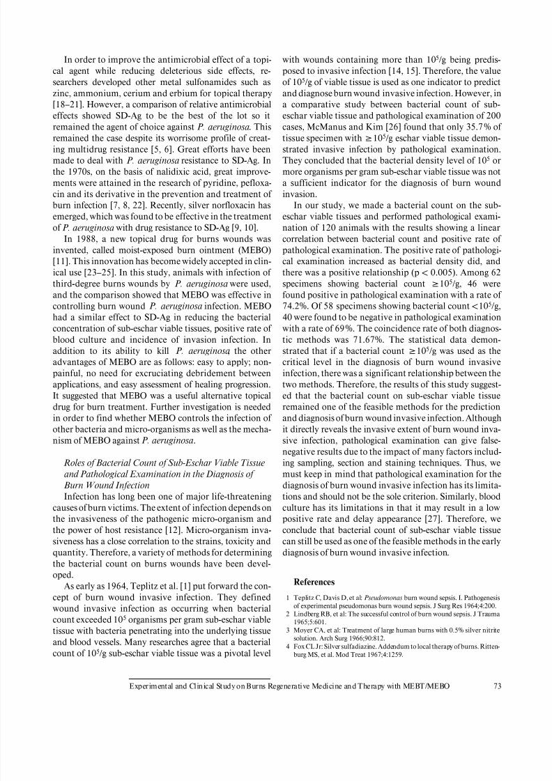
8/3/2019 Luka Bakar Book
http://slidepdf.com/reader/full/luka-bakar-book 79/157
Experimental and Clinical Study on Burns Regenerative Medicine and Therapy with MEBT/MEBO 73
In order to improve the antimicrobial effect of a topi-
cal agent while reducing deleterious side effects, re-
searchers developed other metal sulfonamides such as
zinc, ammonium, cerium and erbium for topical therapy
[18–21]. However, a comparison of relative antimicrobial
effects showed SD-Ag to be the best of the lot so it
remained the agent of choice against P. aeruginosa. Thisremained the case despite its worrisome profile of creat-
ing multidrug resistance [5, 6]. Great efforts have been
made to deal with P. aeruginosa resistance to SD-Ag. In
the 1970s, on the basis of nalidixic acid, great improve-
ments were attained in the research of pyridine, pefloxa-
cin and its derivative in the prevention and treatment of
burn infection [7, 8, 22]. Recently, silver norfloxacin has
emerged, which was found to be effective in the treatment
of P. aeruginosa with drug resistance to SD-Ag [9, 10].
In 1988, a new topical drug for burns wounds was
invented, called moist-exposed burn ointment (MEBO)
[11]. This innovation has become widely accepted in clin-ical use [23–25]. In this study, animals with infection of
third-degree burns wounds by P. aeruginosa were used,
and the comparison showed that MEBO was effective in
controlling burn wound P. aeruginosa infection. MEBO
had a similar effect to SD-Ag in reducing the bacterial
concentration of sub-eschar viable tissues, positive rate of
blood culture and incidence of invasion infection. In
addition to its ability to kill P. aeruginosa, the other
advantages of MEBO are as follows: easy to apply; non-
painful, no need for excruciating debridement between
applications, and easy assessment of healing progression.
It suggested that MEBO was a useful alternative topical
drug for burn treatment. Further investigation is needed
in order to find whether MEBO controls the infection of
other bacteria and micro-organisms as well as the mecha-
nism of MEBO against P. aeruginosa.
Roles of Bacterial Count of Sub-Eschar Viable Tissue
and Pathological Examination in the Diagnosis of
Burn Wound Infection
Infection has long been one of major life-threatening
causes of burn victims. The extent of infection depends on
the invasiveness of the pathogenic micro-organism andthe power of host resistance [12]. Micro-organism inva-
siveness has a close correlation to the strains, toxicity and
quantity. Therefore, a variety of methods for determining
the bacterial count on burns wounds have been devel-
oped.
As early as 1964, Teplitz et al. [1] put forward the con-
cept of burn wound invasive infection. They defined
wound invasive infection as occurring when bacterial
count exceeded 105 organisms per gram sub-eschar viable
tissue with bacteria penetrating into the underlying tissue
and blood vessels. Many researches agree that a bacterial
count of 105/g sub-eschar viable tissue was a pivotal level
with wounds containing more than 105/g being predis-
posed to invasive infection [14, 15]. Therefore, the value
of 105/g of viable tissue is used as one indicator to predict
and diagnose burn wound invasive infection. However, in
a comparative study between bacterial count of sub-
eschar viable tissue and pathological examination of 200
cases, McManus and Kim [26] found that only 35.7% of tissue specimen with6105/g eschar viable tissue demon-
strated invasive infection by pathological examination.
They concluded that the bacterial density level of 105 or
more organisms per gram sub-eschar viable tissue was not
a sufficient indicator for the diagnosis of burn wound
invasion.
In our study, we made a bacterial count on the sub-
eschar viable tissues and performed pathological exami-
nation of 120 animals with the results showing a linear
correlation between bacterial count and positive rate of
pathological examination. The positive rate of pathologi-
cal examination increased as bacterial density did, andthere was a positive relationship (p ! 0.005). Among 62
specimens showing bacterial count 6105/g, 46 were
found positive in pathological examination with a rate of
74.2%. Of 58 specimens showing bacterial count !105/g,
40 were found to be negative in pathological examination
with a rate of 69%. The coincidence rate of both diagnos-
tic methods was 71.67%. The statistical data demon-
strated that if a bacterial count 6105/g was used as the
critical level in the diagnosis of burn wound invasive
infection, there was a significant relationship between the
two methods. Therefore, the results of this study suggest-
ed that the bacterial count on sub-eschar viable tissue
remained one of the feasible methods for the prediction
and diagnosis of burn wound invasive infection. Although
it directly reveals the invasive extent of burn wound inva-
sive infection, pathological examination can give false-
negative results due to the impact of many factors includ-
ing sampling, section and staining techniques. Thus, we
must keep in mind that pathological examination for the
diagnosis of burn wound invasive infection has its limita-
tions and should not be the sole criterion. Similarly, blood
culture has its limitations in that it may result in a low
positive rate and delay appearance [27]. Therefore, weconclude that bacterial count of sub-eschar viable tissue
can still be used as one of the feasible methods in the early
diagnosis of burn wound invasive infection.
References
1 Teplitz C, Davis D, et al: Pseudomonas burn wound sepsis. I. Pathogenesis
of experimental pseudomonas burn wound sepsis. J Surg Res 1964;4:200.
2 Lindberg RB, et al: The successful control of burn wound sepsis. J Trauma
1965;5:601.
3 Moyer CA, et al: Treatment of large human burns with 0.5% silver nitrite
solution. Arch Surg 1966;90:812.
4 Fox CL Jr: Silver sulfadiazine. Addendum to local therapy of burns. Ritten-
burg MS, et al. Mod Treat 1967;4:1259.
8/3/2019 Luka Bakar Book
http://slidepdf.com/reader/full/luka-bakar-book 80/157
74 Burns Regenerative Medicine and Therapy
5 Bridges K, et al: Drug resistance in relation to use of silver sulphadiazine
cream in a burn unit. J Clin Pathol 1977;30:160.
6 Heggers JP, et al: The emergence of silver sulphadiazine resistant Pseudo-
monas aeruginosa. Burns 1978;5:184.
7 Ge SD, et al: Experimental study of topical chemotherapy in prevention
and treatment of burn infection. Acad J Sec Military Med Coll 1982;3:46.
8 Ge SD, et al: Experimental study of topical antimicrobial agent in burns.
Chin J Plast Surg Burns 1985;1:255.
9 Darrell RW, et al: Norfloxacin and silver norfloxacin in the treatment of
Pseudomonas corneal ulcer in the rabbit. Trans Am Ophthalmol Soc 1984;82:75.
10 Ge SD, et al: The effect of pyridonic acid derivatives as topical antibiotics
on the prevention and treatment of pyocyaneous infection following burn.
Chin J Plast Surg Burns 1987;3:10.
11 Xu RX: The clinical application of moist exposed burn therapy. Chin J
Integr Tradition West Med 1988;8:204.
12 Krizek TJ, Robson MC: Evolution of quantitative bacteriology in wound
management. Am J Surg 1975;130:579.
13 Yang ZJ, Xu WS, Shi JX: Burn Management, ed 2. Beijing, People’s Health
Press, 1985.
14 Volence FJ, Clark GM, et al: Burn wound biopsy bacterial quantitation: A
statistical analysis. Am J Surg 1979;138:695.
15 Loebel EC, Marvin JA, et al: The method of quantitative burn wound biop-
sy cultures and its routine use in the care of the burned patient. Am J Clin
Pathol 1974;61:20.
16 Wallace AB: Treatment of burn, a return to basic principles. Br J Plast Surg1949;1:232.
17 Wallace AB: The exposure treatment of burns. Lancet 1951;i: 501.
18 Fox CL Jr, et al: Metal sulfonamides as antibacterial agents in topical thera-
py. Scand J Plast Reconstr Surg 1977;13:89.
19 Monafo WW, et al: Control of infection in major burn wounds by cerium
nitrate/silver sulfadiazine. Burns 1977;3:104.
20 Fox CL Jr, et al: Topical chemotherapy for burns using cerium salts and
silver sulfadiazine. SGO 1977;104:668.
21 Ge SD, et al: N1-metal sulfa drugs and zinepalyanemine and its derivatives
in prevention of burn wound sepsis. Chin J Surg 1982;20:264.
22 Modak SM, et al: Control of burn wound infection by pefloxacin and deriv-
ative. Burns 1984;10:170.
23 Zhang LX, Yang KF: Clinical study report: 2076 cases treated with moist
exposed burn therapy. Chin J Burns Wounds Ulcers 1989;1:22.
24 Zhao RY, Tian LX, Zhao GX: Clinical analysis: 50 cases treated with moist
exposed burn therapy. Chin J Burns Wounds Ulcers 1989;1:32.25 Ma EQ, Huang XY, et al: Clinical report of 69 cases treated with moist
exposed burn ointment. Chin J Burns Wounds Ulcers 1990;2:25.
26 McManus AT, Kim SH: Comparison of quantitative microbiology and his-
topathology in divided burn wound biopsy specimens. Arch Surg 1987;
122:74.
27 Baxter CR, Curreri PW, et al: The control of burn wound sepsis by the use
of quantitative bacteriologic studies and sub-eschar lysis with antibiotics.
Surg Clin North Am 1973;53:1509.
Experimental Research on the Mechanism
of the Anti-Infection Effect of BRT withMEBT/MEBO
Introduction
Many basic studies and clinical research have proved
that BRT with MEBT/MEBO has beneficial effects of
anti-infection, promoting burn wound healing and reduc-
ing scar formation to name but a few of its attributes.
However, the mechanisms of these effects of MEBO
remain a bit mysterious. The following study was de-
signed to elucidate the anti-infection effect mechanism of
BRT with MEBT/MEBO.
Materials and Method
Apparatus and Reagents
Fully automatic enzyme labeling analyzer, Multiskan, MS; AC-
920 hemocyte analyzer; MEBO; RPMI-1640, Gibco, USA; Con A,
supplied from Guangzhou Medical Institute; MTT supplied from
Fluka; Pathogenic O111B4 of Escherichia coli supplied from Binzhou
Medical College.
Animals and Methods
Thirty-two BALB/C mice, 8 weeks old, females, weighing 17–
19 g, were supplied from Beijing Medical University. Twenty-eight
Kunming mice of both sexes weighing 21–24 g, 18 rabbits weighing
2–2.5 kg, and 4 guinea pigs weighing 250–300 g were supplied from
Binzhou Medical College.
The animals were divided randomly into a control group and a
MEBO treatment group. In the MEBO treatment group, mice were
depilated (2! 2 cm) on their backs and smeared with MEBO. Guin-
ea pigs were depilated (3! 2.5 cm) on their backs and smeared with
MEBO. MEBO was applied 2 times a day. Biopsy of skin tissue was
done on day 9 to determine the activity of interleukin-1 (IL-1), and
was stained with hematoxylin and eosin (HE) for histopathological
observation.
Examination Indexes
Morphological Variation of Pathogenic E. coli (O111B4 ). The bac-
teria were cultivated on culture medium containing a given concen-
tration of MEBO [1]. Each generation of the bacteria was stained
using the G staining method. Staining reaction and morphological
variation of the bacteria were observed. The results were compared
with that of the E. coli communis cultured on the same kind of
medium containing MEBO.
Determination of IL-1 Level. MTT was used as a substrate. Since
living cells containing active succinate dehydrogenase can reduce
yellow-colored MTT to form violet or blue-colored formazam par-
ticles, the particles were dissolved by adding isopropyl alcohol hy-
drochloride [2]. The OD value was determined by colorimetry. Theamount of formazam was proportional to the number of living cells.
Determination was performed according to the modified methods
described previously [2]. Mice were killed under sterile conditions,
the thymus was removed, cut and screened. After being centrifuged,
a suspension with 107/ml of cells containing 2 Ìg/ml of Con A was
prepared. On a 24-pore plate, we added to each pore 0.5 ml of this
suspension and then added mice skin tissue suspension and blood
plasma at 0.5 ml/pore. Two more control groups, one with 1640-
nutrient fluid (thymocytes plus nutrients) and the other with Con A
plus thymocytes were set. The 24-pore plate was placed into an incu-
bator at 37 °C, 5% CO2 for 68 h. From each pore, 0.5 ml of the
supernatant was drawn away, then 0.1 ml of 5 Ìg/ml MTT solution
was added to each pore and incubated again for 4 h. 0.5 ml of isopro-
pyl alcohol hydrochloride was added to each pore and then the platewas shaken to dissolve the particles. Supernatant from each pore was
drawn and added to a 96-pore plate, 0.2 ml/pore. The plate was
placed into a fully automatic enzyme-labeling analyzer to determine
the OD value, reflecting the level of IL-1 in skin tissue cells and
blood plasma.
Effect of MEBO on Body Temperature of the Rabbit. The body
temperature of the rabbit was taken prior to application of MEBO.
Then, an area of 6.0 ! 6.5 cm was depilated on the back of the
animal and MEBO was applied twice a day while body temperature
was recorded simultaneously. This was done for a total of six mea-
surements. Changes in body temperature were recorded.
Effect of MEBO on the Classification of Mice Leukocytes. Every
mouse in the MEBO and control groups had a 0.2-ml blood sample
taken by enucleation of the eyeball. The blood was added to 10 ml of
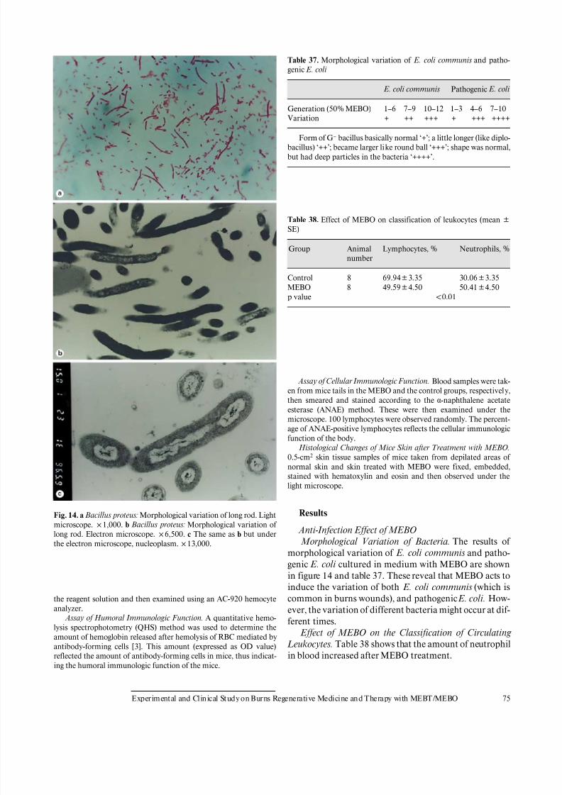
8/3/2019 Luka Bakar Book
http://slidepdf.com/reader/full/luka-bakar-book 81/157
Experimental and Clinical Study on Burns Regenerative Medicine and Therapy with MEBT/MEBO 75
Fig. 14. a Bacillus proteus: Morphological variation of long rod. Light
microscope. !1,000. b Bacillus proteus: Morphological variation of
long rod. Electron microscope. !6,500. c The same as b but under
the electron microscope, nucleoplasm.!13,000.
the reagent solution and then examined using an AC-920 hemocyte
analyzer.
Assay of Humoral Immunologic Function. A quantitative hemo-
lysis spectrophotometry (QHS) method was used to determine the
amount of hemoglobin released after hemolysis of RBC mediated by
antibody-forming cells [3]. This amount (expressed as OD value)
reflected the amount of antibody-forming cells in mice, thus indicat-
ing the humoral immunologic function of the mice.
Table 37. Morphological variation of E. coli communis and patho-
genic E. coli
E. coli communis Pathogenic E. coli
Generation (50% MEBO) 1–6 7–9 10–12 1–3 4–6 7–10
Variation + ++ +++ + +++ ++++
Form of G– bacillus basically normal ‘+’; a little longer (like diplo-
bacillus) ‘++’; became larger like round ball ‘+++’; shape was normal,
but had deep particles in the bacteria ‘++++’.
Table 38. Effect of MEBO on classification of leukocytes (mean B
SE)
Group Animal
number
Lymphocytes, % Neutrophils, %
Control 8 69.94B3.35 30.06B3.35
MEBO 8 49.59B4.50 50.41B4.50
p value !0.01
Assay of Cellular Immunologic Function. Blood samples were tak-
en from mice tails in the MEBO and the control groups, respectively,
then smeared and stained according to the ·-naphthalene acetateesterase (ANAE) method. These were then examined under the
microscope. 100 lymphocytes were observed randomly. The percent-
age of ANAE-positive lymphocytes reflects the cellular immunologic
function of the body.
Histological Changes of Mice Skin after Treatment with MEBO.
0.5-cm2 skin tissue samples of mice taken from depilated areas of
normal skin and skin treated with MEBO were fixed, embedded,
stained with hematoxylin and eosin and then observed under the
light microscope.
Results
Anti-Infection Effect of MEBO Morphological Variation of Bacteria. The results of
morphological variation of E. coli communis and patho-
genic E. coli cultured in medium with MEBO are shown
in figure 14 and table 37. These reveal that MEBO acts to
induce the variation of both E. coli communis (which is
common in burns wounds), and pathogenic E. coli. How-
ever, the variation of different bacteria might occur at dif-
ferent times.
Effect of MEBO on the Classification of Circulating
Leukocytes. Table 38 shows that the amount of neutrophil
in blood increased after MEBO treatment.

8/3/2019 Luka Bakar Book
http://slidepdf.com/reader/full/luka-bakar-book 82/157
76 Burns Regenerative Medicine and Therapy
Table 39. Effect of MEBO on rabbit body temperature ( °C, n = 6,
average)
Days after MEBO treatment
0 1 3 5 7
Body temperature,°C 38.63 39.05 39.31 39.43 39.47
Average elevation of bodytemperature, °C 0.84
Table 40. Effect of MEBO on the production of IL-1 in mouse skin
tissue cells (meanB SE)
Group IL-1 in skin tissue cells
animals OD
IL-1 in blood plasma
animals OD
Control 8 0.043B0.019 4 0.429B0.171
MEBO 8 0.142B0.039 4 0.733B0.105
p value !0.01 !0.05
Table 41. Effect of MEBO on specific immunologic function of mice
(meanB SE)
Group ANAE positive rate, %
(n = 6)
QHS
(n = 8)
Control 58.83B10.61 1.296B0.021
MEBO 54.17B10.23 1.317B0.027
p value 10.05 10.05
Effect of MEBO on Rabbit Body Temperature. 75% of
the rabbits had an increase in body temperature after
MEBO treatment. On day 7, the average elevation of body
temperature was 0.84°C (table 39).
Effects of MEBO on Wound Healing and Scar
Formation
Effect of MEBO on the Production of IL-1 in Mouse
Skin Tissue Cells. It was found that MEBO was effective
in inducing synthesis of IL-1 from IL-1-delivering cells of
the skin. IL-1 is capable of promoting the proliferation of
thymocytes and has a synergistic action with Con A. The
IL-1 levels in both skin tissue and blood plasma of the
MEBO treatment group were significantly higher than
those of the control group (table 40). MEBO Promoted Proliferation of Skin Cells and Cells
at the Margin of the Sebaceous Gland. In this study, we
found that the number of skin basal cells in the division
phase was increased, and the number of juvenile flat cells
at the margin of the sebaceous gland was also increased in
the MEBO group. This finding indicates that the metabo-
lism of the cells was vigorous.
Effect of MEBO on the Specific Immunologic Function
of Mice
Table 41 shows that MEBO did not affect the cellular
and humoral immunologic functions.
Conclusion
The results suggested that: (1) MEBO can prevent
infection; (2) the wound-healing benefit and the reduction
of scar formation in the MEBO group were related to its
effect of increasing the production of IL-1 by skin cells.
Discussion
Clinical practice has strongly proved that MEBO
has anti-infection, pain-killing, wound-healing-promot-
ing and scar-forming-reducing effects. Based on our
study, we discussed the mechanisms of its actions as the
following.
Anti-Infection Effect of MEBO
MEBO has potent ability to control wound infection
and keep the wound moist but not macerated. The active
ingredients in MEBO ointment and its unique dosage
delivery system create an environment hostile to bacterial
growth. In culture medium containing MEBO, morpho-
logical structural and physiological variations of the bac-
teria occurred. MEBO affected the synthesis of the com-
ponents for formation of a bacterial wall and inhibited
related enzymes. Also, the synthesis of DNA was inhibit-
ed and the bacteria proliferation rate was decreased. Deep
pigments were found in the bacteria, this indicated that
the bacteria were in a stable phase of proliferation, during
which a high level of glycogen, lipid, etc. was stored in thebacteria.
MEBO induced morphological and physiological vari-
ations in bacteria while influencing the production of
plasma coagulase of Staphylococcus aureus [4]. Bacterial
pathogenicity is related to the bacterial wall component
and thus MEBO reduced the pathogenicity of the bacte-
ria. The bacterial variation characteristics were varied in
different species of bacteria and at different MEBO con-
centrations. The initiation time of the variation was not
standard.
In clinical care, we found that after treatment with
MEBO, the body temperature of burn patients might rise

8/3/2019 Luka Bakar Book
http://slidepdf.com/reader/full/luka-bakar-book 83/157
Experimental and Clinical Study on Burns Regenerative Medicine and Therapy with MEBT/MEBO 77
by 1–1.5°C during the initial stage. At 3 h after applica-
tion of MEBO, the temperature of burn patients with
superficial second-degree wounds rose. In this study, the
body temperature of rabbits increased after application of
MEBO, and rose by 0.84 °C on day 7. Furthermore,
MEBO induced the production of IL-1, from IL-1-deliver-
ing skin cells, such as epidemic cells, keratinocyte, andLangerhans cells. In the early 1940s, people recognized
that certain extracts from acute inflammatory foci could
cause fever after injection into the body. This type of
inflammatory substance is called endogenous pyrogen
(EP). In 1979, purified EP was first proved to have IL-1
activity, and EP and IL-1 were considered the same mole-
cule [5]. MEBO stimulates local skin cells to produce IL-1,
which in turn was absorbed into the systemic circulation
thereby affecting the temperature-regulating center lead-
ing to elevated body temperature.
The effect of fever on body resistance of mammals is
not clear yet. Some researchers think that fever may pro-mote the immunity of the host. When body temperature is
raised, but still !41°C, the phagocytic power of most
phagocytes is enhanced. We also found that MEBO can
promote phagocytic power of abdominal cavity macro-
phages in mice [6]. In this study, the quantity of neutro-
phils in blood circulation was significantly increased after
treated with MEBO. Bone marrow stimulated by IL-1
may account for this interesting finding.
MEBO Promotes Wound Healing and Reduces Scar
Formation
Clinical data revealed that after treatment with MEBO,
patients with superficial or deep second-degree burns
wounds healed with full epithelization; superficial third-
degree burns wounds healed with a mild scarring that
appeared smooth and soft. In this study, we observed that
the production of IL-1 was increased in mice skin and sub-
dermal tissues after treatment with MEBO. The difference
between the MEBO group and the control group was very
significant. Besides macrophages, many other tissue cells
when stimulated can produce IL-1 in 1 h [9]. IL-1, IL-8 and
tumor necrosis factor (TNF) are cellular factors which are
capable of activating and inducing differentiation of T andB lymphocytes, enhancing the activities of monocytes, NK
cells and killer cells, thus stimulating lysosomal enzyme
activity and phagocytic activity of neutrophils.
Recently, both animal experiments and clinical prac-
tice proved that IL-1 does induce a series of pathophysio-
logical changes. These changes are similar to the host’s
response to infection [6], indicating that IL-1 is an impor-
tant regulatory factor of the body as regards inflammation
and immunologic reaction. We must differentiate be-
tween the local effect of IL-1 and the effect of high lev-
els of IL-1 systemically. These are two totally different
concepts [6]. In this study, the level of IL-1 in mouse skin
and extracellular subdermal tissues in the MEBO treat-
ment group were significantly different from those of the
control group, as well as the IL-1 level in blood plasma.
IL-1 is closely related to wound healing and we know that
wound exudate typically contains IL-1. IL-1 promotes
proliferation of fibroblast and secretion of collagenase [6].
The effect of IL-1 is complicated. It induces inflammationand fever while at the same time promoting wound heal-
ing. Inflammation induced by IL-1 is understood to be a
kind of host defense reaction [7]. After mice were treated
with MEBO, their skin basal cell in division stage in-
creased. Juvenile cells observed around the sebaceous
gland were very metabolically active. This proves that
MEBO promotes wound healing.
Effect of MEBO on Specific Immune Function
In this study, MEBO could increase the quantity of
neutrophils in blood, while relatively decreasing the num-
ber of lymphocytes. In addition, MEBO did not affect thecellular and humoral immunologic function.
References
1 Qu YY, et al: Experimental research on the anti-infective mechanism of
MEBO. Chin J Burns Wounds Surface Ulcers 1996;40:19–23.
2 Mosmann T: Rapid colorimetric assay for cellular growth and survival:
Application to proliferation and cytotoxicity assays. J Immunol Methods
1983;65:55.
3 Bi AH (ed): Medical Immunology. Tongji, Tongji Medical University,
1986, p 7.
4 Yu H: Medical Microbiology. Beijing, People’s Health Publishing House,
1983, p 54.
5 Yang GZ (ed): Outline and Technology of Immunology and Bioengineer-ing. Jilind, Jilin Science & Technology Press, 1991, pp 2–15.
6 Chen WJ (ed): Molecular and Cell Biology of Blood. Beijing, Chinese Med-
ical Science & Technology Press, 1993, vol 126–127, p 362.
7 Yang GZ, et al: Progress in genetic engineering and clinical immunology;
in: Domestic and International Progress in Medical Sciences. Shanghai,
Shanghai Institute of Medical Science & Technology Information, 1987,
p 124.
Primary Exploration on the Mechanism of the
Anti-Infection Effect of BRT with MEBT/MEBO
Introduction
Clinical practice has revealed that MEBO has a re-
markable anti-infection effect [1]. During the period May
to June 1992, fourteen burns cases were treated in the
Burns Department, Affiliated Hospital of this College,
and bacteria isolated from the burns wounds were exam-
ined. We found that Bacillus proteus had the morphologi-
cal Hauch-ohne Hauch (H-O) variation, and the plasma
coagulation ability of S. aureus decreased. In order to
investigate the mechanism of the anti-infection effect of
MEBO, we studied the biological variability of some com-
mon bacteria, such as Bacillus proteus, P. aeruginosa,

8/3/2019 Luka Bakar Book
http://slidepdf.com/reader/full/luka-bakar-book 84/157
78 Burns Regenerative Medicine and Therapy
E. coli and S. aureus, cultured in medium containing a
certain amount of MEBO. The effect of MEBO on non-
specific immunity in vivo was also observed.
Materials and Methods
Clinical Data
During the period from May to June 1992, 14 cases of burns were
treated with MEBO (hospitalized for 4–20 days). Swab samples were
taken from the upper and lower (contact with wound) layers of the
MEBO ointment, before changing the dressing. Bacteria were iso-
lated and cultured.
Reagents, Bacterium Species and Culture
Antibiotic sensitivity test paper and nutrient agar were purchased
from Shanghai Medical Chemistry Institute. B. proteus, P. aerugino-
sa, E. coli and S. aureus were prepared in our department.
The four above bacteria were cultured on ordinary culture me-
dium and medium containing different concentrations of MEBO,
respectively, and continuously transferred to 10–15 generations.Each generation of the bacteria was checked. The biological charac-
teristic and drug sensitivity of bacteria were examined.
Animal Experiment
Forty healthy adult mice of both sexes weighing 20–24 g were
randomly divided into 3 groups, i.e. blank control group (group 1),
liquid paraffin control group (group 2) and MEBO group (group 3).
Animals in groups 2 and 3 were depilated (2.5 ! 2.5 cm) on their
backs and liquid paraffin or MEBO was applied on the depilated
area. The frequency was twice a day for 10 successive days. On the
11th day, the mice were sacrificed and abdominal cavity fluids were
sampled 30 min after intraperitoneal injection of 0.5 ml of 2% sheep
erythrocytes.
Observation Indexes
Bacteria Variation. The four bacteria were cultured and trans-
formed, 18–24 h as one generation. The dynamics of every genera-
tion of the bacteria was observed under a dark-field microscope. The
bacteria were stained with the G method to observe the staining reac-
tion and morphological characteristic. The colony features, biochem-
ical reactions, and the ability of plasma coagulation were also exam-
ined.
Nucleoplasm Staining . The 7th generation of B. proteus was
smeared, fixed with the vapor of 1% molybdic acid solution, hydro-
lyzed DNA with 1% hydrochloric acid for 15 min, then stained with
Löffler’s methylene blue and observed.
Antibiotic Drug Sensitivity Test. The four bacteria were cultured
on ordinary agar medium and medium containing 20% of MEBO,
respectively. Drug sensitivity test paper of eight kinds of antibiotics,
i.e. gentamicin, neomycin, erythromycin, carbenicillin, ampicillin,
kanamycin, chloromycetin and polymyxin, were stuck on the me-
dium, respectively, and observed after 24 h. The diameter of the bac-
terial inhibition zone was measured [2].
Determination of Phagocytic Function of the Intraperitoneal
Phagocytes. The abdominal cavity fluids were smeared and stained
with Wright’s method. The percentage of phagocytes was determined
[3].
Determination of Lysozyme Activity in Abdominal Cavity Fluid
and Serum. The agar plate method was used: 2 ml of Micrococcus
solution (9 ! 1010/ml) were added to 1% agar at 70 °C, then after
mixing well, this was poured into a Petri dish. After cooling, holes
(3 mm in diameter) were bored into the substance. Mouse serum
from 3 groups was added to the holes in one set of Petri dishes and
abdominal cavity fluid was added to the holes in another set of Petri
dishes. These were incubated at 37 °C for 24 h. The diameter of the
bacteriolytic ring was measured [4].
Total Number and Classification of Leukocytes in Peripheral
Blood. Blood samples from the tails were taken from 3 groups of
mice. Leukocytes were counted, stained with Wright’s method and
classification was determined.
Results
Species and Variation of Bacteria Isolated from BurnsWounds
Table 42 shows that after treatment with MEBO, B.
proteus in the wounds had H-O morphological variation.
S. aureus in the wounds turned from positive plasma co-
Table 42. Species and variation of bacteria
isolated from burns wounds1 Case No. Bacteria from upper layer of MEBO Bacteria from lower layer of MEBO
1 S. aureus; P. aeruginosa P. aeruginosa
2 Staphylococcus albus; E. coli E. coli
3 S. albus (negative coagulase) S. albus (negative coagulase)4 S. aureus (positive coagulase) S. aureus (coagulating ability decreased)
5 B. proteus B. proteus (H-O morphological variation)
6 S. aureus (positive coagulase) S. aureus (negative coagulase)
7 S. aureus; E. coli E. coli
8 S. aureus (positive coagulase) S. aureus (positive coagulase)
9 B. amotile B. amotile
10 B. amotile B. amotile
11 P. aeruginosa P. aeruginosa
12 S. albus S. albus
13 P. aeruginosa P. aeruginosa
14 G+ diplococcus G+ diplococcus
1 Samples collected on days 4–20 after MEBO treatment.

8/3/2019 Luka Bakar Book
http://slidepdf.com/reader/full/luka-bakar-book 85/157
Experimental and Clinical Study on Burns Regenerative Medicine and Therapy with MEBT/MEBO 79
agulase to negative, or the plasma coagulating ability de-
creased.
Bacteriostasis of MEBO
Eight species of bacteria, i.e. S. aureus, S. albus, E. coli,
B. proteus, P. aeruginosa, B. typhosus, B. paratyphoid A
and B. dysenteriae, were cultured in simple agar dishes.The scraps of MEBO filter paper were pasted on the bac-
terial surface of streak plating. The results showed that
MEBO had no direct bacteriostasis or bactericidal ac-
tion.
Effect of MEBO on Bacterial Biological
Characteristics
Effect of MEBO on B. proteus Biological Features. B.
proteus was cultured for several generations on medium
containing certain amounts of MEBO. We noted that the
motility of the bacteria gradually decreased before finally
vanishing. Also, we noted that H-O variation occurred.The 7th generation of the bacteria became long and fila-
mentous. In culture medium containing 25% MEBO,
90% of the bacteria became long filamentous or long rod,
and then became small bacillus. Dark pigments ap-
peared, colonies became small and the bacteria grew very
slowly. The decomposition activity of the bacteria to glu-
cose and lactic acid was retarded (table 43; fig. 15). The
effect of MEBO on H antigen of B. proteus is shown in
table 44. Fig. 15. a Normal appearance of E. coli. b Appearance of variant
E. coli cultured in medium containing MEBO for 6 generations.
Table 43. Effect of MEBO on B. proteus biological features
Medium Motility
G 1 G 2 G 3 G 4–10
Morphology
G 1–2 G 3–4 G 5–8 G 9–10
H-O variation
G 1 G 2 G 3–4 G 5–10
H2S test
G 1–7 G 8-10
50% MEBO + B – – + + ++ +++ + + – – + retarded
decomposition
25% MEBO + B B – + ++ ++ * +++ + + + – + retarded
decomposition
+ = Motile;B = weak motility; – = no motility.
+ = Typical G– bacillus; ++ = long rod or filament; +++ = G – with dark pigment.
+ Colonial migration: + = 1–3 cm; – = no migration.
G = Generation; * 90% of the 7th generation of the bacteria became long filamentous.
Table 44. Serological test of B. proteus
cultured in medium containing MEBO Original
bacteria
Medium containing MEBO, 12th generation
10% MEBO 25% MEBO 50% MEBO
B. proteus 1:1,280 1:640 1:640 1:320
H antiserum ++ ++ ++ ++

8/3/2019 Luka Bakar Book
http://slidepdf.com/reader/full/luka-bakar-book 86/157
80 Burns Regenerative Medicine and Therapy
Effect of MEBO on the Biological Characteristic of
P. aeruginosa. It was found that P. aeruginosa cultured in
MEBO-containing medium started to decrease its motili-
ty from the 5th generation, and motility vanished in the
10th generation. Variations of morphology and coloniza-
tion features of the bacteria were also found (table 45).
Effect of MEBO on the Biological Characteristics of
E. coli. From table 46, we can see that after proliferation
to the 10th generation while cultured in MEBO contain-
ing medium, E. coli changed as follows: it lost motility,
became sphere shaped, colonies became smaller, dry and
flat (fig. 16). The decomposition activity of the bacteria to
glucose and lactose was retarded (after 32 h).
Table 45. The effect of MEBO on the biological characteristic of P. aeruginosa
Medium Motility
G 1–4 G 5–9 G 10–12
Morphology
G 1–4 G 5–9 G 10–12
Colonial feature
G 1–5 G 6–9 G 10–12
Oxidase test
G 1–12
MEBO 50% + B – + ++ +++ + ++ ++x +
MEBO 25% + B – + ++ +++ + + ++ +
+ = Typical G– bacillus; ++ = a few became long rod or diplococcus; +++ = deep pigment appeared.
+ = Colony moderate size and smooth; ++ = colony smaller and dry; ++x
= pigment formed but not good.G = Generation.
Table 46. Effect of MEBO on the biological characteristic of E. coli
Medium Motility
G 1–3 G 4–9 G 10–12
Morphology
G 1–6 G 7–9 G 10–12
Colony feature
G 1–3 G 4–8 G 9–12
Fermentation test
glucose
G 1–9 G 10–12
lactose
G 1–9 G 10–12
MEBO 50% +B
– + ++ +++ + ++ +++ + retardeddecomposition + retardeddecomposition
MEBO 25% + B – + ++ +++ + ++ +++ + retarded
decomposition
+ retarded
decomposition
+ = Typical G– bacillus; ++ = long rod (like diplococcus); +++ = bacteria swelling (sphere shaped).
+ = Colony medium size and smooth; ++ = colony medium size, a little flat; +++ = colony small, rough and flat.
G = Generation.
Table 47. Effect of MEBO on the biological characteristic of S. aureus
Medium Morphology
G 1–7 G 8–11 G 12–15
Colony feature
G 1–7 G 8–11 G 12–15
Plasma coagulase test
G 1–7 G 8–12 G 13–15
Manicol test
G 1–11 G 12–15
MEBO 50% + ++ +++ + ++ +++ ++ + B + retarded
decomposition
MEBO 25% + ++ ++ + ++ ++ ++ + + + +
+ = G+ arranged in grape shape; ++ = part of the bacteria became diplococcus-like or arranged in short chain;
+++ = piled up in grape shape and had scattered diplococcus-like and short chain arrangement.
+ = Colony medium size and smooth; ++ = colony small and slightly flat; +++ = colony smaller, flat and dry.
Plasma coagulase test: ++ = Fluid was clear and obviously coagulated; + = fluid turbid and small coagulate;
B = fluid turbid, small and few coagulate.
G = Generation.

8/3/2019 Luka Bakar Book
http://slidepdf.com/reader/full/luka-bakar-book 87/157
Experimental and Clinical Study on Burns Regenerative Medicine and Therapy with MEBT/MEBO 81
Effect of MEBO on the Biological Characteristics of
S. aureus. We found that after 10 generations of S. aureus
cultured in medium containing MEBO, bacteria were
piled up in grape shape and had scattered diplococcus-like
and short chains. Colonies became smaller, flat and dry.
The decomposition activity of the bacteria to mannitol
was retarded (after 32 h), and the variation of plasmacoagulation ability was very significant (table 47) [3].
Nucleoplasm Staining of B. proteus. Cultured in me-
dium containing 25% MEBO and proliferated to the 7th
generation, B. proteus appeared as a long filamentous
variant. Nucleoplasm staining was done and examined.
As the RNA in cytoplasm was hydrolyzed, the nucleo-
plasm was stained blue. The bacteria became long rod or
long filament in the course of binary division, because the
formation of cell wall was slower than the division of the
nucleoplasm. This result proved that bacteria prolifera-
tion was retarded when cultured in medium containing
MEBO.Synergistic Effect of MEBO and Antibiotics on Bacte-
riostasis. When cultured in medium containing 20%
MEBO, P. aeruginosa resistant to carbenicillin and chlo-
romycetin became moderately sensitive and B. proteus
resistant to chloromycetin and ampicillin became sensi-
tive. Both carbenicillin and kanamycin had a synergistic
effect with MEBO against E. coli. Carbenicillin, ampicil-
lin, kanamycin and erythromycin had a synergistic effect
with MEBO against S. aureus (table 48). Fig. 16. a Normal appearance of B. proteus. b Appearance of variant
B. proteus cultured in medium containing MEBO for 2 generations.
Table 48. Synergistic bacteriostasis of MEBO and antibiotics
Culture
medium
Carbenicillin
DR MS S
Ampicillin
DR S
Kanamycin
MS S
Erythromycin
S
Chloramphenicol
DR MS S
Ordinary
S. aureus 30 21 17 20
P. aeruginosa 16 12
E. coli 12 21
B. proteus 5 12
Containing 20% MEBO
S. aureus 38 30 20 25
P. aeruginosa 20 16
E. coli 22 25
B. proteus 20 20
DR = Drug-resistant; MS = moderate sensitivity; S = sensitivity.

8/3/2019 Luka Bakar Book
http://slidepdf.com/reader/full/luka-bakar-book 88/157
82 Burns Regenerative Medicine and Therapy
Table 49. Effect of MEBO on peripheral
blood leukocytes (meanB SE) Group Animals WRC, 109/l PMN, %
Blank control 8 6.87B0.85 26.0B1.53
Vaseline control 8 6.93B1.22 28.3B3.86
MEBO 8 8.75B0.91 48.5B2.56
p value !0.01 !0.01
Table 50. Effect of MEBO on phagocytic
function and lysozyme activity
(meanB SE)
Group Animals Phagocytosis
%
Lysozyme diameter, cm
abdominal cavity fluid serum
Blank control 8 54.23B6.20 1.70B0.16 2.08B0.17
MEBO 8 65.50B4.18 2.33B0.38 2.10B0.11
p value !0.05 !0.05 10.05
Effect of MEBO on Nonspecific Immunity
Effect on Peripheral Blood Leukocytes. MEBO signifi-
cantly increased the peripheral blood count of white blood
cells and polymorphonuclear leukocytes (PMN%) in mice
(table 49).
Effect on Phagocytic Function and Lysozyme Activity.
MEBO significantly promoted the phagocytic function of
phagocytes and increased the lysozyme activity in the
abdominal cavity fluid (table 50).
Conclusion
MEBO can increase the number of WBC in the periph-
eral blood of mice. It can also enhance the function of
phagocytes in the abdominal cavity of mice. MEBO can
induce a variation of bacteria and improve nonspecific
immunity.
Discussion
Bacterial inhibitory test proved that MEBO does not
have a direct bacteriostatic or bacteriocidal effect. Thisfinding may be understood in part due to the oily texture
of MEBO making it very difficult to infiltrate and diffuse
into a watery culture medium. After continuous culture
in medium containing MEBO, many species of bacteria
showed variations in morphological structure and biolog-
ical characteristics that are closely related with the
growth environment. The morphological variation of
bacteria may cause changes in its biochemical character-
istics, antigenicity and toxicity. B. proteus and P. aerugi-
nosa had deep pigmented particles and E. coli became
sphere-shaped after culture in medium containing
MEBO.
These variations are non-genetic. MEBO had a syner-
gistic bacteriostatic effect with antibiotics. This is benefi-
cial to the control of local and systemic infections second-
ary to severe burns. MEBO promoted the phagocytic
function of abdominal cavity phagocytes and release of
lysozymes, and increased the leukocyte and neutrophil
counts in the peripheral blood. This is very important for
clearing out the bacteria and toxins both locally and sys-
temically. In summary, the mechanism of the anti-infec-
tive effect of MEBO includes inducing variation of the
bacteria, decreasing their proliferation rate, reducing bac-
terial pathogenicity and promoting nonspecific immunity
of the body.
References
1 Zhang LX, Yang KF: Clinical report of 2076 burn cases treated with moist
exposed burn therapy. Chin J Burns Wounds Surface Ulcers 1989;1:22–
26.
2 Shanghai Medical Laboratory: Test of Sensitivity to Antibiotics. 1983.
3 Yu H (ed): Medical Microbiology. Beijing, People’s Health Publishing
House, 1983, vol 13–28, p 239.
4 Wang MX (ed): Medical Microbiology and Immunology. Beijing, People’s
Health Publishing House, 1989, p 116.
Experimental Research on the Anti-Anaerobic
and Anti-Fungal Effect of MEBO
Introduction
Clinical research data of BRT with MEBT/MEBO re-
vealed that MEBO has a strong ability to retard wound
infection. Its mechanism is myriad [1]. This paper reports
the effect of MEBO on the morphological structure, colo-

8/3/2019 Luka Bakar Book
http://slidepdf.com/reader/full/luka-bakar-book 89/157
Experimental and Clinical Study on Burns Regenerative Medicine and Therapy with MEBT/MEBO 83
ny character and pathogenicity of anaerobic spore bearing
bacillus (Bacillus tetani), anaerobic non-spore-bearing ba-
cillus (Bacteroides fragilis, Propionibacterium acnes) and
fungi (Candida albicans). MEBO has been proven to pos-
sess strong broad-spectrum antibacterial effects. MEBO
also creates an environment for preserving the residual
surviving cells in the burns area and to promote their pro-liferation [2]. Thus, MEBO offers a dual regulatory ef-
fect.
Materials and Method
Materials
Aquarium Type B224 was designed by the laboratory of the Affil-
iated Hospital of Binzhou Medical College. Culture medium was
supplied from Shanghai Biological Preparation Institute. Bacteroides
fragilis and Propionibacterium acnes were purchased from Shanghai
Medical University; Bacillus tetani and Candida albicans from the
laboratory of the Affiliated Hospital of Binzhou Medical College.
These bacteria were cultured separately in anaerobic agar mediumfor use.
Method
MEBO Group. The above-stated four species of bacteria were
inoculated separately into the medium containing a certain amount
of MEBO. Anaerobic bacteria were incubated at 37 °C for 48–72 h as
one generation. Candida albicans was incubated at 37 °C for 24–48 h
as one generation. After 4–6 successive generations, they were
treated with Gram stain and their staining reaction, morphology and
colony characteristics were observed.
Control Group. The above-stated original bacteria were observed
before being inoculated into the medium containing MEBO.
Examination IndexesVariation of the Bacteria. (1) Bacteroids fragilis, Propionibacte-
rium acnes and Bacillus tetani were inoculated separately into the
medium containing MEBO and cultured for multiple generations
during which morphological and colony variations at each generation
were observed. (2) Variations of Candida albicans were observed
after culture in MEBO-containing media.
Spore Tube Test. Original Candida albicans and the 1st, 2nd, 5th
and 6th generations after being cultured in MEBO-containing media
were inoculated into 0.5 ml human serum medium and cultured at
37°C for 3 h, the fungi were smeared, stained and 500 counts of the
fungi were observed to determine the spore tube producing rates.
Effect of MEBO on Bacterial Growth
Equal amounts of the colonies of the 10th generation of Staphylo-coccus aureus, and Bacillus pyocyaneus cultured in MEBO-contain-
ing media and the original bacteria of the two species were ground
and placed into 0.1 ml of saline, respectively, then 1 ml of saline was
added and the counts of the bacteria were compared.
Effect of MEBO on the Invasive Power of Bacillus pyocyaneus
The 10th generation of B. pyocyaneus cultured in MEBO-con-
taining media and the original B. pyocyaneus cultured in ordinary
media were taken and diluted separately to 3! 106/ml. 0.1 ml of the
bacteria solutions were injected into mice intracutaneously. After
20 h the mice were killed. A block of rectangular skin tissue to the
muscular layer was taken from the injection site of each mouse. The
tissue blocks were made into sections and stained with HE and
observed.
Results
The effect of MEBO on anaerobic bacteria is given in
table 51 and depicted in figures 17–19. The effect of
MEBO on Candida albicans is given in table 52 and
depicted in figure 20. The effect of MEBO on the prolifer-
ation rates of Staphylococcus aureus and Pseudomonas
aeruginosa is given in table 53. The effect of MEBO onthe invasiveness of Pseudomonas aeruginosa is shown in
table 54.
Table 51. Effect of MEBO on anaerobic bacteria
(1) Bacillus tetani
Control group: G positive, slender bacillus, spores could be seen
occasionally on the top, bacteria in the shape of a
group of drumsticks, colonies grown in films
(fig. 17a)MEBO group: 1, 2 generations: most of the bacteria were in a shape
of long rod or long filament1, a few had spores, colo-
nies were flat, rough and dry, none grown in films
(fig. 17b)
3, 4 generations: most of the bacteria were bacillus of
various length, many of them had spores. The bacte-
ria were in a shape of drumstick, a few were long
rods or long filaments, colonies were flat, rough and
dry (fig. 17c)
(2) Propionibacterium acne
Control group: G positive, non-spore-bearing bacillus, straight or
slightly crooked, colonies were small and round with
smooth surface (fig. 18b)MEBO group: 1, 2 generations: basically the same as the control
3, 4 generations: bacillus of different length ap-
peared (like diplobacillus) with aggregation and con-
fluence, colonies were small, slightly flat, rough and
dry (fig. 18c)
5, 6 generations: most of them were small coccoba-
cillus with deep colored particulates, colonies were
small, slightly flat, rough and dry (fig. 18d)
(3 )Bacteroides fragilis
Control group: G positive, non-spore-bearing moderate bacillus,
with obtuse ends, colonies were a little convex with
smooth surface (fig. 18e)
MEBO group: 1, 2 generations: basically the same as the control
3, 4 generations: long bacillus (like diplobacillus)and some coccobacillus appeared, colonies a little
flat with dry and rough surface (fig. 18f)
5, 6 generations: small coccus and coccobacillus
appeared; colonies aggregated to confluence to form
irregular round bodies; colonies were flat, dry and
rough (fig. 18g)
1 Cultured in MEBO-containing media, when MEBO concentra-
tion was 25%, the variation percentage of Bacillus tetani to form long
filaments was higher than that cultured in media containing higher
concentration of MEBO.
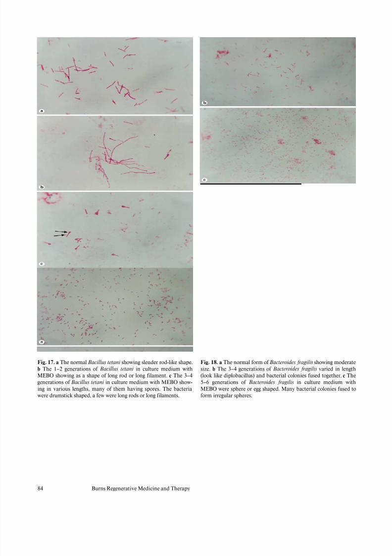
8/3/2019 Luka Bakar Book
http://slidepdf.com/reader/full/luka-bakar-book 90/157
84 Burns Regenerative Medicine and Therapy
Fig. 17. a The normal Bacillus tetani showing slender rod-like shape.
b The 1–2 generations of Bacillus tetani in culture medium withMEBO showing as a shape of long rod or long filament. c The 3–4
generations of Bacillus tetani in culture medium with MEBO show-
ing in various lengths, many of them having spores. The bacteria
were drumstick shaped, a few were long rods or long filaments.
Fig. 18. a The normal form of Bacteroides fragilis showing moderate
size. b The 3–4 generations of Bacteroides fragilis varied in length(look like diplobacillus) and bacterial colonies fused together. c The
5–6 generations of Bacteroides fragilis in culture medium with
MEBO were sphere or egg shaped. Many bacterial colonies fused to
form irregular spheres.

8/3/2019 Luka Bakar Book
http://slidepdf.com/reader/full/luka-bakar-book 91/157
Experimental and Clinical Study on Burns Regenerative Medicine and Therapy with MEBT/MEBO 85
Fig. 19. a The normal shape of Propionibacterium acne showing G+
(some straight or slightly crooked). b The 3–4 generations of Propio-nibacterium acne in culture medium with MEBO showing varied rod
length. c The 5–6 generations of Propionibacterium acne were small
coccobacilli with deep-colored particulates.
Table 52. Effect of MEBO on C. albicans
Control
The fungi were oval with spores and pseudohypha, colonies were mil-
ky with smooth and moisturized surface (fig. 19a)
Spore tube test: spore tube producing rate was 90% (fig. 19b)
MEBO
1, 2 generations: the same as the control, colonies were a little moreflat and small, compared to the control; spore tube producing rate
was about 85% (fig. 19c)
3, 4 generations: some of the fungi were sphere or oval of different
sizes, pseudohypha appeared; colonies were flat, dry and rough
(fig. 19d)
5, 6 generations: the fungi were sphere or oval, filaments with stick or
long rod shape and different length appeared; only few spores;
colonies were flat, dry and hard (fig. 19e)
Spore tube rate was about 0.5–2%1 (fig. 19f)
1 Cultured in media with different concentrations of MEBO, their
spore-producing rates were different. In media containing 25% of
MEBO, the spore tube-producing rate was 2%, a little higher than in
that containing higher concentration of MEBO.
Table 53. Effect of MEBO on the proliferation of S. aureus and P.
aeruginosa
Bacteria Primary bacteria
cultured
counts/ml
10th generation after
cultured in MEBO
containing media
counts/ml
Staphylococcus aureus 1.4!108 1.5!106
Pseudomonas aeruginosa 2!108 6.5!106
Table 54. Effect of MEBO on the invasiveness of P. aeruginosa –
pathological examination
Control group
In subcutaneous tissue, there was congestion and edema, infiltration
of inflammatory cells and a suppurative zone
MEBO groupIn subcutaneous tissue and striated muscles, there was infiltration of
a few inflammatory cells without suppurative phenomenon

8/3/2019 Luka Bakar Book
http://slidepdf.com/reader/full/luka-bakar-book 92/157
86 Burns Regenerative Medicine and Therapy
Fig. 20. a The normal shape of Candida albicans showing egg shape
with many blastospores. b The germ tube tests on normal Candida
albicans gave a producing rate of 90%. c The germ tube producing
rate of 1–2 generations of Candida albicans in culture medium with
MEBO was 85%. d The 3–4 generations of Candida albicans in cul-
ture medium with 25% MEBO showing large oral or few sphere-
shaped and stick- or filament-shaped ones being observed occasion-
ally. e The 5–6 generations of Candida albicans showing in shapes of
stick or long rod. Bacterial filaments in various length and few blas-
tospores being observed. f The germ tube producing rate of 5–6 gen-
erations of Candida albicans in cultured medium with MEBO was
0.5–2%.

8/3/2019 Luka Bakar Book
http://slidepdf.com/reader/full/luka-bakar-book 93/157
Experimental and Clinical Study on Burns Regenerative Medicine and Therapy with MEBT/MEBO 87
Discussion
Micro-organisms proliferate rapidly and are according-
ly susceptible to unfavorable factors in their growing envi-
ronment. These changes in their environment may result
in variations of their characteristics including morpholog-
ical structure, culture feature, toxicity, antigenicity and
drug resistance, etc. Morphological structure variationsare directly related to cell division. Cell division is a com-
plicated process and is more susceptible to unfavorable
factors in the environment than to protein and DNA syn-
theses. For example, Bacillus tetani and Bactorides fragilis
may have polymorphic variations such as polynuclear
filaments or spheres because, in an unfavorable environ-
ment, cell membrane synthesis is delayed and cell divi-
sion cannot proceed in a timely manner, while metabo-
lism proceeds normally. Variation is closely related to the
effect of the strength and the duration of action of the
unfavorable factors. In our experiment, we found that the
same bacteria demonstrated variation in morphological
features when cultured in media containing different con-
centrations of MEBO (data not shown).
In this experiment, we observed the antibacterial effect
of MEBO on Bacillus tetani, Propionibacterium acnes and
Candida albicans. These micro-organisms are normal flo-
ra and opportunistic pathogens in the human body. Un-
der certain conditions they become pathogenic and, in
most clinical cases, the infections are endogenous in etiol-
ogy. Burns patients often receive large doses of antibiot-
ics, which always create an imbalance in native flora
thereby facilitating the overgrowth of opportunisticpathogens such as Candida albicans. Aerobic bacteria
infections such as Staphylococcus aureus, E. coli and
B. pyogeneus predominate in burns wounds though anaer-
obic bacteria infections may also occur since burns
wounds may become ischemic and necrotic. Therefore,
bacterial examination of wounds should always consider
both aerobic and anaerobic bacteria. In 1983, Wang
Dewang reported a positive anaerobic bacteria detection
rate of 23.9% in 34 cases of burns and most of them had
mixed infections.
Bacillus tetani is an anaerobic spore-bearing bacteria.
After being cultured in MEBO-containing media, long
filament variants appeared, and in 3 generations many of
them formed only one spore. This spore is not the vegeta-
tive form of the bacteria as it cannot proliferate undernormal conditions. Spore keeps the bacteria alive though
dormant. Only under favorable conditions can the spore
proliferate and produce exotoxin to cause disease. After
culture in MEBO-containing media for 6 generations, in
Candida albicans the number of blastospores was signifi-
cantly reduced, the spore tube producing rate was only
0.5–2%, while the normal fungi had a spore tube produc-
ing rate of 90%. The above facts prove that MEBO has an
inhibitory effect on the proliferation and pathogenicity of
Bacillus tetani and Candida albicans.
From table 53, we can see the effect on Bacillus pyocya-
neus. After this baceria was cultured in MEBO-containingmedia for 10 generations, we again cultured the 10th gen-
eration for another 20 h and discovered that the bacteria
count/ml was greatly reduced to only 1/30 of that of the
original. The invasiveness of this organism to mice was
also greatly decreased. This proved that under an unfa-
vorable environment, not only morphological structures
but also physiological features of the bacteria are subject
to variations.
Our data as well as many experimental and clinical
data reported by other workers proved that MEBO has a
broad-spectrum antibacterial effect and can also promote
wound healing. Therefore, MEBO is a drug with a dual
regulatory effect.
References
1 Yang K, Ma J, Yang Q, et al: A comprehensive report on the therapeutic
effect of MEBO in treating 4954 cases of various wounds and ulcers. Chin J
Burns Wounds Surface Ulcers 1997;4:15–21.
2 Xu R: China Burns and Wounds Medicine. Burns Wounds Surface Ulcers,
1997, pp 137–140.

8/3/2019 Luka Bakar Book
http://slidepdf.com/reader/full/luka-bakar-book 94/157
88 Burns Regenerative Medicine and Therapy
OOOOOOOOOOOOOOOOOOOOOOOOOOOOOOOOOOOOOOOOOOOOOOOOOOOOOOOOOOOOOOOOOOOOOOOOOOOOOOOOOOOOOOOOOOOOOOOOOOOOOOOOOOOOOOOOOOOOOOOOOOO
Studies on the Effects of BRT with MEBT/MEBO on
Regeneration and Healing of Burns Wounds
A Comparative Study of Fibronectin and
Moist-Exposed Burns Ointment (MEBO) in the
Treatment of Experimental Corneal Alkali Burns
in Rabbits
Introduction
Worldwide reports regarding the basic science and
clinical applications of fibronectin’s (FN) contribution to
the healing of trauma and burns wounds have shown that
FN can enhance the epithelial healing rate by promoting
the adhesion and migration of epithelial cells at the
wound site [1–3]. Clinical application of eye drops made
of FN in treating burned cornea has obtained satisfactory
results. However, we are still years from the widespread
use of FN because the extraction and production of FN
eye drops are time-consuming, expensive and offer spe-
cific preservation challenges. We have used MEBO since
1990 in the treatment of burned cornea and we have
achieved satisfactory results in terms of improving cor-
neal nutrition, promoting wound healing, ameliorating
pain, relieving irritation and reducing the incidence of
corneal ulceration. Therefore, we embarked on an experi-mental study to verify the effect of MEBO as compared
with FN in the management of corneal burns in rabbits.
Materials and Methods
MEBO, developed by Beijing Guangming Chinese Medicine
Institute for Burns, Wounds and Ulcers, is an ointment containing
sesame oil, beeswax and other active ingredients derived from plants
such as Cortex phellodendri and Radix scutellariae. FN produced by
the Shanghai Institute of Biological Products was diluted to a solu-
tion containing 400 Ìg/ml with sterile normal saline.
Five healthy New Zealand white rabbits (body weight 2–3 kg)without eye disease were anesthetized intramuscularly with 20 mg/kg
sodium thiopental and eye solution of 0.4% Novesine two drops in
each eye. 8-mm filter papers, previously soaked completely in 0.5 N
NaOH, were laid on the middle of both corneas, respectively. One
minute later, the paper was removed and the eyes were rinsed imme-
diately with sterile normal saline. Then eye drops made of FN were
administered to the right eyes, four times daily and MEBO was
applied on the left eyes, three times daily. Both courses lasted 2
weeks.
At 6, 24, 36 and 48 h postinjury, the corneal fluorescent staining
zones were photographed with a DF Haiou camera (China) at fixed
focus. After developing the film, we drew the fluorescent staining
zones, analyzed and measured the areas at different hours using a
computer image pattern analyzer, and then calculated the epithelialhealing rate (mm2/h) by linear regression. Observations were made
twice daily on days 3–14 postinjury and fluorescent staining zones
were regarded as a positive indicator. Epithelial damage rate = (fluo-
rescent staining positive number)/(number of eyes examined)
!100%.
Besides corneal fluorescent staining zone, conjunctival conges-
tion and corneal transparency were also observed. (1) Conjunctival
congestion: + = palpebral conjunctival congestion; ++ = palpebral
conjunctival and partial bulbar conjunctival congestion; +++ = whole
palpebral and bulbar conjunctival congestion. (2) Corneal transpar-
ency: + = slightly opacity with distinct structure of underlying iridial
texture; ++ = moderate opacity with blurred structure of iridial tex-
ture; +++ = severe corneal opacity with indistinct structure of iridial
texture.
Results
In the early stage (at 6 h postinjury) epithelial healing
was slow and at 6–48 h the healing rate remained con-
stant. The use of FN on eyes shows an epithelial heal-
ing rate of 1.279 B 0.317 mm2/h compared to 1.285 B
0.128 mm2/h with the MEBO treatment. There is no sig-
nificant difference between the two groups although in the
MEBO group treatment efficacy is a little faster.
Repeated corneal damage occurred from the early
healing stage until 2 weeks postinjury and there is a signif-
icant difference in damage rate in both groups: 58.3% in
the FN group and 33.3% in the MEBO group (u = 5.56,
p ! 0.01).
The cornea showed disk opacity a few minutes follow-
ing burn. Conjunctival congestion with white secretion
was observed at 3 h postinjury. On day 2 postinjury, eye
irritation became obvious with increased secretion and
corneal edema was noted until 1 week postinjury when
edema was still observed in epithelium and matrix. At 2
weeks postinjury, the above symptoms were further re-
lieved. The advantages of MEBO treatment as compared
to FN in the management of burns eyes were demon-
strated both in terms of corneal epithelial exfoliation and
regarding conjunctival congestion and opacity.
Conclusion
Some advantages of MEBO include inexpensive cost,
convenient application, safety and product stability. The
effect of MEBO in the treatment of alkali-burned corneas
compared favorably to that achieved with FN. MEBO
proved more effective than FN in reducing the epithelial
damage rate and was far easier to use given a widespread
application. We suggest that MEBO treatment is remark-
ably advantageous in reducing the repeated epithelial
damage rate of alkali burns eyes compared to FN treat-ment.

8/3/2019 Luka Bakar Book
http://slidepdf.com/reader/full/luka-bakar-book 95/157
Experimental and Clinical Study on Burns Regenerative Medicine and Therapy with MEBT/MEBO 89
Discussion
It has been reported that FN has the effect of promot-
ing epithelial healing rate on corneal defects [4]. This
study verified no significant difference in promoting cor-
neal epithelial healing between the two groups. However,
MEBO treatment is remarkably advantageous in reducing
the repeated epithelial damage rate of alkali burns eyescompared to FN treatment. The repair of relative integri-
ty of the corneal epithelium is beneficial to the stability of
the corneal parenchyma and endothelium. The cornea in
the MEBO group showed slight transparency with slight
congestion. Subsequent to alkali burns, the integrity of
basement membrane underlying corneal epithelium was
damaged. The appropriate replacement of exogenous FN
may reduce the possibility of epithelium damage, as FN is
needed for the repair of basement membrane. This study
demonstrated that MEBO is superior to FN with regard to
firm adherence of corneal epithelium and maintenance
and basement membrane integrity. In addition, MEBO
supplies rich nutrients necessary for repairing and regen-
eration of alkali burns corneas.
FN is a kind of macromolecular glucoprotein which
operates at the wound surface. The surge of enzymes
released subsequent to alkali burn causes the degradation
of FN at the wound surface. The paucity of FN resulting
from enzymatic degradation can be compensated with
exogenous FN, which reduces the epithelial damage rate
by promoting epithelium adhesion. Many combining sites
in FN molecular structure served as a bridge between epi-
thelial cells and basement membrane, thereby improvingepithelium adhesion. Affinity between FN and intracellu-
lar actin may cause the change of epithelial intracellular
actin from sphere to fibriform and then induce cell migra-
tion.
As avascular tissue, the cornea receives the nutrition
and oxygen necessary for metabolism mainly from diffu-
sion of the vascular net in the corneal limbus, from tears
and from aqueous humor. Glucose is the main source of
energy for the oxygenic metabolism of corneal epithelium
and for intraparenchymatous anaerobic metabolism [5].
MEBO contains abundant glucose, organic acid, a variety
of vitamins, proteins and enzymes as well, all of which
directly provide energy and nutrition for alkali-burned
corneal tissue. In this manner, MEBO serves to promote
metabolism of the cornea, to accelerate the prompt re-
moval of necrotic tissue and to facilitate swift growth of
new epithelium. MEBO also has anti-inflammatory prop-
erties, bacterial inhibition, repercussive and analgesic ef-
fects [6]. By relieving local congestion and corneal irrita-
tion, blepharospasm due to pains and nictitation were
alleviated, which reduced the friction to corneal surface
and also improved local resistance.
References
1 Fujikawa LS, Foster CS, Harrist TJ, Lanigan JM, Colvin RB, et al: Fibro-
nectin in healing rabbit corneal wounds. Lab Invest 1981;45:120–129.
2 Nishida T, Nakagawa S, Awata T, Nishibayashi C, Manabe R: Rapid prep-
aration of purified autologous fibronectin eyedrops from patient’s plasma.
Jpn J Ophthalmol 1982;26:410.
3 Shi SM, et al: Comparative physical properties of ophthalmic viscoelastic
materials. J Pract Ophthalmol 1989;7:9.
4 Nishida T, et al: Fibronectin enhancement of corneal epithelial woundhealing of rabbits in vivo. Arch Ophthalmol 1984;102:455.
5 Liu JQ, et al (eds): Practical Ophthalmology. Beijing, People’s Health
Press, 1984, p 43.
6 Xu RX: The medicine of burn and ulcer: A general introduction. Chin J
Burns Wounds Surface Ulcers 1989;1:11.
A Comparative Study of the Effects of
Moist-Exposed Burns Ointment (MEBO) and
Other Drugs on the Healing Rate of Corneal
Epithelial Defect in Rabbits
Introduction
Corneal epithelium consists of three types of cells at
three different levels. They are basal cells in the deep layer,
wing cells in the middle layer and squamous cells at the cor-
neal surface. Microvillus in the superficial layer absorbs
tears to form a tear membrane. Basement membrane be-
hind the deep layer is located in the corneal Bowman mem-
brane (anterior limiting lamina). Corneal epithelial layer is
a natural barrier against micro-organism invasion. Once
injured, it is prone to infection and shedding. Some corneal
diseases such as dendritic keratitis, neuroparalytic kerati-tis, exfoliation of recurrent corneal epithelium, alkali
burns, vesicular keratitis, etc., and surgeries such as scrap-
ing the corneal epithelium for intraocular examination and
operation when performing cutting of the vitreous and re-
pair for retinal detachment, may injure corneal epithelium.
Rapid and complete repair of corneal epithelial defects
plays a very crucial role for the recovery of corneal physio-
logical function and for good eyesight. Corneal physiologi-
cal function contributes greatly to the promotion of deep
corneal wound healing and formation of collagenous fiber
[1]. In this study, rabbits with corneal epithelial defects of
the same size were used and treated with MEBO, homolog-
ous serum, 0.5% dexamethasone, 25,000 U/ml vitamin A.
In addition, the rabbits wore soft corneal contact lenses to
observe the regenerating rate of corneal epithelia and to
assess the therapeutic effects of different measures.
Materials and Methods
Eighty-two healthy adult rabbits of either sex weighting 2–3 kg
were anesthetized by 35% urethane 2.5 ml/kg intravenously and, if
required, by inhalation of ether. Then 1% dicaine was dropped twice
for surface anesthesia into the palpebral fissure which was expanded
with eyelid retractor. The cornea center was constrictively marked at

8/3/2019 Luka Bakar Book
http://slidepdf.com/reader/full/luka-bakar-book 96/157
90 Burns Regenerative Medicine and Therapy
Table 55. Average healing rate of corneal epithelium after injury
(mm2/h, meanB SE)
Groups Number
of eyes
Average healing rate
Group 1 (MEBO) 10 1.184B0.106
Group 2 (dexamethasone) 11 1.087B0.087a
Group 3 (vitamin A) 11 1.065B0.066b
Group 4 (homologous serum) 10 1.114B0.038e
Group 5 (saline) 10 1.037B0.059b,c,f
Group 6 (contact lens) 10 0.770B0.016b,d,e,f,g
a p ! 0.05, b p ! 0.01 compared with group 1; c p ! 0.05, d p ! 0.01
compared with group 2; e p ! 0.05 compared with group 3; f p ! 0.01
compared with group 4; g p ! 0.05 compared with group 5.
surface using an 8-mm corneal trephine and was stained with 1%
fluorescein followed by rinsing with normal saline. Subsequent to the
appearance of a circular mark, an 8-mm stained disk area of epithe-
lial defect was created with a lancet scraping off the whole epithelium
within cycle and by fluorescein staining.
The animals were then divided randomly into 6 groups, 12 in
each. In group 1, the animals were treated with MEBO every 6 h. In
group 2 to group 5, the animals were given eye drops with 0.5% dexa-
methasone, 25,000 U/ml vitamin A, homologous serum and normal
saline, respectively, every 2 h. In group 6, the animals wore soft cor-
neal contact lens without any eye drops, whereby 6 eyes was removed
of lens every 6 h for fluorescein staining and other 6 eyes stained 24 h
later. In addition, 10 rabbits had their secretions cleaned away with
cotton sticks soaked with normal saline and no eye drops were
applied. Corneas were removed at 0, 2, 4, 6, 12, 18, 24, 30, 36 and
48 h for microscopic observation on corneal epithelial healing [2].
Results
The observation record showed the following mean
healing time: 48.6 h in the normal saline group, 47.36 h in
the vitamin A group, 45.18 h in the homologous serum
group, 42.78 h in the MEBO group, 46.5 h in the dexa-
methasone group and 65.3 h in contact lens group. The
results revealed that there was no significant difference of
average healing rate between groups 1 and 4, as well as
groups 2 and 4, groups 2 and 3, groups 3 and 5. However,
there were statistical differences between other each twogroups. MEBO, homologous serum, vitamin A, and dexa-
methasone had superior healing effects compared with
normal saline, while wearing of contact lens retarded the
healing of the defect. Beside homologous serum, MEBO is
remarkably superior to other drugs in promoting corneal
epithelial healing rate (table 55).
Discussion
Cornea has sources of nutrition from the vascular net
in corneal limbus, tears and aqueous humor. Corneal epi-
thelium is a functional barrier between tear membrane
and intraocular tissues through which the fluid output
from the stroma is regulated in order to keep the stroma in
a normal hydration. A corneal epithelial defect caused by
injury or scraping off can repair rapidly in two stages. The
latent period comes first, with a mean time of 5.5B 0.3 h
during which extensive cellular and subcellular changes at
the wound edges are expressed by desquamation of super-ficial cells, by loss of columnar appearance of basal cells,
by damage on hemidesmosome link of the basement
membrane as well as by formation of a cell process, indi-
cating that residual viable epithelial cells are transforming
into functional cells. A healing period is followed after-
wards when epithelial cells around the wound migrate
towards the center at a constant pace without mitochysis.
This process starts from the uninjured epithelial cells
adjacent to wound edge. We see basal cells in particular
being enlarged and flatted with pseudopodium migrating
towards the center and becoming the migratory edge of
monolayer cells, which is followed by two or more layersof epithelial cells. The migration stops as wounds com-
pletely close and a firm hemidesmosome adhesion to the
basement membrane is re-formed. At this point, mitochy-
sis begins and mature corneal stratified epithelium is
finalized.
The experiment revealed us the two periods for epithe-
lial healing. At 2 h postsurgery, the defects did not de-
crease, instead, some increased from 8 to 8.1–8.5 mm,
which may have resulted from the contraction of tissues at
the wound edge and the shedding of injured cells. It was
not until 6 h postsurgery that epithelial defects began to
diminish progressively until complete wound healing was
achieved. Systemic or local administration and either
intra- or extra-ocular agents may be factors contributive
to the successful repairing process of corneal epithelial
defects from the latent period till the end of the healing
period. By means of smearing, eye drops or wearing of
corneal contact lenses, the study investigated various fac-
tors relative to epithelial healing in order to verify the
therapeutic effects of methods and drugs.
MEBO is an ointment containing sesame oil, beeswax
and other active ingredients extracted from plants such as
Cortex phellodendri or Radix scutellariae. It is highlyeffective in the treatment of burns, wounds and ulcers and
its advantages include superior analgesic effects, anti-
inflammation, infection prevention and minimal scar for-
mation post healing [3]. Although recent reports prove
MEBO is significantly superior to other therapies in treat-
ing various burns, wounds and ulcers [4], there is no com-
parative study on the management of eye injury reported.
Therefore, we conducted a preliminary comparative
study on fibronectin (FN) and MEBO in the treatment of
experimental corneal alkali burns in rabbits, which veri-
fied that MEBO was remarkably effective in the treat-
ment of burns on avascular corneal tissue [2, 5]. Further,

8/3/2019 Luka Bakar Book
http://slidepdf.com/reader/full/luka-bakar-book 97/157
Experimental and Clinical Study on Burns Regenerative Medicine and Therapy with MEBT/MEBO 91
we performed this study to investigate the effect of MEBO
on repairing and healing of corneal defect and the roles of
other drugs as well. The results verified that MEBO is
obviously superior to other medications except for homol-
ogous serum in promoting wound healing while no ob-
vious corneal macula was formed posthealing under slit-
lamp microscopic examination.The pharmacological mechanisms of MEBO with re-
spect to its role of promoting healing of corneal wounds
also became understood as follows: (1) Composed of a
macromolecular sticky base, MEBO has an affinity to tis-
sue protein at the wound site. The application of MEBO
to the wound may serve as a bridge to directly stimulate
and induce cell division and migration in an orderly man-
ner that promotes wound healing. (2) MEBO contains
various nutrients necessary for wound healing. For exam-
ple, glucose is an obvious energy source. Vitamins and
organic acids, which are related to the maintenance of tis-
sue metabolism and proliferation of connective tissues,may directly support local nutritional needs, thereby pre-
venting scar formation [6]. Zinc and enzymes may accel-
erate epithelial repair [7]. Protein as the basic element of
the cell membrane may support the growth, differentia-
tion and regulation of cells. (3) MEBO promotes the for-
mation of a unique, integrated automatic drainage circu-
lation system which corrects for local dysfunctional me-
tabolism and circulation resulting from injury. The base
ingredients contained in MEBO absorb metabolic prod-
ucts from the wound and then transport them to the outer
layer of the ointment. Meanwhile, active ingredients of
the ointment continuously penetrate into the wound to
renew the supply of ingredients necessary for tissue repair.
The automatic microparticle transportation and process-
ing of emulsification and dispersion are considered as the
main measure for the treatment of injured avascular tis-
sue [8]. (4) Obaculactone contained in MEBO offers prop-
erties of anti-inflammation, detumescence, analgesic ef-
fects, and enhancing local immunity and controlling in-
fection.
Many reports demonstrate the presence of FN in plas-
ma. FN is a macromolecular glycoprotein that is the con-
junctive medium between cell and extracellular fibers andmatrix. It adheres to collagen, polysaccharide protein, and
receptors on the cell surface and serves as an intercellular
bridge of epithelial cells. It has some correlation with
cytoskeleton structures, e.g. microfilament, actin, to in-
duce the migration of the cell. FN plays an important role
in the firm adhesion between migrating epithelial cells
and wound surface of corneal epithelial defect. Eye chem-
ical burns can be treated with eye drops or subconjuncti-
val injection composed of autoblood. Autoblood is benefi-
cial because it contains macroglobulins that inhibit colla-
genase, release fibrinolysin (which may reduce symble-
pharon, thereby promoting the recovery of the blood ves-
sel net around the cornea as well as restoring sensation in
the injured cornea), improve corneal nutrition and pro-
mote tissue regeneration. Our previous study has verified
that FN can speed up the migration of corneal epithelial
cells [2]. Observation on the rabbit corneal epithelial heal-
ing rate after application of homologous serum in this
study has also suggested an obvious effect of serum in pro-moting the migration of corneal epithelial cells. However,
even serum offers less benefit than does the application of
MEBO.
Various reports can be found about the role of cortico-
steroids in corneal wound healing. It is believed that long-
term administration of corticosteroids at high concentra-
tion may retard epithelial regeneration and that stromal
wound healing though the mechanism remains unclear.
On the other hand, the use of corticosteroids immediately
after corneal burns may have good anti-inflammatory
effects and reduce occurrence of ulcers and neogenetic
vascularization [9]. Others report that corticosteroidsmay inhibit conjunctival cells from migrating towards the
corneal surface without impairing the reformation of cor-
neal epithelium [10]. In this study, eye drops of 0.5%
dexamethasone did not inhibit the migration of corneal
epithelial cells and wound healing. On the contrary, it sig-
nificantly promoted the healing. Therefore, it is feasible to
drop 0.5% dexamethasone on simple corneal epithelial
defects to control inflammation and promote healing,
though even this modality, due to adverse effects, is far
inferior to the effects achieved by MEBO.
Vitamin A plays a role in promoting epithelial growth
and maintaining epithelial normal functions. Many re-
ports have confirmed vitamin A contributing to promote
wound healing, but some researchers reported that vita-
min A failed in promoting epithelial regeneration. The
use of 25,000 U/ml vitamin A in this study revealed little
effect on corneal epithelial healing.
Corneal epithelium, when wearing corneal contact
lenses made of polymethyl methacrylate (PMMA), is pro-
vided with nutrients and oxygen necessary for metabo-
lism which derive primarily from tears penetrating be-
tween the lens and cornea. Eyelid pressure when eyes are
open produces a positive pressure behind the lens to dis-charge tears and the negative pressure produced behind
the lens after winking allows the entrance of the tears to
float the lens. Such a process goes in cycles to achieve the
exchange and renewal of tears. Subsequent to corneal epi-
thelial injury, the ability of local metabolism decreases
and wearing lenses further impairs oxygen absorption
of the cornea, which is unfavorable to wound healing.
Though some reports suggested that wearing corneal con-
tact lenses might be used for treating corneal ulcer, the
observation in this study verified that such management
impaired the corneal wound-healing process and healing
rate was much lower than those in other groups. We con-

8/3/2019 Luka Bakar Book
http://slidepdf.com/reader/full/luka-bakar-book 98/157
92 Burns Regenerative Medicine and Therapy
clude that careful attention should be taken to avoid
harming patients through the application of corneal con-
tact lenses in the treatment of corneal injury.
References
1 Smolin G, et al: Tretinoin and corneal epithelial wound healing. Arch Oph-
thalmol 1979;97:545.
2 Huang QS, et al: A comparative study of fibronectin and MEBO in the
treatment of experimental corneal alkali burn in rabbits. Chin J Burns
Wounds Surface Ulcers 1995;7:18.
3 Xu RX: The medicine of burn and ulcer: A general introduction. Chin J
Burns Wounds Surface Ulcers 1989;1:11.
4 Xu RX: The principle of burn wound treatment. Chin J Burns Wounds
Surface Ulcers 1992;4:8.
5 Huang QS, et al: A dynamic study on MEBO repairing corneal endothe-
lium alkali burn in rabbit. Proceedings of the Fourth National Conference
on Burns, Wounds and Ulcers, Beijing, 1995.
6 Huang QS, et al: Clinical observation of herpes simplex corneal ulcers
treated by combined MEBO and PIC (25 cases report). Chin J Burns
Wounds Surface Ulcers 1993;5:21.
7 Morley TE, et al: Zinc deficiency chronic starvation and hypothalamic
pituitary thyroid function. Am J Clin Nutr 1980;33:176.
8 Du HE: A preliminary introduction on relationship between the biophar-
macy factors and treatment of MEBO. Chin J Burns Wounds Surface
Ulcers 1995;7:8.
9 Woost PG, et al: Effect of growth factors with dexamethasone on healing of
rabbit corneal stromal incisions. Exp Eye Res 1985;40:47.
10 Srinirasan BD: Corneal re-epithelialization and anti-inflammatory agents.
J Am Ophth Soc 1982;80:756.
Exploration of Pathological Changes and
Mechanism of Experimentally Burned Rabbitsafter Treatment with Moist-Exposed Burns
Ointment
Introduction
BRT with MEBT/MEBO, available over the past 10
years, is a remarkable innovation for the management of
burns, wounds and ulcers. Satisfactory results were ob-
tained using this treatment to deal with profound prob-
lems in conventional surgical treatment such as pain,
infection, healing with scar formation, and progressivenecrosis of tissues in the zone of stasis. Clinical applica-
tions worldwide demonstrated that BRT with MEBT/
MEBO is superior to all other therapies and represents the
clear standard of care in burns treatment. To verify the
therapeutic effect of MEBO in treating burns wounds, we
studied a rabbit model of deep second-degree burns
treated with MEBO and with Vaseline, respectively.
Serial histological sections were performed during the
treatment in order to observe the pathomorphological
changes, progression and mechanism of repair. This study
provides references for the prevention and research of
burns, wounds and ulcers.
Material and Method
Thirty healthy adult rabbits of either sex weighing 1.5–2.0 kg
were used in this study. The dorsal hair of each animal was depilated
using 20% sodium sulfide. Rabbits were restrained in a self-made
soaking support frame, and two 4 ! 4 cm deep second-degree
wounds of zygomorphic skin on the back were created via scalding
with 100°C water for 5 s (lesions were determined by pathological
examination). The wounds were then contaminated with 1 ml sus-
pension containing 3.0 ! 108 cfu Staphylococcus aureus. At this
point, the animals were divided randomly into two groups, 15 ani-
mals offering 30 wounds in each group. Animals in the control group
were treated with Vaseline ointment, while animals in the experi-
mental group were treated with MEBO ointment. Both ointments
were applied once every 3 h. The rabbits were caged separately and
freely fed. At six different time phases (days 3–6, 7–9, 10–12, 13–15,
16–18 and 19–22 postburn), five full-thickness wound tissues (0.5!
0.5 cm) were taken from each group. All samples were fixed with 10%
formaldehyde solution, embedded with paraffin, stained with hema-
toxylin and eosin, and studied by the light microscope for pathomor-
phological changes.
Results
Normal rabbit skin demonstrated an absence of der-
mal papilla, but revealed abundant structures of skin hair
and appendages (fig. 21a). Deep second-degree burns
wounds on rabbit back skin involved deep dermis causing
necrosis of full epidermis and partial dermis. Fibers in the
dermis reticular layer and the subcutaneous layer ap-
peared to be thick and sparse with partially survived skin
appendages (fig. 21b). The results of pathomorphological
examinations of two groups at different phases are shown
in table 56.
Conclusion
BRT with MEBT/MEBO treatment can make injured
tissue regenerate in a relatively physiological environ-
ment that conforms to the natural law of tissue regenera-
tion. As a result, scar formation is reduced to the maxi-
mum extent. These experimental results were in accor-
dance with clinical observations.
Discussion
Histologically, the regenerative capacity of skin tissue
has a close correlation with tissue repair. Skin cells can be
classified into two categories according to the different
capability of regeneration: (1) Constantly changing cells,
i.e. epidermal cells that have the ability to divide for in-
definite periods under a normal state and proliferate to
compensate shed and consumed cells. (2) Stable cells as
epithelium in the skin body of a gland that cease prolifera-
tion when the organs mature, but have a continuous
potential for division which is activated after injury to
regenerate [1]. Tissue repair is also achieved by two

8/3/2019 Luka Bakar Book
http://slidepdf.com/reader/full/luka-bakar-book 99/157
Experimental and Clinical Study on Burns Regenerative Medicine and Therapy with MEBT/MEBO 93
Fig. 21. a Normal rabbit skin. HE. !100. b Deep second-degree
burns wound on rabbit back skin with partially survived skin ap-
pendages, epidermis exfoliated. HE.!100.
Table 56. Summary of pathomorphological changes at different time
phases postburn in both the Vaseline and the MEBO groups
Days
postburn
Pathomorphological characteristics
Control group (Vaseline)
3–6 Diffusion inflammation, visible infiltration of many
inflammatory cells aggregating beneath epidermis layer
(fig. 22a)
7–12 Massive infiltration of inflammatory cells, few macro-
phages, few new blood capillary formations, narrow
lumen, poor wound healing with proliferation of fibrous
tissue (fig. 22b)
13–18 Scattered colonies, tissue necrosis patency tending to
worsening (fig. 22c)
19–22 Infiltration of inflammatory cells, wound tissue edema,
disruption of collagen fibers, marked proliferation of
fibrous tissue, progressing to form hyperplastic scars
(fig. 22d)
Experimental group (MEBO)
3–6 New epithelial regeneration on wound, increase of blood
capillaries, active regeneration of granulation tissue,
inconspicuous proliferation of collagen fiber (fig. 23a)
7–9 Active regeneration of epithelia, increased large basal
layer cells, new skin regenerating in varied thickness
(fig. 23b)
10–12 Epidermis regenerating from residual skin appendages,
visible transition from skin appendages to regenerated
epithelia (fig. 23c )
13–15 Gradual decrease of necrotic tissues, contracting to
wound surface (fig. 23d)
16–18 Presence of a lot of macrophages in neoformative granu-lation tissues, limited inflammatory cells (fig. 23e)
19–22 Wounds covered by squamous epithelia and presence of a
few skin appendages in the dermis (fig. 23f)
approaches. First, by the regeneration of tissues similar
both in structure and function – the structure and func-
tion of repaired tissue can be entirely identical to those of
the original [2]. Take, for example, the tissue repair of superficial second-degree burns wounds. On days 3–4
postburn, epithelia began to grow, and continued thusly
on days 5–8 and was mostly completed on days 8–10 post-
burn. Secondly, repair of the damaged tissues can be
achieved through the formation of fibrous tissue, begin-
ning with formation of granular tissues and ending with
scar formation. Microscopic examinations of the tissue
repair of deep second- and third-degree burns wounds
showed an intertexture mainly comprised of fibroblasts
and neoformative blood capillaries. Together with infil-
tration of plasmocytes (such as neutrophil, lymphocyte,
plasma cell, macrophage), we noted neoformative granu-
lation tissues that were then replaced by a great quantity
of closely aligned collagenous intercellular fibers. Subse-
quent to the decrease of fibroblasts, showing a long and
narrow shape, and of blood capillaries, these tissues even-tually developed into scars.
According to our experimental results, in the control
group there was sparse blood capillary formation with
narrow lumen. The tissue was swollen with fiber prolifera-
tion and massive infiltration of inflammatory cells. The
disruption of collagen fiber and absence of regenerated
epidermis to cover wounds eventually resulted in wound
healing by eschar (showed in fig. 22d). In the experimen-
tal group treated with MEBO, residual skin appendages
regenerated into epidermis with multilayers and large
nuclei that progressed and covered the wounds (fig. 23b–
d). Granulation tissue was promoted into regenerative tis-

8/3/2019 Luka Bakar Book
http://slidepdf.com/reader/full/luka-bakar-book 100/157
94 Burns Regenerative Medicine and Therapy
Fig. 22. a In the control group 3–6 days postburn, the wound showed diffusion inflammation, visible infiltration of
much inflammatory cells aggregating beneath epidermis layer. HE.!200. b In the control group 7–12 days postburn,
there was some new blood capillary formation, narrow lumen, poor wound healing with proliferation of fibrous tissue.
HE. !100. c In the control group 13–18 days postburn, colonies were scattered, tissues obviously necrosed. HE.
!100. d In the control group 19–22 days postburn, marked proliferation of fibrous tissue and progressing hyperplas-
tic scars is seen. HE.!100.
Fig. 23. a In the MEBO group 3–6 days postburn, new epithelialregeneration on the wound, increase of blood capillaries, and active
regeneration of granulation tissue can be seen. HE. !100. b In the
MEBO group 7–9 days postburn, epithelia regenerated actively, bas-
al layer cells became larger, and new skin regenerated in varied thick-
ness. HE.!400. c In the MEBO group 10–12 days postburn, epider-
mis regenerated from residual skin appendages, visible transition
from skin appendages to regenerated epithelia. HE. !400. d In the
MEBO group 13–15 days postburn, necrotic tissues gradually de-
crease and contracted to wound surface. HE. !100. e, f In the
MEBO group 3 weeks postburn, there were a lot of macrophages in
neoformative granulation tissues, limited inflammatory cells. HE.
!400. g In the MEBO group 4 weeks postburn, wounds were cov-
ered by squamous epithelia and there were a few skin appendages in
dermis. HE.!200.
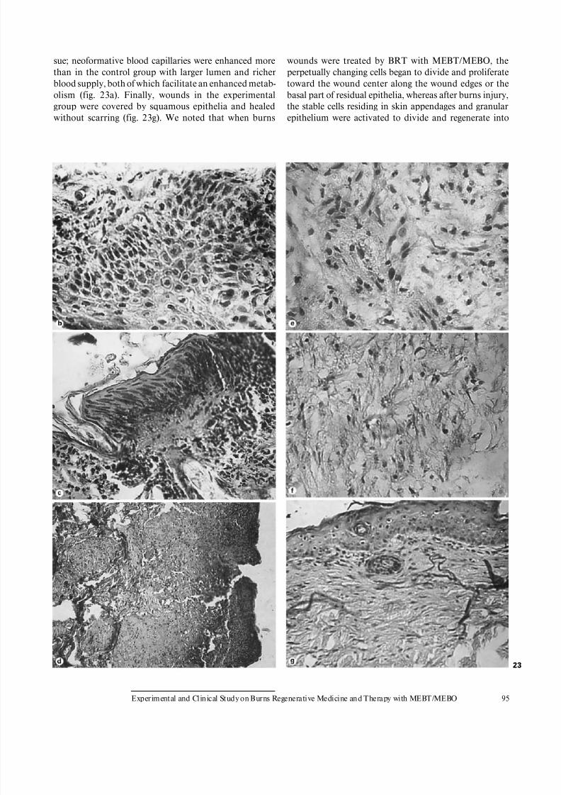
8/3/2019 Luka Bakar Book
http://slidepdf.com/reader/full/luka-bakar-book 101/157
Experimental and Clinical Study on Burns Regenerative Medicine and Therapy with MEBT/MEBO 95
sue; neoformative blood capillaries were enhanced more
than in the control group with larger lumen and richer
blood supply, both of which facilitate an enhanced metab-
olism (fig. 23a). Finally, wounds in the experimental
group were covered by squamous epithelia and healed
without scarring (fig. 23g). We noted that when burns
wounds were treated by BRT with MEBT/MEBO, the
perpetually changing cells began to divide and proliferate
toward the wound center along the wound edges or the
basal part of residual epithelia, whereas after burns injury,
the stable cells residing in skin appendages and granular
epithelium were activated to divide and regenerate into
23

8/3/2019 Luka Bakar Book
http://slidepdf.com/reader/full/luka-bakar-book 102/157
96 Burns Regenerative Medicine and Therapy
epidermic tissue that in turn migrated toward and finally
closed the wound. Pathological examination on days 10–
12 postburn showed a transitional migration from skin
appendages to regenerated epithelia. When epithelia pro-
liferated and divided, wounds were covered by stratified
squamous epithelium. The macroscopic appearance of
healed wounds was initially red or pink and progressivelybecame normal in color.
In clinical management, we observed the elevation of
residual hair follicles and skin islands over the wound sur-
face when deep second-degree burns wounds were treated
with MEBO. Epithelial tissue was the first to achieve the
same height as the wound, followed by skin islands or con-
nective tissues among the hair follicles. Eventually, we
noted wounds healed by epithelialization with mild or no
scarring. We suggested, according to clinical observation
and pathological examination, that if the dermis network
and Leydig cells (interstitial cells of Leydig) are kept
intact, then as regards the treatment of deep burns, epithe-lia regenerated from skin appendages might grow along
the network and eventually recover the normal dermal
architecture without scar formation. However, if the der-
mis network, Leydig cells and granular epithelia were
damaged, then the residual granular epithelia might form
a cell mass with a disordered structure and fail to recover
the normal structure and function of the skin architecture
[3]. This is the case in sweat gland epithelia in adipose
tissue that regenerated and divided into nonsecretory epi-
thelial tissues to close and heal wounds.
Wounds in the control group (Vaseline) showed slow
repairing, obvious proliferation of fibrous tissue and
healed with hyperplastic scars. By comparison, wounds in
the experimental group (MEBO) expressed rapid repair-
ing, active growth of neoformative epidermis, inconspic-
uous proliferation of fibrous tissue and eventually healed
without scarring. These results demonstrate that MEBO
retains optimal wound moisture, while tissue is not im-
mersed. MEBO created a drug membrane that protected
and isolated wound tissue from outer contaminants, al-
lowing native histocytes to propagate in a relatively physi-
ological environment in accordance with the nature re-
generative law of skin. Local microcirculation was alsoimproved and pathological changes of three zones of
burns wounds (necrosis zone, stasis zone and hyperemia
zone) were reversed. These conditions were favorable to
the recovery of tissue in the stasis zone. Therefore, MEBO
was believed to promote epithelial regeneration, control
the increased speed of connective tissue, and keep epithe-
lia and connective tissue in an almost normal rate of pro-
liferation so as to heal deep burns wounds with less or
minimal scarring.
In the experiment, wounds were contaminated by
S. aureus. Microscopic observation showed massive infil-
tration and aggregation of inflammatory cells in the Vase-
line group with few macrophages and scattered colonies
without boundaries (fig. 22c). All wounds were visibly
infected within 1 week. In the MEBO group, inflammato-
ry cells were large in quantity with enhanced capacity of
anti-infection (fig. 23e). Gross observation revealed that
wounds in this group repaired rapidly with absence of
inflammatory response such as red swelling. It was be-lieved that MEBO demonstrated efficacy in promoting
the blood circulation by removing blood stasis, clearing
away heat and toxic material, relieving inflammation and
removing the necrotic tissue while promoting granulation.
The experiment also demonstrated that MEBO might
inhibit or kill the growth of S. aureus.
In this study, the rabbit burns model was kept stable
with zero mortality. Light-microscopic examination re-
vealed a distinct process of histocyte repair. The results
showed that the application of BRT with MEBT/MEBO
in burns management could prevent and control infec-
tion, promote wound repair, minimize scar formation,shorten healing time, avoid complications and relieve
pain as well. BRT with MEBT/MEBO also has the advan-
tages of facilitating the observation of wound repair and
easy application. BRT with MEBT/MEBO is now irrefut-
ably considered to be the standard of care for burns man-
agement worldwide.
References
1 Zhang YM: Experiences in treating facial scars by the combination of abra-
sive technique and excision. Proceedings of Cosmetic Symposium, Wuhan,
1990, p 142.2 Wuhan Medical College (ed): Pathology, ed 1. Beijing, People’s Health
Press, 1982, pp 19–21.
3 Academic Committee of the First National Conference of Moist Exposed
Burn Therapy: A great historical turn in the burn medical science. Chin J
Burns Wounds Ulcers 1989;1:4–10.
Electron-Microscopic Observation of One Case
of Skin Burns Wounds Treated with MEBO
Introduction
To further investigate the mechanism of deep burns
wounds healing without hyperplasic scar formation after
treatment with BRT with MEBT/MEBO, we took a biop-
sy from a deep wound site of a severely burned child
before and after treatment in order to observe it via light
and transmission electron microscopy. The aim of the
study was to find the histological evidence of scar-free
healing.

8/3/2019 Luka Bakar Book
http://slidepdf.com/reader/full/luka-bakar-book 103/157
Experimental and Clinical Study on Burns Regenerative Medicine and Therapy with MEBT/MEBO 97
Case Report
A 12-year-old boy was admitted (PID. 172650) on November 4th
1989 after suffering direct gas flame burns on the face, trunk and
extremities. Clinical assessment indicated a total burns surface area
(TBSA) of 75%, including 45% second-degree and 30% third-degree
wounds. The condition of the patient remained stable during anti-
shock therapy, but he developed sepsis on day 6 postburn. Serial
blood cultures! 3 were negative. On day 10 postburn, escharectomy
and microskin grafting were performed on the left upper and right
lower extremities. On day 20, excision and microskin grafting were
performed on the back again. On day 30, burns wounds of the right
leg and dorsum pedis with mixed second- and third-degree as well as
deep second-degree burns on the back were treated with BRT with
MEBT/MEBO. Wound tissue biopsy was taken from the right leg
before and after treatment, then pathological examinations were car-
ried out light and transmission electron microscopically.
Result
Pathological examination revealed satisfactory healing
of the burns wounds treated with MEBO without forma-
tion of obvious hyperplasic scar tissue.
Light Microscopy
Before treatment, the infiltration of inflammatory cells
was visible around sweat glands and hair follicles, some
having formed local foci (fig. 24a, b). After treatment, skin
recovered to normal structure with regenerative capillar-
ies and fibroblasts in dermis (fig. 24c).
Transmission Electron-Microscopic Observation
Before treatment, a lot of circular vacuoles were pres-ent in the surrounding nucleus that showed irregular
nuclear membrane with disappearance of nucleolus. Elas-
tic fibers in the dermis varied in thickness and had a dis-
orderly arrangement with deposits in the lumen (fig. 25c).
After treatment, cells in the stratum spinosum became
regular, showing distinct nucleus, clear nucleolus and uni-
form distribution of nuclear chromatin. Desmosomes of
the intercellular bridge recovered to normal (fig. 26a–c).
Conclusion
The results proved that after using MEBO, the burns
wounds healed without formation of macroscopic hyper-
plasic scar. The ultrastructure of the healing burns wound
was similar to that of an ordinary traumatic wound.
Discussion
In dermis of normal skin, the dominant cell relating to
traumatic repair and proliferative inflammation is the
fibroblast. It is located adjacent to a collagenous fiber
bundle, showing as fusiform, stellar or polygonal shapes,
and having thick and short cell process. The fibroblast
Fig. 24. a Before MEBO treatment, the infiltration of many neutro-
phils was visible around subcutaneous hair follicles, some havingformed local foci. HE. !200. b Before treatment, infiltration of
inflammatory cells was visible around sweat glands and hair follicles.
HE.!200. c After MEBO treatment, epidermis recovered to normal
structure and there was the presence of regenerative capillaries and
fibroblasts in the dermis. HE.!200.
contains an oval nucleus which occupies one third of
whole cell. It also reveals an obvious nuclear membrane
and one or two nucleoli. There is expanded lumen of
intracytoplasmic rough endoplasmic reticulum (RER).
There are four major types of cell junctions between epi-

8/3/2019 Luka Bakar Book
http://slidepdf.com/reader/full/luka-bakar-book 104/157
98 Burns Regenerative Medicine and Therapy
Fig. 25. a Before MEBO treatment, many circular vacuoles werepresent in the surrounding nucleus that showed irregular nuclear
membrane with disappearance of nucleolus. TEM. !10,000. b Be-
fore treatment, elastic fibers in dermis were in varied thickness and
disorderly arrangement with vacuolar degeneration. TEM.!10,000.
c Before MEBO treatment, appearance of irregular nucleus, presence
of perinuclear vacuoles and disordered elastic fibers with deposit.
TEM.!8,000.
Fig. 26. a After MEBO treatment, intercellular bridge of cells in stra-tum spinosum recovered to normal with distinct nucleus and clear
nucleoli. TEM.!4,000. b After treatment, desmosome of cell junc-
tion almost recovered to normal with clear shape of cell, regular
nucleus and uniform distribution of euchromatin. TEM. !6,000.
c After MEBO treatment, structure of desmosome recovered to nor-
mal with uniform distribution of nuclear chromatin and regular cell
shape with nucleolus in center. TEM.!5,000.

8/3/2019 Luka Bakar Book
http://slidepdf.com/reader/full/luka-bakar-book 105/157
Experimental and Clinical Study on Burns Regenerative Medicine and Therapy with MEBT/MEBO 99
thelial cells, i.e. tight junction, intermediate junction, gap
junction and desmosome.
This study showed that perinuclear vacuoles, disor-
dered elastic and collagenous fibers were presented before
MEBO treatment, comparing to desmosome of intercellu-
lar bridge recovering to normal structure after MEBO
treatment. When in the hyperfunction stage, the intracy-toplasmic RER appeared as small fragmental vesiculi-
form, and when in vigorous synthesization, it appeared
tight with flocculation within the cisternae. The main
function of the RER is to synthesize protein. Smooth
endoplasmic reticulum (SER) has functions correlating
with the synthesis of lipoids and steroids.
Stephen [1] reported the presence of myofibroblasts in
hypertrophic scar tissue according to electron-microscop-
ic observation. Myofibroblast contained incomplete nu-
clear membrane with developed RER. As it has both the
characteristics of smooth muscle cells and the shape of
fibroblasts, it is also termed ‘modified myofibroblast’. Wehave reported the ultrastructure of scar resulting from
burns injuries in 1985 [2]. Comparison of the previous
and present studies indicates that no macroscopic hyper-
trophic scar was formed on burns wounds treated with
BRT with MEBT/MEBO, and the ultrastructure of the
healing burns wound appeared no different from the ordi-
nary traumatic wound. Though we have previously re-
ported the clinical experience of applying MEBO for
treating burns wounds of varying degrees [3], this was
our first presentation of light-microscopic and transmis-
sion electron-microscopic observations regarding burns
wounds. We would like to disclose our research achieve-
ments here in order to stimulate further studies.
References
1 Stephen A: Wound contraction and fibrocontractive disorders. Arch Surg
1978;1:1034–1046.
2 Hong ST, et al: Ultrastructure of scars resulted from burns. Metal Med
1985;1:5–8.
3 Chen SR, Wang Y, Zhang XZ, et al: Clinical observation of the effect of
moist exposed burn ointment (MEBO) on treating one case with extensive
burn. Chin J Burns Wounds Surface Ulcers 1989;1:46.
Pathomorphological Changes of Deep Burns
Wounds Treated with MEBO
Introduction
BRT with MEBT/MEBO has been in wide use for
many years domestically and internationally [1, 2]. Al-
though many clinical practices have confirmed its ad-
vanced and scientific results in burns management, the
mechanism involved in the healing of deep burns wounds
without hypertrophic scar is not yet clear. From March to
November 1994, the authors treated 12 patients sustain-
ing deep burns with MEBO and performed light- and elec-
tron-microscopic observations on wounds before and af-
ter MEBO treatment. The aim was to explore the thera-
peutic effectiveness of MEBO on the healing of deepburns wounds.
Materials and Methods
Twelve patients sustained 2–98% total body surface areas
(TBSA), including 2–82% third-degree burns. Most patients had
burns mainly on the face and extremities and one patient was compli-
cated with inhalation injury. Some patients suffered extremely severe
burns covering the whole body skin. Areas which received MEBO
treatment included chest, back, upper arm, thigh, leg, and instep.
MEBO was applied on burns wounds in accordance with the stan-
dardized MEBO protocol.
All of these patients were initially treated by conventional surgi-
cal therapy in other hospitals. Therefore, this study focused on deep
second-degree and superficial third-degree burns wounds with inter-
mediate and late granulation tissue on days 3–42 postburn that were
treated with MEBO. The ointment application lasted as long as 5–50
days and the wound healed on days 30–92 postburn. Macroscopic
observation of healed wounds showed the coverage of soft, flat and
smooth epithelium or soft and flat scars, without the appearance of
hypertrophic or contractive scars.
Consent to receive MEBO treatment was obtained from patients
or their guardians. Two wound tissue samples 1–2 mm3 in size were
taken from each patient before and after treatment. One of the sam-
ples was fixed with 75% alcohol before being transferred to the
pathology department for hematoxylin and eosin staining and light-
microscopic examination (Olympus). The other was fixed with 2.5%
glutaraldehyde and 1% osmic acid, stained with uranium acetate and
lead citrate, and gradient dehydrated with ethanol and acetone. The
ultrathin sections were examined under a transmission electron
microscope (CM 10, Philips).
Results
Light Microscope
The third-degree burns wounds penetrated to beneath
the dermis and subcutaneous tissue that appeared as uni-
form pink necrosis. Some of the muscular tissue was alsoinvolved, where cross-striation of striated muscle disap-
peared and had the appearance of the pink color of coagu-
lated necrosis. Infiltration of inflammatory cells present-
ed around the sweat glands and hair follicles. During the
course of treatment, collagenous fibers were found to pro-
liferate severely, showing thick and disordered arrange-
ment at the beginning. These progressed to moderate pro-
liferation with thin fasciculi, and finally had the appear-
ance of being delicate and orderly. After healing, the epi-
dermis recovered to normal, and neoregenerated blood
capillaries and fibrocytes appeared in the dermis (ta-
ble 57).
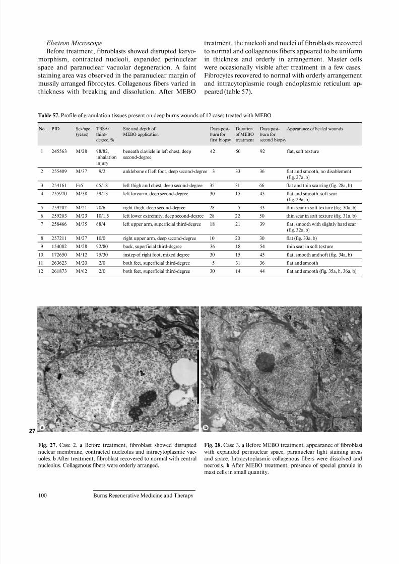
8/3/2019 Luka Bakar Book
http://slidepdf.com/reader/full/luka-bakar-book 106/157
100 Burns Regenerative Medicine and Therapy
Electron Microscope
Before treatment, fibroblasts showed disrupted karyo-
morphism, contracted nucleoli, expanded perinuclear
space and paranuclear vacuolar degeneration. A faint
staining area was observed in the paranuclear margin of
mussily arranged fibrocytes. Collagenous fibers varied in
thickness with breaking and dissolution. After MEBO
treatment, the nucleoli and nuclei of fibroblasts recovered
to normal and collagenous fibers appeared to be uniform
in thickness and orderly in arrangement. Master cells
were occasionally visible after treatment in a few cases.
Fibrocytes recovered to normal with orderly arrangement
and intracytoplasmic rough endoplasmic reticulum ap-
peared (table 57).
Table 57. Profile of granulation tissues present on deep burns wounds of 12 cases treated with MEBO
No. PID Sex/age
(years)
TBSA/
third-
degree, %
Site and depth of
MEBO application
Days post-
burn for
first biopsy
Duration
of MEBO
treatment
Days post-
burn for
second biopsy
Appearance of healed wounds
1 245563 M/28 98/82,
inhalation
injury
beneath clavicle in left chest, deep
second-degree
42 50 92 flat, soft texture
2 255409 M/37 9/2 anklebone of left foot, deep second-degree 3 33 36 flat and smooth, no disablement
(fig. 27a, b)
3 254161 F/6 65/18 left thigh and chest, deep second-degree 35 31 66 flat and thin scarring (fig. 28a, b)
4 255970 M/38 59/13 left forearm, deep second-degree 30 15 45 flat and smooth, soft scar
(fig. 29a, b)
5 259202 M/21 70/6 right thigh, deep second-degree 28 5 33 thin scar in soft texture (fig. 30a, b)
6 259203 M/23 10/1.5 left lower extremity, deep second-degree 28 22 50 thin scar in soft texture (fig. 31a, b)
7 258466 M/35 68/4 left upper arm, superficial third-degree 18 21 39 flat, smooth with slightly hard scar
(fig. 32a, b)
8 257211 M/27 10/0 right upper arm, deep second-degree 10 20 30 flat (fig. 33a, b)
9 154082 M/28 92/80 back, superficial third-degree 36 18 54 thin scar in soft texture
10 172650 M/12 75/30 instep of right foot, mixed degree 30 15 45 flat, smooth and soft (fig. 34a, b)
11 263623 M/20 2/0 both feet, superficial third-degree 5 31 36 flat and smooth
12 261873 M/62 2/0 both feet, superficial third-degree 30 14 44 flat and smooth (fig. 35a, b, 36a, b)
Fig. 27. Case 2. a Before treatment, fibroblast showed disrupted
nuclear membrane, contracted nucleolus and intracytoplasmic vac-
uoles. b After treatment, fibroblast recovered to normal with central
nucleolus. Collagenous fibers were orderly arranged.
Fig. 28. Case 3. a Before MEBO treatment, appearance of fibroblast
with expanded perinuclear space, paranuclear light staining areas
and space. Intracytoplasmic collagenous fibers were dissolved and
necrosis. b After MEBO treatment, presence of special granule in
mast cells in small quantity.
27

8/3/2019 Luka Bakar Book
http://slidepdf.com/reader/full/luka-bakar-book 107/157
Experimental and Clinical Study on Burns Regenerative Medicine and Therapy with MEBT/MEBO 101
Fig. 29. Case 4. a Before treatment, fibroblast showed disrupted
karyomorphism with loose chromatin. Paranuclear light staining
appeared. Intracytoplasmic collagenous fibers was disrupted and dis-
solved. b After treatment, karyomorphism almost recovered to nor-
mal and intracytoplasmic collagenous fibers were uniform in thick-
ness.
Fig. 30. Case 5. a Before treatment, intracytoplasmic collagenous
fibers varied in thickness and were disorderly arranged. b After treat-
ment, collagenous fibers were uniform in thickness and aligned in
order.
28
29
30

8/3/2019 Luka Bakar Book
http://slidepdf.com/reader/full/luka-bakar-book 108/157
102 Burns Regenerative Medicine and Therapy
Fig. 31. Case 6. a Before treatment, presence of much paranuclear
vacuolar with absence of nucleus. b After treatment, presence of mast
cell with much special granules in a shape of wheel and digitiform
arrangement.
Fig. 32. Case 7. a Before treatment, collagenous fibers varied in
thickness and were disorderly arranged. b After treatment, intracyto-
plasmic rough endoplasmic reticulum recovered, collagenous fibers
were uniform in thickness with orderly arrangement.
Fig. 33. Case 8. a Before treatment, collagenous fibers varied in
thickness and were disorderly arranged. b After treatment, intracyto-
plasmic rough endoplasmic reticulum recovered, collagenous fibers
were uniform in thickness.
Fig. 34. Case 10. a Before treatment, elastic fibers in dermis were
disorderly arranged with varied thickness, intracytoplasmic structure
showed vacuolar degeneration. b After treatment, the intercellular
bridge of stratum spinosum epidermidis recovered to normal with
clear nucleus.
31
32
33
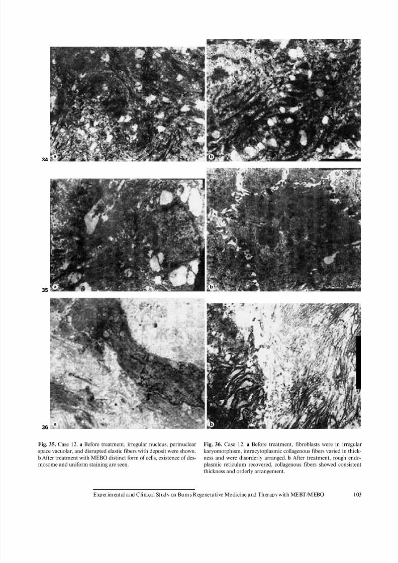
8/3/2019 Luka Bakar Book
http://slidepdf.com/reader/full/luka-bakar-book 109/157
Experimental and Clinical Study on Burns Regenerative Medicine and Therapy with MEBT/MEBO 103
Fig. 35. Case 12. a Before treatment, irregular nucleus, perinuclear
space vacuolar, and disrupted elastic fibers with deposit were shown.
b After treatment with MEBO distinct form of cells, existence of des-
mosome and uniform staining are seen.
Fig. 36. Case 12. a Before treatment, fibroblasts were in irregular
karyomorphism, intracytoplasmic collagenous fibers varied in thick-
ness and were disorderly arranged. b After treatment, rough endo-
plasmic reticulum recovered, collagenous fibers showed consistent
thickness and orderly arrangement.
34
35
36

8/3/2019 Luka Bakar Book
http://slidepdf.com/reader/full/luka-bakar-book 110/157
104 Burns Regenerative Medicine and Therapy
Conclusion
Light- and electron-microscopic observations of 12
cases of deep burns wounds with granulation tissues dem-
onstrated the dynamic comparison of histopathological
alterations before and after MEBO treatment. We noted
especially the changes in fibroblasts, collagenous fibers
and mast cells. No uniform histological changes wereobserved such as are reported in the scientific literature
where burns wounds were treated with conventional sur-
gical therapy resulting in hypertrophic scarring. In our
study, short-term macroscopic observation revealed that
healed burns wounds formed no scars which were ele-
vated from skin surface. This suggests that MEBO offers
excellent prognosis for the treatment of deep second-
degree and superficial third-degree burns wounds without
scarring.
Discussion
Many factors are involved in the wound healing pro-
cess from trauma in general and burns wounds in particu-
lar. Among these factors, the importance of those inherent
to the cell itself has been especially recognized lately by
scholars [3–6]. However, when conducting research to
explore the mechanism of wound healing free of hypertro-
phic scarring, one should attend to histopathology, in oth-
er words, try to understand the alterations of collagenous
fibers, fibroblasts and mast cells [7].
In 1973 and 1974, Linares used light and electron mi-
croscopes for observation and found that hypertrophic
scarring as a result of postburn granulation tissue ap-
peared whirlpool-like or in a nodosity arrangement. In the
early stage after wound healing, there were a paucity of
elastic fibers initially but eventually over years a structure
of lamellar or plexiform shape could be established. Mast
cells took on varied shapes: round, oval or claviform with
deep granular staining. These mast cells released hista-
mine to stimulate the synthesis of collagen and also
secreted many active substances to promote scar prolifer-
ation. The greater the number of mast cells in granulation
tissues, the greater the likelihood of healing with hypertro-phic scarring.
In 1971, using the electron microscope, Gabbiani’s
group noted the presence of fibroblast-like cells in granu-
lation tissues which correlated to wound contraction. In
1980, Rudoph demonstrated that these cells had the same
ultrastructure as fibroblasts and smooth muscle cells.
They named these cells myofibroblasts, the presence of
which in granulation tissues predicted the possibility of
hypertrophic scar formation [7, 8].
In the present study, the authors found absence of
whirlpool-like or nodosity-arranged collagenous fibers
and myofibroblast after MEBO treatment. Small numbers
of mast cells were observed in 2 cases which are consistent
with the number expected in normal skin. Research con-
ducted by He [7] showed the existence of many more mast
cells in hypertrophic scars than in normal skin. Therefore,
the light- and transmission electron-microscopic observa-
tions as well as our pictures after wound healing revealed
wounds free of hypertrophic scars after MEBO treatment.Long-term effects remain to be investigated histologically
as these were short-term observations.
Acknowledgments
The authors would like to thank the Electron Microscopy Depart-
ment of No. 202 PLA Hospital, Burns Department, and the Basic
Research Department of the General Hospital of Benxi Steel Compa-
ny for technical assistance.
References
1 Xu RX: Medical sciences on burns, wounds and ulcers. Chin J Burns
Wounds Surface Ulcers 1994;6:45–51.
2 Zhao JX, Yang GM, Li TY, et al: Clinical analysis of 1003 burn cases
treated with moist exposed burn therapy. Chin J Burns Wounds Surface
Ulcers 1992;4:36–40.
3 Peruccio D, et al: Altered biosynthesis of tumour necrosis factor (TNF)
alpha is involved in post-burn hypertrophic scars. Burns 1994;20:118–
121.
4 Hong ST, et al: Ultrastructure of scars resulted from burns. Metal Med
1985;1:5–8.
5 Wang GS, Jian WG, Xu XS, et al: The exploration of pathological changes
and mechanism of experimental burned rabbits after treatment with moist
exposed burn ointment. Chin J Burns Wounds Surface Ulcers 1992;4:7–
11.
6 Xi SC: Growth regulation of fibroblast. Chin J Burns Wounds Surface
Ulcers 1994;6:5–7.7 He GZ: Pathological changes of hypertrophic scar tissue. Chongqing Med J
1992;21:118–210.
8 Stephen A: Wound contraction and fibrocontractive disorders. Arch Surg
1978;113:1034–1046.
Observation of Microcirculation in Nail Folds at
the Recovery Stage of Burns Wounds Treated
with BRT with MEBT/MEBO
Introduction
From May 1992 to May 1994, we treated 13 cases of
people with nail fold burns and observed the repair of the
microcirculation in the nail folds. The results revealed
that after repair, the microcirculation was normal in
superficial second-degree burns wounds, almost normal
in deep third-degree burns wound and moderately abnor-
mal in mixed second- and third-degree wounds.

8/3/2019 Luka Bakar Book
http://slidepdf.com/reader/full/luka-bakar-book 111/157
Experimental and Clinical Study on Burns Regenerative Medicine and Therapy with MEBT/MEBO 105
Materials and Methods
Object of Observation. 13 young male patients with nail fold burns
were treated with BRT with MEBT/MEBO. As soon as the burns
wounds began healing, the morphology of the microvessels, blood
flow status and repair of the vascular loop surroundings in the nail
fold were observed.
Apparatus. WX-6 multiple position microcirculation photomi-
croscope was manufactured by Xuzhou Factory of Medical Optical
Instruments and supplied by the Department of Pathophysiology of
Binzhou Medical College.
Method of Observation. At a fixed time in the morning, the finger
temperature of the patient was taken using an electron thermometer
at room temperature. The patient was kept in the sitting position
with the arms at the level of the heart. The observation was done
under a mercury lamp light at an angle of 45 ° from the back. Photo-
graphs were taken synchronously and were visualized using a closed-
circuit TV system. If the patient had injured only one hand, the other
hand served as the normal control.
Observation Index. Appearance: vascular clearness, loop number,
input branch, output branch, loop top diameter, loop length, percent-
ages of crossed and deformed loops. Status of blood flow: flow rate,
vasomotion, erythrocyte aggregation, leukocyte number, whitish mi-
cro-thrombus and blood color. Condition at the surroundings of the
loops: exudation, bleeding, sub-papillary venous plexus, papillae and
sweat duct.
Results and Discussion
The microcirculation status was scored and classified
according to the standard for quantitative analysis of nail
fold microcirculation established by Tian [1] (table 58).
Abnormal results were observed in 13 patients (7 cases
with superficial second-degree burns and 6 cases with
deep second-degree burns), and are listed in table 59.
At the initial healing of the wound, the comprehensive
scores of 7 patients with superficial second-degree burns
were in the normal range. Some of them had newly gener-
ated microvessels slightly shorter and smaller than normal
and had exudate in the surroundings. Some individuals
had more leukocytes and their papillae were not clear.
The vessels in the recovery stage demonstrated exudate
and, therefore, papillae were not easy to observe.
Changes in the appearance of the microcirculation of 6
patients with deep second-degree burns wounds were sig-
nificant. Most of the loops were shorter. Two cases had100% crossed loops. One case had a deformed loop ex-
ceeding the normal ratio. As a whole, parts of the micro-
vessels in the nail fold resembled those in the superficial
layer of the skin. Some cases had slower flow rate, which
may be due to the changes of the loop appearance, such as
crossed and deformed loops. One case had remote hemor-
rhage. Three cases had flat papillae, which may be related
to the damage of deep tissue.
This study revealed that the microcirculation was re-
paired after the treatment of burns wounds. This repair
can be attributed to local application of BRT with MEBT/
MEBO and its systemic benefit. MEBO protected the
Table 58. Repair of microcirculation in nail folds of 13 patients
Normal Almost
normal
Slightly
abnormal
Moderately
abnormal
Severely
abnormal
8 1 3 1 0
Table 59. Cases with abnormalities of microcirculation observed at
the recovery stage
Superficial
second-degree
wound (case No.)
Deep second-
degree wound
(case No.)
Clearness 0 0
Loop number 0 2 cases
with 5 loops
Input branch 2 0
Output branch 3 cases withslightly small
0
Loop top 4 cases 0
Loop length 4 cases with
slightly short
4 cases
with short
Crossed loop 0 2
Deformed loop 0 1
Flow rate 0 3
Vasomotion 0 0
Erythrocyte aggregation 0 1 case slight
Leukocyte number 2 cases 130 2 cases 0–50
Whitish microthrombus 0 0
Blood color 0 1 case
with crimson
Exudation 3 0Bleeding 0 1 case with remote
hemorrhage
Sub-papillary venous plexus 0 0
Papillae 2 cases
without clear
3 cases with flat
Sweat duct 0 0
burns wound from dehydration and helped avoid ban-dage pressure. This was favorable to the repair of the
microvessels.
The microcirculation in the normal hands of these
burns patients was examined and nothing abnormal was
found (data not shown). The effect of other diseases could
be excluded.
Reference
1 Tian N (ed): Microcirculation: Basic and Clinical. Beijing, Medical Science
Press of CPLA, 1986, pp 343–345.

8/3/2019 Luka Bakar Book
http://slidepdf.com/reader/full/luka-bakar-book 112/157
106 Burns Regenerative Medicine and Therapy
Physiological Healing Procedure and
Histological Observation on Deep
Second-Degree Burns Treated with BRT with
MEBT/MEBO
Introduction
Moist-exposed burn therapy is a local burns manage-
ment by which burned tissue is exposed to a three-dimen-
sional physiologically moist environment to reject, regen-
erate and repair [1]. We herein present a report on the
dynamic pathomorphological changes of deep second-
degree burns wounds. Studies in this subject are seldom
performed and seldom available.
Materials and Methods
Clinical DataSix cases were enrolled throughout the study, 5 males and 1
female, aged from 21 to 36 years. Total burns surface area varied
from 12 to 45%, with an average of 24.33 B 12.37%. All patients
were admitted within 6 h postburn. After admission, patients
received fluid resuscitation and antibacterial therapy on the basis of
burn severity. Burns wounds were applied with MEBO at a thickness
of 1 mm, which was renewed every 4–6 h till wound healing.
Pathological Observation
Tissue specimens were collected on days 1, 3, 5, 7, 10, 15, and 20
postburn and after healing from wound sites including trunk, upper
limb and both lower limbs. All specimens were allocated into two
parts, one immobilized with 10% formalin, embedded in routine
paraffin, sliced up and HE stained for light-microscopic observation;the other was immobilized with 2.5% glutaraldehyde solution, pre-
pared on routine basis for observation using TEM 1200EZX-model
electron transmission microscope.
Results
Light-Microscopic Observation
Under the light microscope, all burns wounds were
noted to involve deep dermis and the pathological
changes of burns wounds at different time phases were
almost the same.
On day 1 postburn, epidermis became necrotic due tocoagulation and necrotic basal layer cells were seen ar-
ranged in a column-fencing formation. Edema was pres-
ent between epidermis and dermis. Collagenous fibers in
the superficial layer of dermis were denaturated, and
swollen and loosely arranged while epithelia of the dermal
adnexal epithelia were also denaturated. We observed
contracted nucleoli of the capillary endothelium, blood
clot or stasis in lumen with little infiltration of neutrophil
(fig. 37a). Only collagenous fibers and dermal appendages
in deep layer of dermis were approximately normal. On
day 3 postburn, necrotic epidermal cells progressed to
vacuolation with mild edema between the epidermal and
dermal layers. Collagenous fibers in the superficial dermis
had hyaline degeneration while in the deep tissue, the
structure was loose, and blood vessels were slightly swol-
len with congestion. Scattered inflammatory cells (mainly
neutrophils) infiltrated the tissue (fig. 37b). On day 5
37

8/3/2019 Luka Bakar Book
http://slidepdf.com/reader/full/luka-bakar-book 113/157
Experimental and Clinical Study on Burns Regenerative Medicine and Therapy with MEBT/MEBO 107
Fig. 37. a Appearance of deep second-degree burns wound on day 1
postburn. HE. !100. b On day 3 postburn, epidermic cells pro-
gressed to vacuolation, collagenous fibers in superficial dermis had
hyaline degeneration and scattered inflammatory cells infiltrated in
dermis. HE.!100. c On day 5 postburn, superficial tissues in dermis
were liquefactively necrotic and exfoliated. Inflammatory cells,
mainly neutrophil and occasionally with few macrophages, infil-
trated. HE.!100. d On day 10 postburn, the appearance of epithelia
islands was seen. HE.!100. e On day 10 postburn, sweat glands in
dermis proliferated with the infiltration of inflammatory cells all
round, mainly neutrophils, as well as many lymphocytes and mono-
nuclear macrophages. HE.!100. f On day 15 postburn, the adnexal
epithelia in deep dermis grew vertically from the deep, then migrated
toward and covered the wounds. HE. !100. g Most wounds werecovered with stratified squamous epithelium. Actively proliferated
fibroblasts and neoformative capillaries were visible in the dermis,
accompanied by the infiltration of lymphocytes. HE. !100. h Neo-
formative skin has an approximately normal structure. HE.!100.
when the necrotic epidermis exfoliated, superficial tissue
in the dermis liquefied and loosened due to necrosis and
neutrophilic infiltration (occasional lymphocyte and
mononuclear macrophages) (fig. 37c). On day 7 postin-
jury, the aforementioned pathological changes became
more notable. On day 10, the superficial necrotic tissue in
the dermis liquefied, discharged and thinned. A large
amount of neutrophils infiltrated into the liquefied ne-
crotic tissue, interwoven with few mononuclear macro-
phages. The dermal adnexal epithelia in the underlying

8/3/2019 Luka Bakar Book
http://slidepdf.com/reader/full/luka-bakar-book 114/157
108 Burns Regenerative Medicine and Therapy
deep tissue revealed squamous metaplasia with enlarged
cells, deep karyon, rich and red cytoplasm that clustered
to form ‘epithelium islands’ (fig. 37d). Among these is-
lands, fibroblasts proliferated and were characterized by
large volume of cells, conspicuous nucleolus (binucleolate
was frequently present) and basophilic cytoplasm. In ad-
dition, infiltration of many inflammatory cells includingneutrophils (mainly), lymphocytes and mononuclear
macrophages was visible in wounds (fig. 37e). On days 15
and 20 postburn, an inflammatory exudation layer re-
placed the necrotic layer whose liquefaction was now
accomplished. The underlying residual adnexal epithelia,
fibroblasts and endothelia showed active proliferation.
The regenerated ‘epithelial islands’ grew vertically, then
migrated toward and covered the wounds (fig. 37f, g).
Neoformative capillaries were noticed and granulation
tissues were formed. The infiltration of inflammatory
cells (mainly lymphocytes) in the dermis was still present,
especially in the periphery of the regenerated skin ap-pendages. After healing, regenerated skin appeared to be
almost normal in structure and the majority of skin
appendages were restored completely. Infiltration of cer-
tain inflammatory cells and few macrophages in dermis
persisted (fig. 37h).
Electron-Microscopic Observation
Ultrastructural alterations of the superficial dermis
layer were observed on days 1, 3 and 5 postburn, while
those of full-thickness skin were done on days 7, 10, 15
and 20 postburn and after healing.
On day 1 postburn, electron-microscopic observation
indicated loose and disorderly arranged collagenous fibers
in the superficial dermis, and a distinct and recognizable
light-dark zone of the fasciculus. Fibroblasts which had a
cytomembrane profile which remained the same although
being deprived of the cell organ. Nuclear membrane
loomed with increscent nucleopore to cause the interpola-
tion of nuclein and chromatin, leading to lumpish chro-
matin. Microsangial endothelia had similar changes as
did the fibroblasts. The vascular walls appeared as a
lamellar membranous structure. In the lumen, we noted
hemagglutination, frequently joined with blood plateletadherence to the vascular walls (fig. 38a). On day 3 post-
burn, collagenous fiber bundles in the superficial dermis
varied in thickness and appeared disorderly with faint or
absent light-dark zones. The cell ultrastructure interwo-
ven between fibers disappeared. On day 5 postburn, the
superficial collagenous fibers merged to produce floccules
with high electron density surrounded by scattered elec-
tron-dense organelle-like structures and necrotic cell frag-
ments as well as occasionally by varied lower electron-
dense lipid droplets without coating of the limiting mem-
brane (fig. 38b). On day 7, when the ultrastructural
change of the superficial dermis was similar to that on
38
day 5, deep collagenous fibers and the structure of dermal
adnexal epithelia nearly returned to normal, except for a
slight broadening of the intercellular space. On day 10
postburn, fibroblasts in the dermis showed condensed
nuclear chromatin, hypertrophic and shift aside nucleoli.
Rough endoplasmic reticulum (RER) proliferated show-
ing flat vesicular and vesicular-like expansion, while re-
sidual dermal adnexal epithelia were distributed in clus-
ters containing a high amount of tonofibrils (fig. 38c). Ondays 15 and 20 postburn, collagenous fiber bundles of der-
mis varied in thickness and appeared disorderly; fibro-
blasts were binucleated and showed obvious nucleoli with
the presence of karyosome; perinuclear pool demon-
strated beading swelling and the RER proliferated
(fig. 38d). In dermal adnexal epithelia, nucleoli were dis-
tinct and increased numbers of karyosomes were fre-
quently observed.
The chondriosome was rich and intracytoplasmic to-
nofibril showed an increase in cytoplasmic presence. Also,
we observed a gradual establishment and improvement of
the intercellular desmosomal junction. After healing, neo-

8/3/2019 Luka Bakar Book
http://slidepdf.com/reader/full/luka-bakar-book 115/157
Experimental and Clinical Study on Burns Regenerative Medicine and Therapy with MEBT/MEBO 109
Fig. 38. a Vascular wall appeared in the lamellar membranous struc-
ture. In the lumen, there was hemagglutination, frequently joined
with blood platelet adhering to vascular walls. TEM. !6,000. b Col-
lagenous fibers fused each other, occasionally with cell collapse.
TEM. !6,000. c Dermal adnexal epithelia distributed in clusters,
with near-membrane nucleolus and expanded endoplasmic reticu-
lum. Many tonofibrils are contained in the cytoplasm. TEM.
!6,000. d On day 15 postburn, fibroblasts showed active protein
synthesis and metabolism. TEM.!6,000. e After wound healing, the
entire dermis-epidermis junction can be seen. TEM. !8,000. f Fi-
broblasts in dermis showed normal and collagenous fibers were uni-
form in thickness and arranged in an orderly fashion (after healing).TEM.!10,000. g The healing skin after deep second-degree burns is
almost the same as noninjured skin.
formative epidermis cells and intercellular junction re-
covered almost to normal and the integrated dermis-epi-
dermis junction reappeared (fig. 38e). The majority fibro-
blasts in the dermis appeared as a long strip rich in cyto-
plasm with well-developed organelles. However, no addi-
tional active metabolism of the same was observed. Fibro-
blasts were uniform in thickness, fasciculated and well
orientated (fig. 38f). The healing skin of deep second-
degree wounds became almost identical to that of nonin-
jured skin (fig. 38g).

8/3/2019 Luka Bakar Book
http://slidepdf.com/reader/full/luka-bakar-book 116/157
110 Burns Regenerative Medicine and Therapy
Conclusion
Pathological changes of the deep second-degree burn
wounds treated with BRT with MEBT/MEBO can be
divided into three stages: (1) denaturation and necrosis,
(2) liquefaction, and (3) restoration. These stages may
overlap. Burns wounds healed by complete physiological
regeneration.
Discussion
Among the available literature regarding the pathologi-
cal changes of burns wounds treated with BRT with
MEBT/MEBO, the majority report the light microscopic
observation before and after treatment with MEBT [2–4].
There are relatively few monographs or systemic study
reports on the ultrapathologic processes during BRT with
MEBT/MEBO.
Our study demonstrated that the pathological changes
of burns wounds treated with BRT with MEBT/MEBOwere totally different from pathological processes when
treated conventionally [5, 6]. On days 1–3 postburn, the
so-called ‘acute inflammatory response period’ with its
typical denaturation and necrosis occurred in the burned
epidermis and superficial dermis, revealing a slight in-
flammatory response, but without signs of a ‘leukocyte
infiltration zone’. Then the necrotic tissue began to lique-
fy and discharge increasingly as the disease course pro-
gressed. Such changes climaxed around day 10 when der-
mal adnexal epithelial cells, fibroblasts as well as other
repaired cells showed signs of regeneration. Microscopic
observation indicated large cell bodies of fibroblasts,
increased basophil of the cytoplasm, hypertrophic nu-
cleoli, and RER which proliferated, demonstrating the
typical flat vesicular and vesicular-like expansion. At this
time, the proliferation and migration of residual dermal
adnexal epithelia and proliferation of granulation tissues
became dominant. The most spectacular observation was
that the ‘epithelia island’ (formed by the regenerated
adnexal epithelia) initially grew vertically, then migrated
toward and covered the wound as granulation tissues pro-
liferated. This resulted in a flat skin surface. During this
period, either residual adnexal epithelia or fibroblasts dis-played ‘active protein synthesis and metabolism’ [7]. The
prominent characteristic changes involved significant
proliferation of RER and the appearance of increased
karyosomes. Finally, the wounds were noted to be com-
pletely covered by regenerated squamous epithelia as
healing was accomplished.
We observed that inflammatory cells demonstrated a
series of responses during wound healing. On day 1 post-
burn, we observed infiltration of neutrophils into the der-
mis followed by lymphocytes and mononuclear macro-
phages. This infiltration increased on day 3. Neutrophils
were the predominant infiltrating cells in necrotic tissue,
and their numbers grew or declined depending upon the
length of liquefaction time. Meanwhile, lymphocytes and
mononuclear macrophages, mainly located in the residual
dermis, dramatically increased during the period of time
that necrotic tissue was being rejected and wound repair
was initiated. These cells remained the dominant cells
impacting wound repair subsequent to the liquefactionperiod. This study does not resolve questions as regards
the significance of the space-time distribution of these
cells nor does it answer definitively questions about
sequential changes. These questions merit further study.
However, we do note the remarkable truth that, during
wound healing, the function and activity of the above cells
directly or indirectly, alone or cooperatively, participated
in and regulated wound healing.
The study results indicated a staged pathological
change of burns wounds when treated with BRT with
MEBT/MEBO. Wounds changed in three stages accord-
ing to the different time phase postburn: (1) denaturationand necrosis stage, (2) liquefaction stage, and (3) stage of
repair by regeneration, which may overlap during wound
healing.
The first stage began subsequent to burn injury till day
3 and the pathomorphological change was characterized
by the denaturation and necrosis of burns tissue from
which we derived the name ‘denaturation and necrosis
stage’. We postulated that these changes were the result of
direct thermal exposure, local microcirculation blockage
and other secondary injuries. The minor inflammatory
response at this stage had a close correlation with the
effects of MEBT/MEBO. On day 5 postburn, wound
liquefaction became conspicuous and climaxed on day
10. After this time, liquefaction diminished and we noted
less necrotic tissue. The inflammatory response, however,
remained correlated with the liquefaction of necrotic tis-
sue. Repair by regeneration was noted on day 10 postburn
and was manifested mainly by active proliferation of
residual dermal adnexal epithelia. This stimulated the
proliferation of peripheral fibroblasts and endothelia, and
further formed granulation tissues, leading to final heal-
ing by epithelization. After experiencing further differen-
tiation and reformation, the regenerated skin finally at-tained the structure of normal skin. This study demon-
strates that the effect of BRT with MEBT/MEBO deliv-
ered similar pathological changes for both deep second-
degree and third-degree burns wounds. The only differ-
ence lay in the ultimate healing modes.
Remarkably, this study revealed that the healed
wounds appeared as flat fully regenerated skin, featuring
restored, viable hair, almost normal skin elasticity and
no scar formation. Histomorphological observation con-
firmed that the epidermal cells of neoformative skin had
the equivalent structure as normal epidermis and that the
epidermis had good joints with dermis papilla. The healed

8/3/2019 Luka Bakar Book
http://slidepdf.com/reader/full/luka-bakar-book 117/157
Experimental and Clinical Study on Burns Regenerative Medicine and Therapy with MEBT/MEBO 111
skin demonstrated a fully functional and integrated der-
mis-epidermis junction. The initially infantile and active
fibroblasts gradually grew into stable fibrocytes. Arterio-
lae and venulae with thick walls and integrated structure
replaced the neoformative capillaries. Collagenous fibers
were uniform in thickness and orientated fascicularly,
without the presence of whirl-like and nodular arrange-ment. Follicles, sweat glands and sebaceous glands as well
as other dermal accessory organs also regenerated com-
pletely. Therefore, we concluded that treatment of deep
second-degree burns wounds with BRT with MEBT/
MEBO results in complete physiological regeneration
with minimal scar formation.
References
1 Xu RX: Brief introduction on Chinese moist medical science for burns.
Chin J Burns Wounds Surface Ulcers 1996;8:1–6.
2 Wang GS, Jian WG: Pathological changes of rabbit with experimental
burns treated by MEBO and investigation of the mechanism of action.Chin J Burns Wounds Surface Ulcers 1992;4:7–11.
3 Zhang XZ, Cheng SR: A case report: Electron microscopic observation on
burn wound treated with MEBO. Chin J Burns Wounds Surface Ulcers
1992;4:6–9.
4 Zhao YL, Wang Y: Electron microscopic observation on the effect of
MEBO in treating deep burn wound. Chin J Burns Wounds Surface Ulcers
1995;7:1–5.
5 Fang ZY, Wu ZL: Burn Theory and Practice. Shenyang, Liaoning Science
and Technology Publishing House, 1989, pp 12–14.
6 Fu XB, Wang DW: Basis of Trauma Repairing. Beijing, People’s Military
Medical Press, 1997, pp 14–30.
7 Wu ZB: Basis of Ultra-Micro-Pathology. Beijing, People’s Health Press,
1990.
Clinical Procedure and Histological Observation
of Full-Thickness Burns Treated with BRT with
MEBT/MEBO: A Case Report
Introduction
A 20-year-old female patient sustained a 35% TBSAburn secondary to exposure to gasoline fire (15% deep
partial-thickness and 20% full-thickness loss). The patient
was hospitalized at 12 h postburn and was diagnosed as
suffering with full-thickness loss (third-degree) burns on
both lower extremities. The epidermis was necrotic and
detached and the dermal layer was degenerated and
necrotic with a waxy yellow and waxy white appearance
(fig. 39a). The pathological section examination of the
sampled local wound tissues revealed necrosis of full-
thickness epidermis and dermis, degeneration and struc-
tural disturbance of collagenous fibers in dermis, and
microcirculation stasis (fig. 39b).
Results
After admission, the patient was treated initially with
MEBO burns ointment to protect burn tissue before we
performed skin cultivation and relief as per our BRT pro-
tocol. At 48 h postburn, the wounds began to liquefy and
the liquefaction was complete by day 4 (fig. 40). The
liquefied products were gently removed from the woundsurface before MEBO was reapplied every 3–4 h.
Repeat biopsy at the same burned location was per-
formed for pathological examination. The results showed
massive granular tissues among the necrotic epithelial tis-
sue, a proliferation of newly regenerated epithelial cells
with collagenous fibers, as well as the typical skin em-
bryonic base (EB) (fig. 41a, b). After a 10-day application
of MEBO, the comparably primitive epithelial tissues
were observed under pathological examination of epithe-
lial tissues sampled from the wound edge.
Fig. 39. a A 20-year-old female patient sustained a 35% TBSA burn
secondary to exposure to gasoline fire (15% deep partial-thickness
and 20% full-thickness loss). b Necrosis of full-thickness epidermis
and dermis, degeneration and structural disturbance of collagenous
fibers in dermis, and microcirculation stasis. HE.!20.

8/3/2019 Luka Bakar Book
http://slidepdf.com/reader/full/luka-bakar-book 118/157
112 Burns Regenerative Medicine and Therapy
Fig. 40. On day 4, the wounds began to liquefy under the action of
MEBO, and the wound skin began to regenerate.
At 20 days post-treatment with MEBO, the pathologi-
cal examination of deep burns wounds tissue showed the
presence of the newly formed intact stratified squamous
epithelium. The epithelial cells of the superficial layer
appeared normal. Appearance of the collencytes and mi-
croangium in the dermis layer was typical. On day 30
post-treatment, epithelial tissues showed a remarkabledegree of regeneration (fig. 42a), and skin structure was
almost normal (fig. 42b).
Immunohistochemical Examination
Twenty days post-treatment with BRT with MEBT/
MEBO, wound tissue was examined and the results
showed the clear appearance of collagenous fibers in epi-
thelial tissue and subcutaneous tissue. Argentaffin stain
Fig. 41. a Group proliferation of newly regenerated epithelial tissues
and collagenous fibers as well as typical skin embryonic base (EB).
b The skin EB formed by microangium, collagenous fibers and epi-
thelium (stem cell) in newly regenerated epithelial tissues. HE.!40.
Fig. 42. a On day 30 post-treatment, wound tissue showed good pro-
cess of regeneration. b Day 30 post-treatment, epidermis and dermis
showed physiological structure.

8/3/2019 Luka Bakar Book
http://slidepdf.com/reader/full/luka-bakar-book 119/157
Experimental and Clinical Study on Burns Regenerative Medicine and Therapy with MEBT/MEBO 113
indicated active regeneration of reticular fibers. The re-
ticular fibers around the small blood vessels of subcuta-
neous tissue appeared relatively normal. The basal cells of
epidermis regenerated actively (fig. 43a, b).
Thirty days after BRT with MEBT/MEBO treatment,
the regenerated and repaired epithelial tissue was AE3
stained. The pictures showed positive proteins of squa-mous epithelium, brown-stained cytoplasm and blue-
stained nucleus of granular cells in epidermis (fig. 43c, d).
AE1 stain showed negative proteins of glandular epi-
thelium, which is representative of glandular epithelium
already transformed into squamous epithelium (fig. 43e).
Electron-Microscopic Observation
On the day of injury, the epithelium showed necrotic
degeneration, and the monocytes showed a clear nuclearshift with karyopyknosis and even phagocytosis (fig. 44).
Five days post-treatment with MEBO, we noted active
growth of fibrocytes and fibroblasts (fig. 45).
Fig. 43. a Active regeneration of the basement membrane in the basal
lamina of the epidermis. Argentaffin.!40. b Reticular fibers around
the small blood vessel in the dermis stained black. Argentaffin.!40.
c Positive protein of squamous epithelium (indicating spontaneous
self-regeneration and repair). AE3.!20. d Same as figure 46a. AE3.
!40. e Negative protein of glandular epithelium. AE1.!20.
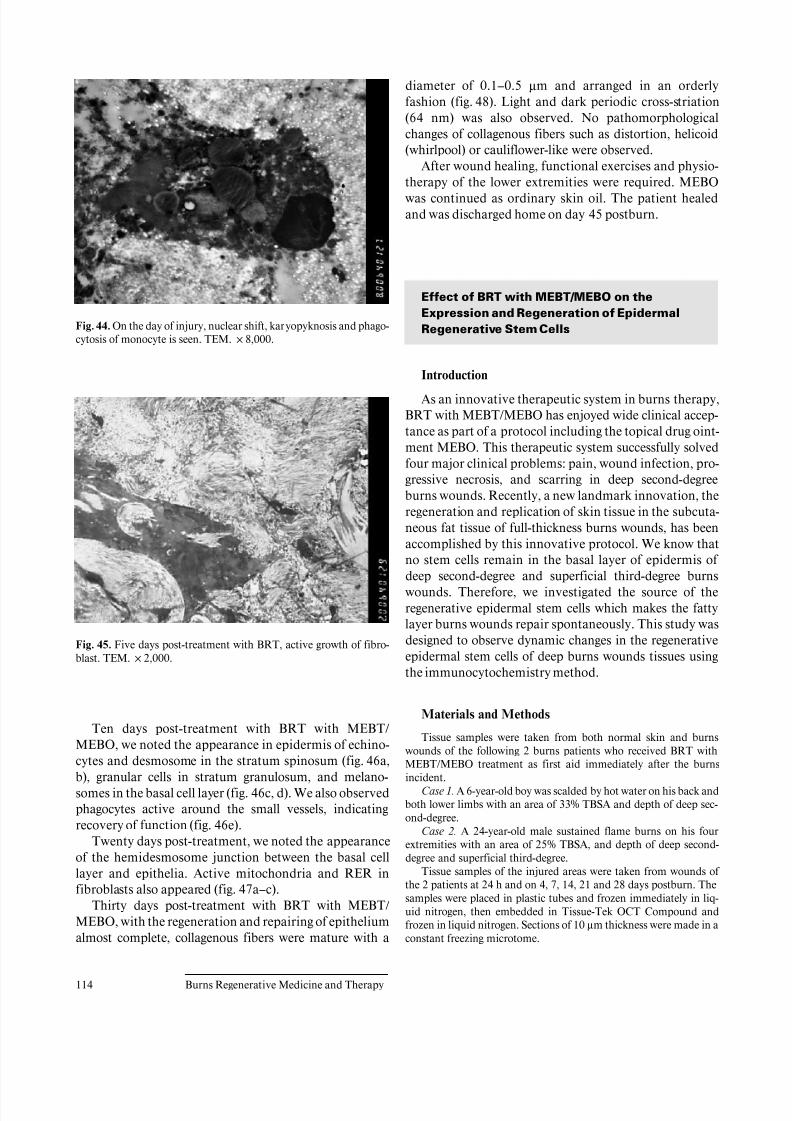
8/3/2019 Luka Bakar Book
http://slidepdf.com/reader/full/luka-bakar-book 120/157
114 Burns Regenerative Medicine and Therapy
Fig. 44. On the day of injury, nuclear shift, karyopyknosis and phago-
cytosis of monocyte is seen. TEM.!8,000.
Fig. 45. Five days post-treatment with BRT, active growth of fibro-
blast. TEM.!2,000.
Ten days post-treatment with BRT with MEBT/
MEBO, we noted the appearance in epidermis of echino-cytes and desmosome in the stratum spinosum (fig. 46a,
b), granular cells in stratum granulosum, and melano-
somes in the basal cell layer (fig. 46c, d). We also observed
phagocytes active around the small vessels, indicating
recovery of function (fig. 46e).
Twenty days post-treatment, we noted the appearance
of the hemidesmosome junction between the basal cell
layer and epithelia. Active mitochondria and RER in
fibroblasts also appeared (fig. 47a–c).
Thirty days post-treatment with BRT with MEBT/
MEBO, with the regeneration and repairing of epithelium
almost complete, collagenous fibers were mature with a
diameter of 0.1–0.5 Ìm and arranged in an orderly
fashion (fig. 48). Light and dark periodic cross-striation
(64 nm) was also observed. No pathomorphological
changes of collagenous fibers such as distortion, helicoid
(whirlpool) or cauliflower-like were observed.
After wound healing, functional exercises and physio-
therapy of the lower extremities were required. MEBOwas continued as ordinary skin oil. The patient healed
and was discharged home on day 45 postburn.
Effect of BRT with MEBT/MEBO on the
Expression and Regeneration of Epidermal
Regenerative Stem Cells
Introduction
As an innovative therapeutic system in burns therapy,
BRT with MEBT/MEBO has enjoyed wide clinical accep-
tance as part of a protocol including the topical drug oint-
ment MEBO. This therapeutic system successfully solved
four major clinical problems: pain, wound infection, pro-
gressive necrosis, and scarring in deep second-degree
burns wounds. Recently, a new landmark innovation, the
regeneration and replication of skin tissue in the subcuta-
neous fat tissue of full-thickness burns wounds, has been
accomplished by this innovative protocol. We know that
no stem cells remain in the basal layer of epidermis of deep second-degree and superficial third-degree burns
wounds. Therefore, we investigated the source of the
regenerative epidermal stem cells which makes the fatty
layer burns wounds repair spontaneously. This study was
designed to observe dynamic changes in the regenerative
epidermal stem cells of deep burns wounds tissues using
the immunocytochemistry method.
Materials and Methods
Tissue samples were taken from both normal skin and burns
wounds of the following 2 burns patients who received BRT withMEBT/MEBO treatment as first aid immediately after the burns
incident.
Case 1. A 6-year-old boy was scalded by hot water on his back and
both lower limbs with an area of 33% TBSA and depth of deep sec-
ond-degree.
Case 2. A 24-year-old male sustained flame burns on his four
extremities with an area of 25% TBSA, and depth of deep second-
degree and superficial third-degree.
Tissue samples of the injured areas were taken from wounds of
the 2 patients at 24 h and on 4, 7, 14, 21 and 28 days postburn. The
samples were placed in plastic tubes and frozen immediately in liq-
uid nitrogen, then embedded in Tissue-Tek OCT Compound and
frozen in liquid nitrogen. Sections of 10 Ìm thickness were made in a
constant freezing microtome.

8/3/2019 Luka Bakar Book
http://slidepdf.com/reader/full/luka-bakar-book 121/157
Experimental and Clinical Study on Burns Regenerative Medicine and Therapy with MEBT/MEBO 115
Fig. 46. a 10 days post-treatment. Desmosome junctions among four
echinocytes. TEM. !3,500. b Desmosome 10 days post-treatment
with MEBO. TEM. !20,000. c Granular cells 10 days post-treat-
ment with BRT with MEBT/MEBO. TEM. !20,000. d Melano-
somes in basal cell layer 10 days post-treatment with BRT with
MEBT/MEBO. TEM.!6,000. e 10 days post-treatment, phagocytes
show active phagocytosis. TEM.!6,000.
Indirect immunofluorescence staining with a biotin-avidin DCS
system was performed. The frozen section was incubated with 10%
horse serum at 4 °C for 20 min, then a diluted (1:20) solution of
mouse anti-human keratin type 19 monoclonal antibody (the 1st
antibody) was added and the mix was again incubated overnight at
4°C. Subsequent to washing with phosphate buffer solution, the sec-
tion was added to 7.5 Ìg/ml of biotinized horse anti-mouse IgG anti-
body (the 2nd antibody, Vector Laboratories, Burlingame, Calif.,
USA) and incubated at 4 °C for 1 h. After another washing, 10 Ìg/ml
of biotin-avidin DCS (Vector Laboratories) was added and incubated
at 4°C for 1 h. The section was rinsed and then mounted with glycer-
in containing 10% PBS and 1% p-phenylenediamine. A control sec-
tion of normal skin was stained in the same way, but without adding
the 1st antibody. All specimens were observed under an Olympus
reflecting fluorescence microscope (Japan) and photos were taken
using ASA400 KODAK films.

8/3/2019 Luka Bakar Book
http://slidepdf.com/reader/full/luka-bakar-book 122/157
116 Burns Regenerative Medicine and Therapy
Fig. 47. a 20 days post-treatment, the epithelia adhered with hemi-
desmosome and fibroblast in the basal cell layer. TEM.!3,000. b 20
days post-treatment, active mitochondrion and rough endoplasmic
reticulum in the fibroblast. TEM. !17,000. c Same as figure 47b.
TEM.!20,000.
Fig. 48. 30 days post-treatment with BRT with MEBT/MEBO, the
collagenous fibers are in orderly arrangement with a diameter of 0.1–
0.5 Ìm and have light and dark periodic cross-striation (64 nm).
TEM.!20,000.
Results
Immunocytochemical examinations were made on
normal skin and burns wounds tissue sections treated
with specific mouse anti-human keratin type 19 mono-
clonal antibody. The results revealed that in the normal
controls of both cases, there were few positive numbers of
epidermal stem cells with keratin type 19 (fig. 49a).
Wound tissue at 24 h postburn showed a moderate
amount of positive epidermal regenerative stem cells
(fig. 49b) and on day 4 postburn, the number of positive
epidermal stem cells around the sweat gland, capillaries
and hair follicles increased (fig. 49c). On days 7 (fig. 49d)
and 14 (fig. 49e), epidermal stem cells containing human
keratin type 19 continually increased and exceeded the
level attained at day 4 postburn, before gradually reaching
a peak level. Prior to days 21 (fig. 49f) and 28 (fig. 49g)
postburn, the number of positive regenerative stem cells
decreased to a certain level as the burns wounds pro-
gressed to healing. The observation showed that, aftertreatment with BRT with MEBT/MEBO, the prolifera-
tion status of the potential regenerative stem cell of the
burns patients changed at a regular rate. We propose that
regenerative stem cells may be the source of epidermal
regenerative stem cells. The glowing fluorescent cells
found observed under the microscope represented the
potential regenerative stem cells in the wound tissues of
deep second- and superficial third-degree burns. After
treatment with MEBO, these stem cells may aid the deep
partial thickness burns wounds to heal without scar for-
mation and aid the superficial full-thickness burns wound
to regenerate skin while healing spontaneously.

8/3/2019 Luka Bakar Book
http://slidepdf.com/reader/full/luka-bakar-book 123/157
Experimental and Clinical Study on Burns Regenerative Medicine and Therapy with MEBT/MEBO 117
Fig. 49. Stained frozen sections using mouse anti-human keratin type
19 monoclonal antibody. Biotin-avidin DCS system, indirect immu-
nofluorescence technique.!200. a Normal skin. No keratin type 19
positive cells. b 24 h postburn. After treatment with BRT, a moderate
amount of keratin type 19 positive cells can be seen. c On day 4 post-
burn after treatment with MEBO, keratin type 19 positive cells
increased. d After treatment with BRT with MEBT/MEBO, on day 7
postburn, the number of keratin type 19 positive cells reached a peak.
e On day 14 postburn after treatment, the number of keratin type 19
positive cells remained at the same peak level as on day 7. f After
treatment with BRT with MEBT/MEBO, on day 21 postburn, the
number of keratin type 19 positive cells decreased. g After treatment
with BRT with MEBT/MEBO, on day 28 postburn, the number of keratin type 19 positive cells decreased significantly.

8/3/2019 Luka Bakar Book
http://slidepdf.com/reader/full/luka-bakar-book 124/157
118 Burns Regenerative Medicine and Therapy
Conclusion
BRT with MEBT/MEBO promotes the activation and
proliferation of epidermal regenerative stem cells in the
residual viable tissue of superficial full-thickness burns
wounds, and these stem cells play a unique role in the
spontaneous wound healing of deep burns.
Discussion
Research on the cell cycle has revealed that cell divi-
sion is closely related to physiological regeneration and
wound repair [1–11]. Some cells stay at phase G0 or G1 for
a long time and will not proliferate unless the condition
becomes favorable. But some cells can undergo contin-
uous division yielding daughters destined to differentiate
to mature cells, while others retain their ability to contin-
uously proliferate. These are termed ‘stem cells’. Stem
cells in the basal layer of the epidermis are capable of pro-liferating continuously. Newly proliferated cells move up-
wards until reaching the deep area of the stratum spino-
sum layer, where they replicate two or three times before
losing their proliferative ability.
In deep second- and third-degree burns wounds, the
whole epidermis and deep part of dermis are damaged so
all resident stem cells in the basal layer of epidermis are
destroyed. The residual viable mesenchymal cells around
the hair follicles, sweat glands and capillaries in subcuta-
neous tissue may provide a source of available regenera-
tive epidermal stem cells. These stem cells can synthesize
specific cellular keratin type 19, and therefore can be
identified immunocytochemically. In this study, anti-
human keratin type 19 monoclonal antibody was used.
With the biotin-avidin DCS system and the indirect
immunofluorescence technique, specific and exact detec-
tion of residual epidermal regenerative stem cells in the
subcutaneous tissue of deep second- and superficial third-
degree burns wounds is accomplished. We observed the
number of potential regenerative stem cells with the posi-
tive label of human keratin type 19 reaching the peak level
on 7–14 days postinjury in burns wounds treated with
BRT with MEBT/MEBO. The immunofluorescent cellswere epidermal stem cells, which induced the fatty layer
burns wound to repair spontaneously. Therefore, we con-
clude that BRT with MEBT/MEBO treatment activates
the dormant potential epidermal regenerative stem cell to
proliferate, thereby ensuring the spontaneous repair and
healing of deep burns wounds without scar formation.
BRT with MEBT/MEBO treatment eliminates the life-
time of pain caused by the hyperplastic scars of patients
with deep burns.
After transformation and differentiation, the epider-
mal regenerative stem cells can yield cells capable of syn-
thesizing other types of keratin, i.e. keratin types 9 and 16.
These cells still have the ability of transformation and can
transform into the cells capable of synthesizing harder
keratin – types 1 and 10. These are the typical types of
keratin contained in mature epidermal cells. This finding
proved that MEBO has the effect of promoting the activa-
tion, proliferation and transformation of epidermal stem
cells.It has been reported that when the cells are awoken
from their dormant phase, the first activated protein is
cyclin D. We know that cyclin D can only be expressed
after being stimulated by growth factors. For eukaryotes,
cells at phase G1 can either enter into the proliferation
state or withdraw from the cell cycle. The main regulator
of the phase G1 limiting point is the cyclin D1/CDK 4 com-
plex. In order to further investigate the mechanisms of the
effect of BRT with MEBT/MEBO, we will continue our
research on the gene regulation for the proliferation cycle
of epidermal regenerative stem cells.
References
1 Xu RX: Conspectus of Chinese moist burn therapy. Chin J Burns Wounds
Surface Ulcers 1996;8:1–7.
2 Zhao JX: Experience with MEBT/MEBO in treating burn wounds. Chin J
Burns Wounds Surface Ulcers 1999;11:16–17.
3 Xiao M: Clinical observation on MEBT/MEBO promoting severe burn
wounds to regenerate and recover. Chin J Burns Wounds Surface Ulcers
1999;11:18–20.
4 Xu ZL, Parker SB, Minkoff R: Distribution of type IV collagen, laminin,
and fibronectin during maxillary process formation in the chick embryo.
Am J Anat 1990;187:232–236.
5 Grana X, Reddy EP: Cell cycle control in mammalian cells: Role of cyclins,
cyclin dependent kinases (CDKS), growth suppressor genes, and cyclin-
dependent kinase inhibitors (CKIS). Oncogene 1995;11:211–215.6 Fuchs E, Green H: Multiple keratins of cultured epidermal cells are trans-
lated from different MRNA molecules. Cell 1979;17:573–577.
7 Lindberg K, Rheinwaid JG: Suprabasal 40 kD keratin (K19) expression as
an immunohistologic marker of premalignancy in oral epithelium. Am J
Pathol 1989;134:89–98.
8 Bartek J, Bartkova J, Schneider J, et al: Patterns of expression of keratin 19
as detected by monoclonal antibodies in human breast tissues and tumors.
Int J Cancer 1985;36:299–306.
9 Cohen IK, Mc Coy BJ: Wound healing; in Goldsmith IA (ed): Biochemistry
and Physiology of the Skin. New York, Oxford University Press, 1983,
vol 1, pp 462–474.
10 Zhang SQ, Wang YC (eds): Cell Cycle Control: Tendency of Cell Biology.
Beijing, Beijing Normal University Press, 1996, vol 1, pp 46–60.
11 De Cherney AH, Dougherty WR, Rodgers KE, et al: Expression of cyclin
protein after thermal skin injury in a guinea pig model. J Burn Care Rehab
1997;18:292–296.

8/3/2019 Luka Bakar Book
http://slidepdf.com/reader/full/luka-bakar-book 125/157
OOOOOOOOOOOOOOOOOOOOOOOOOOOOOOOOOOOOOOOOOOOOOOOOOOOOOOOOOOOOOOOOOOOOOOOOOOOOOOOOOOOOOOOOOOOOOOOOOOOOOOOOOOOOOOOOOOOOOOOOOOO
Clinical Reports of Burns Regenerative Medicine and
Therapy (MEBT/MEBO)
119
Clinical Trial Report of Burns Regenerative
Medicine and Therapy (MEBT/MEBO):
Multicenter Study
Introduction
In order to further examine and evaluate the thera-
peutic effects, main indications and possible side effects
of BRT (MEBT/MEBO) in burns treatment, this multi-
center study was designed to conduct a MEBO phase III
clinical trial. In this trial, MEBO was used for treating
vast numbers of burns patients of both sexes and varied
ages, all suffering burns from different causes, occurring
at different sites, encompassing varied areas and penetrat-
ing to varied depths.
The phase III trail was carried out at five branch cen-
ters of the China National Science and Technology Center
for Burns, Wounds and Ulcers. These centers are the
departments of five general hospitals located, respective-
ly, in the cities of Changsha, Taishan, Dalian, and Na-
nyang, China. The trail period spanned January 1 1996 to
June 30 1999.
Materials and Methods
Clinical Data
General Information
Five hundred and eight hospitalized burns patients were ob-
served. They were divided randomly into two groups: treatment
group (MEBO group) and control group (silver sulfadiazine (SD-Ag)
group). There were 363 patients in the treatment group, 282 males
and 81 females (male:female = 3.5:1), aged 10 days to 73 years (aver-
age 28.4B 15.5 years), who were treated with MEBO. 145 patients
were in the control group, 122 males and 23 females (male:female =
5.3:1), aged 8 months to 72 years (average 29.2B 12.1 years).
Causes of Burns
The most frequent causes of burns injury in this trial were flame
and hot liquid – 211 cases (41.5%) and 202 cases (39.8%), respective-ly. 58 burns were caused by chemicals (11.4%), and among these
chemical burns, 28, 26, and 4 cases were injured by acid (5.5%),
alkali (5.1%), and phosphorus (0.8%), respectively. 22 cases had elec-
tric burns, accounting for 4.3%. The remaining 15 cases (3.0%) were
accidental scalding by hot solid metal. The burn causes of both the
treatment and the control groups were comparable.
Anatomical Regions of Burns
The anatomical regions of burns are shown in table 60. In both
groups, the burns often involved the extremities, hands or the head
and neck regions of the body. The predilection for these smaller ana-
tomic areas sustaining the majority of the burns may be understood
since there are: (1) the most easily exposed at the time of burn,
(2) the upright nature of the human being, and (3) the use of these
extremities in attempts to extinguish the fire or remove scalding
clothing.
Patient Assessment
Extent and Depth of Burns Wounds (table 61). The largest burns
area was 94% TBSA, and the largest third-degree wound was 82%
TBSA. In 508 cases of this trial, there were 106 (20.9%) extensive
burns patients with TBSA 150%, 75 cases in the treatment group and
31 cases in the control group. The wounds show no significant differ-
ences between the two groups (p 1 0.05).
Burn Severity. According to the national standard of classifica-
tion, the burn severity of the patients is shown in table 62. There are
no obvious differences between the two groups (p 1 0.05).
Burn Management
Local Wound Treatment
Generally, exposure therapy was adopted in both groups. The
occlusive method was used unless the individual requirement con-
traindicated it. In both groups, the superficial and deep partial-thick-
ness wounds were permitted to heal spontaneously. Skin grafting
could be performed in order to repair full-thickness burns wounds at
the appropriate time.
In the treatment group, patients received standard burns regener-
ative therapy (MEBT) throughout the treatment procedure, with
MEBO ointment being applied to the wounds at a thickness of 0.5–
1 mm every 4–6 h. No conventional debridement was required.
In the control group, the wounds were treated with 1% silver sul-
fadiazine (SD-Ag) cream or paste according to the typical SD-Ag pro-
tocols after debridement.
Systemic Treatment
Systemic treatment was delivered as required by the patients’
conditions. These systemic treatments included anti-shock, anti-
infection, and nutritional support.
In the control group, the anti-shock regime was the standard re-
suscitation protocol of burns surgery.
In the treatment group, the following formula was recommended
as a guideline for estimating daily fluid requirements during 48–72 h
after injury:
Total amount of fluid infusion (ml) =
Physiological water needs + 1 ml/kg! 1% TBSA
(second and third degree)! 1 kg body weight
Amount of hourly urine output (ml)/1 kg body weight! (ml/kg)
In both adults and children, the desirable denominator should be
monitored, adjusted and maintained at a value of 1 ml.
Clinical Observation Index
Healing Process and Wound Appearance
Any changes in the wound during the whole treatment process
were observed and recorded. We noted the presence or absence of
infection, regeneration and growth of epithelium, need for skin graft-
ing as opposed to spontaneous healing, duration of healing time, and
formation and characteristic of scar, etc.
Standard of Wound Healing. Complete healing of all wounds in
patients with total body surface area (TBSA) !50% burned; residual
wound healing is less than 5% in patients with TBSA 150% burned.

8/3/2019 Luka Bakar Book
http://slidepdf.com/reader/full/luka-bakar-book 126/157
120 Burns Regenerative Medicine and Therapy
Table 60. Frequency of anatomical regions burned in 508 patients of this trial
Head/
face
Neck Left
arm
Right
arm
Left
leg
Right
Leg
Left
Hand
Right
hand
Left
foot
Right
foot
Trunk Perineum/
genital
Treatment group
n 304 293 310 317 243 263 360 373 127 130 135 57
% 59.8 57.8 61.0 62.2 47.2 52.7 70.8 73.4 25.0 25.6 26.6 11.2
Control group
n 92 83 97 84 96 98 90 81 62 54 98 22
% 63.4 57.2 66.9 57.3 66.2 67.6 62.1 55.9 42.8 37.2 67.6 15.1
Table 61. Extent and depth of burns woundsGroup
(n)
TBSA
% (n)
Superficial
second-degree
% (n)
Deep
second-degree
% (n)
Third-degree
% (n)
Treatment group 29.9B18.1 9.2B8.2 17.7B13.7 11.9B11.7
(363) (363) (310) (285) (150)
Control group 31.3B16.4 10.4B7.3 16.9B1.8 12.8B7.4(145) (145) (135) (128) (56)
Table 62. Classification of burns severity
Group (n) Mild
n %
Moderate
n %
Severe
n %
Extraordinarily
severe
n %
Treatment group (363) 96 26.4 110 30.3 83 22.6 75 20.7
Control group (145) 48 33.1 42 29.0 24 16.6 31 21.3
Assessment of Pain during Treatment and Changing Dressing
Pain sensation during treatment was classified by the following
four levels:
– 0: No pain, infant and young patients remaining quiet.
– I: Mild pain, infant and young patients remaining quiet.
– II: Moderate pain but bearable in adults, infant and young pa-
tients crying, wounds bleeding.
– III: Unbearable severe pain, wounds bleeding, analgesic required.
Evaluation of the Analgesic Effect
The analgesic effect was classified as the following five levels:
– Excellent: Relief of wound pain within 10 min after application of MEBO burns ointment.
– Good: Alleviation of wound pain within 10 min and relief within
30 min after application of MEBO.
– Fair: Alleviation of wound pain within 30 min after application,
with no analgesic required.
– Poor: Wound pain not alleviated within 30 min after application,
with analgesic required.
– Very poor: Aggravation of wound pain after application, with
analgesic required.
Complications
Local Complications. These were observed mostly if wound infec-
tion occurred during treatment, such as serious inflammation of nor-
mal tissue surrounding a wound, wound cellulitis, sub-crustal/sub-
eschar empyema, bleeding and necrosis of newly regeneration tissues
resulting in inactive growth, etc.
Systemic Complications.These were observed with the incidence of
any systemic complication such as hypovolemic shock, cardiac failure,
renal failure, respiratory failure and adult respiratory distress syn-
drome, hemorrhage of upper digestive tract, sepsis or septicemia, etc.
Toxicity and Side Effects of the Topical Medicines
Hematological, biochemical and serological tests were carried out
to ascertain the degree of toxicity and side effects of the topical medi-
cines. The potential for localized skin or systemic allergic reactions to
the topical medicines used in this trial was also monitored.
Results
Wound Healing Mode
Superficial Second-Degree Burns Wounds
The average healing time in the treatment group was
determined to be significantly shorter than that of the
control group. Remarkably, whereas no scarring occurred
in the treatment group, by comparison, scar formation of
superficial second-degree wounds did occur in the control
group, which was treated with SD-Ag (table 63).

8/3/2019 Luka Bakar Book
http://slidepdf.com/reader/full/luka-bakar-book 127/157
Experimental and Clinical Study on Burns Regenerative Medicine and Therapy with MEBT/MEBO 121
Table 63. Comparison of healing mode
of superficial second-degree wound Group (n) Area, % Spontaneous
healing time
days
Scar formation
n %
Treatment group (310) 9.7B8.2 9.1B3.6 0 0
Control group (135) 10.4B7.3 13.5B2.7 * 5 3.7 **
Compared with treatment group: * t = 9.582, p ! 0.01; ** ¯2 = 11.612, p ! 0.01.
Table 64. Comparison of healing mode of deep second-degree wounds
Group (n) Area, % Spontaneous
healing time
days
Skin grafting
n %
Scar formation
n %
Treatment group (285) 17.7B13.7 20.3B3.1 2 0.7 38 20.4
Control group (128) 16.9B11.8 27.2B3.4* 11 8.5** 81 63.3***
Compared with treatment group: * t = 11.977, p ! 0.01; ** ¯2= 17.609, p ! 0.01;*** ¯2 = 72.906, p ! 0.01.
Table 65. Comparison of healing mode of third-degree wounds
Group (n) Area, % Healing time
by skin
grafting, days
Skin grafting
n %
Scar formation
n %
Treatment group (150) 11.9B11.7 38.1B7.1 65 43.3 72 48.0
Control group (56) 12.8B7.4 42.9B5.5 47 83.9* 51 91.1**
Compared with treatment group: * ¯
2
= 27.086, p!
0.01; ** ¯
2
= 31.440, p!
0.01.
Deep Second-Degree Burns Wounds
Most deep second-degree wounds in the treatment
group healed spontaneously. The spontaneous healing
time in the treatment group was significant less than that
of the control group, as were the incidences which re-
quired skin grafting and which resulted in scar formation
(table 64). These results indicate that treatment with
MEBO promotes superior wound healing.
Third-Degree Burns Wounds
The majority of third-degree burns wounds in the con-
trol group were healed by skin grafting, in contrast to
43.3% in the treatment group. The rate of scar formation
in the control group was markedly higher than that in the
treatment group (table 65).
Analgesic Effect and Alleviation of Pain Sensation
during Treatment
Regarding both the analgesic effect and severity of pain
sensation during treatment and changing dressing, pa-
tients in the treatment group were all dramatically superi-
or to the control group (tables 66, 67).
Wound Infection
The incidence rate of wound infection in the control
group was significantly higher than that of the treatment
group (table 68). This finding confirmed that treatment
with MEBO controls wound infection effectively.
Toxicity and Side Effects
In this trial, no toxicity, deleterious side effects, local or
systemic allergic reactions were found in either group.
Effects on Extensive Burns Patients
In both groups, the patients suffering TBSA !50%
burns were discharged with complete healing. In each
group, 106 patients were extensively burned. Each of the
75 extensive burns patients (TBSA 150%) in the treat-
ment group was healed. The cure rate for extensive burns
in the treatment group was 100%. There were no deleteri-

8/3/2019 Luka Bakar Book
http://slidepdf.com/reader/full/luka-bakar-book 128/157
122 Burns Regenerative Medicine and Therapy
Table 66. Comparison of analgesic effect after application of topical medicines
Group (n) Excellent
n %
Good
n %
Fair
n %
Poor
n %
Very poor
n %
Treat group (363) 204 56.2 142 39.1 17 4.7 0 0 0 0
Control group (145) 10 6.9* 29 20.0* 65 44.8* 31 21.4* 10 6.9*
Compared with treatment group: * ¯21 50, p ! 0.01.
Table 67. Comparison of pain sensation during the treatment and dressing changes
Group (n) 0
n %
I
n %
II
n %
III
n %
Treatment group (363) 176 48.5 165 45.5 22 6.0 0 0
Control group (145) 5 7.4* 31 21.4* 70 48.3* 39 26.9*
Compared with treatment group: * ¯21 50, p ! 0.01.
Table 68. Comparison of wound infection between the two groups
Group (n) Wound infection
n %
Treatment group (363) 28 15.98
Control group (145) 57 39.31**
Compared with treatment group: ** ¯2 150, p ! 0.01.
ous side effects noted in the treatment group. On the other
hand, 2 of the 31 extensive burns patients in the control
group died and 18 of them were discharged with some
residual wounds. The total cure rate for the control group
was 73.7%, while the rate of improvement was 24% and
mortality rate 2.3%. We conclude from these results that
it is very safe to use burns regenerative medicine and ther-
apy (MEBT/MEBO) for the treatment of patients with
massive burns.
Discussion
Purpose of This Trial
In 1988, the Public Health Ministry of China approved
MEBO as a ‘national grade new topical medicine for
burns wound treatment’. Since then, millions of domestic
and overseas burns patients have been treated with
MEBT/MEBO, and the total cure rate remains more than
99%. In 1991, according to the 10-year plan of the PublicHealth Ministry of China, MEBT/MEBO was listed as
one of the first ten significant medical achievements
which merited announcement across the country. How-
ever, as the changes in pathology and pathophysiology
after burns are very complicated, many questions are not
quite clearly answered as of this printing. For example, it
is asserted by proponents of conventional burns surgery
that deep partial-thickness burns wounds cannot be
healed without scar formation. In our opinion, that asser-
tion is not substantiated. We also suggest a more open-
minded investigation prior to agreeing unquestioningly
with another assertion that full-thickness burns wounds
must be healed by skin grafting. We also query whether
full-thickness burns wounds might be better treated so
that a spontaneous healing mode might be facilitated.
How safe and effective would MEBT/MEBO be is offered
throughout the entire treatment procedure in a patient
with full-thickness burns wounds? Would there be any
deleterious side effects? In order to answer these ques-
tions, we designed a multicenter study on an MEBO phaseIII clinical trial.
Reason for Selecting SD-Ag as a Topical Medicine for
Control
It is well known that since the 1960s, SD-Ag has been
considered the optimal topical medicine for the treatment
of burns wounds. Accordingly, SD-Ag is usually selected
as a classical control to examine and compare new topical
medicines. However, more and more physicians have rec-
ognized that SD-Ag offers only a mild topical antibiotic
benefit. The physiological consequence of SD-Ag exposed
application to a burns wound will promote desiccation

8/3/2019 Luka Bakar Book
http://slidepdf.com/reader/full/luka-bakar-book 129/157
Experimental and Clinical Study on Burns Regenerative Medicine and Therapy with MEBT/MEBO 123
and scarring of the wound. While SD-Ag offers a degree of
prevention of wound infection, it does so at the expense of
wound regeneration and tissue repair because SD-Ag
inhibits these healthy recovery processes. In an effort to
reduce the negative consequences of SD-Ag on tissue
regeneration, a modification of SD-Ag application was
developed whereby more frequent applications in anocclusive manner strove to enhance the retention of tissue
moisture. However, when SD-Ag cream has been used at
an increased frequency to avoid wound desiccation, many
disadvantages occur including toxicity, and other side
effects of using SD-Ag have been reported. These include:
transient leukopenia; sulfonamide hypersensitivity and
kernicterus; argyria; local reactions of burning, itching
and skin rash; delayed separation of burn eschar and
regeneration of epithelial cells resulting in an increase of
scar formation; increase of bacterial resistance, etc. The
pharmacological mechanism of MEBO to treat burns
wounds is completely different from SD-Ag. MEBO canpromote the regeneration of survival viable skin tissue
and cells, then accelerate wound healing by keeping burns
wounds in a three-dimensional physiological moist envi-
ronment while simultaneously facilitating the liquefac-
tion and discharge of necrotic tissues without causing fur-
ther injury to viable tissue. MEBO achieves its infection
control by means of enhancing the resistance of local tis-
sue to infection in the context of an environment which is
no longer conducive to bacterial colonization and prolif-
eration. Rather than exercising a direct bactericidal ac-
tion, MEBO changes the biological characteristics and
decreases the toxicity and invasive activity of bacteria.
Therefore, we selected SD-Ag as a control to clarify the
irrefutably superior therapeutic effects of MEBO as com-
pared to the industry’s standards.
Effects of MEBO on Burns Wounds Management
According to the results of this trial, MEBO promotes
burns wound healing for each depth of wound. Remark-
ably, all of the deep partial-thickness burns wounds
healed spontaneously after treatment with MEBO, there-
by requiring no skin grafting. The rate of scar formation
was also markedly reduced compared to that of the con-trol group. Furthermore, in this trial, more than half of the
clinically diagnosed full-thickness burns wounds could be
healed spontaneously by treating with MEBO throughout
the treatment procedure without requiring any skin graft-
ing. Treatment with MEBO provided burns wounds an
optimum physiological environment for regeneration and
repair. Subsequent to application of MEBO, the wound
would heal spontaneously without further damage to via-
ble tissue. Therefore, the rates of scar formation, deformi-
ty and disability were all significantly decreased com-
pared to those of the control group. Meanwhile, it was
found that in the MEBO treatment group, the incidence
rate of wound infection was significantly reduced com-
pared to that in the control group, indicating that MEBO
is capable of preventing wound infection.
Analgesic Effect and Alleviation of Pain of MEBO
It was determined in this trial that throughout the
treatment and during dressing changes, the analgesic ef-fect and alleviation of pain in the MEBO treatment group
was dramatically superior to that of the SD-Ag control
group. Unlike SD-Ag which required painful peeling away
of crusted and dried dressing from fragile tissue, treat-
ment with MEBO neither aggravated the suffering and
pain sensation during the treatment and changing of
dressing, nor did it fail to offer a satisfactory analgesic
effect. The need for an analgesic agent was rare in the
MEBO-treated group.
Feasibility of MEBO for Treating Extensive Burns
PatientsIn this trial, 75 extensive burns patients with TBSA
150% were treated by BRT with MEBT/MEBO which
resulted in a 100% success rate whereby all patients were
completely healed when discharged from the hospital. No
toxicity, side effects and local or systemic allergic reac-
tions were found. In contrast, 2 of 31 extensive burns
patients in the SD-Ag control group died and 18 of them
were discharged with some residual wounds. Meanwhile,
almost half of the extensive burns patients in the SD-Ag
control group who were failing treatment due to complica-
tions of wound infection, bleeding, severe painful or
delayed healing were switched into the MEBO group for
ethical reasons, and, despite the relatively late access to
MEBO, these patients achieved the same satisfactory
results. Therefore, it was concluded that treatment with
MEBO throughout the clinical procedure for extensive
burns patients was both feasible and safe. In addition, it
indicated that when the treatment with SD-Ag for exten-
sive burns patients appeared to be unsatisfactory, switch-
ing them into a MEBO protocol was reasonable and ap-
propriate. MEBO seemed to promote spontaneous heal-
ing in the delayed-healing wound and was able to prevent
or decrease the need for skin grafting. In conclusion, a clinical display of MEBT/MEBO on
treating superficial and deep partial-thickness burns is
shown in figure 50. In figure 51, the clinical procedure
and display of MEBT/MEBO on treating facial full-thick-
ness burns can be seen. Figure 52 is a clinical graphic
report of the granulation tissue regenerating from burned
bone wounds treated with MEBT/MEBO. Finally, a case
report on the treatment of electrical injury with MEBT/
MEBO is shown in figure 53.

8/3/2019 Luka Bakar Book
http://slidepdf.com/reader/full/luka-bakar-book 130/157
124 Burns Regenerative Medicine and Therapy
Fig. 50. a Burns wounds of superficial and deep partial thicknessdegrees on the 1st day after injury. b Applying MEBO directly, treat-
ment with MEBT. c On the 3rd day after burn. d On the 4th day after
injury, the wound of superficial partial thickness degree has started to
heal. The wound of deep partial thickness degree has started to be
liquefied. e On the 8th day after burn, the wound of superficial par-
tial thickness degree has healed completely. The wound of deep par-
tial thickness degree has liquefied. f On the 8th day after burn, thewound of deep partial thickness degree has liquefied. g On the 12th
day after injury, most of the necrotic tissue has liquefied and dis-
charged and the wound of deep partial thickness degree has started to
heal. h On the 15th day after burn, most of the deep partial-thickness
wounds is healed. i On the 25th day after burn, the wound of deep
partial thickness degree has healed completely.
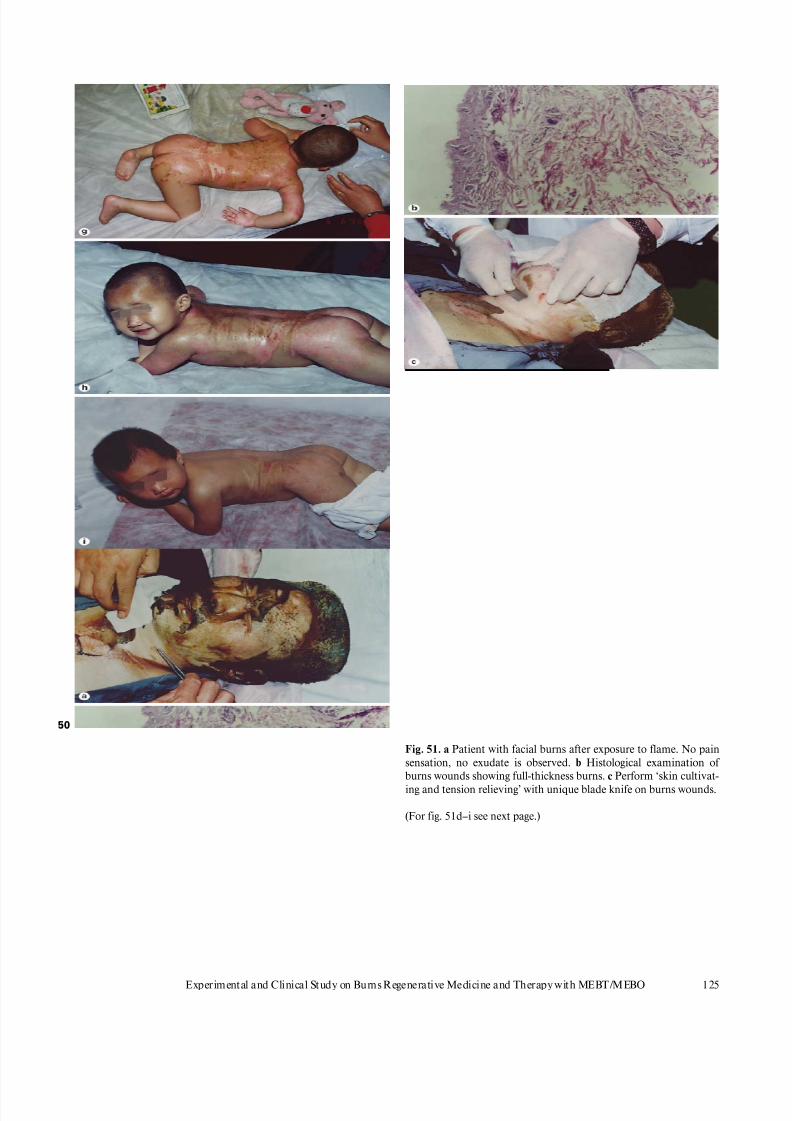
8/3/2019 Luka Bakar Book
http://slidepdf.com/reader/full/luka-bakar-book 131/157
Experimental and Clinical Study on Burns Regenerative Medicine and Therapy with MEBT/MEBO 125
50
Fig. 51. a Patient with facial burns after exposure to flame. No painsensation, no exudate is observed. b Histological examination of
burns wounds showing full-thickness burns. c Perform ‘skin cultivat-
ing and tension relieving’ with unique blade knife on burns wounds.
(For fig. 51d–i see next page.)

8/3/2019 Luka Bakar Book
http://slidepdf.com/reader/full/luka-bakar-book 132/157
126 Burns Regenerative Medicine and Therapy
Fig. 51. d After cultivating, directly apply MEBO according to theprocedure of BRT. e Day 2 postburn. After cleaning the burns
wounds covered with MEBO, perform secondary ‘skin cultivating
and tension relieving’ according to MEBT. f An excellent visual
example of the process of wound liquefaction on day 12 postburn.
g On day 18 postburn, the necrotic tissues were almost liquefied and
painlessly discharged while subcutaneous tissues were allowed toregenerate new skin. h On day 27 postburn, some burns wounds
healed with complete regeneration while other burn lesions were in
the process of healing. i On day 50 postburn, burns wounds healed
and skin regenerated physiologically. Note the symmetrical smile
and the lack of scarring. Full sensation has returned.

8/3/2019 Luka Bakar Book
http://slidepdf.com/reader/full/luka-bakar-book 133/157
Experimental and Clinical Study on Burns Regenerative Medicine and Therapy with MEBT/MEBO 127
Fig. 52. a Appearance of burned tibia of left leg. b Removal of the
necrotic tissues at the surface of the bone. c Removal of the necrotic
periosteum and outer cortex of tibia with scraper and bone chisel.
d Multiple holes drilled in the exposed tibia surface within a distance
of 1.0 cm, deep to viable marrow cavity with minor bleeding.
e Appearance of tibial surface after drilling. f Subsequent to the appli-
cation of preserved soaked MEBO gauze to cover the wound, within
a few days, small granulating buds grew up through the drilled holes.
g With continuous treatment with BRT, the granulation tissue devel-
oped and spread to form a granulated wound. Skin grafting was then
performed to close the wound.

8/3/2019 Luka Bakar Book
http://slidepdf.com/reader/full/luka-bakar-book 134/157
128 Burns Regenerative Medicine and Therapy
Fig. 53. a This case involved sustained elec-
trical injury to wrist and forearm. The pic-
ture shows necrosis of anterior forearm
muscles, radial and ulnar arteries. The in-
terosseous artery was viable. The pressure
pain point of the upper extremity was at the
elbow. b After 55 days of treatment with
BRT, the necrotic tissues were liquefied
and discharged while the wound on anteri-
or wrist at the site of defective muscles was
filled with granulation tissue. The wound is
now healing from its margins by epithelial
migration. c In another case, a 9-year-oldpatient was injured by a 150,00-volt electric
impulse on the head. The injury went deep
into the skull. d Two days after the burn,
the wound was cleaned and the necrotic tis-
sue removed, then multiple holes were
drilled into the skull followed by treatment
with bone BRT. MEBO was applied direct-
ly onto the wound. e Fifteen days after
treatment with MEBT/MEBO, small gran-
ulating buds grew up through the holes.
f Twenty-two days of treatment with BRT and MEBT/MEBO, granulating tissue developed and spread to form a
granulated wound and the new epithelial skin island appeared. g Follow-up picture 2 years later. The wound was
healed and the skull was completely covered.

8/3/2019 Luka Bakar Book
http://slidepdf.com/reader/full/luka-bakar-book 135/157
OOOOOOOOOOOOOOOOOOOOOOOOOOOOOOOOOOOOOOOOOOOOOOOOOOOOOOOOOOOOOOOOOOOOOOOOOOOOOOOOOOOOOOOOOOOOOOOOOOOOOOOOOOOOOOOOOOOOOOOOOOO
Clinical Demonstrations of BurnsRegenerative Medicine and Therapy
(MEBT/MEBO) on SuccessfulTreatment of Extensive Burns
129
Fig. 54. a TBSA 100%, deep second-degree 6%, third-degree 94%,
treatment with MEBT/MEBO. b Prof. Rong Xiang Xu evaluating
and directing BRT treatment. c Posterior view of wounds during
treatment. d The healing image (posterior). e The healing image (an-
terior).
Remarkably, rigorous sterile conditions are not re-quired if burns regenerative medicine and therapy
(MEBT/MEBO) is used in the care of extensive burns
patients. Basic equipment and general surgical conditions
typically suffice. However, ambient room temperature of
32–36°C and careful, well-trained, intelligent nursing
care are required so that removing liquefied product in a
timely manner is accomplished. In general, surgical prin-
ciples of systemic comprehensive treatment should be fol-
lowed. Figure 54a–e shows Prof. Rong Xiang Xu, inven-
tor of burns regenerative medicine and therapy (MEBT/
MEBO), as he assesses and directs the treatment for a
burns patient with deep burns of TBSA 100%.

8/3/2019 Luka Bakar Book
http://slidepdf.com/reader/full/luka-bakar-book 136/157
130 Burns Regenerative Medicine and Therapy
OOOOOOOOOOOOOOOOOOOOOOOOOOOOOOOOOOOOOOOOOOOOOOOOOOOOOOOOOOOOOOOOOOOOOOOOOOOOOOOOOOOOOOOOOOOOOOOOOOOOOOOOOOOOOOOOOOOOOOOOOOO
Extensive Burns Cases with Most Wounds of Superficial
Partial-Thickness
Case 1 (fig. 55a, b)
Fig. 55. Anterior (above) and posterior (below) views. a A 2.5-year-old baby patient, TBSA 70%. b Discharged with
complete healing after treatment with MEBT/MEBO for only 14 days.

8/3/2019 Luka Bakar Book
http://slidepdf.com/reader/full/luka-bakar-book 137/157
Clinical Demonstrations of Burns Regenerative Medicine and Therapy (MEBT/MEBO) on
Successful Treatment of Extensive Burns
131
OOOOOOOOOOOOOOOOOOOOOOOOOOOOOOOOOOOOOOOOOOOOOOOOOOOOOOOOOOOOOOOOOOOOOOOOOOOOOOOOOOOOOOOOOOOOOOOOOOOOOOOOOOOOOOOOOOOOOOOOOOO
Extensive Burns Cases with Most Wounds of Deep
Partial-Thickness
Case 2 (fig. 56a–f)
Fig. 56. a A 21-year-old man, TBSA 98%. b 24 h after treatment with MEBT/MEBO. c, d Regeneration of skin tissue
and wound healing on the 30th day after treatment with MEBT/MEBO.
(For fig. 56e–f see next page.)

8/3/2019 Luka Bakar Book
http://slidepdf.com/reader/full/luka-bakar-book 138/157
132 Burns Regenerative Medicine and Therapy
Fig. 56. e At the present time. f Three years post-treatment, the form, structure and function of the new skin is
identical to normal skin (chest and abdomen).
Case 3 (fig. 57a – d)

8/3/2019 Luka Bakar Book
http://slidepdf.com/reader/full/luka-bakar-book 139/157
Clinical Demonstrations of Burns Regenerative Medicine and Therapy (MEBT/MEBO) on
Successful Treatment of Extensive Burns
133
Fig. 57. a A 28-year-old patient. TBSA 82%. b During the treatment
with MEBT/MEBO. c Wound repair and physiological healing on the
32nd day after treatment with MEBT/MEBO. Anterior and posterior
views. d At the present time.

8/3/2019 Luka Bakar Book
http://slidepdf.com/reader/full/luka-bakar-book 140/157
134 Burns Regenerative Medicine and Therapy
OOOOOOOOOOOOOOOOOOOOOOOOOOOOOOOOOOOOOOOOOOOOOOOOOOOOOOOOOOOOOOOOOOOOOOOOOOOOOOOOOOOOOOOOOOOOOOOOOOOOOOOOOOOOOOOOOOOOOOOOOOO
Extensive Burns Cases with Most Wounds of Full-Thickness
Case 4 (fig. 58a, b)
Fig. 58. a TBSA 94%. Before treatment. b Wound healing on the 40th day after treatment with MEBT/MEBO.

8/3/2019 Luka Bakar Book
http://slidepdf.com/reader/full/luka-bakar-book 141/157
Clinical Demonstrations of Burns Regenerative Medicine and Therapy (MEBT/MEBO) on
Successful Treatment of Extensive Burns
135
Case 5 (fig. 59a – c)
Fig. 59. a TBSA 93%. Before treatment. b On the 9th day after treat-
ment with MEBT/MEBO. c One year later after being healed and
discharged on the 46th day post-treatment with MEBT/MEBO.

8/3/2019 Luka Bakar Book
http://slidepdf.com/reader/full/luka-bakar-book 142/157
136 Burns Regenerative Medicine and Therapy
Case 6 (fig. 60a – c)
Fig. 60. a Upper and lower: TBSA 90%. Before treatment. b Upper
(anterior) and lower (posterior) views: All wounds healed with skinregeneration after 51 days of MEBT/MEBO treatment. c At the
present time.

8/3/2019 Luka Bakar Book
http://slidepdf.com/reader/full/luka-bakar-book 143/157
Clinical Demonstrations of Burns Regenerative Medicine and Therapy (MEBT/MEBO) on
Successful Treatment of Extensive Burns
137
Case 7 (fig. 61a – c, 62a – e)
Fig. 61. a TBSA 90%. Before treatment. b The wound healed on the
43rd day after treatment with MEBT/MEBO. c At the present time.
Fig. 62. Three years later. a The skin healed spontaneously from deep
second-degree wounds (chest and abdomen) and had identical ap-
pearance as normal skin in structure and function. Note the lack of
scar tissue. b The skin healed spontaneously from wounds mixed of
deep second- and superficial third-degree burns (inside of left upper
arm) and recovered to normal structure and function. c The skin
healed spontaneously from superficial third-degree wounds (back)
and almost recovered to normal in structure without obvious scars.
(For fig. 62d–e see next page.)

8/3/2019 Luka Bakar Book
http://slidepdf.com/reader/full/luka-bakar-book 144/157
138 Burns Regenerative Medicine and Therapy
Fig. 62. d The skin healed spontaneously from superficial third-degree wounds (outside of right thigh and knee). There
are few smooth and soft scars with slight hypo-pigmentation but good elasticity and no hyperplasia or dysfunction.
e The scars healed spontaneously from deep third-degree wounds (inside of right thigh and knee). Tissue appeared
smooth and soft without contracture or deformity.
Case 8 (fig. 63a – c, 64a – e)

8/3/2019 Luka Bakar Book
http://slidepdf.com/reader/full/luka-bakar-book 145/157
Clinical Demonstrations of Burns Regenerative Medicine and Therapy (MEBT/MEBO) on
Successful Treatment of Extensive Burns
139
Fig. 63. a TBSA 95%. Before treatment. b On the 55th day after treat-
ment with MEBT/MEBO, the skin tissue regenerated and the wound
healed. c At the present time.
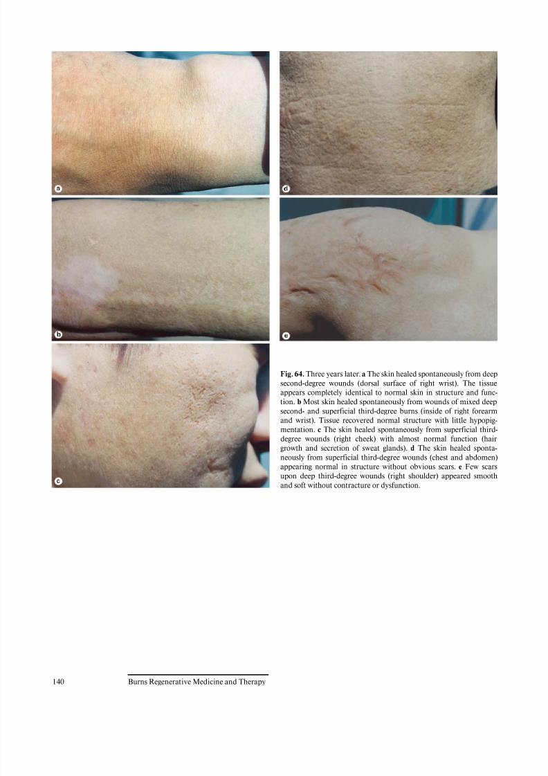
8/3/2019 Luka Bakar Book
http://slidepdf.com/reader/full/luka-bakar-book 146/157
140 Burns Regenerative Medicine and Therapy
Fig. 64. Three years later. a The skin healed spontaneously from deep
second-degree wounds (dorsal surface of right wrist). The tissue
appears completely identical to normal skin in structure and func-
tion. b Most skin healed spontaneously from wounds of mixed deep
second- and superficial third-degree burns (inside of right forearm
and wrist). Tissue recovered normal structure with little hypopig-
mentation. c The skin healed spontaneously from superficial third-
degree wounds (right cheek) with almost normal function (hair
growth and secretion of sweat glands). d The skin healed sponta-
neously from superficial third-degree wounds (chest and abdomen)
appearing normal in structure without obvious scars. e Few scars
upon deep third-degree wounds (right shoulder) appeared smooth
and soft without contracture or dysfunction.

8/3/2019 Luka Bakar Book
http://slidepdf.com/reader/full/luka-bakar-book 147/157
OOOOOOOOOOOOOOOOOOOOOOOOOOOOOOOOOOOOOOOOOOOOOOOOOOOOOOOOOOOOOOOOOOOOOOOOOOOOOOOOOOOOOOOOOOOOOOOOOOOOOOOOOOOOOOOOOOOOOOOOOOO
Clinical Results of Surgical Excisionand Skin Grafting Therapy in the
Treatment of Extensive BurnsPatients
141
Case 1: Male, 23 Years Old. Admission No. 212911
(fig. 65a–c)
Final Diagnosis
(1) Direct flame burns with 92% TBSA (superficial sec-
ond-degree 2%, deep second-degree 19%, third-degree
71%).
(2) Inhalation injury (mild).
(3) Hypovolemic shock postburn.
(4) Septicemia (Pseudomonas aeruginosa); corneal ul-cer ( Pseudomonas, left eye).
Fig. 65. a Before treatment. b Wound healed by multiple skin grafting at 74 days after injury. c 14 months later, the
appearance after plastic and reconstructive operations.
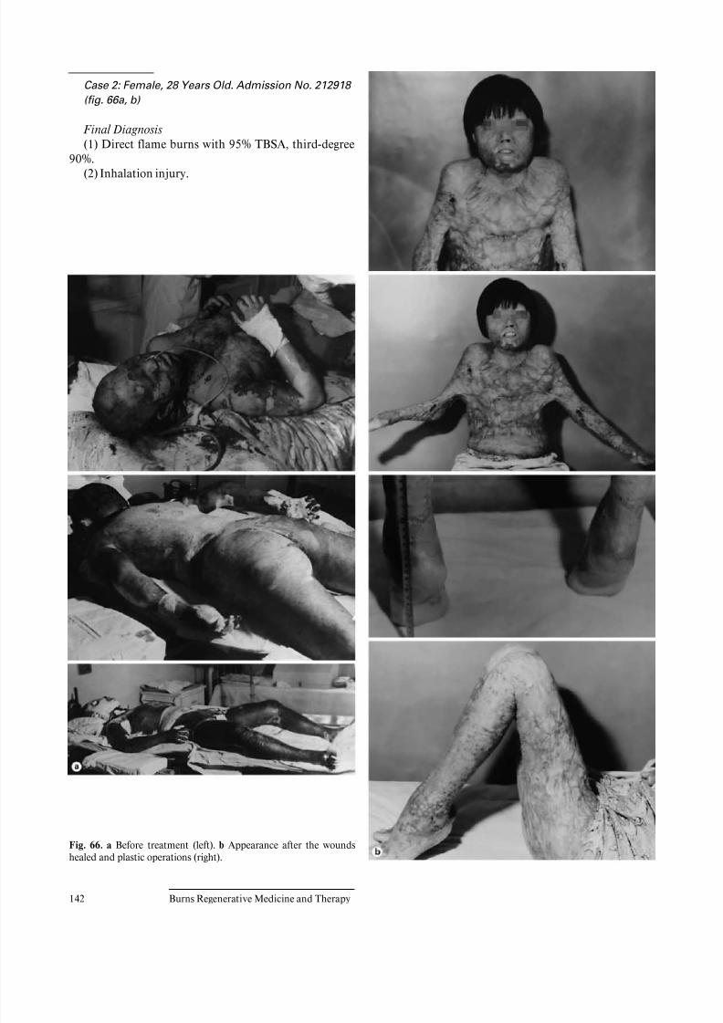
8/3/2019 Luka Bakar Book
http://slidepdf.com/reader/full/luka-bakar-book 148/157
142 Burns Regenerative Medicine and Therapy
Fig. 66. a Before treatment (left). b Appearance after the wounds
healed and plastic operations (right).
Case 2: Female, 28 Years Old. Admission No. 212918
(fig. 66a, b)
Final Diagnosis
(1) Direct flame burns with 95% TBSA, third-degree
90%.
(2) Inhalation injury.

8/3/2019 Luka Bakar Book
http://slidepdf.com/reader/full/luka-bakar-book 149/157
Clinical Results of Surgical Excision and Skin Grafting Therapy in the Treatment of Extensive Burns
Patients
143
Case 3 (fig. 67a, b)
Final Diagnosis
(1) Direct flame burns with 95% TBSA, third-degree
91%.
(2) Inhalation injury.
Fig. 67. a Before treatment. b Most wounds closed after microparticle
autografting. Residual granulation wounds on his chest, back, hands
and feet were still left for skin grafting later.

8/3/2019 Luka Bakar Book
http://slidepdf.com/reader/full/luka-bakar-book 150/157
OOOOOOOOOOOOOOOOOOOOOOOOOOOOOOOOOOOOOOOOOOOOOOOOOOOOOOOOOOOOOOOOOOOOOOOOOOOOOOOOOOOOOOOOOOOOOOOOOOOOOOOOOOOOOOOOOOOOOOOOOOO
A Commentary on Burns Medicaland Regenerative Therapy
145
A Commentary on Surgical Excision and
Skin-Grafting Therapy
Burns therapy with surgical excision and skin graftingis a surgical technique in that it treats the burns wounds
with a surgical method. Surgical technique, in essence,
treats disease through a destructive means while prioritiz-
ing the survival of the patient about the importance of the
appearance and function of the burned limb. Before BRT
with MEBT/MEBO was invented, surgical burns therapy
had become a major method of burns treatment. How-
ever, subsequent to the invention of burns regenerative
medicine and therapy helpful comparisons have been
made between both modalities. Impartial investigators
have learned that deep second-degree burns wounds
should no longer be treated with surgical therapy because
burns regenerative medicine and therapy is objectively
superior to the surgical approach. One remaining indica-
tion for the use of surgical excision and skin grafting for
the treatment of burns may involve third-degree burns
with surviving subcutaneous tissues. This, however, must
only be done after prudent consideration. The indication
of surgical burns therapy should now be defined as: severe
large-area burns reaching the lower layer of superficial fas-
cia. Surgical burns therapy should no longer be the major
method of burns treatment.
This book also introduces the latest technique of skingrafting using cultured composite autografts after surgical
excision. This new technique aims at overcoming the dif-
ficulty of the incorporation of the cultured epithelial auto-
graft into the burns wound. This technique can effectively
prevent ‘autograft exfoliation’ and secondary ulceration.
The doctors of the laboratory of Culture Technology, Inc.,
Sherman Oaks, Calif., USA, harvested two components of
the skin, autologous keratinocytes and fibroblasts from
burns patients and cultured them to enhance prolifera-
tion, and then combined them to form epidermal and der-
mal matrix. Once grown to confluence, the composite
autografts are ready for application to the burn wound.
These results were published in Burns 1999;25:771–779.
This technique had been successfully applied in the treat-
ment of large-area burns after surgical excision in the
Burn Center in Arizona State. While this is a significant
step forward, we must acknowledge that its treating prin-
ciple is the same as that of surgical burns therapy. It pro-
tects the autograft but cannot avoid the damage or dis-
ablement caused by excision. Another comparable disad-
vantage to this technique is its expense. Therefore, indica-
tion for this technique should be third-degree burns and
burns in the muscle layer. This skin grafting using cul-
tured composite autografts after surgical excision should
not be considered a major method of burns treatment.
A Commentary on Moist-Exposed Burns Therapy
BRT with MEBT/MEBO is a comprehensive thera-
peutic technique aiming at treating burns tissue in com-
pliance with the law of burns pathogenesis. Compared
with surgical burns therapy, BRT with MEBT/MEBO is a
technique treating the burns wound in the skin, while sur-
gical burns therapy is a technique treating wounds in the
muscle. Together, these two approaches, when used ap-
propriately, form a complementary therapeutic system.
BRT with MEBT/MEBO can be applied for the treatment
of skin burns while surgical burns therapy can be bestapplied to the treatment of muscle burns. Briefly, BRT
with MEBT/MEBO offers unique therapeutic break-
throughs in treating skin burns as follows:
A BRT with MEBT/MEBO removes the necrotic skin
without causing any damage. Removal of necrotic skin
layer is the first step of burns treatment. Doctors found
no way to remove the necrotic tissue during the past
century, except the destructive method which cut away
the injured wound tissue together with the surrounding
surviving tissues and resulted in further traumatic in-
juries. Taking the advantages of the relevant biochemi-
cal principles, BRT with MEBT/MEBO can spontane-

8/3/2019 Luka Bakar Book
http://slidepdf.com/reader/full/luka-bakar-book 151/157
146 Burns Regenerative Medicine and Therapy
ously remove the necrotic tissue through liquefaction
and drainage without causing further injury to the sur-
rounding surviving tissue. It alone has successfully
resolved this difficult problem.
B BRT with MEBT/MEBO preserves the surviving tis-
sue to the greatest extent currently possible. Burns
wound surface is not smooth and a surgical knife can-not distinguish between injured tissue and surviving
tissue. Surgeons always excise the surviving tissue
together with dead tissue and this is a very serious
attack on the patient – at times it can be more serious
than burns injury itself. Moreover, after excision, the
body surface typically never recovers the loss of subcu-
taneous surviving tissue. However, studies demon-
strate that, if not excised, this recovery can occur. BRT
with MEBT/MEBO takes advantage of the frame
structure of the nutritive base of the drug and the prin-
ciple of biochemistry therewith successfully preserving
the surviving tissue.C BRT with MEBT/MEBO demonstrates that the dream
of skin regeneration has come true. For about a centu-
ry, scientists made great efforts to achieve the regenera-
tion of injured skin. In the early 20th century, doctors
discovered that the subcutaneous tissues survive after
full thickness third-degree burns and may be capable of
regeneration. However, they did not find an adequate
measure to achieve this survival and therefore they
pursued research on in vitro skin cell culture and trans-
plantation of the cultured autograft. By utilizing the
regeneration gene for skin information in the subcuta-
neous tissue, in concert with the creation of a favorable
nurturing environment (one favorable to physiological
regeneration of the skin), BRT with MEBT/MEBO
successfully achieves the skin regeneration within large
areas of deep burns wounds. This achievement greatly
decreased the disablement rate, and increased the sur-
vival rate of large-area burns by 50–80% (compared
with the data published in 1997 and 1994).
D BRT with MEBT/MEBO resolves the problem of pain
in second-degree burns patients. As any person who has
cared for burns patients knows all too well, burns wound
pain is the worst aspect of the suffering of superficialburns patients. Surgical treatment aims at saving the life
without considering the problem of pain. Surgical oper-
ations typically make the pain more serious and many
patients with large-area superficial burns die because
their cases worsen after operation. Severe pain causes
shock and wound stress regulation disturbance which
can tip the scales toward multiple system organ failure
and death. That is why large-area as well as small-area
burns are described as life-threatening in the burns care
textbooks. Pain remains one of the main causes of
burns-related death in all countries. BRT with MEBT/
MEBO takes the advantage of the drug MEBO with
a unique frame structure base to eliminate pain al-
most immediately upon application. MEBO covers the
wound surface, protects the wound from irritations and
relieves the pain. This unique effect of MEBO finally
resolved the problem of burns wound pain.
E BRT with MEBT/MEBO opens up a new approach to
the prevention and treatment of infection. Local andsystemic infection is a difficult problem of burns treat-
ment and today in the era of multidrug resistant patho-
gens, we are scarcely further ahead than we were years
ago. Many antibiotics have been applied but the effica-
cy proves unsatisfactory. BRT with MEBT/MEBO re-
solves this problem by treating the local area in com-
pliance with the pathogenesis of the infection of burns
wound. This treatment controls infection of burns
wound by changing the ecological environment. Con-
currently, by applying BRT with MEBT/MEBO to the
large-area burns, in accordance with the law of system-
atic pathogenesis of infections, we discover that BRTwith MEBT/MEBO is capable of mobilizing and coor-
dinating the potential physiological energy of the sys-
temic wound stress reaction. This alone has successful-
ly advanced a systematic anti-infection principle for
treating large-area burns. To be more specific: At the
shock stage, when wound stress reaction is on the
upsurge, we recommend the systemic application of
broad spectrum antibiotics with no adverse effect on
the kidney. After this stage, when synthetic metabo-
lism of protein begins, we recommend that one stop
the application of any antibiotics. In the whole course
of treatment, if systemic infection occurs occasionally,
a single large dose of broad-spectrum antibiotic (one
with no side effects on kidney) is applied. In this fash-
ion, BRT with MEBT/MEBO offers a systematic
scheme for removing the focus of infection and mini-
mizes the dependence upon antibiotics.
F BRT with MEBT/MEBO allows one to create a new
antishock scheme. It is a common understanding that
shock is a serious disease of burns. For a long time, no
matter what treating method is adopted, the same stan-
dardized fluid infusion antishock scheme is applied.
BRT with MEBT/MEBO considers that there shouldbe different antishock schemes for different treating
methods and different cases. Surgical operation always
makes shock more serious and therefore, fluid infused
to replenish the blood volume is of paramount impor-
tance. Remarkably, BRT with MEBT/MEBO does not
produce any new injury. On the contrary, it helps to
develop spontaneous resuscitation. Antishock mea-
sures mainly aim at protecting and strengthening the
cardiac and renal function. Blood volume replenish-
ment is required only according to the principle of gen-
eral traumatic surgery. Shock, the greatest killer of
burns patients, is finally tamed.

8/3/2019 Luka Bakar Book
http://slidepdf.com/reader/full/luka-bakar-book 152/157
A Commentary on Burns Medical and Regenerative Therapy 147
G BRT with MEBT/MEBO relieves the economic and
mental load of the burns victims. Textbooks over-
emphasize that surgical operation is the only method
for treating burns, so people are frightened of the suf-
ferings during the operation and the high cost of the
treatment. In the US, it typically costs a burns patient
USD 150,000 to be treated in the hospital and thisdoes not include the expense of subsequent plastic sur-
gery. Because surgical operation requires strictly sterile
and isolated wards, such wards are very expensive to
build and maintain. In western countries, treating
burns victims with burns area over 50% BSA is con-
sidered to have no economic value, because most of
the patients will become disabled. BRT with MEBT/
MEBO is very revolutionary in this matter as it does
not require strictly sterile conditions nor does it re-
quire isolation. On the contrary, large-area burns pa-
tients can be treated in ordinary hospital wards or even
in battlefield hospitals and they will recover to becomehealthy and normal people. The cost is extremely low
by comparison and small area burns patients, if treated
with BRT with MEBT/MEBO, do not require hospital-
ization.
BRT with MEBT/MEBO can cure burns of different
causes and different areas, including superficial second-
degree, deep second-degree, and full-thickness third-de-
gree burns. It is also an ideal technique for granulation
tissue regeneration and repair of burns in muscular layer
and bone. BRT with MEBT/MEBO is the major method
of burns treatment.To sum up, what is described above is not speculation.
This is clinically demonstrated and despite the skepticism
of the reader, responsible investigation into these claims
will convince all that burns therapy has now developed
into a new historic stage. In the past, only surgical excision
and skin grafting were the standard of care and offered
great benefit to those whose lives were threatened. Today,
however, with the invention of BRT with MEBT/MEBO
a major method for burns treatment is available. Either
alone or in combination with surgical care, we now offer
an elevation in the standard of care for the treatment of
burns. As we move together into the 21st century, burnstherapy will continue to develop along the lines of BRT
with MEBT/MEBO.

8/3/2019 Luka Bakar Book
http://slidepdf.com/reader/full/luka-bakar-book 153/157

8/3/2019 Luka Bakar Book
http://slidepdf.com/reader/full/luka-bakar-book 154/157
OOOOOOOOOOOOOOOOOOOOOOOOOOOOOOOOOOOOOOOOOOOOOOOOOOOOOOOOOOOOOOOOOOOOOOOOOOOOOOOOOOOOOOOOOOOOOOOOOOOOOOOOOOOOOOOOOOOOOOOOOOO
Conclusion
149
An Inevitable Outcome of Scientific Research
This book provides an introduction to the existing
therapeutic techniques of local treatment of burnswounds and discusses two therapeutic techniques for
treating burns wounds as regards their historical, scien-
tific and technological development. This volume aims at
aiding burns medicine researchers and clinicians in devel-
oping a correct idea regarding the relative indications of
the two therapeutic techniques. It is further hoped that
this volume will assist in the elimination of prejudice
between different schools as well as to improve the level of
the comprehensive burns treatment.
Today, there are only two categories of therapeutic
techniques for burns treatment worldwide. One is BRT
with MEBT/MEBO, which treats skin burns wounds in
such a manner as to achieve both repair and regeneration
by creating a wound environment that optimizes the
potential of remaining viable tissue. Compared to other
treatment protocols, this therapy reduces the rates of scar
formation and disability as well as pain suffered and eco-
nomic burden more than the other technique, which is a
surgical skin grafting technique. This second treatment
protocol, which aims at treating burns involving whole
thickness of the skin and subcutaneous tissues without
leaving any viable wound tissue in place, is well known to
extract a great physiological price in terms of pain, scar-ring and residual suffering. It is also far more expensive
than BRT with MEBT/MEBO. Prior to the invention of
BRT with MEBT/MEBO, widespread utilization of the
surgical technique in the treatment of muscle burns and
skin burns was acceptable. However, now that scientific
studies have demonstrated that BRT with MEBT/MEBO
is superior in every way, surgical technique is only a rea-
sonable standard of care in the treatment of skin burns.
This conclusion is now a consensus within academic cir-
cles worldwide. Surgical burns therapy was born when no
other technique could be applied to treat skin burns. Med-
ical researchers and clinicians had been working hard to
find a technique for treating skin burns. The emergence of
BRT with MEBT/MEBO represents a blessing to every-
one suffering from burns trauma – both the patients and
their loyal caregivers. Anyone who has ever cared for
burns patients will be relieved and grateful to use BRT
with MEBT/MEBO immediately. BRT with MEBT/
MEBO is the realization of the dream of all the medical
workers and is welcome news to burns patients around the
world.
An Inevitable Outcome of the Development of Medical
Science
Life science research spans a history of more than
2,500 years. Medical science is only one part of the life
science. Medical workers and doctors of successive Chi-
nese dynasties took infinite pains in searching for the key
to the door of life science and with it, the ideal methods
for controlling diseases. In ancient Greece, Hippocrates
established anatomy and surgery, and laid the founda-
tions of modern surgery. Since then, medical workers of
the east and the west started using plants and herbs for
treating diseases. After the Renaissance, human biochem-
istry was established, which laid the foundations for the
modern treatment strategy of antagonist chemotherapy.
Medical sciences developed along with the developmentof human civilization but the paradigms of medical
science were slow to change. In the east, the thinking
developed along macro lines of ‘chi’ and patterns of ener-
gy flow. Whole plant extracts and consideration of diet
predominated in the east whereas in the west, the focus
was on more narrow and abstract ideas such as active
principles of plants, essential elements and ultimately
genetic dynamics.
The development of medical treatment methods and
drugs lagged behind the development of human civiliza-
tion. Despite progress in other areas of human endeavor,
the methods and materials for enhancing health and sav-

8/3/2019 Luka Bakar Book
http://slidepdf.com/reader/full/luka-bakar-book 155/157
150 Burns Regenerative Medicine and Therapy
ing human life are still limited to two categories, i.e. sur-
gery and internal medicine. The former, while saving
lives, compromises viable tissue as well as future anatomi-
cal function. The latter, availing itself primarily of chemi-
cals unfavorable to human physiology (e.g. chemothera-
py), remains fundamentally antagonistic to tissue health
and vitality. Neither modality cooperates with the endog-enous vital forces and, therefore, medical workers are ill-
equipped to facilitate regeneration of tissues and mainte-
nance of health. Appreciating the present limitations of
medical science, all health professionals are encouraged to
remain open to new trains of thought, to new methods,
techniques and materials, especially those that purport to
be working in harmony with the principles of human
physiology. The inevitable progress of medical science lies
in this direction.
BRT with MEBT/MEBO treats in accordance with the
principles of human physiology. The techniques and ma-
terials of burns regeneration therapy, by being in com-pliance with the law of human life, and aiming as it does
at maintaining normal physiological functions and physi-
ological activities, becomes a new paradigm for healing.
In this book, we publish the theory of this technique,
research results and examples of its clinical application.
All the data prove that BRT with MEBT/MEBO, with
new methodology and a new drug (MEBO), points to a
new direction in burns medicine research. The success of
BRT with MEBT/MEBO is not only limited to the field of
burns medicine but also sheds light upon the nascent
research of life medicine itself. Burns wounds are typical
traumatic wounds and innovations in burns care are gen-
erally applicable in the treatment of traumatic wounds of
all kinds. In this book, our data proved that after loss of
epidermal tissue, viable epithelial cells in the sweat glands
of the subcutaneous tissues can, given the appropriate
environment, transform into epithelial stem cells and that
these stem cells can, in turn, form new epidermal tissues.
This regeneration of epidermal tissue accomplished the
first cloning of human tissues and organs in the 21st cen-
tury. We have demonstrated the inexpensive and potent
cloning of a ‘tissue stem cell’. Therefore, the establish-
ment of BRT with MEBT/MEBO provides the basis forfurther research in one intriguing aspect of life science –
cloning.
The Formation of Burns Medical Therapy in the
21st Century
The history of burns therapy as a specialized field of
research is less than a hundred years old. While humans
have burned themselves since the dawn of time, no sys-
temic intelligent protocols have been established to serve
mankind in this regard. Hippocrates did record burnstreatment but his methods were not described with any
scientific basis. Subsequently, no specialists in burns
medicine were formed until after 1930. Research into
burns pathogenesis had not produced any impressive
methods or materials for treating burns wounds along
physiologic lines. Surgical methods saved lives but left
patients disfigured and disabled. Surgery, while repre-
senting a big step forward, treats the patients while doing
nothing to encourage regeneration of the skin. Finally, at
the end of the 20th century, Chinese doctors invented
burns regenerative medicine and therapy, which offered
an entirely new therapy operating in compliance with
principles of human physiology. Now, as we enter a new
century and while we pause on that threshold, we all share
the opportunity to cooperate and combine the best of all
approaches from the east and the west. As described
above, the technology of the east, i.e. BRT with MEBT/
MEBO, should be applied for all burns including muscle
burns; while for treating burns in the muscle layer, the
technology of the west, i.e. surgical excision and skin
grafting therapy, should be applied. As a whole, it can be
called integrated east and west burns therapy. In the era of
information, any new technology, thinking, method andmaterial when produced, will immediately be known all
over the world and the information be shared. New tech-
nology is no longer a legendary tree of a doctor that sheds
coins when shaken, and the backward technique will no
longer be applied to produce tragedy. We believe that in
the 21st century, people will make greater progress in the
field of burns medicine and the problems will be com-
pletely solved as people succeed in the cloning of organs.

8/3/2019 Luka Bakar Book
http://slidepdf.com/reader/full/luka-bakar-book 156/157
OOOOOOOOOOOOOOOOOOOOOOOOOOOOOOOOOOOOOOOOOOOOOOOOOOOOOOOOOOOOOOOOOOOOOOOOOOOOOOOOOOOOOOOOOOOOOOOOOOOOOOOOOOOOOOOOOOOOOOOOOOO
Subject Index
151
Antibiotics, infection prophylaxis after burn injury 41, 42
Antimicrobial effects, MEBT/MEBO
anaerobic bacteria infection inhibition 82–87
bacterial counts in viable tissue 68–70
bacteriostasis 77–82fungal infection inhibition 82–87
mechanisms 74–82
Pseudomonas aeruginosa infection 70–73
Blood viscosity, MEBT/MEBO effects 60, 62
Bone marrow, regenerative medicine 9
Burn area assessment, Chinese rule of nines 19
Burn depth
clinical evaluation 20–22
standardization of diagnosis 34
Burn pathogenesis
biochemical injury phase 13
burns regenerative therapy rationale 17
physical injury phase 13reject reaction of necrotic tissues 13, 14
Burn pathology
burns regenerative therapy rationale 17
MEBO response 92–96
morphological changes 14, 15
repair changes
deep second-degree burns 15
superficial second-degree burns 15
third-degree burns 15, 16
standardization of burn depth diagnosis 34
three degree four division method of diagnosis 21, 22
Burn severity, classification 23
Burns regenerative therapy, see also MEBT/MEBO
historical perspective 2, 3, 6, 11, 12overview 12, 13
rationale 17
Burns therapy, historical perspective 1–3
Chinese medicine, historical perspective 5, 6
Chinese rule of nines, burn area assessment 19
Cultured composite autograft, skin graft outcomes 33, 145
Electron microscopy, studies of MEBO response in burn injury
microcirculation 108, 109
scarring 96–104
wound healing 113, 114
Endothelin (ET), MEBT/MEBO effects 61
Erythrocyte agglutinative index, MEBT/MEBO effects 60
Erythrocyte transformation kinetics, MEBT/MEBO effects 60
Extensive burns
MEBT/MEBO outcomes 129–140
surgical excision and skin grafting outcomes 141–143
Extraordinarily severe burns, burn severity classification 23
Fibronectin, MEBO comparative studies in corneal burn model
88–92
First aid, MEBT/MEBO 40
First-degree burns
diagnosis 20
MEBO application guidelines 35
MEBT/MEBO therapy 37
pathology of three degree four division method of diagnosis 21
three degree six division method of diagnosis 22
Gastrointestinal mucosa, regenerative medicine 9
Hematocrit, MEBT/MEBO effects 60, 62Hemorrhoids, MEBO application guidelines 35
Histology, MEBO response studies in burn injury 92–96,
111–114
Immunoglobulin response, MEBT/MEBO effects 63–68
Kidney
protection in shock 40, 41
regenerative medicine 9
MEBT/MEBO
antimicrobial effects
anaerobic bacteria infection inhibition 82–87
bacterial counts in viable tissue 68–70bacteriostasis 77–82
fungal infection inhibition 82–87
mechanisms 74–82
Pseudomonas aeruginosa infection 70–73
antishock mechanisms 49, 50
clinical trial outcome in China 119–128
extensive burns outcomes 129–140
fibronectin comparative studies in corneal burn model 88–92
hematologic parameter effects 60–62
historical perspective 2, 3, 11, 12
immunoglobulin response 63–68
indications 34
limitations 17
microcirculation promotion 55–59, 104–111

8/3/2019 Luka Bakar Book
http://slidepdf.com/reader/full/luka-bakar-book 157/157
MEBT/MEBO (cont.)
ointment
application guidelines 34, 35, 37, 40
components 3
therapeutic effects 3, 36, 37
tissue interactions 36
wound debridement 40
popularity of use 3, 16, 36
procedures and techniques 12
prospects 149, 150
rationale 17, 145–147
standardized local treatment
treatment comparison with MEBT/MEBO 27–29, 33,
40–43, 149
stem cell differentiation promotion 36, 114–118
systemic treatment 37
wound liquefaction, see Wound liquefaction
wound water evaporation prevention 50–54
Microcirculation, MEBT/MEBO promotion 55–59, 104–111
Moderate burns, burn severity classification 23
Moist-exposed burns treatment and moist-exposed burns ointment,
see MEBT/MEBO
Nerve, regenerative medicine 9
Neutrophil, MEBT/MEBO response 77
Nitric oxide, MEBT/MEBO effects 61
Nutritional support, MEBT/MEBO 43
Pancreas, regenerative medicine 9, 10
Phagocyte, MEBT/MEBO response 82
Pseudomonas aeruginosa, MEBT/MEBO effects 70–73
Regenerative medicine
bone marrow regeneration 9
concept 6
pathology of three degree four division method of diagnosis
21, 22
repair changes
deep second-degree burns 15
superficial second-degree burns 15
three degree six division method of diagnosis 22
Severe burns, burns severity classification 23
Shock
management in burn injury
cardiac function enhancement 40
fluid therapy 41
nursing care 41
renal function protection 40, 41
MEBT/MEBO antishock effect and mechanisms in rabbit burn
model 47–50
ß-Sitosterol, MEBO composition 3
Skin, regenerative medicine 10–12
Skin graft
cultured composite autograft 33, 145
extensive burns outcomes 141–143
historical perspective 1, 2, 16
indications 35
rationale 16
rejection 2
standardized local treatment
treatment comparison with MEBT/MEBO 27–29, 33, 149
Skin ulceration, MEBO application guidelines 35
Slight burns, burn severity classification 23
Stem cell
MEBT/MEBO differentiation promotion 36, 114–118
organ cultivation 12
regenerative medicine 6–8
Surgical burns therapy
extensive burns outcomes 141–143
historical perspective 1, 2, 16