Embed Size (px)
Citation preview
Idosa com quadro de sindrome coronariana aguda e síncopes de longa dataOld woman with acute coronary syndrome and history of repetitive syncope
Raimundo Barbosa-Barros, M.D. (nickname “Raymond, the Fox”)Chief of Coronary Center of the Hospital de Messejana Dr. Carlos Alberto Studart Gomes, Fortaleza, Ceará CE, Brazil
Andrés Ricardo Pérez-Riera, M.D. Ph.D.Design of Studies and Scientific Writing Laboratory in the ABC School of Medicine, Santo André, São Paulo, Brazil
https://ekgvcg.wordpress.com
Portuguese Relato de caso
Queixa principal: Dor precordial e desmaioPaciente idosa, (83 anos) diabética, dislipidêmica, e hipotiroidea. Admitida na emergência por quadro de síncope o que refereocorrer periodicamente desde os 38 anos que ela associa a contrariedades. Após a recuperação da conciencia refere dor percordialopressiva e dispnéia. O ECG realizado nesse momento mostrava probable bloqueio completo do ramo do ramo direito, bloqueiodivisional ântero-superior e alterações de repolarização ventricular (ARV), sobrecarga do ventrículo esquerdo (SVE) e aumentodos biomarcadores de necrose miocárdica (troponina T; 0.3/ml). Feita a hipótese diagnóstica de síndrome coronariana aguda semelevação do segmento ST, se solicita cinecoronariografia diagnóstica a qual não foi realizada em consequência de novorebaixamento da consciência, pico hipertensivo e disfunção renal.Refere dor precordial associado a náuseas e dispneia há 3 meses sem investigação prévia.. Referida a UTI para continuidade doscuidados. Da entrada alerta, cooperativa, Glasgow 15 sem déficit com bom padrão respiratório e hipertensão assintomática. PA203/105 mmHg, FC 80bpm RCR me 2 tempos , bulhas rítmicas e normofonéticas sem sopros.
EC0: 16/10 discreto aumento do VVE para o biótipo ( 33mm/m2) hipertrofia concêntrica discreta do VE e contratilidade global esegmentar do VE preservadaRaimundo e AndresPerguntas1. Qual o diagnostico eletrovetorcardiografico ?2. Qual o quais as causas das forcas anteriores proeminentes ? E por que ?
Main complaint: precordial pain and fainting.
Elderly, female patient (83 years old): diabetic, dyslipidemic, and with hypothyroidism. She was admitted in the ER due to symptoms of syncope,
which she mentions has been happening to her since she was 38 years old, associated to different troubles. After recovering consciousness, she
reported oppressive precordial pain and dyspnea. The ECG made at the time revealed complete right bundle branch block, left anterior fascicular
block, and ventricular repolarization alterations (VRA), left ventricular enlargement (LVE) and positive myocardial necrosis biomarkers (troponin
T; 0.3/ml). A diagnostic hypothesis of non-ST segment elevation acute coronary syndrome was made, and diagnostic coronary angiography was
requested, which could not be done as her consciousness got depressed again, presented hypertensive peak and renal dysfunction. She mentioned
precordial pain associated to nausea and dyspnea 3 months before, with no previous investigation. She was sent to the ICU to continue with her
care. She was admitted alert, cooperative, in Glasgow 15 with no deficit, and good respiratory pattern and asymptomatic hypertension. BP 203/105
mmHg, HR 80 bpm, split regular heart rhythm, rhythmic and normal sounds with no murmurs.
Echo: 16/10, discrete increase of left ventricular volume for the biotype (33 mm/m2), discrete concentric LV hypertrophy and preserved overall
and segmentary LV contractility.
Raimundo and Andres.
Questions
1. What is the electro vectorcardiographic diagnosis?
2. What is / are the causes of the prominent anterior QRS forces? And Why?
Case Report

Colleagues opinions

Dear Andrés,
Concentric left ventricular hypertrophy, complete right bundle branch block and a PR too short for a 83 year-old woman: Fabry disease should beruled out. Syncope may come from neurological manifestations of this disease. The QRS axis can be ascribed to the patient thorax morphology(brief and large)? The acute coronary syndrome is expected in old patient with a metabolic syndrome. Hypothyroidism may also play a role in eCGfeatures?I look forward to your comments,Best regards,Philippe [email protected]
Chef du service de Rythmologie GHE, CHU LyonClaude Bernard University Lyon, France

Portuguese:Um caso muito interessante professores.
De maneira resumida, vemos um registro em ritmo sinusal, com associação de BDAS + BDAM + AFC pelo ramo direito. e alterações de repolarização em parede anterolateral.Marcador de doença avançada e importante no sistema de condução, o que deve justificar as síncopes
Obrigado por partilharHorácio Gomes Pereira Filho. University of São Paulo, São Paulo Brazil. Expertise in Cardiology and [email protected]
EnglishA very interesting case teachers.
Briefly, we see a sinus rhythm associated with Left Anterior Fascicular Block(LAFB) + Left Septal Fascicular Block ( LSFB) + Right EndConduction Delay (RECD) in the Right Bundle Branch and repolarization changes in the anterolateral wall.Marker of advanced and important disease in the intraventricular conduction system, which should justify the syncope's episodes.
Thanks for sharingHorácio Gomes Pereira Filho [email protected]
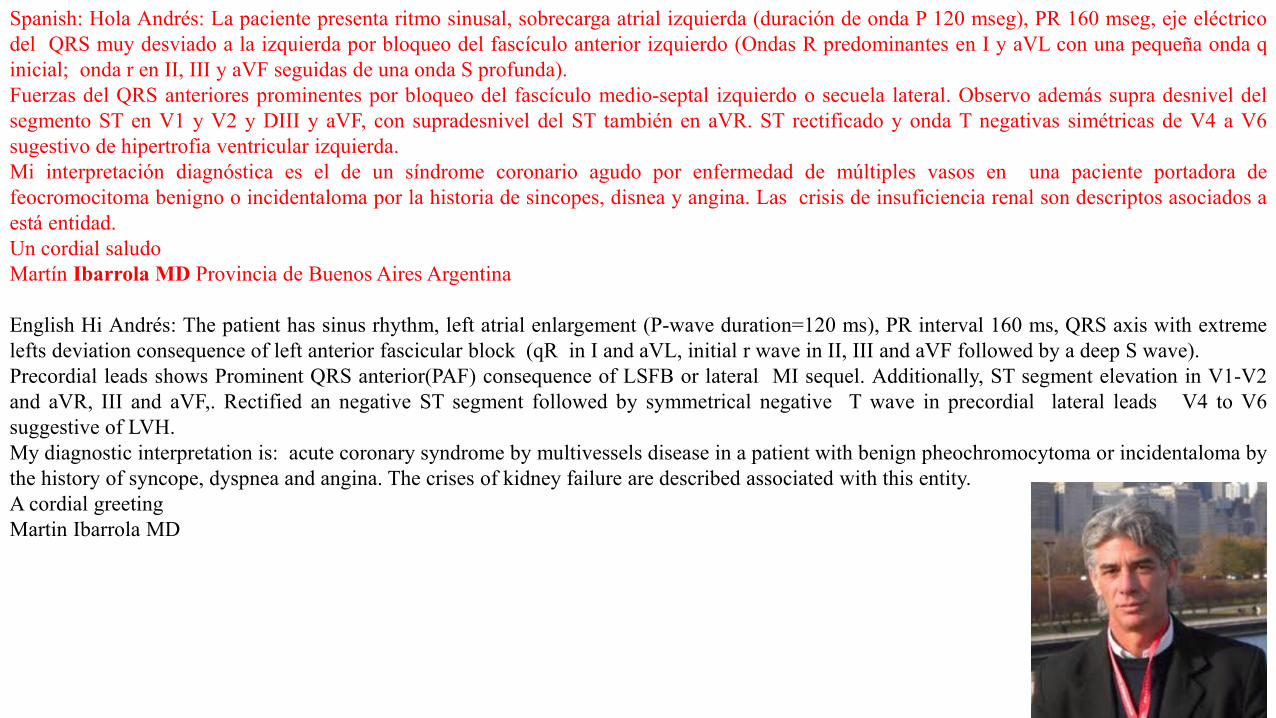
Spanish: Hola Andrés: La paciente presenta ritmo sinusal, sobrecarga atrial izquierda (duración de onda P 120 mseg), PR 160 mseg, eje eléctricodel QRS muy desviado a la izquierda por bloqueo del fascículo anterior izquierdo (Ondas R predominantes en I y aVL con una pequeña onda qinicial; onda r en II, III y aVF seguidas de una onda S profunda).Fuerzas del QRS anteriores prominentes por bloqueo del fascículo medio-septal izquierdo o secuela lateral. Observo además supra desnivel delsegmento ST en V1 y V2 y DIII y aVF, con supradesnivel del ST también en aVR. ST rectificado y onda T negativas simétricas de V4 a V6sugestivo de hipertrofia ventricular izquierda.Mi interpretación diagnóstica es el de un síndrome coronario agudo por enfermedad de múltiples vasos en una paciente portadora defeocromocitoma benigno o incidentaloma por la historia de sincopes, disnea y angina. Las crisis de insuficiencia renal son descriptos asociados aestá entidad.Un cordial saludoMartín Ibarrola MD Provincia de Buenos Aires Argentina
English Hi Andrés: The patient has sinus rhythm, left atrial enlargement (P-wave duration=120 ms), PR interval 160 ms, QRS axis with extremelefts deviation consequence of left anterior fascicular block (qR in I and aVL, initial r wave in II, III and aVF followed by a deep S wave).Precordial leads shows Prominent QRS anterior(PAF) consequence of LSFB or lateral MI sequel. Additionally, ST segment elevation in V1-V2and aVR, III and aVF,. Rectified an negative ST segment followed by symmetrical negative T wave in precordial lateral leads V4 to V6suggestive of LVH.My diagnostic interpretation is: acute coronary syndrome by multivessels disease in a patient with benign pheochromocytoma or incidentaloma bythe history of syncope, dyspnea and angina. The crises of kidney failure are described associated with this entity.A cordial greetingMartin Ibarrola MD

Dear Andrés. Short PQ-interval, maybe tendency to reentry SVT. No delta wave. LVH + “strain” pattern in lateral leads. LAFB+RBBB. Possibly left septal fascicular block.Coronary artery disease can be treated conservatively with medication.Holter indicated. Pheochromocytoma to be ruled out, although rare at old age.Best regards
Kjell Nikus M.D.Ph.D.
Tampere, Finland
Answer to Philippe, Martin and Kjell from Andrés: Fabry disease (FD) is an inherited X-chromosomallysosomal storage disease resulting in intracellular storage of globotriaosylceramide. Cardiac involvementis most frequently manifested as LVH. However, patients with FD may also have various conductionabnormalities before LVH develops. P-wave duration, PR-interval and QRS width are shorter andrepolarization dispersion more pronounced in patients with FD compared with heart rate and age-matchedcontrols. The earliest ECG manifestation are shortened PR interval and resting bradycardia, Thesignificant shortening of the PR-interval in FD occurs because of a marked shortening of the P-waveduration, which in itself demonstrated a high sensitivity and specificity for early detection and treatment ofthis disease.PR or PQ interval: The PR interval duration increases directly with age and inversely with HR increases.The normal duration of the neonatal (birth to 30 days) PR interval is between 70 and 140 ms and averages100 ms. In infants and children from 30 days to 1.5 years the maximal PR interval is 160 ms for HR<70bpm, 150 ms for HR 71-110 bpm, 140 ms for HR 111-130 bpm, and 130 ms for HR>130 bpm. In childrenbetween 1.5 and 6 years of age the maximal PR interval is 170 ms for HR <90 bpm, 160 ms for HR 91-110bpm, 150 ms for HR 111-130 bpm, and 140 ms for HR >130 bpm. From age 7 to 13 years the normalmaximal PR interval is 180 ms for HR <70 bpm, 170 ms for HR 70-90 bpm, 160 ms for HR 91-110 bpm,150 ms for HR 111-130 bpm, and 140 ms for HR >130 bpm.

Table below shows normal values for the PR interval in different age ranges for HRs between 70 and 90 bpm.
\\\\
Normal values of the PR interval in the different age ranges (HR values between 70 bpm and 90 bpm)
Newborn babies 70 to 140 ms
Infants 70 to 160 ms
Children 100 to 160 ms
Teenagers 100 to 180 ms
Adults 120 to 200 ms
Elderly people 120 to 210 ms
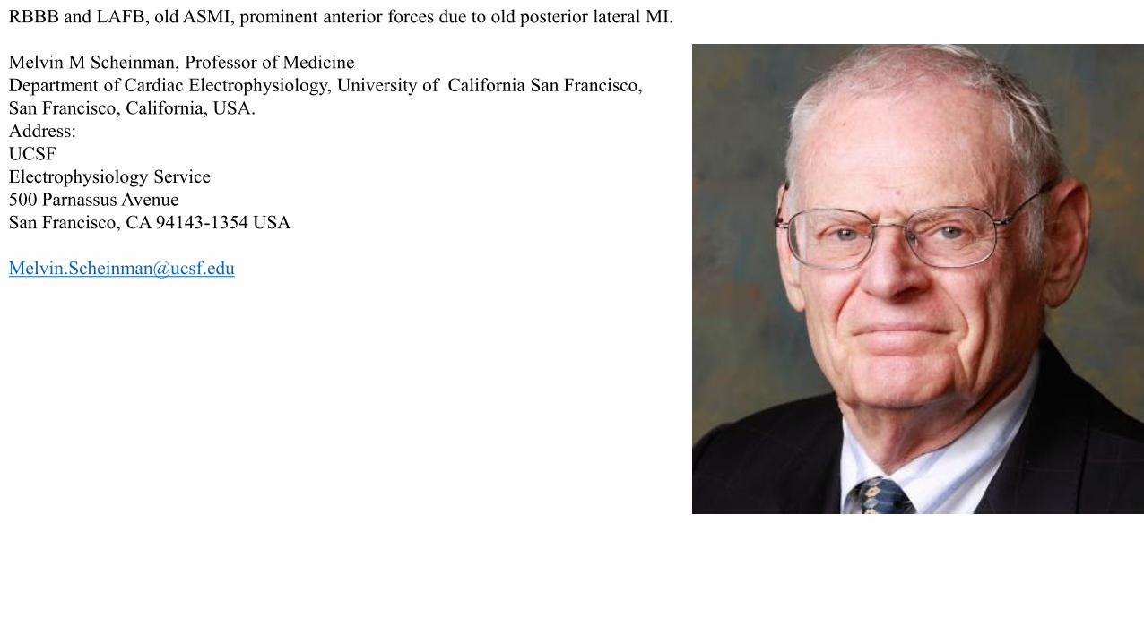
RBBB and LAFB, old ASMI, prominent anterior forces due to old posterior lateral MI.
Melvin M Scheinman, Professor of Medicine Department of Cardiac Electrophysiology, University of California San Francisco, San Francisco, California, USA. Address: UCSFElectrophysiology Service 500 Parnassus AvenueSan Francisco, CA 94143-1354 USA

Final Comments
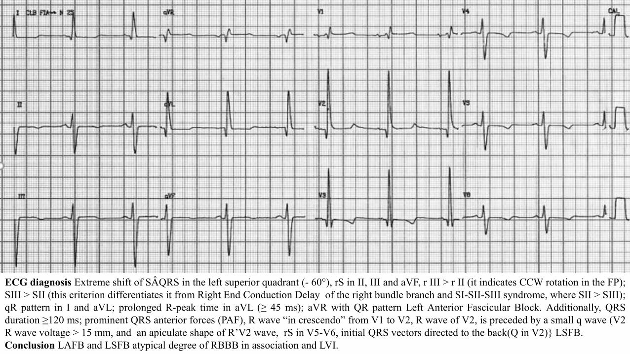
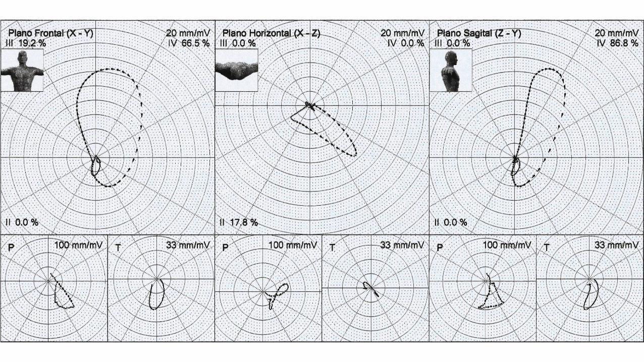
ECG diagnosis Extreme shift of SÂQRS in the left superior quadrant (- 60°), rS in II, III and aVF, r III > r II (it indicates CCW rotation in the FP);SIII > SII (this criterion differentiates it from Right End Conduction Delay of the right bundle branch and SI-SII-SIII syndrome, where SII > SIII);qR pattern in I and aVL; prolonged R-peak time in aVL (≥ 45 ms); aVR with QR pattern Left Anterior Fascicular Block. Additionally, QRSduration ≥120 ms; prominent QRS anterior forces (PAF), R wave “in crescendo” from V1 to V2, R wave of V2, is preceded by a small q wave (V2R wave voltage > 15 mm, and an apiculate shape of R’V2 wave, rS in V5-V6, initial QRS vectors directed to the back(Q in V2)} LSFB.Conclusion LAFB and LSFB atypical degree of RBBB in association and LVI.
IIIIIaVF
X I
Y
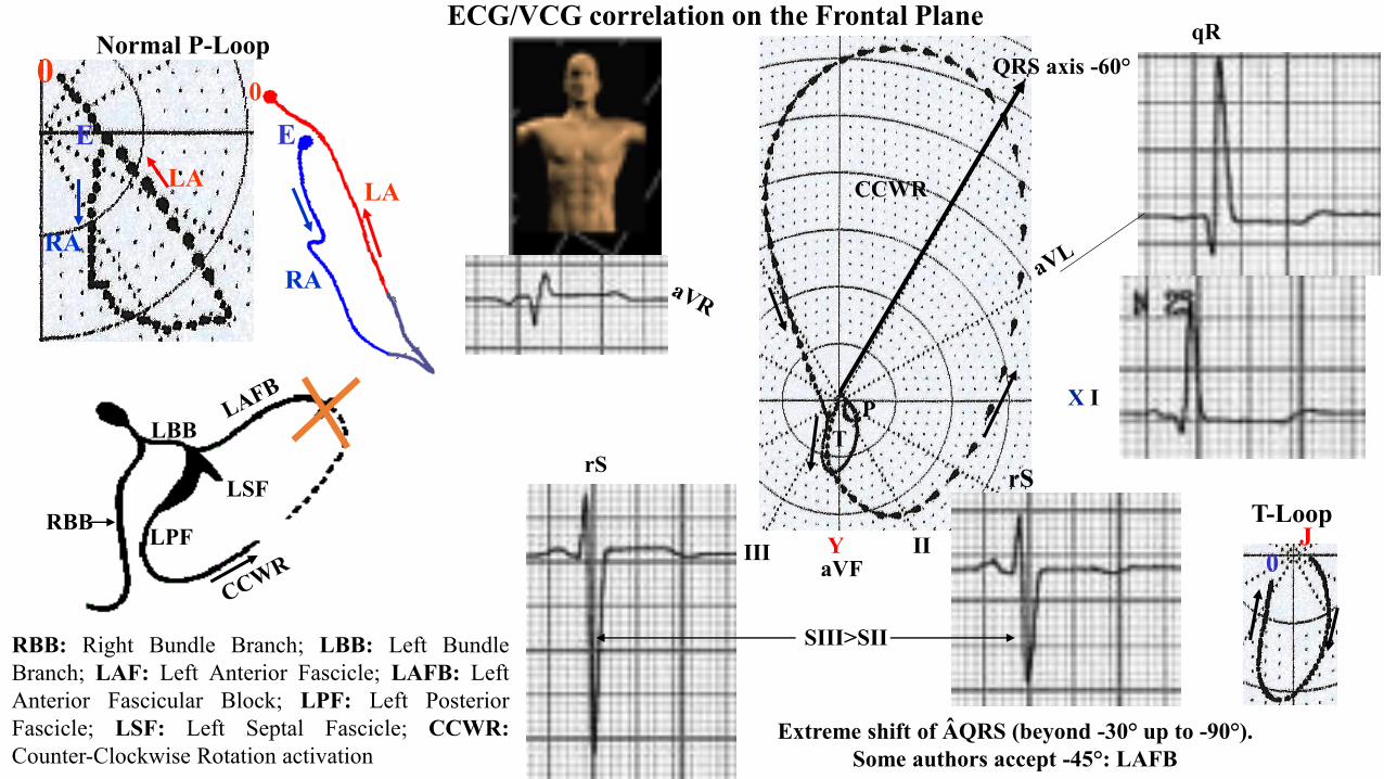
ECG/VCG correlation on the Frontal Plane
QRS axis -60°
PT
SIII>SII
CCWR
LBB
LPF
LSFRBB
RBB: Right Bundle Branch; LBB: Left BundleBranch; LAF: Left Anterior Fascicle; LAFB: LeftAnterior Fascicular Block; LPF: Left PosteriorFascicle; LSF: Left Septal Fascicle; CCWR:Counter-Clockwise Rotation activation
rS rS
qR
Extreme shift of ÂQRS (beyond -30° up to -90°). Some authors accept -45°: LAFB
E
0
RA
LA
EE
RA
LA
0
Normal P-Loop
T-LoopJ
0

R- Wave Peak Time (RWPT) in aVL ≥ 45 ms
aVL
QRS duration 120ms consequence of LAFB(+2ms) and LPFB(+2ms) + degree of RBBB

Each dash or comet represents a time of 2ms, with the present calibration ofthe device. In the present case we have 69 dashes or 120ms
The greater or the lesser distance between dashes indicates the greater or the lesserconduction velocity in the area. Thus, when they are very close to each other, itindicates the presence of conduction delay. To consider the phenomenon as true, it isnecessary for it to be evident in at least 2 planes.
Frontal Plane
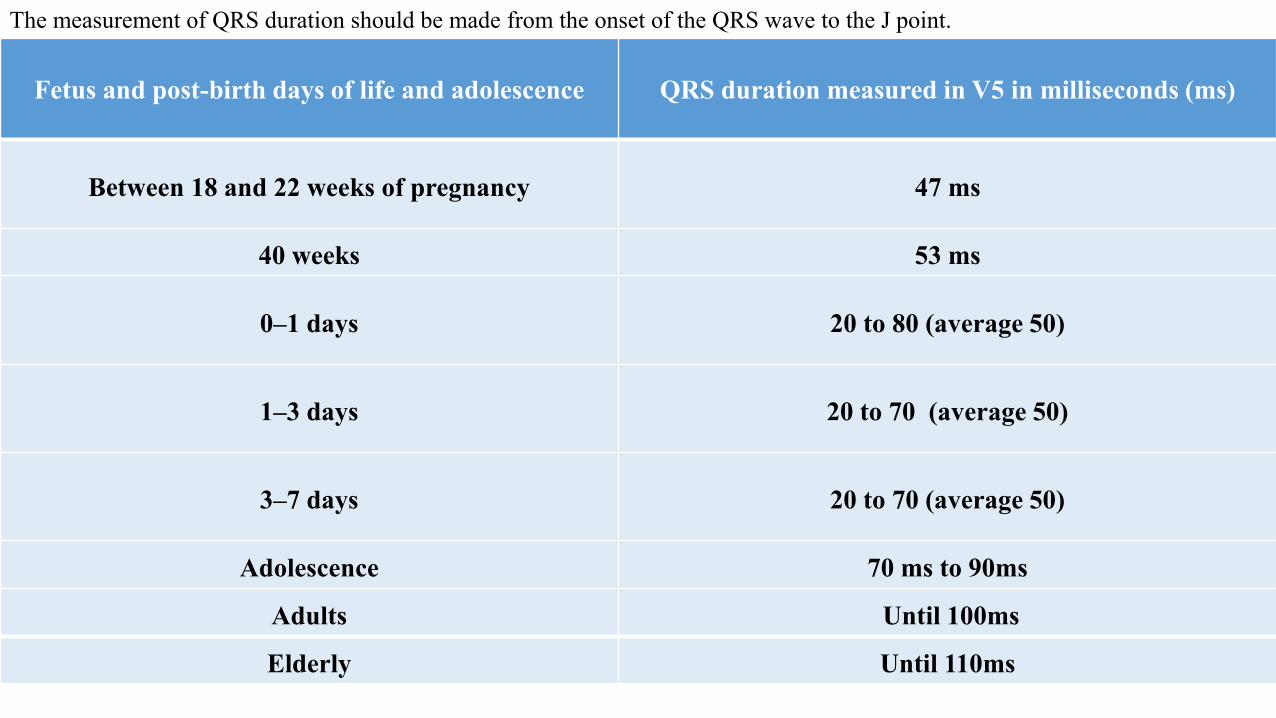
The measurement of QRS duration should be made from the onset of the QRS wave to the J point.
Fetus and post-birth days of life and adolescence QRS duration measured in V5 in milliseconds (ms)
Between 18 and 22 weeks of pregnancy 47 ms
40 weeks 53 ms
0–1 days 20 to 80 (average 50)
1–3 days 20 to 70 (average 50)
3–7 days 20 to 70 (average 50)
Adolescence 70 ms to 90ms
Adults Until 100ms
Elderly Until 110ms
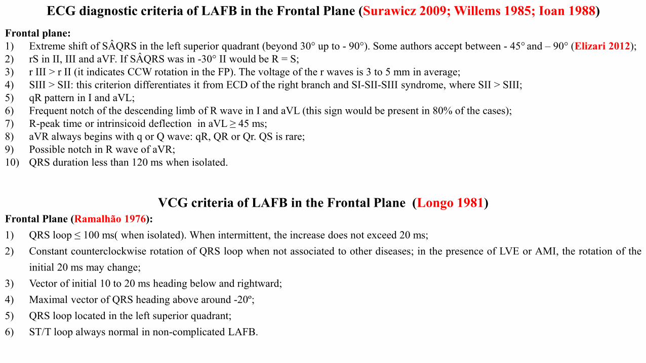
ECG diagnostic criteria of LAFB in the Frontal Plane (Surawicz 2009; Willems 1985; Ioan 1988)Frontal plane:1) Extreme shift of SÂQRS in the left superior quadrant (beyond 30° up to - 90°). Some authors accept between - 45° and – 90° (Elizari 2012);2) rS in II, III and aVF. If SÂQRS was in -30° II would be R = S;3) r III > r II (it indicates CCW rotation in the FP). The voltage of the r waves is 3 to 5 mm in average;4) SIII > SII: this criterion differentiates it from ECD of the right branch and SI-SII-SIII syndrome, where SII > SIII;5) qR pattern in I and aVL;6) Frequent notch of the descending limb of R wave in I and aVL (this sign would be present in 80% of the cases);7) R-peak time or intrinsicoid deflection in aVL ≥ 45 ms;8) aVR always begins with q or Q wave: qR, QR or Qr. QS is rare;9) Possible notch in R wave of aVR;10) QRS duration less than 120 ms when isolated.
VCG criteria of LAFB in the Frontal Plane (Longo 1981)Frontal Plane (Ramalhão 1976):1) QRS loop ≤ 100 ms( when isolated). When intermittent, the increase does not exceed 20 ms;2) Constant counterclockwise rotation of QRS loop when not associated to other diseases; in the presence of LVE or AMI, the rotation of the
initial 20 ms may change;3) Vector of initial 10 to 20 ms heading below and rightward;4) Maximal vector of QRS heading above around -20º;5) QRS loop located in the left superior quadrant;6) ST/T loop always normal in non-complicated LAFB.
X V6
V1
V4
V5
V2 V3
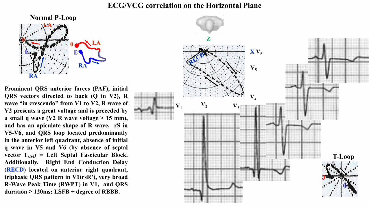
ECG/VCG correlation on the Horizontal Plane
Z
Prominent QRS anterior forces (PAF), initialQRS vectors directed to back (Q in V2), Rwave “in crescendo” from V1 to V2, R wave ofV2 presents a great voltage and is preceded bya small q wave (V2 R wave voltage > 15 mm),and has an apiculate shape of R wave, rS inV5-V6, and QRS loop located predominantlyin the anterior left quadrant, absence of initialq wave in V5 and V6 (by absence of septalvector 1AM) = Left Septal Fascicular Block.Additionally, Right End Conduction Delay(RECD) located on anterior right quadrant,triphasic QRS pattern in V1(rsR’), very broadR-Wave Peak Time (RWPT) in V1, and QRSduration ≥ 120ms: LSFB + degree of RBBB.
EE
RA
LA0E
RA
LA
0
Normal P-Loop
T-Loop
0J
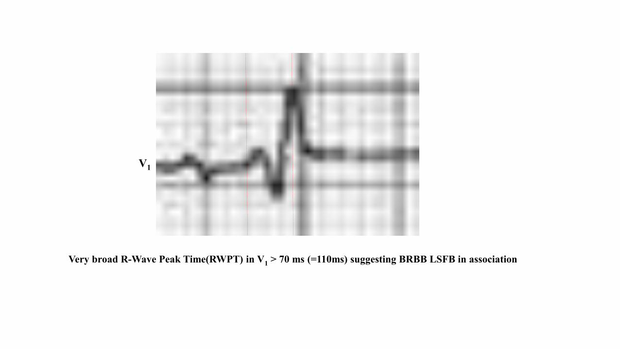
Very broad R-Wave Peak Time(RWPT) in V1 > 70 ms (=110ms) suggesting BRBB LSFB in association
V1
YaVF
Z V2
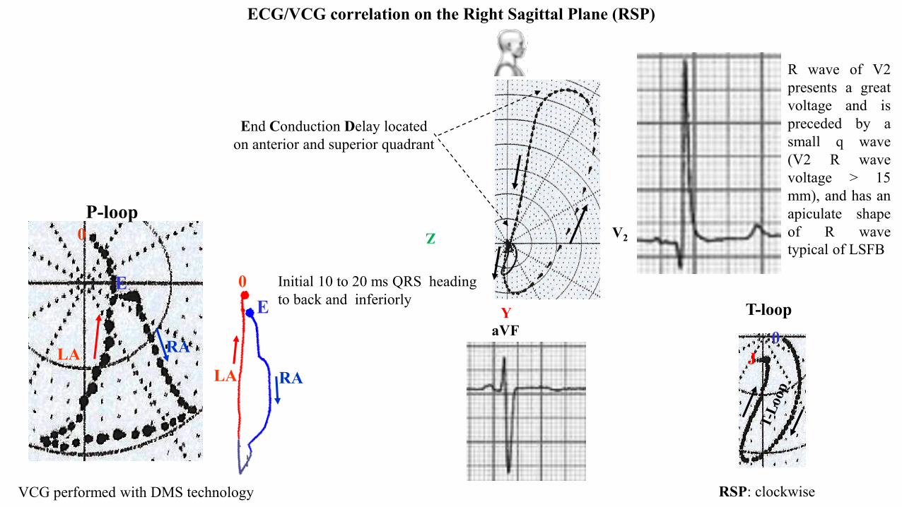
ECG/VCG correlation on the Right Sagittal Plane (RSP)
R wave of V2presents a greatvoltage and ispreceded by asmall q wave(V2 R wavevoltage > 15mm), and has anapiculate shapeof R wavetypical of LSFB
Initial 10 to 20 ms QRS heading to back and inferiorly
End Conduction Delay located on anterior and superior quadrant
E
0
RALA
EE
RALA
0
P-loop
T-loop
J0
RSP: clockwiseVCG performed with DMS technology
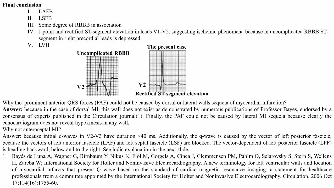
Final conclusionI. LAFBII. LSFBIII. Some degree of RBBB in associationIV. J-point and rectified ST-segment elevation in leads V1-V2, suggesting ischemic phenomena because in uncomplicated RBBB ST-
segment in right precordial leads is depressed.V. LVH
Why the prominent anterior QRS forces (PAF) could not be caused by dorsal or lateral walls sequela of myocardial infarction?Answer: because in the case of dorsal MI, this wall does not exist as demonstrated by numerous publications of Professor Bayés, endorsed by aconsensus of experts published in the Circulation journal(1). Finally, the PAF could not be caused by lateral MI sequela because clearly theechocardiogram does not reveal hypokinesis in any wall.Why not anteroseptal MI?Answer: because initial q-waves in V2-V3 have duration <40 ms. Additionally, the q-wave is caused by the vector of left posterior fascicle,because the vectors of left anterior fascicle (LAF) and left septal fascicle (LSF) are blocked. The vector-dependent of left posterior fascicle (LPF)is heading backward, below and to the right. See ludic explanation in the next slide.1. Bayés de Luna A, Wagner G, Birnbaum Y, Nikus K, Fiol M, Gorgels A, Cinca J, Clemmensen PM, Pahlm O, Sclarovsky S, Stern S, Wellens
H, Zareba W; International Society for Holter and Noninvasive Electrocardiography. A new terminology for left ventricular walls and locationof myocardial infarcts that present Q wave based on the standard of cardiac magnetic resonance imaging: a statement for healthcareprofessionals from a committee appointed by the International Society for Holter and Noninvasive Electrocardiography. Circulation. 2006 Oct17;114(16):1755-60.
Uncomplicated RBBB
V2 V2
The present case
Rectified ST-segment elevation
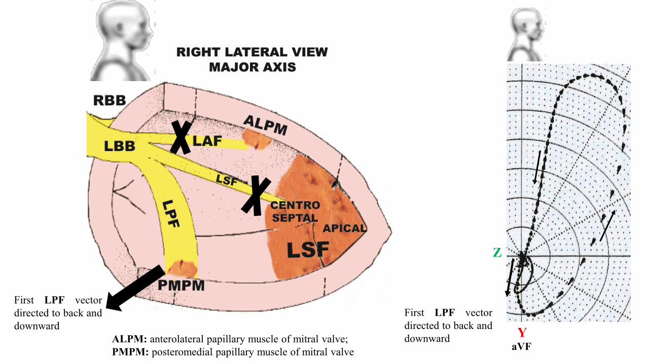
Y
Z
First LPF vectordirected to back anddownward
First LPF vectordirected to back anddownwardALPM: anterolateral papillary muscle of mitral valve;
PMPM: posteromedial papillary muscle of mitral valve aVF
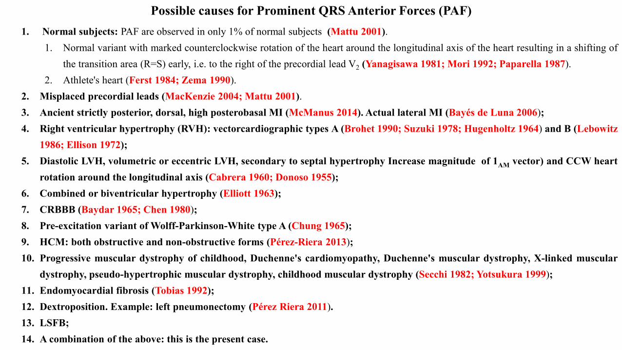
1. Normal subjects: PAF are observed in only 1% of normal subjects (Mattu 2001).1. Normal variant with marked counterclockwise rotation of the heart around the longitudinal axis of the heart resulting in a shifting of
the transition area (R=S) early, i.e. to the right of the precordial lead V2 (Yanagisawa 1981; Mori 1992; Paparella 1987).2. Athlete's heart (Ferst 1984; Zema 1990).
2. Misplaced precordial leads (MacKenzie 2004; Mattu 2001).3. Ancient strictly posterior, dorsal, high posterobasal MI (McManus 2014). Actual lateral MI (Bayés de Luna 2006);4. Right ventricular hypertrophy (RVH): vectorcardiographic types A (Brohet 1990; Suzuki 1978; Hugenholtz 1964) and B (Lebowitz
1986; Ellison 1972);5. Diastolic LVH, volumetric or eccentric LVH, secondary to septal hypertrophy Increase magnitude of 1AM vector) and CCW heart
rotation around the longitudinal axis (Cabrera 1960; Donoso 1955);6. Combined or biventricular hypertrophy (Elliott 1963);7. CRBBB (Baydar 1965; Chen 1980);8. Pre-excitation variant of Wolff-Parkinson-White type A (Chung 1965);9. HCM: both obstructive and non-obstructive forms (Pérez-Riera 2013);10. Progressive muscular dystrophy of childhood, Duchenne's cardiomyopathy, Duchenne's muscular dystrophy, X-linked muscular
dystrophy, pseudo-hypertrophic muscular dystrophy, childhood muscular dystrophy (Secchi 1982; Yotsukura 1999);11. Endomyocardial fibrosis (Tobias 1992);12. Dextroposition. Example: left pneumonectomy (Pérez Riera 2011).13. LSFB;14. A combination of the above: this is the present case.
Possible causes for Prominent QRS Anterior Forces (PAF)
V6X
Z
TP
Afferent limb
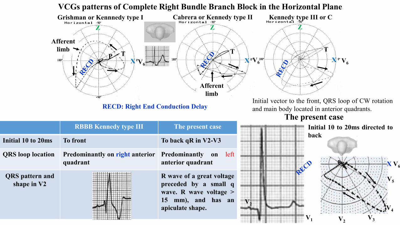
RECD: Right End Conduction Delay
V6X
Z
V6X
Z
T
Afferent limb
T
Grishman or Kennnedy type I Cabrera or Kennedy type II Kennedy type III or C
Initial vector to the front, QRS loop of CW rotationand main body located in anterior quadrants.
VCGs patterns of Complete Right Bundle Branch Block in the Horizontal Plane
X V6
V1
V4
V5
V2V3
V2
RBBB Kennedy type III The present case
Initial 10 to 20ms To front To back qR in V2-V3
QRS loop location Predominantly on right anteriorquadrant
Predominantly on leftanterior quadrant
QRS pattern and shape in V2
R wave of a great voltagepreceded by a small qwave. R wave voltage >15 mm), and has anapiculate shape.
The present caseInitial 10 to 20ms directed toback