Embed Size (px)
Citation preview
Functional Identification of APIP as Human mtnB, a KeyEnzyme in the Methionine Salvage PathwayCamille Mary1*, Paula Duek1, Lisa Salleron2, Petra Tienz3, Dirk Bumann3, Amos Bairoch1,2, Lydie Lane1,2
1 CALIPHO group, SIB-Swiss Institute of Bioinformatics, University of Geneva, Geneva, Switzerland, 2 Department of Human Protein Sciences, University of Geneva,
Geneva, Switzerland, 3 Biologie of infection, Biozentrum, University of Basel, Basel, Switzerland
Abstract
The methionine salvage pathway is widely distributed among some eubacteria, yeast, plants and animals and recycles thesulfur-containing metabolite 5-methylthioadenosine (MTA) to methionine. In eukaryotic cells, the methionine salvagepathway takes place in the cytosol and usually involves six enzymatic activities: MTA phosphorylase (MTAP, EC 2.4.2.28), 59-methylthioribose-1-phosphate isomerase (mtnA, EC 5.3.1.23), 59-methylthioribulose-1-phosphate dehydratase (mtnB, EC:4.2.1.109), 2,3-dioxomethiopentane-1-phosphate enolase/phosphatase (mtnC, EC 3.1.3.77), aci-reductone dioxygenase(mtnD, EC 1.13.11.54) and 4-methylthio-2-oxo-butanoate (MTOB) transaminase (EC 2.6.1.-). The aim of this study was tocomplete the available information on the methionine salvage pathway in human by identifying the enzyme responsible forthe dehydratase step. Using a bioinformatics approach, we propose that a protein called APIP could perform this role. Theinvolvement of this protein in the methionine salvage pathway was investigated directly in HeLa cells by transient andstable short hairpin RNA interference. We show that APIP depletion specifically impaired the capacity of cells to grow inmedia where methionine is replaced by MTA. Using a Shigella mutant auxotroph for methionine, we confirm that theknockdown of APIP specifically affects the recycling of methionine. We also show that mutation of three potentialphosphorylation sites does not affect APIP activity whereas mutation of the potential zinc binding site completely abrogatesit. Finally, we show that the N-terminal region of APIP that is missing in the short isoform is required for activity. Together,these results confirm the involvement of APIP in the methionine salvage pathway, which plays a key role in many biologicalfunctions like cancer, apoptosis, microbial proliferation and inflammation.
Citation: Mary C, Duek P, Salleron L, Tienz P, Bumann D, et al. (2012) Functional Identification of APIP as Human mtnB, a Key Enzyme in the Methionine SalvagePathway. PLoS ONE 7(12): e52877. doi:10.1371/journal.pone.0052877
Editor: Ferdinando Di Cunto, University of Turin, Italy
Received September 18, 2012; Accepted November 22, 2012; Published December 28, 2012
Copyright: � 2012 Mary et al. This is an open-access article distributed under the terms of the Creative Commons Attribution License, which permitsunrestricted use, distribution, and reproduction in any medium, provided the original author and source are credited.
Funding: This project was financed with a grant from the Swiss SystemsX.ch initiative (BattleX), evaluated by the Swiss National Science Foundation. The fundershad no role in study design, data collection and analysis, decision to publish, or preparation of the manuscript.
Competing Interests: The authors have declared that no competing interests exist.
* E-mail: [email protected]
Introduction
Methionine is an essential amino acid involved in major
functions such as protein synthesis, formation of polyamines, DNA
and protein methylation and protection against reactive oxygen
species though the generation of glutathione [1]. In cells, the
methionine that is not used for protein synthesis is converted into
S-adenosylmethionine (SAM), the principal methyl donor
(Figure 1A). Through the methylation cycle pathway, SAM can
be converted back to methionine via the production of homocys-
teine (Hcy). SAM is also the precursor of polyamines such as
spermine and spermidine. Polyamine synthesis leads to the
production of 59-methylthioadenosine (MTA) as a by-product
[2]. The methionine salvage pathway allows cells to recycle the
reduced sulfur in MTA back into methionine (Figure 1A) [3,4].
The methionine salvage pathway and the polyamine synthesis
seem to be tightly coupled, probably in order to maintain
intracellular levels of SAM. For example, it has been shown that
the level and activity of ornithine decarboxylase, the rate-
controlling enzyme in polyamine synthesis, can be modulated by
the first and last metabolites of the methionine salvage pathway:
MTA and 4-methhylthio-2-oxo-butanoate (MTOB) [5–7]. The
methionine salvage pathway may also have an important role in
apoptotic processes as both MTA and MTOB were found to
induce apoptosis [5,8]. In eukaryotic cells, the methionine salvage
pathway takes place in the cytosol and involves six enzymatic
activities: MTA phosphorylase (MTAP, EC 2.4.2.28), 59-
methylthioribose-1-phosphate isomerase (mtnA, EC 5.3.1.23), 59-
methylthioribulose-1-phosphate dehydratase (mtnB, EC 4.2.1.109),
2,3-dioxomethiopentane-1-phosphate enolase/phosphatase (mtnC,
EC 3.1.3.77), aci-reductone dioxygenase (mtnD, EC 1.13.11.54)
and MTOB transaminase (EC 2.6.1.-) [4]. The transamination step
can be catalyzed by a range of transaminases, which preferentially
use aromatic and branched chain amino group donors [9].
The inventory of all enzymes involved in the methionine salvage
has been recently achieved in yeast [9], but the pathway is still
poorly functionally characterized in human. However, it has been
attracting some interest for decades, mainly because its first
enzyme, MTA phosphorylase, is frequently deficient in cancer
cells and primary tumors [10–14].
At the beginning of our study, human MTAP was already
functionally characterized as well as ADI1, that performs mtnD
activity [15,16]. Human mtnC enzyme was found to be ENOPH1
by homology with the Bacillus subtilis enzyme and its structure was
solved by X-ray in the presence of a substrate analog to decipher
its enzymatic mechanism [17]. There is strong evidence that
MTOB transamination is mainly performed by glutamine
transaminases [18–20]. The mtnA enzyme has been recently
PLOS ONE | www.plosone.org 1 December 2012 | Volume 7 | Issue 12 | e52877
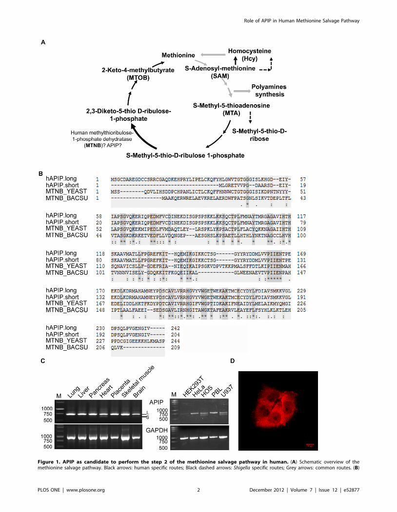
Figure 1. APIP as candidate to perform the step 2 of the methionine salvage pathway in human. (A) Schematic overview of themethionine salvage pathway. Black arrows: human specific routes; Black dashed arrows: Shigella specific routes; Grey arrows: common routes. (B)
Role of APIP in Human Methionine Salvage Pathway
PLOS ONE | www.plosone.org 2 December 2012 | Volume 7 | Issue 12 | e52877
functionally characterized as MRI1, a protein that is induced in
metastatic cells and promotes cell invasiveness [21].
Despite the conservation of the dehydratase step (mtnB, EC
4.2.1.109) in all organisms, it is one of the least studied enzymes in
the pathway. Detailed enzymatic characterization was performed
on recombinant Bacillus subtilis mtnB [22]. Subsequently, although
homologs have been found in many organisms, functional
characterization has been achieved so far only for Pseudomonas
aeruginosa, Saccharomyces cerevisiae and Tetrahymena thermophila ortho-
logs [23,24]. The mtnB enzyme of Tetrahymena thermophila
represents a divergent evolution as it was shown to be fused with
mtnC and able to perform by itself three steps of the pathway
(mtnB, mtnC, mtnD). Another case of gene fusion was observed in
Arabidopsis thaliana, where the bifunctional enzyme encoded by
At5g53850 probably mediates both mtnB and mtnC activities
[22,24]. The lack of detailed enzymatic studies for this enzyme
may be due to the lack of commercial availability of the substrate
and to the instability of the product [22].
The aim of this study was to complete the available information
on the methionine salvage pathway in human cells by identifying
the enzyme responsible for the dehydratase step (mtnB, EC
4.2.1.109). During the development of the HAMAP automatic
annotation platform by the Swiss-Prot group [25] a sequence
profile for the mtnB family was created in collaboration with the
group of Prof. A. Danchin [24]. This profile as well as BLAST
searches demonstrate that the closest human homolog of yeast
MDE1/mtnB and Bacillus subtilis mtnB is a protein called APAF1-
interacting-protein (HGNC gene symbol APIP, Q96GX9). We
therefore changed its functional annotation in the UniProtKB/
Swiss-Prot database into ‘‘Probable methylthioribulose-1-phos-
phate dehydratase (EC 4.2.1.109)’’. One of the goals of our group
being the experimental validation of bioinformatics predictions
relevant to the function of human proteins, we decided to embark
in the functional characterization of APIP. We used RNA
interference in HeLa cells to validate the prediction that APIP is
part of the methionine salvage pathway in human. Mutational
analysis of the protein shows that mutation of the potential zinc
binding site, inferred by the alignment of bacterial and yeast
orthologs, abrogates APIP activity. We also demonstrate that an
alternatively spliced isoform that lacks the first 38 N-terminal
residues is catalytically inactive.
Materials and Methods
Sequence analysisThe putative human ortholog for the mtnB enzymes from
Bacillus subtilis (O31668) and Saccharomyces cerevisiae (P47095) was
searched by BLASTP analysis in the UniProtKB database version
15.14 [26]. Multiple sequence alignments were performed using
Clustal Omega with the default parameters [27].
Chemicals, antibodies, plasmids and cell cultureL-Methionine, Hcy, MTA, MTOB and SAM were purchased
from Sigma. Anti-APIP and anti-V5 antibodies were from Santa-
Cruz Biotechnology (D-20; 1/500 for Western blot) and AbD
Serotec (MCA1360; 1/1000 for Western blot), respectively. Alpha-
tubulin (a-tub) antibody was purchased from Sigma (T6074: 1/
1000 for Western blot). ShRNAs were cloned into pRNAi-H1-
Puro vector from Biosettia. APIP.long and APIP.short cDNAs
were amplified by PCR from HeLa and HOS cells respectively
using specific primers. Cloning into pcDNA3.2-V5 plasmid
(Invitrogen) was achieved using the Gateway system (Invitrogen).
V5APIP3HA, V5APIP3SA and V5APIP3SD were obtained
though several rounds of site-directed mutagenesis using the
Quickchange method (Stratagene). HeLa Kyoto cells (kind gift
from Dr. Cecile Arrieumerlou) were cultured in complete or
methionine free Dulbecco’s modified Eagle’s medium (DMEM,
Invitrogen) supplemented with 10% fetal calf serum (Gibco,
Invitrogen). Cells were incubated at 37uC in an atmosphere of 5%
CO2 in air.
ImmunofluorescenceHeLa cells were fixed in 4% formaldehyde in PBS/pH 4 for
15 minutes at room temperature. After three washings of
5 minutes in PBS, cells were blocked in PBS supplemented with
5% donkey serum, 0.3% triton for 1 hour. APIP primary antibody
(1/100) was incubated overnight in PBS containing 1% BSA,
0.3% Triton. After three washes in PBS, cells were incubated in
Alexa donkey anti-goat secondary antibody (Invitrogen) for
1 hour.
shRNA sequences, PCR and RT-PCR primerssh1APIP: aaaaGAGCATCCAAGATACCTGATCttggatccaa-
GATCAGGTATCTTGGATGCTC
sh2APIP :
aaaaCTGCAAATGGTCACCCTGAATttggatccaaATTCAGG-
GTGACCATTTGCAG
Primers for APIP cDNAs amplification and RT-PCR:
APIP.long sense 59- caccATGTCTGGCTGTGATGCTCG-39,
APIP.short sense 59-caccATGCTCGGGAGGGAGACTGTT-39
APIP.long and APIP.short antisense 59- CTTTCTTTTGGC-
TTAGACAATTCCATTTTC-39
GAPDH sense 59-TGAAGGTCGGTGTCAACGGATTTG-
GC-39
GAPDH antisense 59-CATGTAGGCCATGAGGTCCACCA-
C-39
Primers for the generation of Shigella metA mutant:
MetA_3 and metA_4 were used for the amplification of a
chloramphenicol cassette with flanking 60 bp sequences (lower case)
that were homologous to Shigella chromosomal regions adjacent to
the metA gene. MetA_1 and MetA_2 were used for screening of
chloramphenicol resistant clones for metA replacement.
metA_1: 59- GTGAGCGGCGAATACTA-39
metA_2: 59-TTCACTTGCTGAGGTGC-39
metA_3: 59-gttatcttcagctatctggatgtctaaacgtataagcgtatgtagtgagg-
taatcaggttGTGTAGGCTGGAGCTGCTTCGA-39
metA_4: 59-gcacccgaaggtgcctgaggtaaggtgctgaatcgcttaacgatcgac-
tatcacagaagaCATATGAATATCCTCCTTAGTTCC-39
Sequence alignment of human APIP long and short isoforms (APIP.long and APIP.short) with mtnB enzymes from the yeast Saccharomyces cerevisiaeand the bacteria Bacillus subtilis. (C) Analysis of the expression pattern of APIP mRNA by semi-quantitative RT–PCR. Left panel: Expression in tissues.This experiment was performed using a commercially obtained pre-normalized human multiple tissue cDNA panel (Clontech, panel I). Right panel:Expression in cell lines. The experiment was performed using total RNA preparations derived from human cell lines. The GAPDH gene was used as aninternal control. For APIP at least two bands were detected (arrows) and confirmed to be the short and long isoforms after sequencing (APIP.long:729 bp; APIP.short: 628 bp). M: Size marker; S: APIP.short; L: APIP.long. (D) Immunofluorence analysis of APIP into HeLa cells show a majorcytoplasmic staining.doi:10.1371/journal.pone.0052877.g001
Role of APIP in Human Methionine Salvage Pathway
PLOS ONE | www.plosone.org 3 December 2012 | Volume 7 | Issue 12 | e52877
Semi quantitative RT-PCRA total of 1 mg of total RNA, isolated using the RNeasy kit
(Qiagen), was used for reverse transcription with the Superscript II
RNase H Reverse Transcriptase (Invitrogen) and random primers
(Promega) according to manufacturer’s protocol. The PCR
amplification was performed with an annealing temperature of
56uC, one minute of elongation time and 26 cycles of amplifica-
tion.
Western blottingHeLa cells were lysed with RIPA buffer for 30 minutes on ice
and centrifuged for 20 min at 10 000 g to remove cellular debris.
80 mg of cells extract proteins were loaded on SDS-PAGE gel and
transferred onto PVDF membrane.
Transfection and cell growth assaysAll the transfections were performed with Fugene HD (Roche)
according to manufacturer’s protocol. For transient experiments,
HeLa cells were initially transfected with plasmids expressing
shbGal, sh1APIP or sh2APIP. After 24 hours, cells were submitted
to puromycin selection for 24 additional hours (2 mg/ml,
Invitrogen). At 48 hours, cells were plated equally into 12 wells
dishes (50 000 cells/well) and let to grow for 24 hours. For
complementation experiments, cells were re-transfected with wild
type or mutant V5APIP 72 hours after the initial transfection with
shRNA plasmids. At 96 hours, media were switched to the
different methionine, Hcy, MTA, MTOB, SAM media according
to the experiments. Cells were counted at time point 144 hours
(i.e. 48 hours after the media switch), using Alamarblue (Invitro-
gen) according to manufacturer’s protocol. Alamarblue fluores-
cence was found to correlate very closely with manual cell
counting (data not shown). To establish HeLa cell lines stably
down-regulating APIP, HeLa cells transfected with shRNA vectors
were selected with 1 mg/ml puromycin. Single colonies were
isolated and analyzed for APIP down-regulation by Western
blotting. The cell line with the best knock-down was harvested in
nitrogen liquid. Cell growth assay in the different media was
assessed at least three days after the cell lines were defrosted. For
complementation in stable cell lines, equal amount of cells were
plated in 12 well plates and transfected with 1 mg plasmid DNA/
well. For co-expression experiments, the cells were transfected with
0.5 mg of each plasmid. The medium was changed 24 hours after
transfection. Alamarblue measurements were performed after two
days of incubation in the different media.
Shigella infection experimentExperiments were performed with Shigella flexneri serotype 2a
strain 2457T with a mutation in virG (also called icsA) that prevent
the bacteria move within the HeLa cell and spread from cell to cell
[28]. This strain was a kind gift from Prof. Marcia Goldberg. The
Shigella virG was transformed with the pNF106 vector that harbors
a tetracyclin-inducible GFP construct (TETr-GFP) derived from
the pZS21 vector [29]. The metA mutant was generated using an
adapted method form from Datsenko and Wanner [30]. The
chloramphenicol resistance cassette was amplified by PCR from a
strain already carrying this cassette (see above for the primers).
PCR products were transformed by electroporation into a 2457T
virG strain that carries the pKM208 plasmid with the lambda
recombinase system [31]. Chloramphenicol resistant clones were
screened by PCR using primers located outside the metA gene (see
above for the primers). Correct mutants were used as donors for
transduction into Shigella flexneri serotype 2a strain 2457T virG
using phage P1. The day before infection, HeLa cells were seeded
in 96 wells plates (105 HeLa cells/well). Two hours before
infection, the medium (DMEM+FCS) was removed and replaced
by meth2/MTA DMEM without FCS. Prior to cell infection,
Shigella liquid cultures were grown at 37uC with shaking (200 rpm)
to exponential phase (approximate optical density at
600 nm = 0.5). 1 ml of bacterial culture was spun down and re-
suspended into 0.001% poly-L-Lysine (Sigma) in pre-warmed PBS
and incubated for 15 minutes at 37uC. Treated Shigella were
washed with PBS and re-suspended in pre-warmed meth2/MTA
DMEM. Bacteria were added to cell monolayers at a multiplicity
of infection (MOI) of 10 per HeLa cell, and the samples were
centrifuged for 5 min at 6006 g. After 30 min of incubation at
37uC, extracellular bacteria were killed by adding gentamycin
(50 mg ml21, Gibco) and GFP fluorescence was induced by adding
anhydrotetracycline (aTc, Sigma). Infected cells were incubated at
37uC for up to 4 hours, trypsinized and fixed with 1%
paraformaldehyde before flow cytometry analysis. The samples
were analysed by a four-parameter (two scatter parameters, two
fluorescence colours) flow cytometer equipped with a blue laser
(488 nm) and an automated sample loader for 96 well plates (BD
Bioscience HTS-LSR II SORP). HeLa cells and liberated
individual Shigella were detected based on their particle size
(forward scatter) and granularity (side scatter). The fluorescence
properties of the gated particles were then analysed by exciting
them with the blue laser and detection of the signals in the green
(Ex488 LP502 PB530/30) and orange (Ex488 LP556 BP 585/42)
channels. Data were stored in FCS 3.0 format and several
descriptive statistics (mean, histogram) were calculated by the
DIVA software � 2006, Becton, Dickinson and Company. The
total number of Shigella per infected host cell (bacterial load) was
calculated as follows: (arithmetic mean of total green fluorescence
of infected HeLa cells - arithmetic mean of total green fluorescence
of non-infected HeLa cells)/average green fluorescence of
individual Shigella.
Statistical analysisData are shown as mean +/2 SD of results from at least three
independent experiments. Statistical analysis was done by
performing two-way ANOVA followed by Bonferroni post-test
analysis (star marks in the figure legends: * = p,0.05, ** = p,0.01,
*** = p,0.001).
Results
Identification of APIP as the candidate human mtnBenzyme
To confirm that APIP is the most likely human protein capable
of performing mtnB activity, we performed a BLAST search on
UniProtKB using the sequences of the Bacillus subtilis and
Saccharomyces cerevisiae enzymes. APIP was found to be the best
hit and presents 22.7% and 25.7% identity with the Bacillus subtilis
and yeast enzymes, respectively. BLAST search using the human
APIP sequence retrieves mtnB from both species, confirming that
the three proteins are orthologs.
Two splice isoforms are described for APIP (APIP.long and
APIP.short) [32]. APIP.short has 38 less residues in its N-terminus
(Figure 1B).
We analyzed the expression pattern of the two APIP isoforms in
different tissues and cell lines (Figure 1C). APIP.long was expressed
in all the samples tested, presuming a ubiquitous expression.
APIP.short was expressed at lower levels and detected in heart,
brain, pancreas, liver and placenta. APIP.short was not detected in
HeLa and PBL cells but was well amplified in HOS and U937
cells.
Role of APIP in Human Methionine Salvage Pathway
PLOS ONE | www.plosone.org 4 December 2012 | Volume 7 | Issue 12 | e52877
In accordance with its putative role in the methionine salvage
pathway, APIP was detected mainly in the cytoplasm of HeLa cells
by immunofluorescence (Figure 1D).
Transient silencing of APIP in HeLa cells abrogates theirgrowth in meth2/MTA medium
Cells competent for the methionine salvage pathway, which
recycles MTA to methionine, should be able to grow in meth2/
MTA medium. To verify that HeLa cells have a functional
methionine salvage pathway, we compared their proliferation in
meth+, meth2 or meth2/MTA medium using Alamarblue
fluorescence (Figure 2D). Cells treated with short hairpin RNA
(shRNA) against the b-galactosidase (shbGal) for 96 hours (used as
control cells) presented an average two-fold lower proliferation in
meth2 medium than in meth+ medium. However, proliferation of
cells in meth2/MTA was not significantly different than in meth+,
confirming previous observations [33], and showing that HeLa
cells’ methionine salvage pathway is not affected by potential off-
target effects due to shRNA treatment.
To confirm the involvement of APIP in the methionine salvage
pathway, we transiently silenced APIP using shRNA in HeLa cells
and looked if it affects their growth in meth2/MTA medium. For
this purpose, we used two shRNA constructions: sh1APIP and
sh2APIP, that target the cDNA region of APIP.long and the
39UTR of the two isoforms, respectively (Figure 2A). RT-PCR
analysis showed a significant knockdown of APIP mRNA 48 hours
after transfection and until 144 hours for both sh1APIP and
sh2APIP (Figure 2B). At protein level, both APIP shRNA
constructs induced a slight depletion 48 h after transfection. The
depletion was stronger at 72 hours and lasted until 144 hours
(Figure 2C).
Silencing of APIP with either sh1APIP or sh2APIP reduces the
proliferation of the HeLa cells in meth2/MTA to almost the same
levels as in meth2, indicating that the methionine salvage pathway
was impaired by APIP silencing (Figure 2D). To further confirm
the specificity of the shRNA phenotype, we performed a rescue
experiment by overexpressing N-terminally V5-tagged APIP.long
(V5APIP) in shRNA treated cells. Overexpression of N-terminally
V5-tagged chloramphenicol acetyltransferase (V5CAT) was used
as control. Transfection with rescue plasmids was performed 72 h
after transfection with shRNA plasmids and the proliferation of
the cells in the different media was assessed as described above. As
observed previously, proliferation of HeLa cells in meth2 was
reduced two fold as compared to meth+. Overexpression of
V5APIP or V5CAT in control (shbGal-treated) cells did not
perturb their capacity to grow in meth+. However, overexpression
of V5APIP, but not V5CAT, rescued the proliferation of the HeLa
cells silenced for APIP with sh2APIP in meth2/MTA. Taken
together, these results suggest that APIP is indeed involved in the
methionine salvage pathway.
Stable knockdown of APIP in HeLa cells specificallyaffects growth in MTA and SAM media and depletesintracellular levels of methionine
To characterize further the involvement of APIP in the
methionine salvage, we engineered HeLa cells stably silenced for
APIP. The knockdown was confirmed by Western blot (Figure 3A).
We then studied the capacity of these HeLa cells to grow after two
days incubation in media containing different sources of methi-
onine (Figure 3A). As expected, both control cells and APIP
knockdown cells presented a decreased proliferation of around 2
fold in meth2 medium. As observed during transient experiment,
knockdown of APIP resulted in a reduced cell proliferation in
meth2/MTA, equivalent as the one observed in meth2. As
expected, knockdown of APIP did not affect proliferation in
meth2/MTOB medium, showing that APIP is involved in the
methionine salvage pathway downstream of MTA but upstream of
MTOB generation.
SAM is a precursor for both the methylation cycle and the
methionine salvage pathway (Figure 1A). Knockdown of APIP
slightly but significantly reduced the proliferation of HeLa cells in
meth2/SAM medium, to a rate of 90% relative to control HeLa
cells. Based on this result we can assume that in this condition most
of the methionine is recycled by the methylation pathway. The last
step of the methylation cycle uses homocysteine as a substrate for
methionine synthase. Although HeLa cell growth was slightly
decreased in meth2/Hcy medium as compared to methionine
medium as previously described [34,35], control and APIP
knockdown HeLa cells proliferate similarly in meth2/Hcy
medium. Taken together, these results suggest that down
regulation of APIP acts specifically on the methionine salvage
pathway, without affecting the methylation pathway.
To confirm that intracellular methionine levels were indeed
affected by APIP knockdown, we performed HeLa cells infection
experiments with either a wild type (wt) strain or a methionine
auxotroph mutant (Shigella metA) of Shigella flexneri. Shigella is able to
synthesize methionine from pyruvate via its own biosynthetic
pathway and is also able to recycle methionine through the
methylation cycle pathway [24,36]. However, Shigella (Figure 1A)
does not possess a functional methionine salvage pathway and is
therefore not able to use MTA as a source a methionine. While
not all the transporters capable of importing methionine in
enterobacteria such as Shigella have been fully characterized at the
molecular level, it is known that Shigella is able to use the
methionine from the host [37–41]. The methionine auxotroph
mutant Shigella metA was obtained by deletion of the homoserine
O-acetyltransferase enzyme (EC 2.3.1.31, metA) from the biosyn-
thetic pathway. We first looked at the percentage of HeLa cells
infected by both bacterial strains in MTA medium (Figure 3C). No
significant difference was observed between the two strains,
meaning that the deletion of metA did not affect the invasion
capability of Shigella. The percentage of infected HeLa cells was
also not significantly changed by the silencing of APIP excluding
an involvement of APIP in the entry process of Shigella into the
cells. Next, we looked at Shigella growth inside HeLa cells by
measuring the load of Shigella per HeLa cell over time (Figure 3D).
Wt and Shigella metA grew similarly in control shbGal HeLa cells.
In these cells, Shigella metA probably compensated their deficiency
in methionine biosynthesis by recruiting the host methionine
provided by the methionine salvage pathway from MTA. In APIP
knockdown cells, the growth of wt Shigella was not significantly
affected, but the growth of Shigella metA was dramatically inhibited,
suggesting insufficient availability of methionine inside the host
cells as a consequence of disruption of the methionine salvage
pathway.
Mutational analysis of APIP activity in the methioninesalvage pathway
mtnB is part of the divalent metal ion-dependent aldolase class
II family which includes bacterial L-ribulose-5-phosphate 4-
epimerase (araD), L-fuculose phosphate aldolase (fucA) and
rhamnulose-1-phosphate aldolase (rhaD) (Figure 4A). The X-ray
structures of E.coli AraD, FucA and RhaD are solved and have
helped to decipher their molecular mechanism [42–47]. These
aldolases use zinc as a co-factor. As expected, the three histidines
involved in zinc binding are conserved in APIP. By site-directed
mutagenesis, we changed these histidines into alanines and tested
Role of APIP in Human Methionine Salvage Pathway
PLOS ONE | www.plosone.org 5 December 2012 | Volume 7 | Issue 12 | e52877
the effect of these mutations on APIP activity in the methionine
salvage pathway. As shown in Figure 4B, overexpression of APIP
triple-alanine mutant (V5APIP3HA) failed to rescue the growth
defect of APIP knockdown HeLa cells in meth2/MTA media,
although correct expression of V5APIP3HA was checked by
Western blot (Figure 4B, panel). Therefore, as expected by
homology to other metal ion-dependent aldolase class II enzymes,
these three histidine residues are important for APIP activity. We
next assessed the activity of the APIP.short isoform by overex-
pressing it in APIP knockdown HeLa cells. As shown in Figure 4B,
although expressed at a similar level as V5APIP, V5APIP.short
failed to rescue the growth defect in MTA medium. Despite the
strong similarity between the two isoforms, the absence of the first
38 amino acids disrupts APIP activity in the methionine salvage
pathway. We then investigated if the inactive APIP.short could
compete with APIP for activity. Overexpression of V5APIP.short
did not affect the capacity of control shbGal HeLa cells to grow in
meth2/MTA medium (data not shown). This observation suggests
that V5APIP.short does not affect the activity of endogenous
APIP. To confirm that V5APIP.short does not act as an inhibitor
of full length APIP, we co-expressed both V5APIP and V5APIP.-
short in the APIP knockdown stable cell line. A 1:1 ratio of DNA
plasmids was used during transfection. Controls were obtained by
co-expressing V5CAT with either V5APIP or V5APIP.short at the
Figure 2. Transient silencing of APIP decreases the growth of the HeLa cells in MTA medium. (A) Schematic representation of the twomRNAs isoforms of APIP. Sequence positions of the shRNAs used in the study (sh1APIP and sh2APIP) are indicated by arrows. (B) Semi-quantitativeRT-PCR analysis of APIP silencing 48 h and 144 h after transfection with plasmids expressing shRNAs. A plasmid expressing shRNA against b-galactosidase (shbGal) was used as a negative control. The GAPDH gene was used as an internal control. (C) Western blot analysis of APIP silencing48, 72 and 144 hours after transfection with plasmids expressing shRNAs. All lanes were loaded with 80 mg of cell lysate proteins. Anti a-tubulin (a-tub) was used as a loading control. The bands present below APIP were nonspecifically stained with the anti-APIP antibody. (D, E) Cell growthanalysis of HeLa cells transiently silenced for APIP. Alamarblue fluorescence is expressed relative to the fluorescence of HeLa cells transfected with thesame plasmids and cultured in normal methionine media. (D) 96 hours after transfection with plasmids expressing shRNAs, the same number of cellswas grown for 48 hours either in complete media, or in methionine free media complemented or not with MTA, and their end point growth wasmeasured by Alamarblue fluorescence (E) 72 hours after transfection with plasmids expressing shRNAs, cells were transfected with plasmidsexpressing the N-terminally V5-tagged APIP protein (V5APIP) or the N-terminally V5-tagged chloramphenicol acetyltransferase protein (V5CAT).24 hours later, the same number of cells was grown for 48 hours either in complete media or in methionine free media complemented or not withMTA, and their end point growth was measured by Alamarblue fluorescence.doi:10.1371/journal.pone.0052877.g002
Role of APIP in Human Methionine Salvage Pathway
PLOS ONE | www.plosone.org 6 December 2012 | Volume 7 | Issue 12 | e52877
same ratio. As observed in Figure 4C, co-expression of V5APIP.-
short did not affect the growth rescue by V5APIP in meth2/MTA
medium. From these results, we conclude that the short isoform of
APIP does not act as a negative regulator of the full length isoform.
APIP was found to be phosphorylated at Ser-87 and Ser-89 in
large scale phosphoproteome studies performed on HeLa S3 cells
[48] and human embryonic stem cells HUES 9 [49]. Another
serine located just upstream (Ser-84) might also be phosphorylated
(Prof. C. Arrieumerlou, personal communication). We mutated
these three serines either in alanine (V5APIP3SA), in order to
abrogate potential phosphorylation, or in aspartic acid to mimic
phosphorylation (V5APIP3SD). As shown in Figure 4D, both
V5APIP3SA and V5APIP3SD were able to rescue APIP
knockdown phenotype in meth2/MTA medium similarly as the
wt form of APIP (V5APIP). Hence, phosphorylation at these sites
seems not to be required for APIP activity.
Discussion
Despite its discovery many years ago [3,50], the methionine
salvage pathway is still incompletely described in terms of
enzymatic composition, especially in human. In this study, we
functionally characterized APIP as the human ortholog of mtnB,
the enzyme catalyzing the dehydratase step. While we were
writing this manuscript, a paper came out presenting similar
results and confirming the function of APIP into the methionine
salvage pathway [51]. MtnB belongs to the divalent metal ion-
dependent aldolase class II family [42–47] that comprises mainly
bacterial enzymes. The characteristic features of this family are
conserved in APIP. The three histidines expected to be responsible
for metal binding based on homology with mtnB [22] are present
in APIP and essential for activity. APIP also possesses a conserved
glutamate that was shown to be important for the deprotonation of
the substrate and residues that were shown to be required for
phosphate binding [45]. Bacillus subtilis mtnB as well as related
enzymes RhaD, AraD and FucA were shown to form homo-
Figure 3. Stable knockdown of APIP specifically affects growth in MTA and depletes intracellular levels of methionine. (A) Westernblot analysis of APIP stable knockdown HeLa cells. (B) Alamarblue cell growth analysis of control (shbGal) and APIP knockdown stable cell lines inMTA, MTOB, Hcy and SAM media. Results are expressed relative to the growth of the same cell lines in methionine medium. (C, D) Effects of APIPdepletion on infection by wt Shigella and by a Shigella strain auxotrophic for methionine (Shigella metA). HeLa cells were switched to MTA for twohours and then infected in the same media with wt and mutant form of Shigella flexneri serotype 2a strain 2457T for 4 hours. Parameters of infectionwere monitored by flow cytometry as described in material and methods. (C) Measurement of infection rates (i.e percentage of infected HeLa cells) ineach condition. No significant difference was observed. (D) Time course analysis of the Shigella load shows that the growth of Shigella metA isseverely diminished in HeLa cells silenced for APIP in the absence of methionine in the cell culture medium.doi:10.1371/journal.pone.0052877.g003
Role of APIP in Human Methionine Salvage Pathway
PLOS ONE | www.plosone.org 7 December 2012 | Volume 7 | Issue 12 | e52877
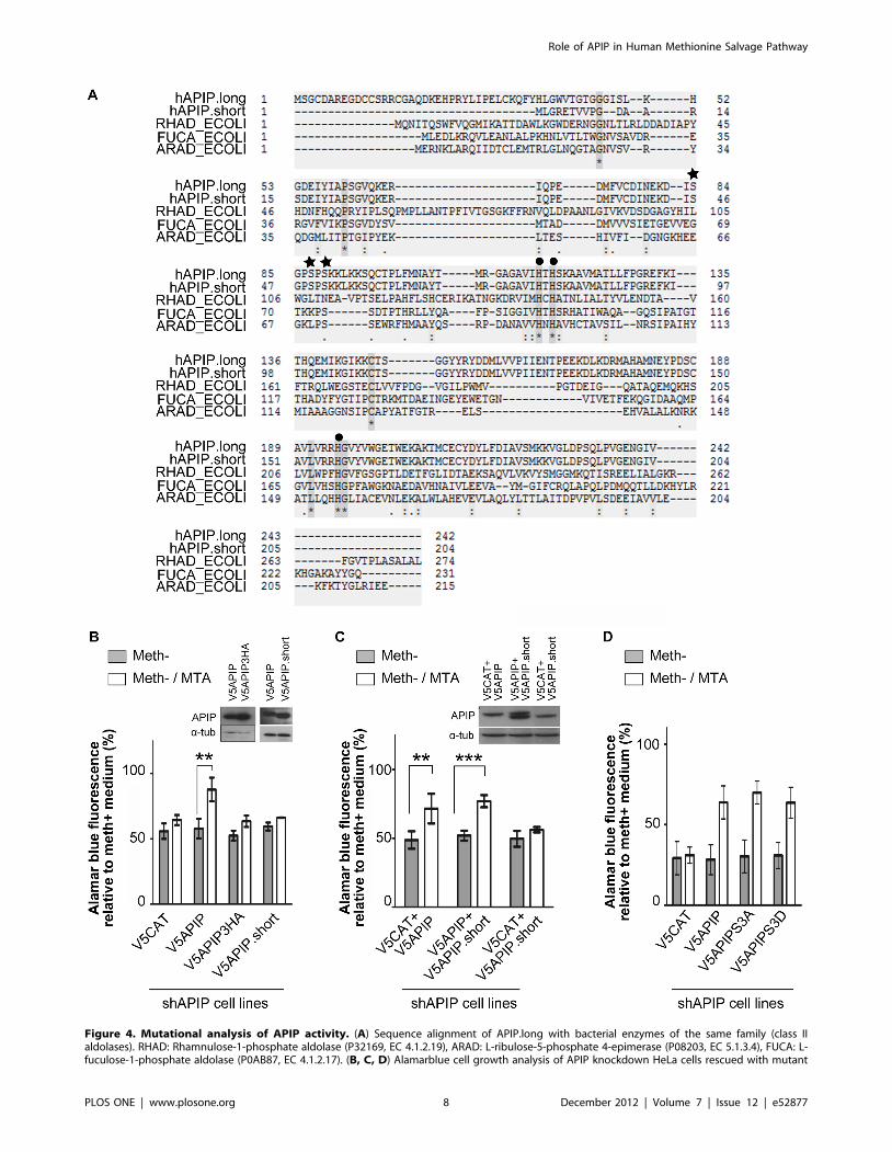
Figure 4. Mutational analysis of APIP activity. (A) Sequence alignment of APIP.long with bacterial enzymes of the same family (class IIaldolases). RHAD: Rhamnulose-1-phosphate aldolase (P32169, EC 4.1.2.19), ARAD: L-ribulose-5-phosphate 4-epimerase (P08203, EC 5.1.3.4), FUCA: L-fuculose-1-phosphate aldolase (P0AB87, EC 4.1.2.17). (B, C, D) Alamarblue cell growth analysis of APIP knockdown HeLa cells rescued with mutant
Role of APIP in Human Methionine Salvage Pathway
PLOS ONE | www.plosone.org 8 December 2012 | Volume 7 | Issue 12 | e52877
tetramers [22,45–47]. The capacity of human APIP to oligomerize
should be investigated further.
APIP.short, which lacks the 38 N-terminal residues, is not able
to replace the long isoform in the methionine salvage pathway.
Some of these N-terminal residues are well conserved across mtnB
eukaryotic orthologs and may be key for enzymatic activity. We
also show that APIP.short does not act as an inhibitor of the full
length isoform, suggesting that it does not bind to APIP nor
compete for the substrate. Interestingly, we were not able to find
this short isoform in ESTs or full length mRNA collections in
organisms other than human.
Both APIP isoforms were previously shown to interact with
APAF-1, thereby competing for binding to caspase-9 [52].
Accordingly, APIP overexpression inhibited mitochondrial apop-
tosis induced by drugs like etoposide and staurosporine whereas
APIP down-regulation increased susceptibility to apoptosis in
mouse skeletal muscle cells C2C12. Moreover, the same study
showed that APIP is induced in mice muscles upon ischemia and
hypoxia, and that its overexpression suppresses ischemia/hypoxia-
induced death. The effects of APIP during ischemia/hypoxia were
first correlated with its APAF-1-related anti-apoptotic activity, but
it was later demonstrated they could occur independently of the
presence of APAF-1, via activation of AKT and extracellular
signal-related kinases MAPK3 (ERK1) and MAPK1 (ERK2) [32].
Although we did not find any effect of APIP up and down-
regulation on apoptosis in HeLa cells (data not shown), the
modulation of apoptosis by APIP has been recently confirmed in
lymphoblastoid and HEK293 cells [51]. Moreover, APIP was also
shown to regulate caspase-1-related inflammatory responses
(pyroptosis) in the same cells. Because of the central role of
methionine and SAM, disruption of the methionine salvage
pathway may affect the cells in many manners. Accordingly, the
same study showed that MTA itself could regulate pyroptosis and
that APIP acts on pyroptosis via the methionine salvage pathway.
Previous studies have shown that many cancer cell lines were
not able to grow in meth2/Hcy media [34]. This phenomenon
was named methionine dependency [53] and led to the
development of new therapeutic strategies, such as methionine
restriction diet [54–58]. This observation has brought interest into
the methionine salvage pathway, firstly, because MTAP is
deficient in many cancer cell lines and secondly, because four
methionine-dependent cell lines could be rescued by the addition
of MTOB in the media [35]. However, it was shown that, despite
the strong correlation between methionine dependency and the
loss of MTAP expression, over-expression of MTAP in deficient
cell lines rescued growth in meth2/MTA but not in meth2/Hcy
[35]. In our study we show that APIP knockdown does not perturb
the growth of the HeLa cells in meth2/Hcy. Taken together, these
two observations indicate that deficiency in methionine salvage
pathway is not responsible for cell methionine dependency.
Finally, during the last 5 years, genetic studies revealed potential
links between APIP expression levels and diseases. For example,
APIP was found to be amplified and up-regulated in squamous
carcinoma cells lines from tongue and larynx [59]. Inversely, APIP
was found to be down-regulated at mRNA and protein levels in
non-small cell lung carcinoma cells and tumors [60]. Genetic
variants located in the 39 UTR region of APIP were found to be
associated with lung disease severity in cystic fibrosis [61]. Finally,
using a genome wide association study, a common SNP (rs514182)
associated with reduced expression of APIP has been shown to be
linked to increased susceptibility to both pyroptosis caused by
Salmonella infection and to the chemotherapeutic agent carboplatin
[51]. Interestingly, this mutation was also linked to improved
survival of individuals with systemic inflammatory response
syndrome. Possible deregulation of methionine metabolism in
those diseases should be investigated further.
Acknowledgments
The authors thank Dr Nicole Freed for help with the Shigella experiments,
Prof Cecile Arrieumerlou for sharing preliminary phosphoproteomics
results and the whole BattleX consortium,CALIPHO team and Gael Panis
for helpful discussions.
Author Contributions
Conceived and designed the experiments: CM LL. Performed the
experiments: CM PT LS. Analyzed the data: CM LL. Contributed
reagents/materials/analysis tools: DB AB LL. Wrote the paper: CM LL.
Performed the bioinformatics studies: AB PD.
References
1. Anderson ME (1998) Glutathione: an overview of biosynthesis and modulation.
Chem Biol Interact 111–112: 1–14.
2. Cavuoto P, Fenech MF (2012) A review of methionine dependency and the role
of methionine restriction in cancer growth control and life-span extension.
Cancer Treat Rev 38: 726–736.
3. Sekowska A, Danchin A (2002) The methionine salvage pathway in Bacillus
subtilis. BMC Microbiol 2: 8.
4. Albers E (2009) Metabolic characteristics and importance of the universal
methionine salvage pathway recycling methionine from 59-methylthioadenosine.
IUBMB Life 61: 1132–1142.
5. Tang B, Kadariya Y, Murphy ME, Kruger WD (2006) The methionine salvage
pathway compound 4-methylthio-2-oxobutanate causes apoptosis independent
of down-regulation of ornithine decarboxylase. Biochem Pharmacol 72: 806–
815.
6. Subhi AL, Diegelman P, Porter CW, Tang B, Lu ZJ, et al. (2003)
Methylthioadenosine phosphorylase regulates ornithine decarboxylase by
production of downstream metabolites. J Biol Chem 278: 49868–49873.
7. Chattopadhyay MK, Tabor CW, Tabor H (2005) Studies on the regulation of
ornithine decarboxylase in yeast: effect of deletion in the MEU1 gene. Proc Natl
Acad Sci U S A 102: 16158–16163.
8. Basu I, Cordovano G, Das I, Belbin TJ, Guha C, et al. (2007) A transition state
analogue of 59-methylthioadenosine phosphorylase induces apoptosis in head
and neck cancers. J Biol Chem 282: 21477–21486.
9. Pirkov I, Norbeck J, Gustafsson L, Albers E (2008) A complete inventory of all
enzymes in the eukaryotic methionine salvage pathway. FEBS J 275: 4111–4120.
10. Subhi AL, Tang B, Balsara BR, Altomare DA, Testa JR, et al. (2004) Loss of
methylthioadenosine phosphorylase and elevated ornithine decarboxylase is
common in pancreatic cancer. Clin Cancer Res 10: 7290–7296.
11. Komatsu A, Nagasaki K, Fujimori M, Amano J, Miki Y (2008) Identification of
novel deletion polymorphisms in breast cancer. Int J Oncol 33: 261–270.
12. Collins CC, Volik SV, Lapuk A, Wang Y, Gout PW, et al. (2012) Next
Generation Sequencing of Prostate Cancer from a Patient Identifies a Deficiency
of Methylthioadenine Phosphorylase (MTAP), an Exploitable Tumor Target.
Mol Cancer Ther 11: 775–783.
forms of V5APIP in MTA medium. Results are expressed relative to the growth of the HeLa cells treated the same way in methionine medium (B)V5APIP mutated for the potential zinc binding site (V5APIP3HA) and V5APIP.short are not able to restore cell growth in MTA medium. Expression ofV5APIP, V5APIP3HA and V5APIP.short was controlled by Western blot in APIP stable knockdown HeLa cells. (C) Co-expression of V5APIP.short did notaffect the growth rescue in MTA conferred by V5APIP. V5CAT was used as control so that the amount of plasmid DNA used for transfection wasconstant in the three conditions. (D) No difference was observed in the rescue efficiency of V5APIP and V5APIP mutated at potential phosphorylationsites (V5APIP3SA and V5APIP3SD).doi:10.1371/journal.pone.0052877.g004
Role of APIP in Human Methionine Salvage Pathway
PLOS ONE | www.plosone.org 9 December 2012 | Volume 7 | Issue 12 | e52877
13. Chow WA, Bedell V, Gaytan P, Borden E, Goldblum J, et al. (2006)
Methylthioadenosine phosphorylase gene deletions are frequently detected byfluorescence in situ hybridization in conventional chondrosarcomas. Cancer
Genet Cytogenet 166: 95–100.
14. Behrmann I, Wallner S, Komyod W, Heinrich PC, Schuierer M, et al. (2003)Characterization of methylthioadenosin phosphorylase (MTAP) expression in
malignant melanoma. Am J Pathol 163: 683–690.15. Della Ragione F, Takabayashi K, Mastropietro S, Mercurio C, Oliva A, et al.
(1996) Purification and characterization of recombinant human 59-methylthioa-
denosine phosphorylase: definite identification of coding cDNA. BiochemBiophys Res Commun 223: 514–519.
16. Hirano W, Gotoh I, Uekita T, Seiki M (2005) Membrane-type 1 matrixmetalloproteinase cytoplasmic tail binding protein-1 (MTCBP-1) acts as an
eukaryotic aci-reductone dioxygenase (ARD) in the methionine salvage pathway.Genes Cells 10: 565–574.
17. Wang H, Pang H, Bartlam M, Rao Z (2005) Crystal structure of human E1
enzyme and its complex with a substrate analog reveals the mechanism of itsphosphatase/enolase activity. J Mol Biol 348: 917–926.
18. Backlund PS, Chang CP, Smith RA (1982) Identification of 2-keto-4-methylthiobutyrate as an intermediate compound in methionine synthesis from
59-methylthioadenosine. J Biol Chem 257: 4196–4202.
19. Cooper AJL, Krasnikov BF, Pinto JT, Kung HF, Li J, et al. (2012) Comparativeenzymology of (2S,4R)4-fluoroglutamine and (2S,4R)4-fluoroglutamate. Comp
Biochem Physiol B Biochem Mol Biol 163: 108–120.20. Cooper AJL (2004) The role of glutamine transaminase K (GTK) in sulfur and
alpha-keto acid metabolism in the brain, and in the possible bioactivation ofneurotoxicants. Neurochem Int 44: 557–577.
21. Kabuyama Y, Litman ES, Templeton PD, Metzner SI, Witze ES, et al. (2009) A
Mediator of Rho-dependent Invasion Moonlights as a Methionine SalvageEnzyme. Molecular cellular proteomics MCP 8: 2308–2320.
22. Ashida H, Saito Y, Kojima C, Yokota A (2008) Enzymatic characterization of 5-methylthioribulose-1-phosphate dehydratase of the methionine salvage pathway
in Bacillus subtilis. Bioscience biotechnology and biochemistry 72: 959–967.
23. Salim HMW, Negritto MC, Cavalcanti ARO (2009) 1+1 = 3: a fusion of 2enzymes in the methionine salvage pathway of Tetrahymena thermophila
creates a trifunctional enzyme that catalyzes 3 steps in the pathway. PLoS Genet5: e1000701.
24. Sekowska A, Denervaud V, Ashida H, Michoud K, Haas D, et al. (2004)Bacterial variations on the methionine salvage pathway. BMC Microbiol 4: 9.
25. Lima T, Auchincloss AH, Coudert E, Keller G, Michoud K, et al. (2009)
HAMAP: a database of completely sequenced microbial proteome sets andmanually curated microbial protein families in UniProtKB/Swiss-Prot. Nucleic
Acids Res 37: D471–8.26. The UniProt Consortium (2012) Reorganizing the protein space at the Universal
Protein Resource (UniProt). Nucleic Acids Res 40: D71–5.
27. Sievers F, Wilm A, Dineen D, Gibson TJ, Karplus K, et al. (2011) Fast, scalablegeneration of high-quality protein multiple sequence alignments using Clustal
Omega. Mol Syst Biol 7: 539. doi:10.1038/msb.2011.75.28. Steinhauer J, Agha R, Pham T, Varga AW, Goldberg MB (1999) The unipolar
Shigella surface protein IcsA is targeted directly to the bacterial old pole: IcsPcleavage of IcsA occurs over the entire bacterial surface. Mol Microbiol 32: 367–
377.
29. Becskei A, Serrano L (2000) Engineering stability in gene networks byautoregulation. Nature 405: 590–593.
30. Datsenko KA, Wanner BL (2000) One-step inactivation of chromosomal genesin Escherichia coli K-12 using PCR products. Proc Natl Acad Sci U S A 97:
6640–6645.
31. Murphy KC, Campellone KG (2003) Lambda Red-mediated recombinogenicengineering of enterohemorrhagic and enteropathogenic E. coli. BMC Mol Biol
4: 11.32. Cho D-H, Hong Y-M, Lee H-J, Woo H-N, Pyo J-O, et al. (2004) Induced
inhibition of ischemic/hypoxic injury by APIP, a novel Apaf-1-interacting
protein. J Biol Chem 279: 39942–39950.33. Della Ragione F, Oliva A, Palumbo R, Russo GL, Gragnaniello V, et al. (1992)
Deficiency of 59-deoxy-59-methylthioadenosine phosphorylase activity inmalignancy. Absence of the protein in human enzyme-deficient cell lines.
Biochem J 281 (Pt 2: 533–538.34. Mecham JO, Rowitch D, Wallace CD, Stern PH, Hoffman RM (1983) The
metabolic defect of methionine dependence occurs frequently in human tumor
cell lines. Biochem Biophys Res Commun 117: 429–434.35. Tang B, Li YN, Kruger WD (2000) Defects in Methylthioadenosine
Phosphorylase Are Associated with but not Responsible for Methionine-dependent Tumor Cell Growth. Cancer Res 60: 5543–5547.
36. Soda K (1987) Microbial sulfur amino acids: an overview. Methods Enzymol
143: 453–459.
37. Zhang Z, Feige JN, Chang AB, Anderson IJ, Brodianski VM, et al. (2003) A
transporter of Escherichia coli specific for L- and D-methionine is the prototype
for a new family within the ABC superfamily. Arch Microbiol 180: 88–100.
38. Merlin C, Gardiner G, Durand S, Masters M (2002) The Escherichia coli metD
locus encodes an ABC transporter which includes Abc (MetN), YaeE (MetI), and
YaeC (MetQ). J Bacteriol 184: 5513–5517.
39. Gal J, Szvetnik A, Schnell R, Kalman M (2002) The metD D-methionine
transporter locus of Escherichia coli is an ABC transporter gene cluster.
J Bacteriol 184: 4930–4932.
40. Kadner RJ, Watson WJ (1974) Methionine transport in Escherichia coli:
physiological and genetic evidence for two uptake systems. J Bacteriol 119: 401–
409.
41. Mantis N, Prevost MC, Sansonetti P (1996) Analysis of epithelial cell stress
response during infection by Shigella flexneri. Infect Immun 64: 2474–2482.
42. Kroemer M, Merkel I, Schulz GE (2003) Structure and catalytic mechanism of
L-rhamnulose-1-phosphate aldolase. Biochemistry 42: 10560–10568.
43. Lee LV, Poyner RR, Vu MV, Cleland WW (2000) Role of metal ions in the
reaction catalyzed by L-ribulose-5-phosphate 4-epimerase. Biochemistry 39:
4821–4830.
44. Johnson AE, Tanner ME (1998) Epimerization via carbon-carbon bond
cleavage. L-ribulose-5-phosphate 4-epimerase as a masked class II aldolase.
Biochemistry 37: 5746–5754.
45. Akana J, Fedorov AA, Fedorov E, Novak WRP, Babbitt PC, et al. (2006) D-
Ribulose 5-phosphate 3-epimerase: functional and structural relationships to
members of the ribulose-phosphate binding (beta/alpha)8-barrel superfamily.
Biochemistry 45: 2493–2503.
46. Luo Y, Samuel J, Mosimann SC, Lee JE, Tanner ME, et al. (2001) The structure
of L-ribulose-5-phosphate 4-epimerase: an aldolase-like platform for epimeriza-
tion. Biochemistry 40: 14763–14771.
47. Dreyer MK, Schulz GE (1993) The spatial structure of the class II L-fuculose-1-
phosphate aldolase from Escherichia coli. J Mol Biol 231: 549–553.
48. Olsen JV, Vermeulen M, Santamaria A, Kumar C, Miller ML, et al. (2010)
Quantitative phosphoproteomics reveals widespread full phosphorylation site
occupancy during mitosis. Sci Signal 3: ra3.
49. Rigbolt KTG, Prokhorova TA, Akimov V, Henningsen J, Johansen PT, et al.
(2011) System-wide temporal characterization of the proteome and phospho-
proteome of human embryonic stem cell differentiation. Sci Signal 4: rs3.
50. Trackman PC, Abeles RH (1983) Methionine synthesis from 59-S-Methylthioa-
denosine. Resolution of enzyme activities and identification of 1-phospho-5-S
methylthioribulose. J Biol Chem 258: 6717–6720.
51. Ko DC, Gamazon ER, Shukla KP, Pfuetzner RA, Whittington D, et al. (2012)
Functional genetic screen of human diversity reveals that a methionine salvage
enzyme regulates inflammatory cell death. Proc Natl Acad Sci U S A.
52. Cho D-H, Lee H-J, Kim H-J, Hong S-H, Pyo J-O, et al. (2007) Suppression of
hypoxic cell death by APIP-induced sustained activation of AKT and ERK1/2.
Oncogene 26: 2809–2814.
53. Kreis W (1991) Methionine dependency of malignant tumors. J Natl Cancer Inst
83: 725.
54. Thivat E, Durando X, Demidem A, Farges M-C, Rapp M, et al. (n.d.) A
methionine-free diet associated with nitrosourea treatment down-regulates
methylguanine-DNA methyl transferase activity in patients with metastatic
cancer. Anticancer Res 27: 2779–2783.
55. Miki K, Al-Refaie W, Xu M, Jiang P, Tan Y, et al. (2000) Methioninase gene
therapy of human cancer cells is synergistic with recombinant methioninase
treatment. Cancer Res 60: 2696–2702.
56. Cellarier E, Durando X, Vasson MP, Farges MC, Demiden A, et al. (2003)
Methionine dependency and cancer treatment. Cancer Treat Rev 29: 489–499.
57. Durando X, Thivat E, Gimbergues P, Cellarier E, Abrial C, et al. (2008)
[Methionine dependency of cancer cells: a new therapeutic approach?]. Bull
Cancer 95: 69–76.
58. Cellarier E, Durando X, Vasson M, Farges M, Demiden A, et al. (2003)
Methionine dependency and cancer treatment. Cancer Treatment Reviews 29:
489–499.
59. Jarvinen A-K, Autio R, Kilpinen S, Saarela M, Leivo I, et al. (2008) High-
resolution copy number and gene expression microarray analyses of head and
neck squamous cell carcinoma cell lines of tongue and larynx. Genes
Chromosomes Cancer 47: 500–509.
60. Down-regulated expression of apoptosis-associated genes APIP and UACA in
non-small cell lung carcinoma (n.d.). International Journal of Oncology 40:
2111.
61. Wright FA, Strug LJ, Doshi VK, Commander CW, Blackman SM, et al. (2011)
Genome-wide association and linkage identify modifier loci of lung disease
severity in cystic fibrosis at 11p13 and 20q13.2. Nat Genet 43: 539–546.
Role of APIP in Human Methionine Salvage Pathway
PLOS ONE | www.plosone.org 10 December 2012 | Volume 7 | Issue 12 | e52877