Embed Size (px)
Citation preview
ER export of the plant K+ channel KAT1:
Identification and functional analysis of acidic
signal motifs
Vom Fachbereich Biologie der Technischen Universität Darmstadt
zur
Erlangung des akademischen Grades
eines Doctor rerum naturalium
genehmigte
Dissertation von
Dipl.-Biol. Melanie Mikosch
aus Köln
Berichterstatter: PD Dr. Ulrike Homann
Mitberichterstatter: Prof. Dr. Gerhard Thiel
Eingereicht am 21.11.2008
Mündliche Prüfung am 23.01.2009
Darmstadt 2009
D17
Die vorliegende Arbeit wurde in der Arbeitsgruppe von PD Dr. Ulrike Homann des
Fachbereichs Biologie der Technischen Universität Darmstadt im Zeitraum von
August 2005 bis November 2008 angefertigt.
Table of Contents
CHAPTER 1 GENERAL INTRODUCTION 1
1.1 The secretory pathway 3 1.1.1 ER 4
1.1.2 Golgi 4
1.2 ER export 5 1.2.1 ER export motifs 6
1.2.2 Interaction with COPII 8
1.3 KAT1 10
1.4 Aim of the work 13
1.5 References 14
CHAPTER 2 DIACIDIC MOTIF IS REQUIRED FOR EFFICIENT TRANSPORT OF THE K+ CHANNEL KAT1 TO THE PLASMA MEMBRANE 19
2.1 Abstract 21
2.2 Introduction 21
2.3 Results 23 2.3.1 Mutation of a diacidic motif reduces the number of active KAT1 K+ channels in the plasma
membrane of guard cell protoplasts 23
2.3.2 Mutation of a diacidic motif results in ER retention of KAT1 26
2.3.3 Mutation of a diacidic motif also affects KAT1 conductance in HEK293 cells 29
2.4 Discussion 31 2.4.1 The diacidic motif of KAT1 is essential for efficient ER export 31
2.4.2 ER retention of KAT1 is very efficient and may be restricted to certain areas 32
2.4.3 Function of the diacidic motif is position‐dependent 33

2.5 Methods 34 2.5.1 Vectors for KAT1 expression 34
2.5.2 Mutagenesis of putative ER export motifs in KAT1 35
2.5.3 Transfection of intact guard cells via particle delivery and isolation of protoplasts 35
2.5.4 Cultivation and transfection of mammalian cell line HEK293 35
2.5.5 Patch clamp measurements 36
2.5.6 CLSM 36
2.6 References 37
CHAPTER 3 EFFICIENCY OF ER EXPORT OF THE K+ CHANNEL KAT1 DEPENDS ON THE NUMBER OF ACIDIC AMINO ACIDS WITHIN A TRIACIDIC MOTIF 40
3.1 Abstract 42
3.2 Introduction 42
3.3 Results and Discussion 45 3.3.1 Number of mutated acidic amino acids affects KAT1 conductance 45
3.3.2 Mutation of acidic amino acids alters localisation of KAT1 in HEK293 cells 49
3.3.3 Rescue of ER export mutant by heterotetramerisation with wildtype KAT1 54
3.4 Conclusion 56
3.5 Methods 56 3.5.1 Construction of pEGFPN2KAT1 56
3.5.2 Cell culture and transfection of HEK293 cells 57
3.5.3 Patch clamp measurements 57
3.5.4 CLSM 57
3.6 References 58
CHAPTER 4 INTERACTION OF THE K+ CHANNEL KAT1 WITH THE COPII COAT COMPONENT SEC24 DEPENDS ON A DIACIDIC ER EXPORT MOTIF 61
4.1 Abstract 63
4.2 Introduction 63
4.3 Results 65 4.3.1 FRET by acceptor bleaching provides a suitable tool for analysis of protein‐protein interaction in
plant cells in vivo 65
4.3.2 FRET measurements indicate interaction between KAT1 and Sec24 at ER export sites of Vicia faba
guard cells 69
4.3.3 FRET between KAT1 and Sec24 depends on the diacidic motif 74
4.4 Discussion 77 4.4.1 The mechanism of cargo recruitment into ER derived COPII carriers is highly conserved among
eukaryotes 77
4.4.2 Export of KAT1 occurs mainly at the perinuclear region of the ER 79
4.5 Methods 80 4.5.1 Expression vectors 80
4.5.2 Transfection of intact guard cells by particle delivery 80
4.5.3 FRET and CLSM 81
4.6 Supplemental Figures 82
4.7 References 85
CHAPTER 5 HOW DO DIACIDIC MOTIFS WORK ON ION CHANNEL TRAFFICKING? 88
5.1 Abstract 90
5.2 Introduction 90
5.3 Diacidic ER export signals of membrane proteins 91
5.4 Interaction of the diacidic motif with the COPII component Sec24 93
5.5 Sequence specificity of the diacidic ER export motif 96
5.6 Role of ER export signals in regulating ion channel density and composition 99
5.7 Conclusions and perspectives 102
5.8 References 104
CHAPTER 6 GENERAL DISCUSSION 108
6.1 Diacidic ER export motifs 110 6.1.1 ER export motifs regulate trafficking of membrane protein in plants 110
6.1.2 ER export affects cargo selection by interaction with Sec24 111
6.1.3 Restriction of the ER export mutant to the perinuclear area 113
6.1.4 mRNA localisation 115
6.2 Tetramerization of ion channels 116 6.2.1 Tetramerization of ion channels most likely occurs at the ER 116
6.2.2 ER export motifs can affect subunit composition of channel tetramers in the plasma membrane 117
6.3 References 120
SUMMARY 124
ZUSAMMENFASSUNG 125
LIST OF PUBLICATIONS IN THIS THESIS 126
DANKSAGUNG 127
CURRICULUM VITAE 128
EIDESSTATTLICHE ERKLÄRUNG 130
1 Chapter 1
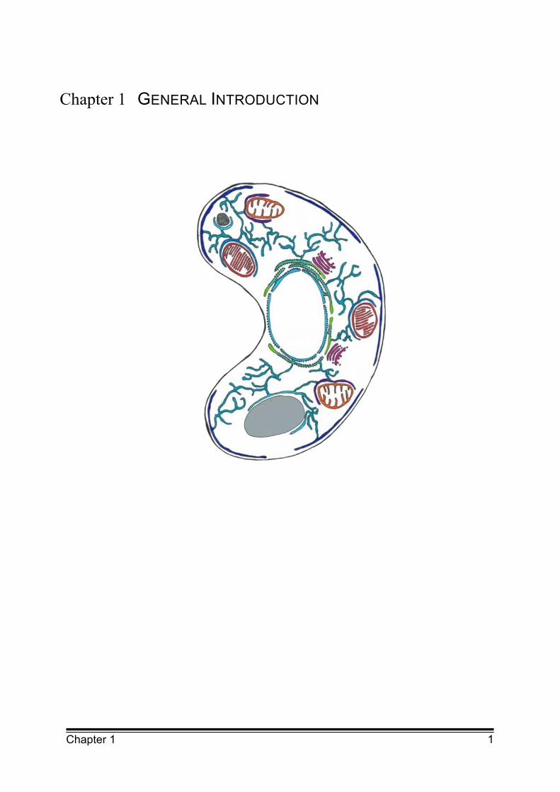
Chapter 1 GENERAL INTRODUCTION
2 Chapter 1
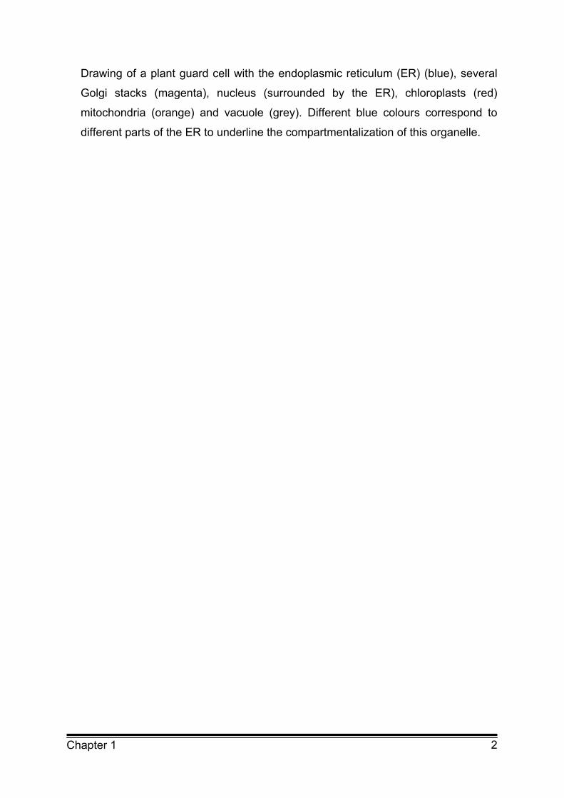
Drawing of a plant guard cell with the endoplasmic reticulum (ER) (blue), several
Golgi stacks (magenta), nucleus (surrounded by the ER), chloroplasts (red)
mitochondria (orange) and vacuole (grey). Different blue colours correspond to
different parts of the ER to underline the compartmentalization of this organelle.
3 Chapter 1
1.1 The secretory pathway
Every single cell is strongly compartmentalised. These compartments differ
immensely in structure and in protein as well as lipid composition. To maintain the
character of each organelle the cell has to regulate the transport of lipids and
proteins.
A substantial proportion of all eukaryotic proteins and most integral membrane
proteins travel through the secretory pathway to their intra- and extracellular
destination.
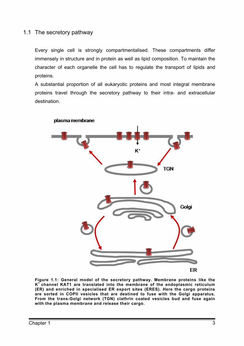
Figure 1.1: General model of the secretory pathway. Membrane proteins like the K+ channel KAT1 are translated into the membrane of the endoplasmic reticulum (ER) and enriched in specialised ER export sites (ERES). Here the cargo proteins are sorted in COPII vesicles that are destined to fuse with the Golgi apparatus. From the trans-Golgi network (TGN) clathrin coated vesicles bud and fuse again with the plasma membrane and release their cargo.
4 Chapter 1
The secretory pathway represents the main route for proteins to the plasma
membrane (Figure 1.1). Membrane proteins are synthesised at ribosomes and
cotranslationally inserted into the membrane of the endoplasmic reticulum (ER).
There they are packed into coat protein II (COPII) coated vesicles that fuse with
the cis-Golgi cisternae (Barlowe et al., 1994). After translocation through the Golgi
apparatus and further transport to the trans-Golgi network the proteins are
transported via clathrin coated vesicles to their final destination.
1.1.1 ER
The ER is the first compartment of the secretory pathway. It forms a large
membrane network which comprises almost half of the total membrane of a cell.
The ER can be divided into several interconnected subcompartments which differ
substantially in their lipid and protein composition. These subcompartments have
different function and morphology. The ER was subdivided by electron microscopy
into the rough ER where ribosomes are attached to the ER membrane and the
smooth ER which is ribosome free. Part of the smooth ER accounts for so called
ER export sites (ERES) where vesicles bud off for further transport to the Golgi
apparatus. These ERES are not found in Saccharomyces cerevisiae, suggesting
that ER export can occur over the whole ER surface. In contrast Pichia pastoris
has clearly defined subdomains of the ER responsible for ER export like plant and
mammalian cells. A structure between the ER and the Golgi apparatus the
ER Golgi intermediate compartment (ERGIC) is unique to higher eukaryotes and
so far has not been found in plant cells (Kirk and Ward, 2007).
1.1.2 Golgi
Nearly all proteins which have been synthesised at the ER are transported to the
Golgi apparatus. The primary function of the Golgi apparatus is to sort and
process proteins and lipids that are synthesised at the ER. The Golgi apparatus is
composed of different cisternae which have individual functions and a
corresponding set of proteins and enzymes. The Golgi apparatus can be divided
into cis-, medial- and trans-Golgi cisternae and the adjacent trans-Golgi network.
5 Chapter 1
Proteins are transported from ERES towards the cis-Golgi cisternae and then they
are subsequently transported through the Golgi apparatus to the trans-Golgi
network from where proteins are again sorted into vesicles to their final
destination.
There is a huge difference between the plant and human Golgi apparatus. Plant
cells contain many individual Golgi stacks that are mobile organelles which travel
along actin filaments. It is not well understood how COPII coated vesicles which
form at the ERES reach the mobile Golgi stacks but it has been shown that the
Golgi stacks in plants move along together with an individual ERES
(daSilva et al., 2004). In mammalian cells the ERES are distributed all over the ER
but the single Golgi apparatus is found at the microtubule organising centre
(MTOC) in the middle of the cell and distinct from the ERES.
1.2 ER export
Regulation of membrane protein transport at the level of the ER is under intense
investigation but major aspects are still unknown. Mechanisms like concentration
of cargo molecules at ERES and recruiting of participating factors are largely
unidentified. Until now several specific amino acid sequences like ER export motifs
have been shown to be involved in cargo concentration and recruiting of the COPII
coat. However, for most of these motifs the mechanism of action is still unclear. As
expected, all motifs are located in cytoplasmic domains of the proteins because
they are supposed to interact with cytoplasmic receptor proteins. In contrast to
membrane proteins which are supposed to interact directly with COPII
components, soluble cargo proteins rely on transmembrane adapter proteins for
regulated ER to Golgi apparatus transport. Therefore soluble cargo proteins
contain specific cargo encoded sorting signals to interact with transmembrane
receptors which in turn interact with the cytosolic components of the COPII coat.
6 Chapter 1
1.2.1 ER export motifs
Only for few highly abundant soluble proteins has the transport out of the ER been
shown to be non selective and to occur via bulk flow (Wieland et al., 1987;
Martinez-Menarguez et al., 1999; Oprins et al., 2001). However, transport via bulk
flow is very inefficient. For most proteins the transport is therefore regulated.
Cargo proteins are actively sorted and enriched into COPII coated vesicles by
means of specific export signals. Up to now, many ER export signals have been
identified. They are frequently found in membrane proteins like ion channels,
receptors and transporters. Among the ER export motifs are four major groups:
diacidic, dihydrophobic, dibasic ER export motifs and a terminal valine (V)
(Table 1.1).
Dihydrophobic signals have been identified in the ERGIC53 receptor that is
necessary for secretion of the coagulation factors V and VII. But these signals are
not sufficient to promote ER export of the protein. For efficient ER export,
oligomerisation of the protein is needed as well (Baines and Zhang, 2007). A
terminal V is very common in mammalian membrane proteins like the stem cell
factor Kit1 (Pauhle et al., 2004).
The diacidic ER export motifs have been identified in several membrane proteins
like ion channel proteins and transporters, most of them finally localised in the
plasma membrane. Nishimura and Balch showed for the first time in 1997 that
diacidic ER export motifs are required for efficient ER export of the vesicular
stomatitis virus glycoprotein (VSVG). Ion channels like Kir2.1 and TASK3
(Ma et al., 2001; Zuzarte et al., 2007) have been found to contain ER export motifs
for regulated ER export. The yeast SNAREs Sys1 and Gap1 also use diacidic
ER export motifs in their cytoplasmic domains for efficient trafficking out of the ER
(Aridor et al., 2001; Votsmeier and Gallwitz, 2001). The function of diacidic
ER export motifs in plant membrane proteins was demonstrated for the Golgi
apparatus localised proteins CASP and GONST1, the aquaporins ZmPIP2,4 and
ZmPIP2,5 and for the K+ channel KAT1 (Hanton et al., 2005; Zelazny et al., 2008;
Mikosch et al., 2006). For some of these motifs a direct interaction with the COPII
coat has been shown.

7 Chapter 1
Table 1.1: Proteins with different identified ER export motifs Protein Origin Motif
Diacidic (D/ExD/E)
CFTR Human DxD Wang et al. (2004)
Kir 1.1 Human ExD Ma et al. (2001)
Kir 2.1 Human ExE Ma et al. (2001)
Kir 2.4 Human ExE Hofherr et al. (2005)
Kir 3.2a Human DxE Ma et al. (2002)
Kir 3.4 Human DxE Ma et al. (2002)
TASK 3 Human ExE Zuzarte et al. (2007)
KAT1 Plant DxE Mikosch et al. (2006)
GAP1 Yeast DxD Malkus et al. (2002)
Sys1 Yeast DxE Votsmeier and Gallwitz (2001)
Can1 Yeast DxD Malkus et al. (2002)
Hip1 Yeast DxD Miller et al. (2003)
Yor1 Yeast DxE Pagant et al (2007)
GONST1 Plant DxE Hanton et al. (2005)
VSVG Virus DxE Nishimura and Balch (1997)
CASP Plant DxE Hanton et al. (2005)
ZmPIP2;4 Plant DxE Zelazny et al. (2008)
ZmPIP2;5 Plant DxE Zelazny et al. (2008)
Dihydrophobic
ERGIC53 Human FF Nufer et al. (2003)
P24 family Human FF Dominguez et al. (1998)
Erv41p Human FF Otte and Barlowe (2002)
Erv46p Human FF Otte and Barlowe (2002)
Dibasic (R/KxR/K)
GAT1 Human RL Farhan et al. (2007)
SERT Human RI Farhan et al. (2007)
DAT Human KL Farhan et al. (2007)
GnTI Plant R Schoberer et al. (2008)
GMII Plant K Schoberer et al. (2008)
Terminal V
Kit1 Human V Pauhle et al. (2004)
HLA-F Human V Boyle et al. (2006)
8 Chapter 1
1.2.2 Interaction with COPII
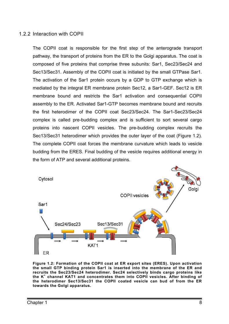
The COPII coat is responsible for the first step of the anterograde transport
pathway, the transport of proteins from the ER to the Golgi apparatus. The coat is
composed of five proteins that comprise three subunits: Sar1, Sec23/Sec24 and
Sec13/Sec31. Assembly of the COPII coat is initiated by the small GTPase Sar1.
The activation of the Sar1 protein occurs by a GDP to GTP exchange which is
mediated by the integral ER membrane protein Sec12, a Sar1-GEF. Sec12 is ER
membrane bound and restricts the Sar1 activation and consequential COPII
assembly to the ER. Activated Sar1-GTP becomes membrane bound and recruits
the first heterodimer of the COPII coat Sec23/Sec24. The Sar1-Sec23/Sec24
complex is called pre-budding complex and is sufficient to sort several cargo
proteins into nascent COPII vesicles. The pre-budding complex recruits the
Sec13/Sec31 heterodimer which provides the outer layer of the coat (Figure 1.2).
The complete COPII coat forces the membrane curvature which leads to vesicle
budding from the ERES. Final budding of the vesicle requires additional energy in
the form of ATP and several additional proteins.
Figure 1.2: Formation of the COPII coat at ER export sites (ERES). Upon activation the small GTP binding protein Sar1 is inserted into the membrane of the ER and recruits the Sec23/Sec24 heterodimer. Sec24 selectively binds cargo proteins like the K+ channel KAT1 and concentrates them into COPII vesicles. After binding of the heterodimer Sec13/Sec31 the COPII coated vesicle can bud of from the ER towards the Golgi apparatus.

9 Chapter 1
COPII vesicle formation occurs in specialised regions –the ERES- that are
enriched in COPII components and cargo molecules. These structures can be
marked with the Sec16 protein (Watson et al., 2006; Bhattacharyya and Glick,
2007). It has been shown that ERES are very dynamic cargo sensitive structures
whose formation is induced by cargo molecules.
ERES have been shown to exist in several eukaryotic organisms like mammalia,
yeast and plant and certain proteins have been identified as markers for ERES.
Cargo selection at ERES occurs through COPII coated vesicles. Several cargo
proteins are concentrated in COPII coated vesicles with the aid of ER export
motifs. For many ER export motifs a direct interaction with the COPII coat has
been shown. Most of them interact with the heterodimer Sec23/Sec24 and in many
cases Sec24 was identified as the receptor. A possible role for the small GTPase
Sar1 in cargo selection has also been suggested (Aridor et al., 2001; Giraudo and
Maccioni, 2003).
Table 1.2: Isoforms of Sec24 in different species (modified after Baines and Zhang, 2007).
Saccharomyces cerevisiae Sec24, LST1 and ISS1
Caenorhabditis elegans Sec24.1 and Sec24.2
Drosophila melanogaster Sec24 and CG10882
Homo sapiens Sec24a, Sec24b, Sec24c, Sec24d
Arabidopsis thaliana At3g07100, At3g44340 and At4g32640
Investigations into the mechanism of recognition of the ER export motif by Sec24
demonstrated that Sec24 harbours at least three cargo binding sites
(Miller et al., 2003; Mossessova et al., 2003) and interacts with several cargo
proteins (Barlowe, 2003).
The B-Site has been shown to interact with diacidic motifs of yeast SNARE Sys1
and mammalian CFTR channel (Mossessova et al., 2003; Wang et al., 2004).
Furthermore, mutation of the B-Site disrupts the binding to the ER export motif of
the yeast SNARE Bet1 (Mossessova et al., 2003). Wang et al. (2004) proved the
binding of CFTR to Sec24 in vitro by homology modelling.
Another element of specificity in cargo recognition is provided by the existence of
homologues of Sec24. In the genome of Homo sapiens four homologous proteins
have been identified, three homologous proteins in Saccharomyces cerevisiae and
10 Chapter 1
three in Arabidopsis thaliana (Table 1.2). Diversity of the COPII coat could be
further increased by alternative splicing. These set of COPII coat components
ensures a wide range of cargo recognition specific for different tissues and cell
types.
1.3 KAT1
K+ channels are very important in cell biology. They belong to a huge mulitigene
family. One subfamily is formed by the inward rectifying Kir K+ channels which form
a tetramer and were originally identified in animal cells. Each subunit of the Kir
channel tetramer has two transmembrane domains with the pore domain in
between the two transmembrane domains. The pore domains of four subunits in
one tetramer provide together the selective ion pore of the channel (Laine et al.,
2004). Another subfamily consists of voltage gated K+ channels (Kv channels).
These channel are made up of four subunits each consisting of six
transmembrane domains. Transmembrane domain four comprises many positively
charged amino acids and provides the voltage sensor. The pore loop is situated
between transmembrane domains five and six and contains the characteristic
K+ channel signature sequence TXXTXGYG (Heginbotham et al., 1994).
K+ channels are particularly important in plant cell biology, since K+ is essential for
plant development, cell growth, photosynthesis and gas exchange. K+ channels
allow rapid flux of K+ ions across the plasma membrane of guard cells and
mediate stomata movement. These ion fluctuations enable guard cells to regulate
the stomata aperture osmotically by water influx and efflux. The K+ channels
mediating K+ flux in guard cells all belong to the Kv type K+ channels. In
Arabidopsis thaliana just a single gene, GORK, encodes for a K+ outward
conducting channel (Hosy et al., 2003) whereas for inward rectifying channels at
least five subunits have been identified (KAT1, KAT2, AKT1, AKT2/3 and AtKC1).
One prominent member is KAT1 which was first cloned by Schachtman et al. in
1992 and became an accepted model channel in plant cell biology. As a member
of the Kv channel family KAT1 function as a tetramer with each subunit consisting
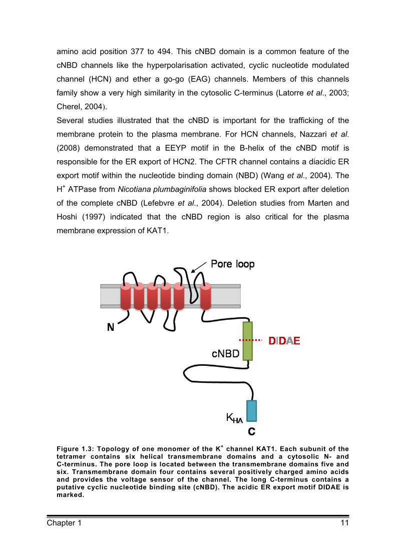
of six transmembrane domains (Figure 1.3). KAT1 has a 297 amino acids long
cytosolic C-terminus with a putative cyclic nucleotide binding domain (cNBD) at
11 Chapter 1
amino acid position 377 to 494. This cNBD domain is a common feature of the
cNBD channels like the hyperpolarisation activated, cyclic nucleotide modulated
channel (HCN) and ether a go-go (EAG) channels. Members of this channels
family show a very high similarity in the cytosolic C-terminus (Latorre et al., 2003;
Cherel, 2004).
Several studies illustrated that the cNBD is important for the trafficking of the
membrane protein to the plasma membrane. For HCN channels, Nazzari et al.
(2008) demonstrated that a EEYP motif in the B-helix of the cNBD motif is
responsible for the ER export of HCN2. The CFTR channel contains a diacidic ER
export motif within the nucleotide binding domain (NBD) (Wang et al., 2004). The
H+ ATPase from Nicotiana plumbaginifolia shows blocked ER export after deletion
of the complete cNBD (Lefebvre et al., 2004). Deletion studies from Marten and
Hoshi (1997) indicated that the cNBD region is also critical for the plasma
membrane expression of KAT1.
Figure 1.3: Topology of one monomer of the K+ channel KAT1. Each subunit of the tetramer contains six helical transmembrane domains and a cytosolic N- and C-terminus. The pore loop is located between the transmembrane domains five and six. Transmembrane domain four contains several positively charged amino acids and provides the voltage sensor of the channel. The long C-terminus contains a putative cyclic nucleotide binding site (cNBD). The acidic ER export motif DIDAE is marked.
12 Chapter 1
An interaction domain (KHA) which is possibly involved in channel clustering in the
plasma membrane (Ehrhardt et al., 1997), resides in the final part of the
C-terminus.
The exact region important for the tetramerization of the KAT1 K+ channel is still
unknown. The shaker channel has a tetramerization domain (T1) in the N-terminus
(Kreusch et al., 1998; Varshney et al., 2004; Robinson and Deutsch, 2005). But
many other channels including the cNBD family of ion channels do not possess
this domain and the tetramerization ability is reflected in parts of the C-terminus
(Tsuruda et al., 2006). Although the transmembrane regions of shaker-type
channels have a high similarity to KAT1, their N- and C-terminal parts are quite
different. This region of KAT1 is much more related to the cNBD channels. The C-
terminus of the cNBD channel HCN2 reveals a high sequence similarity to the
KAT1 C-terminus and crystallisation studies by Zagotta et al. (2003) showed a
tetramerization domain in this part of the protein. Another indication for a
C-terminal tetramerization domain of KAT1 is the ability of a very closely related
plant channel, AKT1, to build tetramers with the C-terminus of the protein only
(Daram et al., 1997).
KAT1 also contains several putative phosphorylation residues. Yang et al. (2008)
showed that phosphorylation of the Kv1.2 channel regulates the transport of the
channels together with other channels in the heterotetramer. This may be due to
the interaction of phosphorylated residues with 14-3-3 proteins. Recently,
Sottocornola et al. (2008) discovered that the KAT1 also interacts with 14-3-3 and
that this interaction influences ion channel trafficking. Although it is not known at
which step of the secretory pathway this interaction occurs.
Altogether KAT1 is one of the best characterised plant ion channels. Therefore, it
provides an excellent model to study ion channel trafficking in plants.

13 Chapter 1
1.4 Aim of the work
The aim of this study was to identify factors regulating the trafficking of membrane
proteins to the plasma membrane. It is widely accepted that the number of
membrane proteins in the plasma membrane - like ion channels, transporters and
receptors - is highly regulated by the rate of endocytosis, recycling and
degradation. Surprisingly, up to now, the role of the secretory pathway in this
process is not fully understood. In order to analyse regulatory steps along the
secretory pathway, the K+ inward rectifying channel KAT1 from
Arabidopsis thaliana was chosen as a model membrane protein. Ion channels are
particularly suitable to study regulation of trafficking because their number in the
plasma membrane has to be tightly controlled.
The first trafficking step after cotranslational insertion of membrane proteins into
the ER membrane is the transport of proteins from the ER to the Golgi apparatus.
This step was chosen as the main focus of the current work.
For the efficient ER export of proteins, their recruitment into COPII vesicles is
essential. This process involves specific amino acid motifs - the so called ER
export signals. For ion channels in yeast and mammalian cells it was shown that
so called diacidic ER export motifs regulate their trafficking at the level of ER
export. As ER export motifs had so far not been identified for plant proteins the
sequence of KAT1 was analysed for specific trafficking determinants. Their role in
ER export was analysed via patch clamp measurements and confocal fluorescent
microscopy.
Yeast membrane proteins have been shown to be incorporated into COPII
vesicles by specific binding to the COPII component Sec24. In order to investigate
the mechanism of ER export of KAT1, the interaction of KAT1 with Sec24 and the
role of the ER export motif in this process were studied. Further analysis
concentrated on the sequence specificity of the acidic motif.
14 Chapter 1
1.5 References
Aridor M, Fish KN, Bannykh S, Weissman J, Roberts TH, Lippincott-Schwartz J, Balch WE (2001) The Sar1 GTPase coordinates biosynthetic cargo
selection with endoplasmic reticulum export site assembly. The Journal of Cell
Biology 152:213-229
Baines AC and Zhang B (2007) Receptor-mediated protein transport in the early
secretory pathway. Trends in Biochemical Sciences 32(8):381-388
Barlowe C, Orci L, Yeung T, Hosobuchi M, Hamamoto S, Salama N, Rexach MF, Ravazzola M, Amherdt M, Schekman R (1994) COPII: a membrane
coat formed by Sec proteins that drive vesicle budding from the endoplasmic
reticulum. Cell 77(6):895-907
Barlowe C (2003) Signals for COPII-dependent export from the ER: what’s the
ticket out? Trends in Cell Biology 13:295-300
Bhattacharyya D and Glick BS (2007) Two mammalian Sec16 homologues have
nonredundant functions in endoplasmic reticulum (ER) export and transitional ER
organization. Molecular Biology of the Cell 18(3):839-849
Boyle LH, Gillingham AK, Munro S, Trowsdale J (2006) Selective export of
HLA-F by its cytoplasmic tail. The Journal of Immunology 176(11):6464-6472
Cherel I (2004) Regulation of K+ channel activities in plants: from physiological to
molecular aspects. The Journal of Experimental Botany 55(396):337-351
Daram P, Urbach S, Gaymard F, Sentenac H, Cherel I (1997) Tetramerization of
the AKT1 plant potassium channel involves its C-terminal cytoplasmic domain.
The EMBO Journal 16:3455-3463
daSilva LL, Snapp EL, Denecke J, Lippincott-Schwartz J, Hawes C, Brandizzi F (2004) Endoplasmic reticulum export sites and Golgi bodies behave
as single mobile secretory units in plant cells. The Plant Cell 16:1753-1771
Dominguez M, Dejgaard K, Füllekrug J, Dahan S, Fazel A, Paccaud JP,
Thomas DY, Bergeron JJ, Nilsson T (1998) gp25L/emp24/p24 protein family
members of the cis-Golgi network bind both COP I and II coatamer. The Journal of
Cell Biology 140:751–765
15 Chapter 1
Ehrhardt T, Zimmermann S, Müller-Röber B (1997) Association of plant K+(in)
channels is mediated by conserved C-termini and does not affect subunit
assembly. FEBS Letters 409:166-170
Farhan H, Reiterer V, Korkhov VM, Schmid JA, Freissmuth M, Sitte HH (2007)
Concentrative export from the endoplasmic reticulum of the γ-amino butyric acid
transporter 1 requires binding to Sec24D. The Journal of Biological Chemistry
282(10):7679–7689
Giraudo CG and Macconi HJ (2003) Endoplasmic reticulum export of
glycosyltransferases depends on interaction of a cytoplasmic dibasic motif with
Sar1. Molecular Biology of the Cell 14:3757-3766
Giraudo C and Contreras I (2003) ER export of glycosyltransferases depends on
interaction of a cytoplasmic dibasic motif with Sar1. Molecular Biology of the Cell
14:3753-3766
Hanton SL, Renna L, Bortolotti LE, Chatre L, Stefano G, Brandizzi F (2005)
Diacidic motifs influence the export of transmembrane proteins from the
endoplasmic reticulum in plant cells. The Plant Cell 17:3081–3093
Heginbotham L, Lu Z, Abramson T, MacKinnon R (1994) Mutations in the
K+ channel signature sequence. The Biophysical Journal 66(4):1061-1067
Hofherr A, Fakler B, Klöcker N (2005) Selective Golgi export of Kir2.1 controls
the stoichiometry of functional Kir2.x channel heteromers. The Journal of Cell
Science 118:1935-1943
Hosy E, Duby G, Véry AA, Costa A, Sentenac H, Thibaud JB (2003) A
procedure for localization and electrophysiological characterization of ion channels
heterologously expressed in a plant context. Plant Methods 19:1-14
Kirk SJ and Ward TH (2007) COPII under the microscope. Seminars in Cell and
Developmental Biology 4:435-447
Kreusch A, Pfaffinger P, Stevens C, Choe S (1998) Crystal structure of
tetramerization domain of shaker potassium channel. Nature 392:945-948
Laine M, Papazian DM, Roux B (2004) Critical assessment of a proposed model
of shaker. FEBS Letters 564(3):257–263
Latorre R, Muñoz F, González C, Cosmelli D (2003) Structure and function of
potassium channels in plants: some inferences about the molecular origin of
inward rectification in KAT1 channels. Molecular Membrane Biology (1):19-25

16 Chapter 1
Lefebvre B, Batoko H, Duby G, Bountry M (2004) Targeting of a Nicotiana
plumbaginifolia H+ ATPase to the plasma membrane is not by default and requires
cytosolic structural determinants. The Plant Cell 16:1772-1789
Ma D, Zerangue N, Lin Y, Collins A, Yu M, Jan Y, Jan LY (2001) Role of ER
export signals in controlling surface potassium channel numbers. Science
291:316–319
Ma D, Zerangue N, Raab-Graham K, Fried SR, Jan YN, Jan LY (2002) Diverse
trafficking patterns due to multiple traffic motifs in G protein-activated inwardly
rectifying potassium channels from brain and heart. Neuron 33(5):715-729
Malkus P, Jiang F, Schekman R (2002) Concentrative sorting of secretory cargo
proteins into COPII coated vesicles. The Journal of Cell Biology 159:915–921
Marten I and Hoshi T (1997) Voltage-dependent gating characteristics of the
K+ channel KAT1 depend on the N- and C-termini. PNAS 94:3448-3453
Martínez-Menárguez JA, Geuze HJ, Slot JW, Klumperman J (1999) Vesicular
tubular clusters between the ER and Golgi mediate concentration of soluble
secretory proteins by exclusion from COPI coated vesicles. Cell 98(1):81-90
Mikosch M, Hurst AC, Hertel B, Homann U (2006) Diacidic motif is required for
efficient transport of the K+ channel KAT1 to the plasma membrane. Plant
Physiology 142:923-930
Miller EA, Beilharz TH, Malkus PN, Lee MCS, Hamamoto S, Orci L, Schekman R (2003) Multiple cargo binding sites on the COPII subunit Sec24p
ensure capture of diverse membrane proteins into transport vesicles. Cell
114:497–509
Mossessova E, Bickford LC, Goldberg J (2003) SNARE selectivity of the COPII
coat. Cell 114:483–495
Nazzari H, Angoli D, Chow SS, Whitaker G, Leclair L, McDonald E, Macri V, Zahynacz K, Walker V, Accili EA (2008) Regulation of cell surface expression of
functional pacemaker channels by a motif in the B-helix of the cyclic nucleotide-
binding domain. American Journal of Physiology: Cell Physiology 295:642-652
Nishimura N and Balch WE (1997) A diacidic signal required for selective export
from the endoplasmic reticulum. Science 277:556–558
Nufer O, Kappeler F, Guldbrandsen S, Hauri HP (2003) ER export of ERGIC53
is controlled by cooperation of targeting determinants in all three of its domains.
The Journal of Cell Science 116:4429-4434
17 Chapter 1
Oprins A, Rabouille C, Posthuma G, Klumperman J, Geuze HJ, Slot JW (2001) The ER to Golgi interface is the major concentration site of secretory
proteins in the exocrine pancreatic cell. Traffic 2(11):831-838
Otte S and Barlowe C (2002) The Erv41p–Erv46p complex: multiple export
signals are required in trans for COPII-dependent transport from the ER. The
EMBO Journal 21:6095–6104
Pagant S, Kung L, Dorrington M, Lee MCS, Miller EA (2007) Inhibiting
endoplasmic reticulum (ER)-associated degradation of misfolded Yor1p does not
permit ER export despite the presence of a diacidic sorting signal. Molecular
Biology of the Cell 18(9):3398-3413
Paulhe F, Imhof BA, Wehrle-Haller B (2004) A specific endoplasmic reticulum
export signal drives transport of stem cell factor (Kit1) to the cell surface. The
Journal of Biological Chemistry 279(53):55545-55555
Robinson JM and Deutsch C (2005) Coupled tertiary folding and oligomerisation
of the T1 domain of Kv channels. Neuron 45(2):223-232
Schachtman DP, Schroeder JI, Lucas WJ, Anderson JA, Gaber RF (1992)
Expression of an inward-rectifying potassium channel by the Arabidopsis thaliana
KAT1 cDNA. Science 258(5088):1654-1658
Schoberer J, Vavra U, Stadlmann J, Hawes C, Mach L, Steinkellner H, Strasser R (2008) Arginine/lysine residues in the cytoplasmic tail promote ER
export of plant glycosylation enzymes. Traffic in press
Sottocornola B, Gazzarrini S, Olivari C, Romani G, Valbuzzi P, Thiel G, Moroni A (2008) 14-3-3 proteins regulate the potassium channel KAT1 by dual
modes. Plant Biology 10(2):231-236
Tsuruda PR, Julius D, Minor DL Jr. (2006) Coiled coils direct assembly of a
cold-activated TRP channel. Neuron 51(2):201-212
Varshney A, Chanda B, Mathew M (2004) Arranging the elements of the
potassium channel: the T1 domain occludes the cytoplasmic face of the channel.
The European Biophysical Journal 33: 370-376
Votsmeier C and Gallwitz D (2001) An acidic sequence of a putative yeast Golgi
membrane protein binds COPII and facilitates ER export. The EMBO Journal
20:6742–6750
Wang X, Matteson J, An Y, Moyer B, Yoo J, Bannykh S, Wilson IA, Riordan JR, Balch WE (2004) COPII-dependent export of cystic fibrosis
18 Chapter 1
transmembrane conductance regulator from the ER uses a diacidic exit code. The
Journal of Cell Biology 167:65–74
Watson P, Townley AK, Koka P, Palmer KJ, Stephens DJ (2006) Sec16
defines endoplasmic reticulum exit sites and is required for secretory cargo export
in mammalian cells. Traffic 7:1678-1687
Wieland F, Gleason M, Serafini T, Rothman J (1987) The rate of bulk flow from
the endoplasmic reticulum to the cell surface. Cell 50: 289-300
Yang JW, Vacher H, Park KS, Clark E, Trimmer JS (2008) Trafficking-
dependent phosphorylation of Kv1.2 regulates voltage-gated potassium channel
cell surface expression. PNAS 104(50):20055-20060
Zagotta WN, Olivier NB, Black KD, Young EC, Olson R, Gouaux E (2003) Structural basis for modulation and agonist specificity of HCN pacemaker
channels. Nature 425(6954):200-205
Zelazny E, Miecielica U, Borst JW, Hemminga MA, Chaumont F (2008) An N-
terminal diacidic motif is required for the trafficking of maize aquaporins ZmPIP2;4
and ZmPIP2;5 to the plasma membrane The Plant Journal in press
Zuzarte M, Rinné S, Schlichthörl G, Schubert A, Daut J, Preisig-Müller R (2007) A diacidic sequence motif enhances the surface expression of the
potassium channel TASK-3. Traffic 8:1093-1100
19 Chapter 2
Chapter 2 DIACIDIC MOTIF IS REQUIRED FOR EFFICIENT
TRANSPORT OF THE K+ CHANNEL KAT1 TO THE PLASMA
MEMBRANE
20 Chapter 2
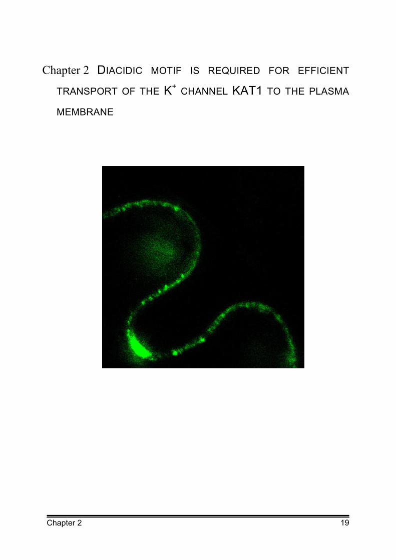
Localisation of the KAT1 K+ channel fused to the green fluorescent protein (GFP)
in the plasma membrane of Vicia faba epidermal cells after transfection by particle
bombardment. The channel is located in distinct punctuate structures which could
be clusters of the K+ channel in the plasma membrane or vesicles containing the
K+ channel close to the plasma membrane.
21 Chapter 2
2.1 Abstract
For a number of mammalian ion channels, trafficking to the plasma membrane
was found to be controlled by intrinsic sequence motifs. Among these sequences
are diacidic motifs that function as endoplasmic reticulum (ER) export signals. So
far it is unclear if similar motifs also exist in plant ion channels. In this study the
function of four diacidic DxE/DxD motifs of the plant potassium (K+) channel KAT1
were analysed. Mutation of the first diacidic DxE motif resulted in a strong
reduction of the KAT1 conductance in both, guard cell protoplasts and HEK293
cells (human embryonic kidney cells). Confocal fluorescence microscopy of guard
cells expressing the mutated KAT1 fused to green fluorescent protein (GFP)
revealed localization of the mutated channel only in intracellular structures around
the nucleus. These structures could be identified as part of the ER via
coexpression of KAT1 fused to yellow fluorescent protein (YFP) with an ER
retained protein (HDEL) fused to cyan fluorescent protein (CFP). Blocking the
vesicle formation from the ER by overexpression of the small GTP-binding protein
Sar1 fixed in its GDP bound form led to retention of wildtype KAT1 in similar parts
of the ER. Mutation of the three other diacidic motifs had no effect.
Together the results demonstrate that one diacidic motif of KAT1 is essential for
ER export of the functional K+ channel in both, guard cell protoplasts and HEK293
cells. This suggests that trafficking of plant plasma membrane ion channels is
controlled via a conserved mechanism.
2.2 Introduction
Plasma membrane K+ channels are crucial for cellular ion homeostasis, osmotic
regulation and excitability of cells. Their correct functioning depends not only on
the control of their activity in the plasma membrane but also on the regulation of
their number in the plasma membrane. Ion channels are transported to the plasma
membrane along the secretory pathway via the endoplasmic reticulum (ER) and
the Golgi apparatus. Until recently, plasma membrane proteins including ion
channels were considered to leave the ER by default (Wieland et al., 1987).
However, studies on trafficking of plasma membrane ion channels in mammalian
22 Chapter 2
cells revealed that transport from the ER to the Golgi apparatus is highly regulated
and plasma membrane channel density may be adjusted by controlling their export
from the ER (Ma et al., 2001; Wang et al., 2004). Among the few motifs identified
as ER export signals in ion channels are the diacidic D/ExD/E motifs which have
also been shown to function as ER export signals in other plasma membrane
proteins in yeast and animal cells (Nishimura and Balch, 1997; Votsmeier and
Gallwitz, 2001). Mutation of these diacidic motifs resulted in a strong reduction of
proteins in the plasma membrane and an accumulation in the ER.
ER export motifs are probably critical for enrichment of cargo proteins into COPII
(coat protein complex II) vesicles which are responsible for the transport of
proteins to the Golgi apparatus. A good candidate for the interaction of cargo
proteins with the COPII coat complex is the coat protein Sec24, which can bind to
a variety of ER export motifs (Bickford et al., 2004).
Homologues of the COPII coat proteins have also been identified in plants (Bar-
Peled and Raikhel, 1997; Movafeghi et al., 1999; Contreras et al., 2004; Yang et
al., 2005). However, knowledge about the molecular mechanism of COPII
mediated transport from the ER in plants is still limited (for review sees Aniento et
al., 2006). The small GTP-binding protein Sar1 which has been shown to be
crucial for formation of COPII in yeast and animal cells has also been found to play
an important role in ER to Golgi apparatus trafficking in plants (Takeuchi et al.,
2000; Phillipson et al., 2001; daSilva et al., 2004). Investigation on the trafficking of
the plasma membrane H+ ATPase from Arabidopsis thaliana has demonstrated
that transport to the plasma membrane is not achieved by default but requires
cytosolic domains (Lefebvre et al., 2004). However, the motif responsible for
targeting the H+ ATPase to the plasma membrane was not identified. ER export
signals in plants have so far only been identified for Golgi apparatus proteins
(Contreras et al., 2004; Yuasa et al., 2005; Hanton et al., 2005). Recently, Hanton
et al. (2005) demonstrated that transport of Golgi apparatus localised membrane
proteins out of the ER was reduced to about 60% by mutation of a diacidic DxE
motif.
Previous studies on trafficking of the plant K+ channel KAT1 from Arabidopsis
thaliana revealed that the K+ channel is subject to a constitutive and pressure-
driven turnover (Hurst et al., 2004; Meckel et al., 2004). Clusters of KAT1 were
found to be inserted into and retrieved from the plasma membrane during
23 Chapter 2
constitutive and pressure-driven exo- and endocytosis of small vesicles. Here the
role of diacidic motifs in trafficking of KAT1 was investigated using patch clamp
measurements and confocal fluorescence microscopy. The former technique
allowed the analysis of the functioning of the channel in its target compartment
plasma membrane while the latter provided information on the subcellular
localization of the channel. Amino acid sequence analysis predicts four diacidic
motifs in the cytosolic C-terminus of KAT1: I. DAE (394-396) located inside the
putative cyclic nucleotide binding domain (cNBD), II. DTE (555-557) and two more
inside the so called KHA domain, III. DLD (662-664) and IV. DGD (668-670), which
is possibly involved in channel tetramerization (Daram et al., 1997) or channel
clustering (Ehrhardt et al., 1997). Here it is shown that the ER export was strongly
dependent on the first diacidic motif. Mutation of this motif almost completely
abolished the transport of functional KAT1 to the plasma membrane and resulted
in the accumulation of the channel in the ER. These results demonstrate that ER
export can act as a site of regulation of ion channel trafficking in plants.
2.3 Results
2.3.1 Mutation of a diacidic motif reduces the number of active KAT1 K+ channels in the plasma membrane of guard cell protoplasts
Sequence analysis of the plant K+ inward rectifier from Arabidopsis thaliana
revealed two diacidic DxE motifs and two diacidic DxD motifs in the cytosolic
carboxyl-terminal tail of the channel. To determine the role of these diacidic motifs
four fusion constructs between green fluorescent protein (GFP) and KAT1 mutants
were constructed. Each of these mutants contained the acidic amino acids
aspartate (D) and glutamate (E) of one DxE or DxD motif substituted by alanine
(A). The resulting mutants KAT1(I)::GFP (DAE[394-396] mutated to AAA),
KAT1(II)::GFP (DTE[555-557] mutated to ATA), KAT1(III)::GFP (DLD[662-664]
mutated to ALA), KAT1(IV)::GFP (DGD[668-670] mutated to AGA) and wildtype
KAT1 fused to GFP (KAT1::GFP) were transiently expressed in guard cell
protoplasts. Transfected cells were analysed by whole cell patch clamp
measurements.
24 Chapter 2
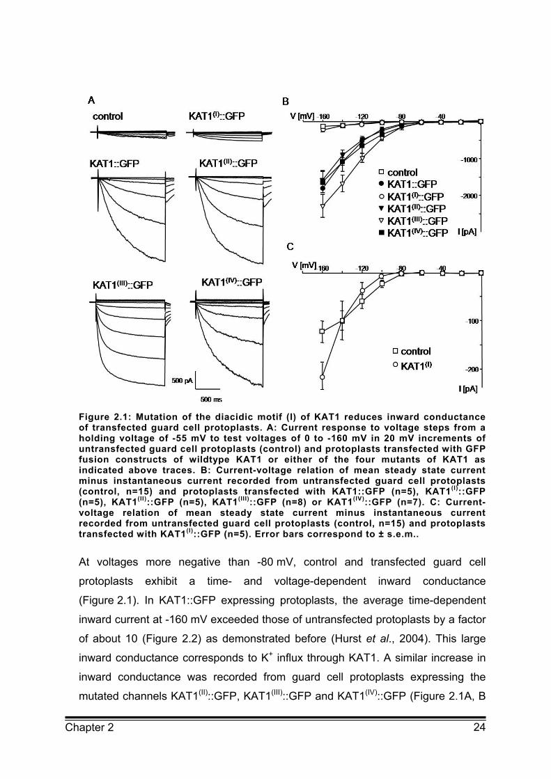
Figure 2.1: Mutation of the diacidic motif (I) of KAT1 reduces inward conductance of transfected guard cell protoplasts. A: Current response to voltage steps from a holding voltage of -55 mV to test voltages of 0 to -160 mV in 20 mV increments of untransfected guard cell protoplasts (control) and protoplasts transfected with GFP fusion constructs of wildtype KAT1 or either of the four mutants of KAT1 as indicated above traces. B: Current-voltage relation of mean steady state current minus instantaneous current recorded from untransfected guard cell protoplasts (control, n=15) and protoplasts transfected with KAT1::GFP (n=5), KAT1(I)::GFP (n=5), KAT1(II)::GFP (n=5), KAT1(III)::GFP (n=8) or KAT1(IV)::GFP (n=7). C: Current-voltage relation of mean steady state current minus instantaneous current recorded from untransfected guard cell protoplasts (control, n=15) and protoplasts transfected with KAT1(I)::GFP (n=5). Error bars correspond to ± s.e.m..
At voltages more negative than -80 mV, control and transfected guard cell
protoplasts exhibit a time- and voltage-dependent inward conductance
(Figure 2.1). In KAT1::GFP expressing protoplasts, the average time-dependent
inward current at -160 mV exceeded those of untransfected protoplasts by a factor
of about 10 (Figure 2.2) as demonstrated before (Hurst et al., 2004). This large
inward conductance corresponds to K+ influx through KAT1. A similar increase in
inward conductance was recorded from guard cell protoplasts expressing the
mutated channels KAT1(II)::GFP, KAT1(III)::GFP and KAT1(IV)::GFP (Figure 2.1A, B
25 Chapter 2
and 2.2). The time-dependent activation of K+ inward currents and the current-
voltage relation of KAT1(II)::GFP and KAT1(IV)::GFP expressing protoplasts
resembled that of KAT1::GFP (Figure 2.1A, B). In protoplasts expressing
KAT1(III)::GFP activation of the K+ inward current already occurred at -80 mV and
was faster than the time-dependent activation of wildtype KAT1 and of the other
two mutants described above (Figure 2.1A, B). This suggests that the
voltage-dependent activation of KAT1 is affected by mutation of the diacidic motif
(III). However, the focus of the present investigation is on trafficking of KAT1 which
seems not to be affected by the mutation.
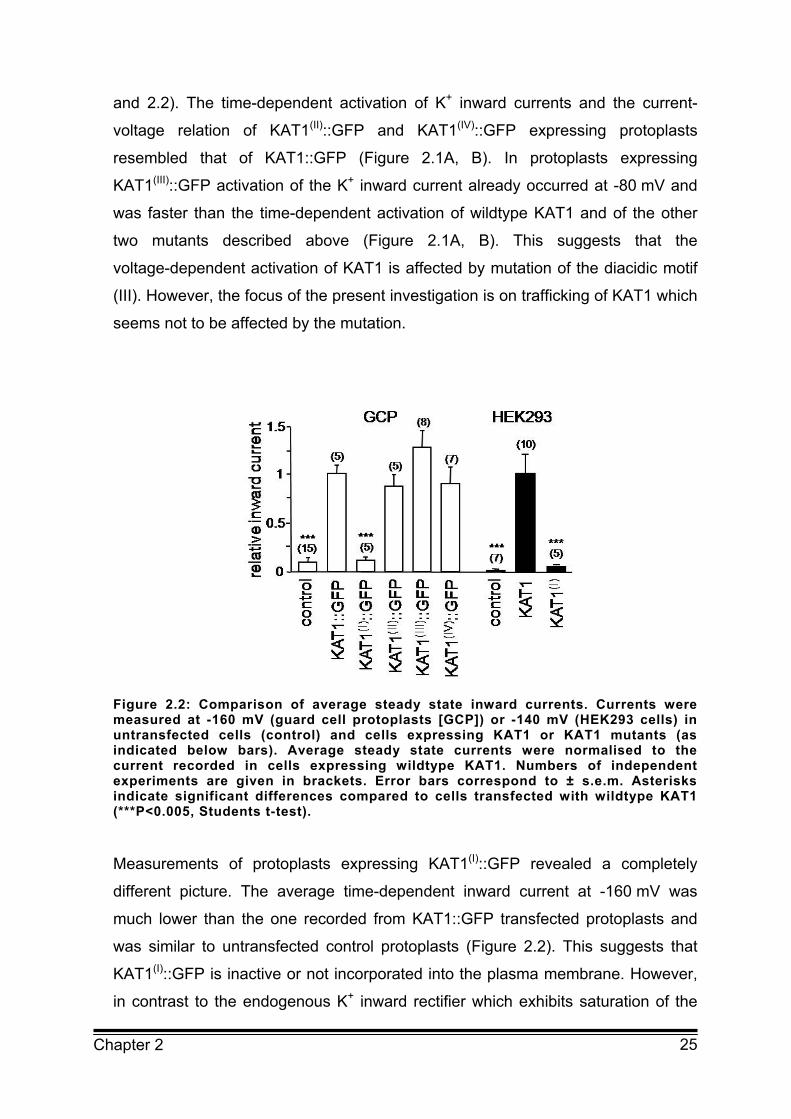
Figure 2.2: Comparison of average steady state inward currents. Currents were measured at -160 mV (guard cell protoplasts [GCP]) or -140 mV (HEK293 cells) in untransfected cells (control) and cells expressing KAT1 or KAT1 mutants (as indicated below bars). Average steady state currents were normalised to the current recorded in cells expressing wildtype KAT1. Numbers of independent experiments are given in brackets. Error bars correspond to ± s.e.m. Asterisks indicate significant differences compared to cells transfected with wildtype KAT1 (***P<0.005, Students t-test).
Measurements of protoplasts expressing KAT1(I)::GFP revealed a completely
different picture. The average time-dependent inward current at -160 mV was
much lower than the one recorded from KAT1::GFP transfected protoplasts and
was similar to untransfected control protoplasts (Figure 2.2). This suggests that
KAT1(I)::GFP is inactive or not incorporated into the plasma membrane. However,
in contrast to the endogenous K+ inward rectifier which exhibits saturation of the

26 Chapter 2
conductance at voltages more negative than -140 mV the inward conductance in
protoplasts transfected with KAT1(I)::GFP revealed no saturation in the voltage
range analysed (Figure 2.1C). The current-voltage relation of KAT1(I)::GFP
expressing protoplasts was very similar to the current-voltage relation of wildtype
KAT1 and the other KAT1 mutants (Figure 2.1B, C). This implies that KAT1(I)::GFP
is indeed incorporated and active in the plasma membrane of guard cells albeit to
a very low extent. Due to the large variability in endogenous K+ inward
conductance the additional KAT1(I)::GFP conductance is not seen as a significant
increase in the average inward current.
Together the results demonstrate that mutation of the diacidic motif (I) largely
reduced the number of active KAT1 channels in the plasma membrane of guard
cells while mutation of the three other diacidic motifs had no effect on the KAT1
conductance in transfected guard cell protoplasts.
2.3.2 Mutation of a diacidic motif results in ER retention of KAT1
The results described above imply that mutation of the diacidic motif (I) of KAT1
strongly affects the number of active channels in the plasma membrane. This can
in principle result from an inhibition of channels in the plasma membrane or from a
reduced incorporation of channels into the plasma membrane. The latter
explanation seems more likely as diacidic motifs have been shown to act as ER
export signals (Barlowe, 2003). To investigate whether the reduction in the number
of channels in the plasma membrane of KAT1(I)::GFP expressing guard cell
protoplasts results indeed from retention of the channel in the ER, the subcellular
distribution of wildtype KAT1 and KAT1 mutants fused to GFP or yellow
fluorescent protein (YFP) were compared in transfected guard cells using confocal
laser scanning microscopy.

27 Chapter 2
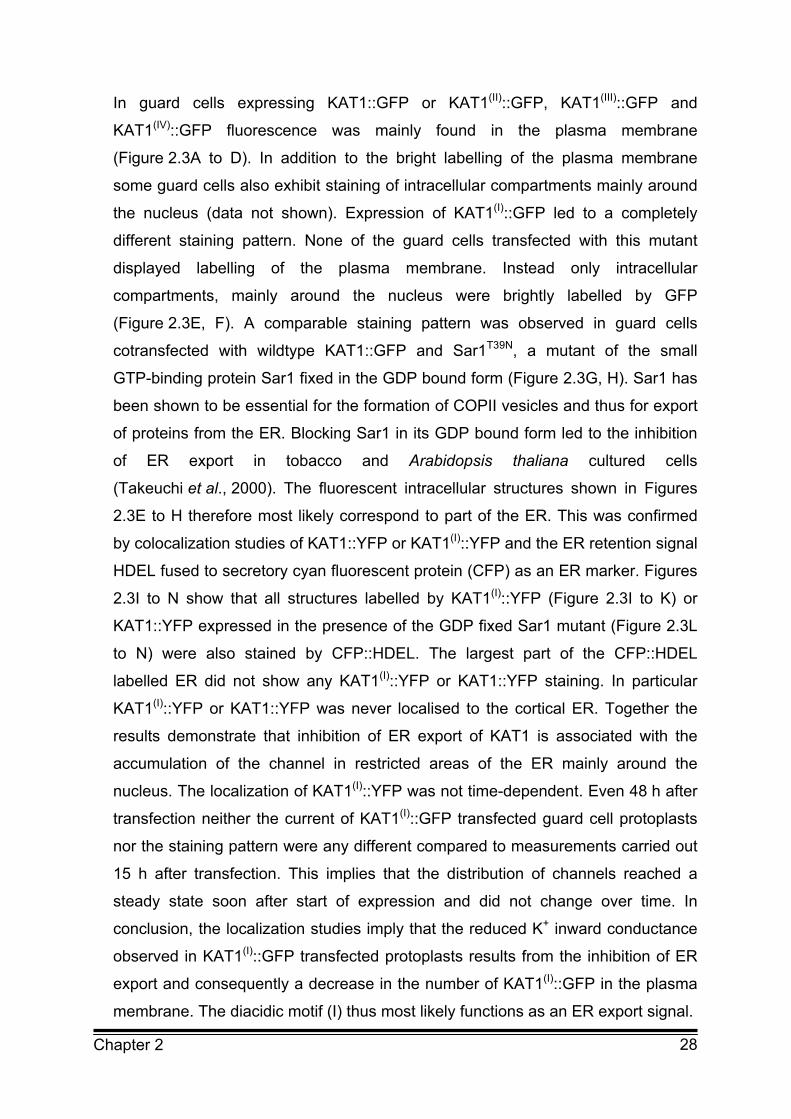
Figure 2.3: Mutation of the diacidic motif (I) of KAT1 results in ER retention A-D: Projection of two optical sections through the equatorial region of guard cells expressing KAT1::GFP (A), KAT1(II)::GFP (B), KAT1(III)::GFP (C) or KAT1(IV)::GFP (D). E-H: Overlay of transparency and fluorescent projection of four optical sections through the equatorial region of a guard cell expressing KAT1(I)::GFP (E,F) or wildtype KAT1::GFP and GDP-fixed Sar1 (G,H). I-K: Projection of four optical sections through the equatorial region of a guard cell cotransfected with KAT1(I)::YFP and CFP::HDEL; KAT1(I)::YFP fluorescence is shown in I, CFP::HDEL fluorescence is shown in J and K represents overlay of both images. L-N: Projection of four optical sections through the equatorial region of a guard cell cotransfected with GDP fixed Sar1, KAT1::YFP and CFP::HDEL; KAT1::YFP fluorescence is shown in L, CFP::HDEL fluorescence is shown in M and N represents overlay of both images. Scale bar corresponds to 10 µm.
28 Chapter 2
In guard cells expressing KAT1::GFP or KAT1(II)::GFP, KAT1(III)::GFP and
KAT1(IV)::GFP fluorescence was mainly found in the plasma membrane
(Figure 2.3A to D). In addition to the bright labelling of the plasma membrane
some guard cells also exhibit staining of intracellular compartments mainly around
the nucleus (data not shown). Expression of KAT1(I)::GFP led to a completely
different staining pattern. None of the guard cells transfected with this mutant
displayed labelling of the plasma membrane. Instead only intracellular
compartments, mainly around the nucleus were brightly labelled by GFP
(Figure 2.3E, F). A comparable staining pattern was observed in guard cells
cotransfected with wildtype KAT1::GFP and Sar1T39N, a mutant of the small
GTP-binding protein Sar1 fixed in the GDP bound form (Figure 2.3G, H). Sar1 has
been shown to be essential for the formation of COPII vesicles and thus for export
of proteins from the ER. Blocking Sar1 in its GDP bound form led to the inhibition
of ER export in tobacco and Arabidopsis thaliana cultured cells
(Takeuchi et al., 2000). The fluorescent intracellular structures shown in Figures
2.3E to H therefore most likely correspond to part of the ER. This was confirmed
by colocalization studies of KAT1::YFP or KAT1(I)::YFP and the ER retention signal
HDEL fused to secretory cyan fluorescent protein (CFP) as an ER marker. Figures
2.3I to N show that all structures labelled by KAT1(I)::YFP (Figure 2.3I to K) or
KAT1::YFP expressed in the presence of the GDP fixed Sar1 mutant (Figure 2.3L
to N) were also stained by CFP::HDEL. The largest part of the CFP::HDEL
labelled ER did not show any KAT1(I)::YFP or KAT1::YFP staining. In particular
KAT1(I)::YFP or KAT1::YFP was never localised to the cortical ER. Together the
results demonstrate that inhibition of ER export of KAT1 is associated with the
accumulation of the channel in restricted areas of the ER mainly around the
nucleus. The localization of KAT1(I)::YFP was not time-dependent. Even 48 h after
transfection neither the current of KAT1(I)::GFP transfected guard cell protoplasts
nor the staining pattern were any different compared to measurements carried out
15 h after transfection. This implies that the distribution of channels reached a
steady state soon after start of expression and did not change over time. In
conclusion, the localization studies imply that the reduced K+ inward conductance
observed in KAT1(I)::GFP transfected protoplasts results from the inhibition of ER
export and consequently a decrease in the number of KAT1(I)::GFP in the plasma
membrane. The diacidic motif (I) thus most likely functions as an ER export signal.

29 Chapter 2
2.3.3 Mutation of a diacidic motif also affects KAT1 conductance in HEK293 cells
To investigate whether the function of the first diacidic motif of KAT1 as an ER
export signal is conserved among the plant and animal kingdom HEK293 cells
(human embryonic kidney cells) transfected with wildtype or mutant KAT1 were
analysed. HEK293 cells exhibit only a low plasma membrane conductance at
negative voltages and no endogenous time-activated K+ inward current (Figure
2.4). They therefore provide an excellent system to study the K+ inward rectifier
KAT1. HEK293 cells expressing wildtype KAT1 showed large time-dependent
inward currents at voltages more negative than -80 mV (Figure 2.4A, B) as
demonstrated before (Hertel et al., 2005). The inward currents displayed the
typical activation kinetic and voltage-dependence recorded from wildtype KAT1
expressing guard cell protoplasts (Hurst et al., 2004). Measurements of HEK293
cells transfected with KAT1(I) showed a completely different current response
which was at first glance similar to the one recorded from untransfected cells
(Figure 2.4A). However, a blow up of the current traces clearly revealed a time and
voltage depended activation of an inward current which was never observed in
untransfected HEK293 cells (Figure 2.4A). The activation kinetic of this current
qualitatively matches the kinetics of the current recorded from wildtype KAT1
transfected cells. All transfected cells exhibit similar voltage-dependence with an
increase in current at voltages negative of -80 mV (Figure 2.4B, C). However, in
KAT1(I) expressing cells the current at -140 mV was reduced to only about 5% of
the current recorded in wildtype KAT1 expressing cells (Figure 2.2). This
demonstrates that KAT1(I) is inserted into the plasma membrane of HEK293 cells
albeit to a lower extent.
30 Chapter 2
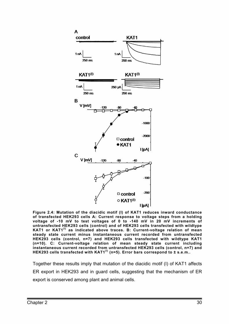
Figure 2.4: Mutation of the diacidic motif (I) of KAT1 reduces inward conductance of transfected HEK293 cells A: Current response to voltage steps from a holding voltage of -10 mV to test voltages of 0 to -140 mV in 20 mV increments of untransfected HEK293 cells (control) and of HEK293 cells transfected with wildtype KAT1 or KAT1(I) as indicated above traces. B: Current-voltage relation of mean steady state current minus instantaneous current recorded from untransfected HEK293 cells (control, n=7) and HEK293 cells transfected with wildtype KAT1 (n=10). C: Current-voltage relation of mean steady state current including instantaneous current recorded from untransfected HEK293 cells (control, n=7) and HEK293 cells transfected with KAT1(I) (n=5). Error bars correspond to ± s.e.m..
Together these results imply that mutation of the diacidic motif (I) of KAT1 affects
ER export in HEK293 and in guard cells, suggesting that the mechanism of ER
export is conserved among plant and animal cells.

31 Chapter 2
2.4 Discussion
2.4.1 The diacidic motif of KAT1 is essential for efficient ER export
In a number of proteins diacidic motifs have been shown to be crucial for efficient
transport of these proteins from the ER to their target compartments (Nishimura
and Balch, 1997; Votsmeier and Gallwitz, 2001; Ma et al., 2001; Wang et al.,
2004; Hanton et al., 2005). In the cytosolic C-terminus of the plant K+ channel
KAT1 four diacidic motifs have been identified. Patch clamp analysis of guard cell
protoplasts and HEK293 cells revealed that only mutation of one (diacidic motif (I))
out of the four diacidic motifs of KAT1 dramatically reduced the inward
conductance of transfected cells. The reduction in inward conductance could be
quantified in HEK293 cells which do not exhibit any endogenous inward
conductance. In HEK293 cells, expressing the mutant channel KAT1(I), the inward
conductance was reduced to only about 5% compared to wildtype KAT1
transfected cells. The reduced current recorded from cells expressing KAT1(I)
showed a qualitatively similar kinetic and voltage-dependence as wildtype KAT1.
This implies that the lower inward conductance of KAT1(I) transfected cells results
from a reduced number of functional channels in the plasma membrane
suggesting that transport but not function of the channel to the plasma membrane
is inhibited.
This was confirmed by confocal images of KAT1(I)::GFP expressing guard cells
which showed a bright staining of intracellular compartments mainly around the
nucleus without any detectable staining of the plasma membrane. Coexpression
studies with the ER marker CFP::HDEL confirmed that these intracellular
structures correspond to the ER, demonstrating that the largest amount of the
mutated channel is indeed retained in the ER. The fluorescence of the remaining
channels that still reached the plasma membrane was too low to be detected by
confocal laser scanning microscopy.
In principle the retention of KAT1(I)::GFP in the ER could result from misfolding of
the mutated protein which is kept in the ER for subsequent degradation.

32 Chapter 2
However, the fact that in guard cell protoplasts as well as in HEK293 cells
functional KAT1(I) channels with similar voltage-dependence and time-dependent
activation kinetics as wildtype KAT1 can be detected in the plasma membrane
argues against this hypothesis. Our results rather implicate that the observed
retention of KAT1(I)::GFP in the ER is due to a reduction of ER export of fully
functional KAT1 channels. This is consistent with our analysis of wildtype KAT1
expressing guard cells where ER export has been blocked by coexpression of the
GDP-fixed Sar1 mutant. These cells showed the same staining pattern as
KAT1(I)::GFP expressing cells. Therefore, it can be concluded that the diacidic
motif (I) of KAT1 acts as an ER export signal in both, HEK293 and guard cells.
This also suggests that the mechanism of ER export is conserved among plant
and animal cells.
2.4.2 ER retention of KAT1 is very efficient and may be restricted to certain areas
Recently, a diacidic motif has been shown to affect ER export of two plant Golgi-
localised membrane proteins (Hanton et al., 2005). Using imaging of transfected
tobacco leaves Hanton et al. (2005) demonstrated that mutation of a diacidic motif
led to a reduction of the Golgi-localization of these proteins by about 40%. The
authors therefore suggest that factors other than diacidic motifs also influence the
ER export of these proteins. In our studies mutation of the diacidic motif (I) nearly
completely blocked transport of the channel to the plasma membrane without
affecting its functioning. This implicates that efficient transport of KAT1 from the
ER to the Golgi apparatus is highly dependent on the first diacidic motif. Similar
results have been described for the function of diacidic motifs in trafficking of
plasma membrane transporters in mammalian cells (Ma et al., 2001; Wang et al.,
2004). Investigation of trafficking of mammalian plasma membrane transporters
suggest that binding of scaffold proteins, such as PDZ domain-containing proteins
can change the relative effectiveness of ER export signals and thus allow
regulation of the number of transporters in the plasma membrane (Ma and Jan,
2002). A tight regulation of the protein density is of particular importance for
plasma membrane ion channels as small changes in the number of channels can
have a pronounced effect on the conductance and thus function of the cell. Most
33 Chapter 2
likely the diacidic motif (I) of KAT1 is part of such a regulatory mechanism that
controls the density of this channel in the plasma membrane of plant cells.
Analysis of the localization of KAT1(I)::GFP reveals that the channel is mainly
retained in the ER around the nucleus. Similar results were found for guard cells
transfected with KAT1::GFP when ER export was blocked by coexpression of
GDP fixed Sar1. This distinct localization of ER retained KAT1 was also found in
epidermal cells (data not shown). As the outer nuclear membrane is continuous
with the ER it is not unexpected that proteins that are retained in the ER can also
be found in the nuclear envelope. However, previous investigations on proteins
retained in the ER revealed localisation of these proteins in the nuclear envelope
only in addition to the distribution throughout the rest of the ER (Batoko et al.,
2000, Hermann et al., 1990). The reasons for retention of KAT1 in restricted areas
around the nucleus remain to be identified.
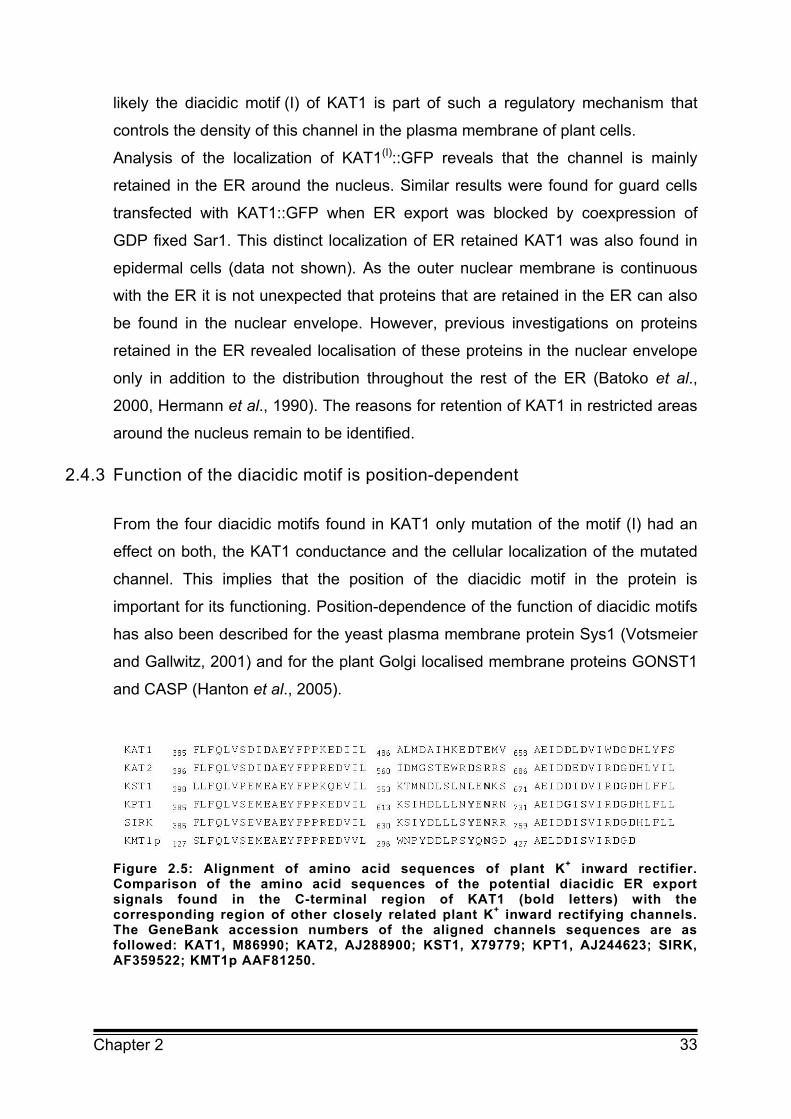
2.4.3 Function of the diacidic motif is position-dependent
From the four diacidic motifs found in KAT1 only mutation of the motif (I) had an
effect on both, the KAT1 conductance and the cellular localization of the mutated
channel. This implies that the position of the diacidic motif in the protein is
important for its functioning. Position-dependence of the function of diacidic motifs
has also been described for the yeast plasma membrane protein Sys1 (Votsmeier
and Gallwitz, 2001) and for the plant Golgi localised membrane proteins GONST1
and CASP (Hanton et al., 2005).
Figure 2.5: Alignment of amino acid sequences of plant K+ inward rectifier. Comparison of the amino acid sequences of the potential diacidic ER export signals found in the C-terminal region of KAT1 (bold letters) with the corresponding region of other closely related plant K+ inward rectifying channels. The GeneBank accession numbers of the aligned channels sequences are as followed: KAT1, M86990; KAT2, AJ288900; KST1, X79779; KPT1, AJ244623; SIRK, AF359522; KMT1p AAF81250.

34 Chapter 2
The diacidic motif (I) of KAT1 is located in the putative cyclic nucleotide binding
domain (cNBD). Recent investigations on disruption of the cNBD in the animal K+
channels HCN and HERG implied that highly conserved regions in the cNBD are
generally critical for ion channel trafficking (Akhavan et al., 2005) This is supported
by the fact that the diacidic ER export motif of the CFTR channel is also located
within the NBD (Wang et al., 2004). Using structural models of the NBD of the
CFTR channel Wang et al. (2004) demonstrated the ability of the loop containing
the diacidic motif to insert directly into the diacidic code binding pocket of the
COPII coat complex Sec23/Sec24. In plant cells the cNBD was found to be
required for efficient transport of a H+ ATPase from Nicotiana plumbaginifolia to
the plasma membrane even though ER export motifs have so far not been
identified (Lefebvre et al., 2004). An alignment of KAT1 with other related plant
K+ inward rectifiers revealed that the diacidic motif (I) and the diacidic motif (IV)
are highly conserved among these channels (Figure 2.5). However, only the
diacidic motif (I) which is located in the cNBD was found to affect ER export
suggesting that the cNBD is per se important for ER export of plant ion channels.
Together the results demonstrate that a diacidic motif of KAT1 acts as an ER
export signal in plant and animal cells probably via a conserved mechanism.
2.5 Methods
2.5.1 Vectors for KAT1 expression
For the expression of a KAT1::GFP fusion protein in guard cells the cDNA of KAT1
was cloned into the pAVA393 expression vector in frame with mGFP5 or YFP
under the control of two strong 35S promoters as previously described by Hurst et
al. (2004).
Expression of KAT1 in the mammalian cell line HEK293 (human embryonic kidney
cells) was obtained with KAT1 cDNA cloned into the pCB6 (Acc.No.: ATCC37274)
eukaryotic expression vector at the NcoI restriction site under control of a CMV
promotor. HEK293 cells were cotransfected with the pEGFPN2 vector (Clontech,
Pharmingen, Germany) to express cytosolic GFP as a transfection control.
35 Chapter 2
2.5.2 Mutagenesis of putative ER export motifs in KAT1
Mutations in the DxE and DxD motif of channel protein were created by PCR
based site directed mutagenesis (QuikChange site directed mutagenesis kit;
Stratagene, LaJolla, USA) and confirmed by sequencing. The expression vectors
pAVA393KAT1 and pCB6KAT1, both containing the KAT1 cDNA as described
above served as templates. The plasmids were cloned into E. coli/DH5α and
isolated with Qiagen high speed Midi-Kit (Qiagen, Germany) for cell transfection.
2.5.3 Transfection of intact guard cells via particle delivery and isolation of protoplasts
Vicia faba L. cv. Bunyan were grown under controlled climate conditions with
18 °C, 70 % relative humidity and a 14/10 h photoperiod at 350-400 µmol
photons/m2/sec. Transfection of intact guard cells via particle delivery was
performed as described earlier (Hurst et al., 2004).
Cotransfection was performed via coating of gold with equal molar amounts of
each plasmid DNA to give a total amount of 15 µg DNA. Guard cell protoplasts
were prepared from transfected leaves after overnight incubation at room
temperature as described previously (Homann, 1998).
2.5.4 Cultivation and transfection of mammalian cell line HEK293
HEK293 cells were grown at 37 °C and 5 % CO2. For transient expression of
KAT1 and KAT1 mutants HEK293 cells were transfected with each 0.75 µg of
pCB6KAT1 and pEGFPN2 vector using the liposomal transfection reagent
Metafectene (Biontex, Munich, Germany) according to manufacturer’s instructions.

36 Chapter 2
2.5.5 Patch clamp measurements
HEK293 cells:
Experiments were performed on cells incubated at 37°C in 5% CO2 for 2-3 days
after transfection (for details see Hertel et al., 2005). Cells were bathed in a
solution containing: 20 mM KCl, 1.8 mM CaCl2, 1 mM MgCl2, 5 mM HEPES at
pH 7.4. Choline-Cl was used to adjust the osmolarity to 300 mOsmol/kg. Patch
pipettes contained: 130 mM K+-gluconate, 10 mM NaCl, 5 mM Hepes, 0.1 mM
Na2GTP, 0.1 µM CaCl2, 2 mM MgCl2, 5 mM Na2Phosphocreatin, 2 mM K2ATP at
pH 7.4 with an osmolarity of approximately 330 mOsmol/kg.
Guard cell protoplasts:
Guard cell protoplasts were bathed in 10 mM KCl, 10 mM CaCl2 and 5 mM MES,
pH 6.0/KOH. The osmolarity was adjusted to 520 mOsmol/kg with Sorbitol. Patch
pipettes were filled with 150 mM K+-gluconate, 10 mM KCl, 2 mM MgCl2, 2 mM
EGTA, 10 mM HEPES, 2 mM K2ATP, pH 7.8/KOH. Osmolarity was adjusted to
560 mOsmol/kg with Sorbitol.
In both cell systems measurements were performed in a standard whole-cell patch
clamp experiment as described in detail previously (Homann and Thiel, 2002;
Hurst et al., 2004; Hertel et al., 2005).
2.5.6 CLSM
Confocal microscopic analysis of transfected turgid guard cells was performed
after overnight incubation as described earlier using a confocal laser scanning
microscope (Leica TCS SP, Leica Microsystems GmbH, Heidelberg, Germany)
(for details see Meckel et al., 2004). For excitation of fluorescent proteins the
following lines of a 25 mW argon laser were used: 488 nm for GFP, 458 nm for
CFP and 514 nm for YFP. Fluorescence was detected at 505-535 nm for GFP,
465-490 nm for CFP and 600-650 nm for YFP. Images were processed using the
Leica Confocal Software 2.00 (LCS, Leica Microsystems GmbH, Heidelberg,
Germany). For the microscopic analysis guard cells were bathed in 0.1 mM CaCl2,
10 mM MES, 45 mM KCl, pH 6.1/KOH.

37 Chapter 2
2.6 References
Akhavan A, Atanasiu R, Noguchi T, Han W, Holder N, Shrier A (2005)
Identification of the cyclic nucleotide binding domain as a conserved determinant
of ion channel cell surface localization. The Journal of Cell Science 118:2803-2812
Aniento F, Matsuoka K, Robinson DG (2006) ER to Golgi pathway: The COPII
pathway. In DG Robinson ed., The Plant Endoplasmic Reticulum. The Plant Cell
Monographs, Vol.4
Barlowe C (2003) Signals for COPII-dependent export from the ER: what's the
ticket out? Trends in Cell Biology 13:295-300
Bar-Peled M and Raikhel NV (1997) Characterization of AtSEC12 and AtSAR1.
Proteins likely involved in endoplasmic reticulum and Golgi transport. Plant
Physiology 114:315–324
Batoko H, Zheng HQ, Hawes C, Moore I (2000) A Rab1 GTPase is required for
transport between the endoplasmic reticulum and Golgi apparatus and for normal
Golgi movement in plants. The Plant Cell 12:2201-2218 Bickford LC, Mossessova E, Goldberg J (2004) A structural view of the COPII
vesicle coat. Current Opinion in Structural Biology 14:147-153
Contreras I, Yang Y, Robinson DG, Aniento F (2004) Sorting signals in the
cytosolic tail of plant p24 proteins involved in the interaction with the COPII coat.
Plant Cell Physiology 45:1779–1786 Daram P, Urbach S, Gaymard F, Sentenac H, Cherel I (1997) Tetramerization of
the AKT1 plant potassium channel involves its C-terminal cytoplasmic domain.
The EMBO Journal 16:3455-3463
daSilva LL, Snapp EL, Denecke J, Lippincott-Schwartz J, Hawes C, Brandizzi F (2004) Endoplasmic reticulum export sites and Golgi bodies behave
as single mobile secretory units in plant cells. The Plant Cell 16:1753-1771
Ehrhardt T, Zimmermann S, Müller-Röber B (1997) Association of plant K+(in)
channels is mediated by conserved C-termini and does not affect subunit
assembly. FEBS Letters 409:166-170
Hanton SL, Renna L, Bortolotti LE, Chatre L, Stefano G, Brandizzi F (2005)
Diacidic motifs influence the export of transmembrane proteins from the
endoplasmic reticulum in plant cells. The Plant Cell 17:3081-3093
38 Chapter 2
Herman EM, Tague BW, Hoffman LM, Kjemtrup SE, Chrispeels MJ (1990).
Retention of phytohemagglutinin with carboxy-terminal tetrapeptide KDEL in the
nuclear envelope and the endoplasmic reticulum. Planta 182:305–312
Hertel B, Horváth F, Wodala B, Hurst AC, Moroni A, Thiel G (2005) KAT1
inactivates at sub-millimolar concentrations of external potassium. The Journal of
Experimental Botany 56:3103-3110
Homann U (1998) Fusion and fission of plasma membrane material
accommodates for osmotically induced changes in the surface area of guard cell
protoplasts. Planta 206:495-499
Homann U and Thiel G (2002) The number of K+ channels in the plasma
membrane of guard cell protoplasts changes in parallel with the surface area.
PNAS 99:10215-10220
Hurst AC, Meckel T, Tayefeh S, Thiel G, Homann U (2004) Trafficking of the
plant potassium inward rectifier KAT1 in guard cell protoplasts of Vicia faba. The
Plant Journal 37:391-397
Lefebvre B, Batoko H, Duby G, Boutry M (2004) Targeting of a Nicotiana
plumbaginifolia H+ ATPase to the plasma membrane is not by default and requires
cytosolic structural determinants. The Plant Cell 16:1772-1789
Ma D, Zerangue N, Lin Y, Collins A, Yu M, Jan Y, Jan L (2001) Role of ER
export signals in controlling surface potassium channel numbers. Science
291:316-319
Ma D and Jan LY (2002) ER transport signals and trafficking of potassium
channels and receptors. Current Opinion in Neurobiology 12:287–292
Meckel T, Hurst AC, Thiel G, Homann U (2004) Endocytosis against high turgor:
intact guard cells of Vicia faba constitutively endocytose fluorescently labelled
plasma membrane and GFP-tagged K+ channel KAT1. The Plant Journal
39:182-193 Movafeghi A, Happel N, Pimpl P, Tai GH, Robinson DG (1999) Arabidopsis
thaliana Sec21p and Sec23p homologues. Probable coat proteins of plant COP-
coated vesicles. Plant Physiology 199:1437-1446
Nishimura N and Balch WE (1997) A diacidic signal required for selective export
from the endoplasmic reticulum. Science 277:556-558

39 Chapter 2
Phillipson BA, Pimpl P, daSilva LL, Crofts AJ, Taylor JP, Movafeghi A, Robinson DG, Denecke J (2001) Secretory bulk flow of soluble proteins is
efficient and COPII-dependent. The Plant Cell 13:2005-2020
Takeuchi M, Ueda T, Sato K, Abe H, Nagata T, Nakano A (2000) A dominant
negative mutant of Sar1 GTPase inhibits protein transport from the endoplasmic
reticulum to the Golgi apparatus in tobacco and Arabidopsis thaliana cultured
cells. The Plant Journal 23:517-525
Votsmeier C and Gallwitz D (2001) An acidic sequence of a putative yeast Golgi
membrane protein binds COPII and facilitates ER export. The EMBO Journal
20:6742-6750
Wang X, Matteson J, An Y, Moyer B, Yoo JS, Bannykh S, Wilson IA, Riordan JR, Balch WE (2004) COPII-dependent export of cystic fibrosis
transmembrane conductance regulator from the ER uses a diacidic exit code. The
Journal of Cell Biology 167:65-74
Wieland F, Gleason M, Serafini T, Rothman J (1987) The rate of bulk flow from
the endoplasmic reticulum to the cell surface. Cell 50:289-300
Yang YD, Elamawi R, Bubeck J, Pepperkok R, Ritzenthaler C, Robinson DG (2005) Dynamics of COPII vesicles and the Golgi apparatus in cultured Nicotiana
tabacum BY-2 cells provides evidence for transient association of Golgi stacks
with endoplasmic reticulum exit sites. The Plant Cell 17:1343-1359
Yuasa K, Toyooka K, Fukuda H, Matsuoka K (2005) Membrane anchored prolyl
hydroxylase with an export signal from the endoplasmic reticulum. The Plant
Journal 41:81-94
40 Chapter 3
Chapter 3 EFFICIENCY OF ER EXPORT OF THE K+ CHANNEL
KAT1 DEPENDS ON THE NUMBER OF ACIDIC AMINO ACIDS
WITHIN A TRIACIDIC MOTIF

41 Chapter 3
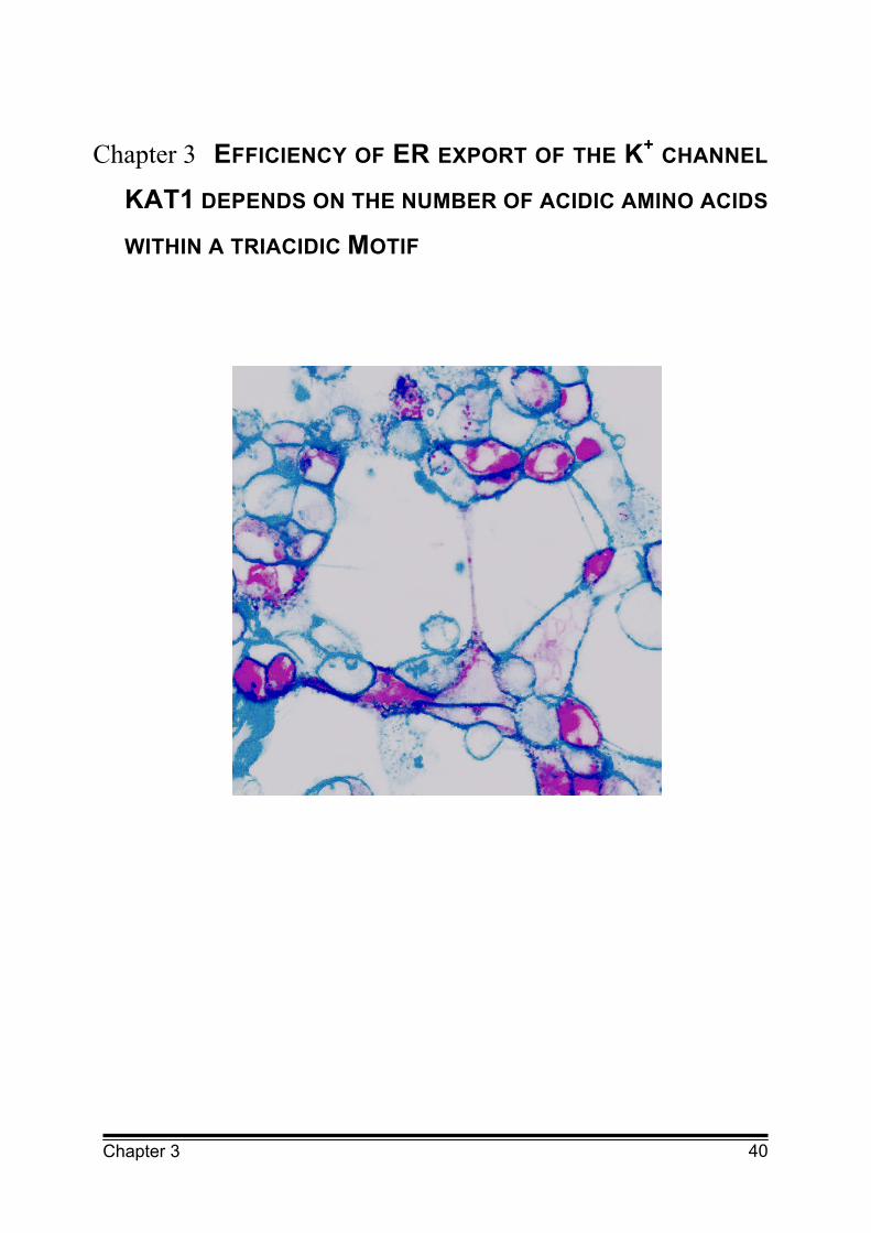
HEK293 cells transiently expressing KAT1 fused to the green fluorescent protein
(purple) stained with the plasma membrane marker FM4-64 (light blue).
Colocalisation of both fluorescent proteins in the plasma membrane is indicated in
dark blue.

42 Chapter 3
3.1 Abstract
For a number of ion channels, including the potassium (K+) inward rectifying
channel KAT1 from Arabidopsis thaliana, diacidic ER export motifs have been
identified. The aim of this study was to specify the role of single acidic amino acids
for efficient ER export of KAT1. Therefore, a sequence of KAT1, containing two
aspartate (D) and one glutamate (E), was analysed. This sequence included the
originally identified diacidic ER export motif (DxE) of KAT1 and an additional D just
downstream of the diacidic motif. Analysis of single, double and triple mutations of
the acidic amino acids of the DxDxE motif revealed a gradual reduction of ER
export depending on the number of mutated acidic residues. The amount of
reduction in ER export was not related to the position but only to the number of
mutated acidic amino acids. Furthermore, it could be demonstrated that plasma
membrane expression of ER export mutants can be rescued by heterotetrameric
assembly with wildtype KAT1. This implies that not all subunits of the KAT1
tetramer need to carry a functional ER export motif for trafficking to the plasma
membrane. Together this study shows that a triacidic motif functions as an ER
export signal of KAT1. In addition, it suggests that ER export may not only function
in regulation of channel density but also in controlling channel composition in the
plasma membrane.
3.2 Introduction
Ion channels are integral membrane proteins which facilitate the diffusion of ions
across biological membranes. They play a key role in many cellular processes
including signal transduction. Therefore, the control of ion channel activity but also
regulation of the number and composition of channels in the membrane is of
particular importance for cell functioning. Recent investigations have highlighted
the transport of ion channel from the endoplasmic reticulum (ER) to the Golgi
apparatus as an important step for regulation of surface expression of ion
channels (Schwappach, 2008). For the efficient ER export proteins are recruited
into coat protein complex II (COPII) vesicles which bud off the ER membrane. This
process involves recognition of specific amino acid motifs, so-called ER export

43 Chapter 3
signals, by components of the COPII coat (Otte and Barlowe, 2004). Until now
several ER export motifs have been identified in membrane proteins including ion
channels, receptors and transporters. Most of them fall into four groups: diacidic,
dihydrophobic and dibasic signal sequences as well as a terminal valine (V) at the
C-terminus. The diacidic ER export motif consist of the two acidic amino acids
aspartate (D) or glutamate (E) separated by any amino acid (x) (D/ExD/E). Ion
channels and transporters are accounting for the largest part of proteins for which
diacidic ER export motifs have been identified. For several members of the
K+ channel family (Ma et al., 2001 and 2002; Hofherr et al., 2005; Zuzarte et al.,
2007) and also for the chloride channel CFTR (Wang et al., 2004) diacidic ER
export motifs where found to be essential for surface expression of the channel.
Recently, it was demonstrated that ER export of the plant K+ channel KAT1 also
strongly depended on a diacidic motif. Mutation of the DxE (394-396) motif in the
C-terminus of the K+ channel reduced the transport of functional KAT1 to the
plasma membrane by about 90 % and resulted in the accumulation of the channel
in the ER (Mikosch et al., 2006).
Further investigations regarding the function of the ER export signal revealed a
direct interaction of the diacidic motif of KAT1 with the COPII component Sec24
(Sieben et al., 2008). Sec24 contains at least three binding sites for ER export
motifs. The B-site was found to be the binding site for the diacidic ER export motif
of a number of proteins like Sys1 and Yor1 (Mossessova et al., 2003; Miller et al.,
2003; Pagant et al., 2007). However, mutation of the B-site of Sec24 from yeast
suggests that other proteins like the amino acid permease Gap1 which also
contains a diacidic motif, do not bind to the B-site (Miller et al., 2003). These
studies suggest that recognition of the diacidic ER export motif by Sec24 may not
be consistent for all diacidic motifs. This may also explain the sometimes
contradictory results on sequence specificity of the diacidic ER export motif
(Mikosch and Homann, 2009).
Therefore, the role of single acidic amino acids for ER export of KAT1 was
analysed. Sequence comparison of several plant ion channels from different
organisms showed that the diacidic ER export motif is located in a highly
conserved part of the protein. Noticeably a third acid residue ahead of the diacidic
motif is found in nearly all of these channels (Figure 3.1). Hence, this residue was
also included in the site directed mutagenesis studies. To complete the
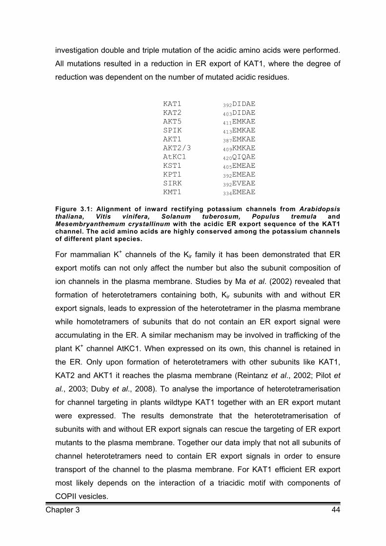
44 Chapter 3
investigation double and triple mutation of the acidic amino acids were performed.
All mutations resulted in a reduction in ER export of KAT1, where the degree of
reduction was dependent on the number of mutated acidic residues.
KAT1 392DIDAE KAT2 403DIDAE AKT5 411EMKAE SPIK 413EMKAE AKT1 387EMKAE AKT2/3 409KMKAE AtKC1 420QIQAE KST1 405EMEAE KPT1 392EMEAE SIRK 392EVEAE KMT1 334EMEAE
Figure 3.1: Alignment of inward rectifying potassium channels from Arabidopsis thaliana, Vitis vinifera, Solanum tuberosum, Populus tremula and Mesembryanthemum crystallinum with the acidic ER export sequence of the KAT1 channel. The acid amino acids are highly conserved among the potassium channels of different plant species. For mammalian K+ channels of the Kir family it has been demonstrated that ER
export motifs can not only affect the number but also the subunit composition of
ion channels in the plasma membrane. Studies by Ma et al. (2002) revealed that
formation of heterotetramers containing both, Kir subunits with and without ER
export signals, leads to expression of the heterotetramer in the plasma membrane
while homotetramers of subunits that do not contain an ER export signal were
accumulating in the ER. A similar mechanism may be involved in trafficking of the
plant K+ channel AtKC1. When expressed on its own, this channel is retained in
the ER. Only upon formation of heterotetramers with other subunits like KAT1,
KAT2 and AKT1 it reaches the plasma membrane (Reintanz et al., 2002; Pilot et
al., 2003; Duby et al., 2008). To analyse the importance of heterotetramerisation
for channel targeting in plants wildtype KAT1 together with an ER export mutant
were expressed. The results demonstrate that the heterotetramerisation of
subunits with and without ER export signals can rescue the targeting of ER export
mutants to the plasma membrane. Together our data imply that not all subunits of
channel heterotetramers need to contain ER export signals in order to ensure
transport of the channel to the plasma membrane. For KAT1 efficient ER export
most likely depends on the interaction of a triacidic motif with components of
COPII vesicles.
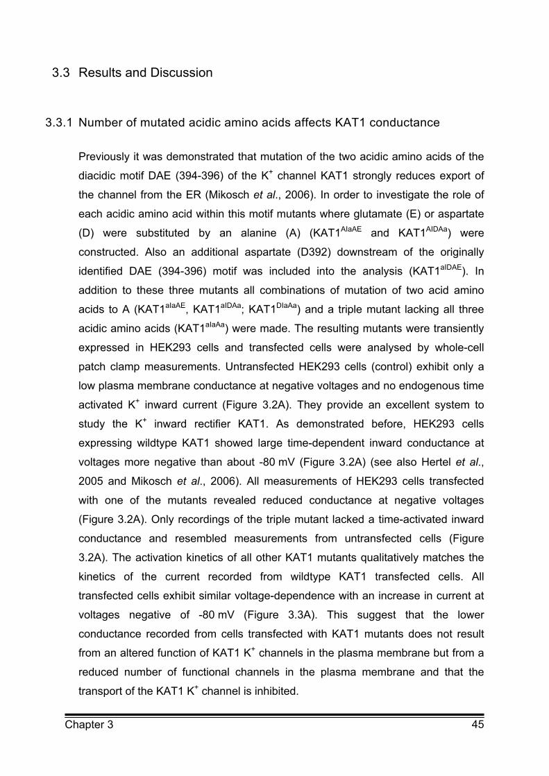
45 Chapter 3
3.3 Results and Discussion
3.3.1 Number of mutated acidic amino acids affects KAT1 conductance
Previously it was demonstrated that mutation of the two acidic amino acids of the
diacidic motif DAE (394-396) of the K+ channel KAT1 strongly reduces export of
the channel from the ER (Mikosch et al., 2006). In order to investigate the role of
each acidic amino acid within this motif mutants where glutamate (E) or aspartate
(D) were substituted by an alanine (A) (KAT1AIaAE and KAT1AIDAa) were
constructed. Also an additional aspartate (D392) downstream of the originally
identified DAE (394-396) motif was included into the analysis (KAT1aIDAE). In
addition to these three mutants all combinations of mutation of two acid amino
acids to A (KAT1aIaAE, KAT1aIDAa; KAT1DIaAa) and a triple mutant lacking all three
acidic amino acids (KAT1aIaAa) were made. The resulting mutants were transiently
expressed in HEK293 cells and transfected cells were analysed by whole-cell
patch clamp measurements. Untransfected HEK293 cells (control) exhibit only a
low plasma membrane conductance at negative voltages and no endogenous time
activated K+ inward current (Figure 3.2A). They provide an excellent system to
study the K+ inward rectifier KAT1. As demonstrated before, HEK293 cells
expressing wildtype KAT1 showed large time-dependent inward conductance at
voltages more negative than about -80 mV (Figure 3.2A) (see also Hertel et al.,
2005 and Mikosch et al., 2006). All measurements of HEK293 cells transfected
with one of the mutants revealed reduced conductance at negative voltages
(Figure 3.2A). Only recordings of the triple mutant lacked a time-activated inward
conductance and resembled measurements from untransfected cells (Figure
3.2A). The activation kinetics of all other KAT1 mutants qualitatively matches the
kinetics of the current recorded from wildtype KAT1 transfected cells. All
transfected cells exhibit similar voltage-dependence with an increase in current at
voltages negative of -80 mV (Figure 3.3A). This suggest that the lower
conductance recorded from cells transfected with KAT1 mutants does not result
from an altered function of KAT1 K+ channels in the plasma membrane but from a
reduced number of functional channels in the plasma membrane and that the
transport of the KAT1 K+ channel is inhibited.
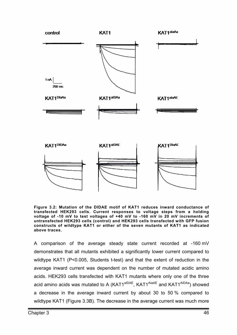
46 Chapter 3
Figure 3.2: Mutation of the DIDAE motif of KAT1 reduces inward conductance of transfected HEK293 cells. Current responses to voltage steps from a holding voltage of -10 mV to test voltages of +40 mV to -160 mV in 20 mV increments of untransfected HEK293 cells (control) and HEK293 cells transfected with GFP fusion constructs of wildtype KAT1 or either of the seven mutants of KAT1 as indicated above traces.
A comparison of the average steady state current recorded at -160 mV
demonstrates that all mutants exhibited a significantly lower current compared to
wildtype KAT1 (P<0.005, Students t-test) and that the extent of reduction in the
average inward current was dependent on the number of mutated acidic amino
acids. HEK293 cells transfected with KAT1 mutants where only one of the three
acid amino acids was mutated to A (KAT1aIDAE, KAT1AIaAE and KAT1AIDAa) showed
a decrease in the average inward current by about 30 to 50 % compared to
wildtype KAT1 (Figure 3.3B). The decrease in the average current was much more

47 Chapter 3
pronounced in cells expressing KAT1 mutants processing only one instead of
three acidic amino acids within the examined array (KAT1aIaAE, KAT1aIDAa;
KAT1DIaAa) (Figure 3.3B). In these cells the current was reduced by 65 to 85 %.
Expression of the triple mutant KAT1aIaAa lead to the strongest reduction in
conductance with an average inward current reduced to 2-3 % of wildtype KAT1
(Figure 3.3B). The dependence of the inward conductance on the number of
amino acids becomes even more obvious when the measured average current is
plotted against the number of acidic amino acids (Figure 3.3C). The wildtype and
mutant K+ channels clearly fall into four groups represented by K+ channels with
zero, one, two or three acidic amino acids located within the examined array.
Together these results demonstrate that all three acidic amino acids analysed
affect KAT1 conductance and that the extent of reduction in conductance is
determined by the number of mutated acidic amino acids with all three acidic
amino acids analysed being equally important.

48 Chapter 3
Figure 3.3: Reduction in inward current depends on the number of mutated acidic amino acids. A: Current-voltage relation of mean steady state current recorded from untransfected HEK293 cells (control, n=5) and HEK293 cells transfected with KAT1::GFP (n=10), KAT1aIDAE::GFP (n=7), KAT1DIaAE::GFP (n=9), KAT1DIDAa::GFP (n=8), KAT1AIDAa::GFP (n=11), KAT1DIaAa::GFP (n=8), KATaIaAE::GFP (n=10) and KAT1aIaAa::GFP (n=6). Error bars correspond to ± s.e.m.. B: Comparison of the average steady state inward current recorded at -160 mV from untransfected HEK293 cells (control) and HEK293 cells expressing wildtype KAT1 and KAT1 mutants (as indicated beside bars). Average steady state currents were normalised to the current recorded in cells expressing wildtype KAT1. Relative size of the average current is given next to the bars. Error bars correspond to ± s.e.m.. C: Correlation between number of acidic amino acids contained in the analysed motif and average current recorded at -160 mV from HEK293 cells expressing wildtype KAT1 or one of the KAT1 mutants. Error bars correspond to ± s.e.m..
49 Chapter 3
3.3.2 Mutation of acidic amino acids alters localisation of KAT1 in HEK293 cells
Judging from previous studies on diacidic motifs of KAT1 (Mikosch et al. 2006) the
observed reduction of the inward conductance of the KAT1 mutants is most likely
due to a reduced number of active channels in the plasma membrane. Therefore,
the subcellular distribution of wildtype KAT1 and KAT1 mutants fused to GFP in
HEK293 cells was compared using confocal laser scanning microscopy. In
HEK293 cells expressing KAT1::GFP the channel was located in the plasma
membrane but also to a great extent in intracellular compartments (Figure 3.4A). A
similar distribution has previously been described for expression of KAT1::GFP in
Chinese hamster ovary (CHO) cells (Szabo et al., 2000). Colocalisation of KAT1
with the plasma membrane marker FM4-64 confirmed its plasma membrane
localisation (Figure 3.4A and B). Figure 3.4B also reveals some intracellular
colabelling of KAT1::GFP and FM4-64 located close to the plasma membrane.
These internal structures most likely correspond to some endosomal
compartments because KAT1 as well as FM4-64 are known to be internalised via
endocytosis (Meckel et al., 2004). To identify the largest part of the intracellular
compartments labelled by KAT1::GFP but not by FM4-64, also colocalisation
studies with the ER tracker red were performed. The partial colocalisation of
KAT1::GFP with the ER tracker demonstrates that some of the intracellular
retained channel is located in the ER but it can also be found in other intracellular
compartments which may correspond to the Golgi apparatus and/or endosomes
(Figure 3.5A).
Localisation analysis was also performed for the KAT1 mutants. HEK293 cells
expressing KAT1 mutants with only one acidic amino acid mutated to alanine
showed a very similar distribution of the K+ channel compared to cells expressing
wildtype KAT1 (Figure 3.4C and Figure 3.6C, E and G). Again the K+ channel is
found in intracellular compartments as well as in the plasma membrane. As a
representative example for these mutants a more detailed analysis of the
distribution of KAT1aIDAE::GFP is displayed in Figure 3.5C. Colocalisation analysis
with FM4-64 clearly confirmed the plasma membrane localisation (Figure 3.4C and
D).
50 Chapter 3
Figure 3.4: Localisation of KAT1 in the plasma membrane depends on acidic amino acids. A: HEK293 cell expressing wildtype KAT1, staining of cells with the plasma membrane marker FM4-64 and overlay of both images. KAT1 colocalises with FM4-64 in the plasma membrane B: Magnification of the plasma membrane region of a HEK293 cell transfected with wildtype KAT1 supporting the localization of the channel in the plasma membrane. Some wildtype KAT1 can also be found in endosomal compartments (arrow) C: HEK293 cell expressing KAT1aIDAE as a representative example of KAT1 mutants where only one of the acidic of the DIDAE motif is mutated to alanine, staining of cells with the plasma membrane marker FM4-64 and overlay of both images. D: Magnification of part of the cell shown in C. This construct also clearly colocalises with the plasma membrane marker FM4-64. E: HEK293 cell expressing the mutant KAT1DIaAa as a representative example of KAT1 mutants where two of the acidic amino acids of the DIDAE motif are mutated to alanine, staining of cells with the plasma membrane marker FM4-64 and overlay of both images. KAT1DIaAa mutant does not localise to the plasma membrane. Scale bars correspond to 10 µm.

51 Chapter 3
Expression of the mutants of KAT1 with two of the three acidic amino acids being
mutated to A led to a completely different staining pattern (Figure 3.4E,
Figure 3.5B and Figure 3.6D, F and H). As a representative example for this group
of mutants the distribution of KAT1DIaAa was analysed in more detail.
KAT1DIaAa::GFP was restricted to a small perinuclear ring with no detectable
fluorescence in the plasma membrane as revealed by staining with the plasma
membrane marker FM4-64 (Figure 3.4E). Almost no additional fluorescent
intracellular structures were observed. Using the ER tracker red the perinuclear
ring could be identified as part of the ER (Figure 3.5B). This mutant has previously
been identified as an ER export mutant (Mikosch et al., 2006). The distribution of
KAT1DIaAa::GFP in HEK293 cells is very similar to the localisation of the same
mutant in Vicia faba guard cells (Mikosch et al., 2006). A retention of the channel
exclusively in the perinuclear region of the ER was also found for the mutant of
KAT1 where all three acidic amino acids in the analysed motif have been replaced
by A (Figure 3.6B). Thus, the localisation studies support the hypothesis that the
reduced K+ inward conductance observed in HEK293 cells expressing one of the
KAT1 mutants results from the inhibition of transport to the plasma membrane and
consequently a decrease in the number of K+ channels in the plasma membrane.
The inhibition of transport most likely occurs at the side of ER export. Previously,
diacidic motifs have been identified as ER export signals in a number of ion
channels including KAT1 (Mikosch and Homann, 2009). The consensus motif of
these diacidic ER export motifs is generally believed to be D/ExD/E. The results
presented above demonstrate that in addition to the two acidic amino acids
identified before as part of the ER export motif of KAT1 (Mikosch et al., 2006) a
third acidic amino acid is equally important. The complete ER export motif is
therefore most likely represented by the triacidic motif DIDAE (392-396). This is
supported by the observation that the extent of ER retention was strictly
dependent on the number of mutated amino acids and only mutation of all three
acidic amino acids completely abolished the KAT1 conductance. This leads to the
question how the ER export motif may function. In order to affect ER export the
motif has to be recognised by Sec24, a component of the COPII coat which
selects proteins for the anterograde transport from the ER to the Golgi apparatus.
Using FRET measurement it was recently demonstrated that KAT1 indeed
interacts with Sec24 at the ER and that this interaction was dependent on the

52 Chapter 3
diacidic DAE (394-392) motif (Sieben et al., 2008). The fact, that KAT1 mutants
with two mutated acidic amino acids still elicited a small inward conductance may
suggest that some mutated channels are exported out of the ER via bulk flow.
Alternatively, the mutated KAT1 may still exhibit a weak binding to Sec24 which
cannot be detected via FRET measurements but still allows some channels to be
incorporated into COPII vesicles for further transport to the plasma membrane.
The latter hypothesis is more likely since bulk flow should also enable the triple
mutant to reach the plasma membrane with an equal efficiency. However, this
mutant remained nearly completely in the ER. This also implies that binding of
KAT1 to Sec24 involves all three acidic amino acids and that bulk flow does not
play a significant role in transport of the channel out of the ER.
Figure 3.5: Mutation of two acidic amino acids of KAT1 results in ER retention. A: HEK293 cell expressing wildtype KAT1, staining of cells with ER tracker red and overlay of both images. The overlay shows a partial colocalisation. B: HEK293 cells expressing the KAT1DIaAa mutant, staining of cells with ER tracker red and overlay of both images. The overlay shows complete colocalisation of the mutated channel with the part of the ER around the nucleus. Scale bars correspond to 10 µm.
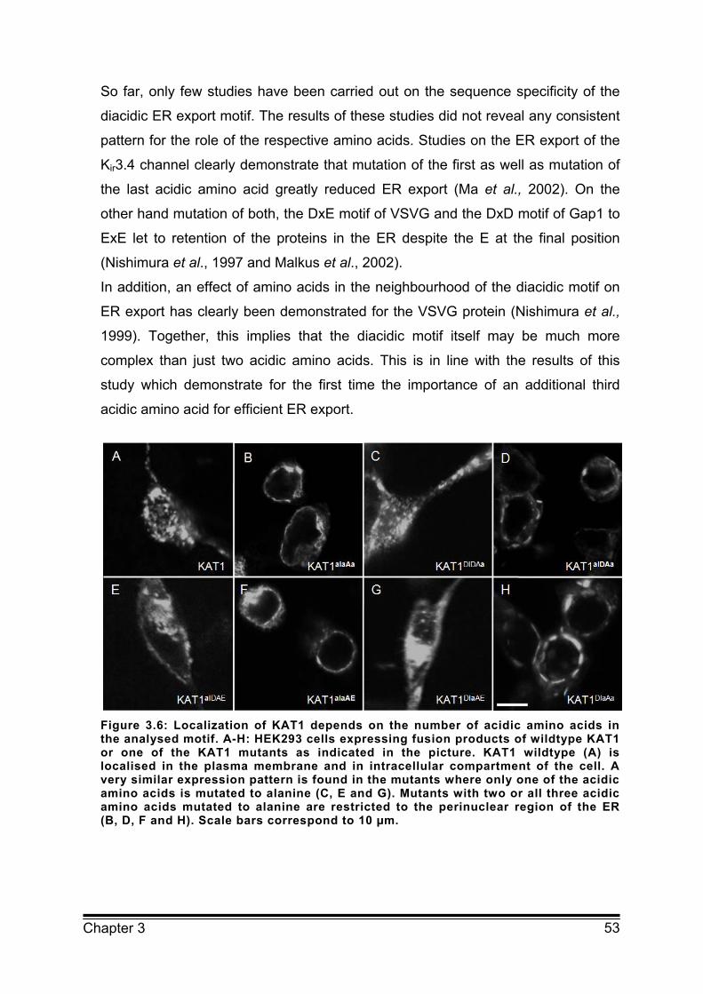
53 Chapter 3
So far, only few studies have been carried out on the sequence specificity of the
diacidic ER export motif. The results of these studies did not reveal any consistent
pattern for the role of the respective amino acids. Studies on the ER export of the
Kir3.4 channel clearly demonstrate that mutation of the first as well as mutation of
the last acidic amino acid greatly reduced ER export (Ma et al., 2002). On the
other hand mutation of both, the DxE motif of VSVG and the DxD motif of Gap1 to
ExE let to retention of the proteins in the ER despite the E at the final position
(Nishimura et al., 1997 and Malkus et al., 2002).
In addition, an effect of amino acids in the neighbourhood of the diacidic motif on
ER export has clearly been demonstrated for the VSVG protein (Nishimura et al.,
1999). Together, this implies that the diacidic motif itself may be much more
complex than just two acidic amino acids. This is in line with the results of this
study which demonstrate for the first time the importance of an additional third
acidic amino acid for efficient ER export.
Figure 3.6: Localization of KAT1 depends on the number of acidic amino acids in the analysed motif. A-H: HEK293 cells expressing fusion products of wildtype KAT1 or one of the KAT1 mutants as indicated in the picture. KAT1 wildtype (A) is localised in the plasma membrane and in intracellular compartment of the cell. A very similar expression pattern is found in the mutants where only one of the acidic amino acids is mutated to alanine (C, E and G). Mutants with two or all three acidic amino acids mutated to alanine are restricted to the perinuclear region of the ER (B, D, F and H). Scale bars correspond to 10 µm.

54 Chapter 3
3.3.3 Rescue of ER export mutant by heterotetramerisation with wildtype KAT1
KAT1 belongs to the shaker-type K+ channel family which functions as homo- or
heterotetramers. Tetramerization is supposed to already occur in the ER. Previous
investigations on trafficking of mammalian Kir channels which also function as
tetramers have demonstrated that not all subunits of heterotetramers need to
contain ER export signals in order to ensure efficient transport to the plasma
membrane (Ma et al., 2001). To analyse the effect of heterotetramerisation on
targeting of ER export mutants of KAT1, fusions of KAT1 and KAT1DIaAa with GFP
and RFP were constructed.
Coexpression of both wildtype KAT1 fusion constructs or of both ER export
mutants fused to RFP and GFP, respectively, resulted in a distribution of the
channels which resembled those found upon expression of each construct on its
own (Figure 3.7A and B). KAT1::RFP and KAT1::GFP were both located in the
plasma membrane and in some intracellular compartments while KAT1DIaAa::RFP
and KAT1DIaAa::GFP were retained in a part of the ER (Figure 3.7A and B).
In contrast, coexpression of the ER export mutant KAT1DIaAa fused to either GFP
or RFP together with the RFP or GFP fusion construct of wildtype KAT1 led to a
completely different distribution of KAT1DIaAa. As shown in a representative
example in Figure 3.7C and D the ER export mutant was no longer retained in part
of the ER but showed the same distribution as wildtype KAT1 including localisation
at the plasma membrane. This implies that plasma membrane localisation of the
ER export mutant could be rescued via heterotetramerisation with subunits that do
contain an ER export signal. More generally, the results suggest that subunits,
which are retained in the ER as homotetramers, can be targeted to the plasma
membrane via heterotetramerisation with subunits that do contain an ER export
signal. The physiological importance of such a mechanism is revealed by
investigations of AtKC1, which is a member of the K+ inward rectifying family from
Arabidopsis thaliana. AtKC1 is only targeted to the plasma membrane when
coexpressed with other members of the K+ channel family (Reintanz et al., 2002,
Pilot et al., 2003, Duby et al., 2008). When expressed on its own, AtKC1 localised
to the ER (Duby et al., 2008). This conditional targeting of AtKC1 most likely plays
an important role in the regulation of the physiological activity of other K+ channel
subunits in Arabidopsis thaliana (Duby et al., 2008).
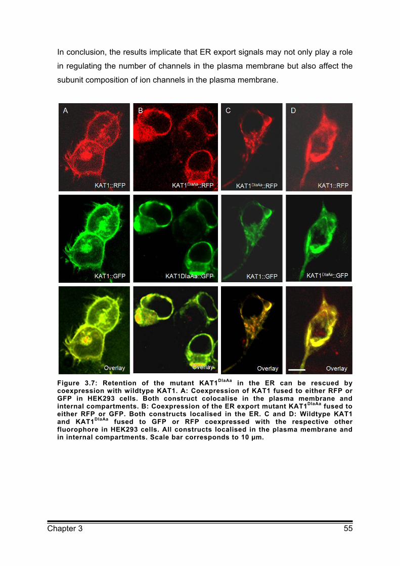
55 Chapter 3
In conclusion, the results implicate that ER export signals may not only play a role
in regulating the number of channels in the plasma membrane but also affect the
subunit composition of ion channels in the plasma membrane.
Figure 3.7: Retention of the mutant KAT1DIaAa in the ER can be rescued by coexpression with wildtype KAT1. A: Coexpression of KAT1 fused to either RFP or GFP in HEK293 cells. Both construct colocalise in the plasma membrane and internal compartments. B: Coexpression of the ER export mutant KAT1DIaAa fused to either RFP or GFP. Both constructs localised in the ER. C and D: Wildtype KAT1 and KAT1DIaAa fused to GFP or RFP coexpressed with the respective other fluorophore in HEK293 cells. All constructs localised in the plasma membrane and in internal compartments. Scale bar corresponds to 10 µm.
56 Chapter 3
3.4 Conclusion
Our results show that the previously identified diacidic ER export motif (DxE) of
KAT1 more likely corresponds to a triacidic ER export motif (DxDxE). It was
demonstrated that all three acidic residues have a similar impact on the function of
the motif and that their effect on ER retention is almost additive. This implies that
all acidic amino acids interact with a binding site of Sec24. However it is still
unknown, which site of Sec24 this interaction occurs at and which role each acidic
amino acid plays in this process. Coexpression analysis of wildtype and ER export
mutant of KAT1 suggests that heterotetramers of wildtype and mutant KAT1 are
still recognised by Sec24 for sorting into COPII vesicles. How many functional
motifs have to be present in the heterotetramer for effective ER export remains to
be shown. Overall, the results underline the importance of a triacidic ER export
motif for density and subunit composition of channels in the plasma membrane.
3.5 Methods
3.5.1 Construction of pEGFPN2KAT1
For expression as C-terminal GFP or RFP fusion in HEK293 cells the KAT1 gene
was cloned into the XhoI and BamHI sites of the pEGFPN2 or pmRFPN1 vector
(Clontech) in frame with the downstream GFP or RFP gene. Mutations in the
DIDAE motif of KAT1 were achieved by PCR based site directed mutagenesis
(QuikChange site directed mutagenesis kit; Stratagene) and confirmed by
sequencing. The expression vectors pEGFPN2KAT1 and pCB6KAT1 served as
templates. The plasmids were cloned into E. coli/DH5α and isolated with Qiagen
high speed Midi-Kit (Qiagen) for cell transfection.
57 Chapter 3
3.5.2 Cell culture and transfection of HEK293 cells
HEK293 cells were grown at 37°C and 5% CO2. For transient expression of KAT1
and KAT1 mutants HEK293 cells were transfected with 1 µg of pEGFPN2KAT1 or
pmRFPN1KAT1 using the liposomal transfection reagent Transfektin (Biorad)
according to manufacturer’s instructions.
3.5.3 Patch clamp measurements
Recordings of membrane conductance in HEK293 cells were performed in a
standard whole cell patch clamp experiment as described as described previously
(Hertel et al., 2005).
3.5.4 CLSM
HEK293 cells were investigated approximately 24 h after transfection with a Leica
TCS SP spectral confocal microscope (Leica Microsystems). Images were
acquired with an HCX PL APO 63×/1.2w objective. GFP was excited with the
488 nm line of a 25 mW argon laser and fluorescence was detected at
505-530 nm. RFP was excited with a 543 nm line of a helium neon laser and
emission collected at 600–630 nm. For plasma membrane staining, cells were
incubated in 10 µM FM4-64 (Invitrogen) before imaging. For ER staining cell were
incubated in 10 µM ER Tracker red (Invitrogen). Images and colocalisation were
analysed with ImageJ software (National Institutes of Health) and the Leica
Confocal Software 2.00 (LCS, Leica Microsystems)
58 Chapter 3
3.6 References
Duby G, Hosy E, Fizames C, Alcon C, Costa A, Sentenac H, Thibaud JB
(2008) AtKC1, a conditionally targeted shaker-type subunit, regulates the activity
of plant K+ channels. The Plant Journal 53(1):115-123
Hertel B, Horváth F, Wodala B, Hurst A, Moroni A, Thiel G (2005) KAT1
inactivates at submillimolar concentrations of external potassium. The Journal of
Experimental Botany 56(422):3103-3110
Hofherr A, Fakler B, Klöcker N (2005) Selective Golgi export of Kir2.1 controls
the stoichiometry of functional Kir2.x channel heteromers. The Journal of Cell
Science 118:1935-1943
Ma D, Zerangue N, Lin Y, Collins A, Yu M, Jan Y, Jan LY (2001) Role of ER
export signals in controlling surface potassium channel numbers. Science
291:316–319
Ma D, Zerangue N, Raab-Graham K, Fried SR, Jan YN, Jan LY (2002) Diverse
trafficking patterns due to multiple traffic motifs in G protein-activated inwardly
rectifying potassium channels from brain and heart. Neuron 33(5):715-729
Malkus P, Jiang F, Schekman R (2002) Concentrative sorting of secretory cargo
proteins into COPII coated vesicles. The Journal of Cell Biology 159:915–921
Meckel T, Hurst AC, Thiel G, Homann U (2004) Endocytosis against high turgor:
intact guard cells of Vicia faba constitutively endocytose fluorescently labelled
plasma membrane and GFP-tagged K+ channel KAT1. The Plant Journal
39:182-193 Mikosch M and Homann U (2009) How do diacidic motifs work on ion channel
trafficking? In preparation
Mikosch M, Hurst AC, Hertel B, Homann U (2006) Diacidic motif is required for
efficient transport of the K+ channel KAT1 to the plasma membrane. Plant
Physiology 142:923–930
Miller EA, Beilharz TH, Malkus PN, Lee MCS, Hamamoto S, Orci L, Schekman R (2003) Multiple cargo binding sites on the COPII subunit Sec24
ensure capture of diverse membrane proteins into transport vesicles. Cell
114:497–509
59 Chapter 3
Mossessova E, Bickford LC, Goldberg J (2003) SNARE selectivity of the COPII
coat. Cell 114:483–495
Nishimura N and Balch WE (1997) A diacidic signal required for selective export
from the endoplasmic reticulum. Science 277:556–558
Nishimura N, Bannykh S, Slabough S, Matteson J, Altschuler Y, Hahn K, Balch WE (1999) A diacidic (DxE) code directs concentration of cargo during
export from the endoplasmic reticulum. The Journal of Biological Chemistry
274:15937-15946
Otte S and Barlowe C (2004) Sorting signals can direct receptor-mediated export
of soluble proteins into COPII vesicles. Nature Cell Biology 6(12):1189-1194
Pagant S, Kung L, Dorrington M, Lee MCS, Miller EA (2007) Inhibiting
endoplasmic reticulum (ER)-associated degradation of misfolded Yor1p does not
permit ER export despite the presence of a diacidic sorting signal. Molecular
Biology of the Cell 18(9):3398-3413
Pilot G, Gaymard F, Mouline K, Chérel I, Sentenac H (2003) Regulated
expression of Arabidopsis thaliana shaker K+ channel genes involved in K+ uptake
and distribution in the plant. Plant Molecular Biology 51(5):773-787
Reintanz B, Szyroki A, Ivashikina N, Ache P, Godde M, Becker D, Palme K, Hedrich R (2002) AtKC1, a silent Arabidopsis thaliana potassium channel alpha
subunit modulates root hair K+ influx. PNAS 99(6):4079-4084
Schwappach B (2008) An overview of trafficking and assembly of
neurotransmitter receptors and ion channels. Molecular Membrane Biology
25(4):270-278
Sieben C, Mikosch M, Brandizzi F, Homann U (2008) Interaction of the K+
channel KAT1 with the COPII coat component Sec24 depends on a diacidic ER
export motif. The Plant Journal 56(6):997-1006
Szabò I, Negro A, Downey PM, Zoratti M, Lo Schiavo F, Giacometti GM (2000)
Temperature-dependent functional expression of a plant K+ channel in mammalian
cells. Biochemical and Biophysical Research Communities 274(1):130-135
Wang X, Matteson J, An Y, Moyer B, Yoo J, Bannykh S,Wilson IA, Riordan JR, Balch WE (2004) COPII-dependent export of cystic fibrosis
transmembrane conductance regulator from the ER uses a diacidic exit code. The
Journal of Cell Biology 167:65–74
60 Chapter 3
Zuzarte M, Rinné S, Schlichthörl G, Schubert A, Daut J, Preisig-Müller R (2007) A diacidic sequence motif enhances the surface expression of the
potassium channel TASK-3. Traffic 8:1093-1100

61 Chapter 4
Chapter 4 INTERACTION OF THE K+ CHANNEL KAT1 WITH
THE COPII COAT COMPONENT SEC24 DEPENDS ON A
DIACIDIC ER EXPORT MOTIF
62 Chapter 4
Maximum projection of a guard cell of Vicia faba transfected with the ER marker
HDEL::CFP via particle bombardment. The network like structure is well visible in
addition to the enlarged perinuclear ER with a ring like extension in the right cell
due to overexpression of a KAT1 ER export mutant.
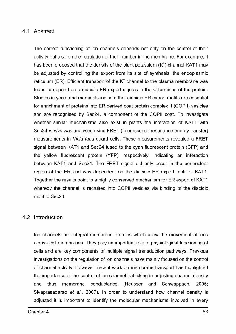
63 Chapter 4
4.1 Abstract
The correct functioning of ion channels depends not only on the control of their
activity but also on the regulation of their number in the membrane. For example, it
has been proposed that the density of the plant potassium (K+) channel KAT1 may
be adjusted by controlling the export from its site of synthesis, the endoplasmic
reticulum (ER). Efficient transport of the K+ channel to the plasma membrane was
found to depend on a diacidic ER export signals in the C-terminus of the protein.
Studies in yeast and mammals indicate that diacidic ER export motifs are essential
for enrichment of proteins into ER derived coat protein complex II (COPII) vesicles
and are recognised by Sec24, a component of the COPII coat. To investigate
whether similar mechanisms also exist in plants the interaction of KAT1 with
Sec24 in vivo was analysed using FRET (fluorescence resonance energy transfer)
measurements in Vicia faba guard cells. These measurements revealed a FRET
signal between KAT1 and Sec24 fused to the cyan fluorescent protein (CFP) and
the yellow fluorescent protein (YFP), respectively, indicating an interaction
between KAT1 and Sec24. The FRET signal did only occur in the perinuclear
region of the ER and was dependent on the diacidic ER export motif of KAT1.
Together the results point to a highly conserved mechanism for ER export of KAT1
whereby the channel is recruited into COPII vesicles via binding of the diacidic
motif to Sec24.
4.2 Introduction
Ion channels are integral membrane proteins which allow the movement of ions
across cell membranes. They play an important role in physiological functioning of
cells and are key components of multiple signal transduction pathways. Previous
investigations on the regulation of ion channels have mainly focused on the control
of channel activity. However, recent work on membrane transport has highlighted
the importance of the control of ion channel trafficking in adjusting channel density
and thus membrane conductance (Heusser and Schwappach, 2005;
Sivaprasadarao et al., 2007). In order to understand how channel density is
adjusted it is important to identify the molecular mechanisms involved in every

64 Chapter 4
step along the secretory and endocytotic pathway of ion channels. Recently,
particular attention has been paid to the first station of the secretory pathway
where ion channels are transported from the endoplasmic reticulum (ER) to the
Golgi apparatus (Ma and Jan, 2002). Cargo transport from the ER to the Golgi
apparatus is mediated by coat protein complex II (COPII) vesicles that bud from
the ER. For the efficient ER export of proteins their recruitment into COPII vesicles
is essential. This process involves specific amino acid motifs so called ER export
signals (Otte and Barlowe, 2004). For a number of plasma membrane ion
channels including the plant K+ channel KAT1 diacidic D/ExD/E motifs have been
shown to function as ER export signals (Ma et al., 2001; Stockklausner et al.,
2001; Wang et al., 2004; Mikosch et al., 2006; Zuzarte et al., 2007). Mutation of
these diacidic motifs resulted in a strong reduction of the channel in the plasma
membrane and an accumulation in the ER. Even though regulation of ER export of
ion channels is now widely recognised as a mechanism for adjusting the
composition and density of channels in the plasma membrane detailed information
about the underlying mechanism are still very limited. In general, ER export
signals are thought to be recognised by distinct cargo binding sites of Sec24, a
component of the COPII coat. In yeast the B-site of Sec24 has been identified as a
binding site of the diacidic motif of the vesicular stomatitis virus glycoprotein
(VSVG) and the yeast membrane protein Sys1 (Mossessova et al., 2003; Miller et
al., 2003). So far, information about the recognition of diacidic ER export motifs of
ion channels relies on in silico analysis. Using homology modelling of the CFTR
channel Wang et al. (2004) have illustrated the ability of diacidic motif of CFTR to
insert into the binding pocket at the B-site of yeast Sec24.
To further investigate the mechanism of COPII-dependent ER export of ion
channels the interaction of a plant Sec24 and the plant ion channel KAT1 in vivo
was analysed using FRET (fluorescence resonance energy transfer)
measurements in Vicia faba guard cells. The interaction between KAT1 and Sec24
fused to the fluorophores CFP and YFP, respectively, could be demonstrated. This
interaction was restricted to subdomains of the ER and was dependent on the
diacidic ER export motif of KAT1. These results provide new insights into the
mechanisms that regulate trafficking of ion channels from the plant ER and
consequently affect the density of K+ channels in the plasma membrane.
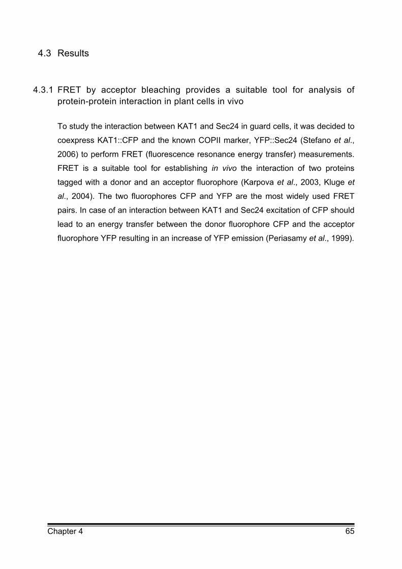
65 Chapter 4
4.3 Results
4.3.1 FRET by acceptor bleaching provides a suitable tool for analysis of protein-protein interaction in plant cells in vivo
To study the interaction between KAT1 and Sec24 in guard cells, it was decided to
coexpress KAT1::CFP and the known COPII marker, YFP::Sec24 (Stefano et al.,
2006) to perform FRET (fluorescence resonance energy transfer) measurements.
FRET is a suitable tool for establishing in vivo the interaction of two proteins
tagged with a donor and an acceptor fluorophore (Karpova et al., 2003, Kluge et
al., 2004). The two fluorophores CFP and YFP are the most widely used FRET
pairs. In case of an interaction between KAT1 and Sec24 excitation of CFP should
lead to an energy transfer between the donor fluorophore CFP and the acceptor
fluorophore YFP resulting in an increase of YFP emission (Periasamy et al., 1999).
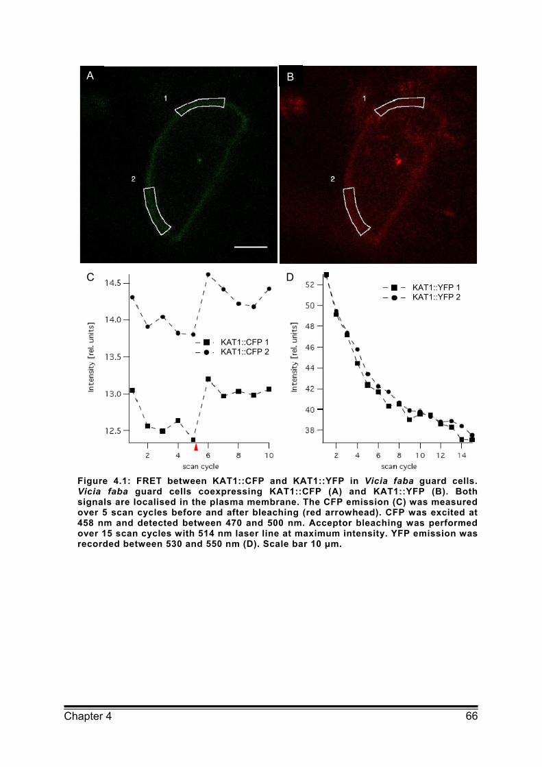
66 Chapter 4
Figure 4.1: FRET between KAT1::CFP and KAT1::YFP in Vicia faba guard cells. Vicia faba guard cells coexpressing KAT1::CFP (A) and KAT1::YFP (B). Both signals are localised in the plasma membrane. The CFP emission (C) was measured over 5 scan cycles before and after bleaching (red arrowhead). CFP was excited at 458 nm and detected between 470 and 500 nm. Acceptor bleaching was performed over 15 scan cycles with 514 nm laser line at maximum intensity. YFP emission was recorded between 530 and 550 nm (D). Scale bar 10 μm.
A B
C D
KAT1::CFP 1 KAT1::CFP 2
KAT1::YFP 1 KAT1::YFP 2
67 Chapter 4
Figure 4.2: Spectral-FRET between KAT1::CFP and KAT1::YFP in Vicia faba guard cells. Guard cells coexpressing KAT1::CFP (A) and KAT1::YFP (B). CFP was excited at 458 nm and detected between 470 and 600 nm. The spectrum was measured before and after acceptor bleaching (C). Between the spectra the acceptor was bleached over 15 scan cycles with 514 nm excitation at maximum intensity and its emission was recorded between 530 and 600 nm (D). Scale bar 10 μm.
To verify the reliability of FRET measurements the extent of cross-talk caused by
excitation of YFP using settings for CFP excitation, and the amount of bleed-
through of CFP fluorescence into the YFP channel were estimated. For cross-talk
analysis Vicia faba guard cells expressing YFP::Sec24 were excited with 458 nm
argon laser line for CFP excitation and the emitted YFP fluorescence was filtered
at 530-550 nm. These measurements revealed a high amount of YFP emission
after excitation at 458 nm (Figure 4.8). In addition, bleed-through led to the
detection of a considerable amount of CFP emission (about 30 %) in the YFP
A B
C D KAT1::CFP prebleach KAT1::CFP postbleach KAT1::YFP 1
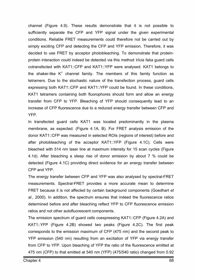
68 Chapter 4
channel (Figure 4.9). These results demonstrate that it is not possible to
sufficiently separate the CFP and YFP signal under the given experimental
conditions. Reliable FRET measurements could therefore not be carried out by
simply exciting CFP and detecting the CFP and YFP emission. Therefore, it was
decided to use FRET by acceptor photobleaching. To demonstrate that protein-
protein interaction could indeed be detected via this method Vicia faba guard cells
cotransfected with KAT1::CFP and KAT1::YFP were analysed. KAT1 belongs to
the shaker-like K+ channel family. The members of this family function as
tetramers. Due to the stochastic nature of the transfection process, guard cells
expressing both KAT1::CFP and KAT1::YFP could be found. In these conditions,
KAT1 tetramers containing both fluorophores should form and allow an energy
transfer from CFP to YFP. Bleaching of YFP should consequently lead to an
increase of CFP fluorescence due to a reduced energy transfer between CFP and
YFP.
In transfected guard cells KAT1 was located predominantly in the plasma
membrane, as expected. (Figure 4.1A, B). For FRET analysis emission of the
donor KAT1::CFP was measured in selected ROIs (regions of interest) before and
after photobleaching of the acceptor KAT1::YFP (Figure 4.1C). Cells were
bleached with 514 nm laser line at maximum intensity for 15 scan cycles (Figure
4.1d). After bleaching a steep rise of donor emission by about 7 % could be
detected (Figure 4.1C) providing direct evidence for an energy transfer between
CFP and YFP.
The energy transfer between CFP and YFP was also analysed by spectral-FRET
measurements. Spectral-FRET provides a more accurate mean to determine
FRET because it is not affected by certain background components (Goedhart et
al., 2000). In addition, the spectrum ensures that indeed the fluorescence ratios
determined before and after bleaching reflect YFP to CFP fluorescence emission
ratios and not other autofluorescent components.
The emission spectrum of guard cells coexpressing KAT1::CFP (Figure 4.2A) and
KAT1::YFP (Figure 4.2B) showed two peaks (Figure 4.2C). The first peak
corresponds to the emission maximum of CFP (475 nm) and the second peak to
YFP emission (540 nm) resulting from an excitation of YFP via energy transfer
from CFP to YFP. Upon bleaching of YFP the ratio of the fluorescence emitted at
475 nm (CFP) to that emitted at 540 nm (YFP) (475/540 ratio) changed from 0.92
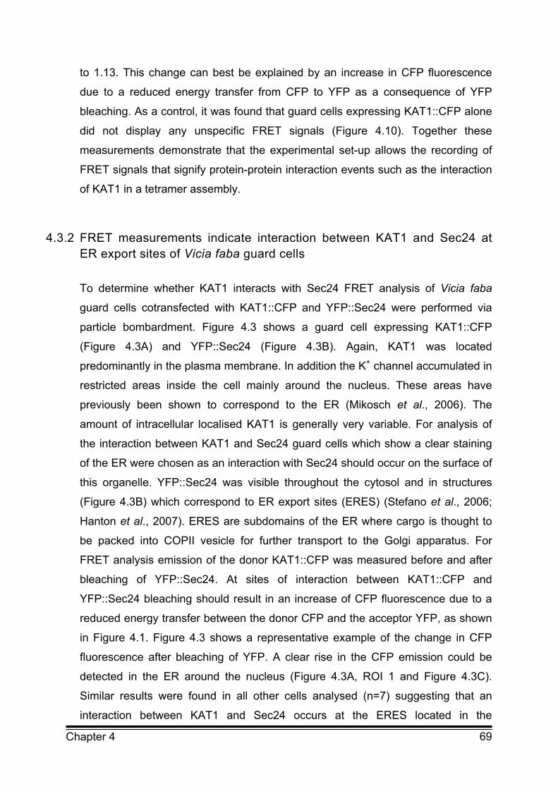
69 Chapter 4
to 1.13. This change can best be explained by an increase in CFP fluorescence
due to a reduced energy transfer from CFP to YFP as a consequence of YFP
bleaching. As a control, it was found that guard cells expressing KAT1::CFP alone
did not display any unspecific FRET signals (Figure 4.10). Together these
measurements demonstrate that the experimental set-up allows the recording of
FRET signals that signify protein-protein interaction events such as the interaction
of KAT1 in a tetramer assembly.
4.3.2 FRET measurements indicate interaction between KAT1 and Sec24 at ER export sites of Vicia faba guard cells
To determine whether KAT1 interacts with Sec24 FRET analysis of Vicia faba
guard cells cotransfected with KAT1::CFP and YFP::Sec24 were performed via
particle bombardment. Figure 4.3 shows a guard cell expressing KAT1::CFP
(Figure 4.3A) and YFP::Sec24 (Figure 4.3B). Again, KAT1 was located
predominantly in the plasma membrane. In addition the K+ channel accumulated in
restricted areas inside the cell mainly around the nucleus. These areas have
previously been shown to correspond to the ER (Mikosch et al., 2006). The
amount of intracellular localised KAT1 is generally very variable. For analysis of
the interaction between KAT1 and Sec24 guard cells which show a clear staining
of the ER were chosen as an interaction with Sec24 should occur on the surface of
this organelle. YFP::Sec24 was visible throughout the cytosol and in structures
(Figure 4.3B) which correspond to ER export sites (ERES) (Stefano et al., 2006;
Hanton et al., 2007). ERES are subdomains of the ER where cargo is thought to
be packed into COPII vesicle for further transport to the Golgi apparatus. For
FRET analysis emission of the donor KAT1::CFP was measured before and after
bleaching of YFP::Sec24. At sites of interaction between KAT1::CFP and
YFP::Sec24 bleaching should result in an increase of CFP fluorescence due to a
reduced energy transfer between the donor CFP and the acceptor YFP, as shown
in Figure 4.1. Figure 4.3 shows a representative example of the change in CFP
fluorescence after bleaching of YFP. A clear rise in the CFP emission could be
detected in the ER around the nucleus (Figure 4.3A, ROI 1 and Figure 4.3C).
Similar results were found in all other cells analysed (n=7) suggesting that an
interaction between KAT1 and Sec24 occurs at the ERES located in the
70 Chapter 4
perinuclear ER. This supports a previous hypothesis that this region of the ER may
be implicated in the export of KAT1 (Mikosch et al., 2006). As a control, the
emission of a FRET signal from the plasma membrane (Figure 4.3A, ROI 3 and 4)
was tested as well as from an area just below the plasma membrane (Figure 4.3A,
ROI 2). It was expected that a FRET signal would not be generated as Sec24
should not interact with protein cargo in distal compartments of the secretory
pathway. Consistent with this hypothesis, emission of CFP recorded from these
areas did not change after photobleaching (Figure 4.3C and E) suggesting that
KAT1 does not interact with Sec24 at or near the plasma membrane.
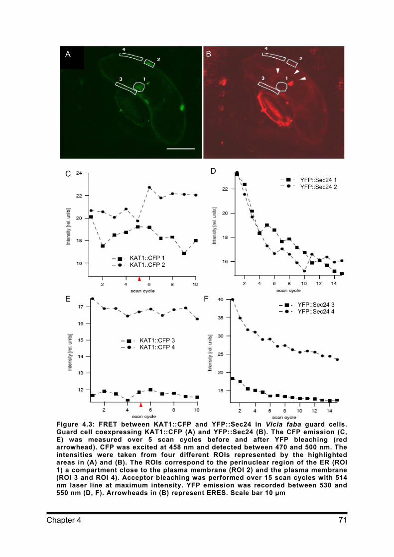
71 Chapter 4
Figure 4.3: FRET between KAT1::CFP and YFP::Sec24 in Vicia faba guard cells. Guard cell coexpressing KAT1::CFP (A) and YFP::Sec24 (B). The CFP emission (C, E) was measured over 5 scan cycles before and after YFP bleaching (red arrowhead). CFP was excited at 458 nm and detected between 470 and 500 nm. The intensities were taken from four different ROIs represented by the highlighted areas in (A) and (B). The ROIs correspond to the perinuclear region of the ER (ROI 1) a compartment close to the plasma membrane (ROI 2) and the plasma membrane (ROI 3 and ROI 4). Acceptor bleaching was performed over 15 scan cycles with 514 nm laser line at maximum intensity. YFP emission was recorded between 530 and 550 nm (D, F). Arrowheads in (B) represent ERES. Scale bar 10 µm
A B
C D
E F
KAT1::CFP 1 KAT1::CFP 2
YFP::Sec24 1 YFP::Sec24 2
YFP::Sec24 3 YFP::Sec24 4
KAT1::CFP 3 KAT1::CFP 4
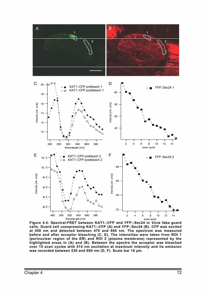
72 Chapter 4
Figure 4.4: Spectral-FRET between KAT1::CFP and YFP::Sec24 in Vicia faba guard cells. Guard cell coexpressing KAT1::CFP (A) and YFP::Sec24 (B). CFP was excited at 458 nm and detected between 470 and 600 nm. The spectrum was measured before and after acceptor bleaching (C, E). The intensities were taken from ROI 1 (perinuclear region of the ER) and ROI 2 (plasma membrane) represented by the highlighted areas in (A) and (B). Between the spectra the acceptor was bleached over 15 scan cycles with 514 nm excitation at maximum intensity and its emission was recorded between 530 and 600 nm (D, F). Scale bar 10 µm.
A B
C D
E F
KAT1::CFP prebleach 1 KAT1::CFP postbleach 1
KAT1::CFP prebleach 2 KAT1::CFP postbleach 2
YFP::Sec24 1
YFP::Sec24 2
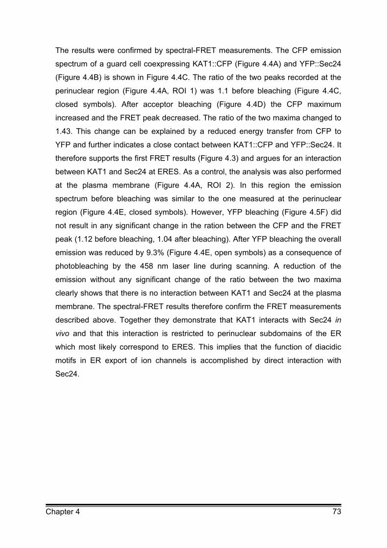
73 Chapter 4
The results were confirmed by spectral-FRET measurements. The CFP emission
spectrum of a guard cell coexpressing KAT1::CFP (Figure 4.4A) and YFP::Sec24
(Figure 4.4B) is shown in Figure 4.4C. The ratio of the two peaks recorded at the
perinuclear region (Figure 4.4A, ROI 1) was 1.1 before bleaching (Figure 4.4C,
closed symbols). After acceptor bleaching (Figure 4.4D) the CFP maximum
increased and the FRET peak decreased. The ratio of the two maxima changed to
1.43. This change can be explained by a reduced energy transfer from CFP to
YFP and further indicates a close contact between KAT1::CFP and YFP::Sec24. It
therefore supports the first FRET results (Figure 4.3) and argues for an interaction
between KAT1 and Sec24 at ERES. As a control, the analysis was also performed
at the plasma membrane (Figure 4.4A, ROI 2). In this region the emission
spectrum before bleaching was similar to the one measured at the perinuclear
region (Figure 4.4E, closed symbols). However, YFP bleaching (Figure 4.5F) did
not result in any significant change in the ration between the CFP and the FRET
peak (1.12 before bleaching, 1.04 after bleaching). After YFP bleaching the overall
emission was reduced by 9.3% (Figure 4.4E, open symbols) as a consequence of
photobleaching by the 458 nm laser line during scanning. A reduction of the
emission without any significant change of the ratio between the two maxima
clearly shows that there is no interaction between KAT1 and Sec24 at the plasma
membrane. The spectral-FRET results therefore confirm the FRET measurements
described above. Together they demonstrate that KAT1 interacts with Sec24 in
vivo and that this interaction is restricted to perinuclear subdomains of the ER
which most likely correspond to ERES. This implies that the function of diacidic
motifs in ER export of ion channels is accomplished by direct interaction with
Sec24.
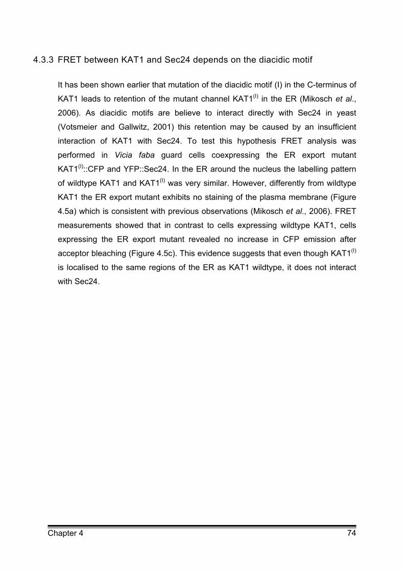
74 Chapter 4
4.3.3 FRET between KAT1 and Sec24 depends on the diacidic motif
It has been shown earlier that mutation of the diacidic motif (I) in the C-terminus of
KAT1 leads to retention of the mutant channel KAT1(I) in the ER (Mikosch et al.,
2006). As diacidic motifs are believe to interact directly with Sec24 in yeast
(Votsmeier and Gallwitz, 2001) this retention may be caused by an insufficient
interaction of KAT1 with Sec24. To test this hypothesis FRET analysis was
performed in Vicia faba guard cells coexpressing the ER export mutant
KAT1(I)::CFP and YFP::Sec24. In the ER around the nucleus the labelling pattern
of wildtype KAT1 and KAT1(I) was very similar. However, differently from wildtype
KAT1 the ER export mutant exhibits no staining of the plasma membrane (Figure
4.5a) which is consistent with previous observations (Mikosch et al., 2006). FRET
measurements showed that in contrast to cells expressing wildtype KAT1, cells
expressing the ER export mutant revealed no increase in CFP emission after
acceptor bleaching (Figure 4.5c). This evidence suggests that even though KAT1(I)
is localised to the same regions of the ER as KAT1 wildtype, it does not interact
with Sec24.
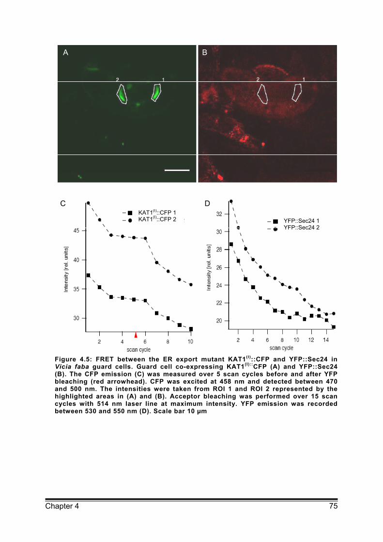
75 Chapter 4
Figure 4.5: FRET between the ER export mutant KAT1(I)::CFP and YFP::Sec24 in Vicia faba guard cells. Guard cell co-expressing KAT1(I)::CFP (A) and YFP::Sec24 (B). The CFP emission (C) was measured over 5 scan cycles before and after YFP bleaching (red arrowhead). CFP was excited at 458 nm and detected between 470 and 500 nm. The intensities were taken from ROI 1 and ROI 2 represented by the highlighted areas in (A) and (B). Acceptor bleaching was performed over 15 scan cycles with 514 nm laser line at maximum intensity. YFP emission was recorded between 530 and 550 nm (D). Scale bar 10 µm
A B
C DKAT1(I)::CFP 1 KAT1(I)::CFP 2 YFP::Sec24 1
YFP::Sec24 2

76 Chapter 4
Figure 4.6: Spectral-FRET between the ER export mutant KAT1(I) and Sec24 in Vicia faba guard cells. Guard cell coexpressing KAT1::CFP (A) and YFP::Sec24 (B). CFP was excited at 458 nm and detected between 470 and 600 nm. The spectrum was measured before and after acceptor bleaching (C). The intensities were taken from the ROI represented by the highlighted area in (A) and (B). Between the spectra the acceptor was bleached over 15 scan cycles with 514 nm excitation at maximum intensity and its emission was recorded between 530 and 600 nm (D). Scale bar 10 µm.
The interaction of Sec24 with the mutant channel was also analysed by spectral-
FRET as described above for the KAT1 wildtype. Figure 4.6 shows a guard cell
coexpressing KAT1(I)::CFP (Figure 4.6A) and YFP::Sec24 (Figure 4.6B). Again
KAT1(I) was mainly located in internal compartments. The emission spectrum
recorded from the perinuclear region before photobleaching of YFP (Figure 4.6C,
A B
C D
KAT1(I)::CFP prebleach KAT1(I)::CFP postbleach YFP::Sec24
77 Chapter 4
closed symbols) was similar to the one measured in guard cell expressing KAT1
wildtype. After bleaching of YFP the overall emission was reduced by 18.9 %
(Figure 4.6C, open symbols). The ratio between the two maxima did not change
significantly after bleaching (1.34 before bleaching, 1.31 after bleaching). This
implies that KAT1(I) did not interact with Sec24 and that the interaction between
KAT1 and Sec24 depends on the first diacidic motif in the C-terminus of KAT1.
The lack of interaction of KAT1(I) and Sec24 most likely leads to the retention of
the ER export mutant in the ER. Together the results support the hypothesis that
the interaction between the channel protein and Sec24 depends on the diacidic
ER export motif and that this interaction is essential for ER export.
4.4 Discussion
4.4.1 The mechanism of cargo recruitment into ER derived COPII carriers is highly conserved among eukaryotes
For the efficient ER export of proteins their recruitment into ER derived COPII
carriers is essential. This process involves specific amino acid motifs so called ER
export signals (Otte and Barlowe, 2004), which are recognised by components of
the COPII coat. The first signal found to be involved in selection of cargo into
COPII vesicles was the diacidic ER export motif of the VSVG (Nishimura and
Balch, 1997). Since then diacidic signals have been proven to be necessary for
export of a number of proteins in yeast and mammals (see for example Votsmeier
and Gallwitz, 2001; Miller et al., 2003; Ma et al., 2001; Wang et al., 2004). Among
these proteins are several ion channels from different families. Current knowledge
of protein export in plant cells indicates that for type I, II and multispanning
membrane proteins such as the K+ channel KAT1 the presence of intact diacidic
motifs is also crucial for ER export (Hanton et al., 2005; Mikosch et al., 2006).
Together this underlines the importance of the diacidic signal sequence for
trafficking of proteins in general and for efficient targeting of ion channels to the
plasma membrane in particular. However, how the function of these signals is
accomplished in plants has yet to be demonstrated. Moreover, information about
the mechanism of ER export of ion channels is limited to results from in silico
analysis (Wang et al., 2004). In this study it was aimed to advance the
78 Chapter 4
understanding of the mechanisms that lead to ER export of ion channels in order
to improve our knowledge of the regulation of channel density and trafficking of
plant membrane proteins in general. Using an in vivo approach based on FRET
analyses it could be shown that KAT1 interacts with the COPII coat component
Sec24. Mutants of KAT1 bearing a disrupted ER export signal failed to interact
with Sec24 and to be exported from the ER. Taken together these results suggest
that KAT1 is exported via a COPII-mediated mechanism and that the interaction of
KAT1 with COPII proteins is mediated by the diacidic motif DxE. Therefore, these
results provide evidence for a direct role of a DxE signal of ion channels in COPII
coat recruitment in vivo. They furthermore advance our understanding of COPII
regulation of ER export in plants. To date, evidence of COPII mediated ER export
in plants has been based on the interference of the pathway using dominant
negative mutants of the small regulatory GTPase Sar1 or overexpression of the
Sar1-GAP, Sec12 (Takeuchi et al., 2000; Phillipson et al., 2001; daSilva et al.,
2004; Yang et al., 2005). This approach has provided additional insights in the
mechanisms of COPII mediated transport in plants by showing that a productive
Sec24 cargo interaction via diacidic motifs leads to COPII mediated export from
the ER. In particular, these data argue that the diacidic motif in the C-terminus of
KAT1 provides the binding site for Sec24. It is known that the B-site of yeast
Sec24 binds to the Lxx-L/M-E motifs in yeast SNAREs Bet1 and Sed5 as well as
to the diacidic motif in Sys1 (Mossessova et al., 2003). Furthermore, homology
modelling of the mammalian ion channel CFTR suggests that the B-site is also
important for binding of ion channels to Sec24 (Wang et al., 2004). The
mechanism of cargo binding by Sec24 in plants is still unknown. There are three
homologous of Sec24 in plants (Robinson et al., 2007) compared to four in
humans and two in yeast (Pagano et al., 1999). Sequence comparison of the
B-site of Sec24 isoforms from plants, yeast and humans revealed a high homology
(Figure 4.7). In particular, the amino acids which have been demonstrated to be
crucial for binding of cargo to the B-site in yeast (Miller et al., 2003) are highly
conserved. Whether these domains are important for establishment of an
interaction of Sec24 with the DxE motif of KAT1 has yet to be shown. It seems
however highly plausible considering the likelihood of conservation of ER export
mechanisms shown in our study and in previous investigations of ER export of
KAT1 (Mikosch et al., 2006).

4
Chapte
FiguamocargSec2cere
4.4.2 Exp
Thes
in ad
prev
YFP
com
ER i
inhib
path
chan
resu
whe
Whe
by th
this
sign
perin
The
sam
two
This
into
subser 4
re 4.7: Seng differeno binding
24D are frovisiae, At3
port of KA
se results
ddition to i
vious findin
P::Sec24 a
partments
is export c
bition of E
hway or by
nnel exclus
ults reinforc
re KAT1 i
ether the e
he data. In
region had
al from the
nuclear ER
observatio
e subdom
step proce
step is no
COPII ves
sequent ste
equence cont species
in yeast om Homo
3g07100, At
T1 occurs
show that
its localisa
ngs (Mikos
and KAT1:
located c
competent.
ER export
mutation o
sively in th
ce the hyp
s package
entire perin
order to re
d to be ana
e perinucle
R membran
on that wil
ain of the
ess. During
ot affected
sicles for f
ep. This pr
omparison . Amino aare highlisapiens, S
t3g44340 a
s mainly a
KAT1 acc
ation at the
sch et al.
::CFP are
lose to the
This is su
of KAT1
of the diac
he perinucl
pothesis th
ed in COP
nuclear reg
esolve a F
alysed. Be
ear area m
nes.
ldtype and
ER sugge
g the first s
by the ER
further tran
rocess wou
of the B-acids whichghted by Sec24, Iss
and At4g32
at the per
cumulates p
e plasma m
, 2006). T
only gen
e plasma m
upported b
either by
cidic ER ex
lear region
hat the pe
PII carriers
gion is ER
RET signa
ecause of w
may be du
d ER expo
ests that th
step, the c
export mo
nsport alon
uld involve
-site of Seh have beboxes. Se
s1 and Lst640 are fro
rinuclear
preferentia
membrane
The fact th
nerated in
membrane
by previous
y inhibition
xport signa
n (Mikosch
rinuclear r
s to facilita
export co
al the fluore
working wi
ue to prote
ort mutant
he export
channel pro
otif. Sorting
ng the sec
e interactio
ec24 showen shown c24A, Sect1 are fromom Arabido
region of
ally at the p
which is i
hat FRET
this regio
argues th
s observat
n of the
al led to ac
h et al., 20
region of t
ate its exp
ompetent c
escence fr
th live cell
ein mobility
of KAT1 a
of the cha
otein accum
g and conc
cretory pat
n of the ch
ws high hoto be crit
c24B, Sec2m Saccharopsis thalia
the ER
perinuclea
n agreeme
signals b
on rather
hat this par
ions show
COPII-dep
ccumulatio
06). Toget
he ER is
port from t
cannot be
rom a large
s, a diffuse
y in and o
accumulate
annel occu
mulates at
centration o
hway may
hannel with79
omology tical for 24C and romyces ana.
r region
ent with
between
than in
rt of the
wing that
pendent
n of the
ther the
the site
the ER.
inferred
e part of
e FRET
over the
e in the
urs as a
t ERES.
of KAT1
y be the
h Sec24

80 Chapter 4
mediated by diacidic ER export motif. The evidence that both, FRET between
KAT1 and Sec24 and ER export of the channels occurred only for wildtype KAT1,
supports this hypothesis.
Together the results clearly show that mutation of the diacidic ER export motif
does not prevent accumulation of KAT1 at ERES but impedes concentration of the
channel into COPII vesicles. While the factors responsible for the first process are
unknown our present results imply that the COPII mediated ER export occurs via a
highly conserved mechanism involving interaction of the channel with the COPII
component Sec24.
4.5 Methods
4.5.1 Expression vectors
For expression of KAT1::CFP and KAT1::YFP in Vicia faba guard cells, the cDNA
of KAT1 was cloned into the pAVA393 expression vector in frame with CFP or
YFP under the control of a 35S promoter as described previously by Hurst et al.
(2004). The binary vector pVKH18En6 was used for expression of Sec24 in guard
cells (Stefano et al., 2004).
4.5.2 Transfection of intact guard cells by particle delivery
Vicia faba L. cv. Bunyan were grown under controlled climate conditions with
18 °C, 70 % relative humidity, and a 14/10 day/night cycle at 350 to 400 µmol
photons m-2s-1. Transfection of intact guard cells via particle delivery was
performed as described previously (Hurst et al., 2004). Cotransfection was
performed via coating of gold with equal molar amounts of each plasmid DNA to
give a total amount of 15 µg DNA.
81 Chapter 4
4.5.3 FRET and CLSM
Confocal microscopy analysis of transfected turgid guard cells was performed after
overnight incubation as described earlier (Meckel et al., 2004) using a confocal
laser scanning microscope (Leica TCS SP). The following 25 mW argon laser lines
were used for excitation of fluorescent proteins: 458 nm for CFP and 514 nm for
YFP. The fluorescence of CFP was detected at 470 to 500 nm and of YFP at 530
to 550 nm. FRET was performed by acceptor photo-bleaching. CFP fluorescence
was monitored for 5 scan cycles before and after YFP bleaching. Acceptor
bleaching was performed for 15 scan cycles at 514 nm excitation with maximum
laser intensity. For spectral-FRET the CFP fluorescence was detected before and
after bleaching between 470 and 600 nm. Image acquisition was carried out by
Leica LSM 5 image browser. The images were analysed and processed using
ImageJ.
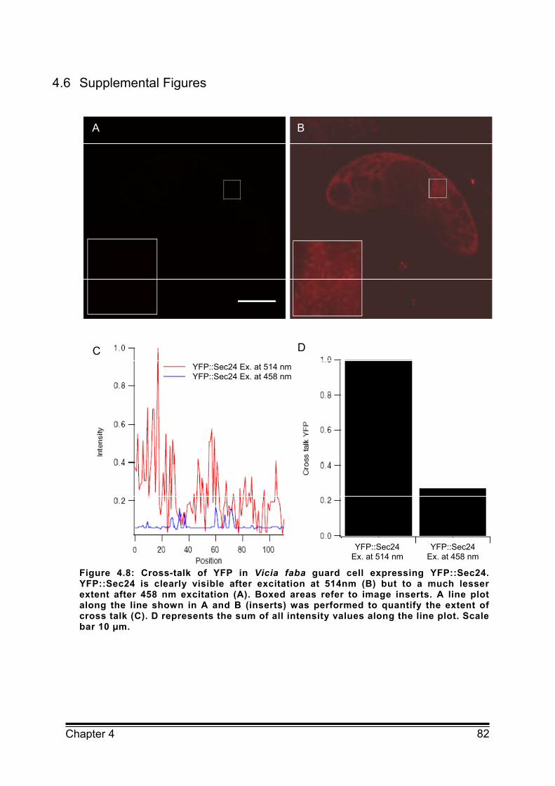
82 Chapter 4
4.6 Supplemental Figures
Figure 4.8: Cross-talk of YFP in Vicia faba guard cell expressing YFP::Sec24. YFP::Sec24 is clearly visible after excitation at 514nm (B) but to a much lesser extent after 458 nm excitation (A). Boxed areas refer to image inserts. A line plot along the line shown in A and B (inserts) was performed to quantify the extent of cross talk (C). D represents the sum of all intensity values along the line plot. Scale bar 10 µm.
A B
C DYFP::Sec24 Ex. at 514 nm YFP::Sec24 Ex. at 458 nm
YFP::Sec24 Ex. at 514 nm
YFP::Sec24 Ex. at 458 nm
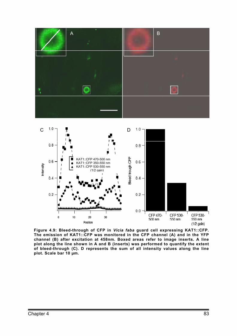
83 Chapter 4
Figure 4.9: Bleed-through of CFP in Vicia faba guard cell expressing KAT1::CFP. The emission of KAT1::CFP was monitored in the CFP channel (A) and in the YFP channel (B) after excitation at 458nm. Boxed areas refer to image inserts. A line plot along the line shown in A and B (inserts) was performed to quantify the extent of bleed-through (C). D represents the sum of all intensity values along the line plot. Scale bar 10 µm.
A B
C D
KAT1::CFP 470-500 nm KAT1::CFP 350-550 nm KAT1::CFP 530-550 nm
(1/2 gain)
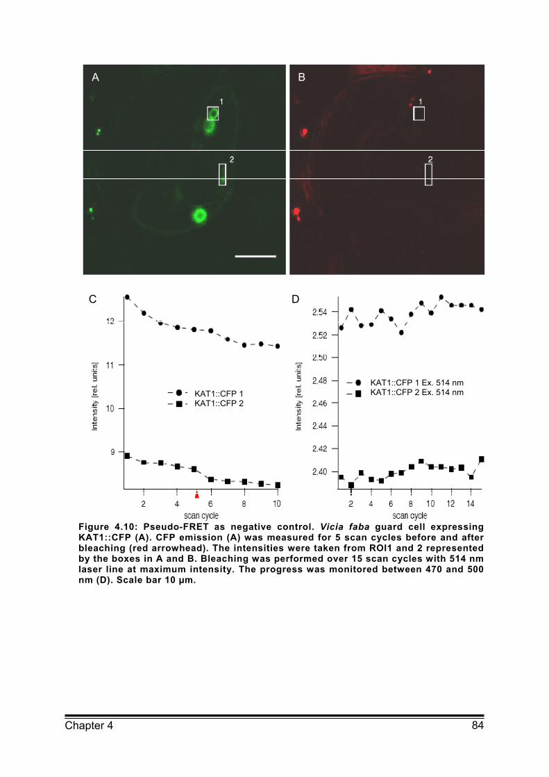
84 Chapter 4
Figure 4.10: Pseudo-FRET as negative control. Vicia faba guard cell expressing KAT1::CFP (A). CFP emission (A) was measured for 5 scan cycles before and after bleaching (red arrowhead). The intensities were taken from ROI1 and 2 represented by the boxes in A and B. Bleaching was performed over 15 scan cycles with 514 nm laser line at maximum intensity. The progress was monitored between 470 and 500 nm (D). Scale bar 10 µm.
A B
C D
KAT1::CFP 1 KAT1::CFP 2
KAT1::CFP 1 Ex. 514 nm KAT1::CFP 2 Ex. 514 nm

85 Chapter 4
4.7 References
daSilva LL, Snapp EL, Denecke J, Lippincott-Schwartz J, Hawes C, Brandizzi F (2004) Endoplasmic reticulum export sites and Golgi bodies behave
as single mobile secretory units in plant cells. The Plant Cell 16:1753-1771
Goedhart J, and Gadella TWJ Jr. (2000) Advanced microspectroscopic methods
for monitoring fluorescent molecules in living plant root hairs. In Cell and molecular
biology of living plant root hairs (Ridge, R.W. and Emons, A.M.C., eds.). Tokyo:
Springer-Verlag, pp. 65-93 Hanton SL, Renna L, Bortolotti LE, Chatre L, Stefano G. Brandizzi F (2005)
Diacidic motifs influence the export of transmembrane proteins from the
endoplasmic reticulum in plant cells. The Plant Cell 17:3081–3093
Hanton SL, Chatre L, Renna L, Matheson LA, Brandizzi F (2007) De novo
formation of plant endoplasmic reticulum export sites is membrane cargo induced
and signal mediated. Plant Physiology 143:1640-1650 Heusser K and Schwappach B (2005) Trafficking of potassium channels. Current
Opinion in Neurobiology 15:364-369 Hurst AC, Meckel T, Tayefeh S, Thiel G, Homann U (2004) Trafficking of the
plant potassium inward rectifier KAT1 in guard cell protoplasts of Vicia faba. The
Plant Journal 37:391–397
Karpova TS, Baumann CT, He L, Wu X, Grammer A, Lipsky P, Hager GL, McNally JG (2003) Fluorescence resonance energy transfer from cyan to yellow
fluorescent protein detected by acceptor photobleaching using confocal
microscopy and a single laser. The Journal of Microscopy 209:56–70
Kluge C, Seidel T, Bolte S, Sharma SS, Hanitzsch M, Satiat-Jeunemaitre B, Ross J, Sauer M, Golldack D, Dietz KJ (2004) Subcellular distribution of the V-
ATPase complex in plant cells, and in vivo localisation of the 100 kDa subunit
VHA-a within the complex. BMC Cell Biology 5:29 Ma D, Zerangue N, Lin Y, Collins A, Yu M, Jan Y, Jan L (2001) Role of ER
export signals in controlling surface potassium channel numbers. Science
291:316-319 Ma D and Jan LY (2002) ER transport signals and trafficking of potassium
channels and receptors. Current Opinion in Neurobiology 12:287–292

86 Chapter 4
Meckel T, Hurst AC, Thiel G, Homann U (2004) Endocytosis against high turgor:
intact guard cells of Vicia faba constitutively endocytose fluorescently labelled
plasma membrane and GFP tagged K+ channel KAT1. The Plant Journal
39:182-193
Mikosch M, Hurst AC, Hertel B, Homann U (2006) Diacidic motif is required for
efficient transport of the K+ channel KAT1 to the plasma membrane. Plant
Physiology 142:923–930
Miller EA, Beilharz TH, Malkus PN, Lee MCS., Hamamoto S, Orci L, Schekman R (2003) Multiple cargo binding sites on the COPII subunit Sec24p
ensure capture of diverse membrane proteins into transport vesicles. Cell
114:497–509
Mossessova E, Bickford LC, Goldberg J (2003) SNARE selectivity of the COPII
coat. Cell 114:483–495
Nishimura N, Balch WE (1997) A diacidic signal required for selective export from
the endoplasmic reticulum. Science 277:556–558
Nishimura N, Bannykh S, Slabough S, Matteson J, Altschuler Y, Hahn K, Balch WE (1999) A diacidic (DxE) code directs concentration of cargo during
export from the endoplasmic reticulum. The Journal of Biological Chemistry
274:15937-15946
Otte S and Barlowe C (2004) Sorting signals can direct receptor-mediated export
of soluble proteins into COPII vesicles. Nature Cell Biology 6:1189-1194
Pagano A, Letourneur F, Garcia-Estefania D, Carpentier JL, Orci L, Paccaud JP (1999) Sec24 proteins and sorting at the endoplasmic reticulum. The
Journal of Biological Chemistry 274:7833–7840
Periasamy A and Day RN (1999) Visualizing protein interactions in living cells
using digitised GFP imaging and FRET microscopy. Methods in Cell Biology
58:293–314
Phillipson BA, Pimpl P, daSilva LL, Crofts AJ, Taylor JP, Movafeghi A, Robinson DG, Denecke J (2001) Secretory bulk flow of soluble proteins is
efficient and COPII-dependent. The Plant Cell 13:2005–2020
Robinson DG, Herranz MC, Bubeck J, Pepperkok R, Ritzenthaler C (2007)
Membrane dynamics in the early secretory pathway. Critical Reviews Plant
Science 26:199–225
Sivaprasadarao A, Taneja TK, Mankouri J, Smith AJ (2007) Trafficking of ATP-
87 Chapter 4
sensitive potassium channels in health and disease. Biochemical Society
Transactions 35:1055-1059
Stefano G, Renna L, Chatre L, Hanton S, Moreau P, Hawes C, Brandizzi F (2006) In tobacco leaf epidermal cells, the integrity of protein export from the
endoplasmic reticulum and of ER export sites depends on active COPI machinery.
The Plant Journal 46:95–110
Stockklausner C, Ludwig J, Ruppersberg JP, Klöcker N (2001) A sequence
motif responsible for ER export and surface expression of Kir2.0 inward rectifier
K+ channels. FEBS Letters 493:129-133
Takeuchi M, Ueda T, Sato K, Abe H, Nagata T, Nakano A (2000) A dominant
negative mutant of Sar1 GTPase inhibits protein transport from the endoplasmic
reticulum to the Golgi apparatus in tobacco and Arabidopsis thaliana cultured
cells. The Plant Journal 23:517-525
Tang BL, Kausalya J, Low DY, Lock ML and Hong W (1999) A family of
mammalian proteins homologous to yeast Sec24p. Biochemical and Biophysical
Research Communications 258:679-684 Votsmeier C and Gallwitz D (2001) An acidic sequence of a putative yeast Golgi
membrane protein binds COPII and facilitates ER export. The EMBO Journal
20:6742–6750
Wang X, Matteson J, An Y, Mayer B, Yoo J, Bannykh S, Wilson I, Riordan J, Balch W (2004) COPII-dependent export of cystic fibrosis transmembrane
conductance regulator from the ER uses a diacidic exit code. The Journal of
Biological Chemistry 167:65-74 Wendeler MW, Paccaud JP, Hauri HP (2007) Role of Sec24 isoforms in selective
export of membrane proteins from the endoplasmic reticulum. EMBO Reports
8:258–264
Yang YD, Elamawi R, Bubeck J, Pepperkok R, Ritzenthaler C, Robinson DG
(2005) Dynamics of COPII vesicles and the Golgi apparatus in cultured Nicotiana
tabacum BY-2 cells provides evidence for transient association of Golgi stacks
with endoplasmic reticulum exit sites. The Plant Cell 17:1513-1531
Zuzarte M, Rinné S, Schlichthörl G, Schubert A, Daut J, Preisig-Müller R (2007) A diacidic sequence motif enhances the surface expression of the
potassium channel TASK-3. Traffic 8:1093-110

Chapte
Chapte
TRA
er 5
er 5 HO
AFFICKI
OW DO D
NG?
DIACIDIC
C MOTIFFS WORRK ON IOON CHA
88
ANNEL
89 Chapter 5
Structure model illustrating the C-terminus of the K+ channel KAT1 according to
the crystal structure of HCN2. A ribbon presentation with the two acidic amino
acids D394 and E396 marked.
.
90 Chapter 5
5.1 Abstract
The function of cells is strongly affected by the type and number of ion channels in
the plasma membrane. Recent investigations have highlighted the complexity of
the regulation of ion channel trafficking and uncovered several trafficking
determinants including diacidic motifs that influence surface expression of ion
channels. The large number of ion channels for which functional diacidic motifs
have already been identified underlines their general importance and has led to
increasing research into the molecular function of these motifs. This chapter will
summarise recent progress in identifying the molecular basis for recognition of ER
export signals and the physiological relevance of regulated ER export of ion
channels and its role in targeting of channel subunits.
5.2 Introduction
Ion channels are integral membrane proteins which allow the movement of ions
across cell membranes. They play an important role in physiological functioning of
cells and are key elements of multiple signal transduction pathways. The correct
functioning of ion channels depends not only on the control of their activity but also
on the number of channels in the membrane. The density of ion channels is
determined jointly by the secretory and endocytic pathway. Recent investigations
have uncovered the transport of ion channel from the endoplasmic reticulum (ER)
to the Golgi apparatus as an important step for regulation of surface expression of
ion channels (for review see for example Ma et al., 2002; Heusser and
Schwappach, 2005; Jarvis and Zamponi, 2007). The ER is the first compartment
of the secretory pathway. There processes like protein synthesis, folding, quality
control and assembly into protein complexes take place. For further movement
along the secretory pathway proteins exit the ER either by unspecific bulk flow or
by selective recruitment into vesicles. The budding of these vesicles and the
specific incorporation of cargo into the forming vesicles are both mediated by coat
protein II (COPII) coats (Barlowe et al., 1994). For the efficient ER export of
proteins their recruitment into COPII vesicles is essential. This process involves
91 Chapter 5
specific amino acid motifs so called ER export signals which are recognised by
components of the COPII coat (Otte and Barlowe, 2004).
Until now several classes of ER export motifs have been identified in the
cytoplasmic part of transmembrane proteins: a terminal valine (V) at the
cytoplasmic C-terminus as well as dihydrophobic, dibasic and diacidic motifs. In
addition to these four classes of ER export signals a number of other motifs have
been shown to be essential for recruitment of proteins into COPII vesicles. This
chapter will focus on diacidic ER export signals and their importance for trafficking
of plasma membrane proteins in particular ion channels. This chapter will
summarise recent findings regarding the molecular mechanism of the interaction
of the diacidic motif with COPII components and analyse the sometimes
contradicting results on sequence specificity of diacidic ER export motifs. In
addition possible physiological implications of regulated ER export are discussed.
5.3 Diacidic ER export signals of membrane proteins
Integral membrane proteins can be selected for incorporation into COPII vesicles
by specific binding to the heterodimeric COPII component Sec23/Sec24
(Wendeler et al., 2007). The first signal found to be involved in selection of cargo
into COPII vesicles was the diacidic ER export motif. In 1997 it was identified in
the vesicular stomatitis virus glycoprotein (VSVG) and consists of the two acidic
amino acids aspartate (D) and glutamate (E) separated by any amino acid (x)
(DxE) (Nishimura and Balch, 1997). Upon mutation of the two acidic amino acids
to alanine (A) the protein remained largely in the ER with only about 10% still
reaching the plasma membrane (Nishimura and Balch, 1997). This implies that
sorting of proteins into COPII vesicles via the diacidic motif is very efficient. Over
the past 10 years related diacidic ER export motifs have been identified in a
number of proteins with ion channels and transporters accounting for the largest
part of these proteins (Table 5.1). The importance of the signal for channel
trafficking has first been demonstrated for human potassium (K+) inward rectifying
channels of the Kir family (Ma et al., 2001 and 2002). Subsequently, a diacidic ER
export motif has also been identified for the cystic fibrosis transmembrane
conductance regulator (CFTR). Malfunction of this channel causes the childhood

92 Chapter 5
hereditary disease cystic fibrosis. The most common mutation among cystic
fibrosis patients is a deletion of phenylalanine (F) at position 508 (ΔF508). This
mutant likely fails to exit the ER because of improper folding that prevents
exposure of a diacidic ER export motif towards the cytoplasm (Wang et al., 2004).
Table 5.1: Proteins for which functional diacidic ER export motif have been identified. References are given as numbers in brackets. 1)vesicular stomatitis virus glycoprotein; 2) type II Golgi matrix protein
protein origin motif
Ion channels
CFTR human DxD Wang et al. (2004)
Kir 1.1 human ExD Ma et al. (2001)
Kir 2.1 human ExE Ma et al. (2001)
Kir 3.2 a human DxE Ma et al. (2002)
Kir 3.4 human DxE Ma et al. (2002)
TASK 3 human ExE Zuzarte et al. (2007)
KAT1 plant DxE Mikosch et al. (2006)
Transporters
GAP1 yeast DxD Malkus et al. (2002)
Sys1 yeast DxE Votsmeier and Gallwitz (2001)
Yor1 yeast DxE Pagant et al (2007)
GONST1 plant DxE Hanton et al. (2005)
Others
VSVG1) virus DxE Nishimura and Balch (1997)
CASP2) plant DxE Hanton et al. (2005)
Recently, the functionality of diacidic ER export motifs has also been
demonstrated for plant ion channels. Analysis of four diacidic motifs in the
C-terminal tail of the K+ inward rectifier KAT1 from Arabidopsis thaliana revealed
that the first diacidic DxE motif is essential for efficient ER export of the functional
channel (Mikosch et al., 2006). Interestingly, mutation of the first diacidic motif did
not only affect trafficking of KAT1 in plant cells but also in human cells suggesting
that that ER export of plasma membrane ion channels is controlled via a
conserved mechanism. Together with the identification of the diacidic motifs in a
93 Chapter 5
variety of ion channels belonging to different families this underlines the relevance
of the diacidic motif for efficient targeting of ion channels to the plasma membrane.
5.4 Interaction of the diacidic motif with the COPII component Sec24
In order to function as ER export signals diacidic motifs have to be recognised by
Sec24, a component of the COPII coat which selects proteins for the anterograde
transport from the ER to the Golgi apparatus.
The best characterised organism regarding the function of Sec24 and its
homologues is the yeast Saccharomyces cerevisiae. In yeast three different
isoforms of Sec24 have been identified (Pagano et al., 1999). The yeast Sec24 is
an essential gene whereas the homologues Lst1 and Iss1 are not. Overexpression
of Lst1 cannot compensate for loss of Sec24 function in yeast (Peng et al., 2000).
Analysis of the cargo recognition in yeast revealed that different cargo molecules
are selected by different Sec24 isoforms. Sec24 can bind to several proteins
containing various ER export motifs including the LxxLE motif of the v-SNARE
Bet1 which is not recognised by the Sec24 isoform Lst1. Lst1, on the other hand,
specifically selects the plasma membrane H+ ATPase Pma1 and a number of
major secretory proteins into COPII vesicles (Pagano et al., 1999; Shimoni et al.,
2000). Depending on the Sec24 isoforms involved in COPII formation the size of
the vesicles can also differ. Vesicles containing both Sec24 and Lst1 are for
example significantly larger then vesicles containing only Sec24 proteins (Miller et
al., 2002).
Studies on the recognition of the yeast SNARE proteins Bet1, Sed5 and Sec22 by
Sec24 revealed three independent cargo binding domains: The A-site which is the
binding site for the YNNSNPF motif of Sed5, the B-site that recognises the
LXXL/ME motif in Bet1 and Sed5 and the C-site found to bind to the NIE motif
present in Sec22. The B-site is also the binding site of the diacidic DxE motif of
Sys1 whereas the diacidic DID motif of the amino acid permease GAP1 does not
use the B-site (Mossessova et al., 2003; Miller et al., 2003; Mancias and Goldberg,
2007). Recent investigations by Pagant et al (2007) demonstrate that recognition
of the diacidic ER export motif of the yeast ATP-binding cassette transporter Yor1
also requires the B-site of Sec24.
94 Chapter 5
Together the results imply that different Sec24 isoforms contain specific binding
domains for selection of different cargo molecules. This is confirmed by
investigations of the human Sec24 homologues. The human genome contains four
isoforms of Sec24 which show different cargo recognition. The human SNARE
protein Sec22 is for example only recognised by the human Sec24 isoforms A and
B whereas the isoforms C and D failed to incorporate Sec22 into COPII vesicles
(Mancias and Goldberg, 2007). Furthermore, studies of the ER export of the
ERGIC53 protein suggests that Sec24A is responsible for export of proteins
containing a dihydrophobic, dileucin ER export signal (Wendeler et al., 2007).
So far, information about cargo specificity of Sec 24 isoforms in plants is missing.
However, sequence comparison of the three different Sec24 isoforms identified in
A. thaliana (At3g07100, At3g44340 und At4g32640) with yeast and human Sec24
isoforms shows that the amino acids which have been demonstrated to be critical
for binding of cargo to the B-site in yeast are highly conserved (Sieben et al.,
2008). This suggests that the mechanism of cargo recognition by Sec24 is also
conserved across species. Therefore recruitment of ion channels into COPII
vesicles most likely also occurs via binding of the diacidic motif to the B-side of
Sec24. Accordingly, diacidic ER export signals should be accessible for the
binding pocket at the B-site of Sec24. This hypothesis is supported by the
observation that for many proteins including the ion channels TASK3 and KAT1
and the transporter Yor1 the localization of the functional diacidic ER export motif
is important and that the motif cannot be rescued by other diacidic motifs identified
within the protein (Mikosch et al., 2006; Pagant et al., 2007; Zuzarte et al.,2007).
Furthermore, homology modelling of the CFTR channel illustrates the ability of the
diacidic motif in the nucleotide binding domain (NBD) of CFTR to insert directly
into the diacidic binding pocket at the B-site of Sec24 (Wang et al., 2004). The
modelling also implies that the localisation of the diacidic motif in the NBD ensures
accessibility of the motif to Sec24. Indeed, the importance of the NBD for efficient
trafficking has been demonstrated for a number of ion channels and also
transporters. Deletion of the cNBD in mammalian channels like HCN2, HERG and
ERG3 causes loss of functional channels in the plasma membrane (Proenza et al.,
2002; Ayder and Palme, 2001; Akhavan et al., 2005). Similar results were found
for plant transporters and channels. The plant plasma membrane H+ ATPase from
Nicotiana plumbaginifolia is retained in the ER after deletion of the whole putative
95 Chapter 5
cNBD in the cytoplasmic C-terminus (Lefebvre et al., 2004). The K+ channel KAT1
could only be functionally expressed in the plasma membrane when the deletion
was behind the putative cNBD (Marten and Hoshi, 1997). A closer inspection of
ER export signals of KAT1 shows that the functional diacidic ER export motif of
KAT1 is indeed located in the cNBD of the channel (Mikosch et al., 2006).
The close relation (identity: 22.66 %, similarity: 37.47 %) between the C-terminus
of KAT1 and the C-terminus of the mammalian HCN2 channel crystallised by
Zagotta et al. (2003) allowed us to perform homology modelling of the cNBD of
KAT1. The resulting model structure presented in Figure 5.1 shows that the
functional diacidic ER export motif (marked red) is exposed on the surface and
thus accessible for the Sec24 protein. This strongly suggests that the ER export of
KAT1 is associated with binding of the channel to a specific site of Sec24. Recent
studies on the interaction of KAT1 and Sec24 from Arabidopsis thaliana strongly
support this hypothesis. FRET (fluorescence resonance energy transfer)
measurements of guard cell cells co-expressing KAT1::CFP and YFP::Sec24
demonstrate that KAT1 interacts with Sec24 at distinct sites of the ER and that this
interaction depends on the first diacidic ER export motif of KAT1 (Sieben et al.,
2008). The results thus provide in vivo evidence for a Sec24 cargo interaction via
a diacidic motif that leads to COPII-mediated ER export. Whether binding of KAT1
occurs indeed at the B-site of Sec24 remains to be shown.

Chapte
FiguRibbB: MmotiKATusin(http(http
5.5 Seq
The
D/Ex
D/Ex
subs
sequ
Deta
yeas
et al
the c
moti
SNA
of th
and
C-teer 5
re 5.1: Strbon presenMolecular s
f is locate1 was aligng Mode
p://salilab.op://www.py
quence sp
consensu
xD/E. How
xD wherea
stituted by
uence spec
ailed inves
st imply tha
l., 2003) a
complex c
f demonst
ARE Bet1 b
he DxE mo
Sys1 sho
rminal par
A
ructure motation withurface rep
ed at the sned with theller wiorg/modellemol.org).
pecificity
us motif o
wever, a nu
as other in
y D. In th
cificity of th
tigations o
at the bind
nd that bin
onsisting o
trates that
binds to the
otif of Sys1
ow some s
rt of the m
odel of theh the two aresentatio
surface of he HCN2 seith the er/modelle
of the dia
f diacidic
umber of st
nvestigatio
his parag
he diacidic
on the bind
ing domain
nding requ
of Sec24 t
the C-term
e B-site of
(Mossess
structural s
motif so th
C-terminuacidic aminn of the mthe proteinequence w
pdb r.html). Th
acidic ER
ER expor
tudies imp
ns sugges
raph the
c ER expor
ding of diff
n for the di
uires an E
together w
minal E of
f Sec24 in
sova et al.
similarities
hat the E f
B
us of the pno acids D3onomer whn. The seqith ClustalX
of HChe Figure w
R export m
rt motifs is
ly that D/E
st that E a
sometime
rt motif are
ferent prote
iacidic mot
at the fina
with peptide
f the ER e
the same
, 2003). Th
: both form
fits into a
potassium 394 and E3hich showsquence of X. The mod
CN2 C-twas genera
motif
s generall
ExE is more
at the fina
es contrad
summariz
eins and m
tif is the B-
al position.
es containi
export moti
manner as
he ER exp
m a helica
conserved
channel K
396 markeds that the dthe C-termdel was geterminus ated using
ly believed
e compatib
al position
dicting res
zed.
motifs to S
-site (Moss
Crystalliz
ing the ER
if LxxLE o
s the C-ter
port motifs
al structure
d basic po96
KAT1. A: d in red. diacidic
minus of enerated
1Q5O PYMOL
d to be
ble than
can be
ults on
Sec24 in
sessova
ation of
R export
of the v-
rminal E
of Bet1
e at the
ocket. It
97 Chapter 5
appears that a D cannot fit into this pocket. In accordance with this observation the
C-terminal E is conserved in all the Bet1 proteins in yeast, humans and Drosophila
melanogaster. The signal sequence of the ER export motif might thus be D/ExE
(Mossessova et al., 2003, Mancias and Goldberg, 2005). Indeed, most of the
functional diacidic motifs identified so far contain an E at the third position (see
also Table 5.1). However, for various proteins like the CFTR channel and the
amino acid permease Gap1 a DxD motif was found to function as an ER export
signal (see Table 5.1 for references). This suggests that both, D and E at the final
position can be essential for functioning of the diacidic ER export signal. A
possible explanation for the contradicting results on the importance of the final E
may be that there is more than one binding site for diacidic motifs. This is
supported by the observation that upon mutation of the B-side of yeast Sec24 the
proteins Bet1 and Sys1 which both contain a DxE motif could not bind to Sec24
anymore. In contrast the amino acid permease Gap1 which contains the diacidic
motif DxD was still recognised by the mutated Sec24 (Miller et al., 2003). Binding
studies carried out by Mossessova et al. (2003) led to the hypothesis that the
A-site of Sec24 performs a shape-based recognition of the motif which might be
much more tolerant regarding the signal sequence than the side chain chemistry
based binding at the B-site.
The question of sequence specificity gets even more complicated when looking at
the importance of the first amino acid of the diacidic motif. An alignment of the
identified ER export motif DxE of the plant K+ channel KAT1 with other closely
related K+ inward rectifiers from Arabidopsis thaliana shows that only the final E is
conserved in all K+ channels whereas the D is mostly substituted by lysine (K) or in
the case of AtKC1 by glutamine (Q) (Figure 5.2). The presence of a K at the first
position of the “diacidic” is likely to preclude binding to the Sec24 B-site,
suggesting that in these plant ion channels either a different signal is at work or
that a different site on Sec24 binds this signal. Interestingly, AtKC1, having a Q at
the first position of the “diacidic” motif, are retained within the ER whereas all other
K+ inward rectifier are transported to the plasma membrane even as
homotetramers (see also next section). This could suggest that only the last amino
acid of the “diacidic” motif has to be acidic. However, mutation of both, the DxE
motif of VSVG and the DxD motif of Gap1 to ExE let to retention of the proteins in
the ER despite of the E at the final position (Nishimura and Balch, 1997; Malkus et
98 Chapter 5
al., 2002). Furthermore, studies on the ER export of the Kir3.4 channel clearly
demonstrate that the first acidic amino acid is important for functioning. Mutation of
D to N in the DxE motif of Kir3.4 greatly reduced ER export of the channel similar
to the mutation of E to Q (Ma et al., 2002). For most diacidic ER export motifs
identified so far it has not been demonstrated as to whether mutation of only one
of the acidic residues is sufficient to stop ER export. It thus remains to be shown if
the diacidic ER export really requires two acidic amino acids.
When looking at the function of diacidic motifs one also has to keep in mind that
the surrounding amino acids may have an impact on the signal sequence. An
effect of amino acids in the neighbourhood of the diacidic motif on ER export has
clearly been demonstrated for the VSVG protein (Nishimura et al., 1999). Such an
effect may also explain the studies on ER export of the dopamine transporter
DAT1. The transporter DAT1 has a putative diacidic ER export motif DxE in the
cytoplasmic C-terminus but only mutation of the D results in inhibition of ER
export. Interestingly, mutation of another not related residue G in the cytoplasmic
C-terminus results in a strong reduction of ER export (Miranda et al., 2004). The
authors reasoning is that the DxE motif is not a diacidic ER export motif. However,
it may also imply that surrounding amino acids influence the function of the
diacidic ER export motif.
Together the results imply that the diacidic motif itself may be much more complex
than just two acidic amino acids. It is also possible, that ER export signals can be
more variable than previously thought and that this variation may contribute to the
regulation of trafficking (see also next paragraph).
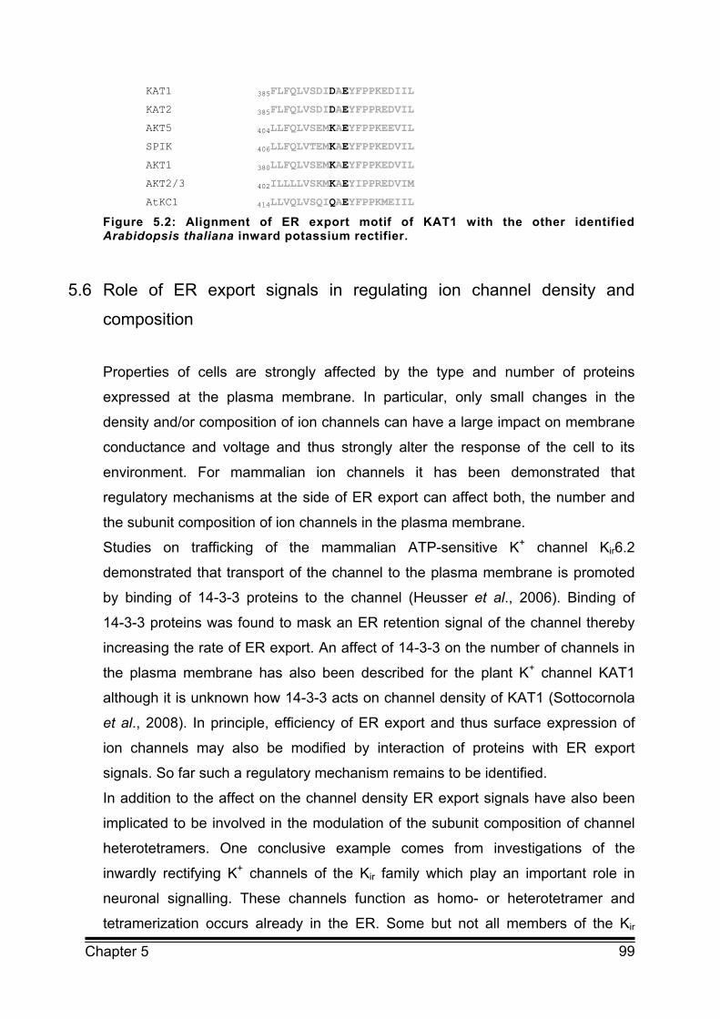
99 Chapter 5
KAT1 385FLFQLVSDIDAEYFPPKEDIIL
KAT2 385FLFQLVSDIDAEYFPPREDVIL
AKT5 404LLFQLVSEMKAEYFPPKEEVIL
SPIK 406LLFQLVTEMKAEYFPPKEDVIL
AKT1 380LLFQLVSEMKAEYFPPKEDVIL
AKT2/3 402ILLLLVSKMKAEYIPPREDVIM
AtKC1 414LLVQLVSQIQAEYFPPKMEIIL
Figure 5.2: Alignment of ER export motif of KAT1 with the other identified Arabidopsis thaliana inward potassium rectifier.
5.6 Role of ER export signals in regulating ion channel density and
composition
Properties of cells are strongly affected by the type and number of proteins
expressed at the plasma membrane. In particular, only small changes in the
density and/or composition of ion channels can have a large impact on membrane
conductance and voltage and thus strongly alter the response of the cell to its
environment. For mammalian ion channels it has been demonstrated that
regulatory mechanisms at the side of ER export can affect both, the number and
the subunit composition of ion channels in the plasma membrane.
Studies on trafficking of the mammalian ATP-sensitive K+ channel Kir6.2
demonstrated that transport of the channel to the plasma membrane is promoted
by binding of 14-3-3 proteins to the channel (Heusser et al., 2006). Binding of
14-3-3 proteins was found to mask an ER retention signal of the channel thereby
increasing the rate of ER export. An affect of 14-3-3 on the number of channels in
the plasma membrane has also been described for the plant K+ channel KAT1
although it is unknown how 14-3-3 acts on channel density of KAT1 (Sottocornola
et al., 2008). In principle, efficiency of ER export and thus surface expression of
ion channels may also be modified by interaction of proteins with ER export
signals. So far such a regulatory mechanism remains to be identified.
In addition to the affect on the channel density ER export signals have also been
implicated to be involved in the modulation of the subunit composition of channel
heterotetramers. One conclusive example comes from investigations of the
inwardly rectifying K+ channels of the Kir family which play an important role in
neuronal signalling. These channels function as homo- or heterotetramer and
tetramerization occurs already in the ER. Some but not all members of the Kir

100 Chapter 5
family contain diacidic ER export signals (Ma et al., 2008; Stockklausner et al.,
2001). Homotetramers of subunits which do not contain an ER export signal like
Kir3.1 and Kir3.3 are accumulating in the ER. In contrast, Kir2.1 which does contain
a diacidic ER export signal localises to the plasma membrane. Ma et al. (2002)
have demonstrated that formation of heterotetramers containing both, Kir subunits
with and without ER export signals leads to expression of the heterotetramer in the
plasma membrane. These results demonstrate that not all subunits of
heterotetramers need to contain ER export signals in order to ensure efficient
transport to the plasma membrane. More importantly, the ER export signal of one
channel subunit can obviously affect targeting of other Kir channels subunits thus
leading to changes in the composition of heterotetramers in the plasma
membrane. Changes in subunit composition alter the electrical characteristics of
the functional K+ channel and can therefore have a strong impact on the
conductance of the plasma membrane and consequently on the function of the
cell.
Heterotetramerisation is found for many members of different K+ channel families
and the impact of heterotetramerisation on the efficiency of ER export may well be
of general importance for the control of membrane conductance.
This hypothesis is supported by studies on ER export mutants of both, mammalian
and plant K+ channels. Efficient trafficking of HCN channels to the plasma
membrane has been demonstrated to depend on the cNBD (see previous
paragraph). Deletion of the B-helix of the cNBD of HCN2 was found to disrupt ER
export of the channel leading to retention of the channel in the ER (Proenza et al.,
2002). However, coexpression of wildtype HCN1 together with the ER export
mutant of HCN2 led to surface expression of the HCN1/HCN2 heterotetramer.
Similar results have been found for the plant K+ channel KAT1. When KAT1
wildtype and the ER export mutant of KAT1 were coexpressed in HEK293 cells
plasma membrane localisation of the ER export mutant could be rescued (Mikosch
et al., 2009). This again implicates that those subunits which are retained in the
ER as homotetramers can be targeted to the plasma membrane via
heterotetramerisation with subunits that do contain an ER export signal. The
physiological importance of such a mechanism is revealed by investigations of
AtKC1, a member of the K+ inward rectifying family from Arabidopsis thaliana.
Several members of this family have been shown to form functional
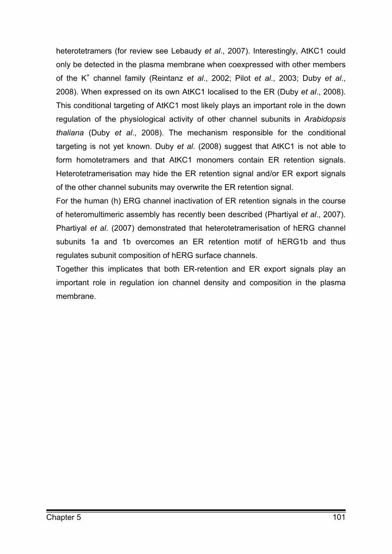
101 Chapter 5
heterotetramers (for review see Lebaudy et al., 2007). Interestingly, AtKC1 could
only be detected in the plasma membrane when coexpressed with other members
of the K+ channel family (Reintanz et al., 2002; Pilot et al., 2003; Duby et al.,
2008). When expressed on its own AtKC1 localised to the ER (Duby et al., 2008).
This conditional targeting of AtKC1 most likely plays an important role in the down
regulation of the physiological activity of other channel subunits in Arabidopsis
thaliana (Duby et al., 2008). The mechanism responsible for the conditional
targeting is not yet known. Duby et al. (2008) suggest that AtKC1 is not able to
form homotetramers and that AtKC1 monomers contain ER retention signals.
Heterotetramerisation may hide the ER retention signal and/or ER export signals
of the other channel subunits may overwrite the ER retention signal.
For the human (h) ERG channel inactivation of ER retention signals in the course
of heteromultimeric assembly has recently been described (Phartiyal et al., 2007).
Phartiyal et al. (2007) demonstrated that heterotetramerisation of hERG channel
subunits 1a and 1b overcomes an ER retention motif of hERG1b and thus
regulates subunit composition of hERG surface channels.
Together this implicates that both ER-retention and ER export signals play an
important role in regulation ion channel density and composition in the plasma
membrane.
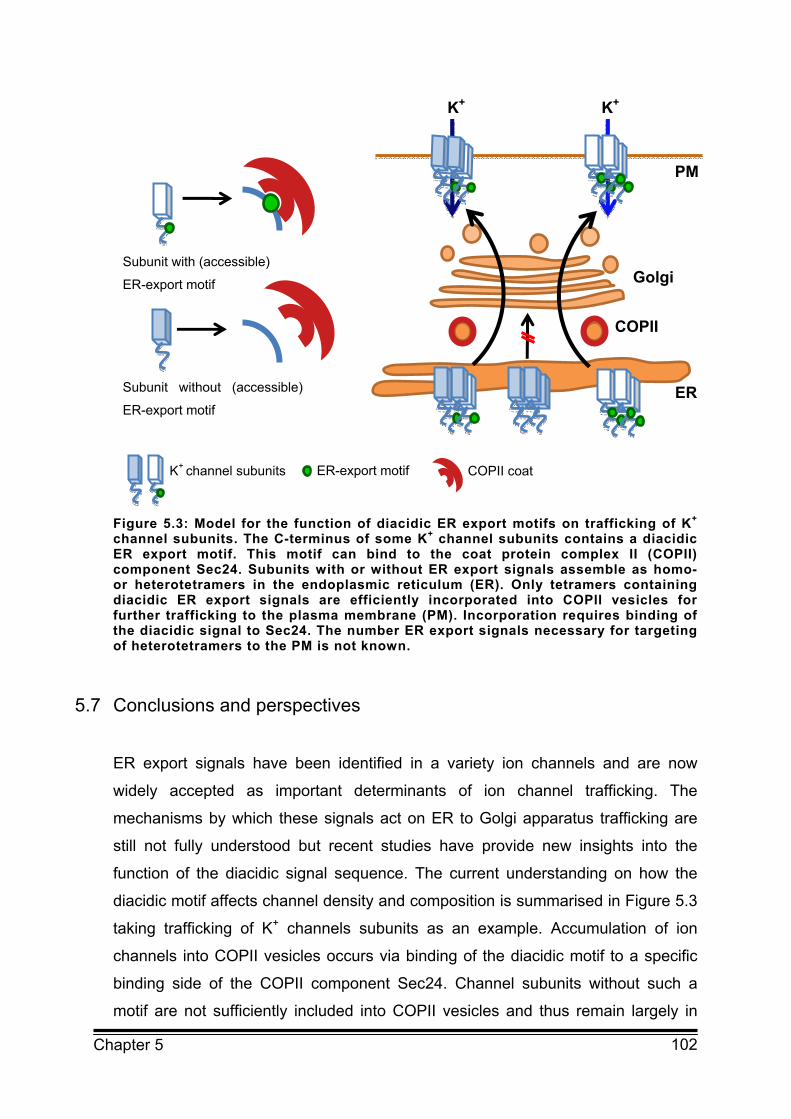
102 Chapter 5
Figure 5.3: Model for the function of diacidic ER export motifs on trafficking of K+ channel subunits. The C-terminus of some K+ channel subunits contains a diacidic ER export motif. This motif can bind to the coat protein complex II (COPII) component Sec24. Subunits with or without ER export signals assemble as homo- or heterotetramers in the endoplasmic reticulum (ER). Only tetramers containing diacidic ER export signals are efficiently incorporated into COPII vesicles for further trafficking to the plasma membrane (PM). Incorporation requires binding of the diacidic signal to Sec24. The number ER export signals necessary for targeting of heterotetramers to the PM is not known.
5.7 Conclusions and perspectives
ER export signals have been identified in a variety ion channels and are now
widely accepted as important determinants of ion channel trafficking. The
mechanisms by which these signals act on ER to Golgi apparatus trafficking are
still not fully understood but recent studies have provide new insights into the
function of the diacidic signal sequence. The current understanding on how the
diacidic motif affects channel density and composition is summarised in Figure 5.3
taking trafficking of K+ channels subunits as an example. Accumulation of ion
channels into COPII vesicles occurs via binding of the diacidic motif to a specific
binding side of the COPII component Sec24. Channel subunits without such a
motif are not sufficiently included into COPII vesicles and thus remain largely in
PM
K+ K+
Golgi
ER
COPII
Subunit without (accessible)
ER-export motif
Subunit with (accessible)
ER-export motif
ER-export motif COPII coat K+ channel subunits

103 Chapter 5
the ER. However, these subunits can be targeted to the plasma membrane via
heterotetramerisation with subunits containing an ER export signal. As the
electrical properties of heterotetramers may differ from that of homotetramers this
in turn can lead to changes in the membrane conductance and consequently alter
the function of the cell.
There is increasing evidence that the ER export signal itself is also subject to
regulation which may involve phosphorylation or binding to scaffold proteins.
These proteins may act via steric masking or unmasking of ER export signals. In
order to fully understand the regulation of ion channel trafficking it will be important
to uncover the mechanisms affecting the function of the ER export signal. For
future research it will also be of major relevance to investigate the physiological
significance of these mechanisms.

104 Chapter 5
5.8 References Akhavan A, Atanasiu R, Noguchi T, Han W, Holder N, Shrier A (2005)
Identification of the cyclic nucleotide binding domain as a conserved determinant
of ion channel cell surface localization. The Journal of Cell Science 118:2803-2812
Aydar E and Palmer C (2001) Functional characterization of the C-terminus of the
human ether-à-go-go-related gene K+ channel (HERG). The Journal of Physiology
534:1-14
Barlowe C, Orci L, Yeung T, Hosobuchi M, Hamamoto S, Salama N, Rexach MF, Ravazzola M, Amherdt M, Schekman R (1994) COPII: a membrane
coat formed by Sec proteins that drive vesicle budding from the endoplasmic
reticulum. Cell 77(6):895-907
Duby G, Hosy E, Fizames C, Alcon C, Costa A, Sentenac H, Thibaud JB
(2008) AtKC1, a conditionally targeted shaker-type subunit, regulates the activity
of plant K+ channels. The Plant Journal 53(1):115-123
Hanton SL, Renna L, Bortolotti LE, Chatre L, Stefano G, Brandizzi F (2005)
Diacidic motifs influence the export of transmembrane proteins from the
endoplasmic reticulum in plant cells. The Plant Cell 17(11):3081-3093
Heusser K and Schwappach B (2005) Trafficking of potassium channels. Current
Opinion in Neurobiology 15:364-369
Heusser K, Yuan H, Neagoe I, Tarasov AI, Ashcroft FM, Schwappach B (2006)
Scavenging of 14-3-3 proteins reveals their involvement in the cell surface
transport of ATP-sensitive K+ channels. The Journal of Cell Science
119:4353-4363
Jarvis SE and Zamponi GW (2007) Trafficking and regulation of neuronal
voltage-gated calcium channels. Current Opinion in Cell Biology 19(4):474-482
Lebaudy A, Very A, Sentenac H (2007) K+ channel activity in plants: Genes,
regulations and functions. FEBS Letters 581(12):2357-2366
Lefebvre B, Batoko H, Duby G, Boutry M (2004) Targeting of a Nicotiana
plumbaginifolia H+ ATPase to the plasma membrane is not by default and requires
cytosolic structural determinants. The Plant Cell 16(7):1772-1789
Ma D and Jan LY (2002) ER transport signals and trafficking of potassium
channels and receptors. Current Opinion in Neurobiology 12(3):287-292
Ma D, Zerangue N, Lin YF, Collins A, Yu M, Jan YN, Jan LY (2001) Role of ER
105 Chapter 5
export signals in controlling surface potassium channel numbers. Science
291(5502):316-319
Ma D, Zerangue N, Raab-Graham K, Fried SR, Jan YN, Jan LY (2002) Diverse
trafficking patterns due to multiple traffic motifs in G protein activated inwardly
rectifying potassium channels from brain and heart. Neuron 33(5):715-729
Malkus P, Jiang F, Schekman R (2002) Concentrative sorting of secretory cargo
proteins into COPII coated vesicles. The Journal of Biological Chemistry
159(6):915-921
Mancias JD and Goldberg J (2005) Exiting the endoplasmic reticulum. Traffic
6:278-285
Mancias JD and Goldberg J (2007) The transport signal on Sec22 for packaging
into COPII coated vesicles is a conformational epitope. Molecular Cell 26(3):403-
414
Marten I and Hoshi T (1997) Voltage-dependent gating characteristics of the K+
channel KAT1 depend on the N- and C-termini. PNAS 94(7):3448-3453
Mikosch M, Hurst AC, Hertel B, Homann U (2006) Diacidic motif is required for
efficient transport of the K+ channel KAT1 to the plasma membrane. Plant
Physiology 142(3):923-930
Mikosch M, Käberich K, Homann U (2009) Efficiency of ER export of the K+
channel KAT1 depends on the number of acidic amino acids within a triacidic
motif. In preparation
Miller E, Antonny B, Hamamoto S, Schekman R (2002) Cargo selection into
COPII vesicles is driven by the Sec24p subunit. The EMBO Journal 21(22):6105-
6113
Miller EA, Beilharz TH, Malkus PN, Lee MCS, Hamamoto S, Orci L, Schekman R (2003) Multiple cargo binding sites on the COPII subunit Sec24p
ensure capture of diverse membrane proteins into transport vesicles. Cell
114(4):497-509
Miranda M, Sorkina T, Grammatopoulos TN, Zawada WM, Sorkin A (2004)
Multiple molecular determinants in the carboxyl terminus regulate dopamine
transporter export from endoplasmic reticulum. The Journal of Biological
Chemistry 279(29):30760-30770
Mossessova E, Bickford LC, Goldberg J (2003) SNARE selectivity of the COPII
coat. Cell 114(4):483-495

106 Chapter 5
Nishimura N and Balch WE (1997) A diacidic signal required for selective export
from the endoplasmic reticulum. Science 277(5325):556-558
Nishimura N, Bannykh S, Slabough S, Matteson J, Altschuler Y. Hahn K, Balch WE (1999) A diacidic (DxE) code directs concentration of cargo during
export from the endoplasmic reticulum The Journal of Biological Chemistry
274(22):15937-15946
Otte S and Barlowe C (2004) Sorting signals can direct receptor mediated export
of soluble proteins into COPII vesicles. Nature Cell Biology 6(12):1189-1194
Pagano A, Letourneur F, Garcia-Estefania D, Carpentier JL, Orci L, Paccaud JP (1999) Sec24 proteins and sorting at the endoplasmic reticulum. The
Journal of Biological Chemistry 274(12):7833-7840
Pagant S, Kung L, Dorrington M, Lee MCS, Miller EA (2007) Inhibiting
endoplasmic reticulum (ER)-associated degradation of misfolded Yor1p does not
permit ER export despite the presence of a diacidic sorting signal. Molecular
Biology of the Cell 18(9):3398-3413
Peng R, De Antoni A, Gallwitz D (2000) Evidence for overlapping and distinct
functions in protein transport of coat protein Sec24p family members. The Journal
of Biological Chemistry 275(15):11521-11528
Phartiyal P, Sale H, Jones EMC, Robertson GA (2007) ER retention and rescue
by heteromeric assembly regulates hERG 1a/1b surface channel composition. The
Journal of Biological Chemistry 282:9874-9882
Pilot G, Gaymard F, Mouline K, Chérel I, Sentenac H (2003) Regulated
expression of Arabidopsis thaliana shaker K+ channel genes involved in K+ uptake
and distribution in the plant. Plant Molecular Biology 51(5):773-787
Proenza C, Tran N, Angoli D, Zahynacz K, Balcar P, Accili EA (2002) Different
roles for the cyclic nucleotide binding domain and amino terminus in assembly and
expression of hyperpolarisation activated, cyclic nucleotide gated channels. The
Journal of Biological Chemistry 277(33):29634-29642
Reintanz B, Szyroki A, Ivashikina N, Ache P, Godde M, Becker D, Palme K, Hedrich R (2002) AtKC1, a silent Arabidopsis thaliana potassium channel alpha -
subunit modulates root hair K+ influx. PNAS 99(6):4079-4084
Shimoni Y, Kurihara T, Ravazzola M, Amherdt M, Orci L, Schekman R (2000)
Lst1p and Sec24p cooperate in sorting of the plasma membrane ATPase into
COPII vesicles in Saccharomyces cerevisiae. The Journal of Biological Chemistry

107 Chapter 5
151(5):973-984
Sieben C, Mikosch M, Brandizzi F, Homann U (2008) Interaction of the K+
channel KAT1 with the COPII coat component Sec24 depends on a diacidic ER
export motif. The Plant Journal 56(6):997-1006
Sottocornola B, Gazzarrini S, Olivari C, Romani G, Valbuzzi P, Thiel G, Moroni A (2008) 14-3-3 proteins regulate the potassium channel KAT1 by dual
modes. Plant Biology 10(2):231-236
Stockklausner C, Ludwig J, Ruppersberg JP, Klöcker N (2001) A sequence
motif responsible for ER export and surface expression of Kir2.0 inward rectifier K+
channels. FEBS Letters 493:129-133
Votsmeier C and Gallwitz D (2001) An acidic sequence of a putative yeast Golgi
membrane protein binds COPII and facilitates ER export. The EMBO Journal
20(23):6742-6750 Wang X, Matteson J, An Y, Moyer B, Yoo J, Bannykh S,Wilson IA, Riordan JR, Balch WE (2004) COPII-dependent export of cystic fibrosis
transmembrane conductance regulator from the ER uses a diacidic exit code. The
Journal of Biological Chemistry 167(1):65-74
Wendeler MW, Paccaud J, Hauri H (2007) Role of Sec24 isoforms in selective
export of membrane proteins from the endoplasmic reticulum. EMBO Reports
8(3):258-264
Zagotta WN, Olivier NB, Black KD, Young EC, Olson R, Gouaux E (2003)
Structural basis for modulation and agonist specificity of HCN pacemaker
channels. Nature 425(6954):200-205
Zuzarte M, Rinné S, Schlichthörl G, Schubert A, Daut J, Preisig-Müller R (2007) A diacidic sequence motif enhances the surface expression of the
potassium channel TASK-3. Traffic 8:1093-1100
108 Chapter 6
Chapter 6 GENERAL DISCUSSION
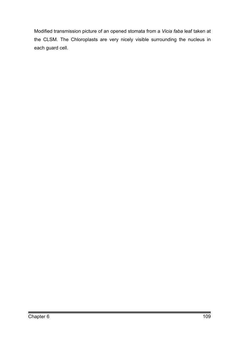
109 Chapter 6
Modified transmission picture of an opened stomata from a Vicia faba leaf taken at
the CLSM. The Chloroplasts are very nicely visible surrounding the nucleus in
each guard cell.

110 Chapter 6
6.1 Diacidic ER export motifs
6.1.1 ER export motifs regulate trafficking of membrane protein in plants
The current work shows for the first time that ER export motifs are functional in ion
channel trafficking in plant cells. A diacidic ER export motif was identified in the
plant potassium (K+) channel KAT1 from Arabidopsis thaliana (Mikosch et al.,
2006). Expression of the KAT1 mutant with aspartate (D394) and glutamate
(E396) mutated to alanine (A) (KAT1DiaAa) in guard cells as well as in HEK293 cells
led to a strong reduction of the plasma membrane inward conductance compared
to the KAT1 wildtype channel.
This reduction in plasma membrane conductance may occur due to several
reasons incorporated in three parameters. The net plasma membrane current I
can be described by Equation (1), where i is the single channel current, Po the
open probability of the channel and n the number of ion channels in the plasma
membrane. A change in the net plasma membrane current I can be caused by a
change in the single channel current i, a reduction of the number of channels in
the plasma membrane, or by changes of the open probability Po of the channel.
I = i * Po * n (1)
Rapid changes in I are very often accomplished by changes in the open probability
Po of the channel. For example, in case of KAT1 the open probability Po is strongly
affected by changes in membrane voltage. On the other hand, long time changes
are more likely due to an altered number of ion channels in the plasma membrane.
Kinetic analysis of the analysed ER export mutant revealed similar kinetics as the
wildtype KAT1 K+ channel. Therefore, the changes in plasma membrane
conductance, observed in the ER export mutant, were most likely caused by a
reduced number of ion channels.
This reduction of KAT1 K+ channels in the plasma membrane was verified by
confocal analysis of the localisation of the KAT1 K+ channel and its mutants in
Vicia faba guard cells as well as in HEK293 cells, which demonstrated that the
analysed motif is truly functional as an ER export signal in both cell types. This

111 Chapter 6
also implies that the mechanisms of ER export are very similar in plant and animal
cells.
During this study a number of other ER export motifs have been discovered in
plant membrane proteins which underlines the importance of the results. One of
them is the diacidic ER export motif in the N-terminus of the maize plasma
membrane aquaporins ZmPIP2;4 and ZmPIP2;5 (Zelazny et al., 2008). Schoberer
et al. (2008) identified an arginine/lysine (RK) motif which is responsible for ER
export of the Golgi resident glycosylation enzymes GnTI and GMII. ER export of
the Golgi proteins GONST1 and CASP is also regulated by diacidic ER export
motifs (Hanton et al., 2005). This shows that regulation of ER export of membrane
proteins by intrinsic signal sequences is a highly conserved mechanism in all
eukaryotic cells.
Further analysis of the diacidic motif and the surrounding amino acids of KAT1
revealed that the diacidic motif was not only diacidic but contained at least three
acid residues at position D392, D394 and E396 (Mikosch et al., 2009). Single
mutation of the amino acids led to the conclusion that all acidic residues have a
similar influence on ER export. The amount of reduction in KAT1 conductance was
clearly dependent on the number of mutated acidic residues. Mutants with only
one of the residues mutated to alanine (A) had a slightly decreased inward
conductance compared to the wildtype K+ channel. Mutation of two of the residues
results in a much stronger reduction of inward conductance. Only when all three
acidic amino acids were mutated to A the ER export was completely inhibited and
almost no KAT1 current could be measured.
6.1.2 ER export affects cargo selection by interaction with Sec24
The selection and concentration of cargo into vesicles at ER export sites (ERES)
depends on interaction of the COPII coat with cargo transport signals. For most of
the ER export signals identified so far an interaction with the Sec24 subunit of the
COPII coat could be shown (Miller et al., 2002 and 2003; Mossessova et al.,
2003). Sec24 contains at least three binding sites for several cargo proteins with
different ER export motifs. The Sec24 A-site binds the NPF motif of the SNARE
protein Sed5 (Miller et al., 2005; Mossessova et al., 2003). The B-site binds to the
LxxLE motif of the SNARE proteins Sed5 and Bet1 as well as to several diacidic
112 Chapter 6
ER export motifs like of the protein Sys1 (Nishimura et al., 1999; Votsmeier and
Gallwitz, 2001). The C-site binds the NIE motif of the SNARE Sec22 (Miller et al.,
2003; Mancias and Goldberg, 2007). An additional binding site was identified for
binding of GAT1 and two other members of the SLC transporter family which
exhibit an arginie/leucine (RL) ER export motif (Farhan et al., 2007).
The first indication for the participation of the COPII coat in ER export of KAT1
came from coexpression analysis of wildtype KAT1 K+ channel with Sar1T39N, the
dominant negative form of Sar1. In these cells ER export of KAT1 was inhibited
and the channel was found in the same perinuclear structures as several ER
export mutants with at least two acidic amino acids replaced by A. Binding of the
K+ channel KAT1 to the COPII subunit Sec24 could be demonstrated by FRET
measurements. After mutation of the amino acids D394 and E396 to A the
interaction between Sec24 and the KAT1DIaAa channel at the ER was abolished
(Sieben et al., 2008). This reveals that KAT1 is recognised by Sec24 and selected
for ER export with the acidic ER export sequence. As the B-site of Sec24 protein
was identified as the binding site for diacidic ER export motifs in yeast, which is
highly conserved between plant, yeast and human Sec24 homologues, it would be
interesting to analyse the importance of the B-site for binding of the acidic ER
export motif of KAT1.
In future studies it will be necessary to show the interaction of the acidic ER export
motif of KAT1 with Sec24 in a more direct way. Regarding the complicated nature
of the identified ER export motif of KAT1 it would also be interesting to analyse the
importance of the single acidic residues for Sec24 binding and address the
question whether they are functionally resistant to exchanges by the other acidic
amino acid. Another aspect is the possibility that the amino acids, surrounding the
acidic motif, may also be of importance for efficient ER export. Therefore, analysis
of these amino acids would also be of major interest. In this context it is worth
mentioning that cargo recognition may rely on multivalent interactions and the
three dimensional structure of the signals which is only revealed upon proper
folding and assembly of KAT1 (Mikosch and Homann, 2009). Further information
on crystal structure would be needed to get an impression of the requirements for
functional ER export motifs. Analysis of the crystal structures of the Sec22 NIE ER
export motif which binds to the C-site of Sec24 demonstrated that the motif is most
likely a folded epitope (Mancias and Goldberg, 2007).

113 Chapter 6
The situation gets even more complex when considering the different Sec24
isoforms present in cells. For example in mammalian cells four different Sec24
isoforms (Sec24A-Sec24D) exist which can be further diversified by alternative
splicing. The ER Golgi intermediate compartment protein 53 (ERGIC53) with the
dihydrophobic diphenylalanine (FF) motif binds to all four Sec24 isoforms
(Wendeler et al., 2007). But the transporter GAT1 is only recognised by the
Sec24D subunit (Farhan et al., 2007). In Arabidopsis thaliana three isoforms of
Sec24 have been identified but so far nothing is known about varying recognition
of cargo by different Sec24 isoforms. Mancias and Goldberg (2008) showed by
comparative analysis of crystal structures of the four human Sec24 isoforms that a
conserved IxM ER export motif binds to a surface groove of mammalian Sec24C
and Sec24D, but the groove is not accessible in the Sec24A and Sec24B subunits.
On the other hand, the LxxLE ER export and the DxE signal of VSVG are
selectively bound by Sec24A and Sec24B subunits. Sec22 is bound only by
Sec24A and Sec24B with an NIE motif (Mancias and Goldberg, 2007).
Therefore, it is necessary to analyse the interaction of KAT1 with all three Sec24
isoforms present in Arabidopsis thaliana to get an insight into the regulation of ER
export through the employment of different Sec24 receptors.
6.1.3 Restriction of the ER export mutant to the perinuclear area
ER export mutants of KAT1 with at least two of the acidic residues mutated to
alanine show a special localization in HEK293 as well as in Vicia faba guard cells.
The GFP fluorescence is restricted to parts of the ER in a perinuclear area.
This area may correspond to ER export sites (ERES). ERES are defined as
ribosome free parts of the ER that label for the COPII coat complex. In mammalian
cells it could be demonstrated that almost 50-60 % of ERES are actively
positioned in the perinuclear area while others are distributed on the periphery of
the cell (Bannykh et al., 1997; Stephens, 2003). In plant cells the situation is quite
different. The Golgi stacks in plants move along together with individual ERES but
these sites are also very often located in the perinuclear area (daSilva et al.,
2004). Intense debate is going on about the nature of the ERES Golgi interface
because several studies refer to this either as a tubular structure or as a more
vesicular arrangement (Kirk and Ward, 2007). It seems likely that the sheet-like

114 Chapter 6
structures, found in cells where ER export of KAT1 was prevented either by
mutation of the ER export motif of the channel or by overexpression of Sar1T39N,
corresponds to ERES in the perinuclear area. Furthermore special arrangements
of the ER may prevent free diffusion of KAT1 in the ER membrane and contribute
to the restriction of the KAT1 K+ channel to these ERES. A similar pattern of ER
retention has been demonstrated for the G-protein coupled receptors α2B-AR and
AT1R when coexpressed with Sar1H79G. Both receptors show a perinuclear
staining (Dong et al., 2008). In addition, mutants of the HERG K+ channel which
lack the cNBD domain which is important for ER export also showed a perinuclear
staining (Akhavan et al., 2005). In contrast, the G-protein coupled receptor β2-AR
displayed a totally different localisation upon coexpression with Sar1H79G. The
receptor was retained in the entire ER (Dong et al., 2008). Experiments
demonstrated that unlike KAT1, ER retention of proteins like the plasma
membrane H+ ATPase or the artificial plasma membrane marker TM23 by
coexpression with Sar1T39N led to a distribution of these proteins in the entire ER
(unpublished results). The restriction of some membrane proteins to specialised
ER subdomains can be due to special interaction with ER resident proteins.
The nature of the protein itself may also contribute to the ER localisation because
it could be shown that length and hydrophobicity of the transmembrane domains of
membrane proteins play an important role in protein sorting within the ER and
throughout the secretory pathway (Ronchi et al., 2008; Brandizzi et al., 2002).
Ronchi et al. (2008) expressed two proteins that differ in transmembrane domain
length: FP-17 and FP-22. FP-17 was retained in the ER and FP-22 was located in
the plasma membrane. Their respective distribution in the ER differs to a great
extend although both of them are freely diffusible in the ER. It seems that FP-17 is
excluded from ER export site and FP-22 not. As a result the authors assume that
self organising principles are an important factor of the distribution within the ER.
In addition, the curvature of the membrane can influence the probability of protein
localisation. To achieve a thermodynamically stable state, proteins tend to locate
in plane or curved membranes according to the length and hydrophobicity of their
transmembrane domains (dePlanque and Kilian, 2003). The protein distribution in the ER can also be altered by ER residential proteins, by
modification of lipids or direct protein-protein interaction. An interesting candidate
conferring ER localisation is the ER resident transmembrane protein calnexin
115 Chapter 6
which interacts with several neurotransmitter transporters like GAT1 in specialised
regions of the ER. Korkhov et al. (2008) assumed that calnexin plays a role in the
control of oligomeric assembly of these transporters. calnexin and the interacting
proteins segregate into so called organised smooth ER membranes (OSER) which
consist of multiple stacked ER membranes. They are supposed to act as a fence
to prevent aggregation of membrane proteins in the OSER. These OSER are
visible in confocal images as concentric bodies close to or associated with the
perinuclear ER. Especially upon expression of a GAT1E101D mutant, which cannot
form oligomers anymore, the localisation in the OSER is dramatically increased
(Korkhov et al., 2008).
The ER export mutants of KAT1 are frequently found in concentric bodies in
addition to the perinuclear area in both HEK293 cells and Vicia faba guard and
epidermal cells. Therefore, it would be interesting to explore whether KAT1 also
interacts with calnexin and if the observed concentric bodies are related to OSER.
6.1.4 mRNA localisation
There are indications that the localisation of mRNA within the cell is of general
importance for cell function. The cotranslational insertion of locally synthesised
proteins could be necessary especially for the formation of oligomeric protein
complexes. The localisation of ion channel mRNA within the cell could have an
enormous impact on the localisation of the channel within the ER and determine
the availability of subunits for oligomerisation. This could be achieved by
information in the noncoding regions of the mRNA. Targeting of mRNA to certain
subdomains involves special mechanism like RNA binding proteins which interact
with the cytoskeleton to ensure the localisation. In mammalian cells it could be
shown that especially mRNA of membrane proteins is typically targeted to specific
regions of the ER. For example the mRNA of the chloride channel ClC1 is directed
to the perinuclear ER. Upon coexpression of the ClC1 channel with the dominant
negative Sar1H79G the channel is trapped in the perinuclear region of the ER
(Papponen et al., 2008). The ER region where the mRNA is localised may be
connected to a kind of channel tetramerization domain in the ER. Also in plants the
localisation of mRNA to special subdomains of the ER has been reported
(Hamada et al., 2003). Considering that the ER localisation pattern of the ClC1

116 Chapter 6
protein resembles that of KAT1, it would be very interesting to analyse whether the
specific localisation of KAT1 to the perinuclear region of the ER can be explained
by the distribution of KAT1 mRNA in the cell.
6.2 Tetramerization of ion channels
6.2.1 Tetramerization of ion channels most likely occurs at the ER
The insight into ion channel tetramerization is based on overexpression
experiments in cell culture or Xenopus laevis oocytes. However, nothing is known
about the availability of subunits within the ER under normal conditions. It seems
very likely that ion channel subunits are not very abundant in the ER because the
number of ion channels in the plasma membrane is generally low. Accordingly,
one may assume that mechanisms have been developed to support the formation
of tetramers. These may either occur through the involvement of specialised
proteins like chaperones or through the differential localisation of subunits within
the ER. Maybe there are specific ion channel assembly domains in the ER where
ion channel subunits are concentrated because tetramerization needs at least the
close proximity of four subunits.
So far it has not been clearly revealed where and when ion channel
tetramerization takes places in the ER. It is most likely that the tetramers are
directly composed in the ER and only fully assembled ion channels can pass the
quality control system of the ER. For a few oligomeric membrane proteins it has
been shown that tetramerization is indeed a prerequisite for ER export (Farhan et
al., 2007; Becker et al., 2007).
But how is the oligomeric state of a protein controlled in the ER? Several other
proteins must be involved in the tetramerization control of ion channels in the ER.
Their nature is quite often undetermined. Recent investigation revealed a very
interesting protein in the ER, Rer1, which recognises polar residues facing the lipid
bilayer (Sato et al., 2003 and 2004). Not yet assembled ion channel subunits are
possible candidates for such a protein because they exhibit several polar as well
as positively charged amino acids in the transmembrane and filter region. Also
117 Chapter 6
calnexin, an ER resident membrane protein, can play a role in oligomeric
assembly of proteins (Korkhov et al., 2008).
COPII can be involved in the control of proper oligomerisation, too. The kinetic
stability of the prebudding complex consisting of Sar1, Sec24 and Sec23 bound to
cargo molecules depends on interaction of the cargo with Sec24. Properly
assembled cargo molecules enhance the stability of the prebudding complex.
Unassembled cargo leads to Sar1-GTP hydrolysis and subsequently the
prebudding complex collapses (Sato et al., 2005). Sato et al. (2004) concludes
that by this process of Sar1-GTP hydrolysis unassembled cargo is actively
excluded from forming COPII vesicles.
So far nothing is known about the tetramerization of KAT1 and related ion
channels in plants. It is necessary to understand whether the KAT1 K+ channel
needs to tetramerise within the ER for efficient ER export and how the
heterotetramerisation influences these processes.
6.2.2 ER export motifs can affect subunit composition of channel tetramers in the plasma membrane
The acidic amino acid sequence DxDxE (392-396) which is responsible for ER
export of KAT1 is highly conserved among K+ channels of Arabidopsis thaliana
and several K+ channels of other plant species. Nevertheless, there are major
differences in the motif between the channels (Figure 3.1). The acidic nature of
D392 is conserved among all depicted ion channel except AKT2/3 and AtKC1.
D394 is replaced by lysine (K) in AKT5, SPIK, AKT1, AKT2/3 and AtKC1 from
Arabidopsis thaliana. Residue E396 is totally conserved among all channels
analysed. It seems that in many cases the motif is disrupted but the channel can
reach the plasma membrane (AKT5, SPIK, AKT1 and especially AKT2/3). The
differences in the ER export motifs of the various plant K+ channels may result in
different affinities of the channel to Sec24 and therefore may play a role in defining
the rate of ER export of different subunits and subsequently render the
composition of channel heterotetramers in the plasma membrane. In particular,
this seems true for channel subunits which lack an ER export motif and are
retained in the ER like AtKC1 from Arabidopsis thaliana. Expression of AtKC1 in
different cell types like COS cells, Xenopus laevis oocytes, tobacco mesophyll
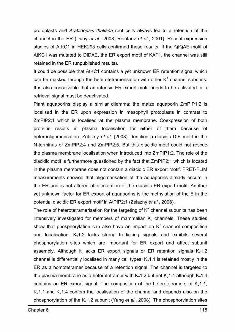
118 Chapter 6
protoplasts and Arabidopsis thaliana root cells always led to a retention of the
channel in the ER (Duby et al., 2008; Reintanz et al., 2001). Recent expression
studies of AtKC1 in HEK293 cells confirmed these results. If the QIQAE motif of
AtKC1 was mutated to DIDAE, the ER export motif of KAT1, the channel was still
retained in the ER (unpublished results).
It could be possible that AtKC1 contains a yet unknown ER retention signal which
can be masked through the heterotetramerisation with other K+ channel subunits.
It is also conceivable that an intrinsic ER export motif needs to be activated or a
retrieval signal must be deactivated.
Plant aquaporins display a similar dilemma: the maize aquaporin ZmPIP1;2 is
localised in the ER upon expression in mesophyll protoplasts in contrast to
ZmPIP2;1 which is localised at the plasma membrane. Coexpression of both
proteins results in plasma localisation for either of them because of
heterooligomerisation. Zelazny et al. (2008) identified a diacidic DIE motif in the
N-terminus of ZmPIP2;4 and ZmPIP2;5. But this diacidic motif could not rescue
the plasma membrane localisation when introduced into ZmPIP1;2. The role of the
diacidic motif is furthermore questioned by the fact that ZmPIP2;1 which is located
in the plasma membrane does not contain a diacidic ER export motif. FRET-FLIM
measurements showed that oligomerisation of the aquaporins already occurs in
the ER and is not altered after mutation of the diacidic ER export motif. Another
yet unknown factor for ER export of aquaporins is the methylation of the E in the
potential diacidic ER export motif in AtPIP2;1 (Zelazny et al., 2008).
The role of heterotetramerisation for the targeting of K+ channel subunits has been
intensively investigated for members of mammalian Kv channels. These studies
show that phosphorylation can also have an impact on K+ channel composition
and localisation. Kv1.2 lacks strong trafficking signals and exhibits several
phosphorylation sites which are important for ER export and affect subunit
assembly. Although it lacks ER export signals or ER retention signals Kv1.2
channel is differentially localised in many cell types. Kv1.1 is retained mostly in the
ER as a homotetramer because of a retention signal. The channel is targeted to
the plasma membrane as a heterotetramer with Kv1.2 but not Kv1.4 although Kv1.4
contains an ER export signal. The composition of the heterotetramers of Kv1.1,
Kv1.1 and Kv1.4 confers the localisation of the channel and depends also on the
phosphorylation of the Kv1.2 subunit (Yang et al., 2008). The phosphorylation sites
119 Chapter 6
of Kv1.2 are located within a putative 14-3-3 binding site and phosphorylation of
the channel may be linked to binding of 14-3-3. Phosphorylation-dependent
binding of 14-3-3 may mask an intrinsic ER exit or retrieval signal. Such a
regulatory effect of 14-3-3 binding on ER export has been demonstrated for
KCNK3 and K2p3.1 channels (O’Kelly et al., 2002 and 2008). In case of the Kir4.2
channel phosphorylation has been proven to function as a negative regulator of
trafficking (Wade et al., 2006).
KAT1 contains several putative 14-3-3 binding sites and Sottocornola et al. (2008)
demonstrated that 14-3-3 proteins not only control the voltage-dependency of the
KAT1 channel but also the number of channels in the plasma membrane. Future
studies have to identify the binding site for 14-3-3 in the sequence of KAT1 and to
analyse the regulatory function of 14-3-3 for KAT1 trafficking. Phosphorylation and
dephosphorylation is obviously an important parameter in the regulation of ion
channel trafficking and necessitates a broadening of the investigative scope to
regulatory factors such as kinases and phosphatases (Sehnke et al., 2002).

120 Chapter 6
6.3 References
Akhavan A, Atanasiu R, Noguchi T, Han W, Holder N, Shrier A (2005)
Identification of the cyclic nucleotide binding domain as a conserved determinant
of ion channel cell surface localization. The Journal of Cell Science 118:2803-2812
Bannykh SI, Rowe T, Balch WE (1996) The organization of endoplasmic
reticulum export complexes. The Journal of Cell Biology 135:19–35
Becker D, Müller M, Leuner K, Jendrach M (2007) The C-terminal domain of
TRPV4 is essential for plasma membrane localisation. Molecular Membrane
Biology 25(2):139-151
Brandizzi F, Frangne N, Marc-Martin S, Hawes C, Neuhaus JM, Paris N (2002)
The destination for single pass membrane proteins is influenced markedly by the
length of the hydrophobic domain. The Plant Cell 14(5):1077-1079
daSilva LL, Snapp EL, Denecke J, Lippincott-Schwartz J, Hawes C, Brandizzi F (2004) Endoplasmic reticulum export sites and Golgi bodies behave
as single mobile secretory units in plant cells. The Plant Cell 16:1753-1777
De Planque MRR and Killian JA (2003) Protein lipid interactions studied with
designed transmembrane peptides: role of hydrophobic matching and interfacial
anchoring. Molecular Membrane Biology 20:271-284
Dong C, Zhou F, Fugetta EK, Filipeanu CM, Wu G (2008) Endoplasmic
reticulum export of adrenergic and angiotensin II receptors is differentially
regulated by Sar1 GTPase. Cell Signal 6:1035-1034
Duby G, Hosy E, Fizames C, Alcon C, Costa A, Sentenac H, Thibaud JB
(2008) AtKC1, a conditionally targeted shaker-type subunit, regulates the activity
of plant K+ channels. The Plant Journal 53(1):115-123
Farhan H, Reiterer V, Korkhov VM, Schmid JA, Freissmuth M, Sitte HH (2007)
Concentrative export from the endoplasmic reticulum of the γ-amino butyric acid
transporter 1 requires binding to Sec24D. The Journal of Biological Chemistry
282(10):7679–7689
Hamada S, Ishiyama K, Choi SB, Wang C, Singh S, Kawai N, Franceschi VR, Okita TW (2003) The transport of prolamine RNAs to prolamine protein bodies in
living rice endosperm cells. The Plant Cell 10:2253-2264
121 Chapter 6
Hanton SL, Renna L, Bortolotti LE, Chatre L, Stefano G, Brandizzi F (2005)
Diacidic motifs influence the export of transmembrane proteins from the
endoplasmic reticulum in plant cells. The Plant Cell 17:3081–3093
Kirk SJ and Ward TH (2007) COPII under the microscope. Seminars in Cell
Developmental Biology 8(4):435-447
Korkhov VM, Milan-Lobo L, Zuber B, Farhan H, Schmid JA, Freissmuth M, Sitte HH (2008) Peptide based interactions with calnexin target misassembled
membrane proteins into endoplasmic reticulum derived multilamellar bodies. The
Journal of Molecular Biology 378:337–352
Mancias JD and Goldberg J (2007) The transport signal of Sec22 for packaging
into COPII coated vesicles is a conformational epitope. Molecular Cell 26:403-414
Mancias JD and Goldberg J (2008) Structural basis of cargo membrane protein
discrimination by the human COPII coat machinery. The EMBO Journal
27(21):2918-2928
Mikosch M and Homann U (2009) How do diacidic motifs work on ion channel
trafficking? In preparation
Mikosch M, Hurst AC, Hertel B, Homann U (2006) Diacidic motif is required for
efficient transport of the K+ channel KAT1 to the plasma membrane. Plant
Physiology 142: 923-930
Mikosch M, Käberich K, Homann U (2009) Efficiency of ER export of the K+
channel KAT1 depends on the number of acidic amino acids within a triacidic
motif. In preparation
Miller E, Antonny B, Hamamoto S, Schekman R (2002) Cargo selection into
COPII vesicles is driven by the Sec24p subunit. The EMBO Journal 21(22):6105-
6113
Miller E, Liu Y, Barlowe C, Schekman R (2005). ER Golgi transport defects are
associated with mutation in the Sed5p binding domain of the COPII coat subunit
Sec24p. Molecular Biology of the Cell 16:3719–3726
Miller EA, Beilharz TH, Malkus PN, Lee MCS, Hamamoto S, Orci L, Schekman R (2003) Multiple cargo binding sites on the COPII subunit Sec24p
ensure capture of diverse membrane proteins into transport vesicles. Cell
114:497–509
Mossessova E, Bickford LC, Goldberg J (2003) SNARE selectivity of the COPII
coat. Cell 114:483–495
122 Chapter 6
Nishimura N, Bannykh S, Slabough S, Matteson J, Altschuler Y, Hahn K, Balch WE (1999) A diacidic (DxE) code directs concentration of cargo during
export from the endoplasmic reticulum. The Journal of Biological Chemistry
274:15937-15946
O’Kelly I and Goldstein SA (2008) Forward transport of K2p3.1: mediation by
14-3-3 and COPI, modulation by p11. Traffic 1:72-78
O’Kelly I, Butler M, Zilberberg N, Goldstein S (2002) Forward transport: 14-3-3
binding overcomes retention in ER by dibasic signals. Cell 111:577-588
Papponen H, Nissinen M, Kaisto T, Myllylä VV, Myllylä R, Metsikkö K (2008)
F413C and A531V but not R894X myotonia congenita mutations cause defective
endoplasmic reticulum export of the muscle-specific chloride channel CLC1.
Muscle Nerve 37(3):317-325
Reintanz B, Szyroki A, Ivashikina N, Ache P, Godde M, Becker D, Palme K, Hedrich R (2002) AtKC1, a silent Arabidopsis thaliana potassium channel alpha -
subunit modulates root hair K+ influx. PNAS 99(6):4079-4084
Ronchi P, Colombo S, Francolini M, Borgese N (2008) Transmembrane domain
dependent partitioning of membrane proteins within the endoplasmic reticulum.
The Journal of Cell Biology 181(1):105-118
Sato K and Nakano A (2005) Dissection of COPII subunit-cargo assembly and
disassembly kinetics during Sar1-GTP hydrolysis. Nature Structural Molecular
Biology 12:167-175
Sato K, Sato M, Nakano A (2003) Rer1p, a retrieval receptor for ER membrane
proteins, recognises transmembrane domains in multiple modes. Molecular
Biology of the Cell 14:3605-3616
Sato M, Sato K, Nakano A (2004) Endoplasmic reticulum quality control of
unassembled iron transporter depends on Rer1p mediated retrieval from the Golgi.
Molecular Biology of the Cell 15:1417-1424
Sato M, Sato K, Nakano A (2004) Reconstitution of coat protein complex II
(COPII) vesicle formation from cargo reconstituted proteoliposomes reveals the
potential role of GTP hydrolysis by Sar1p in protein sorting. The Journal of
Biological Chemistry 279:1330-1335
Schoberer J, Vavra U, Stadlmann J, Hawes C, Mach L, Steinkellner H, Strasser R (2008) Arginine/lysine residues in the cytoplasmic tail promote ER
export of plant glycosylation enzymes. Traffic in press
123 Chapter 6
Sehnke P, DeLille J, Ferl R (2002) Consummating signal transduction: The role
of 14-3-3 proteins in the completion of signal induced transitions in protein activity.
The Plant Cell 14:339-354
Sieben C, Mikosch M, Brandizzi F, Homann U (2008) Interaction of the K+
channel KAT1 with the COPII coat component Sec24 depends on a diacidic ER
export motif. The Plant Journal 56(6):997-1006
Sottocornola B, Gazzarrini S, Olivari C, Romani G, Valbuzzi P, Thiel G, Moroni A (2008) 14-3-3 proteins regulate the potassium channel KAT1 by dual
modes. Plant Biology 10(2):231-236
Stephens DJ (2003) De novo formation, fusion and fission of mammalian COPII
coated endoplasmic reticulum exit sites. EMBO Reports 4:210–217
Votsmeier C and Gallwitz D (2001) An acidic sequence of a putative yeast Golgi
membrane protein binds COPII and facilitates ER export. The EMBO Journal
20:6742–6750
Wade LP, Skatchkov SN, Eaton MJ, Nichols CG (2006) T-terminal determinants
of Kir4.2 channel expression. The Journal Membrane Biology 213:187-193
Wendeler MW, Paccaud J, Hauri H (2007) Role of Sec24 isoforms in selective
export of membrane proteins from the endoplasmic reticulum. EMBO Reports
8(3):258-264
Yang JW, Vacher H, Park KS, Clark E, Trimmer JS (2007) Trafficking-
dependent phosphorylation of Kv1.2 regulates voltage gated potassium channel
surface expression. PNAS 104:20055-20060
Zelazny E, Miecielica U, Borst JW, Hemminga MA, Chaumont F (2008) An N-
terminal diacidic motif is required for the trafficking of maize aquaporins ZmPIP2;4
and ZmPIP2;5 to the plasma membrane. The Plant Journal in press

124 Summary
SUMMARY Membrane proteins, like the K+ channel, are transported to the plasma membrane
through the secretory pathway. In this study it was shown for the first time that ER
export motifs, which play an important role in the transport of membrane proteins,
are functional in plant cells. A diacidic ER export motif was identified in the
K+ channel KAT1 from Arabidopsis thaliana. This motif was shown to be required
for efficient transport of the protein to the plasma membrane. Mutation of the
diacidic motif resulted in ER retention of KAT1 in Vicia faba guard cells and in
HEK293 cells. Further analysis of the diacidic motif and the surrounding amino
acids revealed that the diacidic motif was not only diacidic but contained at least
three acid residues at position D392, D394 and E396. Patch clamp studies
demonstrated that the ER export was almost completely inhibited only when all
three acidic amino acids were mutated to alanine. Mutation of one or two of the
three residues led to a reduction of conductance corresponding to the number of
mutated residues. Confocal analysis of these mutants revealed an almost
complete ER retention for K+ channels with three and two mutated acidic amino
acids but a clear plasma membrane staining for mutation of only one acidic amino
acid. Plasma membrane localisation of the ER export mutant of KAT1 could be
rescued upon heterotetramerisation with the wildtype KAT1 channel. Therefore,
ER export motifs can affect also subunit composition of K+ channel tetramers in
the plasma membrane.
Functional analysis of the identified ER export motif by FRET measurements
demonstrated that KAT1 binds to Sec24, a subunit of COPII. The interaction of the
K+ channel KAT1 with Sec24 was dependent on the acidic ER export motif. This
implies that binding of the diacidic motif of KAT1 to Sec24 is essential for
recruitment of the channel into COPII vesicles and efficient export out of the ER.
Altogether, the experiments show that ER export signals are crucial for the
regulation of ion channel density and composition. Analysis of ER export of KAT1
in plant and animal cells implies that the mechanism of ER export is highly
conserved among eukaryotes.
125 Zusammenfassung
ZUSAMMENFASSUNG Membranproteine wie der Kaliumkanal KAT1 werden entlang des sekretorischen
Weges zur Plasmamembran transportiert. Diese Studie konnte zum ersten Mal
zeigen, dass auch in pflanzlichen Zellen ER Exportmotive an der Regulation
dieses Transportes beteiligt sind. Im C-Terminus des Kaliumkanals KAT1 wurde
eine Signalsequenz aus zwei sauren Aminosäuren identifiziert, die für den
effizienten Transport des Kanals zur Plasmamembran verantwortlich ist. Eine
Mutation des Motivs an den Positionen D394 und E396 zu Alanin führte zur
Retention des Kanals im perinukleären ER. Eine genauere Analyse der
Signalsequenz zeigte, dass eine weitere saure Aminosäure (D392) Einfluss auf
den ER Export des Kaliumkanals hat. Damit entspricht die Signalsequenz einem
Motiv aus drei sauren Aminosäuren. Durch Patch Clamp Messungen konnte
gezeigt werden, dass nur die Mutation aller drei saurer Aminosäuren zu
vollständiger Inhibierung des ER Exports führt. Die elektrophysiologischen
Untersuchungen belegen außerdem, dass das Ausmaß der Reduktion der
K+ Leitfähigkeit von der Anzahl der mutierten sauren Aminosäuren abhängig ist.
Mit Hilfe von FRET Messungen konnte die Bindung des Kaliumkanals KAT1 an die
COPII Untereinheit Sec24 nachgewiesen werden, wobei die Interaktion von KAT1
mit Sec24 abhängig vom ER Exportmotiv war. Die Signalsequenz ist somit
verantwortlich für die Rekrutierung des Kanals in COPII Vesikel und damit für den
effizienten Export aus dem ER.
Die Plasmamembranlokalisation der ER Exportmutante konnte in einem Rescue
Experiment durch Heterotetramerisierung mit dem Wildtyp KAT1 wiederhergestellt
werden. Dieses Rescue Experiment macht deutlich, dass nicht alle Untereinheiten
in einem KAT1 Tetramer ein ER Exportmotiv tragen müssen, damit der Kanal an
die Plasmamembran transportiert wird.
ER Exportmotive sind somit in der Lage, nicht nur die Dichte sondern auch die
Zusammensetzung von Kanälen in der Plasmamembran zu beeinflussen. Ein
Vergleich des ER Exports von KAT1 in tierischen und pflanzlichen Zellen zeigt,
dass der Mechanismus des ER Exports in eukaryotischen Zellen hoch konserviert
ist.
126 List of publications
LIST OF PUBLICATIONS IN THIS THESIS
Chapter 2
Mikosch M, Hurst AC, Hertel B, Homann U (2006) Diacidic motif is required for
efficient transport of the K+ channel KAT1 to the plasma membrane. Plant
Physiology 142: 923-930
Chapter 4
Sieben C, Mikosch M, Brandizzi F, Homann U (2008) Interaction of the K+
channel KAT1 with the COPII coat component Sec24 depends on a diacidic ER
export motif. The Plant Journal 56(6):997-1006
Intended Submission in 2008/2009:
Chapter 3
Mikosch M, Käberich K, Homann U (2009) Efficiency of ER export of the K+
channel KAT1 depends on the number of acidic amino acids within a triacidic
Motif. In preparation
Chapter 5
Mikosch M and Homann U (2009) How do diacidic motifs work on ion channel
trafficking? In preparation
127 Danksagung
DANKSAGUNG
An dieser Stelle möchte ich mich bei allen bedanken, die im Laufe der Jahre ihren
Teil zum Gelingen dieser Arbeit beigetragen haben.
Mein Dank gilt Ulrike Homann für die Betreuung dieser interessanten Arbeit
sowie für die beständige Geduld, Hilfe, Motivation, Diskussion und besonders für
den sehr angenehmen und immer freundschaftlichen Umgang.
Außerdem danke ich Gerhard Thiel für die ständige Unterstützung, die hilfreichen
Diskussionen und die Übernahme des Zweitgutachtens für diese Arbeit.
Muriel Reuff danke ich dafür, eine wirklich gute Freundin zu sein, und für die
vielen Jahre gemeinsamer Arbeit, Frustrations- wie Erfolgserlebnisse während der
Studienzeit.
Herzlich danken möchte ich Christian Sieben, Stefanie Mahr, Kathrin Käberich, Aline Fiedler, Sascha Tayefeh und Anna Beckmann für die Hilfe bei der
Erstellung vieler Daten für diese Arbeit und den angenehmen Laboralltag.
Ich danke Vera Bandmann, Brigitte Hertel, Manuela Gebhardt, Dirk Baumeister, Jörg Balss, Timo Greiner, Jenny Hewing, Michael Henkel, Barbara Reinhards, Elke Fischer-Schliebs, Annette Hurst und Karl Schuller zusammen mit einer wundervollen und sehr kooperativen Arbeitsgruppe die mich
unterstützt und immer wieder zum Lachen gebracht hat. Ich habe die gesamte Zeit
im Thiel/Homann Labor wirklich genossen. Vielen Dank dafür!
Dank gebührt auch all denjenigen die neben der Universität zu dieser Arbeit
beigetragen haben, indem sie immer für mich da waren.
Besonders danken möchte ich meinen Eltern Barbara und Helmut Mikosch und
Geschwistern Maike, Monika, Jascha und Anita für ihre langjährige
Unterstützung und Liebe.
Zum guten Schluss danke ich Klaus Wersching für all die Liebe und
Freundschaft.

128 Curriculum vitae
CURRICULUM VITAE Name Melanie Mikosch
Date of Birth 11.February 1980
Place of Birth Cologne, Germany
Professional and Scientific Career:
08/2005-06/2009 Technical University of Darmstadt
Dissertation in plant cell biology
Supervisor: PD Dr. Ulrike Homann
10/1999-07/2005 Technical University of Darmstadt
Diploma in biology
(cell biology, microbiology and plant physiology)
07/1986-08/1999 Primary and Grammar School in Cologne
Grants and scholarship:
09/2003-12/2003 Scholarship of the DAAD
08/2005-06/2008 Associated member of the interdisciplinary DFG
Graduate College 340 "Communication in Biological
Systems”
List of publications, talks and poster presentations:
Publications:
Mikosch M and Homann U (2009) How do diacidic motifs work on ion channel
trafficking? In preparation
Mikosch M, Käberich K, Homann U (2009) Efficiency of ER export of the K+
channel KAT1 depends on the number of acidic amino acids within a triacidic
Motif. In preparation

129 Curriculum vitae
Sieben C, Mikosch M, Brandizzi F, Homann U (2008) Interaction of the K+
channel KAT1 with the COPII coat component Sec24 depends on a diacidic ER
export motif. The Plant Journal 56(6):997-1006
Mikosch M, Hurst AC, Hertel B, Homann U (2006) Diacidic motif is required for
efficient transport of the K+ channel KAT1 to the plasma membrane. Plant
Physiology 142:923-930
Kumari N, Sharma V, Mikosch M, Unfried C, Gessler A, Fischer-Schliebs E, Luettge U (2005) Seasonal photosynthetic performance and nutrient relations of
Butea monosperma Taub. in comparison to two other woody species of a
seasonal deciduous forest in SE-Rajasthan and to planted trees in the area. Indian
Journal of Forestry 28(2):116-126
Talks and poster presentation:
• Rhein-Main Colloquium (2005) Mainz, Germany, talk
• European plant endomembrane meeting (2005) Paris; France, talk
• Summer school of the interdisciplinary DFG Graduate College 340
"Communication in Biological Systems", (2005) Rizlern, Austria; talk
• European plant endomembrane Meeting (2006) Mosel; Germany, talk
• Summer school of the interdisciplinary DFG Graduate College 340
"Communication in Biological Systems" (2006) Rizlern, Austria; talk
• Plant transport group meeting (2006) Wye, Great Britain; talk
• DFG Schwerpunkttreffen (2006) Hirschberg, Germany, talk
• International workshop on plant membrane biology (2007) Valencia; Poster
presentation
• Botaniker Tagung (2007) Hamburg, Germany, Poster presentation; best poster
award
• European plant endomembrane meeting (2007) Oxford; Great Britain, talk
• 31. Jahrestagung der Deutschen Gesellschaft für Zellbiologie (2008) Marburg,
Germany, Poster presentation
• DFG Schwerpunkttreffen (2008) Hirschberg, Germany
• European plant endomembrane meeting (2008) Lecce, Italy, talk
130 Eidesstattliche Erklärung
EIDESSTATTLICHE ERKLÄRUNG
Ich erkläre hiermit an Eides statt, dass ich die vorliegende Dissertation
selbstständig und nur mit den angegebenen Hilfsmitteln angefertigt habe.
Ich habe bisher noch keinen Promotionsversuch unternommen.
Darmstadt, den 21.November 2008
Melanie Mikosch