Embed Size (px)
Citation preview
UNF Digital Commons
UNF Graduate Theses and Dissertations Student Scholarship
2015
Molecular Identification and FunctionalCharacteristics of Peptide Transporter 1 (PEPT1)in the Bonnethead Shark (Sphyrna tiburo)Hannah HartUniversity of North Florida
This Master's Thesis is brought to you for free and open access by theStudent Scholarship at UNF Digital Commons. It has been accepted forinclusion in UNF Graduate Theses and Dissertations by an authorizedadministrator of UNF Digital Commons. For more information, pleasecontact Digital Projects.© 2015 All Rights Reserved
Suggested CitationHart, Hannah, "Molecular Identification and Functional Characteristics of Peptide Transporter 1 (PEPT1) in the Bonnethead Shark(Sphyrna tiburo)" (2015). UNF Graduate Theses and Dissertations. 610.https://digitalcommons.unf.edu/etd/610
MOLECULAR IDENTIFICATION AND FUNCTIONAL CHARACTERISTICS OF
PEPTIDE TRANSPORTER 1 (PEPT1) IN THE BONNETHEAD SHARK (SPHYRNA
TIBURO)
by
Hannah Hart
A thesis submitted to the Department of Biology
in partial fulfillment of the requirements for the degree of
Masters of Science in Biology
UNIVERSITY OF NORTH FLORIDA
COLLEGE OF ARTS AND SCIENCES
August 2015
Unpublished work, © Hannah Hart
ii
CERTIFICATE OF APPROVAL
The thesis “Molecular identification and functional characteristics of peptide transporter 1
(PEPT1) in the bonnethead shark (Sphyrna tiburo)” submitted by Hannah Hart
Approved by the thesis committee: Date
Dr. Greg Ahearn
Committee Chair
Dr. Jim Gelsleichter
Dr. Andrew Evans
Accepted for the Department of Biology:
Dr. Cliff Ross
Department Chair
Accepted for the College of Arts and Sciences:
Dr. Barbara A. Hetrick
Dean
Accepted for the University:
Dr. John Kantner
Dean of the Graduate School
iii
ACKNOWLEDGEMENTS
First and foremost I would like to thank my advisor and committee members: Dr. Greg Ahearn,
Professor at University of North Florida; Dr. Jim Gelsleichter, Associate Professor at University of
North Florida; and Dr. Andrew Evans, Assistant Professor at the University of Southern
Mississippi Gulf Coast Research Laboratory. Without their help and support this project would not
have been possible. Each of them have played such a significant role in helping me to achieve
success throughout my graduate career. With their knowledge and assistance this research
flourished and we were able to discover novel findings.
I would also like to thank the University of North Florida for funding that helped support this
project, as well as the Martha Green scholarship from the Alpha Phi foundation. I would also like
to thank Dr. Hannalore Daniel, Professor at the TUM School of Life Sciences, for providing the
primary antibody used in the immunohistochemistry aspect of this project.
Last, I would like to thank all of my lab mates that supported me throughout this project, in
addition to Dr. Andrew Evans’ lab for housing me and helping me to complete the molecular
identification portion of my thesis research.
4
TABLE OF CONTENTS
Content Page
List of Figures
5
Abstract
6
Component I: 8
Introduction 8
Methodology 13
Results 24
Discussion 35
Work Cited
39
Vitae
43
5
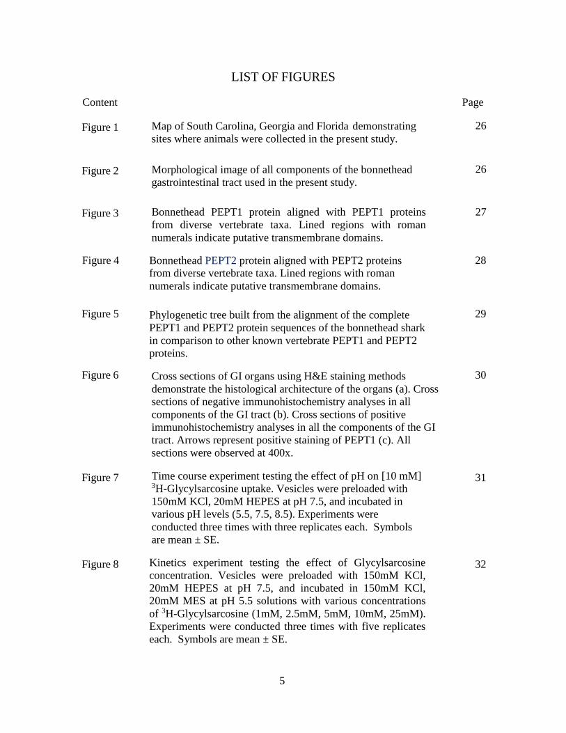
LIST OF FIGURES
Content Page
Figure 1 Map of South Carolina, Georgia and Florida demonstrating
sites where animals were collected in the present study.
26
Figure 2 Morphological image of all components of the bonnethead
gastrointestinal tract used in the present study.
26
Figure 3 Bonnethead PEPT1 protein aligned with PEPT1 proteins
from diverse vertebrate taxa. Lined regions with roman
numerals indicate putative transmembrane domains.
27
Figure 4 Bonnethead PEPT2 protein aligned with PEPT2 proteins
from diverse vertebrate taxa. Lined regions with roman
numerals indicate putative transmembrane domains.
28
Figure 5 Phylogenetic tree built from the alignment of the complete
PEPT1 and PEPT2 protein sequences of the bonnethead shark
in comparison to other known vertebrate PEPT1 and PEPT2
proteins.
29
Figure 6 Cross sections of GI organs using H&E staining methods
demonstrate the histological architecture of the organs (a). Cross
sections of negative immunohistochemistry analyses in all
components of the GI tract (b). Cross sections of positive
immunohistochemistry analyses in all the components of the GI
tract. Arrows represent positive staining of PEPT1 (c). All
sections were observed at 400x.
30
Figure 7 Time course experiment testing the effect of pH on [10 mM] 3H-Glycylsarcosine uptake. Vesicles were preloaded with
150mM KCl, 20mM HEPES at pH 7.5, and incubated in
various pH levels (5.5, 7.5, 8.5). Experiments were
conducted three times with three replicates each. Symbols
are mean ± SE.
31
Figure 8 Kinetics experiment testing the effect of Glycylsarcosine
concentration. Vesicles were preloaded with 150mM KCl,
20mM HEPES at pH 7.5, and incubated in 150mM KCl,
20mM MES at pH 5.5 solutions with various concentrations
of 3H-Glycylsarcosine (1mM, 2.5mM, 5mM, 10mM, 25mM).
Experiments were conducted three times with five replicates
each. Symbols are mean ± SE.
32
6
ABSTRACT
Many elasmobranchs are considered top predators with worldwide distribution, and in general
these fish play an important role in the transfer of energy from the lower to the upper trophic
levels within the marine ecosystem. Despite this, little research has been done regarding the rates
of prey ingestion, digestion, and the processes of energy and nutrient absorption. Specifically
understudied is enzymatic digestion within the intestinal brush border, which functions to break
down macromolecules into smaller subunits for luminal absorption across the gastrointestinal
epithelium. Given their carnivorous diet, the present study sought to expand knowledge on
nutrient intake in elasmobranchs by focusing on the uptake of products of protein metabolism. To
accomplish this, sequence encoding Peptide Transporter 1 (PepT1), a protein found within the
brush border membrane (BBM) of higher vertebrates that is responsible for the translocation and
absorption of small peptides released during digestion by luminal and membrane-bound
proteases, was molecularly identified in the bonnethead shark (Sphyrna tiburo) using degenerate
primers based on conserved portions of known PEPT1 sequences from other vertebrates.
Sequence encoding Peptide Transporter 2 (PepT2) was also isolated from the S. tiburo scroll
valve intestine using the same methodology. PepT1 was then localized using
immunocytochemistry with rabbit polyclonal anti-rat PEPT1 in the esophagus, stomach,
duodenum, scroll valve intestine, rectum, and pancreas. Vesicle studies were used to identify the
apparent affinity of the transporter, and to quantify the rate of uptake by its H+-dependent
cotransporter properties, using 3H-glycylsarcosine as a model dipeptide. The results of this study
provide insight into the rate and properties of food passage within S. tiburo, and can lead to
7
future work on topics such as physiological regulation of protein metabolism and absorption and
how it may vary in elasmobranchs that exhibit different feeding strategies.
Key Words: Elasmobranch, Peptide transporter 1, Peptide transporter 2, scroll valve intestine,
gastrointestinal tract, absorption.
8
INTRODUCTION
Sharks, skates and rays form a group of saltwater- and freshwater-dwelling fish called
elasmobranchs. This group represents primitive vertebrates that evolved at least 400 million
years ago, and are considered to be some of the first jawed vertebrates (Moy-Thomas 1938;
Maisey 1980; Wilga et al. 2001). The jaw is made up of a series of homologous branchial arches
as part of the visceral skeleton, which has acquired the function of biting (Moy-Thomas 1938).
This morphological and functional development led to more complex feeding mechanisms and
allowed for a shift in diet (Moy-Thomas 1938; Wilga et al. 2001). This ability to consume
comparatively larger and more nutrient-valuable prey called for absorptive modifications within
the gastrointestinal system (Holmgren 1999; Wilga et al. 2001). As seen in agnathans (jawless
vertebrates) the gastrointestinal tract is made up of an esophagus and a gut tube (intestine) that
leads to the cloaca. Comparatively however, the gnathostome (jawed vertebrate) GI tract such as
that of elasmobranchs is more complex and consists of an esophagus, stomach, duodenum,
spiral/scroll valve intestine, and colon (Fishbeck et al. 2008).
The elasmobranch spiral/scroll valve intestine is a unique organ that increases surface area
without increasing length by the infolding of the mucosa and submucosa. It is commonly thought
that due to the presence of this organ elasmobranchs undergo slow food passage, which is often
associated with a low rate of consumption, therefore limiting growth and reproductive rates. A
study on juvenile lemon sharks examined digestive capability using an indirect method via an
inert, naturally occurring marker in their food (Cortes and Gruber 1990). The authors found that
lemon sharks are capable of absorbing energy with an average efficiency of about 80%, which is
9
similar to the absorption efficiency of a carnivorous teleost. However, the rate of digestion was
prolonged in comparison, taking between 70 to 100 hours (Cortes and Gruber 1990). Feeding
frequency has also been examined in various species of sharks, and it has been found that the rate
of food passage varies among species (Bush 2002; Lowe 2001; Wetherbee et al. 2004). Species
such as the spiny dogfish are known to gorge themselves every 10 to 16 days (Wetherbee et al.
2004). Conversely, the scalloped hammerhead feeds much more frequently ranging from every
10 to 11 hours (Bush 2002; Lowe 2001). Moreover, although all elasmobranchs are carnivores,
the diet across species can range from microscopic phytoplankton to large pelagic fish. A
number of studies have used quantitative diet analysis to understand what elasmobranchs
consume and different predator-prey interactions important to sharks (Bethea et al. 2007; Cortes
et al. 2006; NMFS 1999). Through such research and different technological advancements
current research has been able to estimate species-specific metabolic rates for a number of
elasmobranch species, and such information can be used to better grasp bioenergetics (Carlson et
al. 2004). However most fish, including sharks, do not initially break down their food prior to
ingestion into the gastrointestinal tract. Thus, it is important to go beyond diet itself, to
understand the chemical means of digestion and absorption (Papastamatiou and Lowe 2004;
Papastamatiou and Lowe 2005; Clements and Raubenheimer 2006; German 2011). For example,
Jhaveri et al. (2015) examined digestive enzyme activity along the gut of the bonnethead shark
(Sphyrna tiburo). This study specifically looked at what compounds the bonnethead shark could
digest and applied this information to infer the digestive strategy within the hindguts of S. tiburo.
Results demonstrated that gut content was concentrated more within the intestinal region
(duodenum, spiral valve intestine, and colon), with greater concentrations within the spiral valve
intestine in comparison to the duodenum and colon. They also found that pancreatic enzyme
10
activity was elevated in the duodenum and spiral valve, whereas brush border membrane (BBM)
enzymatic activity peaked in the spiral valve and colon. However, microbial enzymes were
highest on or within the colon. From such results, they inferred that the spiral valve intestine is
the most active, absorptive section of the shark intestinal region. An additional/final conclusion
was that the bonnethead shark has adapted a yield-maximizing digestive strategy, meaning that
they consume relatively large meals infrequently and thus have enzymatic patterns as described
above (Jhaveri et al. 2015).
Although diet composition and general aspects of enzymatic digestion within the elasmobranch
gut have been studied, studies regarding the breakdown of dietary macromolecules and their
absorption across intestinal epithelium within the BBM are significantly lacking.
Microscopically, the epithelial cells and microvilli that make up the BBM in the elasmobranch
GI tract are remarkably similar to those of the mammalian and teleost gastrointestinal tracts
(Crane 1978). Based on previous studies, it is known that the mammalian BBM increases surface
area for nutritive absorption, and contains enzymes near transporters that facilitate the absorption
of digested nutrients into the intestine (Schmitz et al. 1973; Hauser et al. 1980). However, in
regards to elasmobranchs, little information is available about the functional properties of the
components of the gastrointestinal tract. In one of the few studies on this topic, Crane (1979)
demonstrated the presence of potential BBM D-glucose transporters within small dogfish
(Scyliorhinus canicula) spiral valve intestine. That study showed that glucose uptake by S.
canicula spiral valve BBM is stimulated by a Na+ gradient and inhibited by the monosaccharide
α- methylglucoside, an alternative substrate for glucose transporters, or by phlorizin, a specific
inhibitor of intestinal glucose transporters. Such information provides insight into the cellular
11
mechanisms of the elasmobranch intestine, and similarities to the teleost and mammalian
digestive mechanisms.
Sharks have a carnivorous diet, and therefore protein absorption is an important aspect to
understand. However, in contrast to the modest research conducted on sugar uptake, there are no
publications regarding the mechanisms by which peptides cross the epithelial cells of the
spiral/scroll valve and other digestive organs. It is known that dietary proteins can be degraded
into free amino acids within the digestive tract. However, a great deal of studies provide
evidence that within the mammalian and teleost intestine, most dietary proteins are broken down
into small peptides rather than being broken down to single amino acids (Shimakura et al. 2006).
Thus intestinal peptide transport is an important aspect to understand and such knowledge will
provide further insight into the ability of sharks to absorb amino acids or short peptides for
energy usage, and also lead to further research on topics such as the physiological regulation of
protein metabolism and absorption (Matthews, 1975; Daniel, 2004).
Research has shown that peptide absorption is markedly influenced by an inwardly directed
proton gradient, stimulated by an inside-negative membrane potential and inhibited by an inside-
positive membrane potential; implying that the transport of peptides across a membrane is
associated with the transfer of positively charged ions (Leibach et al., 1996). Peptide transporter
1 (PEPT1 or SLC15A1) and Peptide transporter 2 (PEPT2 or SLC15A2) are both a critical
peptide transporters within the mammalian and teleost digestive and renal tracts that depend on
this driving force. These transporters have also been identified as part of the major facilitator
superfamily of 12 transmembrane domain transporter proteins. However, although these two
12
peptide transporters share similarities in function and structural characteristics, the distribution of
PEPT1 and PEPT2 differ. PEPT1 has been successfully identified and described in a number of
teleost and mammalian species within the gastrointestinal tract, rectum, gallbladder, pancreas,
nuclei of smooth muscle cells, the liver, and throughout the renal tract (Leibach and Ganapathy
1996; Verri et al. 2010). However, studies report that PEPT2 is predominately located in the
kidney BBM, brain, gonads, lungs, gallbladder, and liver (Leibach and Ganapathy 1996; Saito et
al. 1996). PEPT2 has not been detected in the mammalian or teleost intestinal tract. It is
important to note that studies have also shown that PEPT2 possesses a higher affinity than
PEPT1 for diverse dipeptides (Leibach and Ganapathy 1996). In 2003, Verri et al. confirmed the
presence of PepT1 in fish by successfully cloning and functionally characterizing it within the
Danio rerio intestinal BBM. Specifically, PEPT1 is a Na+- independent, H+ - dependent
cotransporter protein present in the intestinal BBM that is responsible for the translocation and
absorption of di- and tripeptides released during digestion by luminal and membrane- bound
proteases.
As there are consistencies seen in the distribution of PEPT1 and PEPT2 across vertebrates
overall, we expect to find PepT1 to have the same range of expression in elasmobranchs,
specifically in the spiral valve intestine and other GI organs. The purpose of this study was to
molecularly identify the presence of PepT1 within the elasmobranch spiral valve intestine and to
histologically assess the distribution of this transporter in all components of the GI tract and the
pancreas. In addition, this study will report the uptake capabilities of the intestinal peptide
transporter. For this particular study bonnethead sharks (Sphyrna tiburo) were used as the model
species due to their abundance and availability.
13
METHODOLOGY
Sample collection
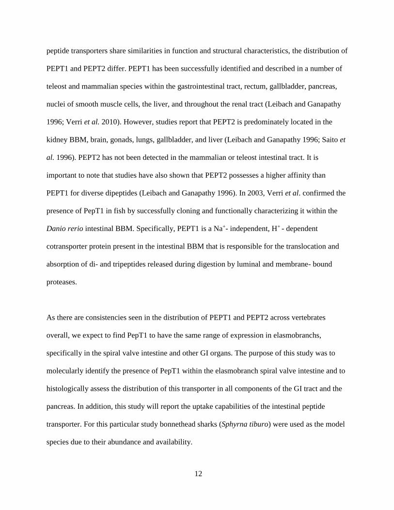
S. tiburo were collected from sites ranging from Charleston, South Carolina to Cape Canaveral,
Florida (Fig. 1) using gillnet and longline fishing. Animals were euthanatized using IACUC-
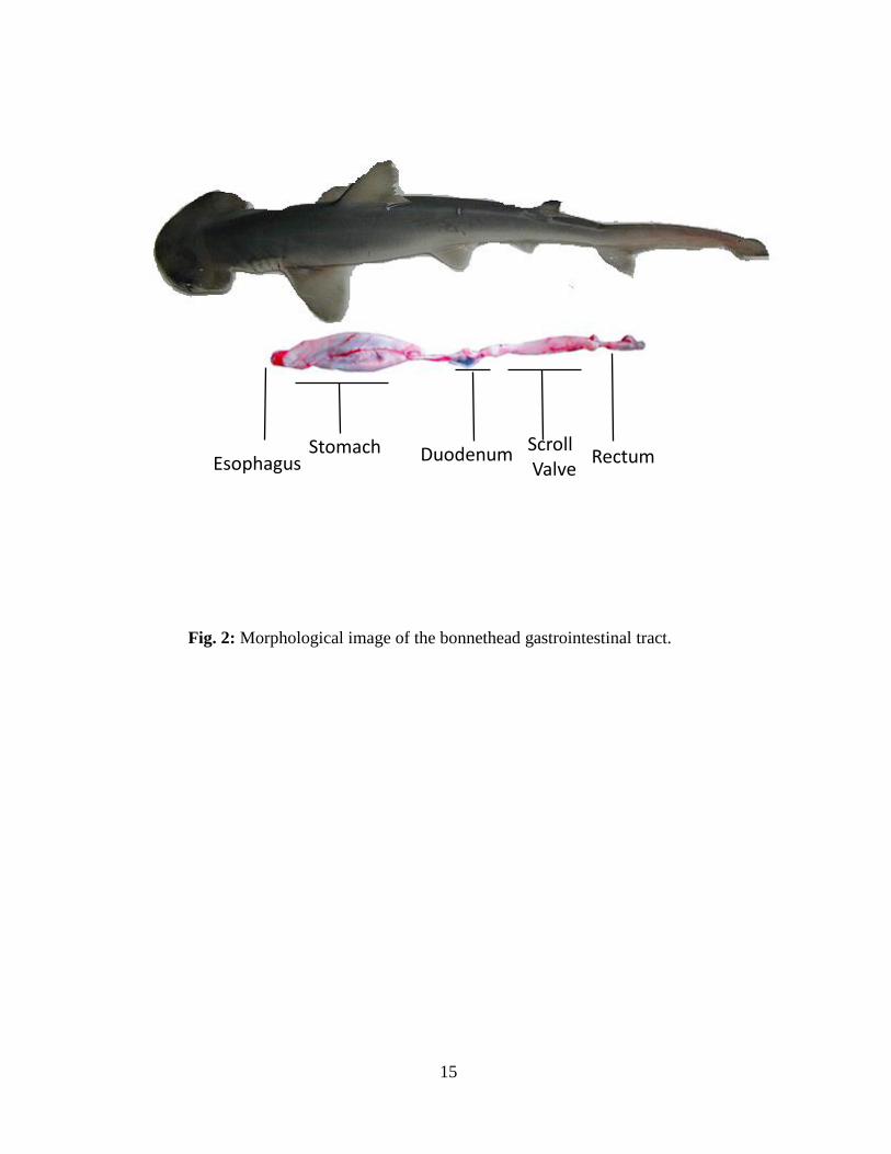
approved methodology, and the gastrointestinal tract was obtained by dissection (Fig. 2). Small
portions of the scroll valve intestine were sampled and stored in RNAlater at -4˚C for use in
molecular studies. Samples (~2-3 mm) of the gastrointestinal tract and accessory organs
(esophagus, stomach, pancreas, duodenum, scroll valve intestine, and rectum) were also obtained
and fixed in 10% formalin in elasmobranch-modified saline for about 48 hrs., then rinsed and
transferred to 70% ethanol for long-term storage until used for histology and
immunocytochemistry. Last, whole intestines were obtained from some animals and stored at -
80˚C until used for vesicle experiments.
14
Fig. 1: Map of South Carolina, Georgia and Florida demonstrating sites where animals
(representative of blue circles) were collected in the present study. Animals were collected
mainly in coastal waters of Charleston, SC, Jacksonville, FL, and Cape Canaveral, FL.
15
Fig. 2: Morphological image of the bonnethead gastrointestinal tract.
Esophagus Stomach Duodenum
Scroll Valve
Rectum
16
Molecular Identification
1. Isolation of S. tiburo pept1 and pept2 cDNA sequences
To isolate total RNA, approximately 50 mg of scroll valve intestine was minced, placed in 750
µL of Trizol reagent and homogenized. The homogenate was centrifuged for 1 minute at 12,000
x g to pellet any remaining solid tissue, with 700 µl of the supernatant subsequently placed into a
microcentrifuge tube. The Direct-Zol RNA Miniprep kit (Zymo Research, Irvine, CA) was then
used for total RNA extraction. The concentration and purity of RNA was determined using a
NanoDrop spectrophotometer and gel electrophoresis. Reverse transcription of 1 µg of total
RNA into cDNA was conducted using Superscript III reverse transcriptase (Life Technologies)
following the manufacturer’s instructions.
For degenerate PCR, degenerate primers were designed based on conserved portions of known
PEPT1 sequences from chicken (NM_204365.1), salmon (NM_001146692.1), eel (AB7762417),
and zebrafish (NM_198064). Four combinations from the mentioned taxa were created, two
different forward and two different reverse primers, to maximize the possibility of at least one
combination annealing to bonnethead cDNA and amplifying a portion of pept1. PCR was
performed using six different cycling parameters, to increase the range of annealing temperatures
tested. Cycling parameters included: 95˚C for 2 min followed by initial annealing temperatures
of 52.5, 53.3, 54.9, 57.2, 60.1 or 62.5 (a different starting temperature for each reaction) for 30
sec and then 72 ˚C for 1 min. For the first 25 cycles, the annealing temperature was decreased
each cycle by 0.5˚C, followed by 15 cycles (for 40 total) at an annealing temperature of 40 ˚C for
all reactions. Following the PCR a 1% gel electrophoresis was run to determine the optimal
17
temperature and combination of primers, with products of the appropriate size isolated, cloned
and sequenced as described below.
The primers that yielded the largest product were 5’-GAGTTCTGYGARMGDTTCTCCTACT-
3’ as the forward primer and 5’-TGGTCAAANARDGYCCAGAACAT-3’ as the reverse primer.
Products from PCR reactions using this primer combination were ligated into the pGEM-T
vector (Promega) and used to transform competent Escherichia coli. Twenty-five µl of
transformed cells were then plated on agar plates coated with isopropyl-beta-D-
thiogalactopyranoside (IPTG) and 5-bromo-4-chloro-3-indolyl-beta-D-galactopyranoside (X-gal)
for blue-white screening and incubated at 37 ˚C overnight. White (positive) colonies were then
selected, placed in 2 mL of LB broth and incubated overnight at 37˚C with shaking at 250 rpm.
Overnight incubations were transferred into capped tubes and centrifuged at high speed for about
30 sec. The supernatant was poured out and the pellet then resuspended in 600 µl of sterile water
The Zyppy Plasmid Miniprep Kit (Zymo Research) was then used to isolate plasmid DNA from
for sequencing preparation. Selected plasmids were then sequenced by Genewiz, and sequences
were analyzed using the CLC Main Workbench 7 software (CLCbio, Qiagen). Although the
target cDNA was pept1, the methods described above yielded clones positive for both pept1 and
pept2 sequences, likely due to high sequence similarity; therefore full length cDNA sequences
were obtained for both genes using rapid amplification of cDNA ends (RACE).
To obtain the remaining 3’ and 5’ coding sequence as well as the 5’ and 3’ untranslated regions
of pept1 and pept2, the FirstChoice RLM-RACE kit (Life Technologies) was used. Total RNA
from scroll valve intestine was extracted as described above, and 5’ and 3’ RACE-ready cDNA
was then generated following the manufacturer’s instructions. 5’ and 3’ RACE PCR products
were then obtained using nested PCR reactions that included kit-specific primers and species-

18
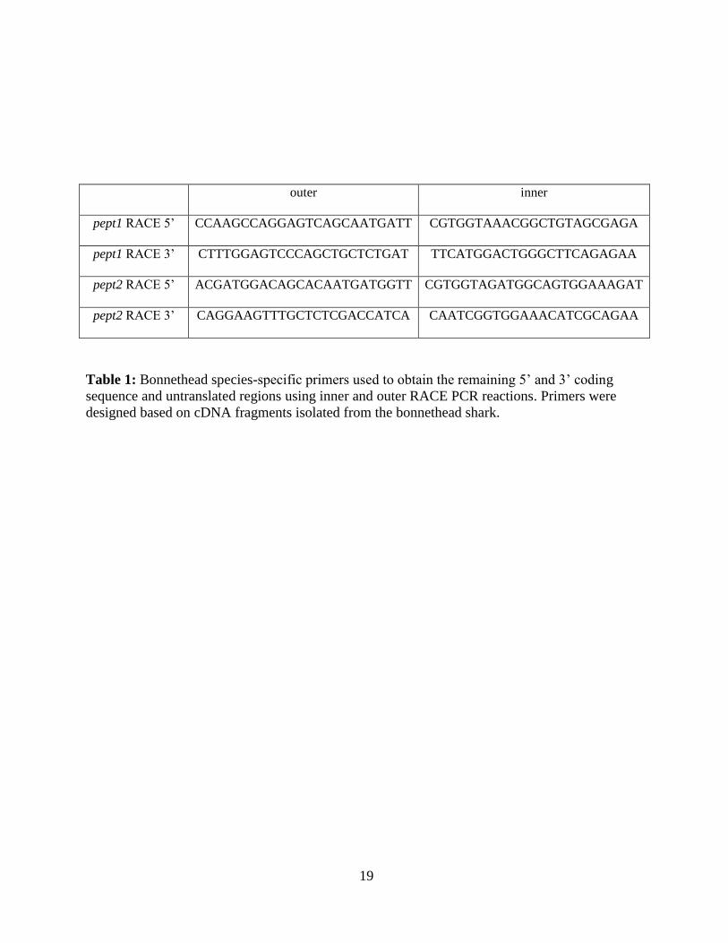
specific primers (Table 1) for pept1 and pept2 designed using the cDNA fragments isolated as
described above. RACE products were then cloned and sequenced as described above, and
sequences were assembled using CLC Main Workbench 7.
19
outer inner
pept1 RACE 5’ CCAAGCCAGGAGTCAGCAATGATT CGTGGTAAACGGCTGTAGCGAGA
pept1 RACE 3’ CTTTGGAGTCCCAGCTGCTCTGAT TTCATGGACTGGGCTTCAGAGAA
pept2 RACE 5’ ACGATGGACAGCACAATGATGGTT CGTGGTAGATGGCAGTGGAAAGAT
pept2 RACE 3’ CAGGAAGTTTGCTCTCGACCATCA CAATCGGTGGAAACATCGCAGAA
Table 1: Bonnethead species-specific primers used to obtain the remaining 5’ and 3’ coding
sequence and untranslated regions using inner and outer RACE PCR reactions. Primers were
designed based on cDNA fragments isolated from the bonnethead shark.
20
2. Sequence and phylogenetic analyses
BLAST analysis was used to confirm the identity of the presumed S. tiburo PepT1 and PepT2
protein sequences (http://www.ncbi.nlm.nih.gov/BLAST). S. tiburo sequences were then aligned
with other elasmobranchs, teleost, and higher vertebrates using the Clustal W algorithm in CLC
Main Workbench for analysis of gene homology (Figures 3 and 4). Potential transmembrane
domains for both pept1 and pept2 were defined using TMprep computational resources
(http://www.bioinformatics.utep.edu/BIMER/tools/transmembrane.html). For phylogenetic
analysis, both S. tiburo protein sequences were aligned with PEPT1 and PEPT2 sequences from
diverse vertebrate taxa using the Clustal W algorithm in MEGA version 6 (Tamura et al. 2013);
phylogenetic relationships were then determined using the Neighbor-Joining method with 2000
iterations to generate a bootstrap consensus tree (Figure 5).
Histology
Fixed tissue samples were processed for routine paraffin histology as described by Gelsleichter
et al. (2003). Once embedded in paraffin, samples were sectioned (5 µm) using a rotary
microtome and mounted on poly-L-lysine-coated slides. They were then stained with Harris
hematoxylin and eosin to examine general cellular architecture.
Immunocytochemistry
Gastrointestinal tract and accessory organ samples were fixed and sectioned as described above.
Immunocytochemistry was then performed to examine the cellular location of PEPT1 using a
rabbit polyclonal anti-rat PEPT1 (SLC15a1, Millipore, Berlin, Germany) and the Vector
ImmPRESS anti-rabbit kit (Vector Laboratories, Burlingame, CA). Tissue sections were
21
incubated in a limonene-based solvent for deparaffinization, rehydrated by submerging in a
descending series of graded alcohol concentrations (100%- 95%), and then rinsed for 10 minutes
in a running tap water bath. Sections were then incubated in an antigen retrieval solution (10 mM
sodium citrate, pH 6.0) at 95˚C for 20 minutes to expose any epitopes of the target antigen that
may have been masked by the fixation process. Sections were then removed from the bath and
brought to room temperature. They were then rinsed in reverse osmosis (RO) water, followed by
phosphate buffered saline (PBS), and then blocked for nonspecific reactivity with primary
antibodies by overnight incubation in 2% normal goat serum in PBS (Vector laboratories) at 4˚C .
After blocking, slides were washed with PBS and endogenous peroxidase activity was then
quenched by incubation in a 1:1 mixture of 3% hydrogen peroxide and methanol for 15 minutes.
Sections were then rinsed again in two separate baths of PBS. Sections were then incubated
overnight in primary antibody diluted 1/1000 in a PBS solution containing 0.1% gelatin and
0.1% sodium azide (G-PBS). Negative control sections were incubated in diluent only.
Following incubation, sections were rinsed in a PBS bath containing 0.05% TWEEN-20 (PBS-
T), followed by two additional PBS rinses. Afterwards, slides were incubated for approximately
30 minutes with secondary antibody, anti-rabbit Ig. Following this incubation, sections were
rinsed in three separate PBS baths and then incubated in the chromogen 3.3’-diaminobenzidine
(DAB), using the ImmPACT DAB Peroxidase (HRP) substrate kit. Slides were rinsed in tap
water and then counterstained in 2% methyl green (Vector laboratories) for 15-60 minutes at
37˚C. Afterwards, they were rinsed briefly in tap water, dehydrated in an ascending series of
graded alcohols (95%-100%), cleared using limonene-based solvent, and then mounted using
Cytoseal 60 (Thermo Scientific, Fair Lawn, NJ). Microscopy was then used to examine the
distribution of immunoreactive PEPT1 in the bonnethead digestive system.
22
Vesicle experiments
1. Preparation of Brush Border Membrane Vesicles:
Whole intestines stored at -80˚C were thawed on ice in PBS. Subsamples of intestine were then
cut and scraped using a razor blade to free epithelial cells into 60 mL of a 300 mM mannitol, 20
mM Tris HCl, 50 mM EGTA, 1 mM PMSF buffer adjusted to pH 7.0 (buffer 1). For Brush
Border Membrane Vesicle (BBMV) purification, an experimental design used on Mozambique
tilapia (Oreochromis mossambicus) intestine (Thamotharan et al. 1996) was implemented on S.
tiburo. Similar methodology was used by Crane et al. (1979), showing that elasmobranch BBMV
can be isolated using this approach. The expanded technique has two additional washing steps
for discarding stored cytoplasmic digestive enzymes (Thamotharan et al. 1996). Purified BBMV
were then used for transport measurements.
2. Transport measurements
Transport experiments were conducted using the scroll intestine BBMV and the Millipore
filtration technique (Thamotharan et al. 1996). [3H] Glycylsarcosine (Moravek Biochemicals,
Brea, California 92821) (Gly-Sar) was used in 120 min uptake experiments by mixing 20 µL of
membrane suspension with 180 µL of radiolabelled incubation medium. Composition of
incubation medium varied with the nature of each experiment. Effect of pH on uptake of
radiolabeled Gly-Sar in intestinal BBMV was examined using incubation media made up of 150
mM KCl and 20 mM HEPES adjusted to a pH of 7.5 or a pH of 8.5, and a third medium
contained 150 mM KCl and 20 mM MES adjusted to pH 5.5. Measurements were taken at
intervals of 0.25, 1, 2, 5, 10, 60, and 120 min.
Kinetics of [3H] Gly-Sar influx at 1 min was then examined using media composed of 150 mM
KCl, 20 mM HEPES at concentrations of 1, 2.5, 5, 10, and 25 mM Gly-Sar.
23
In these two separate experiments, uptake of [3H] Gly-Sar was terminated by injecting 20 µL of
the reaction into 2 mL of a stop solution (composed of same solution as the incubation medium
without the radiolabelled dipeptide). The solution was then filtered using a Millipore filter (0.65
µm pore diameter) and washed with an additional 3 mL of stop solution. Filters were then placed
in 3 mL of Beckman Volume scintillation cocktail and counted using a Beckman LS-6100
scintillation spectrometer. The average of each set of replicates was determined and graphed in
SigmaPlot10.0. A one-way ANOVA was then ran to statistically test the significance between
the mean values of GlySar uptake among the given ion gradients where the overshoot is present.
24
RESULTS
Sequence analysis
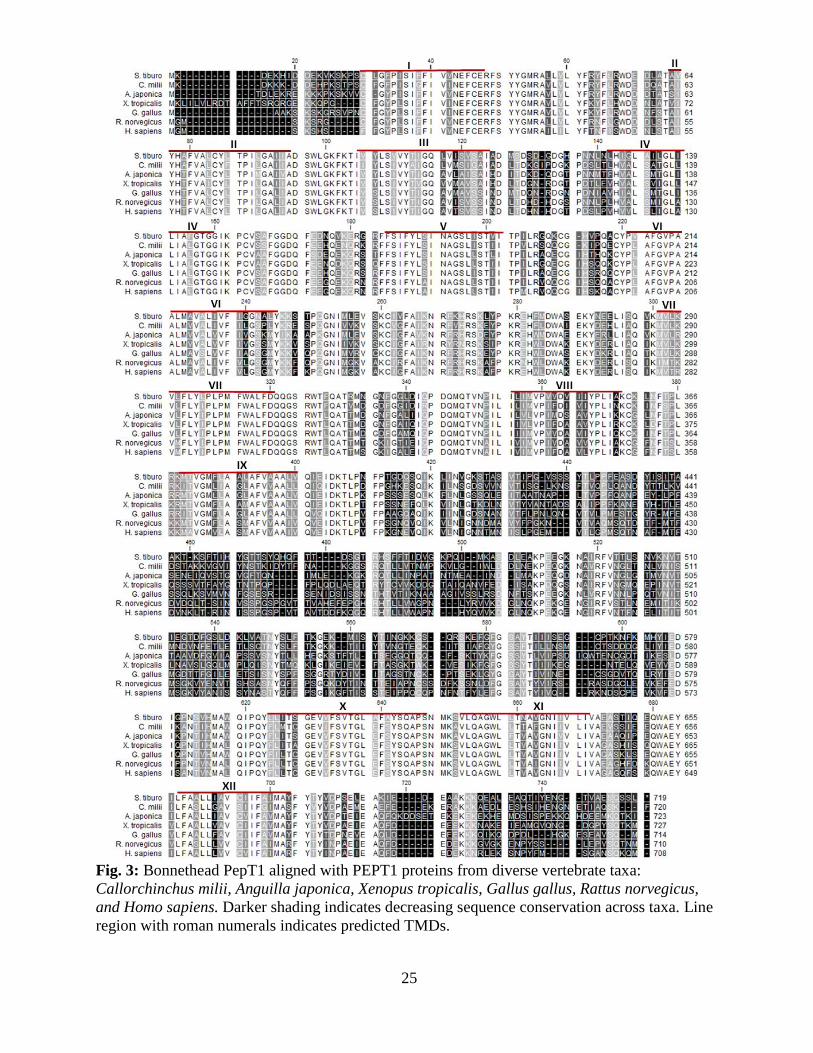
The bonnethead pept1 cDNA was 4,055 bp, with an ORF of 2,157 bp encoding a putative protein
of 718 aa. The sequence also included 455 bp of 5’ untranslated region (UTR) sequence and
1,443 bp of 3’UTR sequence with a polyadenosine mRNA tail. Hydropathy analysis predicts 12
potential transmembrane domains (TMD) with an extracellular loop between TMD IX and X
(Fig. 3).When compared to previously characterized PEPT1 sequences from other vertebrates
using BLAST, the predicted bonnethead sequence shows high sequence identity, ranging from
60 to 67.3 percent.
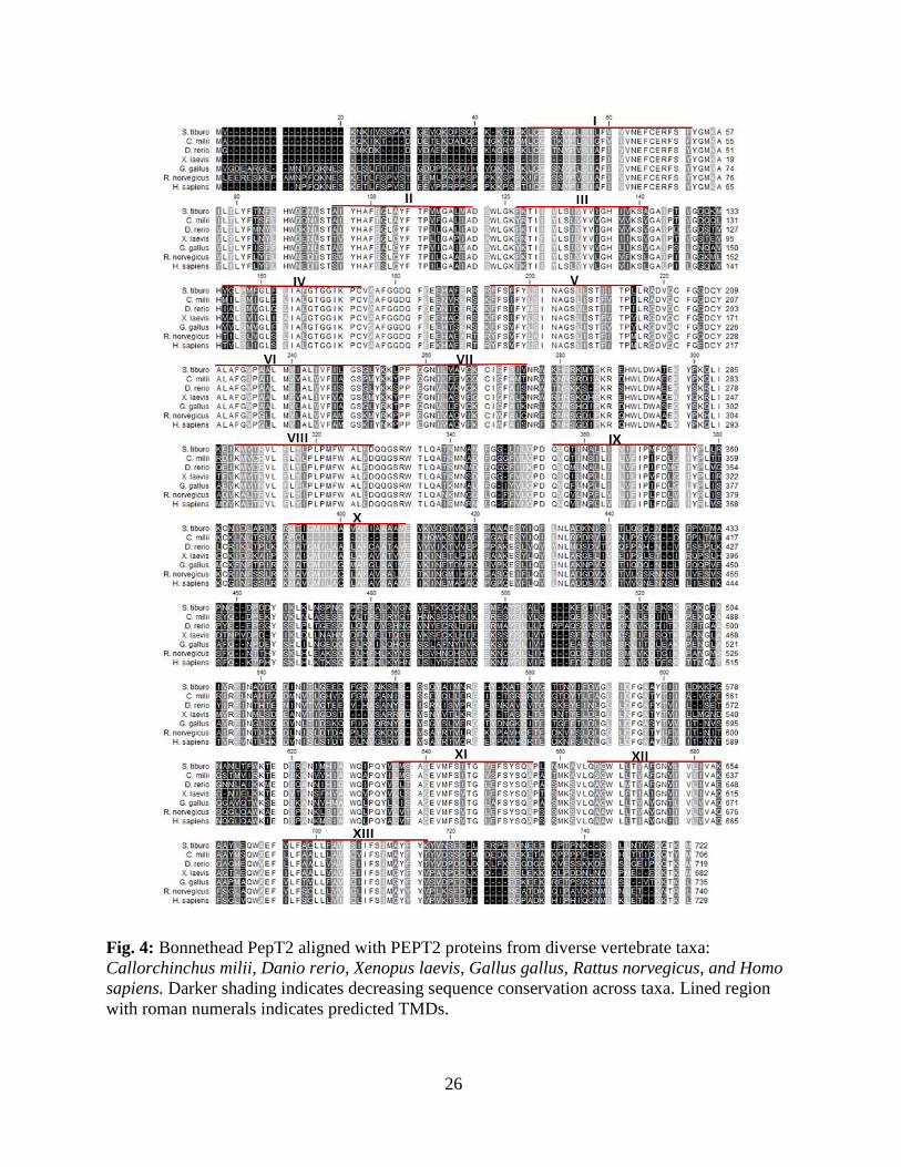
The pept2 cDNA was also isolated from the bonnethead shark intestine. The sequence was 2,549
bp, with an ORF of 2,169 bp encoding a putative protein of 722 aa. The sequence included 91 bp
of 5’ UTR sequence and 289 bp of 3’UTR sequence with a polyadenosine mRNA tail.
Hydropathy analysis predicts 13 potential TMD with an extracellular loop between TMD X and
XI (Fig. 4). When compared to previously characterized PEPT2 sequences from other species
using BLAST, the predicted bonnethead sequence shows high sequence identity, ranging from
61.8 to 66 percent.
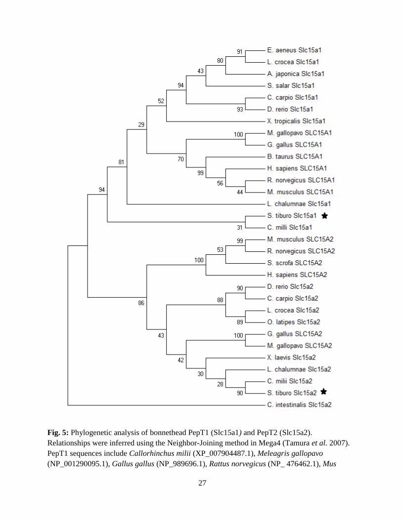
Percent identity between the two isolated intestinal peptide transporters was only 51%. Also, the
phylogenetic reconstruction of the vertebrate peptide transporter proteins assigned the two
isolated bonnethead shark peptide sequences to separate clades and within the PEPT1 and
PEPT2 branches of the phylogenetic tree, which also demonstrated early divergence of the
elasmobranch protein sequences from those of the teleost and mammalian groups (Fig. 5).
25
Fig. 3: Bonnethead PepT1 aligned with PEPT1 proteins from diverse vertebrate taxa:
Callorchinchus milii, Anguilla japonica, Xenopus tropicalis, Gallus gallus, Rattus norvegicus,
and Homo sapiens. Darker shading indicates decreasing sequence conservation across taxa. Line
region with roman numerals indicates predicted TMDs.
26
Fig. 4: Bonnethead PepT2 aligned with PEPT2 proteins from diverse vertebrate taxa:
Callorchinchus milii, Danio rerio, Xenopus laevis, Gallus gallus, Rattus norvegicus, and Homo
sapiens. Darker shading indicates decreasing sequence conservation across taxa. Lined region
with roman numerals indicates predicted TMDs.
27
Fig. 5: Phylogenetic analysis of bonnethead PepT1 (Slc15a1) and PepT2 (Slc15a2).
Relationships were inferred using the Neighbor-Joining method in Mega4 (Tamura et al. 2007).
PepT1 sequences include Callorhinchus milii (XP_007904487.1), Meleagris gallopavo
(NP_001290095.1), Gallus gallus (NP_989696.1), Rattus norvegicus (NP_ 476462.1), Mus
28
musculus (NP_444309.2), Bos taurus (NP_001092848.1), Homo sapiens (NP_005064.1)
Latimeria chalumnae (XP_005992366.1) Anguilla japonica (BAM67012.1), Xenopus tropicalis
(XP_002935692.2), Cyprinus carpio (AEX13747.1), Epinephelus aeneus (AFP33141.1),
Larimichthys crocea (NP_001290295.1), Danio rerio (NP_932330.1), and Salmo salar
(NP_001140154.1). PepT2 sequences include Mus musculus (NP_067276.2), Homo sapiens
(NP_066568.3), Gallus gallus (AGZ02797.1), Danio rerio (NP_001034917.1), Xenopus laevis
(NP_001080398.1), Larimichthys crocea (KKF11892.1), Oryzias latipes (XP_004081581.1),
Cyprinus carpio (ADM48102.1), Latimeria chalumnae (XP_006004055.1), Sus scrofa
(NP_001090983.1), Rattus norvegicus (NP_113860.2), Meleagris gallopavo (XP_010712188.1),
and Callorhinchus milii (XP_007907469.1). The tree is rooted using the PepT2 sequence from
Ciona intestinalis (XP_002121251.1). Numbers at branch points indicate the percentage of 2000
bootstrap replicates supporting the division.
29
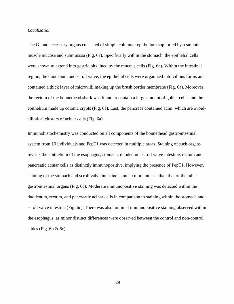
Localization
The GI and accessory organs consisted of simple columnar epithelium supported by a smooth
muscle mucosa and submucosa (Fig. 6a). Specifically within the stomach, the epithelial cells
were shown to extend into gastric pits lined by the mucous cells (Fig. 6a). Within the intestinal
region, the duodenum and scroll valve, the epithelial cells were organized into villous forms and
contained a thick layer of microvilli making up the brush border membrane (Fig. 6a). Moreover,
the rectum of the bonnethead shark was found to contain a large amount of goblet cells, and the
epithelium made up colonic crypts (Fig. 6a). Last, the pancreas contained acini, which are ovoid-
elliptical clusters of acinar cells (Fig. 6a).
Immunohistochemistry was conducted on all components of the bonnethead gastrointestinal
system from 10 individuals and PepT1 was detected in multiple areas. Staining of such organs
reveals the epithelium of the esophagus, stomach, duodenum, scroll valve intestine, rectum and
pancreatic acinar cells as distinctly immunopositive, implying the presence of PepT1. However,
staining of the stomach and scroll valve intestine is much more intense than that of the other
gastrointestinal organs (Fig. 6c). Moderate immunopositive staining was detected within the
duodenum, rectum, and pancreatic acinar cells in comparison to staining within the stomach and
scroll valve intestine (Fig. 6c). There was also minimal immunopositive staining observed within
the esophagus, as minor distinct differences were observed between the control and non-control
slides (Fig. 6b & 6c).
30
a b c
ESO
PH
AG
US
PA
NC
REA
S
REC
TUM
DU
OD
ENU
M
SCR
OLL
VA
LVE
INTE
STIN
E
E
STO
MA
CH
31
Fig. 6: Cross sections of GI organs using H&E staining methods demonstrate the histological
architecture of the organs (column a). Cross sections of negative immunohistochemistry analyses
in all components of the GI tract (column b). Cross sections of positive immunohistochemistry
analyses in all the components of the GI tract. Arrows represent positive staining of PepT1
(column c). All sections were observed at 400x.
32
Function
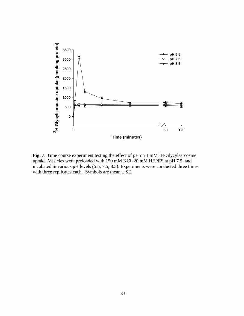
Figure 7 illustrates the effects of a pH gradient on the time course of 1mM [3H] Glycylsarcosine
uptake by the bonnethead scroll valve BBMV. This time course was characterized by an
overshoot at 1 minute that was highest when the pH inside was 7.5 and the pH outside was 5.5
(Fig. 7). No overshoots were seen when the outside pH was 7.5 or 8.5. Overshoots in the
presence of proton gradient suggest that the transmembrane concentration gradient of hydrogen
ions provides the driving force for the uptake of peptides by the scroll valve brush border
membrane vesicles.
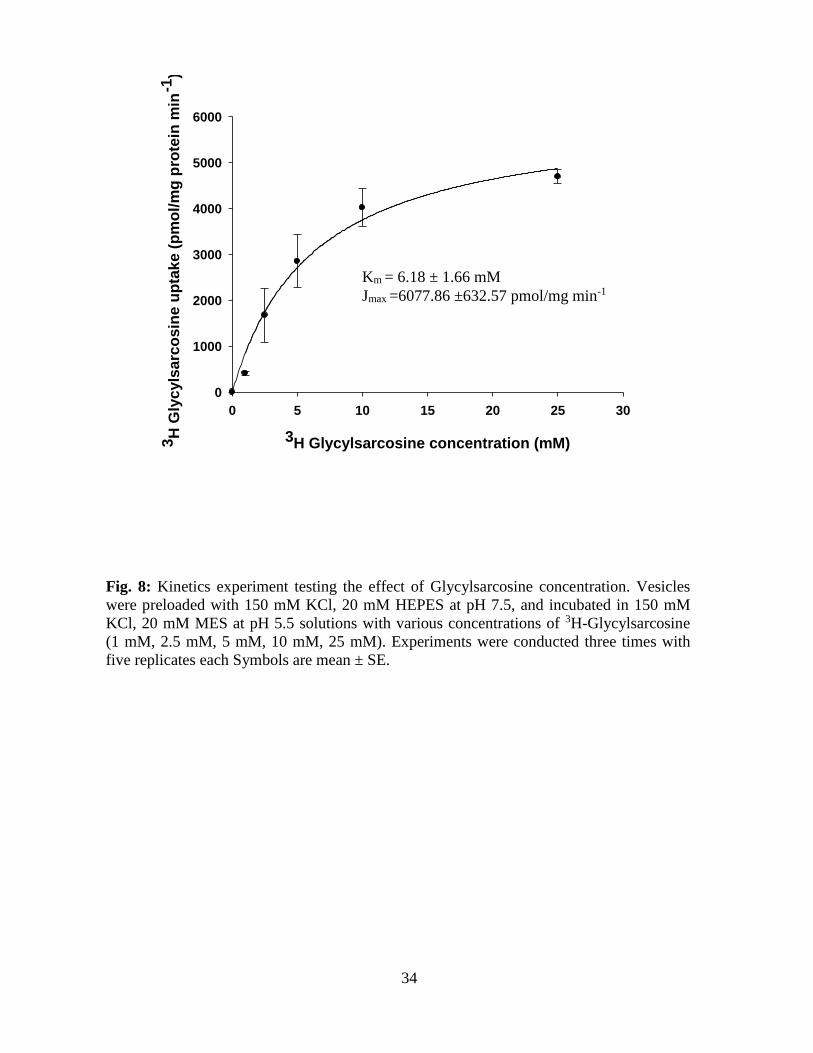
Influxes (1 min uptakes) of [3H] Gly-Sar into the scroll valve BBMVs were measured over a
concentration range of 1- 25mM [3H] Gly-Sar in the presence of an inwardly-directed proton
gradient (inside pH 7.5 and outside pH 5.5). Influxes for this concentration range were used in
the Michaelis-Menten kinetics equation (Joi ={(Jmax * [S])/(Km + [S])}), where Joi is peptide
influx, Jmax is maximal influx, Km is the Gly-Sar concentration at ½ Jmax, and S is Gly-Sar
concentration. This analysis yielded Michaelis- Menten values for a saturable, low- affinity
system with a Km = 6.18 ±1.66 mM and a Jmax = 6077.86 ± 632.57 pmol/ mg protein per 1 min
(n=3).
33
Fig. 7: Time course experiment testing the effect of pH on 1 mM 3H-Glycylsarcosine
uptake. Vesicles were preloaded with 150 mM KCl, 20 mM HEPES at pH 7.5, and
incubated in various pH levels (5.5, 7.5, 8.5). Experiments were conducted three times
with three replicates each. Symbols are mean ± SE.
Time (minutes)
0 60 1203H
-Gly
cy
lsa
rco
sin
e u
pta
ke
(p
mo
l/m
g p
rote
in)
0
500
1000
1500
2000
2500
3000
3500
pH 5.5
pH 7.5
pH 8.5
34
Fig. 8: Kinetics experiment testing the effect of Glycylsarcosine concentration. Vesicles
were preloaded with 150 mM KCl, 20 mM HEPES at pH 7.5, and incubated in 150 mM
KCl, 20 mM MES at pH 5.5 solutions with various concentrations of 3H-Glycylsarcosine
(1 mM, 2.5 mM, 5 mM, 10 mM, 25 mM). Experiments were conducted three times with
five replicates each Symbols are mean ± SE.
3H Glycylsarcosine concentration (mM)
0 5 10 15 20 25 30
3H
Gly
cyls
arc
osin
e u
pta
ke (
pm
ol/
mg
pro
tein
min
-1)
0
1000
2000
3000
4000
5000
6000
Km = 6.18 ± 1.66 mM
Jmax =6077.86 ±632.57 pmol/mg min-1
35
DISCUSSION
Multiple aspects of this study, including the molecular identification, immunohistochemistry,
and BBMV experiments support the identification of cDNAs encoding two individual and
functional S. tiburo peptide transporters of the PTR family. The two encoded proteins,
designated as PepT1 and PepT2, represent the products of orthologous genes in other vertebrate
species. When compared to other known members of the PTR family in vertebrates, the
predicted bonnethead PepT1 and PepT2 share significantly higher overall identity to other
known PEPT1 and PEPT2 sequences, respectively, and are assigned to the expected
monophyletic groups of the reconstructed phylogenetic tree. The analysis of domains also
reveals that the bonnethead PepT1, like other vertebrate PEPT1 proteins, includes 12 TMDs and
the overall major area of difference lies within the large extracellular loop between TMDs 9 and
10. However, unlike PEPT2 in other vertebrates, the bonnethead PepT2 sequence consists of 13
predicted TMDs rather than 12, and the overall major area of difference lies within the large
extracellular loop between TMDs 10 and 11 instead of between TMDs 9 and 10. However, TMD
1 is very weak with a score of 524; therefore it is possible that this TMD is not a major structural
component and that there are in fact 12 TMDs with an extracellular loop between TMDs 9 and
10 as seen in other vertebrate PEPT2 proteins. This idea is further suported by a lysine present
half way down what would be TMD 7, which will later be discussed in detail.
PepT1 in the bonnethead shark was localized primarily in the epithelial cells of multiple
gastrointestinal organs. The presence of this transporter within the stomach, duodenum, scroll
valve intestine, and rectum gives insight into the absorptive qualities of such organs. With PepT1
present in the epithelial linning of such organs, it can be concluded that in each of these organs
36
there is some level of absorption of dietary peptides as they digest their prey throughout the
entire gastrointestinal tract. Also, the minimal expression of PepT1 within the esophagus
provides further support that elasmobranchs typically engulf their prey whole and therefore there
is minimal need for peptide absorption in the esophagus, with initial break down instead
occuring within the elasmobranch stomach. The elasmobranch stomach is the first organ used for
major digestion through the release of hydrochloric acid (HCl), which converts the inactive
zymogen pepsinogen into the active protease enzyme pepsin, initiating the digestion and
absorption of protiens (Papastamatiou & Lowe 2005, Papastamatiou 2007). Within the stomach
the prey is processed into chyme, an acidic fluid consisting of gastric juices and partly digested
food, and then passed into the intestine (Camilleri et al. 1986). Once chyme enters the shark
spiral/scroll valve intestine, it has been found that pancreatic enzyme activity largely decrease
moving down the gut while brush border enzyme activities peak, suggesting that the spiral/ scroll
valve intestine is the primary site of absorption (Jhaveri et al. 2015). Therefore, supporting that
the intensity of PepT1 expression within the stomach and scroll valve intestine epithelial lining is
likely correlated with the importance of absorption of dietary peptides within these organs.
Unfortunately, the localization of PepT2 was not explored in this study as it was not expected
that the pept2 sequence would be isolated. However, the differential distribution of these two
peptide transporters has been explored in a number of vertebrates, and PEPT1 is characterized as
mainly the intestinal peptide transport system (Winckler et al. 1999; Verri et al. 2003; Shimakura
et al. 2006) with PEPT2 characterized as the renal peptide transporter (Leibach and Ganapathy
1996; Saito et al. 1996). However, both transporters are also important in other organs and parts
of the body. For instance, both PEPT1 and PEPT2 are found within the renal proximal tubules. It
is thought that this organ may play a significant role in conserving peptide-bound amino acid and
37
amino nitrogen via the peptide transport process, which may otherwise be lost in urine.
Therefore, the presence of both PEPT1 and PEPT2 maximizes the amount of peptides conserved
before leaving the body (Schlagheck and Webb1984; Matthews 1991; Seal and Parker 1991;
Gardner 1994). With this in mind, unlike the teleost and mammalian intestine, the presence of
both pept1 and pept2 mRNA within the bonnethead scroll valve intestine may be critical for
absorbing the necessary amount of peptides needed to carry out life. The scroll valve intestine
has been described as the most active and absorptive section of the shark intestinal system
(Jhaveri et al. 2015) due to its unique structure that conserves space within the body cavity by
the infolding of the mucosa and submucosa in a spiral or scroll-like fashion. It may be necessary
for elasmobranchs, which are known to consume large meals, to hold those meals for an
extended period of time in the stomach (Wetherbee et al. 1987; Holmgren and Nilsson 1999;
Papastamatiou 2007) and to have multiple active peptide transporters within the intestine. This
arrangement may maximize the amount of peptides absorbed before leaving the body, enabling
these large cartilaginous fish to abosorb the nutrients nessesary to carry out life.
The funtional aspects of mammalian peptide transporters have been well-examined in the past
decade, using BBMV techniques, which have allowed detailed characterization of the kinetics
and ion- dependent properies of such transporters. The functional results from this study provide
new insight into the mechanism and driving force for dipeptide transport in a shark scroll valve
intestine. These data show that the uptake of [3H]Gly-Sar in S. tiburo was stimulated by a proton
gradient, in the absence of sodium. This is further supported by the presence of a lysine amino
acid half way down TMD 7, specifically at amino acid 294 or PepT1 (Fig. 3) and at amino acid
297 of PepT2 (Fig. 4). Meredith (2009) found that the mutation of this residue to anything other
than a positively-charged residue (such as arginine or lysine) abolishes the stimulation of
38
transport by a proton electrochemical gradient. This author identified that the loss of this
positively charged residue is linked to the stoichiometry (1proton: 1 dipeptide) of a proton
coupled transport system. Therefore, rather than a true channel being formed, to allow peptide
transit, there is a small slipage of ion during the conformational change which occurs and
prevents the translocation of the peptide to the other side of the membrane (Meredith 2009).
Gly-Sar uptake by intestinal BBMV in the bonnethead shark appears to be mediated by a low-
affinity, high-capacity type carrier system. This low-affinity carrier system exhibited consistent
quantitative Km kinetic constant binding values with those of substrate binding from mammalian
and teleost studies ranging from 0.2-10 mM (Thamotharan et al. 1996; Daniel et al. 1991;
Skopicki et al. 1991).
In conclusion, the identification of both pept1 and pept2 mRNA within the bonnethead scroll
valve intestine provides a new understanding of the elasmobranch gastrointestinal system, and
gives insight into the absorptive capabilities of this unique organ. With this information along
with the distribution and functional qualities of the PepT1 protein, we can conclude that the
scroll valve intestine, stomach, duodenum, and rectum all appear to play significant roles in
peptide absorption. It is important to continue to research such topics within elasmobranchs,
because the speed of a physiological response and rate at which digestion occurs determine
whether the response is relevant to daily variations in an individual animal’s foraging behavior,
growth and development, and evolutionary potential. There is an overall interest in sharks, and to
better grasp their ecological role it is important to understand what they are eating, digesting, and
excreting back into the environment in order to better predict how sharks function within their
environment and implement appropriate management strategies.
39
REFERENCES
Bethea, D.M., Hale, L., Carson, J.K., Cortes, E., Manire, C.A., Gelsleichter, J. (2007).
Geographic and ontogeneic variation in the diet and daily ration of the bonnethead shark,
Sphyrna tiburo, from the eastern Gulf of Mexico. Mar. Bio. Vol. 152: 1009-1020.
Bush, A.C., Holland, K.H. (2002). Food limitation in a nursery area: estimates of daily ration in
juvenile scalloped hammerhead, Sphyrna lewini, in Kaneohe Bay, Oahu, Hawaii. J. Exp.
Mar. Biol. Ecol. 278: 157-178.
Camilleri, M., Brown, M.L., Malagelada, J.R. (1986). Impaired transit of chime in chronic
intestinal pseudoobstruction. Gastroenterology. 91(3): 619-626.
Carlson, J.K., Goldman, K., Lowe, C. (2004). Metabolism, energetic demands, and endothermy.
In: Carrier J.C., Musick, J.A., Heithaus, M.R. (Ed.) Biology of sharks and their relative.
CRC Press, Boca Raton, FL, pp. 203-224.
Clements, K.D., Raubenheimer, D. (2006). Feeding and nutrition. In: Evans, D.H. (Ed.), The
Physiology of Fishes. CRC Press, Boca Raton, FL, pp. 47-82.
Cortes, E., Manire, C.A., Hueter, R.E. (1996). Diet, feeding, habits, and diel feeding chronology
of the bonnethead shark, Sphyrna tiburo, in southwest Florida. Bull. Mar. Sci. 58:
353-367.
Crane, R.K., Boge, G., Rigal, A. (1979). Isolation of brush border membranes in veicular form
from the intestinal spiral valve of small dogfish (Scyliorhinus canicula). Biochimica et
Biophysica Acta. 554: 264-267.
Daniel, H., Morse, E.L., Adibi, S.A. (1991). The high and low affinity transport system for
dipeptides in kidneys brush border membrane respond differently to alterations in pH
gradient and membrane potential. J. Biol. Chem. 263: 917-924.
Fishbeck, Dale W., and Aurora M. Sebastiani. Comparative Anatomy: Manual of Vertebrate
Dissection. 2nd ed. Englewood, CO: Morton Pub., 2008. Print.
Gardner, M.L.G. (1994). Absorption of intact proteins and peptides. In: Johnson, L.R. (Ed.)
Physiology of the Gastrointestinal Tract. CRC Press, New York, New York, pp. 1795-
1820.
Gelsleichter, J., Rasmussen, L.E.L., Manire, C.A., Tyminski, J., Chang, B., Lombardi-Carlson,
L.
(2003). Serum steroid concentrations and development of reproductive organs during
puberty in male bonnethead sharks, Sphyrna tiburo. Fish Physiology and Biochemistry.
26:389-401.
German, D.P., Weintraub, M.N, Grandy, A.S., Lauber, C.L., Rinkes, Z.L., Allison, S.D. (2011).
Optimization of hydrolytic and oxidative enzyme methods for ecosystem studies. Soil
Biol. Biochem. 43: 1387-1397.
Holmgren, S., Nilsson, S. (1999). Digestive system. In: Hamlett W.C. (Ed.), Sharks, skates, and
rays; the biology of elasmobranch fishes. The Johns Hopkins University Press, Baltimore,
MD, pp. 144-173.
Jhaveri, P., Papastamatiou, Y.P., and German, D.P. (2015). Digestive enzyme activities in the gut
of bonnethead sharks (Sphyrna tiburo) provide insight into their digestive strategy and
evidence for microbial digestion in their hindguts. Comp. Biochem. Physiol. A. In Press.
Leibach, F.H., Ganapathy, V. (1996). Peptide transporters in the intestine and the kidney. Annu.
Rev. Nutr. 16: 99-199.
Lowe, C.G. (2001). Metabolic rates of juvenile scalloped hammerhead sharks (Sphyrna lewini).
40
Mar. Biol. 139: 447-453.
Maisey, J.G. (1980). An Evaluation of Jaw Suspension in Sharks. American Museum of Natural
History. 2706: 1-17.
Matthews, D.M. (1975). Intestinal absorption of peptides. Physil. Rev. 55: 537-608.
Matthews, D.M. (1991). Protein Absorption. In: Wiley-Liss (Ed.) Development and Present state
of the subject. New York, New York.
Meredith, D. (2009). The mammalian proton-coupled peptide cotransporter PEPT1: sitting on the
transporter-channel fence? Phil. Trans. R. Soc. B. 364: 203-207.
National Marine Fisheries Service (NMFS) (1999a). Fishery management plan of the Atlantic
tunas, swordfish and sharks. Silver Spring, National oceanic and atmospheric
administration, US Department of Commerce, Washington.
Papastamatiou, Y.P. (2007). The potential influence of gastric acid secretion during fasting on
digestion time in leopard sharks (Triakis semifasciata). Comp. Biochem. Physiol. A.
147: 37-42.
Papastamatiou, Y.P., Lowe, C.G. (2004). Postprandial response of gastric pH in leopard sharks
(Triakis semifasciata) and its use to study foraging ecology. J. Exp. Biol. 207: 225-
232.
Papastamatiou, Y.P., Lowe, C.G. (2005). Variations in gastric acid secretion during periods of
fasting between two species of shark. Comp. Biochem. Physiol. A. 141: 201-214.
Saito, H., Terada, T., Okuda, M., Sasaki, S., Inui, K. (1996). Molecular cloning and tissue
distribution of rat peptide transporter PEPT2. Biochimica et Biophysic Acta. 1280:
173-177.
Schlagheck, T.G., Webb, K.E. (1984). Characterization of peptides from the gastrointestinal tract
of calves. Fed. Proc. 43: 671.
Seal, C.J., Parker, D.S. (1991). Isolation and characterization of circulating low molecular weight
peptides in steer, sheep, and rat portal and peripheral blood. Comp. Biochem. Physiol.
99B: 679-685.
Skopicki, H.A., Fisher, A.K., Zikos, D., Bloch, R., Flouret, G., Peterson, D.R. (1991). Multiple
carrier-mediated transport of glycyl-L-proline in renal BBMV. Am. J. Physiol. 261:
670-678.
Tamura, K., Stecher, G., Peterson, D., Filipski, A., Kumar, S. (2013). MEGA5: Molecular
evolutionary genetics analysis version 6.0 Molecular Biology and Evolution: 30: 2725-
2729.
Thamotharan, M., Gomme, J., Zonno, V., Maffia, M., Storelli, C., Ahearn, G.A. (1996).
Electrogenic, proton-coupled, intestinal dipeptide transport in herbivorous and
carnivorous teleosts. Amer. Physiol. Soc. 939-947.
Verri, T., Kottra, G.m, Romano, A., Tiso, N., Peric, M., Affia, M., Boll, M., Argenton, F.,
Daniel, H., Storelli, C. (2003). Molecular and functional characterization of the zebrafish
(Danio rerio) PEPT1- type peptide transporter. FEBS Letters. 549: 115-122.
Verri, T.m Romano, A.m, Barca, A., Kottra, G., Daniel, H., Storelli, C. (2010). Transport of di-
and tripeptides in teleost fish intestine. Aquaculture Research. 41: 641-653.
Wetherbee, B.M., Gruber, S.H. (1993). Absorption efficiency of lemon shark Negaprion
brevirostris at varying rates of energy intake. Copeia 1993. 416-425.
Wetherbee, B.M., Gruber, S.H., Ramsey, A.L. (1987). X- radiographic observations of food
passage through digestive tracts of lemon sharks. Trans. Am. Fish. Soc. 116: 763-
41
767.
Wilga, C.D., Hueter, R.E, Wainwright, P.C., Motta, P.J. (2001) Evolution of Upper Jaw
Protrusion Mechanisms in Elasmobranchs. American Zoology. Vol. 41 (6): 1248-1257.
42
Hannah R. Hart
Department of Biology
University of North Florida
1 UNF Drive
Jacksonville, FL 32246
EDUCATION
B.S. in Biology
Florida Institute of Technology, Melbourne, FL
RESEARCH EXPERIENCE
University of North Florida
Graduate Research, August 2013- December 2015
Physiological and molecular analysis of the peptide transporter 1 in the elasmobranch
spiral valve intestine
Florida Fish and Wildlife Conservation Commission, FWRI Crustaceans
Research Biologist I, September 2015- Present
Coordinate with commercial fishermen to set and collect crab traps for life history
analysis
Blue crab dissection and tissue sample extraction (blood, gonads, and walking legs)
Data entry and analysis for reproductive study on the Northeastern Blue crab population
Florida Fish and Wildlife Conservation Commission, Fisheries Independent monitoring
Research Biologist I, May 2014- Present
Run data analysis and compile data for annual reports
Data entry and Compile/send out monthly reports
Assist with scientific fisheries surveys using seine and trawl nets
Fish identification, dissections, and tissue sample extractions (otoliths, gonads, stomach
content, and other tissue samples)
Florida Institute of Technology
Undergraduate Research, 2012-2013
Meta-analysis concerning the temporal and spatial patterns of exotic and native species of
catfish within the St. John’s River
43
ORAL PRESENATIONS
Hart, H., Evans, A., Gelsleichter, J., Ahearn, G. July 2014. The molecular identification and
functional characteristics of peptide transporter 1 (PEPT1) in the bonnethead shark (Sphyrna
tiburo). American Elasmobranch Society, Reno, NV.
POSTER PRESENTATIONS
Hart, H., Evans, A., Gelsleichter, J., Ahearn, G. July 2014. The molecular identification and
functional characteristics of peptide transporter 1 (PEPT1) in the bonnethead shark (Sphyrna
tiburo). Society of Integrative and Comparative Biology, West Palm Beach, FL.
Hart, H., Evans, A., Gelsleichter, J., Ahearn, G. July 2014. The molecular identification and
functional characteristics of peptide transporter 1 (PEPT1) in the bonnethead shark (Sphyrna
tiburo). University of North Florida Research Week, Jacksonville, FL.
Hart, H., Evans, A., Gelsleichter, J., Ahearn, G. July 2014. Transport of di- and tripeptides in the
Sphyrna tiburo spiral valve intestine. American Elasmobranch Society, Chattanooga, TN.
PUBLICATIONS
Works in Progress:
“The molecular identification and functional characteristics of peptide transporter 1 (PEPT1) in
the bonnethead shark (Sphyrna tiburo)”
A molecular and physiological analysis on an elasmobranch gastrointestinal tract with a focus on
the peptide transporters.
TEACHING AND OUTREACH EXPERIENCE
University of North Florida
Graduate Teaching Assistant, January 2015- May 2015
Independely taught two Biology 1 laboratories
Created lesson plans, held weekly office hours, and presented curriculum for weekly
classes
Maintained lab and prepped material for upcoming lab experiments
Graded all course materials (labs, quizzes, writing assignments, and tests)
Expanding Your Horizons
Assist in teaching scientific modules to young woman that include hands on learning
exercises
44
Brevard County Zoo Intern/ volunteer
Directed and helped assist with learning modules for high school students on the
importance of conservation and biological research
REFERENCES
Jim Gelsleichter
Assistant Professor, Department of Biology
University of North Florida
Andrew Evans
Assistant Professor, Department of Coastal Sciences
Gulf Coast Research Laboratory
Greg Ahearn
Professor, Department of Biology
University of North Florida