Embed Size (px)
Citation preview
17
Current Possibilities for Detection of Loosening of Total Hip Replacements
and How Intelligent Implants Could Improve Diagnostic Accuracy
Cathérine Ruther et al.* Department of Orthopaedics, University of Rostock
Germany
1. Introduction
Where pain is experienced following a total hip replacement (THR), there needs to be
clarification as to whether the cause is due to an infected or mechanically loose THR. The
major complication after implantation of a THR is aseptic loosening, caused by stress
shielding and wear-particle induced osteolysis with an incidence of 75 % (Malchau et al.,
2002). A further prevalent reason for implant loosening is a sepsis due to infection of the
periprosthetic membrane.
The optimal management in case of hip pain is an often discussed controversy. Currently,
several diagnostic methods are used to identify the loosening status of the THR and to
establish a basis for revision management. All these techniques are based on imaging
methods. An overview of the main imaging methods used is given in figure 1. Although the
devices and technology are highly developed, a 100 % diagnostic accuracy is not available
(Temmerman et al., 2005). Plain radiographs are mainly used to identify the loosening status
of a THR and most decisions on how to treat disorders after THR can be made (Ostlere &
Soin, 2003). The time period between e.g. the onset of an infection and the possibility to
identify any changes within the THR can be very long (Itasaka et al., 2001). Hence, in early
loosening diagnosis, identifying radiolucent lines or increased uptake in radionuclide
scanning can be very complex owing to the difficulty with excluding loosening (Love et al.,
2001; Udomkiat et al., 2001). Therefore, surgeons cannot verify the actual loosening status
accurately until the point of surgical intervention. Thus, the surgeon carries the risk of
revising a sufficiently integrated THR. A major clinical problem in diagnosing loosening of a
THR is to identify the moment where revision surgery is required. Loosening of THR should
be diagnosed precisely and early in order to avoid massive osteolysis of the femur.
* Ulrich Timm2, Hartmut Ewald2, Wolfram Mittelmeier1, Rainer Bader1, Rico Schmelter3, Armin Lohrengel3 and Daniel Kluess1 1Department of Orthopaedics, University of Rostock, Germany 2Department of General Electrics, University of Rostock, Germany 3Institute of Mechanical Engineering, University of Technology Clausthal-Zellerfeld, Germany
www.intechopen.com
Recent Advances in Arthroplasty
364
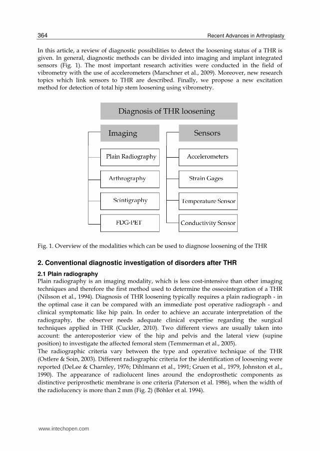
In this article, a review of diagnostic possibilities to detect the loosening status of a THR is given. In general, diagnostic methods can be divided into imaging and implant integrated sensors (Fig. 1). The most important research activities were conducted in the field of vibrometry with the use of accelerometers (Marschner et al., 2009). Moreover, new research topics which link sensors to THR are described. Finally, we propose a new excitation method for detection of total hip stem loosening using vibrometry.
Fig. 1. Overview of the modalities which can be used to diagnose loosening of the THR
2. Conventional diagnostic investigation of disorders after THR
2.1 Plain radiography Plain radiography is an imaging modality, which is less cost-intensive than other imaging
techniques and therefore the first method used to determine the osseointegration of a THR
(Nilsson et al., 1994). Diagnosis of THR loosening typically requires a plain radiograph - in
the optimal case it can be compared with an immediate post operative radiograph - and
clinical symptomatic like hip pain. In order to achieve an accurate interpretation of the
radiography, the observer needs adequate clinical expertise regarding the surgical
techniques applied in THR (Cuckler, 2010). Two different views are usually taken into
account: the anteroposterior view of the hip and pelvis and the lateral view (supine
position) to investigate the affected femoral stem (Temmerman et al., 2005).
The radiographic criteria vary between the type and operative technique of the THR
(Ostlere & Soin, 2003). Different radiographic criteria for the identification of loosening were
reported (DeLee & Charnley, 1976; Dihlmann et al., 1991; Gruen et al., 1979, Johnston et al.,
1990). The appearance of radiolucent lines around the endoprosthetic components as
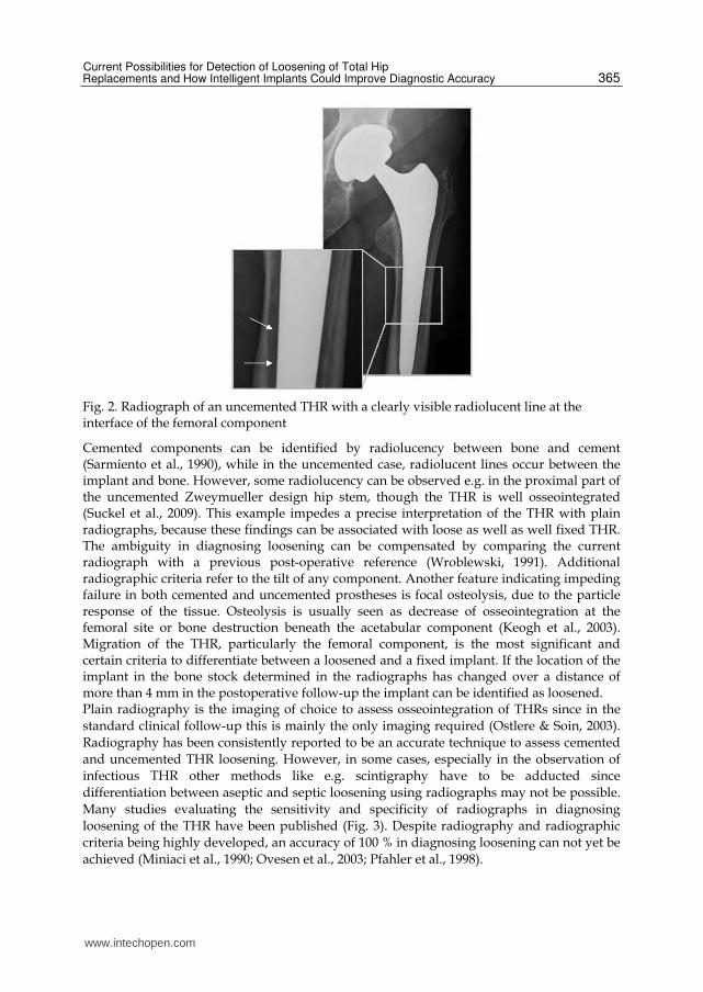
distinctive periprosthetic membrane is one criteria (Paterson et al. 1986), when the width of
the radiolucency is more than 2 mm (Fig. 2) (Böhler et al. 1994).
www.intechopen.com
Current Possibilities for Detection of Loosening of Total Hip Replacements and How Intelligent Implants Could Improve Diagnostic Accuracy
365
Fig. 2. Radiograph of an uncemented THR with a clearly visible radiolucent line at the interface of the femoral component
Cemented components can be identified by radiolucency between bone and cement (Sarmiento et al., 1990), while in the uncemented case, radiolucent lines occur between the implant and bone. However, some radiolucency can be observed e.g. in the proximal part of the uncemented Zweymueller design hip stem, though the THR is well osseointegrated (Suckel et al., 2009). This example impedes a precise interpretation of the THR with plain radiographs, because these findings can be associated with loose as well as well fixed THR. The ambiguity in diagnosing loosening can be compensated by comparing the current radiograph with a previous post-operative reference (Wroblewski, 1991). Additional radiographic criteria refer to the tilt of any component. Another feature indicating impeding failure in both cemented and uncemented prostheses is focal osteolysis, due to the particle response of the tissue. Osteolysis is usually seen as decrease of osseointegration at the femoral site or bone destruction beneath the acetabular component (Keogh et al., 2003). Migration of the THR, particularly the femoral component, is the most significant and certain criteria to differentiate between a loosened and a fixed implant. If the location of the implant in the bone stock determined in the radiographs has changed over a distance of more than 4 mm in the postoperative follow-up the implant can be identified as loosened. Plain radiography is the imaging of choice to assess osseointegration of THRs since in the
standard clinical follow-up this is mainly the only imaging required (Ostlere & Soin, 2003).
Radiography has been consistently reported to be an accurate technique to assess cemented
and uncemented THR loosening. However, in some cases, especially in the observation of
infectious THR other methods like e.g. scintigraphy have to be adducted since
differentiation between aseptic and septic loosening using radiographs may not be possible.
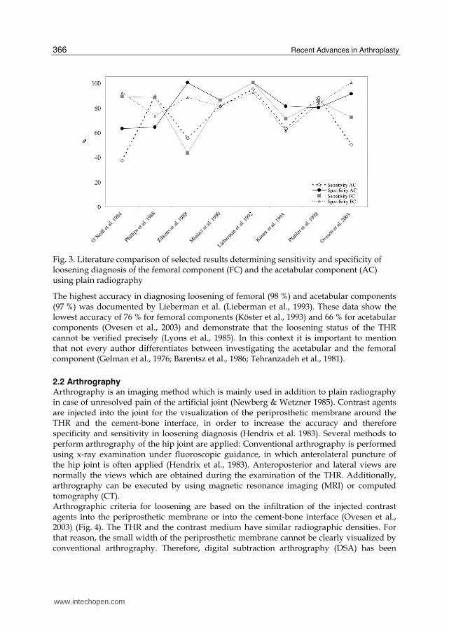
Many studies evaluating the sensitivity and specificity of radiographs in diagnosing
loosening of the THR have been published (Fig. 3). Despite radiography and radiographic
criteria being highly developed, an accuracy of 100 % in diagnosing loosening can not yet be
achieved (Miniaci et al., 1990; Ovesen et al., 2003; Pfahler et al., 1998).
www.intechopen.com
Recent Advances in Arthroplasty
366
Fig. 3. Literature comparison of selected results determining sensitivity and specificity of loosening diagnosis of the femoral component (FC) and the acetabular component (AC) using plain radiography
The highest accuracy in diagnosing loosening of femoral (98 %) and acetabular components (97 %) was documented by Lieberman et al. (Lieberman et al., 1993). These data show the lowest accuracy of 76 % for femoral components (Köster et al., 1993) and 66 % for acetabular components (Ovesen et al., 2003) and demonstrate that the loosening status of the THR cannot be verified precisely (Lyons et al., 1985). In this context it is important to mention that not every author differentiates between investigating the acetabular and the femoral component (Gelman et al., 1976; Barentsz et al., 1986; Tehranzadeh et al., 1981).
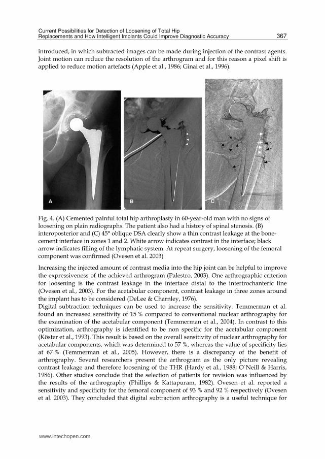
2.2 Arthrography Arthrography is an imaging method which is mainly used in addition to plain radiography in case of unresolved pain of the artificial joint (Newberg & Wetzner 1985). Contrast agents are injected into the joint for the visualization of the periprosthetic membrane around the THR and the cement-bone interface, in order to increase the accuracy and therefore specificity and sensitivity in loosening diagnosis (Hendrix et al. 1983). Several methods to perform arthrography of the hip joint are applied: Conventional arthrography is performed using x-ray examination under fluoroscopic guidance, in which anterolateral puncture of the hip joint is often applied (Hendrix et al., 1983). Anteroposterior and lateral views are normally the views which are obtained during the examination of the THR. Additionally, arthrography can be executed by using magnetic resonance imaging (MRI) or computed tomography (CT). Arthrographic criteria for loosening are based on the infiltration of the injected contrast agents into the periprosthetic membrane or into the cement-bone interface (Ovesen et al., 2003) (Fig. 4). The THR and the contrast medium have similar radiographic densities. For that reason, the small width of the periprosthetic membrane cannot be clearly visualized by conventional arthrography. Therefore, digital subtraction arthrography (DSA) has been
www.intechopen.com
Current Possibilities for Detection of Loosening of Total Hip Replacements and How Intelligent Implants Could Improve Diagnostic Accuracy
367
introduced, in which subtracted images can be made during injection of the contrast agents. Joint motion can reduce the resolution of the arthrogram and for this reason a pixel shift is applied to reduce motion artefacts (Apple et al., 1986; Ginai et al., 1996).
Fig. 4. (A) Cemented painful total hip arthroplasty in 60-year-old man with no signs of loosening on plain radiographs. The patient also had a history of spinal stenosis. (B) interoposterior and (C) 45° oblique DSA clearly show a thin contrast leakage at the bone-cement interface in zones 1 and 2. White arrow indicates contrast in the interface; black arrow indicates filling of the lymphatic system. At repeat surgery, loosening of the femoral component was confirmed (Ovesen et al. 2003)
Increasing the injected amount of contrast media into the hip joint can be helpful to improve
the expressiveness of the achieved arthrogram (Palestro, 2003). One arthrographic criterion
for loosening is the contrast leakage in the interface distal to the intertrochanteric line
(Ovesen et al., 2003). For the acetabular component, contrast leakage in three zones around
the implant has to be considered (DeLee & Charnley, 1976).
Digital subtraction techniques can be used to increase the sensitivity. Temmerman et al. found an increased sensitivity of 15 % compared to conventional nuclear arthrography for the examination of the acetabular component (Temmerman et al., 2004). In contrast to this optimization, arthrography is identified to be non specific for the acetabular component (Köster et al., 1993). This result is based on the overall sensitivity of nuclear arthrography for acetabular components, which was determined to 57 %, whereas the value of specificity lies at 67 % (Temmerman et al., 2005). However, there is a discrepancy of the benefit of arthrography. Several researchers present the arthrogram as the only picture revealing contrast leakage and therefore loosening of the THR (Hardy et al., 1988; O`Neill & Harris, 1986). Other studies conclude that the selection of patients for revision was influenced by the results of the arthrography (Phillips & Kattapuram, 1982). Ovesen et al. reported a sensitivity and specificity for the femoral component of 93 % and 92 % respectively (Ovesen et al. 2003). They concluded that digital subtraction arthrography is a useful technique for
www.intechopen.com
Recent Advances in Arthroplasty
368
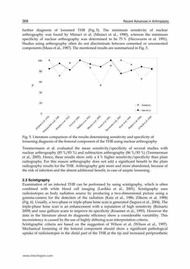
further diagnosis of loosened THR (Fig. 5). The minimum sensitivity of nuclear arthrography was found by Miniaci et al. (Miniaci et al., 1990), whereas the minimum specificity of nuclear arthrography was determined to be 75 % (Herzwurm et al. 1991). Studies using arthrography often do not discriminate between cemented or uncemented components (Maus et al., 1987). The mentioned results are summarized in Fig. 5.
Fig. 5. Literature comparison of the results determining sensitivity and specificity of loosening diagnosis of the femoral component of the THR using nuclear arthrography
Temmermann et al. evaluated the mean sensitivity/specificity of several studies with nuclear arthrography (85 %/83 %) and subtraction arthrography (86 %/85 %) (Temmerman et al., 2005). Hence, these results show only a 4 % higher sensitivity/specificity than plain radiographs. For this reason arthrography does not add a significant benefit to the plain radiography results for the THR. Arthrography gets more and more abandoned, because of the risk of infection and the absent additional benefit, in case of aseptic loosening.
2.3 Scintigraphy Examination of an infected THR can be performed by using scintigraphy, which is often combined with white blood cell imaging (Larikka et al., 2001). Scintigraphy uses radioisotopes as body radiation source by producing a two-dimensional picture using a gamma-camera for the detection of the radiation (Katz et al., 1986; Zilkens et al. 1988) (Fig. 6). Usually, a two-phase or triple-phase bone scan is generated (Segura et al., 2004). The triple-phase bone scan is an enhancement with a reputation of high sensitivity (Reinartz 2009) and uses gallium scans to improve its specificity (Kraemer et al., 1993). However the data in the literature about its diagnostic efficiency show a considerable variability. This inconsistency is caused by the use of highly differing scan interpretation criteria. Scintigraphic criteria are based on the suggestion of Wilson et al. (Wilson et al., 1997). Mechanical loosening of the femoral component should show a significant pathological uptake of radioisotopes in the distal part of the THR at the tip and increased periprosthetic
www.intechopen.com
Current Possibilities for Detection of Loosening of Total Hip Replacements and How Intelligent Implants Could Improve Diagnostic Accuracy
369
uptake. A second substantial lesion in the region of the lesser trochanter is a further sign for loosening and supports the first presumption.
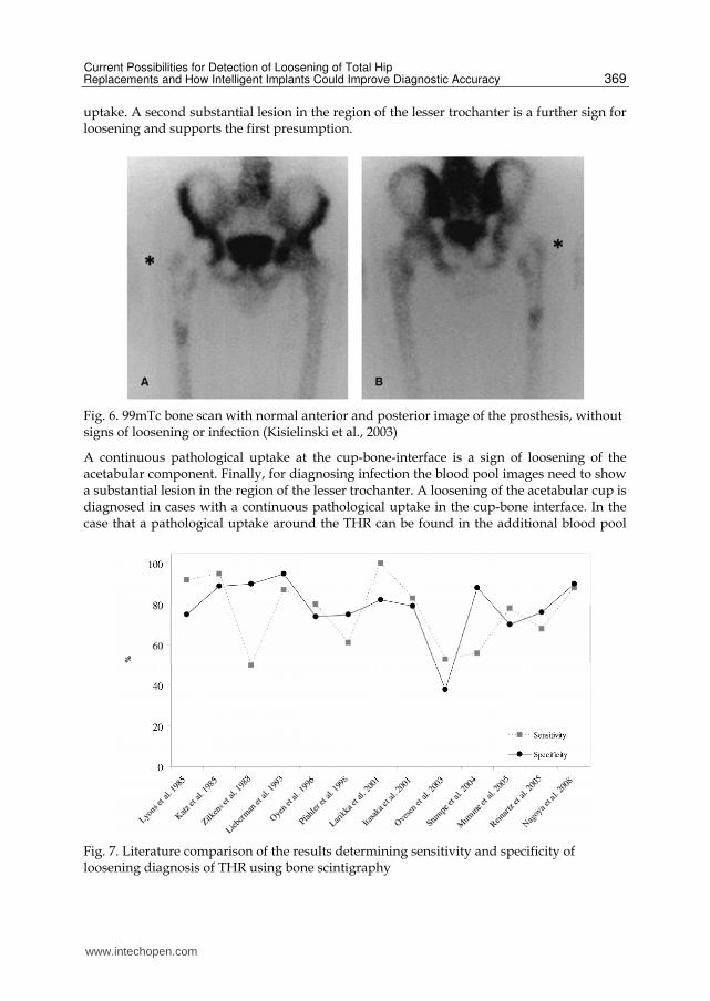
Fig. 6. 99mTc bone scan with normal anterior and posterior image of the prosthesis, without signs of loosening or infection (Kisielinski et al., 2003)
A continuous pathological uptake at the cup-bone-interface is a sign of loosening of the acetabular component. Finally, for diagnosing infection the blood pool images need to show a substantial lesion in the region of the lesser trochanter. A loosening of the acetabular cup is diagnosed in cases with a continuous pathological uptake in the cup-bone interface. In the case that a pathological uptake around the THR can be found in the additional blood pool
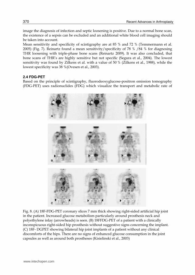
Fig. 7. Literature comparison of the results determining sensitivity and specificity of loosening diagnosis of THR using bone scintigraphy
www.intechopen.com
Recent Advances in Arthroplasty
370
image the diagnosis of infection and septic loosening is positive. Due to a normal bone scan, the existence of a sepsis can be excluded and an additional white blood cell imaging should be taken into account. Mean sensitivity and specificity of scintigraphy are at 85 % and 72 % (Temmermann et al. 2005) (Fig. 7). Reinartz found a mean sensitivity/specificity of 78 % /84 % for diagnosing THR loosening with triple-phase bone scans (Reinartz 2009). It was also concluded, that bone scans of THR’s are highly sensitive but not specific (Segura et al., 2004). The lowest sensitivity was found by Zilkens et al. with a value of 50 % (Zilkens et al., 1988), while the lowest specificity was 38 %(Ovesen et al., 2003).
2.4 FDG-PET Based on the principle of scintigraphy, fluorodeoxyglucose-positron emission tomography (FDG-PET) uses radionuclides (FDG) which visualize the transport and metabolic rate of
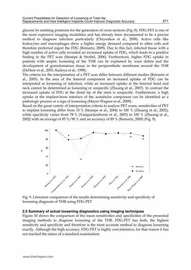
Fig. 8. (A) 18F-FDG-PET coronary slices 7 mm thick showing right-sided artificial hip joint in the patient. Increased glucose metabolism particularly around prosthesis neck and polyethylene inlay (arrowheads) is seen. (B) 18FFDG-PET of a patient with a clinically inconspicuous right-sided hip prosthesis without suggestive signs concerning the implant. (C) 18F- DGPET showing bilateral hip joint implants of a patient without any clinical discomforts of the hips. There are no signs of enhanced glucose consumption in the joint capsules as well as around both prostheses (Kisielinski et al., 2003)
www.intechopen.com
Current Possibilities for Detection of Loosening of Total Hip Replacements and How Intelligent Implants Could Improve Diagnostic Accuracy
371
glucose by emitting positrons for the generation of cross-sections (Fig. 8). FDG-PET is one of the most expensive imaging modalities and has already been documented to be a precise method to diagnose infection particularly (Chryssikos et al., 2008). Active cells like leukocytes and macrophages show a higher energy demand compared to other cells and therefore preferred ingest the FDG (Reinartz, 2009). Due to this fact, infected tissue with a high number of active cells revealed an increased uptake of FDG, which leads to a positive finding in the PET scan (Stumpe & Strobel, 2006). Furthermore, higher FDG uptake in patients with aseptic loosening of the THR can be explained by wear debris and the development of granulomatous tissue in the periprosthetic membrane around the THR (DeHeer et al., 2001; Kadoya et al., 1998). The criteria for the interpretation of a PET scan differ between different studies (Reinartz et al., 2005). In the area of the femoral component an increased uptake of FDG can be interpreted as loosening or infection, while an increased uptake at the femoral head and neck cannot be determined as loosening or unspecific (Zhuang et al., 2007). In contrast the increased uptake of FDG at the distal tip of the stem is unspecific. Furthermore, a high uptake at the implant-bone interface of the acetabular component can be identified as a pathologic process or a sign of loosening (Mayer-Wagner et al., 2009). Based on the great variety of interpretation criteria to analyse PET scans, sensitivities of PET to implant loosening differ from 33 % (Stumpe et al., 2004) to 100 % (Zhuang et al., 2002), while specificity varies from 78 % (Vanquickenborne et al., 2003) to 100 % (Zhuang et al., 2002) with an average of 85 %/90 % and an accuracy of 89 % (Reinartz, 2009) (Fig. 9).
Fig. 9. Literature comparison of the results determining sensitivity and specificity of loosening diagnosis of THR using FDG-PET
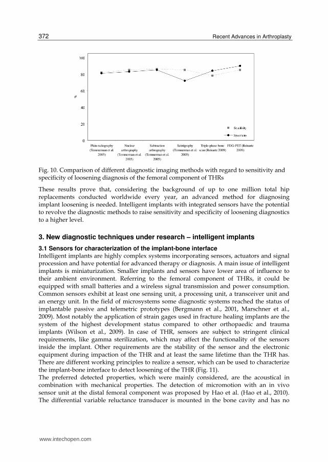
2.5 Summary of actual loosening diagnostics using imaging techniques Figure 10 shows the comparison of the mean sensitivities and specificities of the presented
imaging methods to diagnose loosening of the THR. FDG-PET has both, the highest
sensitivity and specificity and therefore is the most accurate method to diagnose loosening
exactly. Although the high accuracy, FDG-PET is highly cost-intensive, for that reason it has
not reached the status of a standard examination.
www.intechopen.com
Recent Advances in Arthroplasty
372
Fig. 10. Comparison of different diagnostic imaging methods with regard to sensitivity and specificity of loosening diagnosis of the femoral component of THRs
These results prove that, considering the background of up to one million total hip replacements conducted worldwide every year, an advanced method for diagnosing implant loosening is needed. Intelligent implants with integrated sensors have the potential to revolve the diagnostic methods to raise sensitivity and specificity of loosening diagnostics to a higher level.
3. New diagnostic techniques under research – intelligent implants
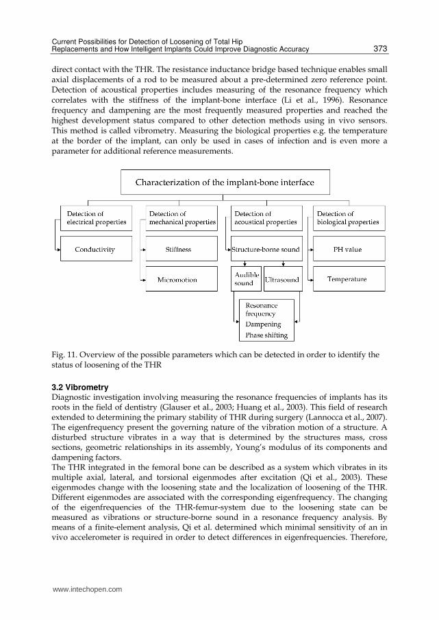
3.1 Sensors for characterization of the implant-bone interface Intelligent implants are highly complex systems incorporating sensors, actuators and signal procession and have potential for advanced therapy or diagnosis. A main issue of intelligent implants is miniaturization. Smaller implants and sensors have lower area of influence to their ambient environment. Referring to the femoral component of THRs, it could be equipped with small batteries and a wireless signal transmission and power consumption. Common sensors exhibit at least one sensing unit, a processing unit, a transceiver unit and an energy unit. In the field of microsystems some diagnostic systems reached the status of implantable passive and telemetric prototypes (Bergmann et al., 2001, Marschner et al., 2009). Most notably the application of strain gages used in fracture healing implants are the system of the highest development status compared to other orthopaedic and trauma implants (Wilson et al., 2009). In case of THR, sensors are subject to stringent clinical requirements, like gamma sterilization, which may affect the functionality of the sensors inside the implant. Other requirements are the stability of the sensor and the electronic equipment during impaction of the THR and at least the same lifetime than the THR has. There are different working principles to realize a sensor, which can be used to characterize the implant-bone interface to detect loosening of the THR (Fig. 11). The preferred detected properties, which were mainly considered, are the acoustical in combination with mechanical properties. The detection of micromotion with an in vivo sensor unit at the distal femoral component was proposed by Hao et al. (Hao et al., 2010). The differential variable reluctance transducer is mounted in the bone cavity and has no
www.intechopen.com
Current Possibilities for Detection of Loosening of Total Hip Replacements and How Intelligent Implants Could Improve Diagnostic Accuracy
373
direct contact with the THR. The resistance inductance bridge based technique enables small axial displacements of a rod to be measured about a pre-determined zero reference point. Detection of acoustical properties includes measuring of the resonance frequency which correlates with the stiffness of the implant-bone interface (Li et al., 1996). Resonance frequency and dampening are the most frequently measured properties and reached the highest development status compared to other detection methods using in vivo sensors. This method is called vibrometry. Measuring the biological properties e.g. the temperature at the border of the implant, can only be used in cases of infection and is even more a parameter for additional reference measurements.
Fig. 11. Overview of the possible parameters which can be detected in order to identify the status of loosening of the THR
3.2 Vibrometry Diagnostic investigation involving measuring the resonance frequencies of implants has its roots in the field of dentistry (Glauser et al., 2003; Huang et al., 2003). This field of research extended to determining the primary stability of THR during surgery (Lannocca et al., 2007). The eigenfrequency present the governing nature of the vibration motion of a structure. A disturbed structure vibrates in a way that is determined by the structures mass, cross sections, geometric relationships in its assembly, Young’s modulus of its components and dampening factors. The THR integrated in the femoral bone can be described as a system which vibrates in its multiple axial, lateral, and torsional eigenmodes after excitation (Qi et al., 2003). These eigenmodes change with the loosening state and the localization of loosening of the THR. Different eigenmodes are associated with the corresponding eigenfrequency. The changing of the eigenfrequencies of the THR-femur-system due to the loosening state can be measured as vibrations or structure-borne sound in a resonance frequency analysis. By means of a finite-element analysis, Qi et al. determined which minimal sensitivity of an in vivo accelerometer is required in order to detect differences in eigenfrequencies. Therefore,
www.intechopen.com
Recent Advances in Arthroplasty
374
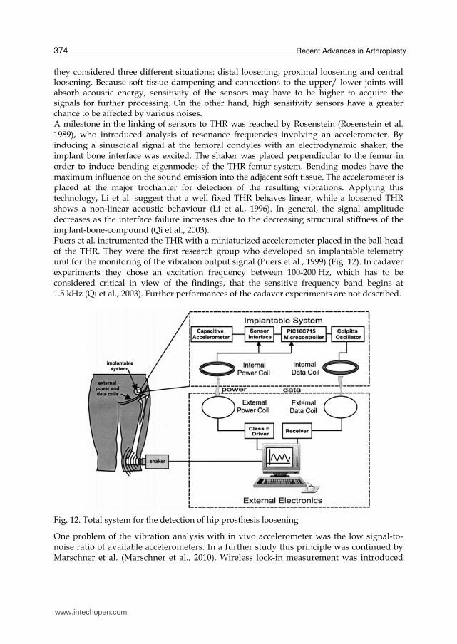
they considered three different situations: distal loosening, proximal loosening and central loosening. Because soft tissue dampening and connections to the upper/ lower joints will absorb acoustic energy, sensitivity of the sensors may have to be higher to acquire the signals for further processing. On the other hand, high sensitivity sensors have a greater chance to be affected by various noises. A milestone in the linking of sensors to THR was reached by Rosenstein (Rosenstein et al. 1989), who introduced analysis of resonance frequencies involving an accelerometer. By inducing a sinusoidal signal at the femoral condyles with an electrodynamic shaker, the implant bone interface was excited. The shaker was placed perpendicular to the femur in order to induce bending eigenmodes of the THR-femur-system. Bending modes have the maximum influence on the sound emission into the adjacent soft tissue. The accelerometer is placed at the major trochanter for detection of the resulting vibrations. Applying this technology, Li et al. suggest that a well fixed THR behaves linear, while a loosened THR shows a non-linear acoustic behaviour (Li et al., 1996). In general, the signal amplitude decreases as the interface failure increases due to the decreasing structural stiffness of the implant-bone-compound (Qi et al., 2003). Puers et al. instrumented the THR with a miniaturized accelerometer placed in the ball-head of the THR. They were the first research group who developed an implantable telemetry unit for the monitoring of the vibration output signal (Puers et al., 1999) (Fig. 12). In cadaver experiments they chose an excitation frequency between 100-200 Hz, which has to be considered critical in view of the findings, that the sensitive frequency band begins at 1.5 kHz (Qi et al., 2003). Further performances of the cadaver experiments are not described.
Fig. 12. Total system for the detection of hip prosthesis loosening
One problem of the vibration analysis with in vivo accelerometer was the low signal-to-noise ratio of available accelerometers. In a further study this principle was continued by Marschner et al. (Marschner et al., 2010). Wireless lock-in measurement was introduced
www.intechopen.com
Current Possibilities for Detection of Loosening of Total Hip Replacements and How Intelligent Implants Could Improve Diagnostic Accuracy
375
and the THR was instrumented with a coil, amplifier, microcontroller and telemetry unit in the distal end of the hip stem. The lock-in amplifier separates the measurement signal from noise. The availability of results which compare different loosening states of the THR and
therefore sensitivity of the proposed sensor systems is rare. Indeed, the presented devices
are available, but only few results verify the possibility to detect a difference between a
loose and a well fixed implant. Currently sensitivity and specificity of vibration analysis in
THR can only be estimated. Georgiou et al. stated a 20 % higher sensitivity measured with
vibration analysis (80 %) than with radiographs (60 %) (Georgiou et al., 2001). The finite
element analysis of Qi et al. showed that a loose THR can be identified reliable if 1/3 of the
femoral component has no direct bone contact (Qi et al., 2003). One of the main problems of
the resonance frequency analysis of THR is the soft tissue damping of the output vibration
signal. Furthermore, connection to the tibia and the pelvis can decrease the energy rate of
the acoustic sound waves.
In another in vitro study the accelerometer was replaced by a blood flow ultrasound probe
which enabled consistent detection of the vibrations for a loose and a fixed THR (Rowlands
et al., 2008). The ultrasound probe lead to increased amplitudes of the output vibrations
compared to the accelerometer signal. Dahl et al. applied ultrasound to detect osseous
integration of the total ankle replacement (Dahl et al., 2010). The cadaveric testing
demonstrated that the ultrasound technique could distinguish between loose and fixed
implant components. In a further study, a trans-femoral implant was measured with
accelerometers. It could be shown that the eigenfrequency increases with the weight bearing
rehabilitation time (Shao et al., 2007).
Paech et al investigated the natural frequency of four different types of THR and determined
these sound patterns in further cadaver tests with bovine and human bone (Paech et al.,
2007). The excitation of the THR was reached by using an acoustically neutral steel ball of
120 g as activating impact for sound emission. However, this is not reproducible for
application in clinical studies.
Besides problems in signal analysis and soft tissue dampening, functional sustainment of
the integrated electronics during sterilization and impaction of the THR seems to be a
significant challenge. Nevertheless detailed research into simulation of different loosening
states and significant test results are expected. Furthermore the excitation has to be
optimized in order to avoid pressure pain induced by the electrodynamic shaker (Georgiou
et al., 2001).
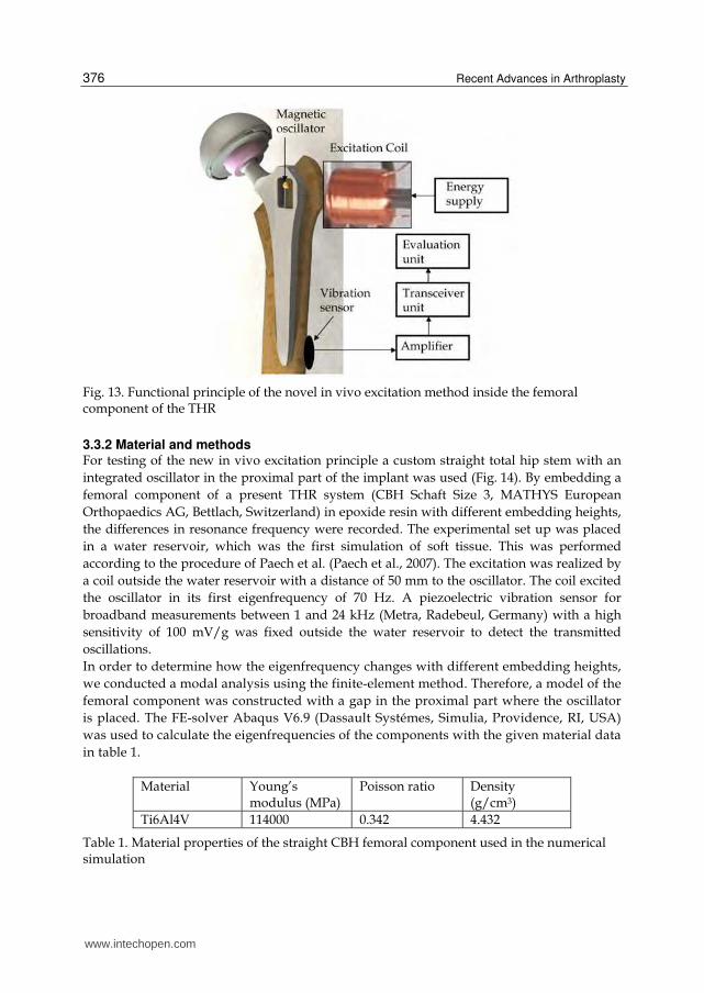
3.3 A new excitation method to detect aseptic loosening 3.3.1 Principle of the novel excitation concept In the proposed concept, an oscillator integrated in the THR is used. The oscillator consists
of a magnetic body which is fixed on a flat steel spring (Ruther et al., 2010) (Fig. 13). This
oscillator is excited by a coil placed outside the patient. The oscillator impinges inside the
THR and excites the THR to vibrate in its eigenfrequency. This can be considered as an
alternative to the electrodynamic shaker, which was used by different research groups. The
excitation in the implant bending modes leads to a sound emission to the surrounding bone
and soft tissue. The sound waves can be detected by a vibration sensor, which is applied
outside the patient’s hip opposite to the excitation coil.
www.intechopen.com
Recent Advances in Arthroplasty
376
Fig. 13. Functional principle of the novel in vivo excitation method inside the femoral component of the THR
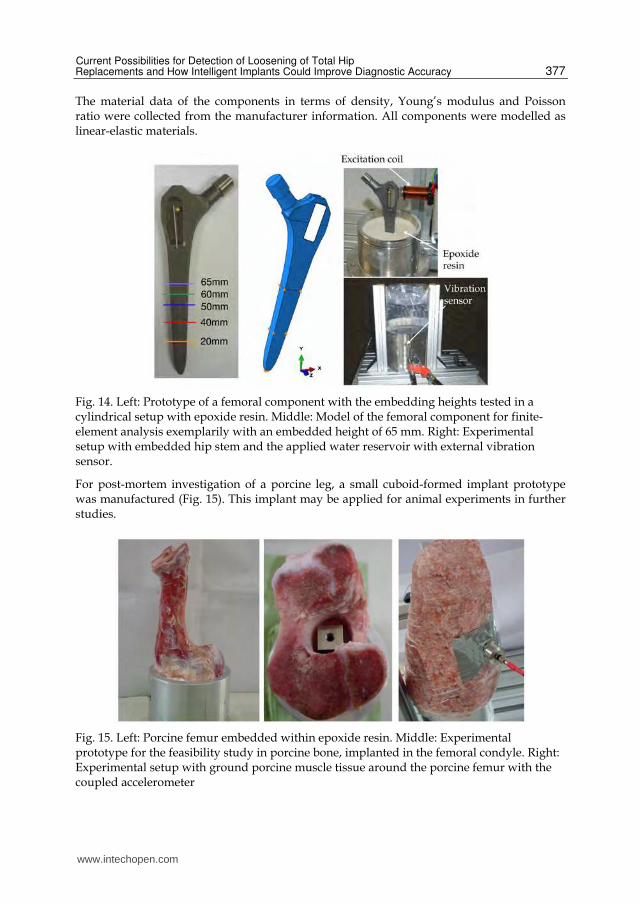
3.3.2 Material and methods For testing of the new in vivo excitation principle a custom straight total hip stem with an
integrated oscillator in the proximal part of the implant was used (Fig. 14). By embedding a
femoral component of a present THR system (CBH Schaft Size 3, MATHYS European
Orthopaedics AG, Bettlach, Switzerland) in epoxide resin with different embedding heights,
the differences in resonance frequency were recorded. The experimental set up was placed
in a water reservoir, which was the first simulation of soft tissue. This was performed
according to the procedure of Paech et al. (Paech et al., 2007). The excitation was realized by
a coil outside the water reservoir with a distance of 50 mm to the oscillator. The coil excited
the oscillator in its first eigenfrequency of 70 Hz. A piezoelectric vibration sensor for
broadband measurements between 1 and 24 kHz (Metra, Radebeul, Germany) with a high
sensitivity of 100 mV/g was fixed outside the water reservoir to detect the transmitted
oscillations.
In order to determine how the eigenfrequency changes with different embedding heights,
we conducted a modal analysis using the finite-element method. Therefore, a model of the
femoral component was constructed with a gap in the proximal part where the oscillator
is placed. The FE-solver Abaqus V6.9 (Dassault Systémes, Simulia, Providence, RI, USA)
was used to calculate the eigenfrequencies of the components with the given material data
in table 1.
Material Young’s modulus (MPa)
Poisson ratio Density (g/cm3)
Ti6Al4V 114000 0.342 4.432
Table 1. Material properties of the straight CBH femoral component used in the numerical simulation
www.intechopen.com
Current Possibilities for Detection of Loosening of Total Hip Replacements and How Intelligent Implants Could Improve Diagnostic Accuracy
377
The material data of the components in terms of density, Young’s modulus and Poisson ratio were collected from the manufacturer information. All components were modelled as linear-elastic materials.
Fig. 14. Left: Prototype of a femoral component with the embedding heights tested in a cylindrical setup with epoxide resin. Middle: Model of the femoral component for finite-element analysis exemplarily with an embedded height of 65 mm. Right: Experimental setup with embedded hip stem and the applied water reservoir with external vibration sensor.
For post-mortem investigation of a porcine leg, a small cuboid-formed implant prototype was manufactured (Fig. 15). This implant may be applied for animal experiments in further studies.
Fig. 15. Left: Porcine femur embedded within epoxide resin. Middle: Experimental prototype for the feasibility study in porcine bone, implanted in the femoral condyle. Right: Experimental setup with ground porcine muscle tissue around the porcine femur with the coupled accelerometer
www.intechopen.com
Recent Advances in Arthroplasty
378
In a first experimental in vitro set-up, the implant was inserted in the femoral condyles with press fit fixation. In order to simulate an advanced loosening the bore whole was reamed, filled with water and then the implant was reinserted. For simulation of soft tissue, ground porcine muscle tissue was arranged around the femur. The vibration sensor was placed perpendicular to the femur to detect bending eigenfrequencies.
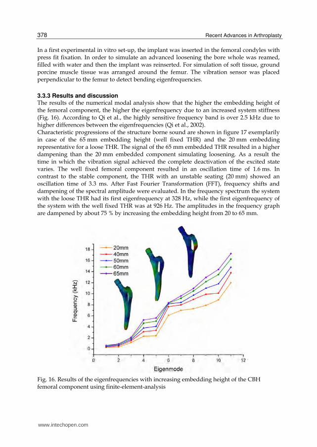
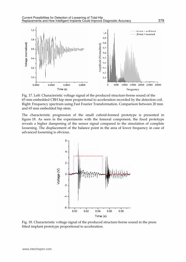
3.3.3 Results and discussion The results of the numerical modal analysis show that the higher the embedding height of the femoral component, the higher the eigenfrequency due to an increased system stiffness (Fig. 16). According to Qi et al., the highly sensitive frequency band is over 2.5 kHz due to higher differences between the eigenfrequencies (Qi et al., 2002). Characteristic progressions of the structure borne sound are shown in figure 17 exemplarily in case of the 65 mm embedding height (well fixed THR) and the 20 mm embedding representative for a loose THR. The signal of the 65 mm embedded THR resulted in a higher dampening than the 20 mm embedded component simulating loosening. As a result the time in which the vibration signal achieved the complete deactivation of the excited state varies. The well fixed femoral component resulted in an oscillation time of 1.6 ms. In contrast to the stable component, the THR with an unstable seating (20 mm) showed an oscillation time of 3.3 ms. After Fast Fourier Transformation (FFT), frequency shifts and dampening of the spectral amplitude were evaluated. In the frequency spectrum the system with the loose THR had its first eigenfrequency at 328 Hz, while the first eigenfrequency of the system with the well fixed THR was at 926 Hz. The amplitudes in the frequency graph are dampened by about 75 % by increasing the embedding height from 20 to 65 mm.
Fig. 16. Results of the eigenfrequencies with increasing embedding height of the CBH femoral component using finite-element-analysis
www.intechopen.com
Current Possibilities for Detection of Loosening of Total Hip Replacements and How Intelligent Implants Could Improve Diagnostic Accuracy
379
Fig. 17. Left: Characteristic voltage signal of the produced structure-borne sound of the 65 mm embedded CBH hip stem proportional to acceleration recorded by the detection coil. Right: Frequency spectrum using Fast Fourier Transformation. Comparison between 20 mm and 65 mm embedded hip stem.
The characteristic progression of the small cuboid-formed prototype is presented in figure 18. As seen in the experiments with the femoral component, the fixed prototype reveals a higher dampening of the sensor signal compared to the simulation of complete loosening. The displacement of the balance point in the area of lower frequency in case of advanced loosening is obvious.
Fig. 18. Characteristic voltage signal of the produced structure-borne sound in the press fitted implant prototype proportional to acceleration.
www.intechopen.com
Recent Advances in Arthroplasty
380
These first test results demonstrate differences between two different states of fixation of
both the femoral component and a prototype for latter animal experiments. Hence, this
confirms the potential usability of the novel non-invasive approach for detecting implant
loosening. In further tests, the influence of the femur-tibia and the femur-pelvis connection
has to be investigated. Additionally, the femoral component will be integrated in a human
cadaveric femur and measured with defined loosening zones. Moreover, the oscillators
could be used in animal studies in order to determine the quality of osseointegration e.g. of
new implant coatings and materials.
4. Conclusion
In loosening diagnostics of THR using imaging, an accuracy of 100 % is currently not
available. This fact raises the demand for a new system to diagnose loosening exactly.
Different approaches have been taken into consideration. Vibrometry, where accelerometers
are used to detect resonance frequencies in vivo, seems to be a promising approach. Due to
the disadvantage of the excitation with an electrodynamic shaker, we proposed a small
mechanical oscillator as novel excitation approach for vibrometry. In the experiments with a
Zweymueller femoral component a higher dampening coefficient could be identified in case
of a fixed implant. Furthermore, within in vitro experiments using an implant inserted in the
porcine femur the same promising results could be achieved.
5. Acknowledgment
This research project was funded by the Deutsche Forschungsgemeinschaft (DFG - German
Research Foundation) under Reg. No. KL 2327/1.
Reprinted from Publication “The diagnostic value of digital subtraction arthrography and
radionuclide bone scan in revision hip arthroplasty, Vol.18, No.6, pp. 735–740, Ovesen, O.,
Riegels-Nielsen, P., Lindequist, S., Jensen, I., Munkner, T., Torfing, T., et al. (2003), Journal of
Arthroplasty,” and “Fluordeoxyglucose positron emission tomography detection of
inflammatory reactions due to polyethylene wear in total hip arthroplasty, Vol.18, No.4, pp.
528–532, Kisielinski, K., Cremerius, U., Reinartz, P., & Niethard, F. U. (2003), Journal of
Arthroplasty” and “A telemetry system for the detection of hip prosthesis loosening by
vibration analysis, Vol.85, No.1-3, pp. 42-47, Puers, R., Catrysse, M., Vandevoorde, G.,
Collier, R. J., Louridas, E., Burny, F., Donkerwolcke, M., Moulart, F., (2000)., Sensors and
Actuators A: Physical, ” with permission from Elsevier.
6. References
Apple, J. S., Roberts, L. J., Gamba, J., Martinez, S., Khoury, M., & Ford, K. (1986). Digital subtraction arthrography of the prosthetic hip. Southern Medical Journal, Vol.79, No.7, pp. 808–810, ISSN 0038-4348
Barentsz, J. O., Lemmens, J. M., & Slooff, T. J. (1986). The use of subtraction arthrography in total hip arthroplasties. Roefo, Vol.144, No.4, pp. 440–446, ISSN 1438-9010
www.intechopen.com
Current Possibilities for Detection of Loosening of Total Hip Replacements and How Intelligent Implants Could Improve Diagnostic Accuracy
381
Bergmann G, Deuretzbacher G, Heller M, Graichen F, Rohlmann A, Strauss J, Duda GN (2001). Hip contact forces and gait patterns from routine activities. Journal of Biomechanics, Vol.34, No.7, pp. 859-871, ISSN 1435-2443
Böhler, M., Knahr, K., Riegler, M., & Salzer, M. (1994). Ten-year results of cemented Weller-type total hip arthroplasties. Analysis using different definitions of failure. Archives of Orthopaedic and Trauma Surgery, Vol.113, No.2, pp. 57–60, ISSN 0936-8051
Chryssikos, T., Parvizi, J., Ghanem, E., Newberg, A., Zhuang, H., & Alavi, A. (2008). FDG-PET imaging can diagnose periprosthetic infection of the hip, Clinical Orthopaedics and Related Research, Vol. 466, No.6, pp.1338-1342, ISSN 0009-921X
Cuckler, J. M. (2010). Unexplained pain after THR: what should i do? Orthopedics, Vol.33, No.9, p.648, ISSN 0147-7447
Dahl, M. C., Kramer, P. A., Reinhall, P. G., Benirschke, S. K., Hansen, S. T., & Ching, R. P. (2010). The efficacy of using vibrometry to detect osteointegration of the Agility total ankle. Journal of Biomechanics, Vol.43, No.9, pp. 1840–1843, ISSN 0021-9290
DeHeer, D. H., Engels, J. A., DeVries, A. S., Knapp, R. H., & Beebe, J. D. (2001). In situ complement activation by polyethylene wear debris. Journal of Biomedical Materials Research, Vol.54, No.1, pp. 12–19, ISSN 0021-9304
DeLee, J. G., & Charnley, J. (1976). Radiological demarcation of cemented sockets in total hip replacement. Clinical Orthopaedics and Related Research, No.121, pp. 20–32, ISSN 0009-921X
Dihlmann, W., Dihlmann, S. W., & Hering, L. (1991). Alloarthroplasty of the hip joint. Radiologic diagnosis of loosening and infection in cemented total endoprostheses. Radiologe, Vol.31, No.10, pp. 496–505, ISSN 0033-832X
Gelman, M. I., Coleman, R. E., Stevens, P. M., & Davey, B. W. (1978). Radiography, radionuclide imaging, and arthrography in the evaluation of total hip and knee replacement. Radiology, Vol.128, No.3, pp. 677–682, ISSN 0033-8419
Georgiou, A. P., & Cunningham, J. L. (2001). Accurate diagnosis of hip prosthesis loosening using a vibrational technique. Clinical Biomechanics, Vol.16, No.4, pp. 315–323, ISSN 0268-0033
Ginai, A. Z., van Biezen, F. C., Kint, P. A., Oei, H. Y., & Hop, W. C. (1996). Digital subtraction arthrography in preoperative evaluation of painful total hip arthroplasty. Skeletal Radiology, Vol.25, No.4, pp. 357–363, ISSN 0364-2348
Glauser, R., Sennerby, L., Meredith, N., Ree, A., Lundgren, A. K., Gottlow, J., et al. (2004). Resonance frequency analysis of implants subjected to immediate or early functional occlusal loading. Successful vs. failing implants. Clinical Oral Implants Research, Vol.15, No.4, pp. 428–434, ISSN 0905-7161
Gruen, T. A., McNeice, G. M., & Amstutz, H. C. (1979). "Modes of failure" of cemented stem-type femoral components: a radiographic analysis of loosening. Clinical Orthopaedics and Related Research, No.141, pp. 17–27, ISSN 0009-921X
Hardy, D. C., Reinus, W. R., Totty, W. G., & Keyser, C. K. (1988). Arthrography after total hip arthroplasty: utility of postambulation radiographs. Skeletal Radiology, Vol.17, No.1, pp. 20–23, ISSN 0364-2348
Hendrix, R. W., Wixson, R. L., Rana, N. A., & Rogers, L. F. (1983). Arthrography after total hip arthroplasty: a modified technique used in the diagnosis of pain. Radiology, Vol.148, No.3, pp. 647–652, ISSN 0033-8419
www.intechopen.com
Recent Advances in Arthroplasty
382
Huang, H. - M., Chiu, C. - L., Yeh, C. - Y., Lin, C. - T., Lin, L. - H., & Lee, S. - Y. (2003). Early detection of implant healing process using resonance frequency analysis. Clin Oral Implants Res, Vol.14, No.4, pp. 437–443, ISSN 0905-7161
Itasaka, T., Kawai, A., Sato, T., Mitani, S., & Inoue, H. (2001). Diagnosis of infection after total hip arthroplasty. Journal of Orthopaedic Science, Vol.6, No 4, pp. 320–326, ISSN 0949-2658
Kadoya, Y., Kobayashi, A., & Ohashi, H. (1998). Wear and osteolysis in total joint replacements. Acta Orthopaedica Scandinavica Supplementum, Vol.278, pp. 1–16, ISSN 0300-8827
Katz, G., Morscher, E., Fridrich, R., & Kentsch, A. (1986). [Evaluation of scintigraphic procedures for clarifying pain conditions in hip joint prostheses]. Schweizerische Medizinische Wochenschrift, Vol.116, No.21, pp. 703–707, ISSN 0036-7672
Keogh, C. F., Munk, P. L., Gee, R., Chan, L. P., & Marchinkow, L. O. (2003). Imaging of the painful hip arthroplasty. American Journal of Roentgenolgy, Vol.180, No.1, pp. 115–120, ISSN 0361-803X
Kisielinski, K., Cremerius, U., Reinartz, P., & Niethard, F. U. (2003). Fluordeoxyglucose positron emission tomography detection of inflammatory reactions due to polyethylene wear in total hip arthroplasty. Journal of Arthroplasty, Vol.18, No.4, pp. 528–532, ISSN 0883-5403
Koster, G., Munz, D. L., & Kohler, H. P. (1993). Clinical value of combined contrast and radionuclide arthrography in suspected loosening of hip prostheses. Archives of Orthopaedic and Trauma Surgery, Vol.112, No.5, pp. 247–254, ISSN 0936-8051
Kraemer, W. J., Saplys, R., Waddell, J. P., & Morton, J. (1993). Bone scan, gallium scan, and hip aspiration in the diagnosis of infected total hip arthroplasty. Journal of Arthroplasty, Vol.8, No.6, pp. 611–616, ISSN 0883-5403
Lannocca, M., Varini, E., Cappello, A., Cristofolini, L., & Bialoblocka, E. (2007). Intra-operative evaluation of cementless hip implant stability: a prototype device based on vibration analysis. Medical Engineering and Physics, Vol.29, No.8, pp. 886–894, ISSN 1350-4533
Larikka, M. J., Ahonen, A. K., Junila, J. A., Niemela, O., Hamalainen, M. M., & Syrjala, H. P. (2001). Extended combined 99mTc-white blood cell and bone imaging improves the diagnostic accuracy in the detection of hip replacement infections. European Journal of Nuclear Medicine, Vol.28, No.3, pp. 288–293, ISSN 0340-6997
Li, P. L., Jones, N. B., & Gregg, P. J. (1996). Vibration analysis in the detection of total hip prosthetic loosening. Medical Engineering and Physics, Vol.18;No.7, pp. 596–600, ISSN 1350-4533
Lieberman, J. R., Huo, M. H., Schneider, R., Salvati, E. A., & Rodi, S. (1993). Evaluation of painful hip arthroplasties. Are technetium bone scans necessary? Journal of Bone and Joint Surgergy, Vol.75, No.3, pp. 475–478, ISSN 0301-620X
Love, C., Tomas, M. B., Marwin, S. E., Pugliese, P. V., & Palestro, C. J. (2001). Role of nuclear medicine in diagnosis of the infected joint replacement. Radiographics, Vol.21, No.5, pp. 1229–1238, ISSN 0271-5333
Lyons, C. W., Berquist, T. H., Lyons, J. C., Rand, J. A., & Brown, M. L. (1985). Evaluation of radiographic findings in painful hip arthroplasties. Clinical Orthopaedics and Related Research, No.195, pp. 239–251, ISSN 0009-921X
www.intechopen.com
Current Possibilities for Detection of Loosening of Total Hip Replacements and How Intelligent Implants Could Improve Diagnostic Accuracy
383
Malchau, H., Herberts, P., Eisler, T., Garellick, G., & Soderman, P. (2002). The Swedish Total Hip Replacement Register. Journal of Bone and Joint Surgergy Am, Vol.84-A Suppl 2, pp. 2–20, ISSN 0021-9355
Maus, T. P., Berquist, T. H., Bender, C. E., & Rand, J. A. (1987). Arthrographic study of painful total hip arthroplasty: refined criteria. Radiology, Vol.162, No.3, pp. 721–727 ISSN 0033-8419
Mayer-Wagner, S., Mayer, W., Maegerlein, S., Linke, R., Jansson, V., & Muller, P. E. (2010). Use of 18F-FDG-PET in the diagnosis of endoprosthetic loosening of knee and hip implants. Archives of Orthopaedics and Trauma Surgery, Vol.130, No.10, pp. 1231–1238, ISSN 0936-8051
Miniaci, A., Bailey, W. H., Bourne, R. B., McLaren, A. C., & Rorabeck, C. H. (1990). Analysis of radionuclide arthrograms, radiographic arthrograms, and sequential plain radiographs in the assessment of painful hip arthroplasty. Journal of Arthroplasty, Vol.5, No.2, pp. 143–149, ISSN 0883-5403
Mumme, T., Reinartz, P., Alfer, J., Muller-Rath, R., Buell, U., & Wirtz, D. C. (2005). Diagnostic values of positron emission tomography versus triple-phase bone scan in hip arthroplasty loosening. Archives of Orthopaedic and Trauma Surgery, Vol.125, No.5, 322–329, ISSN 0936-8051
Nagoya, S., Kaya, M., Sasaki, M., Tateda, K., & Yamashita, T. (2008). Diagnosis of peri-prosthetic infection at the hip using triple-phase bone scintigraphy. Journal of Bone and Joint Surgergy Br, Vol.90, No.2, pp. 140–144, ISSN 0301-620X
Newberg, A. H., & Wetzner, S. M. (1985). Digital subtraction arthrography. Radiology, Vol.154, No.1, pp. 238–239, ISSN 0033-8419
Nilsson, L. T., Frazen, H., Carlsson, A. S., & Onnerfalt, R. (1994). Early radiographic loosening impairs the function of a total hip replacement. The Nottingham Health Profile of 49 patients at five years. Journal of Bone and Joint Surgergy Br, Vol.76, No.2, pp. 235–239, ISSN 0301-620X
O'Neill, D. A., & Harris, W. H. (1984). Failed total hip replacement: assessment by plain radiographs, arthrograms, and aspiration of the hip joint. Journal of Bone and Joint Surgergy Am, Vol.66, No.4, pp. 540–546, ISSN 0021-9355
Ovesen, O., Riegels-Nielsen, P., Lindequist, S., Jensen, I., Munkner, T., Torfing, T., et al. (2003). The diagnostic value of digital subtraction arthrography and radionuclide bone scan in revision hip arthroplasty. Journal of Arthroplasty, Vol.18, No.6, pp. 735–740, ISSN 0883-5403
Oyen, W. J., Lemmens, J. A., Claessens, R. A., van Horn, J. R., Slooff, T. J., & Corstens, F. H. (1996). Nuclear arthrography: combined scintigraphic and radiographic procedure for diagnosis of total hip prosthesis loosening. Journal of Nuclear Medicine, Vol.37, No.1, pp. 62–70, ISSN 0161-5505
Palestro, C. J. (2003). Nuclear medicine, the painful prosthetic joint, and orthopedic infection. Journal of Nuclear Medicine, Vol.44, No.6, pp 927–929, ISSN 0161-5505
Paterson, M., Fulford, P., & Denham, R. (1986). Loosening of the femoral component after total hip replacement. The thin black line and the sinking hip. Journal of Bone and Joint Surgergy Br, Vol.68, No.3, pp. 392–397, ISSN 0301-620X
Pfahler, M., Schidlo, C., & Refior, H. J. (1998). Evaluation of imaging in loosening of hip arthroplasty in 326 consecutive cases. Archives of Orthopaedic and Trauma Surgery, Vol.117, No.4-5, pp. 205–207, ISSN 0936-8051
www.intechopen.com
Recent Advances in Arthroplasty
384
Phillips, W. C., & Kattapuram, S. V. (1982). Prosthetic hip replacements: plain films and arthrography for component loosening. American Journal of Roentgenology, Vol.138, No.4, 677–682, ISSN 0361-803X
Puers, R., Catrysse, M., Vandevoorde, G., Collier, R. J., Louridas, E., Burny, F., Donkerwolcke, M., Moulart, F., (2000). A telemetry system for the detection of hip prosthesis loosening by vibration analysis, Sensors and Actuators A: Physical, Vol.85, No.1-3, pp. 42-47, ISSN 0924-4247
Qi, G., Mouchon, W. P., & Tan, T. E. (2003). How much can a vibrational diagnostic tool reveal in total hip arthroplasty loosening? Clinical Biomechanics (Bristol, Avon), Vol.18, No.5, pp. 444–458, ISSN 0268-0033
Reinartz, P. (2009). FDG-PET in patients with painful hip and knee arthroplasty: technical breakthrough or just more of the same. Quaterly Journal of Nuclear Medicine and Molecular Imaging, Vol.53, No.1, pp. 41–50, ISSN 1824-4785
Reinartz, P., Mumme, T., Hermanns, B., Cremerius, U., Wirtz, D. C., Schaefer, W. M., et al. (2005). Radionuclide imaging of the painful hip arthroplasty: positron-emission tomography versus triplephase bone scanning. Journal of Bone and Joint Surgergy Br, Vol.87, No.4, pp. 465–470, ISSN 0301-620X
Rosenstein A.D., McCoy G.F., Bulstrode C.J., McLardy-Smith P.D., Cunningham J.L., Turner-Smith A.R (1989). The differentiation of loose and secure femoral implants in total hip replacement using a vibrational technique: an anatomical and pilot clinical study. Proceedings of Institute of Mechanical Engineering, Vol.203, No.2, pp. 77-81, ISSN 0954-4119
Rowlands, A., Duck, F. A., & Cunningham, J. L. (2008). Bone vibration measurement using ultrasound: application to detection of hip prosthesis loosening. Medical Engineering and Physics, Vol.30, No.3, pp. 278–284, ISSN 1350-4533
Ruther, C., Ruther, C., Biemann, A., Nierath, H., Ewald, H., Mittelmeier, W., Bader, R., Kluess, D., (2010). A new concept for non-invasive radiation-free detection of implant loosening, 56th Annual Meeting of the Orthopaedic Research Society, New Orleans, Louisiana, USA, March 6-9, 2010, pp. 2413.
Sarmiento, A., Ebramzadeh, E., Gogan, W. J., & McKellop, H. A. (1990). Total hip arthroplasty with cement. A long-term radiographic analysis in patients who are older than fifty and younger than fifty years. Journal of Bone and Joint Surgergy Am, Vol.72, No.10, 1470–1476, ISSN 0021-9355
Segura, A. B., Munoz, A., Brulles, Y. R., Hernandez Hermoso, J. A., Diaz, M. C., Bajen Lazaro, M. T., et al. (2004). What is the role of bone scintigraphy in the diagnosis of infected joint prostheses? Nuclear Medicine Communications, Vol.25, No.5, pp. 527–532, ISSN 0143-3636
Shao F. Xu W., Crocombe A., Ewins D. (2007). Natural frequency analysis of osseointegration for trans-femoral implant. Annals of Biomedical Engineering, Vol.35, No.5, pp. 817-824, ISSN 0090-6964
Stumpe, K. D. M., & Strobel, K. (2006). 18F FDG-PET imaging in musculoskeletal infection. Quaterly Journal of Nuclear Medicine and Molecular Imaging, Vol.50, No.2, pp. 131–142, ISSN 0033-8419
Stumpe, K. D. M., Notzli, H. P., Zanetti, M., Kamel, E. M., Hany, T. F., Gorres, G. W., et al. (2004). FDG PET for differentiation of infection and aseptic loosening in total hip
www.intechopen.com
Current Possibilities for Detection of Loosening of Total Hip Replacements and How Intelligent Implants Could Improve Diagnostic Accuracy
385
replacements: comparison with conventional radiography and three-phase bone scintigraphy. Radiology, Vol.231, No.2, pp. 333–341, ISSN 1824-4785
Suckel, A., Geiger, F., Kinzl, L., Wulker, N., & Garbrecht, M. (2009). Long-term results for the uncemented Zweymuller/Alloclassic hip endoprosthesis. A 15-year minimum follow-up of 320 hip operations. Journal of Arthroplasty, Vol.24, No.6, pp. 846–853, ISSN 0883-5403
Tehranzadeh, J., Schneider, R., & Freiberger, R. H. (1981). Radiological evaluation of painful total hip replacement. Radiology, Vol.141, No.2, pp. 355–362, ISSN 0033-8419
Temmerman, O. P. P., Raijmakers, P. G. H. M., Berkhof, J., David, E. F. L., Pijpers, R., Molenaar, M. A., et al. (2006). Diagnostic accuracy and interobserver variability of plain radiography, subtraction arthrography, nuclear arthrography, and bone scintigraphy in the assessment of aseptic femoral component loosening. Archives of Orthopaedic and Trauma Surgery, Vol.126, No.5, pp. 316–323, ISSN 0936-8051
Temmerman, O. P. P., Raijmakers, P. G. H. M., Berkhof, J., Hoekstra, O. S., Teule, G. J. J., & Heyligers, I. C. (2005). Accuracy of diagnostic imaging techniques in the diagnosis of aseptic loosening of the femoral component of a hip prosthesis: a meta-analysis. Journal of Bone and Joint Surgergy Br, Vol.87, No.6, pp. 781–785, ISSN 0301-620X
Temmerman, O. P. P., Raijmakers, P. G. H. M., David, E. F. L., Pijpers, R., Molenaar, M. A., Hoekstra, O. S., et al. (2004). A comparison of radiographic and scintigraphic techniques to assess aseptic loosening of the acetabular component in a total hip replacement. Journal of Bone and Joint Surgergy Br, Vol.86-A, No.11, pp. 2456–2463, ISSN 0021-9355
Udomkiat, P., Wan, Z., & Dorr, L. D. (2001). Comparison of preoperative radiographs and intraoperative findings of fixation of hemispheric porous-coated sockets. Journal of Bone and Joint Surgergy Br, Vol.83-A, No.12, pp. 1865–1870, ISSN 0021-9355
Uri, G., Wellman, H., Capello, W., Robb, J., & Greenman, G. (1984). Scintigraphic and X-ray arthrographic diagnosis of femoral prosthesis loosening: concise communication. Journal of Nuclear Medicine, Vol.25, No.6, pp. 661–663, ISSN 0161-5505
Vanquickenborne, B., Maes, A., Nuyts, J., Van Acker, F., Stuyck, J., Mulier, M., et al. (2003). The value of (18)FDG-PET for the detection of infected hip prosthesis. European Journal of Nuclear Medicine Molecular Imaging, Vol.30, No.5, pp. 705–715, ISSN 1619-7070
Walker, C. W., FitzRandolph, R. L., Collins, D. N., & Dalrymple, G. V. (1991). Arthrography of painful hips following arthroplasty: digital versus plain film subtraction. Skeletal Radiology, Vol.20, No.6, pp. 403–407, ISSN 0364-2348
Wilson, D. J., Morgan, R. L., Hesselden, K. L., Dodd, J. R., Janna, S. W., & Fagan, M. J. (2009). A single-channel telemetric intramedullary nail for in vivo measurement of fracture healing. Journal of Orthopaedics and Trauma, Vol.23, No.10, pp. 702–709, ISSN 0890-5339
Wroblewski, B. M. (1991). Clinical and radiographic evaluation of total hip replacement. A standard system of terminology for reporting results. Journal of Bone and Joint Surgery Am, Vol.73, No.6, p. 948, ISSN 0021-9355
www.intechopen.com

Recent Advances in Arthroplasty
386
Zhuang, H., Yang, H., & Alavi, A. (2007). Critical role of 18F-labeled fluorodeoxyglucose PET in the management of patients with arthroplasty. Radiologic Clinics of North America, Vol.45, No.4, pp. 711–8, vii, ISSN 0033-8389
Zilkens, K. W., Wicke, A., Zilkens, J., & Bull, U. (1988). Nuclear imaging in loosening of hip-joint endoprostheses. Archives of Orthopaedic and Trauma Surgery, Vol.107, No.5, pp. 288–292, ISSN 0344-8444
www.intechopen.com
Recent Advances in ArthroplastyEdited by Dr. Samo Fokter
ISBN 978-953-307-990-5Hard cover, 614 pagesPublisher InTechPublished online 27, January, 2012Published in print edition January, 2012
InTech EuropeUniversity Campus STeP Ri Slavka Krautzeka 83/A 51000 Rijeka, Croatia Phone: +385 (51) 770 447 Fax: +385 (51) 686 166www.intechopen.com
InTech ChinaUnit 405, Office Block, Hotel Equatorial Shanghai No.65, Yan An Road (West), Shanghai, 200040, China
Phone: +86-21-62489820 Fax: +86-21-62489821
The purpose of this book was to offer an overview of recent insights into the current state of arthroplasty. Thetremendous long term success of Sir Charnley's total hip arthroplasty has encouraged many researchers totreat pain, improve function and create solutions for higher quality of life. Indeed and as described in a specialchapter of this book, arthroplasty is an emerging field in the joints of upper extremity and spine. However,there are inborn complications in any foreign design brought to the human body. First, in the chapter oninfections we endeavor to provide a comprehensive, up-to-date analysis and description of the management ofthis difficult problem. Second, the immune system is faced with a strange material coming in huge amounts ofmicro-particles from the tribology code. Therefore, great attention to the problem of aseptic loosening hasbeen addressed in special chapters on loosening and on materials currently available for arthroplasty.
How to referenceIn order to correctly reference this scholarly work, feel free to copy and paste the following:
Cathérine Ruther, Ulrich Timm, Hartmut Ewald, Wolfram Mittelmeier, Rainer Bader, Rico Schmelter, ArminLohrengel and Daniel Kluess (2012). Current Possibilities for Detection of Loosening of Total HipReplacements and How Intelligent Implants Could Improve Diagnostic Accuracy, Recent Advances inArthroplasty, Dr. Samo Fokter (Ed.), ISBN: 978-953-307-990-5, InTech, Available from:http://www.intechopen.com/books/recent-advances-in-arthroplasty/current-possibilities-for-detection-of-loosening-of-total-hip-replacements-and-how-intelligent-impla