Embed Size (px)
Citation preview
Case report
Open Access
Early loosening and secondary dislocation due to a brokentrochanteric osteotomy wire following a Charnley totalhip arthroplasty: a case reportYousef Shahin*, Rakesh Choudhary, Saeed Al-Naser and Mark Mullins
Address: Department of Trauma and Orthopaedics Morriston Hospital, Heol Maes Eglwys, Morriston, Swansea, SA6 6NL, United Kingdom
Email: YS* - [email protected]; RC - [email protected]; SAN - [email protected];MM - [email protected]
*Corresponding author
Published: 29 May 2009 Received: 7 April 2009Accepted: 30 April 2009
Cases Journal 2009, 2:7117 doi: 10.1186/1757-1626-2-7117
This article is available from: http://casesjournal.com/casesjournal/article/view/7117
© 2009 Shahin et al; licensee Cases Network Ltd.This is an Open Access article distributed under the terms of the Creative Commons Attribution License (http://creativecommons.org/licenses/by/3.0),which permits unrestricted use, distribution, and reproduction in any medium, provided the original work is properly cited.
Abstract
We report a case of interposition of a broken trochanteric wire in the hip joint. This caused earlywear of the prosthesis and dislocation of the Charnley total hip arthroplasty. The patient was treatedwith a revision total hip arthroplasty. This rare complication should be taken into consideration whenperforming a trochanteric osteotomy fixation with wiring in Charnley total hip arthroplasty.
IntroductionTrochanteric osteotomy is a known method used inCharnley total hip arthroplasty. The advantage of thetranstrochanteric approach is that it gives a better view ofthe hip joint. The disadvantages of this method areincreased operative time, increased blood loss, trochan-teric non union and trochanteric bursitis [1,2]. We report arare complication associated with trochanteric osteotomy.
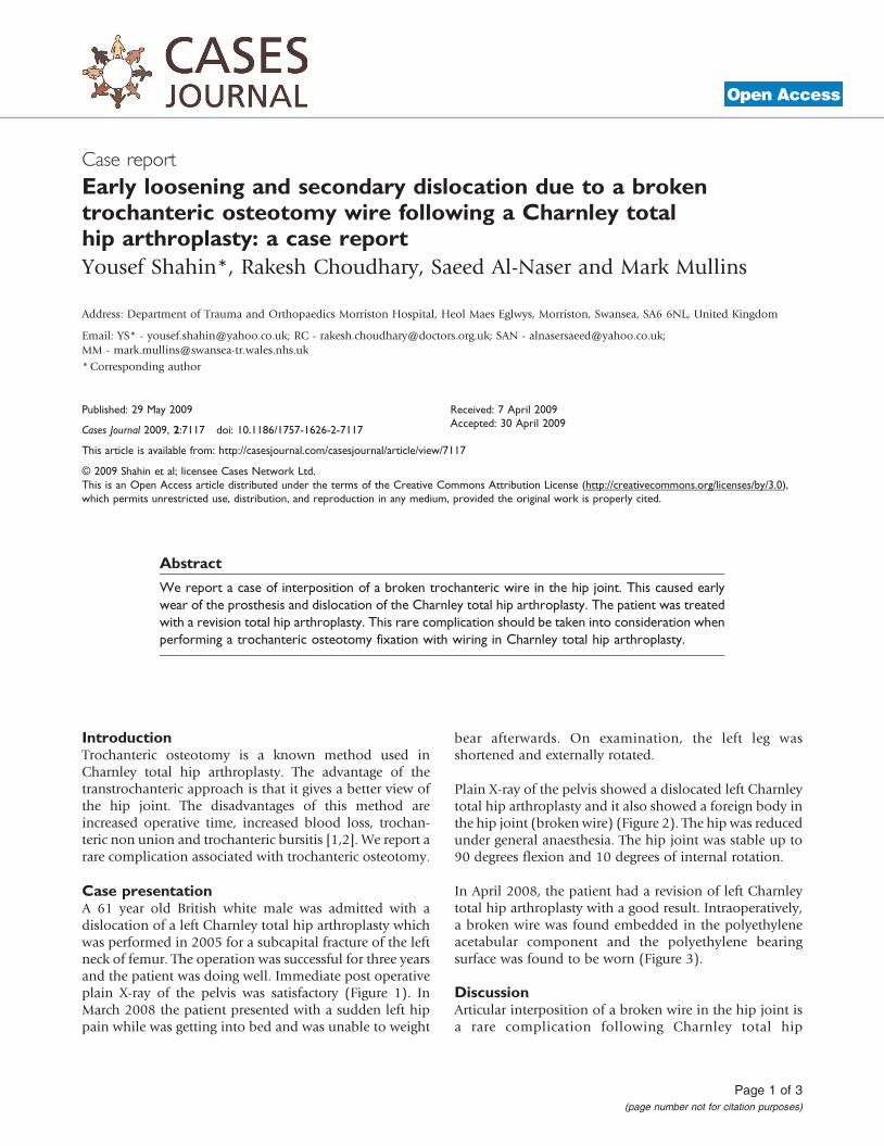
Case presentationA 61 year old British white male was admitted with adislocation of a left Charnley total hip arthroplasty whichwas performed in 2005 for a subcapital fracture of the leftneck of femur. The operation was successful for three yearsand the patient was doing well. Immediate post operativeplain X-ray of the pelvis was satisfactory (Figure 1). InMarch 2008 the patient presented with a sudden left hippain while was getting into bed and was unable to weight
bear afterwards. On examination, the left leg wasshortened and externally rotated.
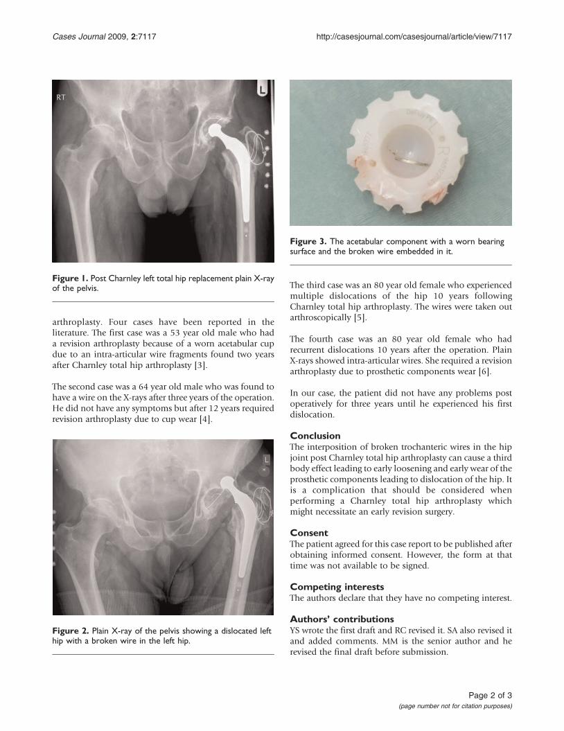
Plain X-ray of the pelvis showed a dislocated left Charnleytotal hip arthroplasty and it also showed a foreign body inthe hip joint (broken wire) (Figure 2). The hip was reducedunder general anaesthesia. The hip joint was stable up to90 degrees flexion and 10 degrees of internal rotation.
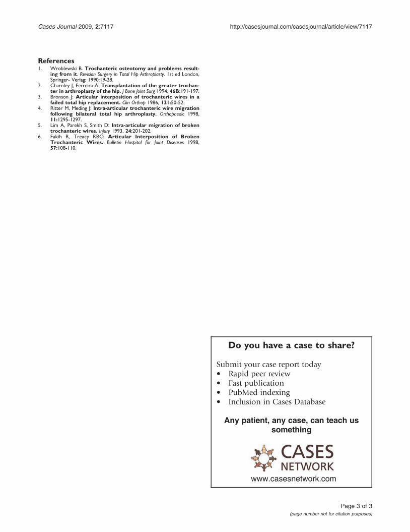
In April 2008, the patient had a revision of left Charnleytotal hip arthroplasty with a good result. Intraoperatively,a broken wire was found embedded in the polyethyleneacetabular component and the polyethylene bearingsurface was found to be worn (Figure 3).
DiscussionArticular interposition of a broken wire in the hip joint isa rare complication following Charnley total hip
Page 1 of 3(page number not for citation purposes)
arthroplasty. Four cases have been reported in theliterature. The first case was a 53 year old male who hada revision arthroplasty because of a worn acetabular cupdue to an intra-articular wire fragments found two yearsafter Charnley total hip arthroplasty [3].
The second case was a 64 year old male who was found tohave a wire on the X-rays after three years of the operation.He did not have any symptoms but after 12 years requiredrevision arthroplasty due to cup wear [4].
The third case was an 80 year old female who experiencedmultiple dislocations of the hip 10 years followingCharnley total hip arthroplasty. The wires were taken outarthroscopically [5].
The fourth case was an 80 year old female who hadrecurrent dislocations 10 years after the operation. PlainX-rays showed intra-articular wires. She required a revisionarthroplasty due to prosthetic components wear [6].
In our case, the patient did not have any problems postoperatively for three years until he experienced his firstdislocation.
ConclusionThe interposition of broken trochanteric wires in the hipjoint post Charnley total hip arthroplasty can cause a thirdbody effect leading to early loosening and early wear of theprosthetic components leading to dislocation of the hip. Itis a complication that should be considered whenperforming a Charnley total hip arthroplasty whichmight necessitate an early revision surgery.
ConsentThe patient agreed for this case report to be published afterobtaining informed consent. However, the form at thattime was not available to be signed.
Competing interestsThe authors declare that they have no competing interest.
Authors’ contributionsYS wrote the first draft and RC revised it. SA also revised itand added comments. MM is the senior author and herevised the final draft before submission.
Figure 1. Post Charnley left total hip replacement plain X-rayof the pelvis.
Figure 2. Plain X-ray of the pelvis showing a dislocated lefthip with a broken wire in the left hip.
Figure 3. The acetabular component with a worn bearingsurface and the broken wire embedded in it.
Page 2 of 3(page number not for citation purposes)
Cases Journal 2009, 2:7117 http://casesjournal.com/casesjournal/article/view/7117
References1. Wroblewski B. Trochanteric osteotomy and problems result-
ing from it. Revision Surgery in Total Hip Arthroplasty. 1st ed London,Springer- Verlag; 1990:19-28.
2. Charnley J, Ferreira A: Transplantation of the greater trochan-ter in arthroplasty of the hip. J Bone Joint Surg 1994, 46B:191-197.
3. Bronson J: Articular interposition of trochanteric wires in afailed total hip replacement. Clin Orthop 1986, 121:50-52.
4. Ritter M, Meding J: Intra-articular trochanteric wire migrationfollowing bilateral total hip arthroplasty. Orthopaedic 1998,11:1295-1297.
5. Lim A, Parekh S, Smith D: Intra-articular migration of brokentrochanteric wires. Injury 1993, 24:201-202.
6. Fakih R, Treacy RBC: Articular Interposition of BrokenTrochanteric Wires. Bulletin Hospital for Joint Diseases 1998,57:108-110.
Page 3 of 3(page number not for citation purposes)
Cases Journal 2009, 2:7117 http://casesjournal.com/casesjournal/article/view/7117
Do you have a case to share?
Submit your case report today• Rapid peer review• Fast publication• PubMed indexing• Inclusion in Cases Database
Any patient, any case, can teach ussomething
www.casesnetwork.com