Embed Size (px)
Citation preview
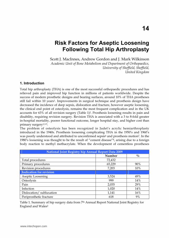
14
Risk Factors for Aseptic Loosening Following Total Hip Arthroplasty
Scott J. MacInnes, Andrew Gordon and J. Mark Wilkinson Academic Unit of Bone Metabolism and Department of Orthopaedics,
University of Sheffield, Sheffield, United Kingdom
1. Introduction
Total hip arthroplasty (THA) is one of the most successful orthopaedic procedures and has relieved pain and improved hip function in millions of patients worldwide. Despite the success of modern prosthetic designs and bearing surfaces, around 10% of THA prostheses still fail within 10 years1. Improvements in surgical technique and prosthesis design have decreased the incidence of deep sepsis, dislocation and fracture, however aseptic loosening, the clinical end point of osteolysis, remains the most frequent complication and in the UK accounts for 63% of all revision surgery (Table 1)2. Prosthesis loosening results in pain and disability, requiring revision surgery. Revision THA is associated with a 3 to 8-fold greater in-hospital mortality, poorer functional outcome, longer hospital stay, and higher cost than primary surgery1,3-5. The problem of osteolysis has been recognized in Judet’s acrylic hemiarthroplasty introduced in the 1940s. Prosthesis loosening complicating THA in the 1950’s and 1960’s was poorly understood and attributed to unconfirmed sepsis6 and prosthesis motion7. In the 1980’s loosening was thought to be the result of “cement disease”8, arising due to a foreign body reaction to methyl methacrylate. When the development of cementless prostheses
National Joint Registry hip Annual Report Data 2009
Number %
Total procedures 72,432
Primary procedures 65,229 90%
Revision procedures 7,203 10%
Indication for revision
Aseptic Loosening 3,524 49%
Osteolysis 999 14%
Pain 2,035 29%
Infection 1,020 14%
Dislocation/ subluxation 1,141 16%
Periprosthetic fracture 618 9%
Table 1. Summary of hip surgery data from 7th Annual Report National Joint Registry for England and Wales2
www.intechopen.com
Recent Advances in Arthroplasty 276
failed to eliminate this problem, wear at the bearing couple was subsequently identified as the main source of particulate debris giving rise to osteolysis. Advances in prosthesis materials, design and surgical technique have improved the wear performance of prostheses, which will decrease the future incidence of osteolysis. However, an ageing population combined with younger more active patients now undergoing joint arthroplasty suggests that osteolysis and resulting prosthesis loosening will continue to be the major complication of THA.
2. Pathophysiology of osteolysis
The term aseptic loosening describes mechanical failure of the prosthesis-host interface, and arises primarily as the end result of focal periprosthetic inflammatory bone loss occurring at this interface. This pro-inflammatory microenvironment is driven by particulate wear debris, which is generated primarily at the articular bearing surface and at other non-articular prosthesis or cement surfaces9. Willert first proposed the involvement of prosthetic debris in the development of oesteolysis. He identified a resultant foreign body reaction and granuloma formation which included macrophages and multinucleated giant cells10. This foreign body reaction has subsequently been reproduced in animal models11. Once particulate wear debris has been dispersed into the joint fluid it may initiate a foreign body reaction at contact surfaces with the host tissues. Schmalzried coined the term “effective joint space” to describe all areas where open communication with the joint pseudo-capsule may allow circulation of the joint fluid and particulate debris12. The effective joint space is thus dynamic and may advance along a tissue plane as osteolysis progresses. Variations in hydrostatic pressure within the joint space during activity may contribute to this circulation12. As well as its role in the migration of wear particles, hydrostatic fluid pressure changes within the joint have been implicated as an osteolytic stimulus. Aspenberg showed in an animal model that fluid pressure alone can lead to osteolysis13. Skoglund also showed that the osteolytic effect of fluid pressure on the bone was greater than that of particles14. However, it remains unclear what contribution this potential mechanism makes to the development of osteolysis clinically. Early migration of the femoral component may predict early and mid-term prosthesis failure. It has been suggested that this migration may lead to instability resulting in locally high fluid pressures which may, in turn, lead to osteolysis15. However, it is also likely that the predictive value of early migration measurements is due to the identification of failures of initial prosthesis fixation, resulting in loosening due to technical failure.
3. The biology of osteolysis
The process of aseptic loosening is characteristically accompanied with the development of a
fibrous membrane at the bone-cement interface. Histological analysis of this membrane has
shown a synovial-like fibrovascular tissue containing cells including macrophages,
fibroblasts and foreign body giant cells9,16. The predominant cell types driving osteolysis,
the macrophage and fibroblast, signal through various pro-inflammatory cytokines
(including the interleukins, TNF alpha, and vascular endothelial growth factor VEGF)
following either phagocytosis of the particles or through surface contact17.
The biological process through which wear particles induce this inflammatory response is still not fully understood. It has become clear that the innate immune system is involved in
www.intechopen.com
Risk Factors for Aseptic Loosening Following Total Hip Arthroplasty 277
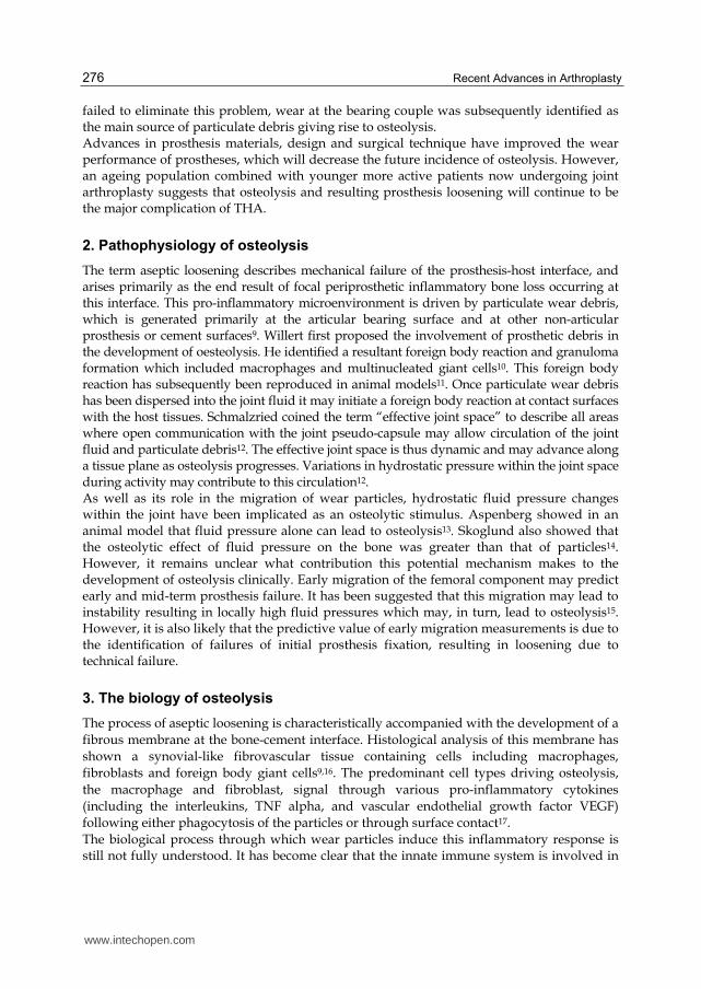
the initiation of the biological response. The innate immune system is the body’s first defense against foreign pathogens. Its ability to recognize and eliminate pathogens relies on pattern recognition receptors (PRR). PRRs are expressed by several cells in the monocyte cell lineage and include toll-like receptors (TLR) and the NOD-like receptors (NLR). These subfamilies evoke an inflammatory response either through the activation of transcription factors or through the formation of inflammasomes (Figure 1). Inflammasomes are large cytoplasmic complexes that activate inflammatory caspases required for the catalysis of pro-IL-1β and pro-IL-18 into their active forms18. Disorders of inflammasome signaling are associated with a number of auto-inflammatory conditions.
Fig. 1. Summary of pattern recognition receptors and their effector pathways. NALP =
NACHT, LRR and PYD domain-containing proteins, IPAF = Ice protease activating factor,
NAIP = neuronal apoptosis inhibitory protein, NOD = nucleotide-binding oligomerization
domain proteins, CIITA = Major histocompatibility complex class-2 transactivator
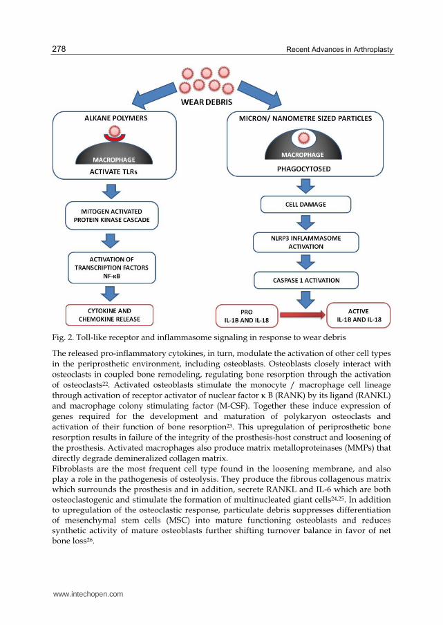
Caicedo et al found that metal implant debris stimulated an inflammatory response in
macrophages through inflammasome signaling (Figure 2)19. Maitra found that UHMWPE
wear particles are phagocytosed causing intracellular activation of NACHT, LRR and PYD
domains-containing protein 3 (NLRP3) leading to inflammasome formation. In addition
alkane polymers generated by UHMWPE activate TLRs through cell surface contact. This
leads to the activation of transcription factors including NF-KB resulting in cytokine
release20. St Pierre et al showed in a mouse model that titanium particles also induce an
inflammatory response through the activation of the NLRP3 inflammasome21.
www.intechopen.com
Recent Advances in Arthroplasty 278
Fig. 2. Toll-like receptor and inflammasome signaling in response to wear debris
The released pro-inflammatory cytokines, in turn, modulate the activation of other cell types
in the periprosthetic environment, including osteoblasts. Osteoblasts closely interact with
osteoclasts in coupled bone remodeling, regulating bone resorption through the activation
of osteoclasts22. Activated osteoblasts stimulate the monocyte / macrophage cell lineage
through activation of receptor activator of nuclear factor κ B (RANK) by its ligand (RANKL)
and macrophage colony stimulating factor (M-CSF). Together these induce expression of
genes required for the development and maturation of polykaryon osteoclasts and
activation of their function of bone resorption23. This upregulation of periprosthetic bone
resorption results in failure of the integrity of the prosthesis-host construct and loosening of
the prosthesis. Activated macrophages also produce matrix metalloproteinases (MMPs) that
directly degrade demineralized collagen matrix.
Fibroblasts are the most frequent cell type found in the loosening membrane, and also play a role in the pathogenesis of osteolysis. They produce the fibrous collagenous matrix which surrounds the prosthesis and in addition, secrete RANKL and IL-6 which are both osteoclastogenic and stimulate the formation of multinucleated giant cells24,25. In addition to upregulation of the osteoclastic response, particulate debris suppresses differentiation of mesenchymal stem cells (MSC) into mature functioning osteoblasts and reduces synthetic activity of mature osteoblasts further shifting turnover balance in favor of net bone loss26.
www.intechopen.com
Risk Factors for Aseptic Loosening Following Total Hip Arthroplasty 279
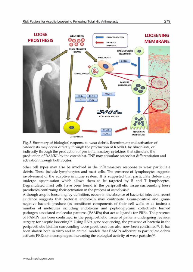
Fig. 3. Summary of biological response to wear debris. Recruitment and activation of osteoclasts may occur directly through the production of RANKL by fibroblasts, or indirectly through the production of pro-inflammatory cytokines that stimulate the production of RANKL by the osteoblast. TNF may stimulate osteoclast differentiation and activation through both routes.
other cell types may also be involved in the inflammatory response to wear particulate
debris. These include lymphocytes and mast cells. The presence of lymphocytes suggests
involvement of the adaptive immune system. It is suggested that particulate debris may
undergo opsonisation which allows them to be targeted by B and T lymphocytes.
Degranulated mast cells have been found in the periprosthetic tissue surrounding loose
prostheses confirming their activation in the process of osteolysis27
Although aseptic loosening, by definition, occurs in the absence of bacterial infection, recent
evidence suggests that bacterial endotoxin may contribute. Gram-positive and gram-
negative bacteria produce (as constituent components of their cell walls or as toxins) a
number of molecules including endotoxins and peptidoglycans, collectively termed
pathogen associated molecular patterns (PAMPs) that act as ligands for PRRs. The presence
of PAMPs has been confirmed in the periprosthetic tissue of patients undergoing revision
surgery for aseptic loosening28. Using RNA gene sequencing, the presence of bacteria in the
periprosthetic biofilm surrounding loose prostheses has also now been confirmed29. It has
been shown both in vitro and in animal models that PAMPs adherent to particulate debris
activate PRRs on macrophages, increasing the biological activity of wear particles30.
www.intechopen.com
Recent Advances in Arthroplasty 280
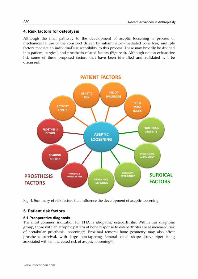
4. Risk factors for osteolysis
Although the final pathway to the development of aseptic loosening is process of
mechanical failure of the construct driven by inflammatory-mediated bone loss, multiple
factors mediate an individual’s susceptibility to this process. These may broadly be divided
into patient, surgical, and prosthesis-related factors (Figure 4). Although not an exhaustive
list, some of these proposed factors that have been identified and validated will be
discussed.
Fig. 4. Summary of risk factors that influence the development of aseptic loosening
5. Patient risk factors
5.1 Preoperative diagnosis
The most common indication for THA is idiopathic osteoarthritis. Within this diagnosis
group, those with an atrophic pattern of bone response to osteoarthritis are at increased risk
of acetabular prosthesis loosening31. Proximal femoral bone geometry may also affect
prosthesis survival, with large non-tapering femoral canal shape (stove-pipe) being
associated with an increased risk of aseptic loosening31.
www.intechopen.com
Risk Factors for Aseptic Loosening Following Total Hip Arthroplasty 281
Higher rates of prosthesis loosening also occur in patients who have undergone arthroplasty for post-traumatic arthritis and osteonecrosis when compared with primary osteoarthritis. However, it is thought that this finding may relate to higher activity levels and increased bearing surface wear, rather than being a function of the pre-operative diagnosis32,33. A number of preoperative diagnoses carry a possible increased risk of prosthesis failure through associated medication. Patients taking systemic steroids have been found to have a higher risk of reoperation34. Non-steroidal anti-inflammatory drugs (NSAIDs) have been implicated in impaired bone healing, and patients taking NSAIDs have higher reoperation rates, although NSAID use may be acting as a marker of a painful prosthesis rather than contributing directly to prosthesis failure34. Poorer prosthesis survival might be expected in patients with inflammatory arthropathy due to its inflammatory pathogenesis and the historic frequent use of corticosteroids in its treatment (that are associated with loss of bone mass through osteoblast suppression). However, Furnes et al, in a large arthroplasty registry-based study, found no difference in THA survival between patients with rheumatoid arthritis versus those with osteoarthritis35. Rud-Sorensen et al found that the risk of stem revision due to aseptic loosening was lower in rheumatoid patients versus primary osteoarthritis, whilst acetabular prosthesis survival was similar36. Furnes et al and Bordini et al have reported higher acetabular revision rates due to aseptic loosening in patients with a primary diagnosis of developmental dyplasia of the hip compared to primary osteoarthritis37,38. Rates of acetabular prosthesis failure are higher in younger patients and those with greater graft coverage of the cup39. The role of these factors is unclear, but may relate to activity levels, or mechanical factors influencing prosthesis support.
5.2 Body mass index and obesity
The Health Survey for England 2009 showed that over the last 16 years there has been
marked increase in the proportion of the population that are obese. This proportion
increased from 13% of men in 1993 to 22% in 2009 and from 16% of women in 1993 to 24% in
200940. The mean BMI of a patient undergoing THA in England and Wales has increased
over the last 5 years from 27.4 to 28.4. Likewise the percentage of patients classed as either
obese or morbidly obese has risen from 29% in 2004 to 37%
Historically, obesity has been deemed a relative contraindication for THA41, as the joint
reaction force experienced at the hip is directly proportional to body weight, and thus
obesity was considered a risk factor for prosthesis failure. Obesity is associated with a
higher incidence of perioperative complications including cardiovascular and respiratory
events42, venous thrombosis43, wound infection44, and dislocation45. However, despite the
increase in joint load in these patients, no consistent increase in bearing wear or osteolysis
has been shown across study populations46,47 and thus obesity is not a clear risk factor for
osteolysis.
5.3 Bearing-surface wear and activity level
Patient activity level associates with osteolysis. It is thought this association operates
primarily though the production of wear of the bearing surface. Flugsrud showed that
patients who undertake intermediate to intense activity are four times more likely than the
less-active to develop acetabular prosthesis loosening48. A recent study with five to ten year
www.intechopen.com
Recent Advances in Arthroplasty 282
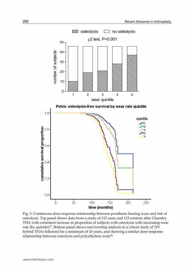
Fig. 5. Continuous dose-response relationship between prosthesis bearing wear and risk of osteolysis. Top panel shows data from a study of 115 cases and 115 controls after Charnley THA with consistent increase in proportion of subjects with osteolysis with increasing wear rate (by quintile)57. Bottom panel shows survivorship analysis in a cohort study of 319 hybrid THAs followed for a minimum of 10 years, and showing a similar dose-response relationship between osteolysis and polyethylene wear58.
2 test, P<0.001
1 2 3 4 50
10
20
30
40
50
no osteolysisosteolysis
wear quintile
num
ber
of
subje
cts
www.intechopen.com
Risk Factors for Aseptic Loosening Following Total Hip Arthroplasty 283
follow up has shown that 24% of patients who have engaged in high levels of activity
developed femoral osteolysis, and had higher revision rates49.
Traditionally the rate of polyethylene wear has been reported as a function of time. The results from ex-vivo hip simulator experiments have shown that the number of hip cycles is proportional to the rate of wear of prosthesis surface50. In vivo, there is a great range of wear rates between individual as a consequence of differing activity levels51. Several validated assessment tools have been developed to measure activity levels in arthroplasty populations52, and Schmalzried et al showed that wear in patients is a function of activity53. There are no clear guidelines outlining what levels of activity can be undertaken
following THA although the proportion of patients participating in athletic activity
following THA ranges between 52 – 83%54-56. Whilst low-impact activities such as
walking, swimming and cycling have always been recommended following THA, some
patients participate in more high-impact and competitive sports. The increasing
participation in athletic activity and higher post-operative expectations can partly be
explained by the increasing numbers of younger patients undergoing THA. 42% of men
and 31% of women who underwent THA in England and Wales in 2009 were under the
age of 65 years2. A large number of patients over the age of 65 are also participating in
high levels of activity49.
Several investigators have shown a relationship between high levels of polyethylene wear
and osteolysis/aseptic loosening, and the concept of a wear-rate ‘threshold’ (commonly
defined as 0.1mm/year) below which osteolysis occurs very rarely, has been suggested.
Wilkinson et al quantitated the association between wear and osteolysis and found no
evidence to support this concept. In a case-control study of 230 hips after cemented
Charnley THA with a metal on polyethylene bearing they showed that the risk of osteolysis
increased with each quintile increase in wear, from very low levels of wear, below the
suggested threshold, through to high levels57. They subsequently showed that the risk of
osteolysis showed a similar pattern of consistently increasing risk ratio with each wear rate
quintile in a separate cohort study of patients with 319 hybrid THAs using a metal on
conventional polyethylene bearing (Figure 5).
5.4 Genetic factors
Within a given ethnic population the sequence of DNA between individuals is 99.5%
identical. However, variability within the code does occur and gives rises to the phenotypic
variability within the population. These variants occur at approximately every 1000
nucleotide base pairs of the code. This variation, where it occurs in >1% of the population is
termed a polymorphism. The most common type of variant is a single letter change in the
DNA sequence, termed a single nucleotide polymorphism (SNP). There are thought to be
around 10 million common SNPs in the human genome. The individual specific risk of
common diseases is thought to be influenced by the sum of many genetic variations, each
potentially causing small changes in biological function and consequently subtle changes in
phenotype59.
Patients vary in their osteolytic response to particulate wear debris. Some show little bone
resorption in the presence of marked prosthesis wear whereas others undergo marked
osteolysis following a small amount of prosthesis wear (Figure 6)57. Macrophage
responsiveness to in-vitro particulate debris stimulation also varies between individual60,
www.intechopen.com

Recent Advances in Arthroplasty 284
and monocytes (PBMCs) taken from patients with a susceptibility to osteolysis exhibit
quantitatively greater inducible cytokine responses to particulate debris in-vitro versus
patients without this susceptibility61 . It is suggested that this inter-patient variability may
have a genetic basis.
Fig. 6. Patients exhibit variable osteolytic responses to wear debris. a) radiograph showing marked polyethylene wear, but no osteolytic response, b) radiograph showing mild wear but pronounced femoral and acetabular osteolysis with prosthesis loosening.
Variation within the genes encoding inflammatory cytokines have been associated with
osteolysis. Wilkinson et al showed an association between variability within the DNA
encoding the tumor necrosis factor (TNF) promoter region (dbSNP rs361525) and risk of
osteolysis following THA62. Subjects with osteolysis were approximately twice as likely to
carry the variant DNA code as those subjects with no osteolysis. This association has been
replicated in an independent population by Ambruzova et al63. Gordon et al have reported
genetic variation within the genes encoding Interleukin-1 receptor antagonist (IL-1RN) and
IL-6 is also associated with osteolysis64. Similar associations have also been identified in
other populations65-67.
Variation within genes that regulate bone turnover also associate with osteolysis. Gordon et
al showed that carriage of the dbSNP rs288326 variant in the FRZB gene encoding secreted
frizzled-related protein-3 (Frp3), a regulatory glycoprotein within the osteogenic Wnt
signaling pathway that modulates mesenchymal stem cell differentiation of osteoblasts68,
associated with susceptibility to osteolysis following THA69. Its carriage also associated with
www.intechopen.com
Risk Factors for Aseptic Loosening Following Total Hip Arthroplasty 285
the development of heterotopic ossification following THA. Malik et al have also shown
associations between aseptic loosening and other candidate loci within the genes encoding
matrix metalloproteinase 1 and the vitamin D receptor67, mannose-binding lectin70, and the
RANK/OPG pathway71.
Recent studies using beadchip assays have shown that many genes are differentially expressed in wear debris-induced cells and tissues72-74, and have highlighted our limited understanding of the spectrum of biological mediators involved in the pathogenesis of osteolysis. The identification of further risk loci is required to further understanding of the pathogenesis of aseptic loosening. This would potentially allow for the development of screening tools, and provide investigational targets for prophylaxis or treatment with the aim of reducing the need for revision surgery, and its associated morbidity and mortality.
6. Prosthesis risk factors
6.1 Prosthesis design
Prosthesis design factors, aside from those that modulate wear, contribute to risk of osteolysis. Modularity allows intra-operative adjustment of bearing surfaces, prosthesis length and offset. However, it also creates additional interfaces within the construct at which generation of debris through wear may occur. Such interfaces include the trunion between the femoral head and stem at which corrosive wear may occur, and backside wear between an acetabular liner and its shell at which abrasive wear may occur, and potentially several other prosthetic component junctions in highly modular systems. Hydroxyapatite coating of the prosthesis may prevent osteolysis following injection of intra-articular particles by sealing the implant-bone interface from their ingression though the promotion of osseointegration at this interface75,76, but may also be a source of third-body wear. Selection of bearing diameter is also a factor. The use of larger head sizes reduces the risk of dislocation, but increase volumetric wear77. The need for a thinner liner to accommodate the larger head may also cause increased contact stresses and an increase in wear.
6.2 Polyethylene wear
The metal on polyethylene bearing couple remains the gold standard for THA. However, the manufacturing and sterilization process of polyethylene has changed over time with the aim of improving its wear rate characteristics. The earliest prostheses were made with non-cross-linked ultra-high molecular weight polyethylene (UHWPE) that was irradiated to render it sterile for patient use. The process of sterilization with ionizing radiation leads to cross-linking within the polymer. Cross-linking improves wear resistance of the material, but also causes the formation of free radicals. Free radical species cause the oxidation of UHMWPE over time. Polyethylene oxidation degrades UHMWPE, and decreases its wear resistance. Several production techniques have been developed to reduce the generation of free radicals, including annealing and melting. Melting reduces free radical concentration more than annealing but adversely affects the yield stress and fatigue resistance of the polymer. Annealing below melting point has a less adverse effect on the mechanical properties, but is less effective than melting at free radical removal. Sterilization in an oxygen-free environment also produces more cross-linking and reduces free radical production78. Irradiation in an inert gas and vacuum packing is also now routinely carried out to reduce pre-implantation oxidation, however this does not prevent oxidation occurring in vivo. Faris
www.intechopen.com
Recent Advances in Arthroplasty 286
et al compared the wear rates of UHMWPE produced using three combinations of polyethylene production and sterilization techniques79 and found the best wear rates were achieved in sterilization by radiation in an inert gas with molded polyethylene. Irradiation sterilization of ram extruded components in an inert gas and in air had 11% and 16% more wear respectively. Highly cross-linked polyethylene has exhibited reduced wear rates clinically in short-term studies80,81, and thus their potential role in reducing the incidence of osteolysis is promising. Further developments in polyethylene modification techniques are currently being explored to further reduce oxidization in-vivo and optimize the wear performance of UHMWPE without compromising its other mechanical properties, and include doping with anti-oxidants such as vitamin E and cycling of annealing and irradiating. However, the macrophage response in osteolysis is influenced by the size, composition and number of wear particles82,83. Particle size and number vary with the extent of cross-linking within the material. Although cross-linking reduces the total amount of wear debris generated versus conventional UHMWPE, the particle size produced is smaller, and the number of particles is increased, which may enhance their osteolytic potential in-vivo. Also, whilst increased cross-linking results in enhanced wear resistance there is a reduction in fatigue strength potentially leading to mechanical failure84.
6.3 Alternate bearing couples
Although metal on polyethylene bearings have most commonly been used in THA, there is a long history of use of other bearing couples, including metal on metal, ceramic on ceramic, and ceramic on polyethylene. Metal on metal bearings have reduced wear rates compared with metal on polyethylene.
Jacobbson reported a 77% 20-year survivorship of the metal on metal McKee Farrar THA
compared to 73% for the Charnley THA85. Metal on metal prostheses also have the
advantages of allowing a larger bearing diameter, improving stability characteristics, and
are self-polishing. Although the volumetric wear rate of metal on metal bearings is low, the
particles generated are in the nanometer range and the number of particles is far greater86.
These particles circulate widely within the body and their systemic effects remain unclear.
At a local level metal release can cause an adverse surrounding tissue reaction, termed
aseptic lymphocytic vasculitis associated lesions (ALVAL), and inflammatory masses87,88.
Metal hypersensitivity may also occur87.
Ceramic on polyethylene and ceramic on ceramic bearing couples have lower wear and
osteolysis rates versus metal on polyethylene bearings in some long-term studies89,90. Most
ceramic wear particles are also in the nanometer range and wear volume is lower than that
of metal on metal bearing couples. A prospective randomized multicenter study of 930 hips
comparing alumina-on-alumina with cobalt chromium-on-polyethylene bearing couples
reported an alumina-alumina survival rate of 96.8% at 10 years91. However, cases of
osteolysis have also been reported in poorly functioning ceramic on ceramic prostheses.
Yoon reported osteolysis rates of 22% in a series of patients with ceramic on ceramic
prostheses92. Nam reported a case of alumina debris induced pelvic and femoral osteolysis
in a well-functioning prosthesis93. Ceramics are also expensive, have a small fracture risk
due to their brittleness, and are sensitive to component mal-positioning that may result in
impingement damage and stripe wear. There are also some reports of squeaking associated
with ceramic on ceramic bearing couples94.
www.intechopen.com
Risk Factors for Aseptic Loosening Following Total Hip Arthroplasty 287
7. Surgical risk factors
Regardless of prosthesis design and bearing surface, surgical technique is an important factor that affects prosthesis survival. Data from large national joint registries has recently facilitated examination of these factors in relation to prosthesis survival.
7.1 Hospital type and surgeon operating volume
Type of hospital and the surgeon undertaking the procedure can influence THA survival.
Fowles et al showed that low operating volume is associated with increased risk of THA
revision95. Similarly, Espehaug et al, using data from the Norwegian arthroplasty register,
found the lowest revision rates amongst surgeons with the highest THA volume96. In the
same study, university hospitals had higher revision rates than local and central hospitals.
This may be attributed to the lower number of operations per surgeon at these hospitals or
possible centralization of high-risk patients and more complex cases. Bordini et al found that
prosthesis survival was negatively associated with lower surgeon skill38.
7.2 Prosthesis alignment and soft tissue balancing
Malalignment of prostheses may alter the articulation of prosthesis components with the
potential to increase contact stresses and increase wear, this increases the incidence of edge
loading and results in stripe wear in hard on hard bearing couples. Despite the advantage of
larger femoral head size, soft tissue balancing remains important in the reduction of
dislocation of the femoral head. Subluxation of the femoral head during the swing phase of
gait, especially in metal on polyethylene couples, causes socket edge contact resulting in
wear97. Complete dislocation of the femoral head may damage the head during dislocation-
relocation, and can increase wear rates.
7.3 Prosthesis dislocation and interface micromotion
Prosthesis stability influences the development of aseptic loosening. Motion between the
prosthesis and bone contributes to the formation of a fibrous membrane rather than bone98.
Bechtold et al found that particulate wear debris prevents bone formation in the presence of
prosthesis instability 99. In addition, prosthesis motion alters local joint fluid pressures and
can transport particles along the periprosthetic space.
7.4 Cementing techniques
Improvements in prosthesis survival have accompanied advances in cementation
technique100. First generation cementing techniques involved finger packing of the cement
without bone preparation, pressurization or use of a medullary plug. In the mid-seventies
second generation techniques were adopted which involved improved canal preparation by
pulsatile lavage that increased cement penetration and interdigitation, retrograde insertion
of cement using a gun to reduce blood lamination, and the use of an intramedullary plug to
limit the size of the cement column. Studies with 10 year follow up have shown that 2nd
generation techniques were associated with a reduced the incidence in femoral loosening
with rates of 3 to 7%101,102 compared with rates of approximately 30% at 10 years in first
generation reports103,104. Third generation techniques included vacuum mixing of cement to
reduce cement porosity and increase fatigue strength105, and cement pressurization to
www.intechopen.com
Recent Advances in Arthroplasty 288
further improve cement interdigitation. Subsequently 4th generation cementation techniques
have added distal and proximal prosthesis centralizers to improve the stem position
allowing for an optimal and even cement mantle. Herbert, in a review of the Swedish THA
Register examining 160,000 cases, reported that the evolution from 1st to 3rd generation
cementing techniques over a 20 year period was associated with a reduced incidence of
revision for aseptic loosening100.
8. Summary and future directions
Aseptic loosening is the end result of a complex interaction of variables leading to development of osteolysis. Although the last 30 years has seen many advances in the understanding of these factors, osteolysis will remain a problem for the foreseeable future. Newer bearing surfaces have shown potential in wear rate reduction. However, wear particles from all materials have the potential to trigger an inflammatory response. The local and systemic consequences of metal release also need to be more clearly defined and quantitated. Further studies looking at prosthesis bone anchorage in conjuction with particle and pressure effects need to be explored, and the factors that influence loosening membrane formation. Currently the only effective treatment for aseptic loosening is revision surgery. Future advances in our understanding of the biological response to wear particles may lead to the development of biological markers for better prediction and early detection of osteolysis, and the development of non-surgical solutions for prophylaxis and therapy. Advances in genomic and bioinformatics technology have provided us with the opportunity to identify investigational targets for prophylaxis or treatment. Pharmocological and biological agents used in the treatment of osteolysis in metastatic disease and metabolic bone disease may have potential in osteolysis following THA.
9. References
[1] Kurtz SM, Ong KL, Schmier J, Mowat F, Saleh K, Dybvik E, et al. Future clinical and economic impact of revision total hip and knee arthroplasty. J Bone Joint Surg 2007 Oct;89-A Suppl 3:144-51.
[2] The NJR centre HH. National joint registry for England and Wales. 7th annual report, 2010. Available from URL: www.njrcentre.co.uk.
[3] Mahomed NN, Barrett JA, Katz JN, Phillips CB, Losina E, Lew RA, et al. Rates and outcomes of primary and revision total hip replacement in the United States medicare population. J Bone Joint Surg 2003 Jan;85-A(1):27-32.
[4] Doro C, Dimick J, Wainess R, Upchurch G, Urquhart A. Hospital volume and inpatient mortality outcomes of total hip arthroplasty in the United States. J Arthroplasty 2006 Sep;21(6 Suppl 2):10-6.
[5] Zhan C, Kaczmarek R, Loyo-Berrios N, Sangl J, Bright RA. Incidence and short-term outcomes of primary and revision hip replacement in the United States. J Bone Joint Surg 2007 Mar;89-A(3):526-33.
[6] Charnley J, Follacci FM, Hammond BT. The long-term reaction of bone to self-curing acrylic cement. J Bone Joint Surg. Br. 1968: 50; 822-29.
[7] McKee GK, Watson-Farrar J. Replacement of arthritic hips by the McKee Farrar prosthesis. J Bone Joint Surg. Br. 1966; 48 (2): 245-59.
[8] Jones LC, Hungerford DS. “Cement disease.” Clin Orthop 1987; 225: 192-206.
www.intechopen.com
Risk Factors for Aseptic Loosening Following Total Hip Arthroplasty 289
[9] Goldring SR, Schiller AL, Roelke M, Rourke CM, O'Neill DA, Harris WH. The synovial-like membrane at the bone-cement interface in loose total hip replacements and its proposed role in bone lysis. J Bone Joint Surg 1983 Jun;65-A(5):575-84.
[10] Willert HG, Semlich M. Reactions of the articular capsule to wear products of artificial joint prostheses. J Biomed Mater Res. 1977; 11:157-164.
[11] Goodman SB, Fornasier VL, Lee J, Kei J. The histological effects of the implantation of different sizes of polyethylene particles in the rabbit tibia. J Biomed Mater Res. 1990; 24: 517 – 524.
[12] Schmalzried TP, Jasty M, Harris WH. Periprosthetic bone loss in total hip arthroplasty. Polyethylene wear debris and the concept of the effective joint space. J Bone Joint Surg Am 1992a; 74: 849 – 63.
[13] Aspenberg P, Van der Vis H. Fluid pressure may cause periprosthetic osteolysis. Particles are not the only thing. Acta Orthop Scand 1998; 69: 1-4.
[14] Skoglund B, Aspenberg P. PMMA particles and pressure – a study of the osteolytic properties of two agents proposed to cause prosthetic loosening. J Orthop Res 2003; 21: 196-201.
[15] Aspenberg P, Van der Vis H. Migration, particles, and fluid pressure. A discussion of causes of prosthetic loosening. Clin Orthop Rel Res 1998; 352: 75-80.
[16] Harris WH, Schiller AL, Scholler J, Freiberg RA, Scott R. Extensive localized bone resorption in the femur following total hip replacement. J Bone Joint Surg Am 1976; 58: 612-618. .
[17] Tuan RS, Lee FY, Konttinen YT, Wilkinson JM, Smith RL. What are the local and systemic biologic reactions and mediators to wear debris, and what host factors determine or modulate the biologic response to wear particles? J Am Acad Orthop Surg 2008; 16(supl 1): S42-S48.
[18] Martinon F, Mayor A, Tschopp J. The inflammasomes: guardians of the body. Annu Rev Immunol. 2009; 27: 229-65.
[19] Caicedo, M. S. et al. Soluble and particulate Co-Cr-Mo alloy implant metals activate the inflammasome danger signaling pathway in human macrophages: a novel mechanism for implant debris reactivity. J Orthop Res 27, 847-854, doi:10.1002/jor.20826 (2009).
[20] Maitra R, Clement CC, Scharf B, Crisi GM, Chitta S, Paget D, Purdue PE, Cobelli N, Santambrogio L. Endosomal damage and TLR2 mediated inflammasome activation by alkane particles in the generation of aseptic osteolysis. Mol Immunol. 2009 Dec;47(2-3):175-84.
[21] St Pierre CA, Chan M, Iwakura Y, Ayers DC, Kurt-Jones EA, Finberg RW. Periprosthetic osteolysis: characterizing the innate immune response to titanium wear-particles. J Orthop Res. 2010 Nov; 28(11): 1418-1424.
[22] Rodan GA, Martin TJ. Role of osteoblasts in hormonal control of bone resorption- A hypothesis. Calcif Tissue Int 1981;33: 349-351.
[23] Boyle WJ, Simone WS, Lacey DL. Osteoclast differentation and activation. Nature 2003; 423: 337-342.
[24] Wei X, Zhang X, Zuscik MJ, Drissi MH, Schwarz EM, O'Keefe RJ Fibroblasts express RANKL and support osteoclastogenesis in a COX-2-dependent manner after stimulation with titanium particles. J Bone Miner Res 2005;20:1136-48.
[25] Sakai H, Jingushi S, Shuto T, Urabe K, Ikenoue T, Okazaki K, Kukita T Fibroblasts from the inner granulation tissue of the pseudocapsule in hips at revision
www.intechopen.com
Recent Advances in Arthroplasty 290
arthroplasty induce osteoclast differentiation, as do stromal cells. Ann Rheum Dis 2002;61:103-9).
[26] Wang ML, Tuli R, Manner PA, Sharkey PF, Hall DJ, Tuan RS. Direct and indirect induction of apoptosis in human mesenchymal stem cells in response to titanium particles. J Orthop Res 2003; 21: 697-707.
[27] Solovieva SA, Ceponis A, Konttinen YT, Takagi M, Suda A, Eklund KK, Sorsa T, Santavirta S. Mast cells in loosening of totally replaced hips. Clin Orthop Rel Res. 1996 Jan;(322): 158-165.
[28] Nalepka JL, Lee MJ, Kraay MJ, Marcus RE, Goldberg VM, Chen X, Greenfield E. Lipopolysaccharide found in aseptic loosening of patients with inflammatory arthritis. Clin Orthop Rel Res 2006; 451: 229-235.
[29] Dempsey KE, Riggio MP, Lennon A, Hannah VE, Ramage G, Allan D, Bagg J. Identification of bacteria on the surface of clinically infected and non-infected prosthetic hip joints removed during revision arthroplasties by 16S rRNA gene sequencing and by microbiological culture. Arthritis Res Ther 2007; 9:R46.
[30] Greenfield EM, Bechtold J. What other biologic and mechanical factors might contribute to osteolysis? J Am Acad Orthop Surg 2008; 16 (suppl 1): S56-S62.
[31] Kobayashi S, Saito N, Horiuchi H, Iorio R, Takaoka K Poor bone quality or hip structure as risk factors affecting survival of total-hip arthroplasty. Lancet 2000;355:1499-504.
[32] Tsao AK. What patient and surgical factors contribute to implant wear and osteolysis in total joint arthroplasty. J Am Acad Orthop Surg 2008; 16 (suppl 1):S7–S13.
[33] Letson GD. Activity relationships of THA in patients with osteonecrosis and osteonecrosis. Orthopedics. 1996 Aug; 19 (8):665-8.
[34] Espehaug B, Havelin LI, Engesaeter LB, Langeland N, Vollset SE Patient-related risk factors for early revision of total hip replacements. A population register-based case-control study of 674 revised hips. Acta Orthop Scand 1997;68:207-15.
[35] Furnes, O. et al. Hip disease and the prognosis of total hip replacements. A review of 53,698 primary total hip replacements reported to the Norwegian Arthroplasty Register 1987-99. J Bone Joint Surg Br 83, 579-586 (2001).
[36] Rud-Sorensen, C., Pedersen, A. B., Johnsen, S. P., Riis, A. H. & Overgaard, S. Survival of primary total hip arthroplasty in rheumatoid arthritis patients. Acta Orthop 81, 60-65, doi:10.3109/17453671003685418 (2010).
[37] Furnes O, Lie SA, Vollset, Engesater LB, Havelin LI. Hip disease and the survival of hip prostheses in the Norwegian Arthroplasty Register. Acta Orthop Scand (Suppl 280) 1998;69.
[38] Bordini, B. et al. Factors affecting aseptic loosening of 4750 total hip arthroplasties: multivariate survival analysis. BMC Musculoskelet Disord 8, 69 (2007).
[39] Papachristou G, Hatzigigoris P, Panousis K, Plessas S, Sourlas J, Levidiotis C, Chronopoulos E. Total hip arthroplasty for developmental hip dysplasia. International Orthopaedics. 2006; 30: 21-25.
[40] Health Survey for Enlgland 2009: Trend Tables. The Health and Social Care Information Centre, 2010.
[41] Charnley J. Long term results of low friction arthroplasty. Hip 1982: 42-9. [42] Adams JP, Murphy PG. Obesity in anaesthesia and intensive care. Br J Anaesth 2000;
85: 91-108.
www.intechopen.com
Risk Factors for Aseptic Loosening Following Total Hip Arthroplasty 291
[43] LoweGD, HaverkateF, Thompson SG. Prediction of deep venous thrombosis after hip replacement surgery by preoperative clinical and haemostatic variables: the ECAT DVT study: European concerted action on thrombosis. Thromb Haemostat 2003; 89: 493-498.
[44] Namba RS, Paxton L, Fithian DC, Stone ML. Obesity and perioperative morbidity in total hip and knee arthroplasty patients. J Arthroplasty 2005; 20 (suppl 3): 46-50.
[45] Chee YH. Total hip replacement in the morbidly obese patient with OA. JBJS 2010; 92-B(8): 1066-1071.
[46] Lubbeke A, Garavaglia G, Barea C, Roussos C, Stern R, Hoffmeyet P. Influence of obesity on femoral osteolysis five and ten years following total hip arthroplasty. J Bone Joint Surg Am 2010; 92: 1964 – 1972.
[47] Andrew JG, Palan J, Kurup HV, Gibson P, Murray DW, Beard DJ. Obesity in toal hip replacement. J Bone Joint Surg Br 2008; 90: 424-429.
[48] Flugsrud GB, Nrdsletten L, Espenhaug B, Havelin LI, Meyer HE. The effect of middle-age body weight and physical activity on the risk of early revision hip arthroplasty: a cohort study of 1,535 individuals. Acta Orthop 2007; 86: 963-974.
[49] Lubbeke A, Garavaglia G, Barea, Stern R, Peter R, Hoffmeyer P. Influence of patient activity on femoral osteolysis at five and ten years following hybrid total hip replacement. J Bone Joint Surg Br 2011; 93: 456-463. .
[50] Clark, I C Wear of artificial joint materials. Friction and wear studies. Eng Med 10: 115 – 122, 1981.
[51] Schmalzried TP, Szuszczewicz ES, Northfield MR, Akizuki KH, Frankel RE, Belcher G, Amstutz HC. Quantitative assessment of walking activity after total hip or knee replacement. J Bone Joint Surg Am 1998; 80: 54-59.
[52] Naal FD, Impellizzeri FM, Leunig M. Which is the best activity rayting scale for patient undergoing total joint arthoplasty? Clin Orthop Rel Res 2009; 467: 958-965.
[53] Schmalzried TP, Shepherd EF, Dorey FJ, Jackson WO, dela Rosa M, Fa’vae F, McKellop HA, McClung CD, Martell J, Moreland JR, Amstutz HC. Wear is a function of use, not time. Clin Orthop Rel Res 2000; 381: 36-46.
[54] Huch K, Muller KAC, Sturmer T, Brenner H, Puhl W, Gunther K-P. Sports activities 5 years after total hip or knee arthroplasty: the Ulm osteoarthritis study. Ann Rheum Dis 2005; 64: 1715–1720.
[55] Wylde V, Blom A, Dieppe P, Hewlett S, Learmonth I. Return to sport after joint replacement. J bone Joint Surg Br 2008; 90: 920-923.
[56] Chatterji U, Asworth MJ, Lewis PL, Dobson PJ. Effect of total hip arthroplasty on recreational and sporting activity. ANZ J Surg 2004; 74: 446-449.
[57] Wilkinson JM, Hamer AJ, Stockley I, Eastell R. Polyethylene wear rate and osteolysis: critical threshold versus continuous dose-response relationship. J Orthop Res 2005 May;23(3):520-5.
[58] Emms, N. W., Stockley, I., Hamer, A. J. & Wilkinson, J. M. Long-term outcome of a cementless, hemispherical, press-fit acetabular component: survivorship analysis and dose-response relationship to linear polyethylene wear. J Bone Joint Surg Br 92, 856-861, doi:92-B/6/856 [pii] 10.1302/0301-620X.92B6.23666 (2010).
[59] Misch EA, Hawn TR. Toll –like receptor polymorphisms and susceptibility to human disease. Clin Sci. 2008; 114: 347-360.
[60] Matthews JB, Green TR, Stone MH, Wroblewski BM, Fisher, J, et al. Comparison of the response of primary human peripheral blood mononuclear phagocytes from
www.intechopen.com
Recent Advances in Arthroplasty 292
different donors to challenge with model polyethylene particles of known size and dose. Biomaterials 2000 Oct;21(20):2033-44.
[61] Gordon A, Kiss-Toth E, Greenfield E, Eastell R, Wilkinson J. Differences in cytokine mRNA expression in stimulated mononuclear cells from subjects with previous osteolysis versus non-osteolysis subjects after total hip arthroplasty. Trans ORS 33, 424. 2008.
[62] Wilkinson JM, Wilson AG, Stockley I, Scott IR, Macdonald DA, Hamer AJ, et al. Variation in the TNF gene promoter and risk of osteolysis after total hip arthroplasty. J Bone Miner Res 2003;18(11):1995-2001.
[63] Ambruzova Z, Gallo J, Mrazek F, Kubistova Z, Onderkova J, Petrek M. Association of cytokine gene polymorphisms with expansile periprosthetic osteolysis in total hip arthroplasty. Tissue Antigens 2006;67(6):528.
[64] Gordon A, Kiss-Toth E, Stockley I, Eastell R, Wilkinson JM. Polymorphisms in the Interleukin-1 Receptor Antagonist and Interleukin-6 Genes Affect Risk of Osteolysis in Patients with Total Hip Arthroplasty. Arthritis Rheum 2008;58:3157-65.
[65] Kolundiz R, Orlic D, Truklja V, Pavelic K, Troselj KG. Single nucleotide
polymorphisms in the interleukin-6 gene promoter, tumour necrosis factor- gene
promoter, and transforming growth factor-1 gene signal sequence as predictors of time to onset of aseptic loosening after total hip arthrolasty: preliminary study. J Orthop Sci 2006; 11: 592-600.
[66] Gallo J, Mrazek F, Petrek M. Variation in cytokine genes can contribute to severity of acetabular osteolysis and risk for revision in patients with ABG 1 total hip arthroplasty: a genetic association study. BMC Med Genet 2009; 10:109.
[67] Malik MH, Jury F, Bayat A, Ollier WE, Kay PR. Genetic susceptibility to total hip arthroplasty failure: a preliminary study on the influence of matrix metalloproteinase 1, interleukin 6 polymorphisms and vitamin D receptor. Ann Rheum Dis 2007 Aug;66(8):1116-20.
[68] Martin TJ, Sims NA, Ng KW. Regulatory pathways revealing new approaches to the devlopment of anabolic drugs for osteoporosis. Osteoporos Int 2008; 19: 1125-1138.
[69] Gordon A, Southam L, Loughlin J, Wilson AG, Stockley I, Hamer AJ, et al. Variation in the secreted frizzled-related protein-3 gene and risk of osteolysis and heterotopic ossification after total hip arthroplasty. J Orthop Res 2007 Dec 12;25(12):1665-70.
[70] Malik MH, Bayat A, Jury F, Kay PR, Ollier WE. Genetic susceptibility to total hip arthroplasty failure--positive association with mannose-binding lectin. J Arthroplasty 2007 Feb;22(2):265-70.
[71] Malik MH, Bayat A, Jury F, Ollier WE, Kay PR. Genetic susceptibility to hip arthroplasty failure--association with the RANK/OPG pathway. Int Orthop 2006 Jun;30(3):177-81.
[72] Garrigues GE, Cho DR, Rubash HE, Goldring SR, Herndon JH, Shanbhag AS. Gene expression clustering using self-organizing maps: analysis of the macrophage response to particulate biomaterials. Biomaterials 2005 Jun;26(16):2933-45.
[73] Shanbhag AS, Kaufman AM, Hayata K, Rubash HE. Assessing osteolysis with use of high-throughput protein chips. J Bone Joint Surg 2007 May;89-A(5):1081-9.
[74] Koulouvaris P, Ly K, Ivashkiv LB, Bostrom MP, Nestor BJ, Sculco TP, et al. Expression profiling reveals alternative macrophage activation and impaired osteogenesis in periprosthetic osteolysis. J Orthop Res 2008 Jan;26(1):106-16.
www.intechopen.com
Risk Factors for Aseptic Loosening Following Total Hip Arthroplasty 293
[75] Rahbek O, Kold S, Bendix K, Overgaard S, Soballe K. Superior sealing effect of hydroxyapatite in porous-coated implants: Experimental studies on the migration of polyethylene particles around stable and unstable implants in dogs. Acta Orthop 2005; 76: 375 – 385.
[76] Soballe K, Hansen ES, Brockstedt-Rasmussen H, Bunger C. Hydroxyapatite coating converts fibrous tissue to bone around loaded implants. J Bone Joint Surg Br 1993; 75: 270-278.
[77] Charnley J, Kamangar A, Longfield MD: The optimum size of prosthetic heads in relation to the wear of plastic sockets in total replacement of the hip. Med Biol Eng 1969; 7: 31-39.
[78] Mckellop HA, Shen FW,Campbell P, Ota T. Effect of molecular weight, calcium stearate, and sterilization methods on the wear of ultra high molecular weight polyethylene acetabular cups in a joint simulator. J Orthop Res 1999; 17:329-339.
[79] Faris PM, Ritter MA, Pierce AL, Davis KE, Faris GW. Polyethylene sterilization and production effects wear in total hip arthroplasties. Clin Orthop Rel Res 2006; 453: 305-308.
[80] Dorr LD, Wan Z, Shahrdar C, Sirianni L, Boutary M, Yun A. Clinical performance of a Durasul highly cross linked polyethylene acetabular liner for total hip arthroplasty at five years. J Bone Joint Surg Am 2005; 87: 1816-1821.
[81] Digas G, Karrholm J, Thanner J, Malchau H, Herberts P. The Otto Aufranc Award: Highly cross-linked polyethylene in total hip arthroplasty. Randomized evaluation of penetration rate in cemented and uncemented sockets using radiostereometric analysis. Clin Orthop Rel Res 2004; 429: 6-16.
[82] Shanbhag AS, Jacobs JJ, Black J, Galante JO, Glant TT: Human monocyte response to particlute biomaterials generated in vivo and in vitro. J Orthop Res 1995;13:792-801.
[83] Shanbhag AS, Jacobs JJ, Glant TT, Gilbert JL, Black J, Galante JO. Composition and morphology of wear debris in failed uncemented total hip replacement. J Bone Joint Surg Br; 76: 60-67.
[84] Bradford, L., Baker, D., Ries, M. D. & Pruitt, L. A. Fatigue crack propagation resistance of highly crosslinked polyethylene. Clin Orthop Relat Res, 68-72, doi:00003086-200412000-00011 [pii] (2004).
[85] Jacobsson SA, Djerf K, Wahlstrom O. Tewenty-year results of McKee Farrar versus Charnley prosthesis. Clin Orthop Rel Res 1996; 329: S60-S68.
[86] Doorn PF, Campbell PA, Worrall J, Benya PD, McKellop HA, Amstutz HC. Metal wear particle characterization from metal on metal total hip replacements: Transmission electron microscopy study of periprosthetic tissues and isolated particles. J Biomed Mater Res 1998; 42: 103-111.
[87] Willert, H. G. et al. Metal-on-metal bearings and hypersensitivity in patients with artificial hip joints. A clinical and histomorphological study. J Bone Joint Surg Am 87, 28-36, doi:87/1/28 [pii] 10.2106/JBJS.A.02039pp (2005).
[88] Pandit, H. et al. Pseudotumours associated with metal-on-metal hip resurfacings. J Bone Joint Surg Br 90, 847-851, doi:10.1302/0301-620X.90B7.20213 90-B/7/847 [pii] (2008).
[89] Urban JA, Garvin KL, Boese CK, Bryson L, Pederson DR, Callaghan JJ, Miller RK. Ceramic-on-polyethylene bearing surfaces in total hip arthroplasty. Seventeen to twenty-one-year results. J Bone Joint Surg Am 2001; 83: 1688-1694.
[90] Hannouche D, Hamadouche M, Nizard R, Bozot P, Sedel L. Ceramics in total hip replacement. Clin Orthop Rel Res. 2005; 430: 62-71.
www.intechopen.com
Recent Advances in Arthroplasty 294
[91] Mesko, J. W., D'Antonio, J. A., Capello, W. N., Bierbaum, B. E. & Naughton, M. Ceramic-on-ceramic hip outcome at a 5- to 10-year interval: has it lived up to its expectations? J Arthroplasty 26, 172-177, doi:S0883-5403(10)00261-5 [pii]10.1016/ j.arth.2010.04.029 (2011).
[92] Yoon, TR, Rowe SM, Jung ST, Seon KJ, Maloney WJ. Osteolysis in association with a total hip arthroplasty with ceramic bearing surfaces. J Bone Joint Surg Am 1998; 80: 1459-1468.
[93] Nam KW, Yoo JJ, Lae Kim Y, Kim YM, Lee MH, Kim HJ. Alumina-debris-induced osteolysis in contemporary alumina-on-alumina total hip arthroplasty. A case report. J Bone Joint Surg Am 2007; 89: 2499-2503.
[94] Jarrett, C. A. et al. The squeaking hip: a phenomenon of ceramic-on-ceramic total hip arthroplasty. J Bone Joint Surg Am 91, 1344-1349, doi:91/6/1344 [pii] 10.2106/ JBJS.F.00970 (2009).
[95] Fowles, J., Bunker, J. P. & Schurman, D. J. Hip surgery data yield quality indicators. Bus Health 4, 44-46 (1987).
[96] Espehaug, B., Havelin, L. I., Engesaeter, L. B. & Vollset, S. E. The effect of hospital-type and operating volume on the survival of hip replacements. A review of 39,505 primary total hip replacements reported to the Norwegian Arthroplasty Register, 1988-1996. Acta Orthop Scand 70, 12-18 (1999).
[97] McKellop HA, D’Lima Darryl. How have wear testing and joint simulator studies helped to discriminate among materials and designs? J AM Acad Orthop Surg 2008; 16 (suppl 1): S111-S119.
[98] Bragdon CR, Jasty M, Greene M, Rubash HE, Harris WH. Biologic fixation of total hip implants: Insights gained from a series of canine studies. J Bone Joint Surg Am 2004; 86 (suppl 2): 105-117.
[99] Bechtold JE, Kubic V, Soballe K. Bone ingrowth in the presence of particulate polyethylene: Synergy between interface motion and particulate polyethylene in periprosthetic tissue response. J Bone Joint Surg Br 2002; 84: 915-919.
[100] Herberts P, Malchau H. Long-term registration has improved the quality of hip replacement. A review of the Swedish THR Register comparing 160,000 case. Acta Orthop Scand 2009; 71: 111-121.
[101] Mulroy RD, Harris WH. The effect of inproved cementing techniques on component loosening in total hip replacement: An 11-year radiographic rebiew. J Bone Joint Surg Br 1990; 72: 757-760.
[102] Stauffer RN. Ten year results of second generation femoral cementing in total hip replacement surgery. Read at the Harvard Medical School Postgraduate Course on Total Hip Surgery. Cambridge, MA, Oct, 1991.
[103] Stauffer RN. Ten year follow up study of total hip replacement. J Bone Joint Surg Am 1982; 7: 983-990.
[104] Barrack RL, Mulroy RD, Harris WH. Improved cementing techniques and femoral component loosening in young patients with hip arthroplasty. J Bone Joint Surg 1992; 74: 385-389.
[105] Davies JP, Jasty M, O’Connor DO, Burke DW, Harrigan TP, Harris WH. The effect of centrifuging bone cement. J Bone Joint Surg Br 1989; 71: 39-42.
www.intechopen.com
Recent Advances in ArthroplastyEdited by Dr. Samo Fokter
ISBN 978-953-307-990-5Hard cover, 614 pagesPublisher InTechPublished online 27, January, 2012Published in print edition January, 2012
InTech EuropeUniversity Campus STeP Ri Slavka Krautzeka 83/A 51000 Rijeka, Croatia Phone: +385 (51) 770 447 Fax: +385 (51) 686 166www.intechopen.com
InTech ChinaUnit 405, Office Block, Hotel Equatorial Shanghai No.65, Yan An Road (West), Shanghai, 200040, China
Phone: +86-21-62489820 Fax: +86-21-62489821
The purpose of this book was to offer an overview of recent insights into the current state of arthroplasty. Thetremendous long term success of Sir Charnley's total hip arthroplasty has encouraged many researchers totreat pain, improve function and create solutions for higher quality of life. Indeed and as described in a specialchapter of this book, arthroplasty is an emerging field in the joints of upper extremity and spine. However,there are inborn complications in any foreign design brought to the human body. First, in the chapter oninfections we endeavor to provide a comprehensive, up-to-date analysis and description of the management ofthis difficult problem. Second, the immune system is faced with a strange material coming in huge amounts ofmicro-particles from the tribology code. Therefore, great attention to the problem of aseptic loosening hasbeen addressed in special chapters on loosening and on materials currently available for arthroplasty.
How to referenceIn order to correctly reference this scholarly work, feel free to copy and paste the following:
Scott J. MacInnes, Andrew Gordon and J. Mark Wilkinson (2012). Risk Factors for Aseptic LooseningFollowing Total Hip Arthroplasty, Recent Advances in Arthroplasty, Dr. Samo Fokter (Ed.), ISBN: 978-953-307-990-5, InTech, Available from: http://www.intechopen.com/books/recent-advances-in-arthroplasty/risk-factors-for-aseptic-loosening-following-total-hip-arthroplasty