Embed Size (px)
Citation preview
University of KentuckyUKnowledge
Orthopaedic Surgery and Sports Medicine FacultyPublications Orthopaedic Surgery and Sports Medicine
6-2019
Aseptic Loosening of an APTIS Distal RadioulnarJoint Arthroplasty – Case ReportKaris BennettUniversity of Kentucky, [email protected]
Srinath KamineniUniversity of Kentucky, [email protected]
Right click to open a feedback form in a new tab to let us know how this document benefits you.
Follow this and additional works at: https://uknowledge.uky.edu/orthopaedicsurgery_facpubPart of the Orthopedics Commons, Surgery Commons, and the Trauma Commons
This Article is brought to you for free and open access by the Orthopaedic Surgery and Sports Medicine at UKnowledge. It has been accepted forinclusion in Orthopaedic Surgery and Sports Medicine Faculty Publications by an authorized administrator of UKnowledge. For more information,please contact [email protected].
Repository CitationBennett, Karis and Kamineni, Srinath, "Aseptic Loosening of an APTIS Distal Radioulnar Joint Arthroplasty – Case Report" (2019).Orthopaedic Surgery and Sports Medicine Faculty Publications. 20.https://uknowledge.uky.edu/orthopaedicsurgery_facpub/20
Aseptic Loosening of an APTIS Distal Radioulnar Joint Arthroplasty – Case Report
Notes/Citation InformationPublished in Trauma Case Reports, v. 21, 100196, p. 1-6.
© 2019 Published by Elsevier Ltd.
This is an open access article under the CC BY-NC-ND license (http://creativecommons.org/licenses/BY-NC-ND/4.0/).
Digital Object Identifier (DOI)https://doi.org/10.1016/j.tcr.2019.100196
This article is available at UKnowledge: https://uknowledge.uky.edu/orthopaedicsurgery_facpub/20
Contents lists available at ScienceDirect
Trauma Case Reports
journal homepage: www.elsevier.com/locate/tcr
Case Report
Aseptic loosening of an APTIS distal radioulnar joint arthroplasty –Case reportKaris Bennett, Srinath Kamineni⁎
University of Kentucky (Orthopaedic Surgery & Sports Medicine), Lexington, KY 40536, United States of America
A R T I C L E I N F O
Keywords:APTISAseptic looseningDRUJ
A B S T R A C T
Dysfunction of the distal radioulnar joint can cause significant pain and instability. The self-stabilizing APTIS distal radioulnar joint prosthesis is used as a solution for severe distal radio-ulnar joint pathologies. We present a case of a 60-year-old male, who received an APTIS distalradioulnar joint prosthesis which resulted in aseptic loosening within five years of the initialimplantation. Infection, incorrect implantation, demographic differences and over-activity wereall excluded as the source; therefore, mechanical aseptic loosening was concluded. Ultimately,two surgeries were required to resolve the patient's pain, which resulted in a one-bone forearmonce the implant was extracted. The solution to a failed APTIS implant, a one bone forearm, isdifficult and protracted, so every effort should be attempted to preserve distal ulna bone stockbefore resorting to the implantation of this device.
Introduction
A stable distal radioulnar joint (DRUJ) is one of several crucial elements in load transmission between the hand and elbow, andpathologies that dysfunction the DRUJ cause painful deficits of this weight-bearing function [1,2]. Several pathologies includinginflammatory arthritis and post-traumatic arthritis can cause DRUJ dysfunction and instability and can occasionally lead to anirreparable DRUJ [3,4]. In DRUJ pathologies with significant distal ulna bone loss, forearm function may be restored by performing,among other procedures, a DRUJ arthroplasty [1,3].
Prosthetic DRUJ arthroplasties have become increasingly more common, as they improve the strength of the fingers, forearm andwrist while, also, improving one's ability to lift heavy objects [1]. One such prosthetic DRUJ arthroplasty that is available is the APTISDRUJ implant (Manufactured by APTIS Medical located in Louisville, Kentucky, US), with most published studies reporting a 95% orgreater survival rate after 60months [5]. This prosthesis does not require attachment to the triangular fibrocartilage complex, likemany other DRUJ prostheses, as it replaces the sigmoid notch function; therefore it is self-stabilizing [1,6,7].
We present a case of an APTIS arthroplasty loosening and the subsequent management to resolve the patient's debilitatingsymptoms. We discuss the sequence of procedures performed prior to the DRUJ arthroplasty, while, also describing the revisionprocedures that were required.
https://doi.org/10.1016/j.tcr.2019.100196Accepted 7 April 2019
⁎ Corresponding author at: Room K412, Dept. of Orthopaedics and Sports Medicine, Elbow Shoulder Research Center (ESRC), Kentucky Clinic,University of Kentucky, 740 S. Limestone, Lexington, KY 40536, United States of America.
E-mail address: [email protected] (S. Kamineni).
Trauma Case Reports 21 (2019) 100196
Available online 15 April 20192352-6440/ © 2019 Published by Elsevier Ltd. This is an open access article under the CC BY-NC-ND license (http://creativecommons.org/licenses/BY-NC-ND/4.0/).
Case report
A 60-year-old male presented to the senior author with a painful wrist/forearm, having had an APTIS arthroplasty for distal ulnaloss and DRUJ instability five years previously. Since all conservative managements had failed, he sought pain relief and wrist/DRUJstability.
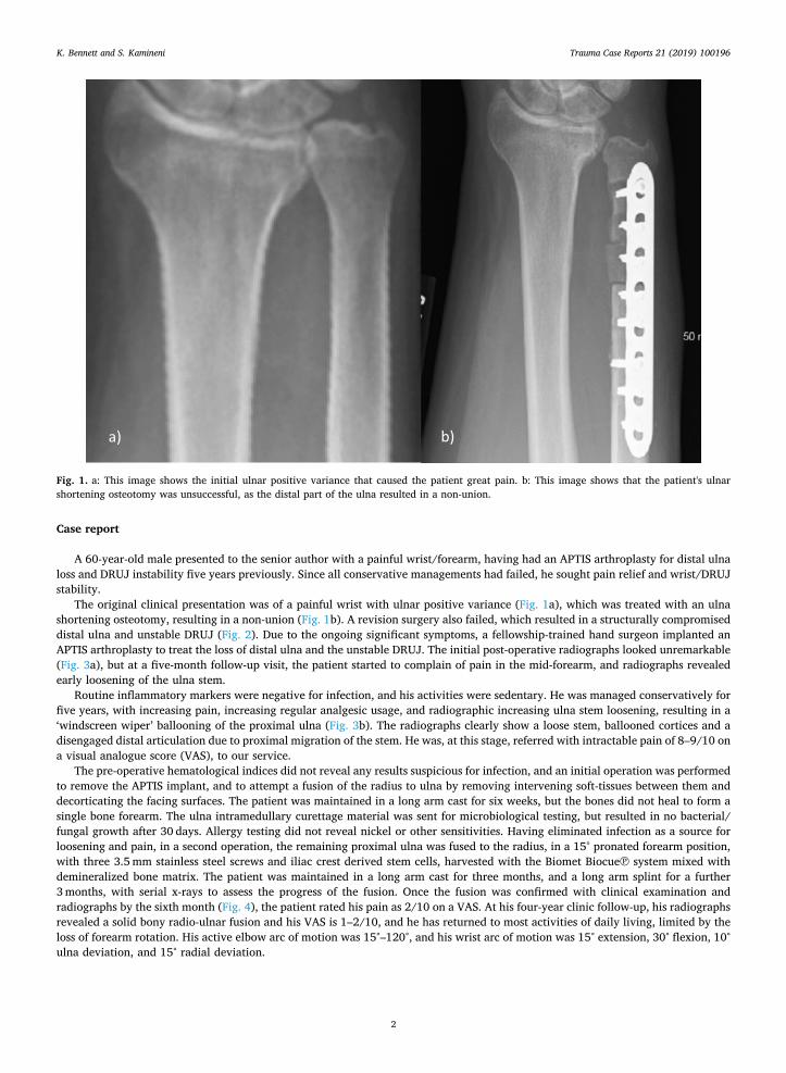
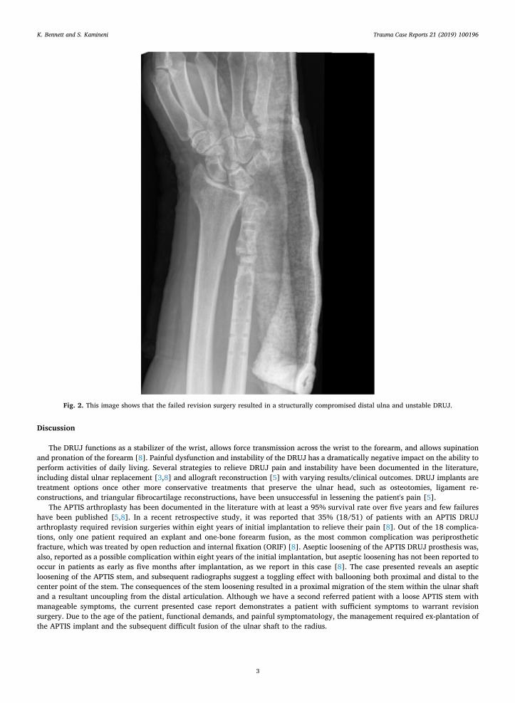
The original clinical presentation was of a painful wrist with ulnar positive variance (Fig. 1a), which was treated with an ulnashortening osteotomy, resulting in a non-union (Fig. 1b). A revision surgery also failed, which resulted in a structurally compromiseddistal ulna and unstable DRUJ (Fig. 2). Due to the ongoing significant symptoms, a fellowship-trained hand surgeon implanted anAPTIS arthroplasty to treat the loss of distal ulna and the unstable DRUJ. The initial post-operative radiographs looked unremarkable(Fig. 3a), but at a five-month follow-up visit, the patient started to complain of pain in the mid-forearm, and radiographs revealedearly loosening of the ulna stem.
Routine inflammatory markers were negative for infection, and his activities were sedentary. He was managed conservatively forfive years, with increasing pain, increasing regular analgesic usage, and radiographic increasing ulna stem loosening, resulting in a‘windscreen wiper’ ballooning of the proximal ulna (Fig. 3b). The radiographs clearly show a loose stem, ballooned cortices and adisengaged distal articulation due to proximal migration of the stem. He was, at this stage, referred with intractable pain of 8–9/10 ona visual analogue score (VAS), to our service.
The pre-operative hematological indices did not reveal any results suspicious for infection, and an initial operation was performedto remove the APTIS implant, and to attempt a fusion of the radius to ulna by removing intervening soft-tissues between them anddecorticating the facing surfaces. The patient was maintained in a long arm cast for six weeks, but the bones did not heal to form asingle bone forearm. The ulna intramedullary curettage material was sent for microbiological testing, but resulted in no bacterial/fungal growth after 30 days. Allergy testing did not reveal nickel or other sensitivities. Having eliminated infection as a source forloosening and pain, in a second operation, the remaining proximal ulna was fused to the radius, in a 15° pronated forearm position,with three 3.5mm stainless steel screws and iliac crest derived stem cells, harvested with the Biomet Biocue℗ system mixed withdemineralized bone matrix. The patient was maintained in a long arm cast for three months, and a long arm splint for a further3months, with serial x-rays to assess the progress of the fusion. Once the fusion was confirmed with clinical examination andradiographs by the sixth month (Fig. 4), the patient rated his pain as 2/10 on a VAS. At his four-year clinic follow-up, his radiographsrevealed a solid bony radio-ulnar fusion and his VAS is 1–2/10, and he has returned to most activities of daily living, limited by theloss of forearm rotation. His active elbow arc of motion was 15°–120°, and his wrist arc of motion was 15° extension, 30° flexion, 10°ulna deviation, and 15° radial deviation.
a) b)
Fig. 1. a: This image shows the initial ulnar positive variance that caused the patient great pain. b: This image shows that the patient's ulnarshortening osteotomy was unsuccessful, as the distal part of the ulna resulted in a non-union.
K. Bennett and S. Kamineni Trauma Case Reports 21 (2019) 100196
2
Discussion
The DRUJ functions as a stabilizer of the wrist, allows force transmission across the wrist to the forearm, and allows supinationand pronation of the forearm [8]. Painful dysfunction and instability of the DRUJ has a dramatically negative impact on the ability toperform activities of daily living. Several strategies to relieve DRUJ pain and instability have been documented in the literature,including distal ulnar replacement [3,8] and allograft reconstruction [5] with varying results/clinical outcomes. DRUJ implants aretreatment options once other more conservative treatments that preserve the ulnar head, such as osteotomies, ligament re-constructions, and triangular fibrocartilage reconstructions, have been unsuccessful in lessening the patient's pain [5].
The APTIS arthroplasty has been documented in the literature with at least a 95% survival rate over five years and few failureshave been published [5,8]. In a recent retrospective study, it was reported that 35% (18/51) of patients with an APTIS DRUJarthroplasty required revision surgeries within eight years of initial implantation to relieve their pain [8]. Out of the 18 complica-tions, only one patient required an explant and one-bone forearm fusion, as the most common complication was periprostheticfracture, which was treated by open reduction and internal fixation (ORIF) [8]. Aseptic loosening of the APTIS DRUJ prosthesis was,also, reported as a possible complication within eight years of the initial implantation, but aseptic loosening has not been reported tooccur in patients as early as five months after implantation, as we report in this case [8]. The case presented reveals an asepticloosening of the APTIS stem, and subsequent radiographs suggest a toggling effect with ballooning both proximal and distal to thecenter point of the stem. The consequences of the stem loosening resulted in a proximal migration of the stem within the ulnar shaftand a resultant uncoupling from the distal articulation. Although we have a second referred patient with a loose APTIS stem withmanageable symptoms, the current presented case report demonstrates a patient with sufficient symptoms to warrant revisionsurgery. Due to the age of the patient, functional demands, and painful symptomatology, the management required ex-plantation ofthe APTIS implant and the subsequent difficult fusion of the ulnar shaft to the radius.
Fig. 2. This image shows that the failed revision surgery resulted in a structurally compromised distal ulna and unstable DRUJ.
K. Bennett and S. Kamineni Trauma Case Reports 21 (2019) 100196
3
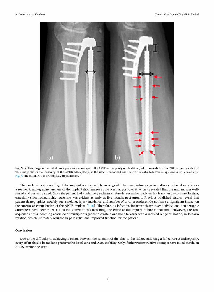
The mechanism of loosening of this implant is not clear. Hematological indices and intra-operative cultures excluded infection asa source. A radiographic analysis of the implantation images at the original post-operative visit revealed that the implant was well-seated and correctly sized. Since the patient had a relatively sedentary lifestyle, excessive load-bearing is not an obvious mechanism,especially since radiographic loosening was evident as early as five months post-surgery. Previous published studies reveal thatpatient demographics, notably age, smoking, injury incidence, and number of prior procedures, do not have a significant impact onthe success or complication of the APTIS implant [9,10]. Therefore, as infection, incorrect sizing, over-activity, and demographicdifferences have been ruled out as the source of this loosening, the cause of the implant failure is indistinct. However, the con-sequence of this loosening consisted of multiple surgeries to create a one bone forearm with a reduced range of motion, in forearmrotation, which ultimately resulted in pain relief and improved function for the patient.
Conclusion
Due to the difficulty of achieving a fusion between the remnant of the ulna to the radius, following a failed APTIS arthroplasty,every effort should be made to preserve the distal ulna and DRUJ stability. Only if other reconstructive attempts have failed should anAPTIS implant be used.
a) b)Fig. 3. a: This image is the initial post-operative radiograph of the APTIS arthroplasty implantation, which reveals that the DRUJ appears stable. b:This image shows the loosening of the APTIS arthroplasty, as the ulna is ballooned and the stem is subsided. This image was taken 5 years afterFig. 4, the initial APTIS arthroplasty implantation.
K. Bennett and S. Kamineni Trauma Case Reports 21 (2019) 100196
4
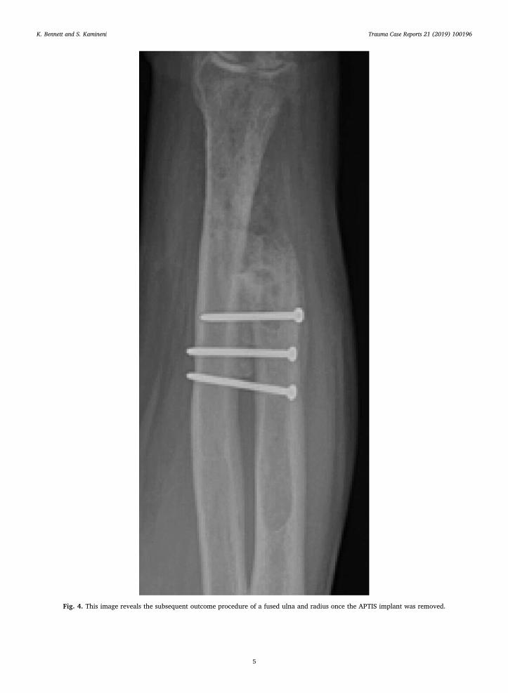
Fig. 4. This image reveals the subsequent outcome procedure of a fused ulna and radius once the APTIS implant was removed.
K. Bennett and S. Kamineni Trauma Case Reports 21 (2019) 100196
5
Acknowledgements
No financial remunerations for either author exist.
References
[1] A. Rampazzo, B.B. Gharb, G. Brock, L.R. Scheker, Functional outcomes of the Aptis-Scheker distal radioulnar joint replacement in patients under 40 years old, J.Hand. Surg. [Am.] 40 (7) (2015) 1397–1403, https://doi.org/10.1016/j.jhsa.2015.04.028.
[2] L.R. Scheker, Arthritis of the distal radioulnar joint, Tech. Orthop. 24 (1) (2009) 32–41, https://doi.org/10.1097/bto.0b013e3181a07edc.[3] E.J. Galvis, J. Pessa, L.R. Scheker, Total joint arthroplasty of the distal radioulnar joint for rheumatoid arthritis, J. Hand. Surg. [Am.] 39 (9) (2014) 1699–1703,
https://doi.org/10.1016/j.jhsa.2014.03.043.[4] G.L. Kaiser, L.S. Bodell, R.A. Berger, Functional outcomes after arthroplasty of the distal radioulnar joint and hand therapy: a case series, J. Hand Ther. 21 (4)
(2008) 398–407, https://doi.org/10.1197/j.jht.2008.06.002.[5] W.P. Cooney III, R.A. Berger, Distal radioulnar joint implant arthroplasty, American Society for Surg of the Hand 5 (4) (2005) 217–219, https://doi.org/10.
1016/j.jassh.2005.08.004.[6] L.R. Scheker, Replacement of distal radioulnar joint, Over Tech Orthop 22 (2012) 112–118, https://doi.org/10.1053/j.oto.2012.04.001.[7] R.M. Strigel, M.L. Richardson, Distal radioulnar joint arthroplasty with a Scheker prosthesis, Radiol. Case Rep. 1 (2) (2006) 65–67, https://doi.org/10.2484/rcr.
vli2.29.[8] K.D. Bellevue, M.K. Thayer, M.P. Pouliot, J.I. Huang, D. Hanel, Complications of Aptis DRUJ arthroplasty, J. Hand. Surg. [Am.] 22 (3) (2017) S14–S15, https://
doi.org/10.1016/j.jhsa.2017.06.039.[9] L.R. Scheker, D.W. Martineau, Distal radioulnar joint constrained arthroplasty, Hand Clin. 29 (1) (2013) 113–121, https://doi.org/10.1016/j.hcl.2012.08.023.[10] Willis AA, Berger RA, Cooney III WP. Arthroplasty of the distal radioulnar joint using a new ulnar head endoprosthesis: preliminary report. J. Hand. Surg. [Am.]
2007;32A:177–89. DOI:https://doi.org/10.1016/j.jhsa.2006.12.004.
K. Bennett and S. Kamineni Trauma Case Reports 21 (2019) 100196
6