Embed Size (px)
Citation preview
��������������� ���������������������
����������� ��������������� ���������������������
A c t a U n i v e r s i t a t i s T a m p e r e n s i s 960U n i v e r s i t y o f T a m p e r e
T a m p e r e 2 0 0 3
��� ��� ����������� �%���������"89������������������
��� �(&!���:�"�(������������������� ������8
����&%!�("��(&�����������&"�����&��� ���-:�"��8
;������!�2<����&�8 ������8���(��%��/,��8/���8���/�=(!�(��
�������������
�����������
��������� �� �������������� ������� ��� �������� ��������� �� ������ ��!��"
#����"���$�%�&��'���
������""������������(��������������� ����������)��*'�+),�-��-,�.�-�*''+��,,-����
��������!��������������&���������� ������/���
�!� 0�,. �1/�, ��,, �� 0�,. � /�,1��.,��2&3&���������455$���&��&�����
6!�(�����("������������(��6!�(�����(�������������� ����������/.,*'�+),�-��-,�./-7*''+��,�-),�7����455�(���&�����
��� ��� � ��������������������������������� ������������ ����� ����!� �"�#� ��
��������� ��$���������%�&�����'� (��������������� ��� �������
������ ��$��������� ����� � )������� ���*����������� ��*���� ��� ������� ���������� �� +�������������������� ��� %� ��,�

To my parents
3
CONTENTS CONTENTS............................................................................................................ 4 LIST OF ORIGINAL PUBLICATIONS .................................................................... 6 ABBREVIATIONS .................................................................................................. 7 ABSTRACT ............................................................................................................ 9 1. INTRODUCTION.............................................................................................. 11 2. REVIEW OF LITERATURE.............................................................................. 12
2.1 Mitochondria............................................................................................ 12 2.1.1 Brief history.................................................................................................... 12 2.1.2 Structure and function of mitochondria.......................................................... 12 2.1.3 Mitochondrial genome organization and replication...................................... 16 2.1.4 Mitochondrial transcription ............................................................................ 22
2.2 Mitochondrial translation system ............................................................. 25 2.2.1 Components of mitochondrial translation machinery .................................... 25 2.2.2 Mitochondrial ribosomes................................................................................ 28 2.2.3 Mitoribosomal protein S12............................................................................. 29
2.2.3.1 S12 in bacteria and its role in translational fidelity................................. 30 2.2.3.2 technical knockout (tko)........................................................................... 32
2.2.4 Co- regulation of nuclear and mitochondrial gene expression ....................... 34 2.3 Mitochondria and disease ....................................................................... 37
2.3.1 Inheritance, heteroplasmy and segregation of mtDNA .................................. 38 2.3.2 Threshold effects ........................................................................................... 39 2.3.3 Animal models of mitochondrial disease ....................................................... 40 2.3.4 Drosophila as a model organism.................................................................... 41
2.4 Genetics of deafness............................................................................... 44 2.4.1 Principles of mechanosensation ..................................................................... 44 2.4.2 Mitochondrial deafness .................................................................................. 48 2.4.3 Aminoglycoside antibiotics and deafness ...................................................... 49
3. AIMS OF THE STUDY ..................................................................................... 52 4. MATERIALS AND METHODS.......................................................................... 53
4.1 Bacterial strains....................................................................................... 53 4.2. Mammalian cell culture........................................................................... 53 4.3 Drosophila strains and P-element mediated transgenesis ...................... 53 4.4 Drosophila behavioural tests ................................................................... 54
4.4.1 Bang-sensitivity .............................................................................................. 54 4.4.2 Reactivity........................................................................................................ 55 4.4.3 Developmental time and antibiotic sensitivity ............................................... 55 4.4.4 Courtship analysis .......................................................................................... 55 4.4.5 Deafness assay................................................................................................ 56
4.5 Respiratory enzyme activities and ATP synthesis ................................... 56 4.6 Translational kinetics in bacteria ............................................................. 57 4.7 Construction of uORF -mutagenised MRPS12 expression plasmids ...... 60
5. RESULTS......................................................................................................... 62 5.1 Mutational analysis of rpsL (I) ................................................................. 62
5.1.1 L56H-rpsL ...................................................................................................... 63 5.1.2 K87Q-rpsL...................................................................................................... 64
5.2 Molecular and biochemical characterization of tko25t (II, III)..................... 64 5.2.1 Decreased mitochondrial 12S:16S rRNAs ratio............................................. 65 5.2.2 Decreased OXPHOS capacity and rate of ATP synthesis.............................. 65
4
5.3 Behavioural and developmental alterations (II) ....................................... 66 5.3.1 Bang-sensitivity .............................................................................................. 66 5.3.2 Hyporeactivity ................................................................................................ 67 5.3.3 Impaired courtship behaviour......................................................................... 67 5.3.4 Impaired sound responsiveness ...................................................................... 68 5.3.5 Developmental delay and doxycyclin hypersensitivity.................................. 69
5.4 Transgenic analysis of tko (II, III) ............................................................ 70 5.4.1 Phenotypic rescue of the tkoH85L revertant strains (II).................................... 71 5.4.2 Dosage effects of tko25t ................................................................................... 71 5.4.3 Tissue variable rescue of tko25t by UAS-tko+ (III) ......................................... 73 5.4.4 Phenotypic effects of the tkoQ116K substitution (II and unpublished data)...... 74
5.5 Regulation of MRPS12 expression in human cells (IV) ........................... 75 5.5.1 MRPS12 is targeted to mitochondria ............................................................. 75 5.5.2 MRPS12 is regulated by alternative splicing ................................................. 76 5.5.3 Translational control of MRPS12................................................................... 77
6. DISCUSSION................................................................................................... 80 6.1 Molecular and biochemical consequences of tko25t (I, II, III) ................... 80 6.2 The mechanosensory defect of tko25t leads to hearing impairment (II, III)81 6.3 Developmental consequences of the tko25t mutation (II, III)..................... 83 6.4 Dosage effects of tko25t (II, III) ................................................................. 85 6.5 Expression of tko+ in the nervous system rescues bang-sensitivity (III) . 87 6.6 Does tko25t manifest as muscle weakness?............................................. 88 6.7 Model of restrictive ribosome: tkoQ116K (I, II) ............................................ 89 6.8 Regulation of MRPS12 in human cells (IV) ............................................. 90 6.9 Possible physiological consequences of mutations in the mitochondrial translation machinery .................................................................................... 93
7. SUMMARY....................................................................................................... 96 8. ACKNOWLEDGEMENTS................................................................................. 98 9. REFERENCES............................................................................................... 100 10. ORIGINAL COMMUNICATIONS.................................................................. 132
5
LIST OF ORIGINAL PUBLICATIONS The thesis is based on following original scientific communications, which are referred to
in the text by their Roman numerals I-IV.
I Toivonen JM, Boocock MR and Jacobs HT (1999). Modelling in Escherichia coli of mutations in mitoribosomal protein S12: novel mutant phenotypes of rpsL. Mol Microbiol 31: 1735-1746.
II Toivonen JM, O’Dell KMC, Petit N, Irvine SC, Knight G, Lehtonen M, Longmuir
M, Luoto K, Touraille S, Wang Z, Alziari S, Shah ZH and Jacobs HT (2001): technical knockout, a Drosophila model of mitochondrial deafness. Genetics 159: 241-254.
III Toivonen JM, Manjiry S, Touraille S, Wang Z, Alziari S, O’Dell KMC and Jacobs
HT (2003): Gene dosage and selective expression modify phenotype in a Drosophila model of human mitochondrial disease. Mitochondrion 3, 83-96.
IV Mariottini P, Shah ZH, Toivonen JM, Bagni C, Spelbrink JN, Amaldi F and Jacobs
HT (1999): Expression of the gene for mitoribosomal protein S12 is controlled in human cells at the levels of transcription, RNA splicing and translation. J Biol Chem 274, 31853-31862.
6
ABBREVIATIONS ADP adenosine diphosphate ATP adenosine trisphosphate ANT adenine nucleotide translocase APP P1,P5-Di(adenosine-5´)pentaphosphate bp base pair CHO chordotonal organ CS Canton S D-loop displacement loop Ef-Ts elongation factor Ts Ef-Tu elongation factor Tu ETC electron transport chain FAD flavin adenine dinucleotide GAL4 yeast transcriptional activator GDP guanosine diphosphate GTP guanosine trisphosphate dB decibel DMEM Dulbecco’s modified Eagle’s medium DMSO dimethyl sulfoxide DmTTF Drosophila melanogaster transcription termination factor DNA deoxyribonucleic acid DOX doxycyclin DTNB dithio-bis-nitrobenzoic acid EDTA ethylenediamine N,N,N’,N’ tetra-acetic acid EF elongation factor ER endoplasmic reticulum EtBr ethidium bromide HCl hydrochloric acid HSP heavy strand promoter H-strand heavy strand IF initiation factor IM inner membrane IMS intermembrane space IPTG isopropylthiogalactoside KCN potassium cyanide L-strand light strand LSP light strand promoter LSU ribosomal large subunit mRNA messenger RNA MRPS12 human mitochondrial protein S12 mtDNA mitochondrial DNA mtSSB mitochondrial single stranded binding protein mRNP messenger ribonucleoprotein mt mitochondrial mtDNA mitochondrial DNA mTERF mitochondrial transcription termination factor MRP mitochondrial RNA processing MRPS12 human mitoribosomal protein S12
7
NAD nicotinamide adenine dinucleotide ND NADH-dehydrogenase nt nucleotide OD optical density oligo(Y) oligopyrimidine OM outer membrane ONP ortho-nitrophenol ONPG ortho-Nitrophenyl-β-D-galactopyranoside OXPHOS oxidative phosphorylation PCR polymerase chain reaction PM paromomycin Pol γ mitochondrial DNA polymerase gamma POLRMT mitochondrial RNA polymerase RI restriction intermediate RNA ribonucleic acid ROS reactive oxygen species rpS12 bacterial ribosomal protein S12 rRNA ribosomal RNA RT-PCR reverse transcriptase PCR RRF ribosome release factor S12 ribosomal protein S12 (general name) SD Shine/Dalgarno SM streptomycin SMR streptomycin resistant SMS streptomycin sensitive SSU ribosomal small subunit TCA tricarboxylic acid cycle (Krebs cycle) TFAM mitochondrial transcription factor A TFB1M mitochondrial transcription factor B1 TFB2M mitochondrial transcription factor B2 tko technical knockout (gene for Drosophila mitoribosomal S12) TMPD N,N,N´,N´-tetramethyl-p-phenylenediamine TOP terminal oligopyrimidine tRNA transfer RNA UAS upstream activating sequence UCP uncoupling protein uORF upstream open reading frame UTR untranslated region VDAC voltage-dependent anoion channel (porin) wt wild-type w/v weight per volume
8
ABSTRACT Mutations in the maternally inherited mitochondrial genome (mtDNA) or in nuclear genes
involved in mitochondrial metabolism manifest with a wide range of clinical phenotypes,
which frequently include sensorineural deafness in syndromic or non-syndromic form. The
pathological mechanisms of such diseases are largely unknown, and their outcome is
known to be affected by environmental and genetic modifiers. Relevant whole animal
models are needed to explore the developmental and biochemical consequences of
mitochondrial dysfunction, as well as mechanisms underlining the high tissue specificity of
the diseases. The main purpose of my study is to elucidate the validity of Drosophila
melanogaster (fruit fly) as a model system for human mitochondrial disorders, particularly
those resulting from mitochondrial translational defects. To develop such models, I have
manipulated the nuclear Drosophila gene technical knockout (tko), encoding
mitoribosomal protein S12, a critical component of the ribosomal accuracy centre involved
in the fidelity of protein synthesis both in bacterial and mitochondrial ribosomes. The
prototypic mutation tko25t results in conserved amino-acid substitution (L85H) and exhibits
some close parallels with human mitochondrial disease, such as developmental delay,
temporary paralysis in response to mechanical stress (followed by seizure-like episodes),
hyporeactivity and defective auditory function. Based on bacterial modelling the mutation
causes defective ribosome assembly and, in agreement with this, tko25t shows decreased
levels of mitochondrial small subunit ribosomal RNA, is hypersensitive to mitochondrial
translational inhibitor doxycyclin, and shows greatly decreased mitochondrial oxidative
phosphorylation capacity and diminished ATP synthesis. I infer that the tko25t mutant
provides a model of mitochondrial hearing impairment resulting from a quantitative
deficiency of mitochondrial translational capacity. To extend our understanding of
mitoribosomal function and to elucidate the critical times and cell types in development for
manifestation of the disease-like phenotype, I have created novel transgenic flies and
analysed their expression and phenotypes in various genetic backgrounds. Transgenic
reversion of the tko25t mutation results in complete rescue of the phenotype, whereas
increased expression of the mutant allele shows partial attenuation of the biochemical
defect. However, the latter is not associated with improved performance. In this sense, the
model mimics biochemical and phenotypical threshold effects associated with
heteroplasmic mtDNA mutations in humans (i.e. those in which mixture of wild-type and
mutant mtDNA is present). Selective spatio-temporal expression of wild-type tko+ in
9
mutant flies results in a diverse range of phenotypes, and indicates tissues and
developmental stages where mitochondrial translational capacity becomes limiting in the
mutant. High level over-expression of tko+ in vivo leads to pre-adult lethality both in
mutant and in wild-type genetic backgrounds, emphasizing the importance of correctly
regulated expression of the components of the mitochondrial translation machinery.
Differential regulation of expression may also be one important variable affecting the
phenotypic outcome in human diseases. Using cultured human cells I have elucidated
mechanisms involved in expression of the human homologue of tko, MRPS12,
demonstrated that it is highly expressed in tissues dependent on oxidative metabolism,
such as heart and skeletal muscle, and is subject to sophisticated regulatory mechanisms
involving transcription, alternative splicing and cell growth-mediated translational control.
10
1. INTRODUCTION
Mitochondria are eukaryotic organelles responsible for cellular oxidative energy
production. They are also involved in various physiological processes such as intermediary
metabolism and cellular signalling events. Due to the endosymbiotic origin of this
organelle, mitochondria have retained a circular (or in some organisms linear) chromosome
(mtDNA) that encodes a small number of polypeptides essential for respiratory chain
function. Additionally, mtDNA encodes RNA species required for expression of the
mitochondrially encoded proteins. Mutations in mitochondrial transfer RNAs (tRNAs) or
ribosomal RNAs (rRNAs) are frequently associated with maternally inherited diseases
manifesting as either syndromic or non-syndromic disorders, often characterized by
seizures, ataxia, muscle weakness and hearing impairment. Each cell contains a large
number of mtDNA molecules, which complicates the genetics of these disorders.
Frequently, the determining factor is the proportion of mutated and the wild-type
molecules in cells or tissues (heteroplasmy). Mitochondrial disorders can also result from a
mutation in nuclear genes involved in mitochondrial metabolism. Animal modelling of
mitochondrial diseases has largely concentrated on nuclear genes, because mtDNA is
intractable for conventional genetic manipulations.
In this thesis I will describe mitochondria, their various functions, gene expression, and
common features behind mitochondrial biogenesis and disease. Particular emphasis will be
laid upon description of the mitochondrial translation system because of its frequent
involvement in human diseases, such as mitochondrial deafness. I will also discuss the use
of the fruit fly, Drosophila melanogaster, as an experimental animal for studying human
mitochondrial disease. The genetic power of D. melanogaster has long been recognised,
but since it is a relatively ‘simple’ invertebrate, its biomedical relevance has been
sometimes overlooked. Recent studies of insulin signalling in aging, and of multiple
neurodegenerative disorders suggest, however, that this insect species might be
physiologically more similar to humans than we have previously thought.
11
2. REVIEW OF LITERATURE 2.1 Mitochondria This chapter describes mitochondria in general, their structure and diverse functions. The
animal mitochondrial genome, its maintenance and expression at the RNA level will also
be more specificly described. A more specific description of the mitochondrial protein
synthesis machinery will be given in section 2.2.
2.1.1 Brief history
Mitochondria are eukaryotic cell organelles that contain their own genome and are
responsible for cellular respiration and aerobic energy production. The name
'mitochondrion' was coined by C. Bender in 1898, and is derived from the Greek mitos
meaning thread and chondrion meaning granule. Occasional descriptions of structures we
now know to be mitochondria appeared here and there in the microscopic literature already
150 years ago. However, the first study characterizing these structures and showing them
to be present in a wide variety of cell types was that of Richard Altmann in 1890, who
considered these "bioblasts" to be independent entities, and suggested them to be tiny
organisms forming colonies within eukaryotic cells. Among others he contributed to The
Serial Endosymbiotic Theory for the origin of mitochondria, which states that in the early
history of eukaryotes a protobacterium capable of aerobic respiration was engulfed by an
anaerobic protoeukaryotic cell. By providing some metabolic advantage to the host (such
as ATP, hydrogen or detoxification of oxygen) it became an endosymbiont currently
known as the mitochondrion. Supportive evidence for this view was the discovery of
deoxyribonucleic acid (DNA) inside mitochondria (Nass and Nass, 1963), and the theory
was later popularised and received its best support and articulation by Lynn Margulis
(Margulis, 1981).
2.1.2 Structure and function of mitochondria The internal space of mitochondria (mitochondrial matrix) is surrounded by two
membranes (Figure 2.1 a). The outer membrane (OM) is freely permeable to ions and most
metabolites due to the presence of non-specific channels formed by porins (VDAC), for
solutes of molecular weight less than 10 kDa (Scheffler, 1999). The inner membrane (IM)
is impermeable to most such molecules and project to the mitochondrial matrix via
12
lamellar and/or tubular invaginations called cristae. Between the inner and outer
membranes lies the intermembrane space (IMS), which was for a long time thought to be
fully contiguous with the intracristal space. However, recent studies using three-
dimensional electron tomography suggest some sort of compartmentalization between
these structures (reviewed by Frey and Mannella, 2000). The cristal membranes seem to
have only small tubular contacts (crista junctions) with the peripheral surface of the IM,
and it is possible that this restriction is sufficient to limit exchanges of metabolites and
proteins between the two compartments and the membranes enclosing them. The OM and
the peripheral IM confront each other periodically by integral membrane protein-mediated
contacts. This kind of contacts is required, for example, for protein import, and is achieved
by interaction of the translocation complexes Tom and Tim of the outer and inner
membrane, respectively (Pfanner and Meijer, 1997). The IM is extremely protein-rich due
to the presence of all the complexes for oxidative phosphorylation (OXPHOS) and a large
number of other proteins that provide import and export of polypeptides, metabolites and
ions. It is also the membrane across which the membrane potential (∆ψ) is built up by the
electron transport chain (ETC) to create proton motive force required for ATP synthesis.
∆ψ is also needed for protein import and for the transport of various substances into and
out of mitochondria (Nicholls and Ferguson, 2002).
The morphology of mitochondria and cristae varies depending on environmental
conditions and cell type, and is likely to reflect the energy demands of different tissues.
Mitochondria are not merely floating around the cell but have non-random localization
controlled by extramitochondrial cytoskeletal elements, such as microtubules (Heggeness
et al., 1978; Yaffe et al., 1996; Yaffe, 1999), and their movements are affected by
mutations in cytoskeletal motor proteins, such as kinesins (Pesavento et al., 1994; Pereira
et al., 1997; Tanaka et al., 1998). In some cells, mitochondria form a convoluted network
(mitochondrial reticulum, figure 2.1 b) whereas in others they can appear as discrete
filaments or ovoid structures (Bereiter-Hahn and Voth, 1994). The form of this dynamic
reticulum is controlled by continuous fission and fusion events mediated by specific
proteins that seem to be conserved among all eukaryotes (Shaw and Nunnari, 2002).
Therefore, a traditional picture of mitochondrion as a static, individual, rod-shaped
structure is at least in most cases misleading.
13
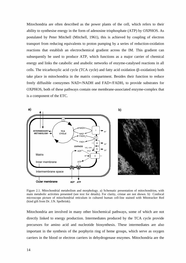
Mitochondria are often described as the power plants of the cell, which refers to their
ability to synthesise energy in the form of adenosine trisphosphate (ATP) by OXPHOS. As
postulated by Peter Mitchell (Mitchell, 1961), this is achieved by coupling of electron
transport from reducing equivalents to proton pumping by a series of reduction-oxidation
reactions that establish an electrochemical gradient across the IM. This gradient can
subsequently be used to produce ATP, which functions as a major carrier of chemical
energy and links the catabolic and anabolic networks of enzyme-catalysed reactions in all
cells. The tricarboxylic acid cycle (TCA cycle) and fatty acid oxidation (β-oxidation) both
take place in mitochondria in the matrix compartment. Besides their function to reduce
freely diffusible coenzymes NAD+/NADH and FAD+/FADH2 to provide substrates for
OXPHOS, both of these pathways contain one membrane-associated enzyme-complex that
is a component of the ETC.
H+
H+
H+
H+
II
TCACYCLE
I
III
IV
V
ADP
ATP
ADP ATP
II
NADH
NAD+
FADH2
FAD+
Outer membrane
Inner membrane
Matrix
½O2 +2H+
2H20
INTERMEDIARYMETABOLISM
OXPHOS
Intermembrane space
a) b)
H+
H+
H+
H+
II
TCACYCLE
I
III
IV
V
ADP
ATP
ADP ATP
II
NADH
NAD+
FADH2
FAD+
Outer membrane
Inner membrane
Matrix
½O2 +2H+
2H20
INTERMEDIARYMETABOLISM
OXPHOS
Intermembrane space
a) b)
H+
H+
H+
H+
II
TCACYCLE
I
III
IV
V
ADP
ATP
ADP ATP
II
NADH
NAD+
FADH2
FAD+
Outer membrane
Inner membrane
Matrix
½O2 +2H+
2H20
INTERMEDIARYMETABOLISM
OXPHOS
Intermembrane space
a) b)
Figure 2.1. Mitochondrial metabolism and morphology. a) Schematic presentation of mitochondrion, with main metabolic activities presented (see text for details). For clarity, cristae are not shown. b) Confocal microscope picture of mitochondrial reticulum in cultured human cell-line stained with Mitotracker Red (kind gift from Dr. J.N. Spelbrink).
Mitochondria are involved in many other biochemical pathways, some of which are not
directly linked to energy production. Intermediates produced by the TCA cycle provide
precursors for amino acid and nucleotide biosynthesis. These intermediates are also
important in the synthesis of the porphyrin ring of heme groups, which serve as oxygen
carriers in the blood or electron carriers in dehydrogenase enzymes. Mitochondria are the
14
site for the synthesis of iron-sulphur (Fe/S) clusters involved in electron transport,
substrate binding and biochemical catalysis, both inside and outside of mitochondria
(Muhlenhoff and Lill, 2000). Intermediates derived from β-oxidation can be used as an
alternative carbon source when glucose is unavailable, such as under starvation (Barger
and Kelly, 2000). For detoxification of tissues, excess ammonia created by metabolism is
transported to liver mitochondria where the nitrogen enters the urea cycle and is ultimately
excreted. The first and rate-limiting step of steroid hormone synthesis from cholesterol
occurs in mitochondria of steroidogenic tissues (Stocco, 2001, and references therein;
Thomson, 2003). In some cases, such as in brown fat of newborn infants, cold adaptive
rodents and hibernating animals, proton flow in mitochondrial membranes can be short-
circuited by uncoupling proteins (UCPs), in order to produce heat instead of ATP (Kozak
and Harper, 2000, and references therein). For a long time it has been known that
mitochondria are also the primary site for the creation of reactive oxygen species (ROS,
Boveris et al., 1972), which might have impact on many pathological states in humans, as
well as on ageing (reviewed by Droge, 2002; Mandavilli et al., 2002). Milder uncoupling
of mitochondria has been suggested to be involved in prevention of ROS formation and
body weight control (Jezek, 2002).
A great number of studies have been published that confirm the importance of
mitochondria in programmed cell death, i.e., apoptosis (reviewed by Wang, 2001). Various
apoptotic stimuli are transduced to mitochondria by BH3-only proteins (including Bad, Bid
and its truncated form tBid), and their action can be either neutralised by anti-apoptotic
proteins (such as Bcl-2 and Bcl-xL), or the signal can be further transduced to
mitochondria by pro-apoptotic proteins (such as Bax or Bak). This signal causes
appearance of the permeability transition pore, which uncouples the IM resulting in the
collapse of the electrochemical gradient and swelling of mitochondria. Components of the
permeability transition pore have been suggested to include VDAC and adenine nucleotide
translocase (ANT), which are also responsible for transport of small metabolites and
nucleotides, respectively, across the mitochondrial membranes. The rupture of the OM
releases many pro-apoptotic mitochondrial proteins (including cytochrome c, Smac/Diablo,
apoptosis inducing factor and endonuclease G) from the IMS to the cytosol and/or the
nucleus, and results in subsequent activation of caspases that degrade various intracellular
substrates. Simultaneously, this release causes caspase-independent nuclear chromatin
condensation and large-scale DNA fragmentation. Other caspase-independent pathways
15
may be involved in programmed cell death, such as loss of mitochondrial function by
uncoupling of electron transfer from OXPHOS, and alterations in calsium homeostasis, as
shown by tBid treatment of isolated mitochondria. (Wang, 2001, and references therein).
Another pro-apoptotic protein, Bad, has been recently shown to be a component of a multi-
enzyme complex localised in the mitochondrial OM, and is required to nucleate the
assembly of this complex (Danial et al., 2003). Other components of the complex are
protein kinase A and protein phosphatase 1, responsible for Bad phosphorylation
(inactivation) and dephosphorylation (activation), respectively, as well as the A kinase
anchoring protein Wawe-1, and the glycolytic enzyme glucokinase. The work described by
Danial et al. (2003) shows that Bad phosphorylation is required for maximal glucokinase
activity, and suggests that in addition to being an integral participant in the apoptotic
pathway, it also might ensure that glycolysis and apoptosis are coordinated.
There is also good evidence that mitochondria serve as temporary cellular calcium
storages, or buffers preventing or delaying the spread of calcium signals in cells (for recent
reviews, see Pozzan et al., 2000, and Rizzuto et al., 2000). Free cellular calcium exists
normally at very low concentrations, but is increased in neuronal activation or by release
from the endoplasmic reticulum (ER) in response to signalling events. The close proximity
of mitochondria to the ER allows them to fine-tune the microenvironment of the calcium
release sites and to rapidly respond in such release (Rizzuto et al., 1998). This can result in
increased ATP production via stimulation of the dehydrogenases of the TCA cycle by
intramitochondrial calcium (Denton et al., 1972). When these divergent tasks of
mitochondria in cellular and physiological processes are taken into consideration it is
perhaps too simplistic to think of them only as the ‘batteries’ of the cell. However, the
bioenergetic functions performed by the OXPHOS machinery are clearly fundamental for
multicellular life.
2.1.3 Mitochondrial genome organization and replication
The genetic information for mitochondrial biogenesis is mostly encoded by the nuclear
DNA, and the proteins involved generally contain an amino-terminal targeting sequence
that directs them to be imported into the organelle. These proteins include all those
involved in transcription and translation, as well as the proteins of the mitochondrial DNA
(mtDNA) replication and maintenance machinery. However, the mtDNA of animals codes
16
for a limited number of RNAs essential for intra-mitochondrial protein synthesis as well as
some proteins needed for the function of ETC. This chapter describes a typical metazoan
mitochondrial gene content and mainly uses human and the fruit fly Drosophila
melanogaster as an example.
The mitochondrial genome in animals is a covalently closed circular molecule that is
present in 103 – 104 copies per cell (Lightowlers et al., 1997). The mtDNA encodes 13
polypeptides (12 in those whose mtDNA do not encode A8), 2 ribosomal RNAs (rRNA)
and 22 transfer RNAs (tRNA) (Anderson et al., 1981; Bibb et al., 1981; Lewis et al.,
1995). Almost all the genes are contiguous and all lack introns; the genetic code used
differs from the universal code in some respects (reviewed by Kurland, 1992). The open
reading frames (ORFs) are generally punctuated by tRNA genes which serve as signals for
precise nucleolytic cleavages to process long precursor RNAs to their mature forms (Ojala
et al., 1981), which involves both 5´ and 3´ processing of the tRNAs (Doersen et al., 1985;
Rossmanith et al., 1995; Nagaike et al., 2001; Puranam and Attardi, 2001). Released
mRNAs and rRNAs are subsequently polyadenylated, a process that also completes the
immature translation termination codon of some ORFs.
The mtDNA gene content is conserved from human to Drosophila, but variations in length,
nucleotide content and organization of the genome exist (Figure 2.2). Mitochondrial ORFs
encode subunits of complex I (NADH-ubiquinol oxidoreductase, EC1.6.5.3), complex III
(ubiquinone-cytochrome-c oxidorteductase, or bc1 complex, EC 1.10.2.2), and complex IV
(cytochrome-c oxidase, 1.9.3.1) of the ETC, as well as two subunits of the complex V
(ATP synthase, EC 3.6.1.3). Complex II (EC 1.3.99.11) of the ETC is the succinate
dehydrogenase, a membrane bound enzyme of the TCA cycle, which has no mtDNA-
encoded subunits.
Major variations between the mtDNAs of mammals and fruit flies are as follows. Firstly,
the mtDNA of Drosophila contains a long, 96% A+T rich region in the equivalent position
to the extensive non-coding region in mammals that contain the key signals for the
initiation and regulation of transcription and DNA replication, as well as the displacement-
loop (D-loop) whose function remains to be properly understood. The region contains the
origin of replication in both species, but unlike in mammals, the length of the stretch varies
substantially between Drosophila species (Goddard and Wolstenholme, 1978; Lewis et al.,
17
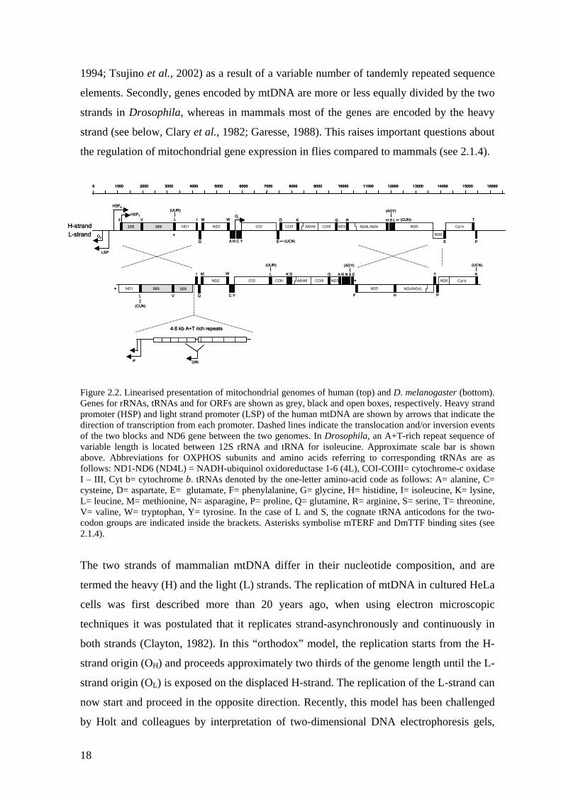
1994; Tsujino et al., 2002) as a result of a variable number of tandemly repeated sequence
elements. Secondly, genes encoded by mtDNA are more or less equally divided by the two
strands in Drosophila, whereas in mammals most of the genes are encoded by the heavy
strand (see below, Clary et al., 1982; Garesse, 1988). This raises important questions about
the regulation of mitochondrial gene expression in flies compared to mammals (see 2.1.4).
igure 2.2. Linearised presentation of mitochondrial genomes of human (top) and D. melanogaster (bottom). s for rRNAs, tRNAs and for ORFs are shown as grey, black and open boxes, respectively. Heavy strand
he two strands of mammalian mtDNA differ in their nucleotide composition, and are
by Holt and colleagues by interpretation of two-dimensional DNA electrophoresis gels,
F
S
D H S L
E P
I M
Q
W K G T
COII A8/A6 COIII ND3ND2 COI Cyt b
ND6
ND5ND4L/ND4
RV
ND116S
A N C Y
HSPR
LSP
(UUR)
L
(UCN)
(AGY)
(CUN)
OH
OLHSPT
12SH-strandL-strand
I M
Q
W
C Y
(UUR)
L K D G
F H P
T
(UCN)
S
ND5 ND4/ND4L
COII A8/A6 COIII ND3ND2 COI Cyt bND6
(AGY)
A R N S E
VL
ND1 16S 12S
(CUN)
4.6 kb A+T rich repeats
ORIP
∗
∗
∗
0 1000 2000 3000 4000 5000 6000 7000 8000 9000 10000 11000 12000 13000 14000 15000 16000
F
S
D H S L
E P
I M
Q
W K G T
COIICOII A8/A6A8/A6 COIIICOIII ND3ND3ND2ND2 COICOI Cyt bCyt b
ND6ND6
ND5ND5ND4L/ND4ND4L/ND4
RV
ND1ND116S16S
A N C Y
HSPR
LSP
(UUR)
L
(UCN)
(AGY)
(CUN)
OH
OLHSPT
12SH-strandL-strand
I M
Q
W
C Y
(UUR)
L K D G
F H P
T
(UCN)
S
ND5 ND4/ND4L
COII A8/A6 COIII ND3ND2 COI Cyt bND6
(AGY)
A R N S E
VL
ND1 16S 12S
(CUN)
4.6 kb A+T rich repeats
ORIP
∗
∗
∗
0 1000 2000 3000 4000 5000 6000 7000 8000 9000 10000 11000 12000 13000 14000 15000 160000 1000 2000 3000 4000 5000 6000 7000 8000 9000 10000 11000 12000 13000 14000 15000 16000
FGenepromoter (HSP) and light strand promoter (LSP) of the human mtDNA are shown by arrows that indicate the direction of transcription from each promoter. Dashed lines indicate the translocation and/or inversion events of the two blocks and ND6 gene between the two genomes. In Drosophila, an A+T-rich repeat sequence of variable length is located between 12S rRNA and tRNA for isoleucine. Approximate scale bar is shown above. Abbreviations for OXPHOS subunits and amino acids referring to corresponding tRNAs are as follows: ND1-ND6 (ND4L) = NADH-ubiquinol oxidoreductase 1-6 (4L), COI-COIII= cytochrome-c oxidase I – III, Cyt b= cytochrome b. tRNAs denoted by the one-letter amino-acid code as follows: A= alanine, C= cysteine, D= aspartate, E= glutamate, F= phenylalanine, G= glycine, H= histidine, I= isoleucine, K= lysine, L= leucine, M= methionine, N= asparagine, P= proline, Q= glutamine, R= arginine, S= serine, T= threonine, V= valine, W= tryptophan, Y= tyrosine. In the case of L and S, the cognate tRNA anticodons for the two- codon groups are indicated inside the brackets. Asterisks symbolise mTERF and DmTTF binding sites (see 2.1.4).
T
termed the heavy (H) and the light (L) strands. The replication of mtDNA in cultured HeLa
cells was first described more than 20 years ago, when using electron microscopic
techniques it was postulated that it replicates strand-asynchronously and continuously in
both strands (Clayton, 1982). In this “orthodox” model, the replication starts from the H-
strand origin (OH) and proceeds approximately two thirds of the genome length until the L-
strand origin (OL) is exposed on the displaced H-strand. The replication of the L-strand can
now start and proceed in the opposite direction. Recently, this model has been challenged
18
which can be used to analyse mtDNA replication intermediates (RIs) in tissues and cell
culture (Holt et al., 2000; Yang et al., 2002). The data suggested initially that two different
mechanisms of mtDNA replication exist simultaneously: the orthodox mode, working
mainly in the maintenance of a given copy number of the mitochondrial genome, and
conventional, strand-coupled replication, with frequent lagging-strand initiation events
predominating when efficient amplification of the mtDNA molecules is required (Holt et
al., 2000). Further analysis from highly purified mtDNA, however, has suggested that
mammalian mtDNA is at least mainly replicated by the strand-coupled mechanism and
previous data can be explained as an artefact of DNA preparation methods (Yang et al.,
2002). Very little is known about mtDNA replication in Drosophila, but the A+T rich
region has been shown to contain an origin of replication by electron microscopic studies
(Goddard and Wolstenholme, 1978). These studies suggested that in the mtDNA of
Drosophila embryos most of the “leading” strand replication has been completed before
the “lagging” strand replication starts, i.e. extreme strand asynchrony. How much this
could be due to purification method artefacts as reported by Yang et al. (2002) is difficult
to estimate, but the evident absence of a D-loop and the highly repetitive nature of the A+T
rich control region suggest that some fundamental differences in the mechanisms are
possible (Goddard and Wolstenholme, 1978; Lewis et al., 1994).
Whatever the exact mechanism of mammalian mtDNA replication, it is putatively primed
ith extensive RNA transcripts derived from the light-strand promoter (LSP), which is
tDNA has been characterized to some extent in
uman and mouse, and quite extensively in Drosophila. DNA polymerase gamma (pol γ) is
w
located directly upstream of OH (reviewed by Clayton, 1991; Shadel and Clayton, 1997;
Lee and Clayton, 1998). RNase MRP recognizes and cleaves RNA-DNA hybrid (the R-
loop) in a region that precisely matches the major observed RNA to DNA transition sites in
the D-loop area, and is most likely providing the free 3´-end for the priming of replication
(Tapper and Clayton, 1981; Lee and Clayton, 1998). Similarly, an RNA-primed
mechanism has been suggested for the initiation of L-strand replication at a conserved stem
loop structure at OL (Hixson et al., 1986).
The enzymatic machinery that replicates m
h
a heterodimer consisting of a catalytic (pol γ-α) and accessory (pol γ-β) subunits and is
believed to be the only DNA polymerase active in mitochondria (Bolden et al., 1977;
Wernette and Kaguni, 1986; Kaguni and Olson, 1989; Lewis et al., 1996; Ropp and
19
Copeland, 1996; Wang et al., 1997; Carrodeguas and Bogenhagen, 2000). The pol γ-α is
related to Pol I of E. coli and to phage T7 DNA polymerase, and contains both polymerase
and proofreading activities (Lewis et al., 1996). In addition, pol γ-α has potential function
in mtDNA repair, since it is active in base excision repair of abasic sites of DNA (Olson
and Kaguni, 1992; Longley et al., 1998; Pinz and Bogenhagen, 2000). pol γ-β is related to
prokaryotic aminoacyl-tRNA synthetases, serves as a processivity factor increasing the
catalytic efficiency of the pol γ-α in vitro, and might have an additional role in primer
recognition during mtDNA replication (Olson et al., 1995; Fan et al., 1999; Wang and
Kaguni, 1999).
Null mutations in the Drosophila gene encoding the catalytic subunit pol γ-α (tamas),
sult in a defect of late larval locomotory behaviour followed by prepupal death, and are
re
associated with aberrations of the visual system and disruption of the mitochondrial
distribution pattern in the central nervous system (Iyengar et al., 1999). Over expression of
pol γ-α in transgenic flies results in late pupal lethality, and is characterised by severely
reduced mtDNA copy number and cuticular defects including altered numbers of
macrochaetae (Lefai et al., 2000a). However, over expression of the same gene to a more
or less similar extent in Drosophila Schneider cells does not cause any obvious functional
defects or decrease in mtDNA levels (Lefai et al., 2000a). This serves as a good example
of fundamental differences that may exist between cell-culture models and the whole
organism with complex developmental requirements. Similarly, over expression of human
wild type pol γ-α (POLG) in cultured human cells causes no alterations in mitochondrial
function or mtDNA, although dominant negative polymerase deficient mutants cause
mtDNA depletion (Spelbrink et al., 2000; Jazayeri et al., 2003). Null mutations and
substitutions of conserved amino-acids in the Drosophila gene for the accessory subunit,
pol γ-β, result in reduced cell proliferation in the central nervous system followed by
lethality during early pupation. This is associated with aberrant mitochondrial morphology
and loss of mtDNA (Iyengar et al., 2002). In contrast to pol γ-α null mutants, pol γ-β
mutants show only mariginal effects on locomotory behaviour as well as later lethality,
which might be explained either by differential temporal expression patterns of the two
genes during development (Lefai et al., 2000b) or by differences in relative maternal
contribution in tissues that are critical for viability, as discussed by Iyengar et al., 2002.
20
Both polymerase and proofreading activities of pol γ, as well as the rate of initiation of
DNA strands, are further stimulated by mitochondrial single-stranded DNA-binding
rotein (mtSSB) in vitro (Thommes et al., 1995; Farr et al., 1999). mtSSB is encoded in
arity to the bacteriophage T7
rimase/helicase gene 4, and is which associated with the human mitochondrial disorder
to be attached to the mitochondrial
ner membrane and are suggested to be the structural units of mtDNA inheritance (Berger
p
Drosophila by the gene lopo (low power, Maier et al., 2001) and shows similar properties
to E. coli SSB, which functions in bacterial DNA replication by stabilizing the displaced
single-stranded segment of DNA during passage of the replication fork and enhancing the
catalytic activity of the polymerase. Insertion of a transposable element into the mtSSB
gene, producing essentially a null mutant, results in a drastic decrease in mtDNA copy
number, loss of respiration capacity, late larval lethality and aberrations of the visual
system similar to pol γ-α mutants. Unlike these, however, it shows no alterations in the
number or structure of mitochondria (Maier et al., 2001).
Other proteins required for mtDNA replication and maintenance include Twinkle
(Spelbrink et al., 2001), a helicase that shows simil
p
adPEO (autosomal dominant progressive external ophthalmoplegia) characterised by
multiple mtDNA deletions. Additionally, topoisomerases are likely to play roles in mtDNA
transactions (Zhang et al., 2001; Wang et al., 2002).
mtDNA is organised in mitochondria as protein-DNA complexes called nucleoids which at
least in yeast, and possibly in mammalian cells, seem
in
and Yaffe, 2000; Jacobs et al., 2000; Lehtinen et al., 2000; MacAlpine et al., 2000;
Garrido et al., 2003). Nucleoid structures, mostly identified in yeast, contain 2-4 copies of
mtDNA and approximately 20 polypeptides and recently it has been shown that in
mammalian cell lines at least Twinkle, mitochondrial transcription factor A (TFAM - see
section 2.1.4 for more detailed discussion of its role in gene expression) and mtSSB
colocalize with mtDNA in punctate structures (Miyakawa et al., 1987; Spelbrink et al.,
2001; Garrido et al., 2003). TFAM in mammalian cell lines and placenta is abundant
enough to cover all of the mtDNA and, indeed, mtDNA is far from naked (Takamatsu et
al., 2002; Alam et al., 2003). Since essentially all TFAM is associated with mtDNA it
seems likely that the stability of both mtDNA and TFAM are dependent on each other
(Alam et al., 2003; Garrido et al., 2003). Supporting this view, TFAM levels seem to be
severely reduced in mtDNA-less (rho0) cell lines whereas mtSSB is not, although its
21
punctate localization disappears (Garrido et al., 2003). In addition, the yeast counterpart of
TFAM (Abf2p) is required for mtDNA maintenance (Diffley and Stillman, 1992;
Zelenaya-Troitskaya et al., 1998) but probably not for gene expression (Lightowlers et al.,
1997), and can be complemented by TFAM (Parisi et al., 1993) or by the bacterial,
histone-like protein HU (Megraw and Chae, 1993).
2.1.4 Mitochondrial transcription
The mitochondrial genome in mammals is transcribed as long polycistronic pre-mRNAs
toya et al., 1982; Montoya et al., 1983). The H-strand
ontains two promoters (Figure 2.2), HSPR for the transcription of the two rRNAs and the
A polymerases (Tiranti et al.,
997). Initiation of transcription requires also mitochondrial transcription factor A (TFAM,
from three different promoters (Mon
c
tRNAs for phenylalanine and valine, and HSPT for the transcription of all genes in the H-
strand except tRNA for phenylalanine. The L-strand contains only one promoter (LSP)
from which all eight tRNAs and a single ORF (ND6) of the L-strand are transcribed
(Figure 2.2). A transcript generated from the LSP is also proposed to prime mtDNA
replication, functionally coupling mitochondrial gene expression with genome
maintenance (Chang and Clayton, 1985; Chang et al., 1985; Shadel and Clayton, 1997)
(see also 2.1.3). In Drosophila, RNA end-mapping has suggested many putative
transcription start sites, and it is possible that in this organism all the ‘blocks’ of genes are
transcribed from their own promoters (Berthier et al., 1986).
Mammalian mitochondrial genes are transcribed by mitochondrial RNA polymerase
(POLRMT), which shows similarity to T3/T7 phage-like RN
1
also known as mtTFA), and mitochondrial transcription factors B1 (TFB1M, also known
as mtTFB) and/or B2 (TFB2M), all of which interact directly with POLRMT (Fisher and
Clayton, 1985; Parisi and Clayton, 1991; Falkenberg et al., 2002; McCulloch et al., 2002).
TFAM is a high mobility group (HMG) box protein that exhibits low sequence specificity
(Parisi and Clayton, 1991), and is required both in transcription and mtDNA maintenance,
and is essential for embryogenesis in mice (Parisi and Clayton, 1991; Larsson et al., 1998).
However, it binds with high affinity to upstream elements of both HSP and LSP in vitro,
where it facilitates specific transcription initiation indicating a role in proper promoter
recognition and recruitment of POLRMT (Fisher and Clayton, 1985). When over
expressed in tissue-culture or imported into isolated mitochondria, TFAM is able to
22
enhance the expression of H-strand transcript levels (12S rRNA and COI) as well as
stimulate the nascent H-strand (7S DNA) synthesis from the LSP (Montoya et al., 1997;
Gensler et al., 2001). The Drosophila homologue of TFAM has been studied by RNA
interference (RNAi) in cell culture, which showed that 95% reduction of TFAM protein
levels deplete mtDNA to less than half of the controls with only mariginal effects on
mitochondrial transcription (Goto et al., 2001). This might not be conclusive for the in vivo
situation, because of differences with respect to developmental requirements of the whole
organism, as discussed earlier (Lefai et al., 2000a). In cell culture models, it is possible that
TFAM can be subjected to substantial down regulation without any obvious effect on
mitochondrial transcription, since it seems to exist in excess for what is required for these
processes (Takamatsu et al., 2002; Alam et al., 2003).
TFB1M and TFB2M further promote mitochondrial transcription in vitro from both HSP
and LSP, TFB2M being at least ten times more active, which might partly account for
exible regulation of mtDNA expression (Falkenberg et al., 2002). These proteins are
(EtBr) is a
pophilic cation that accumulates into mitochondria, intercalates into mtDNA and prevents
fl
homologous to bacterial rRNA dimethyltransferases, and at least TFB1M, showing higher
homology, appears to be a dual-function protein which can methylate bacterial small
subunit (SSU) rRNA in a conserved stem loop structure that seems to be also partially
methylated in mitochondrial SSU rRNA (McCulloch et al., 2002; Seidel-Rogol et al.,
2003). Therefore, these genes have been probably recruited to mitochondrial transcription
during evolution, and since homologues for both TFB1M and TFB2M can be found in
mouse and Drosophila, but only one in C. elegans, a gene duplication event during early
metazoan evolution is the most plausible explanation (Rantanen et al., 2003).
Reduced levels of TFAM are found in muscle fibers of patients with mtDNA depletion and
in rho0-cell lines lacking mtDNA (Larsson et al., 1998). Ethidium bromide
li
its transcription and replication. It can be utilised in tissue culture media to produce stabile
rho0-cell lines, or in more temporary mtDNA depletion-repletion experiments. In HeLa
cells subjected to and recovering from EtBr treatment, TFAM and POLRMT proteins
exhibit similar depletion-repletion profiles suggesting that mitochondrial transcription
machinery is co-ordinately regulated in response to changes in mtDNA copy number, and
that this control is most likely post-transcriptional (Seidel-Rogol and Shadel, 2002).
Currently it is not known if TFB1M and TFB2M expression follows similar patterns, but it
23
is clear that not all factors required for the maintenance of the mitochondrial genetic
system are co-regulated, since transcription and translation of human pol-γ is unaffected by
changes in level or even by the total loss of mtDNA (Davis et al., 1996). As discussed
before (see 2.1.3), in addition to its importance in transcription and transcription-mediated
replication TFAM, but not pol-γ, is also proposed to function as an mtDNA packaging
protein (Alam et al., 2003; Garrido et al., 2003).
Premature termination of the pre-rRNA transcript starting from HSPR is believed to be
brought about in mammals by mitochondrial transcription termination factor mTERF
ruse et al., 1989; Daga et al., 1993), which binds mammalian mtDNA at the site within
the level of initiation, the two
romoters of the H-strand seem to play a role in regulation of the relative abundance of
(K
the gene for tRNALeu(UUR) (Christianson and Clayton, 1988). In Drosophila, a putative
counterpart of mTERF (DmTTF) has recently been characterised (Roberti et al., 2003), but
the conserved sequence corresponding to the human mTERF binding site in Drosophila is
not occupied by DmTTF. Instead, the latter binds specifically homologous, non-coding
sequences at the ends of the convergent gene units of each strand, indicated as asterisks in
Figure 2.2 (Roberti et al., 2003). These sites coincide with regions previously suggested to
be transcription termination sites by RNA mapping (Berthier et al., 1986). Only one
transcription termination site in each strand would imply that, unlike in mammals, the
rRNA transcript would have to be produced by post-transcriptional processing in
Drosophila. The experiments carried out thus far do not exclude the possibility that
mtDNA transcription in Drosophila is initiated and/or attenuated in several positions, but
points out possible differences in the regulation of rRNA versus mRNA genes in this
organism compared to mammals (Berthier et al., 1986).
In mammals, the steady-state levels of the rRNAs compared to H-strand mRNAs are
increased 50-100 fold (Gelfand and Attardi, 1981). At
p
mRNAs and rRNAs, the ratio of which has been shown to be modulated directly by thyroid
hormone (Enriquez et al., 1999). Also, changes in ATP levels might affect the preferential
use of the two promoters, as observed in vitro, although the nature of the ATP-requiring
step is unknown (Gaines et al., 1987). Processing efficiency, differential transcript stability
and changes at the level of termination frequency at the mTERF-dependent termination site
have been proposed to have a role in controlling this process.
24
2.2 Mitochondrial translation system
Mitochondria have their own separate protein synthesis machinery for translation of the
components of this apparatus are mtDNA
eins, translation factors, and aminoacyl-tRNA-synthetases
To get insight into mitochondrial protein synthesis it is helpful to first consider the
998. See also Figure 2.3).
he bacterial translation system contains three translation initiation factors, named IF1-
messages encoded by mtDNA. The RNA
derived, but all ribosomal prot
must be imported into mitochondria, at least in animals.
2.2.1 Components of mitochondrial translation machinery
analogous process in prokaryotes (reviewed by Brock et al., 1
T
IF3. IF3 works essentially as a recycling factor by binding to the free ribosomal small
subunit (SSU) and by inhibiting its association with the large subunit (LSU) after
translation termination. Prior to subsequent ribosomal re-association at a new initiation
site, IF3 promotes the correct positioning of mRNA on the SSU by facilitating the codon-
anticodon interactions between the start codon and the aminoacylated initiator tRNA
(fMet-tRNAfmet) at the peptidyl-site (P-site) of the ribosome. IF2 is a GTPase that binds the
fMet-tRNAfmet and brings it to the initiation complex. Its affinity for the ribosome is
increased by IF1, which also occludes the A-site during the initiation, thus preventing other
tRNAs to bind this position. In the classical E. coli model of the elongation cycle, the
active GTP bound form of elongation factor (EF) -Tu interacts with aminoacylated tRNA
(aa-tRNA) forming a ternary complex which promotes binding of the aa-tRNA to the
aminoacyl site (A-site) of the mRNA-programmed ribosome. Once locked into the A-site
by cognate codon-anticodon interactions (and other rRNA/protein -mediated contacts), the
GTP in the ternary complex is hydrolysed, and EF-Tu is released from the ribosome as an
EF-Tu·GDP complex which is recycled to the active form in a reaction catalysed by EF-Ts.
Peptidyl transfer is catalysed by a reaction centre located in the LSU and a GTPase EF-G is
required for translocation of the peptidyl-tRNA from the ribosomal A-site to the P-site,
during the elongation cycle. (Brock et al., 1998). Four factors are involved in termination
of protein synthesis in bacteria, which are called release factors (RFs). RF1 and/or RF2 are
responsible for recognition of stop codons, a process that is stimulated by RF3, which is a
GTPase (RF1/2-RF3·GTP complex). Ribosome recycling factor (RRF), in combination
with EF-G, is essential for the release of the ribosome from the mRNA at the stop codon.
25
The precise mechanism of this process is not currently understood, although it might
mimic the translocation process (Kim et al., 2000).
Figure 2.3. Schematic presentation of bacterial translation. a) Icomplex bind to the IF3-bound ribosomal small subunit in ra
IF3
IF1
Initiation ternary complex
mRNA
P AP A
SSU
LSU
P A
IF3 IF1
IF2
5´
3´
Start codon
P AE
E
IF2
fMGTP fM
fM
2
GDP
Pi
tRNA
EF-TuGTP
2
Elongationternary complex
IF1
EF-TuGDP
Pi
P AE
fM
2
EF-TuGTP
3
Elongationternary complex
+n additionalelongation cycles
Peptidyl transfer& translocation
IF3
P AE
RF1/2RF3
Terminationcomplex
fM 2
3
n-2
n-1
n
Stopcodon
EF-GGTP
+ PiEF-GGDP
P AE
RF1/2
RF3
fM 2 3
n-2
n-1
nPeptide hydrolysis
GTP
GDP
Pi
P ARRF
IF3E
EF-GGTP
?
Subunit dissociation
a)
b)
c)
IF3
IF1
Initiation ternary complex
mRNA
P AP A
SSU
LSU
P A
IF3 IF1
IF2
5´
3´
Start codon
P AE
E
IF2
fMfMGTP fMfM
fM
2
fMfM
22
GDP
Pi
tRNA
EF-TuGTP
2
EF-TuGTP
22
Elongationternary complex
IF1
EF-TuGDP
Pi
P AE
fM
2
fMfM
22
EF-TuGTP
33
Elongationternary complex
+n additionalelongation cycles
Peptidyl transfer& translocation
IF3
P AE
RF1/2RF3
RF1/2RF3
Terminationcomplex
fM 2
3
n-2
n-1
n
fM 2fM 2
3
n-2
n-1
n
n-2
n-1
n-2
n-1
nn
Stopcodon
EF-GGTP
EF-GGTP
+ PiEF-GGDP
EF-GGDP
P AE
RF1/2
RF3
fM 2 3
n-2
n-1
n
fM 2fM 2 3
n-2
n-1
n
n-2
n-1n-
2
n-1
nnPeptide hydrolysis
GTP
GDP
Pi
P ARRF
IF3E
EF-GGTP
EF-GGTP
?
Subunit dissociation
a)
b)
c)
nitiation. The mRNA and the initiation ternary ndom order, and IF1 occupies the A-site. The
rge ribosomal subunit can now bind the 30S initiation complex to form the 70S initiation complex.
mulli, 1996; Koc and Spremulli, 2002), as well as
laConcomitantly, IF1 and IF3 are ejected, and IF2-bound GTP is hydrolysed. The unoccupied A-site can now bind the first elongation ternary complex. b) Elongation. The EF-Tu-GTP is hydrolysed and its dissociation causes conformational change that leads the initiator tRNA in the P-site to form a peptide bond with an A-site aa-tRNA in a reaction catalysed by the peptidyl transferase center. The A-site is concomitantly occupied by EF-G-GTP, hydrolysis of which promotes translocation of the A-site peptidyl-tRNA to the P-site. Initiator tRNA is ejected via the E-site, and another elongation cycle can start. EF-Tu is reactivated in a reaction catalysed by EF-Ts, and EF-G is reactivated by autocatalysis (not shown). c) Termination. When a termination codon is exposed in the A-site, the RF1/2-RF3 complex recognises it. Hydrolysis of the RF3-bound GTP causes this complex to dissociate and the translated polypeptide to be released. Subsequent binding of ribosome release factor causes mRNA and tRNA to be ejected and results in dissociation of the ribosomal subunits. The exact mechanism of this process is not properly understood, but it seems to be EF-G assisted. The small ribosomal subunit is subsequently bound by IF3, which prevents premature re-association with the large subunit. See text for more details. Mitochondrial orthologues of both IF2 and IF3, but not IF1, have been studied in mammals
Liao and Spremulli, 1991; Ma and Spre(
mitochondrial elongation factors EF-Tu and EF-Ts, and EF-G (Schwartzbach et al., 1996;
Cai et al., 2000; Karring et al., 2002). RF1 have been identified in rat and human
mitochondria, as well as RRF in humans (Lee et al., 1987; Zhang and Spremulli, 1998).
Despite the differences with respect to kinetic properties or apparent lack of homologues of
26
some factors operating in prokaryotes, the mitochondrial translation system seems to be
mechanistically closely related to the bacterial one.
The 13 polypeptides of the mtDNA are translated from nine monocistronic and two
icistronic mRNAs. None of these mRNAs possesses significant 5´- or 3´-untranslated d
regions (UTRs) (Montoya et al., 1981; Ojala et al., 1981). How then, are mitochondrial
mRNAs recognised by the translation initiation complex? Eukaryotic cytoplasmic
ribosomes use a 5´-cap binding and scanning mechanism, and usually contain a conserved
Kozak sequence in the translation start site (Kozak, 1987; Kozak, 1999). In prokaryotes,
ribosomes recognise the start codon using the Shine/Dalgarno (SD) interaction between the
mRNA and the SSU rRNA. The SD region of the mRNA (upstream of the initiation codon)
base pairs with the 3´ end of the 16S rRNA, helping to position the small ribosomal
subunit in relation to the initiation codon (Shine and Dalgarno, 1975). Since mitochondrial
mRNAs are not 5´-capped and do not contain SD-like sequences, it is unlikely that
sequence-directed base pairing between SSU rRNA and mRNA is involved in the initiation
process in the organelle. It is possible, however, that functional homologues of the SD
sequence are located internally to mitochondrial mRNAs and have been therefore
overlooked. Mitochondrial ribosomal SSUs have an intrinsic ability to bind mRNAs in
vitro even in the absence of start codon and initiation factors, and this interaction is
affected by the length of the mRNA but does not require a free 5´-end, as evidenced by
efficient binding of circularised mRNA (Liao and Spremulli, 1989; Liao and Spremulli,
1990; Farwell et al., 1996). It has been suggested that sequence-independent binding is an
early step of translation initiation. IFs may facilitate the re-localisation of the bound SSU
on the mRNA, and the initiation site could be stabilized by recognition of the start codon
by initiator tRNA using codon-anticodon interaction (Farwell et al., 1996). This is
supported by the fact that the addition of mitochondrial extract stimulates the 5´-specific
protection of mRNAs (Denslow et al., 1989). In fact, this mode of initiation would not be
dedicated to mitochondria only, since recent data in E. coli suggest that translation of
leaderless mRNAs is dependent on IF2 and IF3, and that recognition of the initiation codon
is independently achieved by the ribosome-IF2-fmet-tRNAfmet complex (Moll et al., 2002).
A problem in initiation has been suggested as a possible pathological mechanism resulting
from the aminoacylation defect in A3243G-mutated tRNALeu (UUR), since cell lines carrying
this heteroplasmic mutation show decreased association of mRNA with mitochondrial
ribosomes (Chomyn et al., 2000).
27
Mitochondrial translation initiation might also be affected by direct interaction with the
ner membrane, since mitoribosomes are largely associated with IM both in yeast and
ucleoprotein particles that are responsible for the
ndamental process of protein synthesis. Combinations of X-ray crystallography and
in
bovine (Spithill et al., 1978; Liu and Spremulli, 2000). This might relate to the fact that all
mtDNA-encoded polypeptides are hydrophobic membrane proteins, which need to be
assembled into IM to form ETC complexes. It is not known if insertion into the membrane
in animals is a post-translational or co-translational process, but in yeast, examples of both
have been described (Fox, 1996). In yeast mRNAs are activated for translation by mRNA-
specific protein complexes that bind the 5´-UTRs and direct the message to the vicinity of
the IM. Although yeast is an organism in which most significant progress have been made
in understanding translational activation (Fox, 1996), the mechanism of initiation is likely
to be different in mammals due to the lack of mRNA leader sequences.
2.2.2 Mitochondrial ribosomes
Ribosomes are ubiquitous ribon
fu
cryo-electron microscopy have recently produced detailed structures of both of the
ribosomal subunits of Thermus thermophilus and E. coli, sometimes associated with
tRNAs, mRNA, translation factors or antibiotics (reviewed by Ramakrishnan, 2002; Gao et
al., 2003). The composition and properties of mitochondrial 55-60S ribosomes
(mitoribosomes) have mostly been studied using bovine and rat tissues (O'Brien and Kalf,
1967; Denslow and O'Brien, 1984; Koc et al., 2000; Koc et al., 2001a; Koc et al., 2001b;
Patel et al., 2001; Suzuki et al., 2001a; O'Brien, 2002). In contrast to cytoplasmic (80S)
ribosomes, mitoribosomes seem to be more “bacterial (70S)-like” with respect to
sensitivity to some antibiotics, which can be rationalized in the light of their endosymbiotic
origin. However, the size of the rRNAs compared to those of bacteria has been reduced in
mitochondria, and this reduction has apparently been compensated by replacing the lost
RNA segments with additional mitoribosomal proteins, or by enlarging the proteins in
order to maintain the structural and/or functional integrity of the ribosome (Suzuki et al.,
2001b; O'Brien, 2002). More than 80 mitoribosomal proteins have been found using a
proteomic approach from humans, but only a few of them have been characterised in
detail. Like many mitochondrial transcription and replication factors, some of these extra
proteins appear to have been recruited to mitochondrial translation from some other
28
cellular duties in the course of evolution, and might still be bifunctional (Spirina et al.,
2000; O'Brien, 2002). A good example of this is MRP-S29, also known as death associated
protein 3 (DAP-3), which is a pro-apoptotic protein and mediates interferon- and tumor
necrosis factor mediated cell death (Kissil et al., 1999). Structural analysis suggests that it
is a GTP-binding protein, and it possesses a putative phosphorylation site (Koc et al.,
2001c). Interestingly, mammalian mitochondrial ribosomes bind GDP and GTP, which
makes this protein a likely candidate for this action. Mutants deficient for the yeast
orthologue of DAP-3 are defective in the maintenance of mtDNA (Berger et al., 2000),
suggesting loss of mitochondrial translational capacity (Fox, 1996). It is therefore possible,
that mitochondrial translation machinery serves as a component of cellular apoptotic
signalling pathways.
The rRNAs from bacteria and mitochondria show little primary sequence conservation but
ajor secondary structures have been preserved between bacterial and mitochondrial
oli is encoded by the str operon gene rpsL (Funatsu et
l., 1977; Post and Nomura, 1980). Various organellar S12 proteins are closely related to
m
ribosomes, as well as the structures of functionally important proteins in the ribosomal
accuracy centre, involved in the maintenance of faithful protein synthesis (Brakier-Gingras
and Phoenix, 1984; Alksne et al., 1993).
2.2.3 Mitoribosomal protein S12
Ribosomal protein S12 (S12) in E. c
a
E. coli S12 and to their homologues in other organelles, but only distantly related to their
isologues in cytosolic ribosomes (Shah et al., 1997; Johnson et al., 1998). The organellar
S12 orthologues in plants and protists are usually mitochondrial or plastid-encoded, and an
amino-terminal targeting sequence is required for the delivery of the (nuclear-coded)
metazoan S12 orthologues to mitochondria (Shah et al., 1997). There is substantial
variation in the size of the relevant mRNAs in different animal species studied, mainly
because of differences in the length of the 5´-UTR. In humans (MRPS12) and flies (tko)
this region is long (>300 bp), and in humans it contains an upstream open reading frame
(the uORF) indicative of translational regulation (see section 2.2.4, Shah et al., 1997). In
addition, the promoter region of the human MRPS12 gene seems to contain binding sites
for transcriptional regulatory factors NRF-1 and NRF-2 (Johnson et al., 1998). None of
these putative regulatory elements have been functionally elucidated.
29
2.2.3.1 S12 in bacteria and its role in translational fidelity
Bacterial S12 is an important component of the ribosomal decoding centre, which seems to
ars of evolution (Alksne et al.,
d domains of SSU rRNA, such as helix 44,
al susceptibility to agents that
isreading, such as streptomycin (SM) (Brakier-Gingras and Phoenix,
be structurally and functionally conserved over 2 billion ye
1993). The decoding centre consists of conserve
helix 27 and the 530 loop (E. coli nomenclature), plus proteins S4, S5 and most notably
S12. From a structural point of view, S12 belongs to the class of OB-fold proteins (Murzin,
1993) containing a five stranded β-barrel, a motif that is shared with many functionally
unrelated single stranded RNA and DNA binding proteins (Brodersen et al., 2002). The
core C-terminus of S12 is located at the interface of the small and large ribosomal subunits,
which is almost devoid of other proteins. Conserved residues of S12 interact with and
stabilise bases of rRNA (G530, A1492 and A1493 in the E. coli numbering) involved in
recognition of codon-anticodon base pairing when aa-tRNA binds to the ribosomal A-site
(Ogle et al., 2001; Ogle et al., 2003). S12 also interacts directly with EF-Tu bound aa-
tRNA in the A-site, which has been suggested to stabilize the binding of the ternary
complex (Stark et al., 2002). This contact is most likely broken as the aa-tRNA moves into
the peptidyl transferase center in the 50S subunit. S12 can be also crosslinked to IF1 of E.
coli and may therefore interact with it in the initiation complex (Boileau et al., 1983). The
unusually long N-terminal tail of S12 traverses the body of the SSU, contacting proteins S8
and S17 on the other side. This extension is sandwiched between two domains of the
mature SSU, which implies that these domains cannot be packed against each other before
S12 interaction. Therefore, S12 might have an additional role in assembly and stabilisation
of the SSU structure (Brodersen et al., 2002), although S12 mutations affecting assembly
have not been previously described in any organism.
Mutations in the rRNA or protein components of decoding centre can result in increased or
decreased translational fidelity, and hence alter the ribosom
cause translational m
1984; Kurland and Ehrenberg, 1984). Mutations found either from clinical samples or
created intentionally under SM selection have been reported in S12 that result in SM
resistance (SMR) or dependence (SMD) (Funatsu and Wittmann, 1972; van Acken, 1975;
Timms et al., 1992; Finken et al., 1993; Timms and Bridges, 1993; Meier et al., 1994;
Timms et al., 1995; Sreevatsan et al., 1996; Inaoka et al., 2001). Generally these mutants
show increased accuracy of translation and are sometimes associated with decreased
30
ribosomal elongation rate (restrictive mutations, Bilgin et al., 1992). Two conserved
regions of S12 that are affected by mutations locate close to the A-site, namely amino acids
41-42 and 85-91 in E. coli numbering. SMR can also result from conserved SSU rRNA
mutations in bacteria or chloroplasts (Montandon et al., 1985; Honore et al., 1995;
Sreevatsan et al., 1996; Springer et al., 2001). Animals are not normally particularly
sensitive to streptomycin, and all animal and fungal species studied this far carry glutamine
in mitochondrial S12 at the residue corresponding to conserved lysine 87 (K87Q) in E. coli
(Shah et al., 1997). The analogous mutation has been reported in SMR Mycobacteria,
although it occurs together with a SSU rRNA mutation (Meier et al., 1994). This raises the
question as to whether K87Q can confer SMR in its own right, and if so, could it play a part
in the natural SM resistance of mitochondria (Shah et al., 1997)?
Mutations in other residues of the SSU rRNA and in proteins S4 and S5 of the ribosomal
accuracy centre can cause ambiguity in ribosomal decoding (ram). These mutants show
laxed accuracy of translation and are characterised by suppression of nonsense codons
al
mounts of rRNA and ribosomal proteins, and ribosomal operons show feedback inhibition
re
both in bacteria and yeast (Andersson et al., 1986; Shen and Fox, 1989; Alksne and
Warner, 1993; Anthony and Liebman, 1995; O'Connor et al., 1997). ram strains show
increased sensitivity to SM and can be counteracted by restrictive mutations in S12. Recent
structural data (Ogle et al., 2003) implies that, although located in the opposite side of the
subunit, both ram mutations and SM facilitate the rotation of the ribosomal “shoulder”
domain that takes place during the decoding process. This “domain closure” is a major
rearrangement necessary for tRNA selection, and by reducing the energetic cost of the
process ram and SM will facilitate the acceptance of near-cognate (i.e. wrong) tRNAs by
the ribosome. In contrast, restrictive S12 mutations increase the activation barrier of the
rearrangement, hence favouring the selection for cognate tRNAs (Ogle et al., 2003). This
interpretation is supported by the fact that S12 mutations decrease GTP hydrolysis by Ef-
Tu and increase accuracy during initial selection and proofreading (Bilgin et al., 1992).
The str operon in E. coli also encodes three other proteins of the translational machinery,
namely ribosomal protein S7, EF-G and EF-Tu. Normally E. coli maintains roughly equ
a
at the translational level to prevent pools of free ribosomal proteins from accumulating, as
reported for example in the case of S4, S7 and S8 (Nomura et al., 1980). This control is
based on structural similarities between the autoregulated mRNA and the binding site of
31
the corresponding ribosomal protein on the small subunit rRNA. Although there is no
evidence of binding of S12 to its own mRNA, it has been reported that several ribosomal
proteins, most prominently S12, facilitate in vitro and in vivo splicing of the T4 phage td
intron and enhance the activity of the hammerhead ribozyme (Coetzee et al., 1994; Semrad
and Schroeder, 1998). As an RNA-binding factor, it is conceivable that prolonged, high-
level over-expression of S12 could be deleterious to the cell via non-specific binding to
undefined RNA species. Alternatively, this could interfere with ribosome assembly by
changing the stoichiometry of the interplayers.
2.2.3.2 technical knockout (tko)
The orthologue of S12 in Drosophila is encoded by the X-linked gene tko (technical
ed on the phenotype found in screens for behaviour after
duced mutagenesis (Judd et al., 1972; Shannon et al., 1972; Royden et al., 1987). The
knockout). The gene name is bas
in
only viable allele described thus far is tko25t (Judd et al., 1972; Royden et al., 1987; Shah et
al., 1997), which falls into the category of “bang sensitive” (BS) mutants (listed in Table
2.1, see also 2.4.1) (Ganetzky and Wu 1982; Engel and Wu 1994; Pavlidis and Tanoye
1995; Kuebler et al., 2001; Lee and Wu, 2002). tko25t is recessive, and shows lethality in
female hemizygotes, i.e. in heterozygous condition with deficiencies of (large deletions
affecting) the locus (Judd et al., 1972). Homozygous flies are described as semi-lethal, and
show slender bristle phenotype associated with altered mechanosensory function in
response to bristle deflection (Judd et al., 1972; Shannon et al., 1972; Engel and Wu,
1994). Royden et al., (1987) have shown by transgenesis that tko25t can be rescued by a
genomic fragment containing the gene for mitoribosomal protein S12. However, the nature
of the mutation was not identified by this study. Shah and co-workers showed later that the
tko25t allele contains a mutation converting leucine at position 85 (56 in E. coli numbering)
to histidine (L85H). High conservation of this residue suggests that it is responsible for the
phenotype, but formal proof is still lacking because an effect of the mutations, e.g. outside
the coding regions, could not be excluded. In any case, no difference was observed in the
mutant strain in the expression of the tko gene at the mRNA level or in the gross
arrangement of the genomic region, when mutant and wild-type flies were compared (Shah
et al., 1997).
32
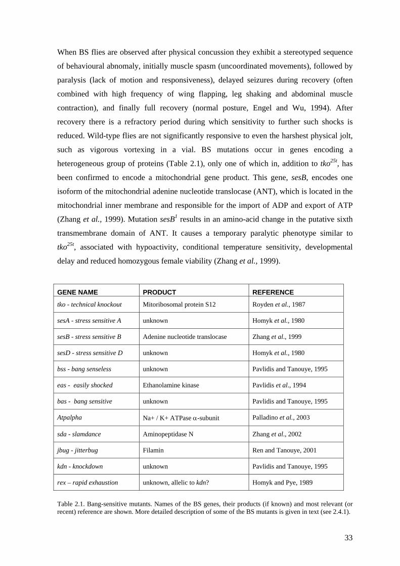
When BS flies are observed after physical concussion they exhibit a stereotyped sequence
of behavioural abnomaly, initially muscle spasm (uncoordinated movements), followed by
aralysis (lack of motion and responsiveness), delayed seizures during recovery (often p
combined with high frequency of wing flapping, leg shaking and abdominal muscle
contraction), and finally full recovery (normal posture, Engel and Wu, 1994). After
recovery there is a refractory period during which sensitivity to further such shocks is
reduced. Wild-type flies are not significantly responsive to even the harshest physical jolt,
such as vigorous vortexing in a vial. BS mutations occur in genes encoding a
heterogeneous group of proteins (Table 2.1), only one of which in, addition to tko25t, has
been confirmed to encode a mitochondrial gene product. This gene, sesB, encodes one
isoform of the mitochondrial adenine nucleotide translocase (ANT), which is located in the
mitochondrial inner membrane and responsible for the import of ADP and export of ATP
(Zhang et al., 1999). Mutation sesB1 results in an amino-acid change in the putative sixth
transmembrane domain of ANT. It causes a temporary paralytic phenotype similar to
tko25t, associated with hypoactivity, conditional temperature sensitivity, developmental
delay and reduced homozygous female viability (Zhang et al., 1999).
GENE NAME PRODUCT REFERENCE
tko - technical knockout Mitoribosomal protein S12 Royden et al., 1987
sesA - stress sensitive A unknown Homyk et al., 1980
sesB - stress sensitive B eotide translocase Adenine nucl Zhang et al., 1999
sesD - stress sensitive D unknown Homyk et al., 1980
bss - bang senseless unknown Pavlidis and Tanouye, 1995
eas - easily shocked Ethanolamine kinase Pavlidis et al., 1994
bas - bang sensitive unknown Pavlidis and Tanouye, 1995
Atpalpha Na+ / K+ ATPase α-subunit Palladino et al., 2003
sda - slamdance Aminopeptidase N Zhang et al., 2002
jbug - jitterbug Filamin Ren and Tanouye, 2001
kdn - knockdown unknown Pavlidis and Tanouye, 1995
rex – rapid exhaustion unknown, allelic to kdn? Homyk and Pye, 1989
Table 2.1. Bang-srecent) reference a
ensitive muta e BS genes, their produ vant (or re shown. M description of some of the B 2.4.1).
nts. Names of thore detailed
cts (if known) and most releS mutants is given in text (see
33
2.2.4 Co- regulation of n ndrial gene
transcriptional processing, mRNA stability, translation, post-translational modifications
tal processes that
the mitochondrial and the nuclear genomes.
etazoan genomes, and they might have a role also as mitochondrial
odifiers (Caceres and Kornblihtt, 2002; Ladd and Cooper, 2002; Nissim-Rafinia
uclear and mitocho expression
Control of gene expression is achieved at various levels, such as transcription, post-
and protein stability. Little is known about physiological and developmen
regulate co-ordinated gene expression of
Because mitochondria are involved in important cellular and physiological processes, it is
plausible to expect that at least some mitochondrially located nuclear genes need to be
controlled in an orchestrated fashion. Many transcriptional regulatory elements have been
shown, or suggested, to play roles in nucleo-mitochondrial communication, most notably
nuclear respiratory factors NRF-1 and NRF2 (reviewed by Scarpulla, 2002), the
OXBOX/REBOX system (Chung et al., 1992) and Mt3/4 elements (Suzuki et al., 1995).
Genes possessing NRF-1/2 binding sites include those encoding many subunits of the ETC
complexes, components of the mitochondrial transcription/replication machinery and genes
involved in protein import and assembly pathways and intermediary metabolism
(Scarpulla, 2002). As already mentioned, human mitoribosomal protein S12 has also
putative NRF-1 and NRF-2 binding sites in its promoter (Johnson et al., 1998). In addition,
DNA replication-related elements have been shown to be important in the regulation of
both nuclear and mitochondrial replication machineries in Drosophila, where they
probably have a role in the regulation of at least Pol γ-β, mtSSB and TFAM (Wang et al.,
1997; Lefai et al., 2000b; Ruiz De Mena et al., 2000; Takata et al., 2001; Takata et al.,
2003).
Alternative splicing and usage of alternative transcription start sites has been shown to be a
major regulator of gene expression and generator of diversity from the relatively small
number of genes in m
disease m
and Kerem, 2002). For example, many enzymes responsible for mtDNA repair are encoded
by the same genes as their nuclear counterparts, and mitochondrial targeting sequences are
generated through alternative splicing of mRNAs, alternative use of transcription initiation
sites, or alternative use of translation initiation sites (Kang and Hamasaki, 2002). Similarly,
human cytosolic and mitochondrial lysyl-tRNA synthetases are encoded by the same gene,
and the mitochondrial version is produced by alternative splicing that results in exon
inclusion and insertion of the mitochondrial targeting peptide (Tolkunova et al., 2000).
34
Well-defined elements that regulate the translational ‘potential’ of many key mRNAs are
5´-terminal oligopyrimidine tracts (TOPs) and upstream open reading frames (uORFs).
TOPs are present in mRNAs for many components of cytosolic translation machinery,
ost notably ribosomal proteins, and in addition to mammals they are found in many
reviewed by Meijer and Thomas, 2002). uORFs generally encode
utative peptides of 5 to 25 amino-acids and are recognised by the scanning initiation
m
mRNAs in other vertebrates, and even in insects (reviewed by Meyuhas, 2000). Growth
arrest in response to a wide variety of signals, such as contact inhibition or amino-
acid/serum starvation, leads to selective repression of the translation of TOP mRNAs and
is characterised by their shift from polysomes (complex of ribosomes bound to a single
mRNA) into the subpolysomal fraction (mRNP particles). Conversely, growth stimuli
cause efficient accumulation of these mRNAs to the actively translated polysomal fraction.
A particularly complex example of a TOP-regulated protein in vertebrates is poly(A)-
binding protein (PABP), that itself has been implicated in translational regulation, where it
assists initiation of translation and controls mRNA stability. Furthermore, PABP is subject
to autoregulation through an A-rich sequence in its 5´-UTR (Meyuhas, 2000, and
references therein).
Probably close to 40 % of vertebrate mRNAs contain at least one uORF in their 5´-UTR,
and large proportion of these messengers are rare and poorly translated at least in normal
growth conditions (
p
complex with varying efficiencies, depending on properties of the uORF itself, the regions
surrounding it and the intercistronic length between the uORF and major ORF (Lovett and
Rogers, 1999; Meijer and Thomas, 2002). In some cases, the uORF-encoded short peptide
might directly interact in cis with translating ribosome and prevent further translation, for
example by inhibiting the peptidyl transferase centre (Lovett and Rogers, 1999). However,
this mechanism is probably specific to certain metabolic pathways, since uORF-encoded
peptides are not generally conserved in their amino-acid sequence. Leaky scanning
(ribosomes do not recognise uORF), or possibly re-initiation after translation of the peptide
encoded by the uORF might result in translation of the downstream ORF (Morris and
Geballe, 2000; Meijer and Thomas, 2002). A good example of these mechanisms is yeast
transcription factor GCN4, which is involved in upregulation of a large group of amino-
acid biosynthetic genes (Ruiz-Echevarria and Peltz, 2000; Dever, 2002; Meijer and
Thomas, 2002). GCN4 contains a total of four uORFs in its 5’-leader, the first (uORF1)
35
and the last (uORF4) of which are functionally most important. During amino-acid
starvation eukaryotic initiation factor eIF2a is phosphorylated, a mechanism that leads to
reduced efficiency of ternary complex formation. Even in these conditions most of the
ribosomes can translate the uORF1, then resume and continue scanning towards uORF4. In
normal conditions ternary complex formation is efficient, and when scanning reaches
uORF4 ribosomes will recognise and translate it, which results in disengagement of the
ribosome from the GCN4 message without producing Gcn4p. Under starvation, however,
the low levels of ternary complexes enable scanning to continue for longer distances
without re-initiation of translation. Ribosomes then scan past the uORF4 and subsequently
acquire the ternary complex in time to initiate at the GCN4 start codon, which results in
production of Gcn4p and transcriptional upregulation of amino-acid biosynthetic genes.
(Meijer and Thomas, 2002). Interestingly, one of these target genes in yeast is ILV5, the
product of which can improve the mtDNA stability in lines lacking Abf2p, the yeast
counterpart of TFAM (Zelenaya-Troitskaya et al., 1995). It turns out that Ilv5p controls the
organization of mtDNA in nucleoids (Kaufman et al., 2000; MacAlpine et al., 2000), and
hence it can be said that regulation of mtDNA organization and transmission in yeast is
indirectly under translational regulation by uORFs. In mice, uncoupling protein 2 (UCP2),
which is expressed in a tissue-specific manner, is under both transcriptional and
translational control, the latter being due to uORFs. Under oxidative stress conditions
increase in UCP2 protein levels occurs without any change in UCP2 mRNA levels, which
allows strong and rapid induction in tissues containing high, basal expression at mRNA
level (Pecqueur et al., 2001).
Through nonsense or frameshift mutations, up to 30% of mutant alleles contributing to
human genetic disease harbour a premature termination signal (Mendell and Dietz, 2001).
In many cases, premature termination codons in the coding sequence or nonsense codons
the 5´-UTR lead to nonsense-mediated mRNA decay, or mRNA surveillance, an mRNA in
quality control mechanism that is ubiquitous in eukaryotes (reviewed by Mendell and
Dietz, 2001). The exact nature and location of this process are still under investigation, but
both nuclear and cytosolic compartments have been suggested (Wilkinson and Shyu,
2002). In principle, uORF-containing mRNAs can be sensitive to this pathway, which
could lead to mRNA degradation. Certain uORFs in yeast are followed by cis-acting
stabilizer element which prevent the transcript from undergoing nonsense-mediated mRNA
decay, such as in the case of GCN4 (Ruiz-Echevarria and Peltz, 2000). The cis-acting
36
stabilizer element is bound by the RNA-binding protein Pub1, mammalian homologues of
which have been shown to play important roles in differentiation and proliferation, and
which has been proposed to prevent rapid decapping and destruction of the bound mRNAs
by the surveillance complex (Ruiz-Echevarria and Peltz, 2000).
2.3 Mitochondria and disease
Mitochondrial disease can be caused by mutations in nuclear DNA or in mtDNA, most of
which target subunits of OXPHOS complexes directly or else indirectly, via genes
involved in mitochondrial translation (reviewed by Smeitink et al., 2001). Mitochondrial
atrilineal inheritance, are usually progressive, and have disorders can show Mendelian or m
no known treatment. Nonsyndromic mitochondrial disorders affect only single organs,
such as the eye or cochlea, whereas syndromic disorders involve multiple organ systems.
Generally, mitochondrial disorders affect highly energy-dependent, post-mitotic tissues,
such as the central nervous system (CNS) and skeletal and cardiac muscle, and are often
characterised by exercise intolerance, seizures and sensorineural deafness. Typical
examples of syndromic manifestations (reviewed by Schmiedel et al., 2003) include
MELAS (mitochondrial myopathy, encephalopathy, lactic acidosis and stroke-like
episodes), MERRF (myoclonic epilepsy with ragged red fibers), LHON (Leber hereditary
optic neuropathy), Leigh syndrome (infantile subacute necrotizing encephalopathy) and
NARP (neuropathy, ataxia and pigmentary retinopathy). However, there is considerable
clinical variability and many patients do not fit into one particular category. Over 100
different mtDNA point mutations and even more mtDNA deletions have been described
since the first mutations associated with disease were described 15 years ago (Holt et al.,
1988; Wallace et al., 1988a; Wallace et al., 1988b). A search for genes causing “nuclear-
mitochondrial disorders” is ongoing, and has revealed already many genes involved in
OXPHOS, mtDNA maintenance, ETC assembly and protein import (Koehler et al., 1999;
reviewed by Zeviani, 2003). Based upon the available data, an estimate for the prevalence
of all mitochondrial diseases is at least 1 in 8000 of the population, making them among
the most common genetically determined diseases (reviewed by Chinnery and Turnbull,
2001). Additionally, mtDNA mutations and impairment of mitochondrial function has been
implied also in cancer (reviewed by Copeland et al., 2002; Tomlinson et al., 2002),
diabetes mellitus (reviewed by Berdanier, 2001), many age-related neurodegenerative
disorders, such as Parkinson’s disease (reviewed by Sherer et al., 2002), Alzheimer’s
37
disease (reviewed by Castellani et al., 2002), Huntignton’s disease (reviewed by Ross,
2002), and the pathogenesis of Down’s syndrome (reviewed by Arbuzova et al., 2002).
2.3.1 Inheritance, heteroplasmy and segregation of mtDNA The human mitochondrial genome is thought to be transmitted strictly from the mother to
the offspring (Birky, 1995; Birky, 2001), although a single case of muscle-specific,
c drift and highly tissue
ependent directional selection for different, seemingly neutral (non-pathogenic) mtDNA
paternal transmission has been recently reported (Schwartz and Vissing, 2002). Because
mtDNA lacks sexual recombination and has approximately 10 times higher mutation
frequency compared to nuclear genes it is in principle susceptible to the accumulation of
deleterious mutations over generations, the so-called Muller’s ratchet (Brown et al., 1979;
Bergstrom and Pritchard, 1998). Mutations in mtDNA can either be inherited or generated
sporadically, and the presence of two or more kinds of mtDNA molecules in the individual,
tissue or single-cell is called heteroplasmy. At the population level mtDNA is highly
polymorphic, but this is not the case at the individual level. During early embryogenesis
only a small proportion of the available mtDNA molecules repopulate the embryonic
tissues. It is thought that this genetic “bottleneck” causes a heteroplasmic pool of mtDNAs
to segregate rapidly. Genotype frequencies in shift greatly in a single transmission from
mother to offspring (reviewed by Chinnery et al., 2000). As a result most individuals are
homoplasmic for a single species of mtDNA. Since mtDNA exists in thousands of copies
per cell, the proportion of pathogenic mtDNA molecules needs to be high enough in order
to cause dysfunction. This can result by unequal partitioning of mtDNA (vegetative
segregation) in dividing cells, or by clonal expansion in non-dividing cells due to cell
cycle-independent replication of mtDNA. Although homoplasmic pathogenic mutations
exist, most disease causing mutations occur in the heteroplasmic state, implying that
energy deficiency can result from absence of complementation by wild-type mtDNA. It is
possible that oocytes containing mutated mtDNA are subjected to negative selection during
oogenesis to eliminate cells with defective OXPHOS capacity (quality control), but this is
not currently supported by available data (Chinnery et al., 2000).
The segregating unit of mtDNA inheritance have been suggested to be a nucleoid (Jacobs
et al., 2000). In mouse models, evidence for both random geneti
d
genotypes in the same animal have been observed (Jenuth et al., 1996; Jenuth et al., 1997).
Since genetic background has an effect on these processes, mtDNA segregation seems to
38
be under complex nuclear control (Battersby et al., 2003). It is still a mystery how
pathogenic mtDNAs are selected (or counter-selected) in the female human germ-line, or if
the predominant mechanism is random genetic drift (Chinnery et al., 2000).
2.3.2 Threshold effects
Clinical diagnosis and genetic counselling of mitochondrial disorders is difficult because
lt in different symptoms. Conversely, similar pathological states
an be caused by different mutations. Both the nature of the mutation and the level of
S capacity at the cellular level. For example, individual muscle
bers from patients exhibiting different clinical manifestations (e.g. MELAS, PEO, DM or
the same mutation can resu
c
heteroplasmy affect the severity of the disease, but the relationship with severity is not
necessarily linear (Morgan-Hughes et al., 1995). Heteroplasmy levels can alter in various
tissues of the same individual or even in different cells of the same tissue. Furthermore, the
same level of heteroplasmy can result as dysfunction of some tissues but not in others.
These differential phenotypic manifestations are explained by varying, tissue-specific
tolerance levels for a particular mutation, i.e. threshold effects (reviewed by Rossignol et
al., 2003). Even homoplasmic (or nearly homoplasmic) mutations do not necessarily cause
disease in all individuals carrying them, but might require involvement of environmental
factors or alterations in nuclear, possibly tissue-specific genetic modifiers (Carelli et al.,
2003). Good examples of homoplasmic mtDNA mutations with variable penetrance are
aminoglycoside induced deafness associated with the mitochondrial SSU rRNA mutation
A1555G (Prezant et al., 1993; Guan et al., 2001) and Leber’s hereditary optic neuropathy
(LHON) most often associated with mtDNA-encoded genes for complex I subunits
(reviewed by Man et al., 2002), both of which are tissue-specific, affecting mainly hearing
and vision, respectively.
Threshold effects have been analysed in more detail by studying consequences of
heteroplasmy for OXPHO
fi
Leigh syndrome) but the same mtDNA mutation (A3243G in tRNALeu(UUR) ) have been
studied by combined quantification of heteroplasmy levels and histochemical detection of
cytochrome c oxidase (COX) activity. In these studies high mutant load was associated
with COX negative fibers, and the differences in the somatic distribution of the mutation
among tissues were suggested to be responsible for the differential phenotypic expression
of the disease (Petruzzella et al., 1994; Koga et al., 2000). Secondly, cytoplasmic hybrid
39
(cybrid) technology (King and Attardi, 1989) has been used to transfer patient-derived
mitochondria to cell-lines lacking mtDNA (rho0) in order to produce cybrids with varying
heteroplasmy levels. In combination, transmitochondrial studies indicate that both
heteroplasmy level and nuclear background affect biochemical capacity of the cells
(Attardi et al., 1995; Koga et al., 1995; Raha et al., 1999; Vergani et al., 1999). Finally,
influences of heteroplasmy have been modelled by inhibiting OXPHOS complexes by
titration with specific inhibitors. The studies carried out mainly in rat tissues have revealed
biochemical threshold effects, i.e. the activity of ETC complexes can be decreased
substantially without affecting respiration or ATP synthesis (Rossignol et al., 1999).
Comparable observations have been made in a Drosophila subobscura strain carrying
partially deleted mtDNA, which manifests decreased activities of some ETC complexes
(particularly complex I), but shows no effect on net ATP synthesis, nor obvious phenotype
(Beziat et al., 1997). However, the affected ETC complexes of these flies are more
susceptible to inhibition, and the threshold level for complex I inhibition, where a large
drop in respiration and ATP synthesis is observed is only 20% in the deletion mutant strain
compared to 70% in wild-type flies (Farge et al., 2002a). Whether or not the mutation
directly affects on ETC subunit, or has a more general impact on production of
mitochondrially encoded polypeptides, migh influence the levels of heteroplasmy that can
be tolerated, since threshold effects can operate at the transcriptional or translational level
or affect enzyme activity via kinetic or assembly defects (Rossignol et al., 2003).
2.3.3 Animal models of mitochondrial disease
Whole animal models are crucial in order to understand complex pathophysiology, tissue
rial disorders, as well as segregation of
tDNA. Although cybrid technology has been useful in defining pathogenic effects of
specificity and developmental aspects of mitochond
m
mtDNA mutations in cellular level, the research has been hindered by the lack of animal
models with mtDNA mutations due to inaccessibility of mtDNA for targeted mutagenesis
(reviewed by Jacobs, 2001). For this reason, animal modelling of mitochondrial disease
has concentrated on manipulations of nuclearly encoded mitochondrial genes, some of
which have been studied in knock-out mice, or alternatively in Drosophila. In Drosophila
the studies have historically used conventional or “forward” genetics, i.e. from phenotype
to genotype, whereas in mice, known mitochondrial genes have been subjected to
(conditional) targeted disruption. Manipulation of mouse embryonic stem cells has
40
produced knockout lines of many nuclear encoded mitochondrial proteins, such as TFAM,
adenine nucleotide translocase 1 (ANT1), uncoupling proteins, Mn superoxide dismutase
and many genes involved in apoptotic pathways (reviewed by Wallace, 2002). Recently the
very first transmitochondrial lines carrying either a partial deletion (Inoue et al., 2000) or
pathogenic point mutation (Sligh et al., 2000) in mtDNA were described. These have
proved that heteroplasmic point mutation producing symptoms of mitochondrial disease in
mouse can be transferred through the female germline, and this approach has given a great
promise for future research (Hirano, 2001).
2.3.4 Drosophila as a model organism
The question arises as to whether relatively simple invertebrates such as fruit-flies or
ve as reasonable models for human disease.
nimal experimentation in general is a trade-off between genetic power and biomedical
early 20th century. This work started
enetics as we know it today, and Drosophila was the first organism in which
worms (Caenorhabditis elegans) can ser
A
relevance (Dow and Davies, 2003). To maximise biomedical relevance studies should be
carried out using human patients or patient-derived tissues. Even if this is possible in some
cases, genetic manipulation of humans is impossible for ethical reasons, and studies in
most closely related vertebrates are hindered by technical problems. Major vertebrate
models offering at least partial genomic sequence data, and in which classical genetics is
possible, are rat (Rattus rattus), mouse (Mus musculus) and zebrafish (Danio rerio). The
mouse, which is regarded generally as the most suitable model organism for human
disorders, shares reasonably conserved physiology with humans and can be subjected to
targeted gene disruption by homologous recombination. However, this is both slow and
very expensive, and might therefore create restraints that could be avoided by using non-
vertebrate models. Additionally, the life cycle of the mouse is far too long to perform
genetic screens routinely (Dow and Davies, 2003).
Drosophila melanogaster has been under intensive investigation since its “domestication”
by Thomas Hunt Morgan and his students in the
g
chromosomes were identified as the carriers of the hereditary material, with the genes
arranged in linear order (Sturtevant, 1913; Bridges, 1914). It was also the first
experimental animal in which ionizing radiation was shown to cause genetic damage,
mutations (Muller, 1927). Today, the Drosophila genome is completely sequenced (Adams
41
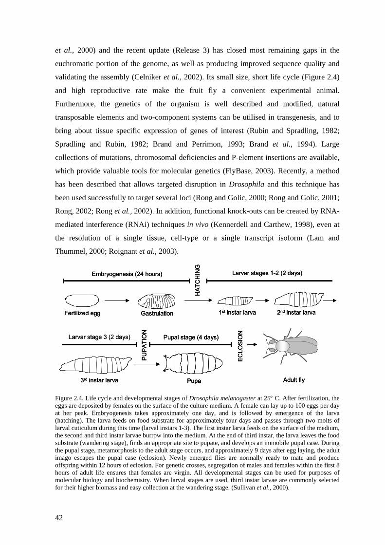
et al., 2000) and the recent update (Release 3) has closed most remaining gaps in the
euchromatic portion of the genome, as well as producing improved sequence quality and
validating the assembly (Celniker et al., 2002). Its small size, short life cycle (Figure 2.4)
and high reproductive rate make the fruit fly a convenient experimental animal.
Furthermore, the genetics of the organism is well described and modified, natural
transposable elements and two-component systems can be utilised in transgenesis, and to
bring about tissue specific expression of genes of interest (Rubin and Spradling, 1982;
Spradling and Rubin, 1982; Brand and Perrimon, 1993; Brand et al., 1994). Large
collections of mutations, chromosomal deficiencies and P-element insertions are available,
which provide valuable tools for molecular genetics (FlyBase, 2003). Recently, a method
has been described that allows targeted disruption in Drosophila and this technique has
been used successfully to target several loci (Rong and Golic, 2000; Rong and Golic, 2001;
Rong, 2002; Rong et al., 2002). In addition, functional knock-outs can be created by RNA-
mediated interference (RNAi) techniques in vivo (Kennerdell and Carthew, 1998), even at
the resolution of a single tissue, cell-type or a single transcript isoform (Lam and
Thummel, 2000; Roignant et al., 2003).
Figure 2.4. Life cycle and developmental stages of Drosophila melanogaster at 25° C. After fertilization, the eggs are deposited by females on the surface of the culture medium. A female can lay up to 100 eggs per day at her peak. Embryogenesis takes approximat(hatching). The larva feeds on food substrate fo
Fertilized egg Gastrulation
Embryogenesis (24 hours)
1st instar larva 2nd instar larva
3rd instar larva
HAT
CH
ING
PUPA
TIO
N
Pupa
ECLO
SIO
N
Adult fly
Larvar stages 1-2 (2 days)
Pupal stage (4 days)Larvar stage 3 (2 days)
Fertilized egg Gastrulation
Embryogenesis (24 hours)
1st instar larva 2nd instar larva
3rd instar larva
HAT
CH
ING
PUPA
TIO
N
Pupa
ECLO
SIO
N
Adult fly
Larvar stages 1-2 (2 days)
Pupal stage (4 days)
Fertilized egg Gastrulation
Embryogenesis (24 hours)
1st instar larva 2nd instar larva
3rd instar larva
HAT
CH
ING
PUPA
TIO
N
Pupa
ECLO
SIO
N
Adult fly
Larvar stages 1-2 (2 days)
Pupal stage (4 days)Larvar stage 3 (2 days)
ely one day, and is followed by emergence of the larva r approximately four days and passes through two molts of
rval cuticulum during this time (larval instars 1-3). The first instar larva feeds on the surface of the medium, lathe second and third instar larvae burrow into the medium. At the end of third instar, the larva leaves the food substrate (wandering stage), finds an appropriate site to pupate, and develops an immobile pupal case. During the pupal stage, metamorphosis to the adult stage occurs, and approximately 9 days after egg laying, the adult imago escapes the pupal case (eclosion). Newly emerged flies are normally ready to mate and produce offspring within 12 hours of eclosion. For genetic crosses, segregation of males and females within the first 8 hours of adult life ensures that females are virgin. All developmental stages can be used for purposes of molecular biology and biochemistry. When larval stages are used, third instar larvae are commonly selected for their higher biomass and easy collection at the wandering stage. (Sullivan et al., 2000).
42
The Drosophila melanogaster genome is ~180 Mb in length (a third of which is centric
heterochromatin) and encodes approximately 13 500 genes. Compared to humans (~ 3.2
Gb, approximately 30 000 genes) it is markedly smaller in size, but contains about half the
num er of genes and transcripts. Importantly, according to the Homophila database (Chien
et al., 2002), approximately 75% of the human disease gene associated sequences found in
the OMIM database (Online Mendelian Inheritance in Man, (Hamosh et al., 2002) have
strong matches to one or more sequences in the Drosophila genome database (FlyBase,
2003). It is clear that at least insights to the basic mechanisms of human disease can be
gained from studies in flies. Good examples of these are neurodegenerative diseases, such
as Huntington’s disease (HD) which is caused by expansions of a polyglutamine repeat
found in the protein huntingtin, leading to neuronal loss and characterized by insoluble
protein aggregates in brains (Ross, 2002; Rubinsztein, 2002). Even if mouse models have
provided valuable data about molecular mechanisms underlying HD, the pathogenesis of
the disease is not currently understood. A powerful approach to identifying pathways
involved in HD pathology and possible protective strategies, is to perform suppressor
screens to identify genes that alleviate or modify the disease, for instance by using P-
element insertions in a Drosophila model (Kazemi-Esfarjani and Benzer, 2000).
Especially where a pathway shares a common evolutionary origin between different
eukaryotic lineages, genetic dissection of its components, combined with com
b
parative
structural studies, can produce valuable data for understanding disease processes. For
example, the insulin signalling pathways in humans, mice, flies and worms shows
remarkable conservation between the organisms (Leevers, 2001; Claeys et al., 2002):
reduction in the function of this pathway seems to increase longevity in all these organisms
(reviewed by Partridge and Gems, 2002). Similarly, developmental and comparative
studies suggest that the organs of mechanoreception, from worms to humans most
probably share a common evolutionary origin (Garcia-Anoveros and Corey, 1997;
Caldwell and Eberl, 2002). Alterations in the human cochlea are difficult to study, a case in
which Drosophila can provide a valuable tool for experimental manipulations (Jarman,
2002), as discussed in the next chapter.
43
2.4 Genetics of deafness
.4.1 Principles of mechanosensation
ms depending on the mechanical input to which
eptors can be found, for example, in skin (touch,
in), joints and muscles (limb proprioception), the vestibular system (balance) and
2
Mechanoreceptors can take various for
they have adapted. In humans mechanorec
sharp pa
cochlea (audition). Hearing is based on the conversion of mechanical vibration
(movement) to electrical signals, which are subsequently transmitted further through
synaptic connections with afferent sensory neurons. In vertebrates sound detection is
mediated by inner-ear hair cells (Hudspeth and Jacobs, 1979). The hair cell (Figure 2.5) is
a sensory receptor that responds to mechanical stimulation of its sensory hair bundle,
which usually consists of numerous large microvilli (stereocilia) and a single true cilium
(the kinocilium). In Drosophila mechanoreceptive sensory cells are also found all over the
body, including the thorax, abdomen, legs, wings and antennae. The most noticeable of
these are the sensory bristles inserted in the cuticle (external sensory organs, Figure 2.5).
Another type of mechanosensors, chordotonal organs (CHOs, Figure 2.5), do not have an
external bristle and are located within the cuticulum. Developmental and comparative
studies suggest that external sense organs and CHOs in Drosophila have a common
evolutionary origin (Merritt, 1997). The most famous assembly of CHOs, Johnston’s
organ, is localised in the antennae, and serves as the principal auditory organ in insects
(Eberl et al., 2000). Fruit-flies use Johnston’s organ to discriminate species-specific
courtship songs at close range, which can be used in genetic screens for mutations that
disrupt auditory function (Eberl et al., 1997). Indeed, many mutations have been found that
disrupt the CHO structure, and hence show defective courtship behaviour, such as atonal
(ato), beethoven (btv) and touch-insensitive-larva-B (tilB, Eberl et al., 1997; Eberl et al.,
2000).
44
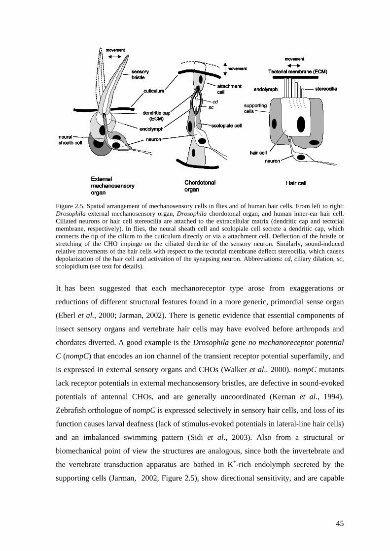
supportingcells
endolymph
neuron
cuticulumattachment cell
External mechanosensoryorgan
Chordotonalorgan
sensorybristle
movement
movement
dendritic cap(ECM)
neural sheath cell
scolopiale cell
stereocilia
Tectorial membrane (ECM)
movement
hair cellneuron
endolymph
Hair cell
cdsc supporting
cells
endolymph
neuron
cuticulumattachment cell
External mechanosensoryorgan
Chordotonalorgan
sensorybristle
movement
movement
dendritic cap(ECM)
neural sheath cell
scolopiale cell
stereocilia
Tectorial membrane (ECM)
movement
hair cellneuron
endolymph
Hair cell
endolymph
neuron
cuticulumattachment cell
External mechanosensoryorgan
Chordotonalorgan
sensorybristle
movement
movement
dendritic cap(ECM)
neural sheath cell
scolopiale cell
stereocilia
Tectorial membrane (ECM)
movement
hair cellneuron
endolymph
Hair cell
endolymph
neuron
cuticulumattachment cell
External mechanosensoryorgan
Chordotonalorgan
sensorybristle
movement
movement
dendritic cap(ECM)
neural sheath cell
scolopiale cellendolymph
neuron
cuticulumattachment cell
External mechanosensoryorgan
Chordotonalorgan
sensorybristle
movement
movement
dendritic cap(ECM)
neural sheath cell
scolopiale cell
stereocilia
Tectorial membrane (ECM)
movement
hair cellneuron
endolymph
Hair cell
stereocilia
Tectorial membrane (ECM)
movement
hair cellneuron
endolymph stereocilia
Tectorial membrane (ECM)
movement
hair cellneuron
endolymph
Hair cell
cdsc
Figure 2.5. Spatial arrangement of mechanosensory cells in flies and of human hair cells. From left to right: Drosophila external mechanosensory organ, Drosophila chordotonal organ, and human inner-ear hair cell. Ciliated neurons or hair cell stereocilia are attached to the extracellular matrix (dendritic cap and tectorial membrane, respectively). In flies, the neural sheath cell and scolopiale cell secrete a dendritic cap, which connects the tip of the cilium to the cuticulum directly or via a attachment cell. Deflection of the bristle or stretching of the CHO impinge on the ciliated dendrite of the sensory neuron. Similarly, sound-induced relative movements of the hair cells with respect to the tectorial membrane deflect stereocilia, which causes depolarization of the hair cell and activation of the synapsing neuron. Abbreviations: cd, ciliary dilation, sc, scolopidium (see text for details).
It has been suggested that each mechanoreceptor type arose from exaggerations or
reductions of different structural features found in a more generic, primordial sense organ
(Eberl et al., 2000; Jarman, 2002). There is genetic evidence that essential components of
insect sensory organs and vertebrate hair cells may have evolved before arthropods and
chordates diverted. A good example is the Drosophila gene no mechanoreceptor potential
C (nompC) that encodes an ion channel of the transient receptor potential superfamily, and
is expressed in external sensory organs and CHOs (Walker et al., 2000). nompC mutants
lack receptor potentials in external mechanosensory bristles, are defective in sound-evoked
potentials of antennal CHOs, and are generally uncoordinated (Kernan et al., 1994).
Zebrafish orthologue of nompC is expressed selectively in sensory hair cells, and loss of its
function causes larval deafness (lack of stimulus-evoked potentials in lateral-line hair cells)
and an imbalanced swimming pattern (Sidi et al., 2003). Also from a structural or
biomechanical point of view the structures are analogous, since both the invertebrate and
the vertebrate transduction apparatus are bathed in K+-rich endolymph secreted by the
supporting cells (Jarman, 2002, Figure 2.5), show directional sensitivity, and are capable
45
to mechanically tune themselves to varying stimulus intensities (Walker et al., 2000;
Gopfert and Robert, 2003).
Deafness can result from developmental, structural or functional defects of the
mechanoreceptors or associated cells. In flies mutant for the proneural gene atonal,
chordotonal organs, photoreceptors, and a subclass of olfactory receptors fail to develop
because their precursors are not specified (Jarman et al., 1993; Jarman et al., 1995; Gupta
and Rodrigues, 1997). atonal flies are devoid of scolopidia, a specific structure in the CHO
(Figure 2.5), although the antennal nerve is present (Jhaveri et al., 2000). Evidence
underlying developmental homology between CHOs and mammalian hair cells is provided
by the mouse homologue of atonal (MATH1) which is expressed in the auditory sensory
epithelium at the time hair cell precursors are specified and is required for hair cell
generation (Bermingham et al., 1999). Furthermore, the atonal mutation in Drosophila is
rescued by MATH1 expression (Ben-Arie et al., 2000). In btv5P1 mutants the chordotonal
cilium of auditory mechanosensory neurons displays an anatomically aberrant ciliary
dilation (Figure 2.5), in the contact zone between the ciliary axoneme and membrane
(Eberl et al., 2000; Gopfert and Robert, 2003). btv5P1 as well as tilB2 affects only the
CHOs, which is shown by complete loss of sound-evoked potentials measured from the
antennal nerve but normal touch-evoked potential due to deflection of thoracic bristles
(Kernan et al., 1994; Eberl et al., 2000). In addition to being deaf, tilB2 males are sterile
due to a motility defect caused by malformation of sperm tail axonemes, the other of the
two ciliary structures in the fly (Eberl et al., 2000). Screens for defects in the larval
response to touch have revealed a set of mutations that affect mechanotransduction both in
external sensory organs and CHOs, such as uncoordinated (unc) (Kernan et al., 1994).
This mutant seems to exhibit more “syndromic” defects of mechanotransduction, since in
addition to affecting both CHOs and external sense organs, it also shows uncoordination of
movement and a sperm axoneme defect similar to that seen in tilB2 (Baker and Kernan,
2003). Despite this, unc shows a spontaneous response to light and to mechanical stimuli
indicating no defects in neuromuscular excitability (Kernan et al., 1994). These variable
phenotypes underlie a wide range of organ systems responsible for mechanotransduction,
touch, balance, and hearing.
Sensitivity to mechanical stress (bang-sensitivity, BS, see Table 2.1, section 2.2.3.2) has
become a classic tool for dissecting neural function in Drosophila. It should be noted that
46
mutations affecting development and structure of sense organs (discussed above) do not
generally show a BS phenotype. However, a decreased frequency of action potentials in
response to external sensory bristle displacement is a common defect in many BS mutants
studied, i.e. bang-sensitive (bas1), bang-senseless (bssMW1), easily-shocked (eas2) and tko25t
(Engel and Wu, 1994).
In addition, BS flies show seizures and paralysis when subjected to direct electrical
stimulation of the brain (Pavlidis and Tanouye, 1995; Lee and Wu, 2002). The giant fibers
(GFs) are a pair of interneurons that project from the fly brain to the thoracic ganglion, and
have several outputs in the thorax (Figure 2.6). The GFs synapse with a peripherally
synapsing interneuron (PSI), which drives flight motor neurons (DLMmns) that synapse
with dorsal longitudinal muscle (DLM) fibers. When an electrical stimulus is applied to the
brain of a fly, the GF pathway is activated, which results in synchronous activation of all
DLM fibers. When neuronal activity was (indirectly) measured from DLM fibers, BS
mutants (including tko25t) showed seizures of ~0.5-3 sec, which are likely to correspond to
the preparalytic seizures after mechanical disturbance (Pavlidis and Tanouye, 1995; Lee
and Wu, 2002). During the paralysis following the seizures, subsequent GF stimulation
could not evoke DLM fiber potentials. However, since muscle fibers could be stimulated
directly from their motor neurons, it seems that this stimulation failure is not due to defects
in flight muscles, motor neurons, or connections between them, but occurs at the level of
PSI-DLMmn synapses (Pavlidis and Tanouye, 1995). After recovery, a refractory period,
during which a stimulus was less effective at producing seizures and paralysis, was
observed with different BS mutants (Pavlidis and Tanouye, 1995; Lee and Wu, 2002).
These events closely follow the characteristics and time-course of mechanically induced
BS behaviour. It has been suggested that BS is due to a similar physiological defect in all
the mutants studied, but there are probably additional processes affected in some mutants
but not others (Pavlidis and Tanouye, 1995). It is also likely that other neural circuits in
addition to GF pathway are involved, which makes the interpretation of the results more
difficult. Mutations in at least two of the most studied BS genes, bas and bss, can also be
suppressed genetically by mutations affecting Na+-channels, K+-channels and electrical
synapses (Kuebler et al., 2001).
47
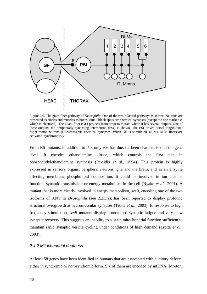
GF PSI
DLMmns
DLMs
1 2 3 4 5 6
HEAD THORAX
eGF PSI
DLMmns
DLMs
1 2 3 4 5 6
HEAD THORAX
e
Figure 2.6. The giant fiber pathway of Drosophila. One of the two bilateral pathways is shown. Neurons are presented as circles and muscles as boxes. Small black spots are chemical synapses (except the one marked e, which is electrical). The Giant fiber (GF) projects from brain to thorax, where it has several outputs. One of these outputs, the peripherally synapsing interneuron (PSI) is shown. The PSI drives dorsal longitudinal flight motor neurons (DLMmns) via chemical synapses. When GF is stimulated, all six DLM fibers are activated synchronously.
From BS mutants, in addition to tko, only eas has thus far been characterised at the gene
level. It encodes ethanolamine kinase, which controls the first step in
phosphatidylethanolamine synthesis (Pavlidis et al., 1994). This protein is highly
expressed in sensory organs, peripheral neurons, glia and the brain, and as an enzyme
affecting membrane phospholipid composition. It could be involved in ion channel
function, synaptic transmission or energy metabolism in the cell (Nyako et al., 2001). A
mutant that is more clearly involved in energy metabolism, sesB, encoding one of the two
isoforms of ANT in Drosophila (see 2.2.3.2), has been reported to display profound
structural overgrowth at neuromuscular synapses (Trotta et al., 2003). In response to high
frequency stimulation, sesB mutants display pronounced synaptic fatigue and very slow
synaptic recovery. This suggests an inability to sustain mitochondrial function sufficient to
maintain rapid synaptic vesicle cycling under conditions of high demand (Trotta et al.,
2003).
2.4.2 Mitochondrial deafness
At least 58 genes have been identified in humans that are associated with auditory defects,
either in syndromic or non-syndromic form. Six of them are encoded by mtDNA (Morton,
48
2002). Mutations in these genes frequently target components of the mitochondrial
translation machinery, such as A1555G mutation in mitochondrial SSU rRNA (for
discussion, see 2.3.2 and 2.4.3). Mutations in mitochondrial tRNAs for serine, leucine and
lysine have been implied in sensorineural hearing impairment and in syndromes including
sensorineural hearing impairment (reviewed by Jacobs, 1997). The tRNA mutations have
been suggested to act at the molecular level in diverse ways, such as impaired rate of tRNA
synthesis, decreased aminoacylation efficiency, increased turnover rate or defects in
processing and nucleotide modifications (Schon et al., 1992; El Meziane et al., 1998;
Borner et al., 2000; Levinger et al., 2001; Toompuu et al., 2002). Generally, mutations that
exist in the homoplasmic or nearly homoplasmic state show only a mild phenotype in cell
culture, such as decreased growth on selective (glucose-free) medium or modest
hypersensitivity to the antibiotic doxycyclin (Reid et al., 1997; Toompuu et al., 1999).
Such mutations may only be pathogenic in certain cell types or tissues, such as in the
cochlea, when the biochemical threshold for the mitochondrial dysfunction is reached. In
heteroplasmic cases associated with more severe mutations, a sharp threshold in the
capacity to support protein synthesis is observed, such as in the case of A3243G in
tRNALeu(UUR) or T7445C in tRNASer(UCN) (Dunbar et al., 1996; Guan et al., 1998).
Knowing the importance of mtDNA-encoded RNAs in deafness, the protein components of
the mitochondrial translation machinery are strong candidates for SHI in their own right, as
well as modifiers of expression of the mitochondrial tRNA/rRNA mutations (Shah et al.,
1997). Chromosome 19q13.1, to which the autosomal deafness locus DFNA4 has been
mapped, contains two genes encoding components of this machinery: ribosomal protein
S12 (MRPS12) (Shah et al., 1997; Johnson et al., 1998) and mitochondrial seryl-tRNA
synthetase (SARSM) (Yokogawa et al., 2000). These genes have been screened for
possible mutations in two DFNA4-linked deafness families, but no plausible disease
mutations were found (Shah et al., 2001). Therefore, at least in DFNA4-families identified
thus far, these genes can be excluded unless distant regulatory mutations are considered.
However, as putative modifiers of mitochondrial disorders these genes are still of interest.
2.4.3 Aminoglycoside antibiotics and deafness The most common mutation associated with maternally inherited non-syndromic deafness
is A1555G in the mitochondrial SSU rRNA (Prezant et al., 1993). The mutation is largely
49
homoplasmic, exists in numerous families with different ethnic origins, and sometimes the
affected members have a history of aminoglycoside antibiotic treatment (Estivill et al.,
1998; Hutchin and Cortopassi, 2000). As mentioned earlier, not all the carriers of the
mutation suffer from SHI, and genetic modifiers of the phenotype have been suggested to
play a role in the pathogenesis (Bu et al., 1993; Guan et al., 1996; Guan et al., 2001).
However, no nuclear modifiers of the disease have thus far been identified in humans.
Moreover, the pathogenic mechanism of the mutation is still largely unknown, although
many studies using patient-derived primary cell-lines and cybrid cell-lines have been
studied.
Aminoglycoside antibiotics have a general capacity to bind to nucleic acids that have
internal ‘bubbles’, bulges and other non-Watson-Crick duplex elements (Ryu and Rando,
2001) and show high affinity for the decoding centre of prokaryotic ribosomes (Cundliffe,
1987). Streptomycin (SM) and paromomycin (PM), for example, cause mistranslation of
mRNAs in bacteria (Ruusala and Kurland, 1984) and this is due to an altered
conformational state favouring acceptance of near-cognate tRNAs, as discussed in section
2.2.3. The mitochondrial A1555G mutation introduces a new base pair into the SSU rRNA
and thereby changes the region involved in decoding to a more bacterial-like conformation
(position 1491 in E. coli). This can bind certain aminoglycosides, such as PM, efficiently
in vitro (Hutchin et al., 1993; Hamasaki and Rando, 1997). Therefore, it has been
suggested that a decreased rate (or mistranslation) of mitochondrially encoded
polypeptides due to interaction with the drug could be the basis for aminoglycoside-
induced deafness (Hutchin et al., 1993). Indeed, cell-culture models have shown an
approximately 30% decrease in the rate of mitochondrial protein synthesis in the presence
of PM in A1555G harbouring lymphoblastoid cell-lines compared to controls (Guan et al.,
2000). More recently, similar experiments were carried out on patient-derived fibroblasts,
transmitochondrial cybrids with uniform nuclear background, and with transmitochondrial
fibroblasts in patient or control nuclear background simultaneously (Giordano et al., 2002).
These studies did not reveal a translational defect in any combination of mtDNA and
nuclear background without antibiotics, but confirmed the PM sensitivity reported
previously by Guan et al. (2000). Surprisingly, SM did not cause any defect in the same
experiments, which contrasted with the previous results suggesting moderate reduction in
mitochondrial translation products in the HeLa cell background (Inoue et al., 1996). These
experiments have further supported the idea that nuclear, possibly tissue-specific modifiers
50
have an important impact on manifestation of the disease, and that potential selective
uptake of the drugs by the cochlea could also be involved in the specificity of the disease.
The role of SM as an environmental inducer of deafness still remains to be elucidated,
because in contrast to PM, this specific mutation does not increase SM binding in vitro
(Hamasaki and Rando, 1997). It is notable that not all effects of this group of antibiotics
are attributable to decoding per se, since aminoglycosides have been shown to inhibit
RNase P RNA cleavage and aminoacylation of yeast tRNAAsp in vitro (Mikkelsen et al.,
1999; Walter et al., 2002).
Modifiers of the paromomycin resistance mutation PR454 in yeast mitochondrial SSU
rRNA, corresponding to human A1555G have been described (Weiss-Brummer and
Huttenhofer, 1989; Li and Guan, 2002; Li et al., 2002). Mto1p and Mss1p are
mitochondrially located tRNA modification enzymes homologous to bacterial gidA and
mnmE, respectively, and mutant alleles of the genes only manifest as respiration deficiency
when combined with the PR454 mutation (Decoster et al., 1993; Colby et al., 1998; Bregeon
et al., 2001). Human homologues of both MTO1 and MSS1 can complement this
deficiency in yeast, and at least MTO1 is expressed highly in the cochlea (Li and Guan,
2002; Li et al., 2002). PR454 has been suggested to result in stringent (restrictive)
proofreading during mitochondrial translation (Weiss-Brummer and Huttenhofer, 1989),
and since many mitochondrial tRNAs contain modifications it is plausible that
hypomodified tRNAs in mto1 and mss1 mutant strains are not functional in the highly
accurate ribosomal environment (Bregeon et al., 2001). Therefore, human homologues of
these proteins could be potentially involved in adjustment of codon-anticodon interactions
that might lead to disease in the context of the A1555G mutation (Li and Guan, 2002; Li et
al., 2002). A possibility that the tRNA modification deficiency could lead to additional
direct susceptibility of tRNAs to aminoglycosides can also not be excluded, since
aminoglycosides are implicated in the inhibition of binding of fMet-tRNAfmet to IF2 (Evans
et al., 2003).
51
3. AIMS OF THE STUDY The main purpose of this study is to elucidate the validity of D. melanogaster as a model
system for human mitochondrial disorders, particularly those resulting from translational
defects. Two different mutations in the Drosophila mitoribosomal protein S12 have been
studied for effects on translation, on mitochondrial OXPHOS function, and on the
development, physiology and behaviour of the fly. Additional experiments to understand
the molecular effects of these mutations on ribosome function were carried out in a
bacterial model system. Genetic and environmental factors affecting the fly phenotype
were investigated, showing that the model exhibits some close parallels with human
mitochondrial disease. One important variable affecting the phenotypic outcome may be
differential regulation of expression of the S12 gene. To investigate this at the molecular
level, the human homologue was studied using cultured human cells. More specifically, the
aims of the study were:
1) to model in E. coli the molecular consequences of the L85H mutation found in
tko25t flies, as well as the potential involvement of a conserved glutamine residue
(Q116) in the natural streptomycin resistance of mitochondria (I)
2) to characterise the mutant phenotype in prototypic tko25t mutants and in various
transgenic variant lines with respect to molecular, biochemical, developmental and
behavioural alterations (II, III)
3) to determine whether the L85H mutation is the sole cause of the tko25t phenotype
by a transgenic reversion test (II)
4) to create and characterise tkoQ116K transgenic lines for streptomycin resistance and
other possible phenotypes (II)
5) to create GAL4-responsive UAS-tko+ transgenic lines and characterize the effects
of tko+ over-expression in both wild-type and tko mutant flies (III)
6) to elucidate the mechanisms of regulation of human mitochondrial S12, especially
with respect to post-transcriptional control (IV)
52
4. MATERIALS AND METHODS
This chapter briefly describes the main methods used. For more detailed descriptions of
general methods, such as PCR, various blotting techniques, transformation, transfection
and molecular cloning, please refer to the original publications I-IV.
4.1 Bacterial strains Streptomycin sensitive (SMS) E. coli strain XL-1 Blue was obtained from Stratagene (La
Jolla, CA). Streptomycin resistant (SMR) strain DS941 was supplied by Dr. Martin R.
Boocock, University of Glasgow, and strains UD1D2 and UD5B6 used in nonsense
suppression analysis, and strain UD8C1 used in elongation rate assays were provided by
Dr. Diarmaid Hughes and Dr. Charles Kurland, University of Uppsala, Sweden. Strains
were stored as 50% v/v glycerol stocks at -70°C. Cultures were grown on L-agar plates or
in L-broth (Sambrook et al., 1989) supplemented with appropriate antibiotics, except for
the experiments carried out with UD1D2, UD5B6 and UD8C1, which were done using M9
minimal medium (Sambrook et al., 1989). To produce competent cells for transformations,
cells were grown to mid-logarithmic phase in a medium supplemented with 10 mM MgCl2, cooled on ice and washed with TB (10mM Pipes, 55mM MnCl2, 15 mM Cacl2, 250 mM
KCl). Cells were resuspended in TB containing 7% DMSO, aliquots were frozen in liquid
nitrogen, and stored at -70°C.
4.2. Mammalian cell culture
HEK293-EBNA cells were maintained in DMEM, 10% FCS, supplemented with 50µg/ml
uridine, 1 mM glutamine, 1 mM sodium pyruvate plus 100 units/ml penicillin and 100
µg/ml streptomycin. Cells were routinely passaged weekly. For serum starvation
conditions, please check original article (IV).
4.3 Drosophila strains and P-element mediated transgenesis
The tko25t mutant strain was obtained from Dr. Chun-Fang Wu and the tko3/FM7 stock was
obtained from the Bloomington stock center (Bloomington, IN). The strains Canton S
(wild-type) and w1118 were obtained from Dr. Kevin O’Dell, University of Glasgow,
Scotland, and the inbred control strain 0713 was supplied by Dr. Serge Alziari, Université
53
Blaise-Pascal, Clermont II, France. The stocks were maintained at 25 °C on a 12 hr:12 hr
light:dark cycle in yeast sugar medium (1% agar, 1.5% sucrose, 3% glucose, 3.5% active
dried yeast, 1.5% maize meal, 1% wheat germ, 1% soya flour, 3% treacle, all w/v)
supplemented with 0.5% propionic acid and 0.1% methyl paraben (Nipagin M), with w/v.
The tko25t stock was maintained both as a homozygous mutant (inbred) and tko25t/FM7
balanced stock (for explanation of balancers, see below). P-element mediated
transformation was carried out essentially as described by Spradling (Spradling, 1986).
Various versions of tko (i.e. tkoL85H, tkoH85L, tkoQ116K and tko+) were cloned into
pP{CaSpeR-4} (Thummel and Pirrotta, 1992) or pP{UAST} (Brand and Perrimon, 1993)
vectors, and co-injected into recipient w1118 embryos with pUChs∆2-3 helper plasmid.
Survivors were mated to the recipient strain, and putative transgenic progeny were selected
on the basis of the w+ eye colour marker. Siblings were crossed in order to produce
homozygous stocks. After molecular characterization, homozygous viable strains were
bred to various endogenous tko backgrounds (e.g. tko25t and tko3).
Insertions in the X, second and third chromosomes were maintained over balancers FM7,
CyO and TM6Sb, respectively. In D. melanogaster recombination occurs in females, and it
can be controlled by balancer chromosomes that contain dominant (and recessive) markers.
Balancers are no longer capable of pairing or recombining with their normal homologue
during meiotic prophase. Most recessive mutations are invisible in heterozygous
conditions. Because of the markers, transmission of balancers to progeny can be tracked
unambiguously and, therefore, also transmission of the homologous chromosome
containing the desired mutation (e.g. tko25t). If the progeny do not have the balancer, it
must have the homologue. By this way, invisible genotypes can be scored in progeny with
virtually 100% reliability.
4.4 Drosophila behavioural tests
4.4.1 Bang-sensitivity
Freshly eclosed flies were collected and separated into fresh food vials, with females and
males separated. After three to four hours flies were briefly CO2-anaesthetised Single flies
were transferred to empty vials and allowed to recover for half an hour. Stress sensitivity
was measured by vortexing single flies in vials for 15 s and by measuring the time taken
54
for the fly to stand on its feet again (recovery time). Stress-sensitivity of the delayed flies
eclosing from the doxycyclin experiment (see 4.4.3) were measured similarly.
4.4.2 Reactivity
Locomotion in response to mild mechanical disturbance was measured by counting the
distance travelled by a single fly in a 10 cm X 10 cm gridded open-field chamber. The
distance travelled during ten minutes after stimulus (in consecutive one minute periods)
was recorded. A total of 50 flies of each genotype were analysed.
4.4.3 Developmental time and antibiotic sensitivity
The stage of the developmental delay was determined by following the growth of larvae on
food plates containing an inert dye (neutral red) at 25 °C. The embryos were synchronised
by letting fertilised females lay eggs, and then replacing the plates at three-hour intervals.
The onset and length of the first and second larval molts, pupariation and eclosion of
imago were followed daily. Two antibiotics known to affect bacterial translation were
tested for potential additional effects on developmental phenotype on yeast-sugar medium
vials containing varying concentrations (0, 1, 3, 10, 30, 100, 300 or 1000 µg / ml food) of
doxycyclin (DOX) or streptomycin (SM). The timing and number of females and males
eclosing under each condition were recorded.
4.4.4 Courtship analysis
Courtship was analysed as in O’Dell et al. (1989). Briefly, virgin flies were collected
within six hours of eclosion and aged for 4 days in vials containing 10 females or males.
Flies were briefly CO2-anaesthetised and the lack of any obvious defects in appearance
(e.g. wing morphology) was confirmed. Males and females were briefly CO2-anaesthetised
and transferred to the opposite sides of round mating chambers (∅ diameter 2.2 cm)
divided in half by a removable separator. After one hour of recovery at 25°C, the separator
was removed and the status of the flies was observed once every minute for one hour at
25°C. The duration of latency (the period preceding courtship), of courtship and of
copulation was measured (see also Figure 5.6 a). Ten pairs of each combination of sex and
genotype were analysed simultaneously in order to minimize the effect of possible
environmental factors (e.g. air pressure).
55
4.4.5 Deafness assay
Hearing was measured (by K. Luoto and K.M.C. O’Dell) by a modification of the assay
used by Von Schilcher (1976b) and Eberl et al., (1997). Canton S and tko25t males were
collected in batches of ten within six hours of eclosion. After one day, flies were briefly
anaesthetised and the wings surgically removed. At the age of 5-7 days six males from
each batch were transferred to a mating chamber and exposed to 5 minutes of silence,
followed by 10 minutes of computer-generated pulse song (Ritchie et al., 1998) of varying
intensities (60, 70, 80, 100 or 110 dB). The number of males exhibiting courting behaviour
(max = 6) was scored before exposure to song (4.40-4.50 min), and at early (6.00-6.10
min) and late (14.30-14.40) timepoints thereafter. The short-term effect was calculated by
subtracting the scores of “before” from “early”, and long term effect by subtracting the
scores of “before” from “late”, in order to eliminate any possible differences caused by
spontaneous (i.e. not song-induced) homosexual mating behaviour between the strains.
4.5 Respiratory enzyme activities and ATP synthesis The principles of respiratory complex activity measurements are shown in Figure 4.1.
Complex I activity was monitored by NADH oxidation at 340 nm in pH 7.2 buffer
containing 35 mM NaH2PO4, 5 mM MgCl2, 2.5 mg/ml BSA, in the presence of 2 µg/ml
antimycin (complex III inhibitor), 2 mM KCN (complex IV inhibitor), 97.5 µM
ubiquinone, 0.13 mM NADH and 40 µg mitochondrial protein. Only the rotenone
(complex I inhibitor) sensitive activity was recorded. For measurements of complex III,
cytochrome c reduction was monitored at 550 nm in pH 7.2 in the same buffer, but in the
presence of 2 mM KCN, 2 µg/ml rotenone, 15 µM ubiquinol, 15 µM cytochrome c and
5 µg mitochondrial protein. The non-enzymatic reduction of cytochrome c with ubiquinol
(in the absence of mitochondria) was subtracted from this measurement. Complex IV
activity was determined by oxidation of partially reduced cytochrome c, monitored at
550 nm in pH 7.4 buffer containing 30 mM KH2PO4, 1 mM EDTA, 56 µM cytochrome c
and 5 µg mitochondrial protein. Citrate synthase activity was assayed by the appearance of
thionitrobenzoic acid (yellow) derived from the reduction of dithio-bis-nitrobenzoic acid
(DTNB-colourless) with coenzyme A, monitored at 412 nm in 100 mM Tris-HCl pH 8
buffer, 2.5 mM EDTA, 37 µM acetyl-CoA, 75 µM DTNB, 300 µM oxaloacetate and 5 µg
mitochondrial protein. All activities were measured at 28 ºC.
56
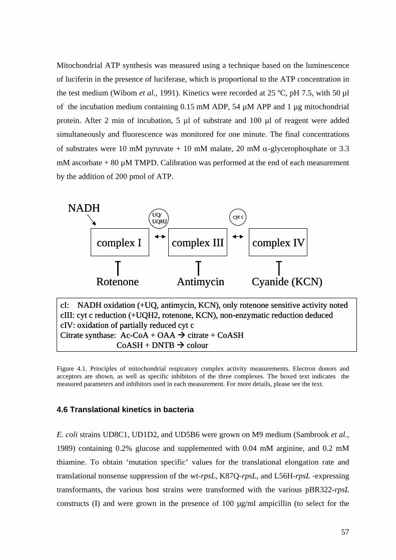
Mitochondrial ATP synthesis was measured using a technique based on the luminescence
of luciferin in the presence of luciferase, which is proportional to the ATP concentration in
the test medium (Wibom et al., 1991). Kinetics were recorded at 25 ºC, pH 7.5, with 50 µl
of the incubation medium containing 0.15 mM ADP, 54 µM APP and 1 µg mitochondrial
protein. After 2 min of incubation, 5 µl of substrate and 100 µl of reagent were added
simultaneously and fluorescence was monitored for one minute. The final concentrations
of substrates were 10 mM pyruvate + 10 mM malate, 20 mM α-glycerophosphate or 3.3
mM ascorbate + 80 µM TMPD. Calibration was performed at the end of each measurement
by the addition of 200 pmol of ATP.
cyt c
complex I complex III complex IV
Rotenone Antimycin Cyanide (KCN)
UQ/UQH2
NADH
cI: NADH oxidation (+UQ, antimycin, KCN), only rotenone sensitive activity notedcIII: cyt c reduction (+UQH2, rotenone, KCN), non-enzymatic reduction deducedcIV: oxidation of partially reduced cyt cCitrate synthase: Ac-CoA + OAA citrate + CoASH
CoASH + DNTB colour
cyt c
complex I complex III complex IV
Rotenone Antimycin Cyanide (KCN)
UQ/UQH2
NADH
cI: NADH oxidation (+UQ, antimycin, KCN), only rotenone sensitive activity notedcIII: cyt c reduction (+UQH2, rotenone, KCN), non-enzymatic reduction deducedcIV: oxidation of partially reduced cyt cCitrate synthase: Ac-CoA + OAA citrate + CoASH
CoASH + DNTB colour Figure 4.1. Principles of mitochondrial respiratory complex activity measurements. Electron donors and acceptors are shown, as well as specific inhibitors of the three complexes. The boxed text indicates the measured parameters and inhibitors used in each measurement. For more details, please see the text.
4.6 Translational kinetics in bacteria
E. coli strains UD8C1, UD1D2, and UD5B6 were grown on M9 medium (Sambrook et al.,
1989) containing 0.2% glucose and supplemented with 0.04 mM arginine, and 0.2 mM
thiamine. To obtain ‘mutation specific’ values for the translational elongation rate and
translational nonsense suppression of the wt-rpsL, K87Q-rpsL, and L56H-rpsL -expressing
transformants, the various host strains were transformed with the various pBR322-rpsL
constructs (I) and were grown in the presence of 100 µg/ml ampicillin (to select for the
57
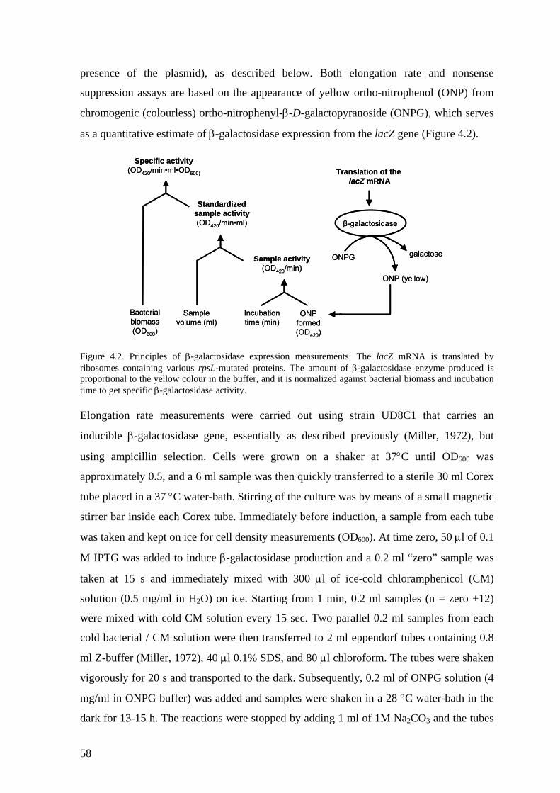
presence of the plasmid), as described below. Both elongation rate and nonsense
suppression assays are based on the appearance of yellow ortho-nitrophenol (ONP) from
chromogenic (colourless) ortho-nitrophenyl-β-D-galactopyranoside (ONPG), which serves
as a quantitative estimate of β-galactosidase expression from the lacZ gene (Figure 4.2).
Specific activity(OD420/min•ml•OD600)
Standardized sample activity(OD420/min•ml)
Sample activity(OD420/min)
Incubationtime (min)
ONPformed(OD420)
Samplevolume (ml)
Bacterial biomass(OD600)
ONPG
β-galactosidase
ONP (yellow)
galactose
Translation of the lacZ mRNA
Specific activity(OD420/min•ml•OD600)
Standardized sample activity(OD420/min•ml)
Sample activity(OD420/min)
Incubationtime (min)
ONPformed(OD420)
Samplevolume (ml)
Bacterial biomass(OD600)
ONPG
β-galactosidase
ONP (yellow)
galactose
Translation of the lacZ mRNA
Figure 4.2. Principles of β-galactosidase expression measurements. The lacZ mRNA is translated by ribosomes containing various rpsL-mutated proteins. The amount of β-galactosidase enzyme produced is proportional to the yellow colour in the buffer, and it is normalized against bacterial biomass and incubation time to get specific β-galactosidase activity. Elongation rate measurements were carried out using strain UD8C1 that carries an
inducible β-galactosidase gene, essentially as described previously (Miller, 1972), but
using ampicillin selection. Cells were grown on a shaker at 37°C until OD600 was
approximately 0.5, and a 6 ml sample was then quickly transferred to a sterile 30 ml Corex
tube placed in a 37 °C water-bath. Stirring of the culture was by means of a small magnetic
stirrer bar inside each Corex tube. Immediately before induction, a sample from each tube
was taken and kept on ice for cell density measurements (OD600). At time zero, 50 µl of 0.1
M IPTG was added to induce β-galactosidase production and a 0.2 ml “zero” sample was
taken at 15 s and immediately mixed with 300 µl of ice-cold chloramphenicol (CM)
solution (0.5 mg/ml in H2O) on ice. Starting from 1 min, 0.2 ml samples (n = zero +12)
were mixed with cold CM solution every 15 sec. Two parallel 0.2 ml samples from each
cold bacterial / CM solution were then transferred to 2 ml eppendorf tubes containing 0.8
ml Z-buffer (Miller, 1972), 40 µl 0.1% SDS, and 80 µl chloroform. The tubes were shaken
vigorously for 20 s and transported to the dark. Subsequently, 0.2 ml of ONPG solution (4
mg/ml in ONPG buffer) was added and samples were shaken in a 28 °C water-bath in the
dark for 13-15 h. The reactions were stopped by adding 1 ml of 1M Na2CO3 and the tubes
58
were centrifuged at 16 000 gmax in a microcentrifuge at 4°C for 15 min. The average rate of
nitrophenol formation (OD420) of these was measured. The data was transformed by
subtracting the average value of parallel zero-time samples, and by normalizing cell
densities at the start of the assay to OD600 = 1.0 and ONPG incubation times to 1 hour, and
values were plotted as square roots of corrected OD420 values. The time required to
produce the first active β-galactosidase monomer was estimated from the point of
intersection of the trend-line and the x-axis (time). Eighteen different clones expressing wt
protein, 12 clones expressing the K87Q mutated protein, and 5 clones expressing the L56H
mutated protein were analysed. Freshly transformed clones were used in order to avoid
additional mutations in plasmid constructs, and each strain used was verified by
sequencing the single colony PCR product of the cell pellet from the sample taken at the
actual time of the β-galactosidase measurements. All results obtained from clones
containing additional mutations in the reading frame or in the upstream sequence, were
discarded.
E. coli strains UD1D2 and UD5B6 were used to measure nonsense suppression in vivo.
UD1D2 carries an F´ plasmid that includes a constitutively expressed, wild-type β-
galactosidase fusion protein (lacIZ∆14), and UD5B6 carries a nonsense mutation in this
same gene (lacIZ∆14, UGA at 189) in an otherwise isogenic background. Because the
chromosomal copy of the lacZ gene has been deleted in these strains, all β-galactosidase
message will be transcribed from the F´ fusion gene, and functional β-galactosidase
produced from strain UD5B6 can be explained only by read-through of a stop codon by
ribosomes. Freshly transformed clones were grown to mid-log phase, growth was stopped
on ice and parallel samples (0.1 ml from UD1D2 clones and 1.0 ml from UD5B6 clones)
were pelleted, and resuspended in 1 ml of ice cold Z buffer (Miller, 1972). From this point
on, β-galactosidase activity was measured as in the case of elongation rate measurements,
but with typical incubation times of 2 h and 15-16 h for UD1D2 and UD5B6 clones,
respectively. Average OD420 values of parallel samples were normalized to the amount of
cells and time of incubation. Values for each UD5B6 clone expressing a given rpsL
mutation were then divided by the values of corresponding UD1D2 clones to get a
suppression value specific for the given mutation in the rpsL-gene. The results were
calculated using the following formulae:
59
STEP 1: OD420 – (OD550 x 1.75) x 1000 Miller unit ( β-gal x 103) values Vol (ml) x OD600 x time (min) STEP 2: Value of ∆ 14 UGA at 189 (UD5 B6) x 10,000 suppression by 104
Value of ∆ 14 (UD1 D2)
Standard deviations for the suppression values of the clones carrying different rpsL mutant
plasmids were obtained using following equation
Var(x) + Var(y) x ρ2
mean(y)2 SD =
where Var(x) = variance of UD5B6 suppression values, Var(y) = variance of UD1D2
suppression values, and ρ = mean(x) / mean(y).
4.7 Construction of uORF -mutagenised MRPS12 expression plasmids
For the 5´-UTR characterization (unpublished), uORF constructs were made using the
same Invitrogen expression vector as in the other constructs (pcDNA3.1(-)/Myc-His B)
described in article IV. Splice variant b was created using combinations of primers rps-d1
(GGTTAGACCTATAGAGGGACGGCCCAGGTG), the two bases surrounding the splice
site underlined) and rps-d2 (CACCTGGGCCGTCCCTCTATAGGTCTAACC) with
commercial primers BGH and T7, respectively, using MRPS12-Myc/B construct as a
template. The two first round PCR products were used as a template for second round PCR
with primers BGH and T7, and the product was then cut with BamHI and HindIII and
ligated to the vector. To mutate the uORF, the following primers were designed: rps-
uORF1 (TCTGTTAGCAGCCTGAGGGCCTGTGG, mutated base underlined) and its
reverse complement rps-uORF2 (CCACAGGCCCTCAGGCTGCTAACAGA) to create
construct b2, rps-uORF3 (CAGCATGGGGCCTGTGGTTTAGACCTA, bases
surrounding the deleted A-residue underlined) and its reverse complement rps-uORF4
(TAGGTCTAAACCACAGGCCCCATGCTG) to create construct b3, and rps-uORF5
(GCAGCATGCGTGCATGCGGGTGAACCT, altered bases underlined) and its reverse
complement rps-uORF6 (TAGGTTCACCCGCATGCACGCATGCTGC) to create
60

construct b4. Primers rps-uORF1, -3 and -5 were used in combination with primer BGH,
and primers rps-uORF2, -4 and -6 were used with primer T7, using cloned construct b as a
template. Second round PCR, digestion, and cloning was performed as above.
61
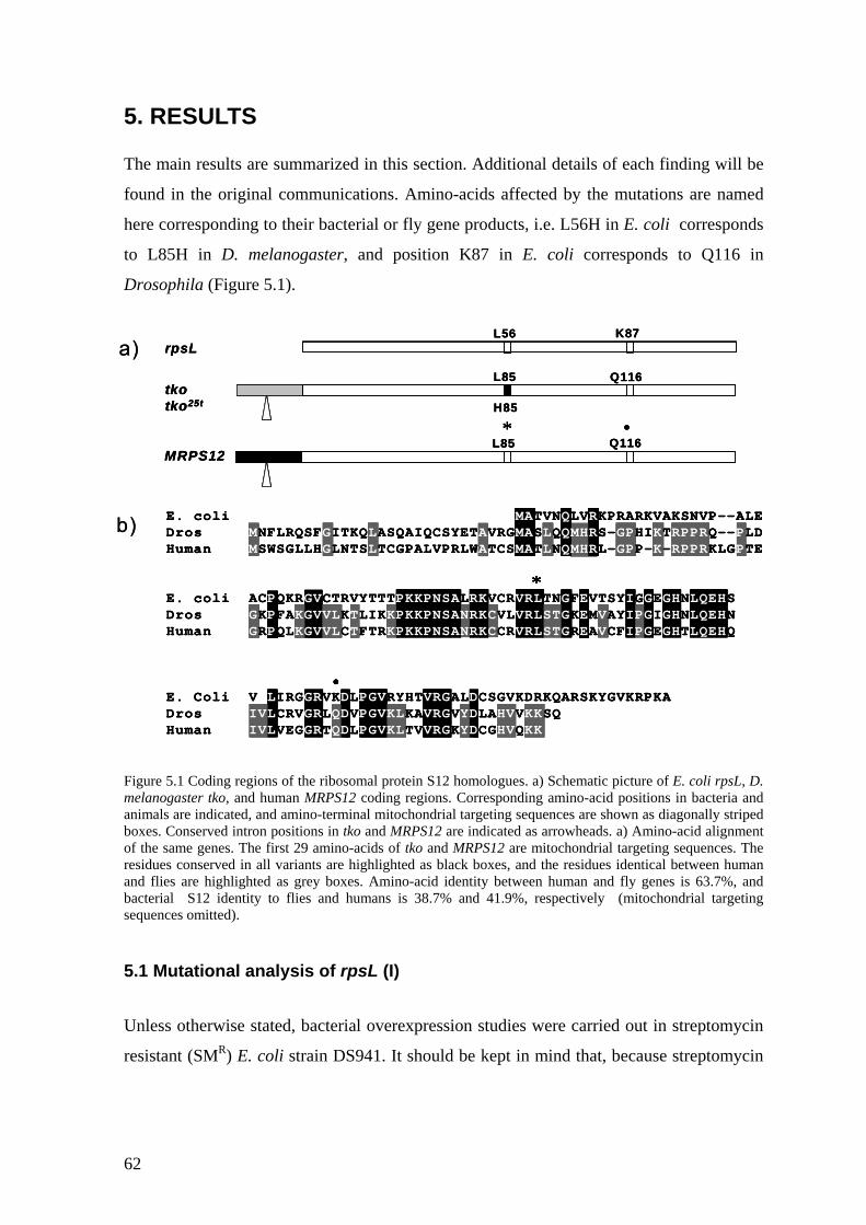
5. RESULTS The main results are summarized in this section. Additional details of each finding will be
found in the original communications. Amino-acids affected by the mutations are named
here corresponding to their bacterial or fly gene products, i.e. L56H in E. coli corresponds
to L85H in D. melanogaster, and position K87 in E. coli corresponds to Q116 in
Drosophila (Figure 5.1).
rpsL
tkotko25t
K87
MRPS12L85 Q116
L56
L85 Q116
H85
* •
•
*
E. coli MATVNQLVRKPRARKVAKSNVP--ALEDros MNFLRQSFGITKQLASQAIQCSYETAVRGMASLQQMHRS-GPHIKTRPPRQ--PLDHuman MSWSGLLHGLNTSLTCGPALVPRLWATCSMATLNQMHRL-GPP-K-RPPRKLGPTE
E. coli ACPQKRGVCTRVYTTTPKKPNSALRKVCRVRLTNGFEVTSYIGGEGHNLQEHSDros GKPFAKGVVLKTLIKKPKKPNSANRKCVLVRLSTGKEMVAYIPGIGHNLQEHNHuman GRPQLKGVVLCTFTRKPKKPNSANRKCCRVRLSTGREAVCFIPGEGHTLQEHQ
E. Coli VILIRGGRVKDLPGVRYHTVRGALDCSGVKDRKQARSKYGVKRPKADros IVLCRVGRLQDVPGVKLKAVRGVYDLAHVVKKSQHuman IVLVEGGRTQDLPGVKLTVVRGKYDCGHVQKK
a)
b)
rpsL
tkotko25t
K87
MRPS12L85 Q116
L56
L85 Q116
H85
* •
rpsL
tkotko25t
K87
MRPS12L85 Q116
L56
L85 Q116
H85
* •
•
*
E. coli MATVNQLVRKPRARKVAKSNVP--ALEDros MNFLRQSFGITKQLASQAIQCSYETAVRGMASLQQMHRS-GPHIKTRPPRQ--PLDHuman MSWSGLLHGLNTSLTCGPALVPRLWATCSMATLNQMHRL-GPP-K-RPPRKLGPTE
E. coli ACPQKRGVCTRVYTTTPKKPNSALRKVCRVRLTNGFEVTSYIGGEGHNLQEHSDros GKPFAKGVVLKTLIKKPKKPNSANRKCVLVRLSTGKEMVAYIPGIGHNLQEHNHuman GRPQLKGVVLCTFTRKPKKPNSANRKCCRVRLSTGREAVCFIPGEGHTLQEHQ
E. Coli VILIRGGRVKDLPGVRYHTVRGALDCSGVKDRKQARSKYGVKRPKADros IVLCRVGRLQDVPGVKLKAVRGVYDLAHVVKKSQHuman IVLVEGGRTQDLPGVKLTVVRGKYDCGHVQKK
•
*
••
**
E. coli MATVNQLVRKPRARKVAKSNVP--ALEDros MNFLRQSFGITKQLASQAIQCSYETAVRGMASLQQMHRS-GPHIKTRPPRQ--PLDHuman MSWSGLLHGLNTSLTCGPALVPRLWATCSMATLNQMHRL-GPP-K-RPPRKLGPTE
E. coli ACPQKRGVCTRVYTTTPKKPNSALRKVCRVRLTNGFEVTSYIGGEGHNLQEHSDros GKPFAKGVVLKTLIKKPKKPNSANRKCVLVRLSTGKEMVAYIPGIGHNLQEHNHuman GRPQLKGVVLCTFTRKPKKPNSANRKCCRVRLSTGREAVCFIPGEGHTLQEHQ
E. Coli VILIRGGRVKDLPGVRYHTVRGALDCSGVKDRKQARSKYGVKRPKADros IVLCRVGRLQDVPGVKLKAVRGVYDLAHVVKKSQHuman IVLVEGGRTQDLPGVKLTVVRGKYDCGHVQKK
a)
b)
Figure 5.1 Coding regions of the ribosomal protein S12 homologues. a) Schematic picture of E. coli rpsL, D. melanogaster tko, and human MRPS12 coding regions. Corresponding amino-acid positions in bacteria and animals are indicated, and amino-terminal mitochondrial targeting sequences are shown as diagonally striped boxes. Conserved intron positions in tko and MRPS12 are indicated as arrowheads. a) Amino-acid alignment of the same genes. The first 29 amino-acids of tko and MRPS12 are mitochondrial targeting sequences. The residues conserved in all variants are highlighted as black boxes, and the residues identical between human and flies are highlighted as grey boxes. Amino-acid identity between human and fly genes is 63.7%, and bacterial S12 identity to flies and humans is 38.7% and 41.9%, respectively (mitochondrial targeting sequences omitted).
5.1 Mutational analysis of rpsL (I)
Unless otherwise stated, bacterial overexpression studies were carried out in streptomycin
resistant (SMR) E. coli strain DS941. It should be kept in mind that, because streptomycin
62
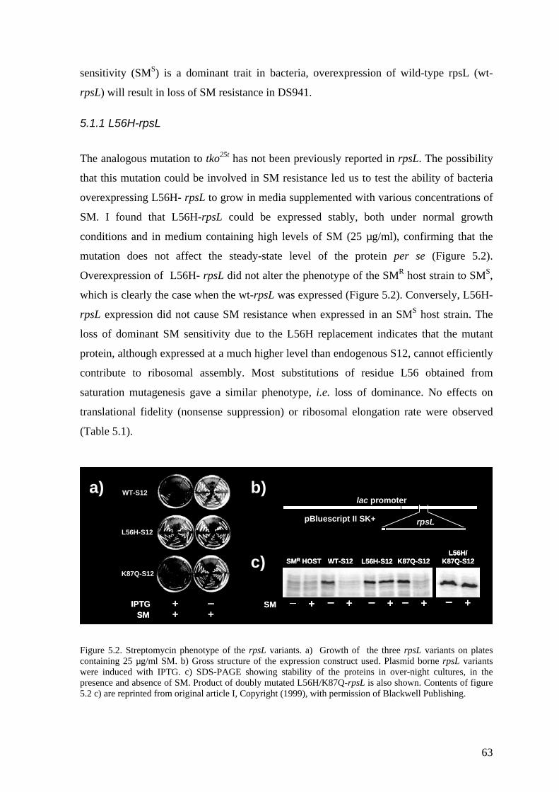
sensitivity (SMS) is a dominant trait in bacteria, overexpression of wild-type rpsL (wt-
rpsL) will result in loss of SM resistance in DS941.
5.1.1 L56H-rpsL
The analogous mutation to tko25t has not been previously reported in rpsL. The possibility
that this mutation could be involved in SM resistance led us to test the ability of bacteria
overexpressing L56H- rpsL to grow in media supplemented with various concentrations of
SM. I found that L56H-rpsL could be expressed stably, both under normal growth
conditions and in medium containing high levels of SM (25 µg/ml), confirming that the
mutation does not affect the steady-state level of the protein per se (Figure 5.2).
Overexpression of L56H- rpsL did not alter the phenotype of the SMR host strain to SMS,
which is clearly the case when the wt-rpsL was expressed (Figure 5.2). Conversely, L56H-
rpsL expression did not cause SM resistance when expressed in an SMS host strain. The
loss of dominant SM sensitivity due to the L56H replacement indicates that the mutant
protein, although expressed at a much higher level than endogenous S12, cannot efficiently
contribute to ribosomal assembly. Most substitutions of residue L56 obtained from
saturation mutagenesis gave a similar phenotype, i.e. loss of dominance. No effects on
translational fidelity (nonsense suppression) or ribosomal elongation rate were observed
(Table 5.1).
K87Q-S12
WT-S12
L56H-S12
IPTGSM + +
+ –
L56H/K87Q-S12K87Q-S12WT-S12 L56H-S12SMR HOST
+– +– +– +–
SM
+–
rpsLpBluescript II SK+
lac promotera) b)
c)K87Q-S12
WT-S12
L56H-S12
IPTGSM
IPTGSM + +
+ –+ ++ –
L56H/K87Q-S12K87Q-S12WT-S12 L56H-S12SMR HOST
+– +– +– +–
SM
+–
L56H/K87Q-S12K87Q-S12WT-S12 L56H-S12SMR HOST
+– +– +– +– +– +– +– +–
SM
+– +–
rpsLpBluescript II SK+
lac promotera) b)
c)
Figure 5.2. Streptomycin phenotype of the rpsL variants. a) Growth of the three rpsL variants on plates containing 25 µg/ml SM. b) Gross structure of the expression construct used. Plasmid borne rpsL variants were induced with IPTG. c) SDS-PAGE showing stability of the proteins in over-night cultures, in the presence and absence of SM. Product of doubly mutated L56H/K87Q-rpsL is also shown. Contents of figure 5.2 c) are reprinted from original article I, Copyright (1999), with permission of Blackwell Publishing.
63
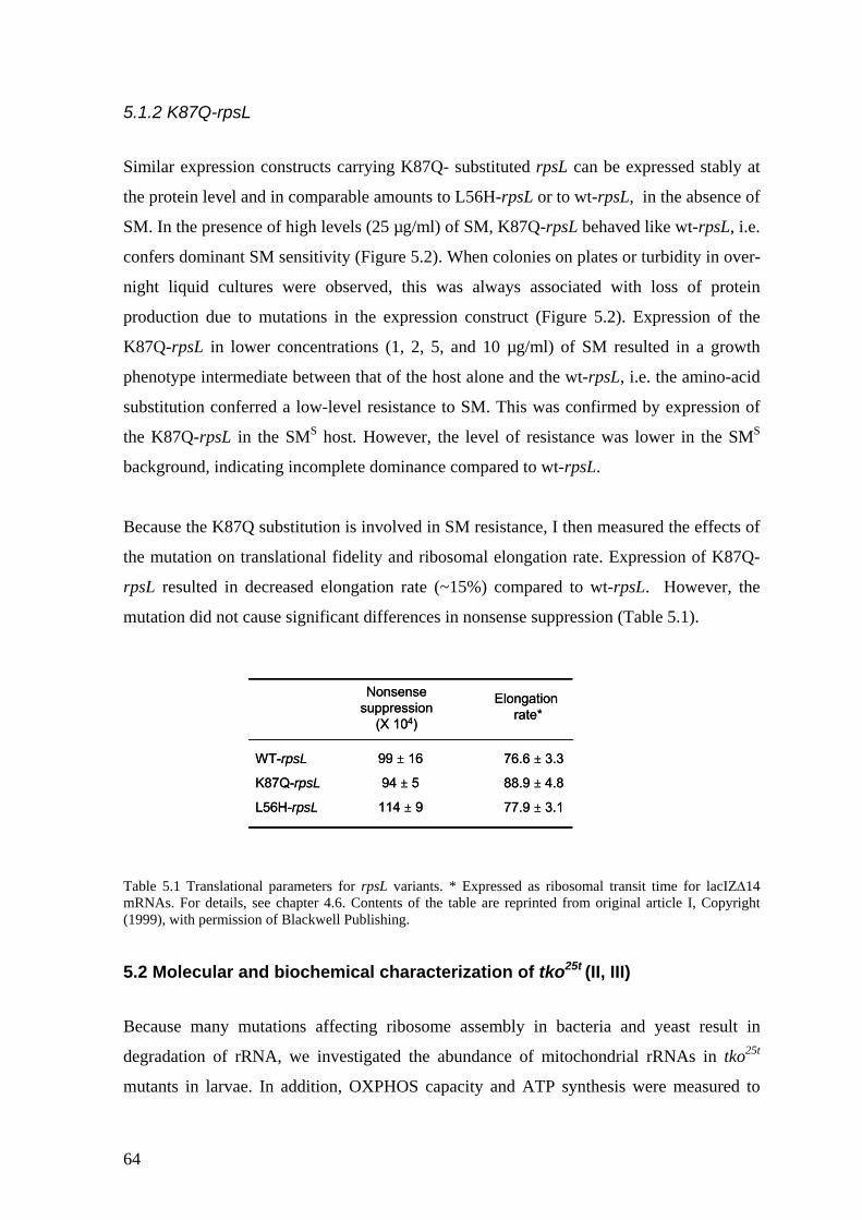
5.1.2 K87Q-rpsL Similar expression constructs carrying K87Q- substituted rpsL can be expressed stably at
the protein level and in comparable amounts to L56H-rpsL or to wt-rpsL, in the absence of
SM. In the presence of high levels (25 µg/ml) of SM, K87Q-rpsL behaved like wt-rpsL, i.e.
confers dominant SM sensitivity (Figure 5.2). When colonies on plates or turbidity in over-
night liquid cultures were observed, this was always associated with loss of protein
production due to mutations in the expression construct (Figure 5.2). Expression of the
K87Q-rpsL in lower concentrations (1, 2, 5, and 10 µg/ml) of SM resulted in a growth
phenotype intermediate between that of the host alone and the wt-rpsL, i.e. the amino-acid
substitution conferred a low-level resistance to SM. This was confirmed by expression of
the K87Q-rpsL in the SMS host. However, the level of resistance was lower in the SMS
background, indicating incomplete dominance compared to wt-rpsL.
Because the K87Q substitution is involved in SM resistance, I then measured the effects of
the mutation on translational fidelity and ribosomal elongation rate. Expression of K87Q-
rpsL resulted in decreased elongation rate (~15%) compared to wt-rpsL. However, the
mutation did not cause significant differences in nonsense suppression (Table 5.1).
WT-rpsL
K87Q-rpsL
L56H-rpsL
99 ± 16
94 ± 5
114 ± 9
76.6 ± 3.3
88.9 ± 4.8
77.9 ± 3.1
Nonsensesuppression
(X 104)
Elongation rate*
WT-rpsL
K87Q-rpsL
L56H-rpsL
99 ± 16
94 ± 5
114 ± 9
76.6 ± 3.3
88.9 ± 4.8
77.9 ± 3.1
Nonsensesuppression
(X 104)
Elongation rate*
Table 5.1 Translational parameters for rpsL variants. * Expressed as ribosomal transit time for lacIZ∆14 mRNAs. For details, see chapter 4.6. Contents of the table are reprinted from original article I, Copyright (1999), with permission of Blackwell Publishing.
5.2 Molecular and biochemical characterization of tko25t (II, III)
Because many mutations affecting ribosome assembly in bacteria and yeast result in
degradation of rRNA, we investigated the abundance of mitochondrial rRNAs in tko25t
mutants in larvae. In addition, OXPHOS capacity and ATP synthesis were measured to
64
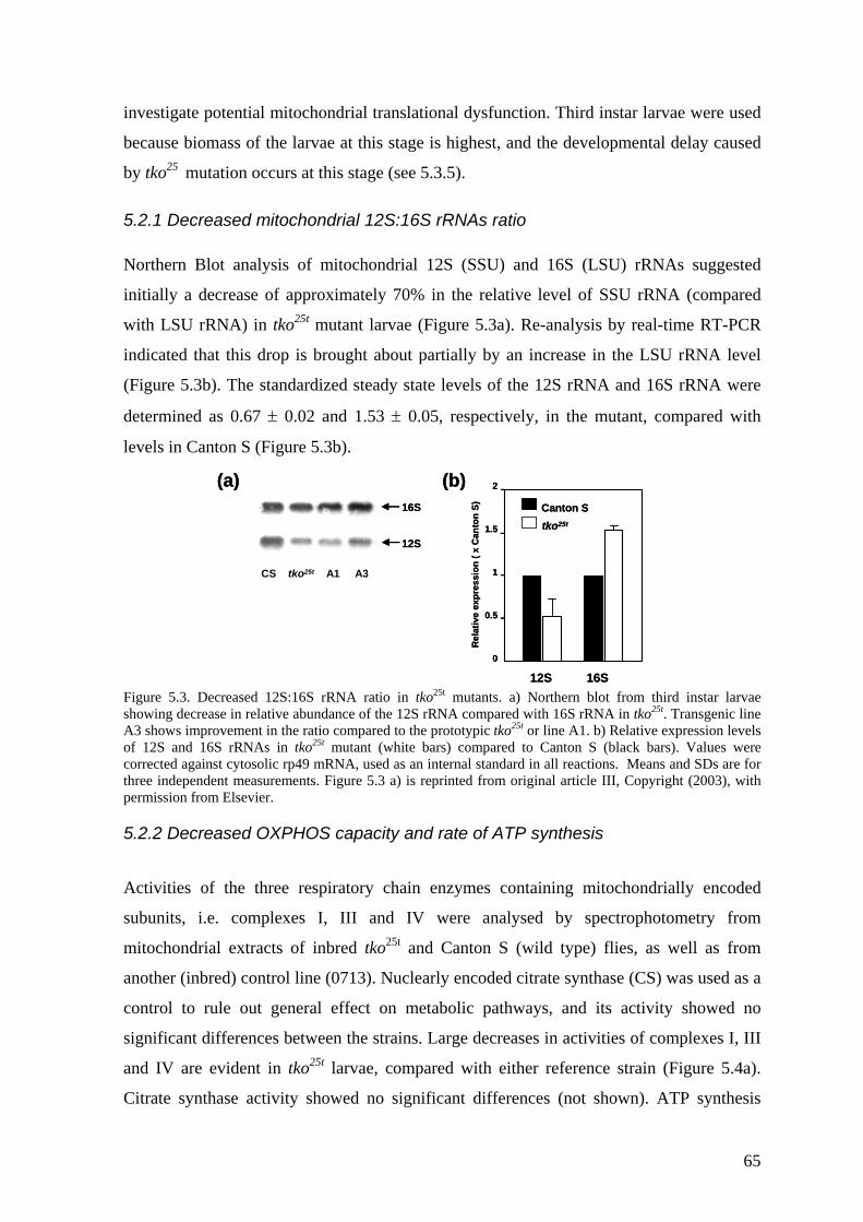
investigate potential mitochondrial translational dysfunction. Third instar larvae were used
because biomass of the larvae at this stage is highest, and the developmental delay caused
by tko25 mutation occurs at this stage (see 5.3.5).
5.2.1 Decreased mitochondrial 12S:16S rRNAs ratio Northern Blot analysis of mitochondrial 12S (SSU) and 16S (LSU) rRNAs suggested
initially a decrease of approximately 70% in the relative level of SSU rRNA (compared
with LSU rRNA) in tko25t mutant larvae (Figure 5.3a). Re-analysis by real-time RT-PCR
indicated that this drop is brought about partially by an increase in the LSU rRNA level
(Figure 5.3b). The standardized steady state levels of the 12S rRNA and 16S rRNA were
determined as 0.67 ± 0.02 and 1.53 ± 0.05, respectively, in the mutant, compared with
levels in Canton S (Figure 5.3b). R
elat
ive
expr
essi
on (
xC
anto
n S)
2
1.5
1
0.5
0
12S 16S
Canton Stko25t
16S
12S
CS tko25t A1 A3
(a) (b)R
elat
ive
expr
essi
on (
xC
anto
n S)
2
1.5
1
0.5
0
12S 16S
Canton Stko25t
16S
12S
CS tko25t A1 A3
(a) (b)
Figure 5.3. Decreased 12S:16S rRNA ratio in tko25t mutants. a) Northern blot from third instar larvae showing decrease in relative abundance of the 12S rRNA compared with 16S rRNA in tko25t. Transgenic line A3 shows improvement in the ratio compared to the prototypic tko25t or line A1. b) Relative expression levels of 12S and 16S rRNAs in tko25t mutant (white bars) compared to Canton S (black bars). Values were corrected against cytosolic rp49 mRNA, used as an internal standard in all reactions. Means and SDs are for three independent measurements. Figure 5.3 a) is reprinted from original article III, Copyright (2003), with permission from Elsevier.
5.2.2 Decreased OXPHOS capacity and rate of ATP synthesis
Activities of the three respiratory chain enzymes containing mitochondrially encoded
subunits, i.e. complexes I, III and IV were analysed by spectrophotometry from
mitochondrial extracts of inbred tko25t and Canton S (wild type) flies, as well as from
another (inbred) control line (0713). Nuclearly encoded citrate synthase (CS) was used as a
control to rule out general effect on metabolic pathways, and its activity showed no
significant differences between the strains. Large decreases in activities of complexes I, III
and IV are evident in tko25t larvae, compared with either reference strain (Figure 5.4a).
Citrate synthase activity showed no significant differences (not shown). ATP synthesis
65
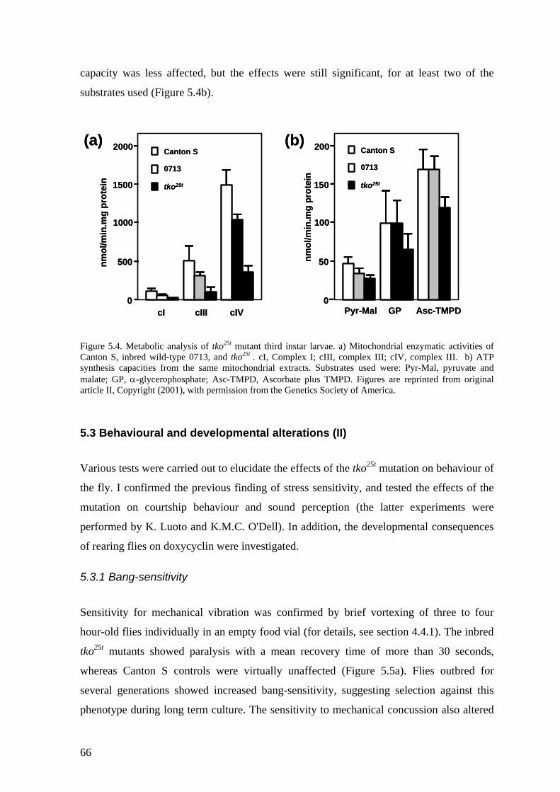
capacity was less affected, but the effects were still significant, for at least two of the
substrates used (Figure 5.4b).
nm
ol/m
in.m
g pr
otei
n
0
500
1000
1500
2000
tko25t
0713
Canton S
cI cIII cIV 0
50
100
150
200
tko25t
0713
Canton S
nmol
/min
.mg
prot
ein
Pyr-Mal GP Asc-TMPD
(a) (b)
nmol
/min
.mg
prot
ein
0
500
1000
1500
2000
tko25t
0713
Canton S
cI cIII cIV 0
50
100
150
200
tko25t
0713
Canton S
nmol
/min
.mg
prot
ein
Pyr-Mal GP Asc-TMPD
(a) (b)
Figure 5.4. Metabolic analysis of tko25t mutant third instar larvae. a) Mitochondrial enzymatic activities of Canton S, inbred wild-type 0713, and tko25t . cI, Complex I; cIII, complex III; cIV, complex III. b) ATP synthesis capacities from the same mitochondrial extracts. Substrates used were: Pyr-Mal, pyruvate and malate; GP, α-glycerophosphate; Asc-TMPD, Ascorbate plus TMPD. Figures are reprinted from original article II, Copyright (2001), with permission from the Genetics Society of America.
5.3 Behavioural and developmental alterations (II) Various tests were carried out to elucidate the effects of the tko25t mutation on behaviour of
the fly. I confirmed the previous finding of stress sensitivity, and tested the effects of the
mutation on courtship behaviour and sound perception (the latter experiments were
performed by K. Luoto and K.M.C. O'Dell). In addition, the developmental consequences
of rearing flies on doxycyclin were investigated.
5.3.1 Bang-sensitivity
Sensitivity for mechanical vibration was confirmed by brief vortexing of three to four
hour-old flies individually in an empty food vial (for details, see section 4.4.1). The inbred
tko25t mutants showed paralysis with a mean recovery time of more than 30 seconds,
whereas Canton S controls were virtually unaffected (Figure 5.5a). Flies outbred for
several generations showed increased bang-sensitivity, suggesting selection against this
phenotype during long term culture. The sensitivity to mechanical concussion also altered
66
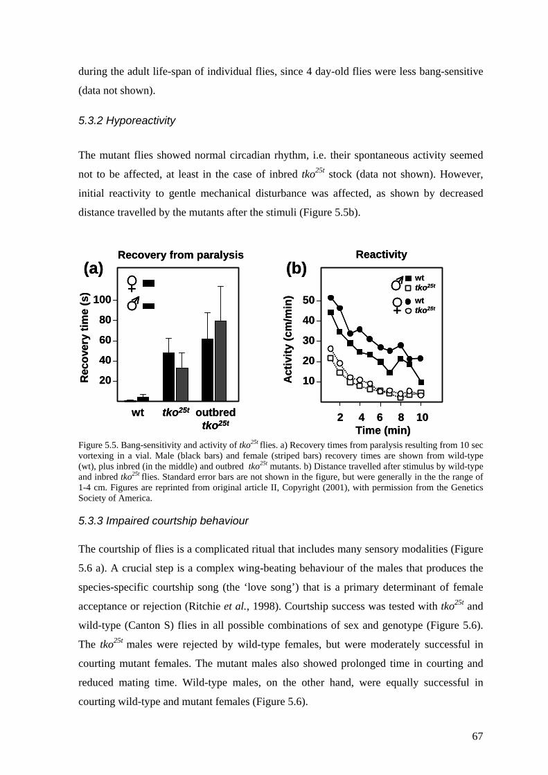
during the adult life-span of individual flies, since 4 day-old flies were less bang-sensitive
(data not shown).
5.3.2 Hyporeactivity
The mutant flies showed normal circadian rhythm, i.e. their spontaneous activity seemed
not to be affected, at least in the case of inbred tko25t stock (data not shown). However,
initial reactivity to gentle mechanical disturbance was affected, as shown by decreased
distance travelled by the mutants after the stimuli (Figure 5.5b).
100
80
60
40
20Rec
over
y tim
e (s
)
wt tko25t outbredtko25t
Recovery from paralysis
50
40
30
20
10Activ
ity (c
m/m
in)
2 4 6 8 10Time (min)
wttko25t
wttko25t
(a) (b)Reactivity
100
80
60
40
20Rec
over
y tim
e (s
)
wt tko25t outbredtko25t
Recovery from paralysis
50
40
30
20
10Activ
ity (c
m/m
in)
2 4 6 8 10Time (min)
wttko25t
wttko25t
(a) (b)Reactivity
Figure 5.5. Bang-sensitivity and activity of tko25t flies. a) Recovery times from paralysis resulting from 10 sec vortexing in a vial. Male (black bars) and female (striped bars) recovery times are shown from wild-type (wt), plus inbred (in the middle) and outbred tko25t mutants. b) Distance travelled after stimulus by wild-type and inbred tko25t flies. Standard error bars are not shown in the figure, but were generally in the the range of 1-4 cm. Figures are reprinted from original article II, Copyright (2001), with permission from the Genetics Society of America.
5.3.3 Impaired courtship behaviour The courtship of flies is a complicated ritual that includes many sensory modalities (Figure
5.6 a). A crucial step is a complex wing-beating behaviour of the males that produces the
species-specific courtship song (the ‘love song’) that is a primary determinant of female
acceptance or rejection (Ritchie et al., 1998). Courtship success was tested with tko25t and
wild-type (Canton S) flies in all possible combinations of sex and genotype (Figure 5.6).
The tko25t males were rejected by wild-type females, but were moderately successful in
courting mutant females. The mutant males also showed prolonged time in courting and
reduced mating time. Wild-type males, on the other hand, were equally successful in
courting wild-type and mutant females (Figure 5.6).
67
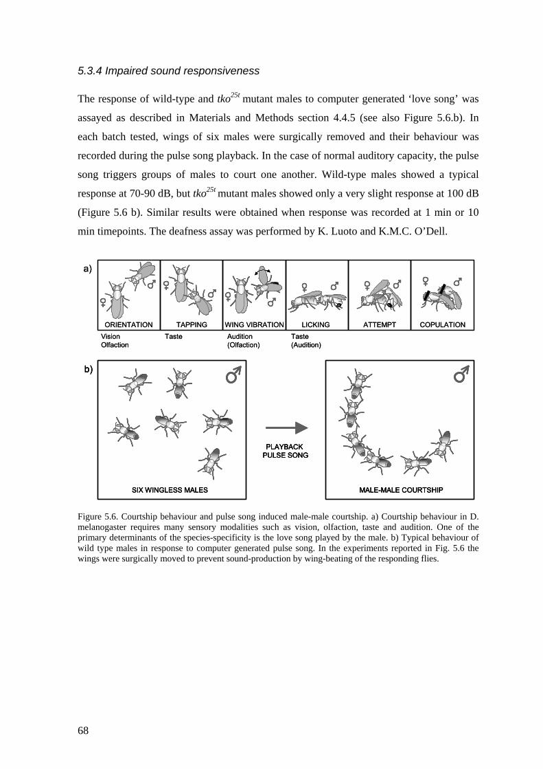
5.3.4 Impaired sound responsiveness The response of wild-type and tko25t mutant males to computer generated ‘love song’ was
assayed as described in Materials and Methods section 4.4.5 (see also Figure 5.6.b). In
each batch tested, wings of six males were surgically removed and their behaviour was
recorded during the pulse song playback. In the case of normal auditory capacity, the pulse
song triggers groups of males to court one another. Wild-type males showed a typical
response at 70-90 dB, but tko25t mutant males showed only a very slight response at 100 dB
(Figure 5.6 b). Similar results were obtained when response was recorded at 1 min or 10
min timepoints. The deafness assay was performed by K. Luoto and K.M.C. O’Dell.
igure 5.6. Courtship behaviour and pulse song induced male-male courtship. a) Courtship behaviour in D.
ORIENTATION TAPPING WING VIBRATION LICKING ATTEMPT COPULATION
a)
MALE-MALE COURTSHIP
PLAYBACKPULSE SONG
SIX WINGLESS MALES
b)
Vision Taste Audition TasteOlfaction (Olfaction) (Audition)
ORIENTATION TAPPING WING VIBRATION LICKING ATTEMPT COPULATION
a)
MALE-MALE COURTSHIP
PLAYBACKPULSE SONG
SIX WINGLESS MALES
b)
MALE-MALE COURTSHIP
PLAYBACKPULSE SONG
SIX WINGLESS MALES
b)
Vision Taste Audition TasteOlfaction (Olfaction) (Audition)
Fmelanogaster requires many sensory modalities such as vision, olfaction, taste and audition. One of the primary determinants of the species-specificity is the love song played by the male. b) Typical behaviour of wild type males in response to computer generated pulse song. In the experiments reported in Fig. 5.6 the wings were surgically moved to prevent sound-production by wing-beating of the responding flies.
68
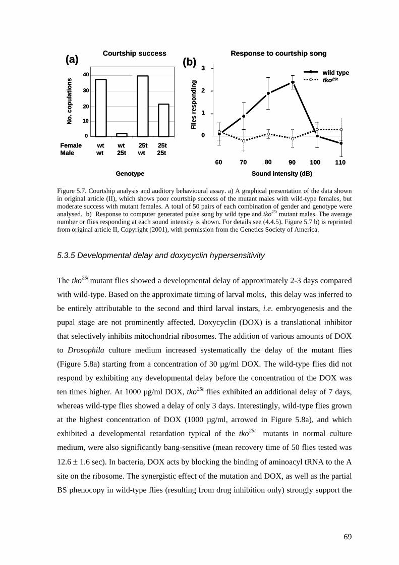
0
10
20
30
40
Female wt wt 25t 25tMale wt 25t wt 25t
No.
cop
ulat
ions
(a)3
2
1
0
Flie
s re
spon
ding
60 70 80 90 100 110
wild typetko25t
Sound intensity (dB)
(b)Courtship success Response to courtship song
Genotype
0
10
20
30
40
Female wt wt 25t 25tMale wt 25t wt 25t
No.
cop
ulat
ions
(a)3
2
1
0
Flie
s re
spon
ding
60 70 80 90 100 110
wild typetko25t
Sound intensity (dB)
(b)Courtship success Response to courtship song
Genotype Figure 5.7. Courtship analysis and auditory behavioural assay. a) A graphical presentation of the data shown in original article (II), which shows poor courtship success of the mutant males with wild-type females, but moderate success with mutant females. A total of 50 pairs of each combination of gender and genotype were analysed. b) Response to computer generated pulse song by wild type and tko25t mutant males. The average number or flies responding at each sound intensity is shown. For details see (4.4.5). Figure 5.7 b) is reprinted from original article II, Copyright (2001), with permission from the Genetics Society of America.
5.3.5 Developmental delay and doxycyclin hypersensitivity
The tko25t mutant flies showed a developmental delay of approximately 2-3 days compared
with wild-type. Based on the approximate timing of larval molts, this delay was inferred to
be entirely attributable to the second and third larval instars, i.e. embryogenesis and the
pupal stage are not prominently affected. Doxycyclin (DOX) is a translational inhibitor
that selectively inhibits mitochondrial ribosomes. The addition of various amounts of DOX
to Drosophila culture medium increased systematically the delay of the mutant flies
(Figure 5.8a) starting from a concentration of 30 µg/ml DOX. The wild-type flies did not
respond by exhibiting any developmental delay before the concentration of the DOX was
ten times higher. At 1000 µg/ml DOX, tko25t flies exhibited an additional delay of 7 days,
whereas wild-type flies showed a delay of only 3 days. Interestingly, wild-type flies grown
at the highest concentration of DOX (1000 µg/ml, arrowed in Figure 5.8a), and which
exhibited a developmental retardation typical of the tko25t mutants in normal culture
medium, were also significantly bang-sensitive (mean recovery time of 50 flies tested was
12.6 ± 1.6 sec). In bacteria, DOX acts by blocking the binding of aminoacyl tRNA to the A
site on the ribosome. The synergistic effect of the mutation and DOX, as well as the partial
BS phenocopy in wild-type flies (resulting from drug inhibition only) strongly support the
69
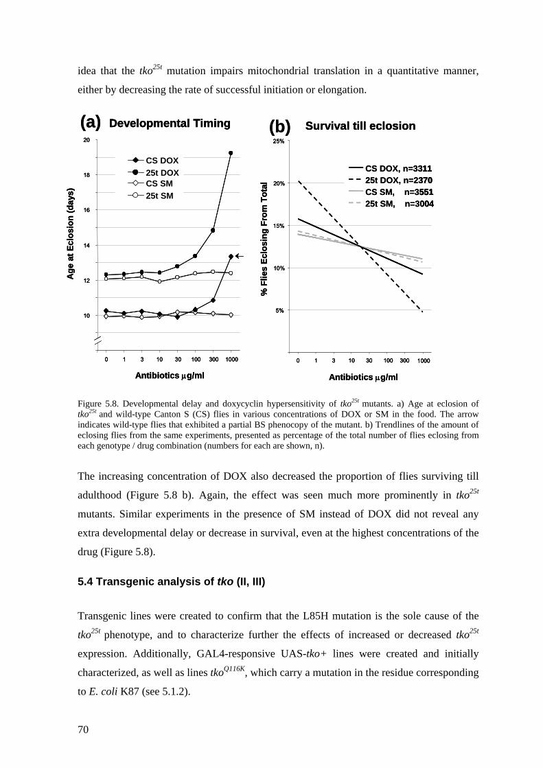
idea that the tko25t mutation impairs mitochondrial translation in a quantitative manner,
either by decreasing the rate of successful initiation or elongation.
Developmental Timing
10
12
14
16
18
20
0 1 3 10 30 100 300 1000
Age
at E
clos
ion
(day
s)
CS SM25t SM
CS DOX25t DOX
5%
10%
15%
20%
25%
0 1 3 10 30 100 300 1000
Survival till eclosion
% F
lies
Eclo
sing
Fro
m T
otal CS SM, n=3551
25t SM, n=3004
CS DOX, n=331125t DOX, n=2370
Antibiotics µg/ml Antibiotics µg/ml
(a) (b)Developmental Timing
10
12
14
16
18
20
0 1 3 10 30 100 300 1000
Age
at E
clos
ion
(day
s)
CS SM25t SM
CS DOX25t DOX
5%
10%
15%
20%
25%
0 1 3 10 30 100 300 1000
Survival till eclosion
% F
lies
Eclo
sing
Fro
m T
otal CS SM, n=3551
25t SM, n=3004
CS DOX, n=331125t DOX, n=2370
Antibiotics µg/ml Antibiotics µg/ml
Developmental Timing
10
12
14
16
18
20
0 1 3 10 30 100 300 1000
Age
at E
clos
ion
(day
s)
CS SM25t SM
CS DOX25t DOX
5%
10%
15%
20%
25%
0 1 3 10 30 100 300 1000
Survival till eclosion
% F
lies
Eclo
sing
Fro
m T
otal CS SM, n=3551
25t SM, n=3004
CS DOX, n=331125t DOX, n=2370CS SM, n=355125t SM, n=3004
CS DOX, n=331125t DOX, n=2370
Antibiotics µg/ml Antibiotics µg/ml
(a) (b)
Figure 5.8. Developmental delay and doxycyclin hypersensitivity of tko25t mutants. a) Age at eclosion of tko25t and wild-type Canton S (CS) flies in various concentrations of DOX or SM in the food. The arrow indicates wild-type flies that exhibited a partial BS phenocopy of the mutant. b) Trendlines of the amount of eclosing flies from the same experiments, presented as percentage of the total number of flies eclosing from each genotype / drug combination (numbers for each are shown, n).
The increasing concentration of DOX also decreased the proportion of flies surviving till
adulthood (Figure 5.8 b). Again, the effect was seen much more prominently in tko25t
mutants. Similar experiments in the presence of SM instead of DOX did not reveal any
extra developmental delay or decrease in survival, even at the highest concentrations of the
drug (Figure 5.8).
5.4 Transgenic analysis of tko (II, III)
Transgenic lines were created to confirm that the L85H mutation is the sole cause of the
tko25t phenotype, and to characterize further the effects of increased or decreased tko25t
expression. Additionally, GAL4-responsive UAS-tko+ lines were created and initially
characterized, as well as lines tkoQ116K, which carry a mutation in the residue corresponding
to E. coli K87 (see 5.1.2).
70
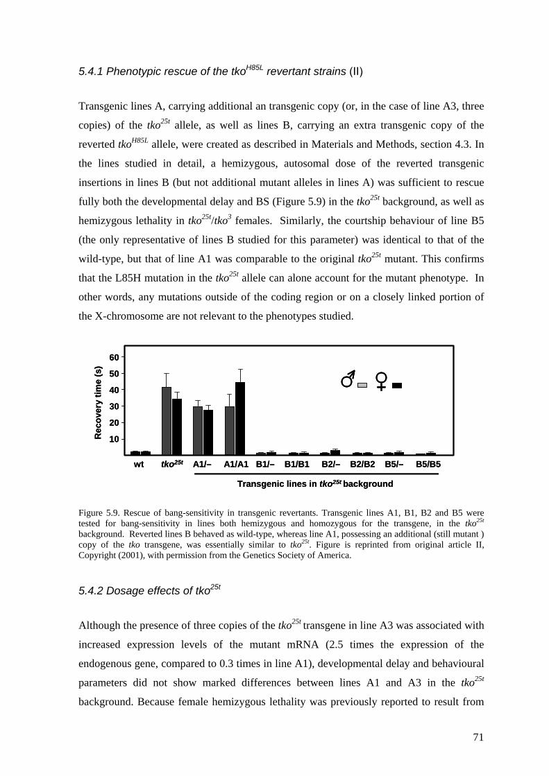
5.4.1 Phenotypic rescue of the tkoH85L revertant strains (II)
Transgenic lines A, carrying additional an transgenic copy (or, in the case of line A3, three
copies) of the tko25t allele, as well as lines B, carrying an extra transgenic copy of the
reverted tkoH85L allele, were created as described in Materials and Methods, section 4.3. In
the lines studied in detail, a hemizygous, autosomal dose of the reverted transgenic
insertions in lines B (but not additional mutant alleles in lines A) was sufficient to rescue
fully both the developmental delay and BS (Figure 5.9) in the tko25t background, as well as
hemizygous lethality in tko25t/tko3 females. Similarly, the courtship behaviour of line B5
(the only representative of lines B studied for this parameter) was identical to that of the
wild-type, but that of line A1 was comparable to the original tko25t mutant. This confirms
that the L85H mutation in the tko25t allele can alone account for the mutant phenotype. In
other words, any mutations outside of the coding region or on a closely linked portion of
the X-chromosome are not relevant to the phenotypes studied.
igure 5.9. Rescue of bang-sensitivity in transgenic revertants. Transgenic lines A1, B1, B2 and B5 were 25t
.4.2 Dosage effects of tko25t
lthough the presence of three copies of the tko25t transgene in line A3 was associated with
60
50
40
30
20
10Rec
over
y tim
e (s
)
wt tko25t A1/– A1/A1 B1/– B1/B1 B2/– B2/B2 B5/– B5/B5
Transgenic lines in tko25t background
60
50
40
30
20
10Rec
over
y tim
e (s
)
wt tko25t A1/– A1/A1 B1/– B1/B1 B2/– B2/B2 B5/– B5/B5
Transgenic lines in tko25t background
Ftested for bang-sensitivity in lines both hemizygous and homozygous for the transgene, in the tko background. Reverted lines B behaved as wild-type, whereas line A1, possessing an additional (still mutant ) copy of the tko transgene, was essentially similar to tko25t. Figure is reprinted from original article II, Copyright (2001), with permission from the Genetics Society of America.
5
A
increased expression levels of the mutant mRNA (2.5 times the expression of the
endogenous gene, compared to 0.3 times in line A1), developmental delay and behavioural
parameters did not show marked differences between lines A1 and A3 in the tko25t
background. Because female hemizygous lethality was previously reported to result from
71
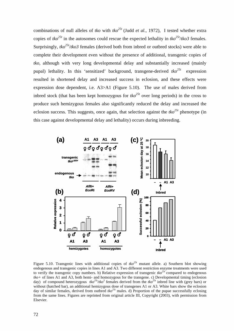
combinations of null alleles of tko with tko25t (Judd et al., 1972), I tested whether extra
copies of tko25t in the autosomes could rescue the expected lethality in tko25t/tko3 females.
Surprisingly, tko25t/tko3 females (derived both from inbred or outbred stocks) were able to
complete their development even without the presence of additional, transgenic copies of
tko, although with very long developmental delay and substantially increased (mainly
pupal) lethality. In this ‘sensitized’ background, transgene-derived tko25t expression
resulted in shortened delay and increased success in eclosion, and these effects were
expression dose dependent, i.e. A3>A1 (Figure 5.10). The use of males derived from
inbred stock (that has been kept homozygous for tko25t over long periods) in the cross to
produce such hemizygous females also significantly reduced the delay and increased the
eclosion success. This suggests, once again, that selection against the tko25t phenotype (in
this case against developmental delay and lethality) occurs during inbreeding.
gure 5.10. Transgenic lines with additional copies of tko mutant allele. a) Southern blot showing
ogenous and transgenic copies in lines A1 and A3. Two diffe nt restriction enzyme treatments were used
100
80
60
40
20
0
Inbred
– – A1 A3
Succ
essf
ul e
clos
ion
(%)
20
18
16
14
12
10
(b) (d)
Mea
n ec
losi
on d
ay a
t 25
ºC
Inbred
– – A1 A3
(c)
Rel
ativ
e ex
pres
sion 4
3
2
1
0
A1 A3 A1 A3
hemizygotes homozygotes
AflII+EcoRV
AflII+EcoRI
A1 A3 A1 A3
endogenoustko
(a)
transgenictkoL85H
100
80
60
40
20
0
Inbred
– – A1 A3
100
80
60
40
20
0
Inbred
– – A1 A3
Succ
essf
ul e
clos
ion
(%)
20
18
16
14
12
10
(b) (d)
Mea
n ec
losi
on d
ay a
t 25
ºC
Inbred
– – A1 A3
(c)
Rel
ativ
e ex
pres
sion 4
3
2
1
0
A1 A3 A1 A3
Rel
ativ
e ex
pres
sion 4
3
2
1
0
A1 A3 A1 A3
hemizygotes homozygotes
AflII+EcoRV
AflII+EcoRI
A1 A3 A1 A3
endogenoustko
(a)
transgenictkoL85H
Fiend
25t
reto verify the transgenic copy numbers. b) Relative expression of transgenic tko25t compared to endogenous tko+ of lines A1 and A3, both hemi- and homozygous for the transgene. c) Developmental timing (eclosion day) of compound heterozygous tko25t/tko3 females derived from the tko25t inbred line with (grey bars) or without (hatched bar), an additional hemizygous dose of transgenes A1 or A3. White bars show the eclosion day of similar females, derived from outbred tko25t males. d) Proportion of the pupae successfully eclosing from the same lines. Figures are reprinted from original article III, Copyright (2003), with permission from Elsevier.
72
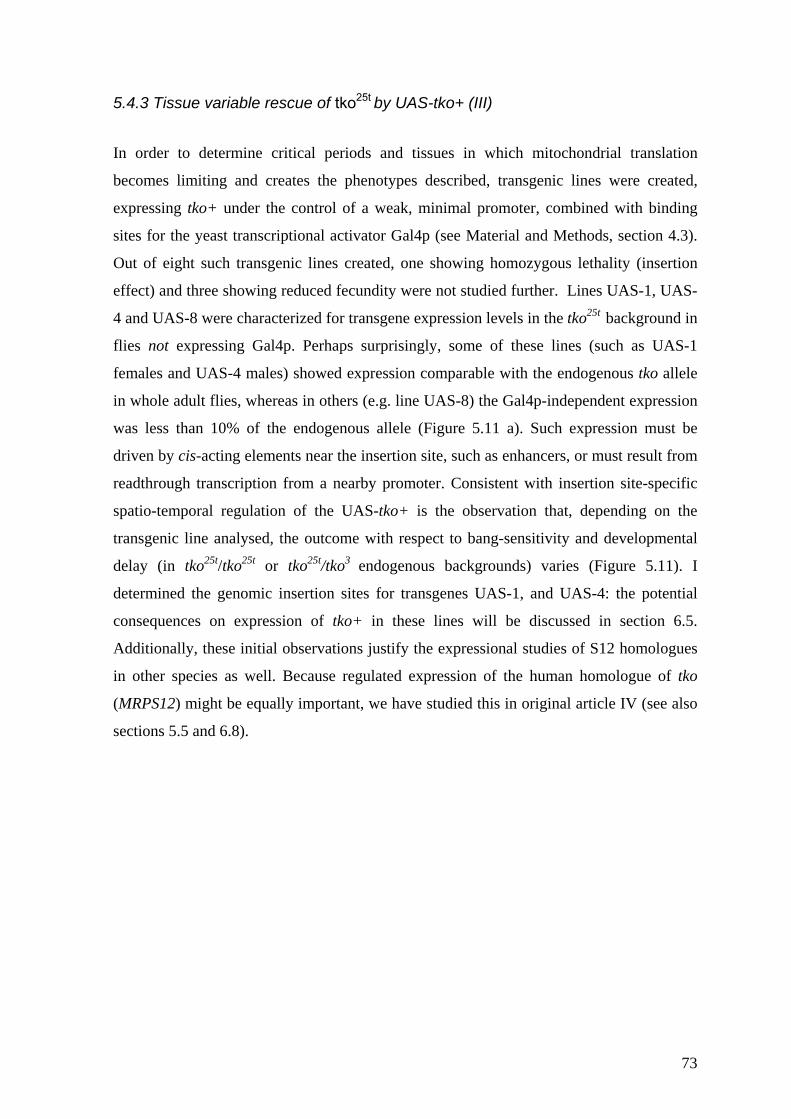
5.4.3 Tissue variable rescue of tko25t by UAS-tko+ (III)
In order to determine critical periods and tissues in which mitochondrial translation
ecomes limiting and creates the phenotypes described, transgenic lines were created,
+
b
expressing tko+ under the control of a weak, minimal promoter, combined with binding
sites for the yeast transcriptional activator Gal4p (see Material and Methods, section 4.3).
Out of eight such transgenic lines created, one showing homozygous lethality (insertion
effect) and three showing reduced fecundity were not studied further. Lines UAS-1, UAS-
4 and UAS-8 were characterized for transgene expression levels in the tko25t background in
flies not expressing Gal4p. Perhaps surprisingly, some of these lines (such as UAS-1
females and UAS-4 males) showed expression comparable with the endogenous tko allele
in whole adult flies, whereas in others (e.g. line UAS-8) the Gal4p-independent expression
was less than 10% of the endogenous allele (Figure 5.11 a). Such expression must be
driven by cis-acting elements near the insertion site, such as enhancers, or must result from
readthrough transcription from a nearby promoter. Consistent with insertion site-specific
spatio-temporal regulation of the UAS-tko+ is the observation that, depending on the
transgenic line analysed, the outcome with respect to bang-sensitivity and developmental
delay (in tko25t/tko25t or tko25t/tko3 endogenous backgrounds) varies (Figure 5.11). I
determined the genomic insertion sites for transgenes UAS-1, and UAS-4: the potential
consequences on expression of tko in these lines will be discussed in section 6.5.
Additionally, these initial observations justify the expressional studies of S12 homologues
in other species as well. Because regulated expression of the human homologue of tko
(MRPS12) might be equally important, we have studied this in original article IV (see also
sections 5.5 and 6.8).
73
2019181716151413121110
(c)
√R
ecov
ery
time
(s)
876543210
CS tko25t UAS-1 UAS-4 UAS-8
Mea
n ec
losi
on d
ay a
t 25
ºC 15
14
13
12
11
10
9
(b)
Rel
ativ
e ex
pres
sion
1.2
1
0.8
0.6
0.4
0.2
0
UAS-1 UAS-4 UAS-8
(a)
Mea
n ec
losi
on d
ay a
t 25
ºC
– UAS-1 UAS-4 wt
(d) tko25t / tko3 bkd
CS tko25t UAS-1 UAS-4 UAS-8
tko25t bkd
tko25t bkd tko25t bkd
2019181716151413121110
(c)
√R
ecov
ery
time
(s)
876543210
CS tko25t UAS-1 UAS-4 UAS-8CS tko25t UAS-1 UAS-4 UAS-8
Mea
n ec
losi
on d
ay a
t 25
ºC 15
14
13
12
11
10
9
(b)
Rel
ativ
e ex
pres
sion
1.2
1
0.8
0.6
0.4
0.2
0
UAS-1 UAS-4 UAS-8
(a)
Mea
n ec
losi
on d
ay a
t 25
ºC
– UAS-1 UAS-4 wt
(d) tko25t / tko3 bkd
CS tko25t UAS-1 UAS-4 UAS-8CS tko25t UAS-1 UAS-4 UAS-8
tko25t bkd
tko25t bkd tko25t bkd
Figure 5.11. Expression and phenotypic analysis of UAS-tko+ lines. a) Relative expression of tko+ compared to endogenous tko25t allele in whole adult flies of lines UAS-1, UAS-4, and UAS-8. b) Bang-sensitivity of the same lines (grey bars) and of Canton S (wild-type, black bars) and prototypic tko25t mutants (white bars). c) Mean eclosion day at 25°C in tko25t background. d) Mean eclosion day of UAS-1 and UAS-4 females in compound heterozygous tko25t/tko3 background (grey bars). Wild-type (black bar) and tko25t/tko3 (white bar) females shown as a reference. Figures are reprinted from original article III, Copyright (2003), with permission from Elsevier.
5.4.4 Phenotypic effects of the tkoQ116K substitution (II and unpublished data) In order to create mitoribosomes with putatively altered translational kinetics, transgenic
lines carrying the novel tkoQ116K allele (changing glutamine at position 116 to lysine, see
also Figure 5.1) were created. Five independent lines were obtained, with two X-linked
and three autosomal insertions. When three autosomal insertions of tkoQ116K were crossed
to the tko3 background they showed sterility in females homozygous for tko3 (i.e.
endogenous tko nulls), but not in their FM7-balanced sisters that contained one copy of
tko+ in the balancer chromosome (for explanation of balancers, see section 4.3). This
recessive female sterility was of great interest, since line B females containing the tkoH85L-
reverted (wild type) transgene in the same tko3 background were unaffected. Furthermore,
when the same tkoQ116K transgenes were transferred to the tko25t mutant background, the
females were fertile and all tko25t-related defects (such as developmental delay) were
rescued. This suggested that mutations L85H and Q116K in tko have completely different
74
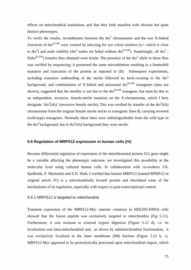
effects on mitochondrial translation, and that they both manifest with obvious but quite
distinct phenotypes.
To verify the results, recombinants between the tko3 chromosome and the two X-linked
insertions of tkoQ116K were created by selecting for eye colour markers (w+ which is close
to tko3) and male viability (tko3 males are lethal without tkoQ116K). Surprisingly, all tko3 ;
P[tkoQ116K] females thus obtained were fertile. The presence of the tko3 allele in these flies
was verified by sequencing: it possessed the same microdeletion resulting in a frameshift
mutation and truncation of the protein as reported in (II). Subsequent experiments,
including extensive outbreeding of the stocks followed by back-crossing to the tko3
background, and combinations of X-linked and autosomal tkoQ116K transgenes (data not
shown), suggested that the sterility is not due to the tkoQ116K transgene, but must be due to
an independent, recessive, female-sterile mutation on the X-chromosome, which I here
designate ‘tko3(rfs)’ (recessive female sterile). This was verified by transfer of the tko3(rfs)
chromosome from the original female sterile stocks to transgenic lines B, carrying reverted
(wild-type) transgenes. Normally these lines were indistinguishable from the wild type in
the tko3 background, but in tko3(rfs) background they were sterile.
5.5 Regulation of MRPS12 expression in human cells (IV)
Because differential regulation of expression of the mitoribosomal protein S12 gene might
be a variable affecting the phenotypic outcome, we investigated this possibility at the
molecular level using cultured human cells. In collaboration with co-workers J.N.
Spelbrink, P. Mariottini and Z.H. Shah, I verified that human MRPS12 (named RPMS12 in
original article IV) is a mitochondrially located protein and elucidated some of the
mechanisms of its regulation, especially with respect to post-transcriptional control.
5.5.1 MRPS12 is targeted to mitochondria Transient expression of the MRPS12-Myc reporter construct in HEK293-EBNA cells
showed that the fusion peptide was exclusively targeted to mitochondria (Fig 5.11).
Furthermore, it was resistant to external trypsin digestion (Figure 5.12 d), i.e. its
localization was intra-mitochondrial and, as shown by submitochondrial fractionation, it
was exclusively localized in the inner membrane (IM) fraction (Figure 5.12 b, c).
MRPS12-Myc appeared to be proteolytically processed upon mitochondrial import, which
75
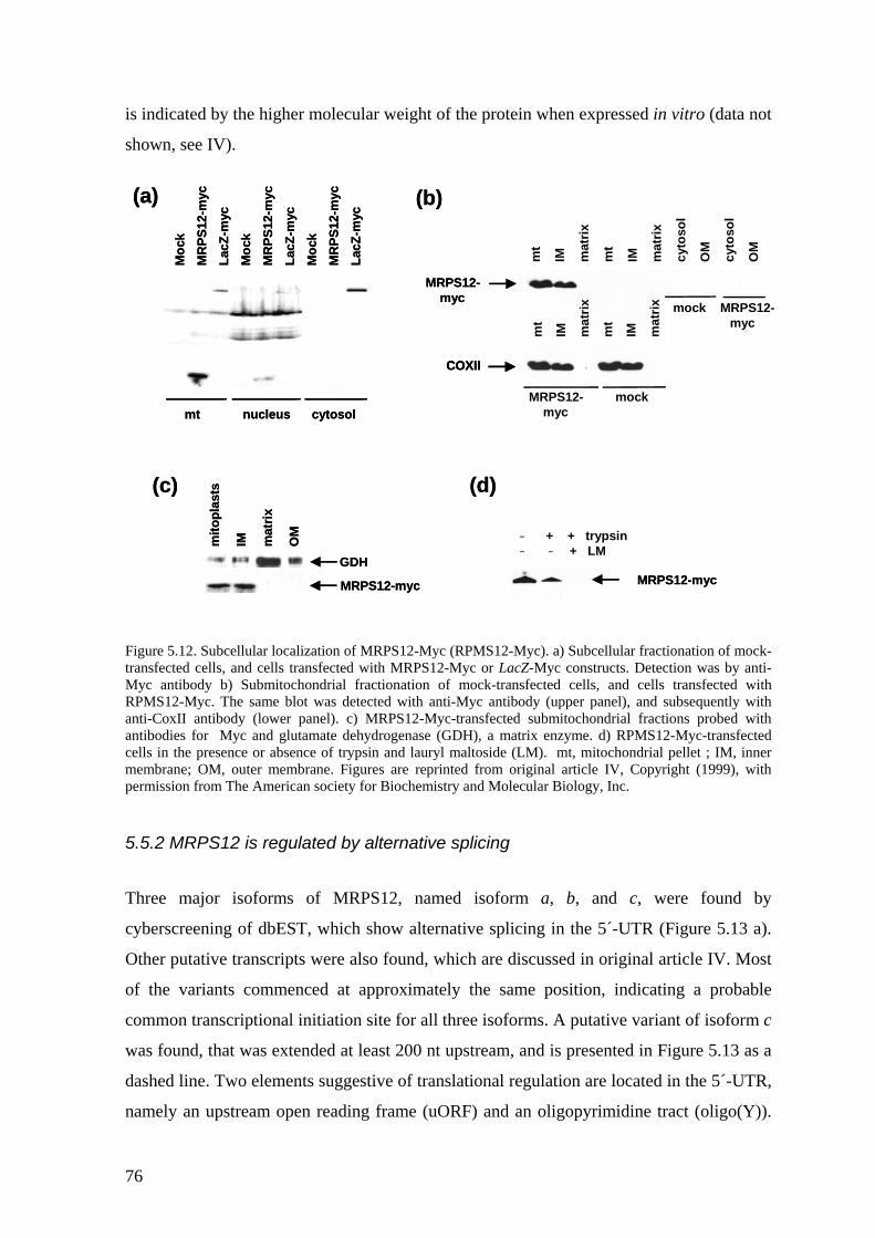
is indicated by the higher molecular weight of the protein when expressed in vitro (data not
shown, see IV).
COXII
MRPS12- mock myc
Moc
kM
RPS
12-m
ycLa
cZ-m
ycM
ock
MR
PS12
-myc
LacZ
-myc
Moc
kM
RPS
12-m
ycLa
cZ-m
yc
mt nucleus cytosol
(a) (b)
MRPS12-myc
mito
plas
ts
IM mat
rix
OM
GDH
(c)
mt
IM mat
rix
mt
IM mat
rix
– + + trypsin– – + LM
MRPS12-myc
MRPS12-myc
(d)
mock MRPS12-myc
mt
IM mat
rix
mt
IM mat
rix
cyto
sol
OM
cyto
sol
OM
COXIICOXII
MRPS12- mock myc
Moc
kM
RPS
12-m
ycLa
cZ-m
ycM
ock
MR
PS12
-myc
LacZ
-myc
Moc
kM
RPS
12-m
ycLa
cZ-m
yc
mt nucleus cytosol
(a) (b)
MRPS12-myc
mito
plas
ts
IM mat
rix
OM
GDH
(c)
mt
IM mat
rix
mt
IM mat
rix
– + + trypsin– – + LM
MRPS12-myc
MRPS12-myc
(d)
mock MRPS12-myc
mt
IM mat
rix
mt
IM mat
rix
cyto
sol
OM
cyto
sol
OM
Figure 5.12. Subcellular localization of MRPS12-Myc (RPMS12-Myc). a) Subcellular fractionation of mock-transfected cells, and cells transfected with MRPS12-Myc or LacZ-Myc constructs. Detection was by anti-Myc antibody b) Submitochondrial fractionation of mock-transfected cells, and cells transfected with RPMS12-Myc. The same blot was detected with anti-Myc antibody (upper panel), and subsequently with anti-CoxII antibody (lower panel). c) MRPS12-Myc-transfected submitochondrial fractions probed with antibodies for Myc and glutamate dehydrogenase (GDH), a matrix enzyme. d) RPMS12-Myc-transfected cells in the presence or absence of trypsin and lauryl maltoside (LM). mt, mitochondrial pellet ; IM, inner membrane; OM, outer membrane. Figures are reprinted from original article IV, Copyright (1999), with permission from The American society for Biochemistry and Molecular Biology, Inc.
5.5.2 MRPS12 is regulated by alternative splicing Three major isoforms of MRPS12, named isoform a, b, and c, were found by
cyberscreening of dbEST, which show alternative splicing in the 5´-UTR (Figure 5.13 a).
Other putative transcripts were also found, which are discussed in original article IV. Most
of the variants commenced at approximately the same position, indicating a probable
common transcriptional initiation site for all three isoforms. A putative variant of isoform c
was found, that was extended at least 200 nt upstream, and is presented in Figure 5.13 as a
dashed line. Two elements suggestive of translational regulation are located in the 5´-UTR,
namely an upstream open reading frame (uORF) and an oligopyrimidine tract (oligo(Y)).
76
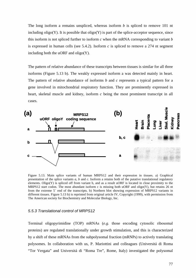
The long isoform a remains unspliced, whereas isoform b is spliced to remove 101 nt
including oligo(Y). It is possible that oligo(Y) is part of the splice-acceptor sequence, since
this isoform is not spliced further to isoform c when the mRNA corresponding to variant b
is expressed in human cells (see 5.4.2). Isoform c is spliced to remove a 274 nt segment
including both the uORF and oligo(Y).
The pattern of relative abundance of these transcripts between tissues is similar for all three
isoforms (Figure 5.13 b). The weakly expressed isoform a was detected mainly in heart.
The pattern of relative abundance of isoforms b and c represents a typical pattern for a
gene involved in mitochondrial respiratory function. They are prominently expressed in
heart, skeletal muscle and kidney, isoform c being the most prominent transcript in all
cases.
igure 5.13. Main splice variants of human MRPS12 and their expression in tissues. a) Graphical resentation of the splice variants a, b and c. Isoform a retains both of the putative translational regulatory
.5.3 Translational control of MRPS12
erminal oligopyrimidine (TOP) mRNAs (e.g. those encoding cytosolic ribosomal
MRPS12uORF oligoY coding sequence
a
b
c
Hea
rtB
rain
Plac
enta
Lung
Live
rSk
el. M
uscl
eK
idne
yPa
ncre
as
b, c
a
(a) (b)MRPS12uORF oligoY coding sequence
a
b
c
Hea
rtB
rain
Plac
enta
Lung
Live
rSk
el. M
uscl
eK
idne
yPa
ncre
as
b, c
a
Hea
rtB
rain
Plac
enta
Lung
Live
rSk
el. M
uscl
eK
idne
yPa
ncre
as
Hea
rtB
rain
Plac
enta
Lung
Live
rSk
el. M
uscl
eK
idne
yPa
ncre
as
b, c
a
(a) (b)
Fpelements. Oligo(Y) is spliced off from variant b, and as a result uORF is located in close proximity to the MRPS12 start codon. The most abundant isoform c is missing both uORF and oligo(Y), but retains 26 nt from the extreme 5´ end of the transcripts. b) Northern blot showing expression of MRPS12 variants in different tissues. Figure 5.13 b) is reprinted from original article IV, Copyright (1999), with permission from The American society for Biochemistry and Molecular Biology, Inc.
5
T
proteins) are regulated translationally under growth stimulation, and this is characterized
by a shift of these mRNAs from the subpolysomal fraction (mRNPs) to actively translating
polysomes. In collaboration with us, P. Mariottini and colleagues (Universitá di Roma
“Tor Vergata” and Universitá di “Roma Tre”, Rome, Italy) investigated the polysomal
77
distribution of the three MRPS12 mRNA isoforms in response to serum starvation by
sucrose density gradient centrifugation of post-mitochondrial fractions. The results of this
experiment are shown in original article IV (Figure 5 therein). In short, Northern blots
probed with the full-length probe showed that MRPS12 is indeed translationally controlled,
and exhibits a prominent shift from the mRNP fraction in serum-starved cells to the
polysomal fraction in growing cells. This was essentially similar to the shift of TOP
mRNA rpL4, although MRPS12 seemed to be translated in slightly smaller polysomes,
even in growing cells. RT-PCR analysis from the same fractions indicated, however, that
only the main isoform c, lacking both of uORF and oligo(Y), was growth controlled,
whereas isoforms a and b showed a more or less uniform distribution under both
conditions.
Because it was somewhat surprising that the putative elements involved in translational
he effect of the uORF on the translational competence of mRNA isoform b was
control were missing from the only variant that seemed to be translationally regulated, I
investigated this more in detail by expression of RPMS12 in cell culture. In this case, two
expression constructs differing in their 5´-UTR were used (Figure 5.14 a). The mRNAs
derived from MRPS12-Myc/S included a 155 nt stretch of vector-derived sequence, fused
directly to the MRPS12 coding sequence, preceded by just 24 nt of immediate upstream
UTR. The second construct, MRPS12-Myc/B included the same vector sequence plus
essentially the entire 5´-UTR of MRPS12, commencing in the region of the major 5´ end of
the fully spliced isoform c. By RT-PCR I verified that, when expressed, MRPS12-Myc/B
derived mRNA was spliced and gave rise to mRNA isoform c, with the exception of the 5´
vector-derived sequence (Figure 5.14 a). RPMS12-Myc was expressed efficiently from
both constructs in the presence of serum in the cell-culture medium, but a much lower
amount of protein product was produced under serum starvation (Figure 5.14 b). Under the
same conditions, control construct LacZ-Myc was only slightly affected. This finding
further supports the previous result that isoform c is translationally controlled.
Furthermore, the 26 nt sequence at the immediate 5´ end of isoform c seems to mediate
this control, since this is the only difference in the two constructs used (Figure 5.14 c).
T
investigated by similar reporter-construct expression (J.M.T., unpublished data). Isoform b
was hardly expressed at all at the protein level unless the start codon of the uORF was
mutated to destroy the translation initiation site (Figure 5.15 a, b). Maximal synonymous
78
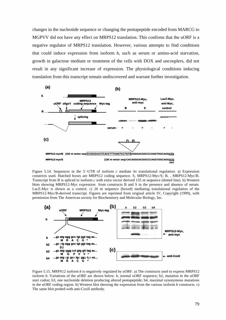
changes in the nucleotide sequence or changing the pentapeptide encoded from MARCG to
MGPVV did not have any effect on MRPS12 translation. This confirms that the uORF is a
negative regulator of MRPS12 translation. However, various attempts to find conditions
that could induce expression from isoform b, such as serum or amino-acid starvation,
growth in galactose medium or treatment of the cells with DOX and uncouplers, did not
result in any significant increase of expression. The physiological conditions inducing
translation from this transcript remain undiscovered and warrant further investigation.
Figure 5.14. Sequences in the 5´-UTR of isoform c mediate its translational regulation. a) Expressi
uORF oligoY coding sequence Myc-tag
B
S
splicing
B S control
serum : + - + - + -
MRPS12-Myc,anti-myc
LacZ-Myc,anti-Myc,
(c)
(b)(a)
MRPS12-myc/B (155 nt vector seq)-CCGCGACCTCACCTTTAGGTCCTGTGAGGGACGGCCCAGGTGGCAGGATG
MRPS12-myc/S (155 nt vector seq)-CACAGGGACGGCCCAGGTGGCAGGATGstart
MRPS12uORF oligoY coding sequence Myc-tag
B
S
splicing
B S control
serum : + - + - + -
MRPS12-Myc,anti-myc
LacZ-Myc,anti-Myc,
(c)
(b)(a)
MRPS12-myc/B (155 nt vector seq)-CCGCGACCTCACCTTTAGGTCCTGTGAGGGACGGCCCAGGTGGCAGGATG
MRPS12-myc/S (155 nt vector seq)-CACAGGGACGGCCCAGGTGGCAGGATGstart
MRPS12
on onstricts used. Hatched boxes are MRPS12 coding sequence. S, MRPS12-Myc/S; B, , MRPS12-Myc/B.
gu RPS12 oform b. Variations of the uORF are shown below. b, normal uORF sequence; b2, mutation in the uORF
cTranscript from B is spliced to isoform c with extra vector derived 155 nt sequence (dotted line). b) Western blots showing MRPS12-Myc expression from constructs B and S in the presence and absence of serum. LacZ-Myc is shown as a control. c) 26 nt sequence (boxed) mediating translational regulation of the MRPS12-Myc/B-derived transcript. Figures are reprinted from original article IV, Copyright (1999), with permission from The American society for Biochemistry and Molecular Biology, Inc.
...gc atg agg gcc tgt ggt tag ac...M R A C G *
...gc ctg agg gcc tgt ggt tag ac...(L)
...gc atg∆ggg cct gtg gtt tag ac...M G P V V *
...gc atg cgt gca tgc ggg tga ac...M R A C G *
b
b2
b3
b4
b
MRPS12-Myc,anti-myc
anti-CoxII
b b2 b3 b4(a) (b)
(c)
uORF MRPS12 Myc-tag
...gc atg agg gcc tgt ggt tag ac...M R A C G *
...gc ctg agg gcc tgt ggt tag ac...(L)
...gc atg∆ggg cct gtg gtt tag ac...M G P V V *
...gc atg cgt gca tgc ggg tga ac...M R A C G *
b
b2
b3
b4
b
MRPS12-Myc,anti-myc
anti-CoxII
b b2 b3 b4(a) (b)
(c)
uORF MRPS12 Myc-tag
Fiis
re 5.15. MRPS12 isoform b is negatively regulated by uORF. a) The constructs used to express M
start codon; b3, one nucleotide deletion producing altered pentapeptide; b4, maximal synonymous mutations in the uORF coding region. b) Western blot showing the expression from the various isoform b constructs. c) The same blot probed with anti-CoxII antibody.
79
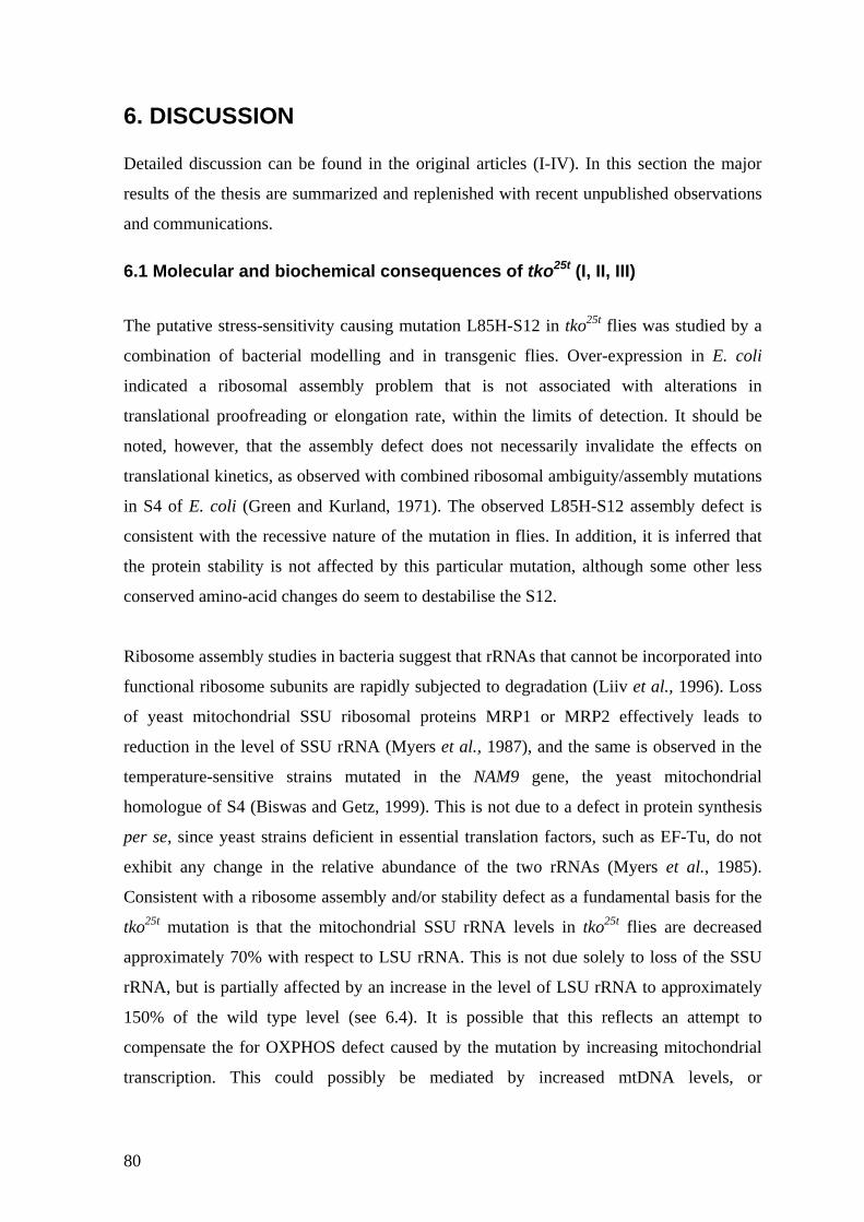
6. DISCUSSION
e found in the original articles (I-IV). In this section the major
sults of the thesis are summarized and replenished with recent unpublished observations
iochemical consequences of tko25t (I, II, III)
was studied by a
ombination of bacterial modelling and in transgenic flies. Over-expression in E. coli
te
cannot be incorporated into
nctional ribosome subunits are rapidly subjected to degradation (Liiv et al., 1996). Loss
Detailed discussion can b
re
and communications.
6.1 Molecular and b
The putative stress-sensitivity causing mutation L85H-S12 in tko25t flies
c
indicated a ribosomal assembly problem that is not associa d with alterations in
translational proofreading or elongation rate, within the limits of detection. It should be
noted, however, that the assembly defect does not necessarily invalidate the effects on
translational kinetics, as observed with combined ribosomal ambiguity/assembly mutations
in S4 of E. coli (Green and Kurland, 1971). The observed L85H-S12 assembly defect is
consistent with the recessive nature of the mutation in flies. In addition, it is inferred that
the protein stability is not affected by this particular mutation, although some other less
conserved amino-acid changes do seem to destabilise the S12.
Ribosome assembly studies in bacteria suggest that rRNAs that
fu
of yeast mitochondrial SSU ribosomal proteins MRP1 or MRP2 effectively leads to
reduction in the level of SSU rRNA (Myers et al., 1987), and the same is observed in the
temperature-sensitive strains mutated in the NAM9 gene, the yeast mitochondrial
homologue of S4 (Biswas and Getz, 1999). This is not due to a defect in protein synthesis
per se, since yeast strains deficient in essential translation factors, such as EF-Tu, do not
exhibit any change in the relative abundance of the two rRNAs (Myers et al., 1985).
Consistent with a ribosome assembly and/or stability defect as a fundamental basis for the
tko25t mutation is that the mitochondrial SSU rRNA levels in tko25t flies are decreased
approximately 70% with respect to LSU rRNA. This is not due solely to loss of the SSU
rRNA, but is partially affected by an increase in the level of LSU rRNA to approximately
150% of the wild type level (see 6.4). It is possible that this reflects an attempt to
compensate the for OXPHOS defect caused by the mutation by increasing mitochondrial
transcription. This could possibly be mediated by increased mtDNA levels, or
80
alternatively the rate of transcription initiation could be affected. Characterisation of these
processes warrants further investigation.
Because the mutation was inferred to decrease the quantitative capacity for mitochondrial
6.2 The mechanosensory defect of tko leads to hearing impairment (II, III)
oth the sensitivity to mechanical shock and the altered electrophysiological response to
translation, I then examined the effects on OXPHOS complex activities. The results
indicated that mutant larvae and adult flies have severely decreased maximal activities in
all OXPHOS complexes containing mtDNA-encoded subunits, whereas the control
enzyme activity, citrate synthase, seemed to be unaffected, or even slightly increased.
Also the ATP synthesis capacity is affected, although to a lesser extent. Similar results
with respect to OXPHOS enzyme activities were obtained from a D. subobscura line
heteroplasmic (80% mutant) for a partially deleted mtDNA, but in this case a more modest
50% and 30% drop, respectively, in the activity of the affected complexes I and III, did not
result in any decrease of ATP synthesis or respiration (Debise et al., 1993; Farge et al.,
2002a). Both of these studies reflect the excess capacity of the OXPHOS complexes with
respect to ATP production, and indicate that sharp biochemical thresholds function at the
enzymatic as well as at the translational level. Furthermore, it is possible that some
metabolic adaptations increase the tolerance for OXPHOS defects, because both the level
of D. subobscura heteroplasmy and tko25t phenotype are modulated by the nuclear
background (Farge et al., 2002b; Le Goff et al., 2002). It should be noted that our
OXPHOS measurements were only carried out using whole flies or larvae, and this would
obviously mask any possible tissue-specific differences in bioenergetic function. In adults,
OXPHOS activities should be largely attributable to muscle and the central nervous
system, which are the major OXPHOS-dependent tissues.
25t
B
bristle deflection (Judd et al., 1972; Shannon et al., 1972; Ganetzky and Wu, 1982; Engel
and Wu, 1994) suggest that the nervous system is affected by the mutation. Additional
preliminary evidence to support this is the complete rescue of BS in our transgenic line
UAS-4, in which tko+ expression is probably induced specifically in adult nervous system
by local enhancers, as discussed below (see 6.5). Is this defect then specific to external
sensory organs, or are the auditory chordotonal organs (CHOs) affected as well? More
81
generally, could the presented mitochondrial translation defects lead to impaired sound
perception in both flies and human?
A courtship analysis (O'Dell et al., 1989) with all possible combinations of sex and
genotype (tko25t and Canton S) was carried out in order to get a first indication of potential
auditory involvement. The courtship success of the tko25t males with wild-type females is
severely reduced. This rejection by wild-type females could imply that the mutant males
fail to produce the correct courtship song, possibly due to defects in their own sound
perception. Sensory feedback has been shown to be important in shaping the courtship
song, which is defective in beethoven (btv) and touch-insensitive-larvaB (tilB) mutants
(Tauber and Eberl, 2001). Males mutant for the gene cachophony (cac), encoding a
voltage-gated calcium channel, also produce aberrant courtship song and show poor mating
success (Von Schilcher, 1976a; Peixoto and Hall, 1998). Some cac mutants show also
temperature dependent convulsions and locomotory defects (Peixoto and Hall, 1998).
Although this is not the case with (inbred) tko25t, they still respond weakly to external
stimuli (hyporeactivity), which could be involved in poor mating success. However, the
mutant males seem to exhibit vigorous courting with no obvious behavioural differences
compared to wild-type males (unpublished observation), although this has not been
analysed statistically. One way to approach this question would be to compare the mating
success of the mutant and wild-type males from which the wings have been surgically
removed, in order to differentiate ‘sluggishness’ from defects in song production.
Despite the obvious problems in courting wild-type females, tko25t males are relatively
successful in courting mutant females, the amount of copulations being 50% that of the
wild-type males. This is likely to be influenced by selection during inbreeding of the tko25t
strain. If the correct ‘love song’ cannot be produced by the males, it is unavoidable that
mutations or polymorphisms that result in enhanced female receptivity will increase in the
isolated population. A perfect example of this is raised (rsd), a mutation that causes the
wings of adults to be held upright and prevents courtship song production (McRobert et al.,
1995). The females from long-established rsd stocks are more receptive to courtship
stimuli and mate more readily with rsd males (and wild-type males) than do wild-type
females (McRobert et al., 1995). Similarly, tko25t females require shorter duration of
courting from the wild-type males than do wild type females. This behavioural evolution is
consistent with our findings that stress sensitivity of tko25t is also counter-selected during
82
long-term inbreeding. When tko25t males do succeed in copulation, the duration of
copulation is decreased to approximately 70% that of the wild-type males. It is possible
that this is due to an additional muscular energy deficit in the mutant males, but any firm
conclusions are currently impossible to draw.
Because courtship involves more than one of the sensory modalities, including both vision
and olfaction (Hall, 1994), a deafness test based on behaviour of the wingless male flies in
response to computer generated courtship song (Von Schilcher, 1976b; Eberl et al., 1997;
Eberl et al., 2000) was used to assay more specifically the potential defect in the auditory
pathway. Because mutations in some genes, such as shibire, that encodes a Drosophila
dynamin, might result in aberrant male-male activity without sound-stimuli (Kitamoto,
2002), the effect of any spontaneous homosexual activity was deducted from the effect of
song-induced courtship in these experiments. Typical song-induced male-male courtship
was shown previously to be defective in btv and tilB mutants (Eberl et al., 2000), and
clearly seems to be lacking in tko25t males. This suggests that the courtship defect is at least
partially due to an impairment of antennal chordotonal organ function. Currently it is
impossible to say if the mutants are completely deaf, or if this hearing impairment is
altered during aging, but at least 5-7 day old males do not show any detectable behavioural
response to song at typical sound intensities. It would be worthwhile to verify this result by
electrophysiological measurements of sound-evoked extracellular potentials from antennal
nerves, as has been done with atonal, btv and tilB mutants (Eberl et al., 2000).
6.3 Developmental consequences of the tko25t mutation (II, III)
The prototypic tko25t mutant shows a developmental delay of 2-3 days compared to wild-
type controls, or to transgenic lines B carrying H85L-reverted transgene. This phenotype,
although to a slightly lesser extend (K.M.C. O’Dell, K. Luoto and H.A. Lindsay, personal
communication), is shared with another BS mutant affecting a mitochondrially located
gene product, sesB, encoding one of the isoforms of adenine nucleotide translocase (ANT)
in Drosophila (Zhang et al., 1999). Moreover, easily shocked (eas), encoding ethanolamine
kinase (Nyako et al., 2001), and slamdance (sda), encoding the homologue of human
aminopeptidase N (Zhang et al., 2002) are also developmentally delayed and BS. Both eas
and sda are expressed highly in the central nervous system, the former affecting the
balance of membrane phospholipids and the latter possibly being involved in calcium
signalling (Nyako et al., 2001; Zhang et al., 2002). It should be noted that my
83
measurements of developmental delay were carried out using the inbred tko25t stock, which
also shows a mitigated BS phenotype compared to outbred flies (Figure 5.5). It seems
likely that prolonged cultivation of the homozygous mutant stock results in selection
against the mutant phenotype and attenuation of the delay. There is increasing evidence
that genetic background plays an important role in the phenotype of any strain (de Belle
and Heisenberg, 1996; Zhang et al., 2002). Indeed, extensive backcrossing of the inbred
flies to the wild-type genetic background causes extreme prolongation of the development,
as well as increased un-coordination and stress-sensitivity (personal observation). This will
potentially allow us to screen for putative tko25t modifier genes in the future.
Further inhibition of mitochondrial translation by doxycyclin (DOX) results in a dose-
dependent increase of the time required for eclosion, as well as increased lethality.
Although a very high dosage of DOX decreased the rate of development of wild-type flies
as well, the synergy between the mutation and the mitoribosome inhibition by DOX is
revealed by a striking increase in sensitivity to the drug in the mutants. That DOX and
tko25t alter the same physiological processes is further supported by the observed partial BS
phenocopy in the wild-type flies fed with high levels of DOX (Figure 5.8). Streptomycin
(SM), which binds to the 30S subunit of bacterial ribosomes and inhibits translational
initiation and accuracy, as well as chloramphenicol (CM) that binds to the bacterial 50S
subunit and inhibits the peptidyltransferase centre, did not have any effect on these
phenotypes, even at the highest concentrations tested. This might be explained by
differential pharmacokinetics of the antibiotics used. At least in humans, tetracyclines such
as DOX penetrate effectively into most tissues from body fluids, but the same is not
necessarily true, at least for polycationic SM, which is poorly absorbed from the
gastrointestinal tract. It is possible that SM, transport of which is dependent upon a system
located in the cell membrane, might be selectively accumulating in tissues not critical for
developmental timing or viability. Alternatively, although cellular import of SM could be
sufficient, it is not clear how these drugs enter mitochondria. As discussed in section 2.4,
cell culture models suggest that SM might not be as efficient in binding mitoribosomes as
some other aminoglycosides (Giordano et al., 2002). Therefore it would be interesting to
test the susceptibility of the tko25t flies to paromomycin (PM), for example.
Consistent with mitochondrial biogenesis being essential for development, lack of
functional mitochondrial zygotically supplied S12 in tko3 mutants leads to lethality,
84
characterised by larval arrest before death, as also observed in the case of pol γ null
mutants (Iyengar et al., 1999). Likewise, ubiquitous high-level expression of tko+ driven
by da-GAL4 leads to larval or pre-pupal lethality in the three lines studied in detail (UAS-
1, UAS-4 and UAS-8). This parallels the finding of Lefai and co-workers (Lefai et al.,
2000) that over expression of the catalytic subunit of pol γ results in late pupal lethality.
Both of these examples stress the importance of correct spatio-temporal regulation of the
expression of mitochondrial gene products during development, even if high levels of the
gene products can be tolerated in tissue-culture models (Lefai et al., 2000). The lethality of
UAS-tko+ lines is likely to be dependent on the expression levels, since rearing the flies at
18°C instead of 25°C greatly reduces the lethality and allows each of the three lines to
pupate (S. Manjiry and J.M. Toivonen, unpublished observations). Furthermore, two of the
lines (UAS-1and UAS-4) are not lethal at 18°C but eclose to a lesser extent and by delayed
timing, supporting the idea that additional spatial or temporal control to the transgene
expression is provided by cis-elements at the insertion site. The profound lethality at 25°C
is most likely due to increased GAL4-driven expression of the transgene, since the da-
GAL4 line used drives the expression of UAS-GFP (green fluorescent protein) much more
efficiently at 25°C than at 18°C (K.M.C. O’Dell and S. Manjiry, personal communication).
6.4 Dosage effects of tko25t (II, III)
The lethality of tko3, and all behavioural and biochemical aspects of tko25t tested, are
rescued by tkoH85L-reverted transgenic lines B, revealing that the L85H mutation in S12 is
the sole cause of the phenotype. In contrast, additional transgenic copies of tko25t in tkoL85H
–transgenic lines A do not improve developmental delay or adult behavioural phenotype
and, surprisingly, do not rescue tko3 lethality even if expressed at much higher levels (2.5
times the wild-type level in A3) than the endogenous gene. Two possible mechanisms
could be involved. Firstly, even if the overall expression of the transgenes is very high in
line A3 carrying three transgenic copies, the transgenes are located in autosomes and might
lack some regulatory information provided by the X-chromosomal environment. This is
unlikely, because all autosomal lines B are virtually wild-type. Secondly, the truncated S12
protein produced by the tko3 allele, which is certainly non-functional, might also produce
deleterious effect in a “sensitized” hypomorphic background such as tko25t. Indeed, even if
tko25t expression in compound heterozygous tko25t/tko3 females carrying additional
hemizygous dose of A3 exceeds the amount of expression in the original mutant, the
85
phenotype is significantly worse (see below). Expression of the truncated protein might
lead to a severe reduction in cell growth, such as observed in the case of human MRPL12
(Marty and Fort, 1996).
Females hemizygous for tko25t have been reported previously to be lethal (Judd et al.,
1972). This can be explained by dosage compensation, which in flies is achieved by
boosting expression from the X-chromosome in males to equal that of the two female X-
chromosomes (Birchler et al., 2003). In our hands, however, compound heterozygous
females for tko3 and tko25t are semi-lethal, i.e. are able to complete their development with
extremely long delay and increased morbidity, and usually show a severely shortened life-
span. The OXPHOS enzyme activities from these flies have not been measured, but it
seems logical that a developmental threshold with respect to ATP production has been
crossed, leading to a much more severe phenotype than in the case of tko25t homozygotes.
In agreement, I found that in this compound heterozygous condition, the addition of an
extra hemizygous dose of the tko25t mutant allele from the A1 or A3 transgenic
chromosome attenuates the developmental delay and decreases the morbidity in dosage-
dependent manner. Also in this case, an inbred genetic background results in less severe
phenotype (see Figure 5.9).
In the tko25t background, the SSU/LSU rRNA ratio is partially restored in larvae of line A3.
The OXPHOS defects are partially restored in line A1 and line A3 larvae, and this
improvement seems roughly to correlate with the expression level. No significant
improvement, however, is observed in ATP synthesis, which is consistent with the fact that
developmental delay and BS are not affected by increased tko25t expression. Interestingly,
even if the SSU/LSU rRNA ratio seems to be smaller in line A1, the actual level of the
SSU rRNA (and obviously LSU rRNA) is higher in line A1 than in line A3. This suggests
that elevated expression of the mutant transgene in line A3 is sufficient to maintain the
ratio of the rRNAs (and OXPHOS enzyme activities) at such a level that putative
compensatory mechanisms affecting mitochondrial transcription are not triggered.
Increased levels of both rRNAs, but decreased SSU/LSU ratio in prototypic tko25t and in
line A1 can lead to more a pronounced imbalance in ribosomal pools than observed in line
A3, with more severe consequences for ETC activity. Despite the improvement in
respiratory chain function, ATP synthesis capacity as well as all the behavioural and
86
developmental phenotypes that go along with it are not improved in line A3, showing
another example of sharp threshold effect.
6.5 Expression of tko+ in the nervous system rescues bang-sensitivity (III) The various aspects of the tko25t mutant phenotype presumably result from failure of those
developmental or physiological processes most dependent upon mitochondrial translational
function. In order to investigate, both spatially and temporally, the nature of such process,
we have created UAS-tko+ transgenic lines that should allow expression of the wild-type
tko gene in a manner defined by selected GAL4 driver lines. The transgene was placed
under the control of a weak promoter, which theoretically should result in only minimal
expression in the absence of a specific GAL4 driver. However, initial characterization of
the transgenic lines suggest, that the lines are expressing tko+ in variable levels, even
without induction by GAL4. Interestingly, the different insertions seem to produce
completely different outcomes depending on their insertion sites, and it is plausible that
this is resulting from the local cis-acting regulatory elements in the vicinity of the
transgene. It should be noted, however, that these differences are not due to disturbance of
another gene at the insertion site, since they do not show marked differences in the wild-
type background.
For example, the inserted transgene in line UAS-4 is located in the 5´ flanking region (or
possibly within a first intron) of the gene Spinophilin (Spn), which is known to be induced
following the metamorphic pulse of ecdysone at the end of the third larval instar, and
subsequently believed to be confined in the adult nervous system (Allen et al., 1997;
Keegan et al., 2001). This line is rescued partially for developmental delay (which takes
place in during second and third larval instar) and completely for BS, strongly suggesting
that it is indeed controlled in the same fashion as Spn. If this proves to be true, we can
restrict the critical period for the developmental delay partly to the third instar (which is
consistent with the previous results), as well as narrow down the tissues responsible for BS
to the nervous system. Line UAS-1, which is inserted within the gene six-banded (sba),
and is expressed in adults at comparable average levels with UAS-4, does not rescue BS.
At the adult stage, sba is expressed uniquely in the eye (Zeidler and Mlodzik, 1997), and if
the same enhancers control the expression of the UAS-1, rescue of BS is not expected.
Retrospectively, the preliminary data obtained from analysis of UAS-tko+ lines justifies
87
many of the previous observations, e.g. bang-sensitivity and altered electrochemical
response to mechanosensory bristle deflection, that both indicated involvement of the
nervous system on the phenotypic outcome (Judd et al., 1972; Shannon et al., 1972;
Ganetzky and Wu, 1982; Engel and Wu, 1994).
Given that ubiquitous high-level expression of tko+ appears to be lethal, more specific
induction at different developmental stages and tissues would probably help us to elucidate
the mechanisms of lethality as well. The nervous system is most likely involved, because
UAS-4 is sufficient to rescue tko3 lethality in males (see original article III, Fig. 5 d). It
should be noted that, at this point, the suggestive patterns of transgene expression are only
speculative. More rigorous experiments, such as in situ hybridisations at different
developmental stages, will hopefully give more insight into stages and tissues critical for
mitochondrial translation capacity.
6.6 Does tko25t manifest as muscle weakness? The involvement of the nervous system in production of the tko25t phenotype is strongly
supported by the data presented above. In humans, however, skeletal muscle is a major
target tissue in mitochondrial translational disorders, and hence the possible impact of the
tko25t mutation on muscle function should be considered. As already mentioned, flies that
have been outbred extensively, i.e. in which the effects of putative genetic modifiers have
been eliminated, show severely uncoordinated movement compared to inbred flies, which
have been subjected to selection against various aspects of the phenotype (BS,
developmental delay, impaired courtship) during long-term culture. In principle, this
movement disorder could result from increased muscle weakness, decreased function of
the nervous system, or a combined effect of the two. Another BS mitochondrial mutant,
sesB1 (see 6.3), mutated in the Drosophila homologue of the human heart- and skeletal
muscle specific isoform of adenine nucleotide translocase (ANT1), is also generally
inactive and uncoordinated (Zhang et al., 1999). When the sesB1 mutant is stimulated to
jump and fly, it does so abnormally, and only for very short distances, suggesting a
muscular defect (although alternative explanations, such as reactivity defect and/or
alterations in the giant fiber pathway are possible). Interestingly, ANT1 in humans is
associated with autosomal dominant progressive external ophthalmoplegia (adPEO), a
disease characterized (among other things) by exercise intolerance, ataxia and hearing
deficit (Kaukonen et al., 2000). Drosophila embryos homozygous for mutations in the
88
gene bent, encoding a myosin light chain kinase involved in muscle development and
function, cannot hatch from the embryonic vitelline membrane due to muscle weakness
(Fyrberg et al., 1992). In addition, heterozygous bent adults have a slight bristle phenotype
(like tko25t homozygotes). Although tko25t flies can hatch more or less normally, in a
’sensitized’ background (e.g. tko25t/tko3), they show increased pupal lethality, characterized
by inability to escape the pupal case (Figure 5.9 d). These observations suggest that muscle
tissue is another potential target for phenotypic manifestation of the tko25t mutation, and it
would be worthwhile to investigate this in detail. Further characterization of the outbred
prototypic tko25t mutants (e.g. for ability jump and fly, and for morphology of the muscles)
and crosses of established UAS-tko+ lines with muscle and nervous system specific
GAL4-driver lines (as well as creation of new lines with different genomic insertion sites)
should help us to investigate potential involvement of the muscle system in the outcome of
the phenotype.
6.7 Model of restrictive ribosome: tkoQ116K (I, II)
Bacterial transformants over expressing the K87Q-rpsL were found to be moderately
resistant to SM compared to transformants expressing the unmutated rpsL from otherwise
identical expression constructs. This suggests that the previously reported high-level SM-
resistance in Mycobacteria (Meier et al., 1994) is at least partly due to the associated rRNA
mutation in this strain. The fact that the endogenous (SMS) rpsL was still present in the
transformants could potentially have some impact on these results, since streptomycin
sensitivity is usually a dominant trait in bacteria (Breckenridge and Gorini, 1969). No
significant decrease in nonsense suppression was found, but the mutation slowed the
translation rate of the ribosome approximately by 15%. The loss of ribosome processivity
is generally correlated with the degree with which mutant ribosomes restrict nonsense
suppression events, i.e. more accurate ribosomes are also slower (Dong and Kurland,
1995). The system we used to measure translational stringency has been developed to
measure nonsense codon suppression, and could be more optimal for detection of
ribosomal ambiguity mutations (ram) instead of increased accuracy, since bacterial
ribosomes in the absence of aminoglycosides are reasonably accurate by nature (Dong and
Kurland, 1995).
89
The low level SM-resistance conferred by the K87Q mutation led us to test the analogous
but opposite (Q116K) mutation in transgenic flies. The rationale for this was to mimic the
putative ribosomal perturbations caused by the A1555G mutation in SSU rRNA, which are
associated with non-syndromic and aminoglycoside induced mitochondrial deafness
(Hutchin et al., 1993; Prezant et al., 1993; Hamasaki and Rando, 1997; Guan et al., 2000;
Giordano et al., 2002). In article (II), recessive female sterility for three independent
tkoQ116K lines was reported. As explained in Results (see 5.4.4.), this is most probably an
artifact resulting from an additional recessive female sterile mutation in the tko3
chromosome. The balancer-system is an excellent tool for Drosophila geneticists in
maintenance of stocks, because invisible genotypes can be scored in progeny with virtually
100% reliability. Balancers (such as FM7) are unable to recombine with their homologue
chromosomes (such as tko3). A lethal allele, such as tko3, needs to be maintained
continuously over a balancer chromosome, such as FM7, and therefore it is not ‘screened’
regularly for additional recessive mutations, such as would give rise to deleterious
phenotypes, such as female sterility. Therefore, it is plausible that during the creation of
the tko3; P[tkoQ116K] lines significant portion of the tko3/FM7 females contained a
chromosome that I designate tko3(rfs), carrying both the tko3 allele and another linked
mutation, causing female sterility, and (possibly through genetic bottlenecks) this
chromosome became the only tko3 chromosome in these lines.
At this point, all that we can say is that transgenic tkoQ116K flies are apparently unaffected
by the mutation, and that they are not sensitive to SM in the tko3 or tko3/FM7 background
(Kevin O’Dell, unpublished observation). This might, however, relate to the
pharmacokinetic issues discussed in 5.3. It would be interesting to test the susceptibility of
these transgenics to DOX and PM in variable allelic backgrounds (i.e. tko+, tko3 and
tko25t). Because of the great interest of the female sterility phenotype (which now seems to
be technical artifact) some less obvious but potentially equally interesting tkoQ116K–related
defects might have been overlooked.
6.8 Regulation of MRPS12 in human cells (IV)
Using reporter constructs in cultured cells we have shown that human mitochondrial
ribosomal protein S12 (MRPS12, in publication IV named RPMS12) is a mitochondrially
located protein. It is imported inside mitochondria, as evidenced by inaccessibility to
90
external protease treatment and size difference compared to the in vitro translated product,
indicating proteolytic processing of the mitochondrial target peptide. The MRPS12-Myc
fusion protein is almost completely located in the inner membrane fraction. This is
consistent with mitoribosome location in yeast (Fox, 1996), and with the documented
interaction of bovine mitochondria with the inner membrane (Liu and Spremulli, 2000).
Our findings in Drosophila indicate the importance of correct regulation of expression of
tko, the fly homologue of human MRPS12. The expression must be sufficient to generate
the required amount of mitochondrial translational capacity to meet the needs of
development, including specific functioning of the peripheral nervous system, but not so
great as to impair mitoribosome biogenesis by the presumed feedback loop.
Three main splice variants of RPMS12 mRNA were found by cyberscreening of dbEST.
Additionally, some (although less prevalent) isoforms might be produced from
heterogenous transcription start sites. Due to use of alternative splice-donor sites, the major
forms of mRNAs differ in their putative 5´-UTRregulatory elements. The non-spliced
isoform a contains both a short upstream open reading frame (uORF) and an
oligopyrimidine tract (oligo(Y)). The isoform b retains only the uORF, which is now
spliced to a position closer to the RPMS12 start codon. From the fully spliced isoform c
both of these elements have been removed. The relative abundance of these variants in
different tissues is more or less similar, although the unspliced form a is mainly expressed
in heart. In general, the tissue distribution is typical for a gene involved in mitochondrial
respiratory function. The fully spliced isoform lacking the putative translational control
elements is the most prominent transcript in all tissues.
The translational control of the various isoforms was studied by investigating the
polysomal distribution of the mRNA variants in growing and non-growing cells. It appears
that the most prominent isoform c is regulated according to growth status, representing a
shift from the polysomal to sub-polysomal fraction during serum starvation. This reminds
the typical regulation exhibited by mRNAs for cytosolic ribosomal proteins, via the
terminal oligopyrimidine tracts (TOPs, Meyuhas, 2000). The observation is confirmed at
the protein level by expression of MRPS12-Myc in the presence and absence of serum.
When MRPS12-Myc mRNA is expressed from the construct roughly corresponding to
non-spliced variant a, it is efficiently spliced to isoform c, which is consistent for this
variant being the most abundant transcript in tissues. The growth responsiveness of
91
isoform c in the absence of both uORF and oligo(Y) suggest that these elements have a
negative function on translational regulation. Accordingly, the polysomal distribution of
the isoforms a and b is not regulated by serum. How does the positive regulation of
isoform c then take place?
A short, 26 nt sequence in the extreme 5´end of the transcript c seems to mediate the
translational control of this message. The element does not resemble typical TOPs
(Meyuhas, 2000), and might direct the growth regulation in a way that differs from the
regulation of the cytosolic ribosomal protein mRNAs, although it might share some
features with it. For example, it can be hypothesised that in addition to growth control, this
element could be responsive to other stimuli, such as bioenergetic needs, availability of
specific substrates or developmental signals. Alternatively, the regulation of expression by
this element could be modulated by tissue specific factors in some conditions. Since this 26
nt element is also present in isoforms a and b, which are not growth controlled, the
additional regulatory elements present in these variants must over-ride the growth control.
Alternatively, some other properties specific for the isoform c, such as the distance
between the 26 nt sequence and the initiation codon in the spliced mRNAs, might be
essential.
The uORF in splice variant b is surrounded by a relatively strong Kozak environment
(Kozak, 1999), which seems to be recognised by most ribosomes. The elimination of the
uORF leads to efficient expression of the reporter construct (unpublished observation, see
Figure 5.15), confirming that the uORF is a negative regulator of MRPS12 translation.
Additionally, splice form b cannot be further spliced to form c, suggesting that the
Oligo(Y) tract might be essential for recognition of the acceptor upstream of the MRPS12
start codon. The behaviour of the construct in any various modified versions of the uORF
suggest that the mechanism is based on dissociation of the ribosomes from the mRNA after
futile initiation and translation of the uORF-encoded peptide. Alternatively, blockage (by a
terminated ribosome) of the subsequent ribosomes scanning the same message could be
involved (Morris and Geballe, 2000), which is supported by the relatively wide distribution
of the message in small polysomes and mRNPs (see original publication IV, Fig 5 c). The
nucleic acid sequence of the uORF coding region or the properties of the nascent peptide
itself do not seem to have an effect on MRPS12-Myc expression (Figure 5.15). Although it
cannot be completely excluded that also the transcript stability could be altered via
92
nonsense-mediated decay, the Northern blots and RT-PCR results do not support this.
Translation of MRPS12, most probably by a leaky scanning mechanism (Morris and
Geballe, 2000), seems to be minimal under the conditions tested. Because our various
attempts to find conditions that could induce the expression from isoform b failed, the
potential signals relieving the control of the transcript remains to be elucidated in the
future. This might be difficult, or even impossible in cell culture, since such mechanisms
very likely exist only in specialised tissues such as heart, skeletal muscle or kidney, where
this isoform is especially prominent.
As mentioned in 2.2.3, there is substantial variation in the size of the mRNAs in different
animal species studied (Shah et al., 1997). Even if the length of the 5´-UTR in both human
and in Drosophila is comparable, there is no evidence of translational regulatory elements
(at least uORFs) in Drosophila mRNA. It is possible, therefore, that mechanisms similar
to human do not exist in flies, although this has not been rigorously explored. It should be
noted, that translational regulatory elements are not exclusively restricted to 5´-UTRs, and
similar outcomes with respect to gene expression in different species may be achieved by
completely different combinations of transcriptional and post-transcriptional regulation.
6.9 Possible physiological consequences of mutations in the mitochondrial translation machinery
The sensitivity of sound perception to mitochondrial dysfunction in humans is
demonstrated by frequent involvement of sensorineural hearing impairment in the
pathology of mitochondrial diseases (see section 2.4.2 and references therein). However,
processes that are affected at the cellular level are still largely unknown. Similarly,
although dysfunction in components of the mitochondrial OXPHOS machinery has now
been established in tko25t mutants, and the effects of the mutation on mitochondrial
translational apparatus have been described, it is not clear what the exact physiological
mechanisms leading to auditory dysfunction and other aspects of the phenotype are.
This study, and studies by Engel and Wu (1994), suggest that signalling from both external
sensory organs (decreased action potentials in response to deflection) and from antennal
chordotonal organs (impaired auditory function), both of which connect to sensory neurons
of the peripheral nervous system, are affected. In the case of the antennal organ it is not
93
clear, however, which cell types and/or neural circuits are affected, and sound-evoked
electrophysiological measurements should be applied to address this question more
specifically. It could be hypothesised that the defects are a direct result of energy
deficiency due to decreased ATP synthesis in these organs and/or associated cells, which
might have an influence on ion recycling in sensory organs or in nerve terminals. Human
inner-ear hair cells, as well as sensory receptors in general, are rich in mitochondria. One
candidate ion that could be involved is calcium, which is directly sequestered by
mitochondria (Pozzan et al., 2000; Rizzuto et al, 2000). Also, some other, ATP-dependent
ion uptake mechanism in nerve terminals could be affected, which might have an
influence, for example on endolymph composition. Electrophysiological studies of the
giant fiber pathway suggest that neuronal dysfunction in BS mutants is probably not
restricted to sensory organs, since electrical stimulation of the central nervous system leads
to paralysis and seizures similar to those observed when mechanical disturbance is used
(Pavlidis and Tanouye, 1995; Lee and Wu, 2002). Therefore, in the case of tko25t it is
likely that the effects of the mutation are pleiotropic. Involvement of many channel
proteins in suppression of the BS phenotype suggests that altered ionic conditions might
underlay both decreased action potentials in response to mechanosensory stimulation and
hyperexcitatory behaviour in the central nervous system (Kuebler et al., 2001). At this
point we cannot exclude that also other sensory modalities than hearing are involved in the
tko25t phenotype, since they have not been rigorously studied. Also changes in cognitive
processes could result from mitochondrial dysfunction, some of which might be age
related. The hyporeactive behaviour of tko25t could potentially be caused by decreased
cognitive processes This could be tested by behavioural assays, such as odor-aversion
testing (for review, see Waddell and Quinn, 2001). In this assay, flies experience an odor
in conjunction with electric-shock punishment, and during subsequent testing flies capable
of learning and/or memorizing this preferentially avoid the shock-associated odor. There
might be pitfalls using this technique with tko25t, since the electric shock used in the assay
could cause these mutants to become paralysed. Additionally, the test assumes a normal
ability to smell, which should be tested first in the case of tko25t. A similar test based on
visual learning has also been developed, which does not include electric shock (Waddell
and Quinn, 2001, and references therein).
It is not surprising that an overall decrease in mitochondrial translation capacity could
result in a complex phenotype in flies, since many mitochondrial diseases in humans are
94
syndromic. For example, myoclonal epilepsy with ragged red fibers (MERRF) is caused
most frequently by the mutation A8344G in the mitochondrial tRNALys (Shoffner et al.,
1990), and results in a defect in translation of all mtDNA-encoded genes and defects in
activities of OXPHOS complexes I, III and IV (Bindoff et al., 1991). Clinical presentation
of MERRF includes neural (ataxia, spasticity) and muscular (muscle weakness, myopathy)
symptoms, in combination with sensorineural hearing loss. Cognitive disturbances are one
feature of mitochondrial encephalomyopathies, and recent microarray study in rats
showing cognitive defects revealed changes in the expression of many mitochondrial
proteins (Blalock et al., 2003). A mutation in tRNALeu(UUR) associated with mitochondrial
encephalomyopathy, lactic acidosis, and strokelike episodes (MELAS) results (among
other things) in hearing impairment, cognitive decline, and short stature (Majamaa et al.,
1998). Further research using mitochondrial disease models, like tko, could be valuable in
studying the basic physiological mechanisms involved in human mitochondrial translation
disorders.
95
7. SUMMARY In this thesis, I have demonstrated the potential of D. melanogaster as a model for human
mitochondrial disorders. Mutations in the tko, mitochondrially located fly homologue of
the bacterial ribosomal protein S12, were characterised with respect to their effects on
phenotypic, biochemical and molecular level alterations. Additionally, regulation of the
expression of the human homologue of tko, MRPS12, was studied in cell culture. My main
conclusions are:
1) Based on bacterial mutation modelling and characterization of mitochondrial ribosomal
RNA levels, the prototypic tko25t mutation causes a defect in mitoribosomal assembly, and
a quantitative decrease in mitochondrial protein synthesis capacity. In agreement with this,
tko25t mutants are hypersensitive to the mitochondrial translation inhibitor doxycyclin, and
show greatly decreased mitochondrial respiratory chain activity and also a decreased ATP
synthesis capacity. In addition to these biochemical defects, the tko25t mutation results in a
complex phenotype including developmental, physiological, and behavioural aspects, such
as developmental delay, bang-sensitivity, impaired courtship behaviour and hearing
impairment. Based on a transgenic reversion test, this complex phenotype is due solely to
the previously reported tko mutation L85H.
2) A high expression level of the tko25t allele is not associated with improved
developmental or behavioural performance, except in a hemizygous tko25t/tko3 background,
tko3 being a null allele. In this background, a higher expression level of the mutant allele
leads to significant attenuation of the defect. Selective spatio-temporal expression of wild-
type tko+ in the tko25t background indicates the times and cell-types critically dependent on
mitochondrial translational capacity. The results suggest that the nervous system is
implicated in manifestation of the mitochondrial translation disorders in flies, as it is in
humans. Moreover, unregulated high-level expression of the wild-type tko+ results in pre-
adult lethality, further emphasizing the importance of the controlled regulation of its
expression.
3) Human MRPS12 is prominently expressed in tissues with high oxidative metabolism,
such as in the heart and in skeletal muscle. MRPS12 shows complex regulation of
expression at the level of transcription, mRNA splicing and translation. Studies in both
96
flies and human cells indicate that mitoribosome biogenesis is a strictly controlled process,
and any alterations in the process can lead to severe consequences on mitochondrial
metabolism.
6) Besides hearing impairment, tko25t mutant shows several features characteristic of
human heteroplasmic mitochondrial disorders, including sharp phenotypic and biochemical
thresholds for mitochondrial translational capacity. The phenotypes of the mutant flies
parallel these of the typical human mitochondrial disorders. Furthermore, like in humans,
the severity of the phenotypic manifestation is dependent on both genetic and
environmental factors. Although physiological similarities between human and mouse
makes it in many cases the model organism of the choice, my results suggest that the
biomedical relevance of Drosophila in modelling human mitochondrial disorders has been
greatly underestimated.
97
8. ACKNOWLEDGEMENTS
The work described in this thesis has been mainly carried out in the Laboratory of Molecular Biology at the Institute of Medical Technology, University of Tampere, Finland. Part of the research has been conducted in the following institutions: Institute of Biomedical and Life Sciences, Division of Molecular Genetics, University of Glasgow, Scotland; Equipe Genome Mitochondrial, Université Blaise-Pascal, Clermont II, France; Departments of Biology at Universitá di “Roma Tre” and Universitá di Roma “Tor Vergata”, Rome, Italy. The research was supported by the Academy of Finland, Juselius Foundation, Tampere University Hospital Medical Research Fund, and the European Union. Printing of the thesis was supported by The Scientific Foundation of The City of Tampere. I am grateful to my external reviewers, Anu Wartiovaara and Rafael Garesse Alarcón, for their valuable comments and advise to improve my thesis, and for being able to do this in a very short period provided. My sincerest appreciation goes to Howy Jacobs, for excellent teaching and supervision. Working with Howy has been both challenging and rewarding. I want to thank him also for the opportunity to attend and present my results in many scientific conferences. I am grateful to Anja “The Lab Cerberus” Rovio for her helping hand in technical trouble-shooting, and to Outi Kurronen for recently solving many of my practical problems in the lab. Special thanks go to Hans Spelbrink for being an infinite technical database, and to him and Herma Renkema for always providing a countryside partyhouse when urgently needed. I thank Zahid Shah for useful discussions, and for launching many of the projects included in my thesis. I was encouraged to initiate my studies in mitochondrial field by Sanna ”The Holy Chicken” Marjavaara, and I want to thank her for enjoyable chats, for sharing my black humour, for great parties and for list of other things. It has been a great pleasure to know the four “Groovy Chicks”: Rimmy Manjiry and Nuria Garrido I thank for brilliant cooking, live music, keeping up the spirit in the lab, as well as being good friends; Helen Lindsay and Kaisa Luoto I thank for friendship and for introducing me to the music of marvellous Westlife and for transcendental “Kaisaisms” with hidden wisdom. I am thankful to my duck-park neighbour Gertjan “The Big G” Hakkaart for interestingly meandering stories at Telakka, and for being a living gig-alert. I sincerely respect Marina Toompuu for tolerance of my chaotic office behaviour during all these years. I want to thank all current and former members of the mito-labs (Anne, Ernesto, Helen C., Henna, Jaakko, Johanna, John, Mikko, Olli, Qiong, Sanna, Sjoerd, Timo et al.) for good company, help and discussions (both scientific and nonsense), and the “fly room” students Esko, Marjo, Mark and Niels, for their valuable assistance in my projects. I am grateful to all the people that helped me in University of Glasgow, and especially to my “second supervisor” Kevin O’Dell, for teaching me almost everything I know about the fruit flies, for conducting the experiments related to fly behaviour and genetics with me, and for memorable trip to Scottish highlands. Similarly, I want to express my gratitude to all the people that assisted my biochemical experiments with “mouches” in the Université Blaise-Pascal, and especially Serge Alziari, for delicious dinners and for mountain bike during my visits in Clermont-Ferrand, and Sylvie Touraille and Monique Renoux for lab-supervision and patience. Big “thank you” also belongs to following people: my stringent biology teacher, Pauli Aaltokallio, for inspiring lessons at Palokka high school; Linda
98
McQueen, for providing a best possible place for lodging in Glasgow; all graduate students and senior scientists from other groups of IMT that have helped me with my practical lab work or attended social activities outside the lab (i.e. pubs). Especially, it has been my pleasure enjoy evenings playing pool and spend whisky-flavoured poker nights with Kimmo and Iivari. Ten credits and papukaijamerkki to our outstanding janitor, Heimo Koskinen, for batteries, CO2, light bulbs, mail and monkey-wrench, and for making everyday-life of all the researchers in the institute much more simple. Life is not just work, and I have been privileged to enjoy a company of numerous people that are not directly involved in my science projects, but who have probably affected to my choices in various ways. Special thanks are reserved for Suharit (Kokko, Kotanen ja Asikainen) for 20+ years of friendship, and for hiking, competing, shivering and starving in the woods with me, as well as for more sophisticated re-unions lately. I am grateful to the ultimate carnivores, Taigan Pojat (Jussi, Otso, Vesku, Päiskäri, Esko, Iippu ja Jokke), for sensational fishing trips to the final frontiers of Säkylä and Lapland, and for financial funding in the form of sökö. I thank all my student friends from Jyväskylä for hilarious company and for kindly making available a couch/floor to stay over during my spontaneous excursions. Thanks to Hemmo and Atte for eastern Finland base camp, and for both for [sarcasm ON] extremely intelligent [sarcasm OFF] chats in Tuupovaara and in Lapland. Big hug goes to my dear friends Niina (✿) for long frendship and kind investments in my paintings, and Anna (★), for ∞ things in recent years. Finally, I want express my deepest gratitude and love to my parents for an adequate set of chromosomes, including mtDNA, and for all the trade-offs (e.g. parental care, provision of environmental resources) they have experienced during my embryonic, post-natal and pre-doctoral development. Tampere, September 2003 Janne Toivonen
99
9. REFERENCES Adams, M. D., Celniker, S. E., Holt, R. A., Evans, C. A., Gocayne, J. D., Amanatides, P. G., Scherer, S. E., Li, P. W., Hoskins, R. A., Galle, R. F., George, R. A., Lewis, S. E., Richards, S., Ashburner, M., Henderson, S. N., Sutton, G. G., Wortman, J. R., Yandell, M. D., Zhang, Q., Chen, L. X., Brandon, R. C., Rogers, Y. H., Blazej, R. G., Champe, M., Pfeiffer, B. D., Wan, K. H., Doyle, C., Baxter, E. G., Helt, G., Nelson, C. R., Gabor, G. L., Abril, J. F., Agbayani, A., An, H. J., Andrews-Pfannkoch, C., Baldwin, D., Ballew, R. M., Basu, A., Baxendale, J., Bayraktaroglu, L., Beasley, E. M., Beeson, K. Y., Benos, P. V., Berman, B. P., Bhandari, D., Bolshakov, S., Borkova, D., Botchan, M. R., Bouck, J., Brokstein, P., Brottier, P., Burtis, K. C., Busam, D. A., Butler, H., Cadieu, E., Center, A., Chandra, I., Cherry, J. M., Cawley, S., Dahlke, C., Davenport, L. B., Davies, P., de Pablos, B., Delcher, A., Deng, Z., Mays, A. D., Dew, I., Dietz, S. M., Dodson, K., Doup, L. E., Downes, M., Dugan-Rocha, S., Dunkov, B. C., Dunn, P., Durbin, K. J., Evangelista, C. C., Ferraz, C., Ferriera, S., Fleischmann, W., Fosler, C., Gabrielian, A. E., Garg, N. S., Gelbart, W. M., Glasser, K., Glodek, A., Gong, F., Gorrell, J. H., Gu, Z., Guan, P., Harris, M., Harris, N. L., Harvey, D., Heiman, T. J., Hernandez, J. R., Houck, J., Hostin, D., Houston, K. A., Howland, T. J., Wei, M. H., Ibegwam, C., Jalali, M., Kalush, F., Karpen, G. H., Ke, Z., Kennison, J. A., Ketchum, K. A., Kimmel, B. E., Kodira, C. D., Kraft, C., Kravitz, S., Kulp, D., Lai, Z., Lasko, P., Lei, Y., Levitsky, A. A., Li, J., Li, Z., Liang, Y., Lin, X., Liu, X., Mattei, B., McIntosh, T. C., McLeod, M. P., McPherson, D., Merkulov, G., Milshina, N. V., Mobarry, C., Morris, J., Moshrefi, A., Mount, S. M., Moy, M., Murphy, B., Murphy, L., Muzny, D. M., Nelson, D. L., Nelson, D. R., Nelson, K. A., Nixon, K., Nusskern, D. R., Pacleb, J. M., Palazzolo, M., Pittman, G. S., Pan, S., Pollard, J., Puri, V., Reese, M. G., Reinert, K., Remington, K., Saunders, R. D., Scheeler, F., Shen, H., Shue, B. C., Siden-Kiamos, I., Simpson, M., Skupski, M. P., Smith, T., Spier, E., Spradling, A. C., Stapleton, M., Strong, R., Sun, E., Svirskas, R., Tector, C., Turner, R., Venter, E., Wang, A. H., Wang, X., Wang, Z. Y., Wassarman, D. A., Weinstock, G. M., Weissenbach, J., Williams, S. M., WoodageT, Worley, K. C., Wu, D., Yang, S., Yao, Q. A., Ye, J., Yeh, R. F., Zaveri, J. S., Zhan, M., Zhang, G., Zhao, Q., Zheng, L., Zheng, X. H., Zhong, F. N., Zhong, W., Zhou, X., Zhu, S., Zhu, X., Smith, H. O., Gibbs, R. A., Myers, E. W., Rubin, G. M., and Venter, J. C. (2000). The genome sequence of Drosophila melanogaster. Science 287, 2185-2195.
Alam, T. I., Kanki, T., Muta, T., Ukaji, K., Abe, Y., Nakayama, H., Takio, K., Hamasaki, N., and Kang, D. (2003). Human mitochondrial DNA is packaged with TFAM. Nucleic Acids Res 31, 1640-1645.
Alksne, L. E., and Warner, J. R. (1993). A novel cloning strategy reveals the gene for the yeast homologue to Escherichia coli ribosomal protein S12. J Biol Chem 268, 10813-10819.
Alksne, L. E., Anthony, R. A., Liebman, S. W., and Warner, J. R. (1993). An accuracy center in the ribosome conserved over 2 billion years. Proc Natl Acad Sci USA 90, 9538-9541.
100
Allen, P. B., Ouimet, C. C., and Greengard, P. (1997). Spinophilin, a novel protein phosphatase 1 binding protein localized to dendritic spines. Proc Natl Acad Sci USA 94, 9956-9961.
Anderson, S., Bankier, A. T., Barrell, B. G., de Bruijn, M. H., Coulson, A. R., Drouin, J., Eperon, I. C., Nierlich, D. P., Roe, B. A., Sanger, F., Schreier, P. H., Smith, A. J., Staden, R., and Young, I. G. (1981). Sequence and organization of the human mitochondrial genome. Nature 290, 457-465.
Andersson, D. I., Andersson, S. G., and Kurland, C. G. (1986). Functional interactions between mutated forms of ribosomal proteins S4, S5 and S12. Biochimie 68, 705-713.
Anthony, R. A., and Liebman, S. W. (1995). Alterations in ribosomal protein RPS28 can diversely affect translational accuracy in Saccharomyces cerevisiae. Genetics 140, 1247-1258.
Arbuzova, S., Hutchin, T., and Cuckle, H. (2002). Mitochondrial dysfunction and Down's syndrome. Bioessays 24, 681-684.
Attardi, G., Yoneda, M., and Chomyn, A. (1995). Complementation and segregation behavior of disease-causing mitochondrial DNA mutations in cellular model systems. Biochim Biophys Acta 1271, 241-248.
Baker, J., and Kernan, M. (2003). Using uncoordinated to search for genes involved in ciliogenesis. Paper presented at: A Dros Res Conf 44 2003 :252C.
Barger, P. M., and Kelly, D. P. (2000). PPAR signaling in the control of cardiac energy metabolism. Trends Cardiovasc Med 10, 238-245.
Battersby, B. J., Loredo-Osti, J. C., and Shoubridge, E. A. (2003). Nuclear genetic control of mitochondrial DNA segregation. Nat Genet 33, 183-186.
Ben-Arie, N., Hassan, B. A., Bermingham, N. A., Malicki, D. M., Armstrong, D., Matzuk, M., Bellen, H. J., and Zoghbi, H. Y. (2000). Functional conservation of atonal and Math1 in the CNS and PNS. Development 127, 1039-1048.
Berdanier, C. D. (2001). Mitochondrial gene expression in diabetes mellitus: effect of nutrition. Nutr Rev 59, 61-70.
Bereiter-Hahn, J., and Voth, M. (1994). Dynamics of mitochondria in living cells: shape changes, dislocations, fusion, and fission of mitochondria. Microsc Res Tech 27, 198-219.
Berger, K. H., and Yaffe, M. P. (2000). Mitochondrial DNA inheritance in Saccharomyces cerevisiae. Trends Microbiol 8, 508-513.
101
Berger, T., Brigl, M., Herrmann, J. M., Vielhauer, V., Luckow, B., Schlondorff, D., and Kretzler, M. (2000). The apoptosis mediator mDAP-3 is a novel member of a conserved family of mitochondrial proteins. J Cell Sci 113 ( Pt 20), 3603-3612. Bergstrom, C. T., and Pritchard, J. (1998). Germline bottlenecks and the evolutionary maintenance of mitochondrial genomes. Genetics 149, 2135-2146.
Bermingham, N. A., Hassan, B. A., Price, S. D., Vollrath, M. A., Ben-Arie, N., Eatock, R. A., Bellen, H. J., Lysakowski, A., and Zoghbi, H. Y. (1999). Math1: an essential gene for the generation of inner ear hair cells. Science 284, 1837-1841.
Berthier, F., Renaud, M., Alziari, S., and Durand, R. (1986). RNA mapping on Drosophila mitochondrial DNA: precursors and template strands. Nucleic Acids Res 14, 4519-4533.
Beziat, F., Touraille, S., Debise, R., Morel, F., Petit, N., Lecher, P., and Alziari, S. (1997). Biochemical and molecular consequences of massive mitochondrial gene loss in different tissues of a mutant strain of Drosophila subobscura. J Biol Chem 272, 22583-22590.
Bibb, M. J., Van Etten, R. A., Wright, C. T., Walberg, M. W., and Clayton, D. A. (1981). Sequence and gene organization of mouse mitochondrial DNA. Cell 26, 167-180.
Bilgin, N., Claesens, F., Pahverk, H., and Ehrenberg, M. (1992). Kinetic properties of Escherichia coli ribosomes with altered forms of S12. J Mol Biol 224, 1011-1027.
Bindoff, L. A., Desnuelle, C., Birch-Machin, M. A., Pellissier, J. F., Serratrice, G., Dravet, C., Bureau, M., Howell, N., and Turnbull, D. M. (1991). Multiple defects of the mitochondrial respiratory chain in a mitochondrial encephalopathy (MERRF): a clinical, biochemical and molecular study. J Neurol Sci 102, 17-24. Birchler, J. A., Pal-Bhadra, M., and Bhadra, U. (2003). Dosage dependent gene regulation and the compensation of the X chromosome in Drosophila males. Genetica 117, 179-190.
Birky, C. W., Jr. (1995). Uniparental inheritance of mitochondrial and chloroplast genes: mechanisms and evolution. Proc Natl Acad Sci USA 92, 11331-11338.
Birky, C. W., Jr. (2001). The inheritance of genes in mitochondria and chloroplasts: laws, mechanisms, and models. Annu Rev Genet 35, 125-148.
Biswas, T. K., and Getz, G. S. (1999). The single amino acid changes in the yeast mitochondrial S4 ribosomal protein cause temperature-sensitive defect in the accumulation of mitochondrial 15S rRNA. Biochemistry 38, 13042-13054.
Blalock, E. M., Chen, K. C., Sharrow, K., Herman, J. P., Porter, N. M., Foster, T. C., and Landfield, P. W. (2003). Gene microarrays in hippocampal aging: statistical profiling identifies novel processes correlated with cognitive impairment. J Neurosci 23, 3807-3819.
102
Boileau, G., Butler, P., Hershey, J. W., and Traut, R. R. (1983). Direct cross-links between initiation factors 1, 2, and 3 and ribosomal proteins promoted by 2-iminothiolane. Biochemistry 22, 3162-3170.
Bolden, A., Noy, G. P., and Weissbach, A. (1977). DNA polymerase of mitochondria is a gamma-polymerase. J Biol Chem 252, 3351-3356.
Borner, G. V., Zeviani, M., Tiranti, V., Carrara, F., Hoffmann, S., Gerbitz, K. D., Lochmuller, H., Pongratz, D., Klopstock, T., Melberg, A., Holme, E., and Paabo, S. (2000). Decreased aminoacylation of mutant tRNAs in MELAS but not in MERRF patients. Hum Mol Genet 9, 467-475.
Boveris, A., Oshino, N., and Chance, B. (1972). The cellular production of hydrogen peroxide. Biochem J 128, 617-630.
Brakier-Gingras, L., and Phoenix, P. (1984). The control of accuracy during protein synthesis in Escherichia coli and perturbations of this control by streptomycin, neomycin, or ribosomal mutations. Can J Biochem Cell Biol 62, 231-244.
Brand, A. H., and Perrimon, N. (1993). Targeted gene expression as a means of altering cell fates and generating dominant phenotypes. Development 118, 401-415.
Brand, A. H., Manoukian, A. S., and Perrimon, N. (1994). Ectopic expression in Drosophila. Methods Cell Biol 44, 635-654.
Breckenridge, L., and Gorini, L. (1969). The dominance of streptomycin sensitivity re-examined. Proc Natl Acad Sci USA 62, 979-985.
Bregeon, D., Colot, V., Radman, M., and Taddei, F. (2001). Translational misreading: a tRNA modification counteracts a +2 ribosomal frameshift. Genes Dev 15, 2295-2306.
Bridges, C. B. (1914). Direct proof through non-disjunction that the sex-linked genes of Drosophila are borne on the X-chromosome. Science 40, 107-109.
Brock, S., Szkaradkiewicz, K., and Sprinzl, M. (1998). Initiation factors of protein biosynthesis in bacteria and their structural relationship to elongation and termination factors. Mol Microbiol 29, 409-417.
Brodersen, D. E., Clemons, W. M., Jr., Carter, A. P., Wimberly, B. T., and Ramakrishnan, V. (2002). Crystal structure of the 30 S ribosomal subunit from Thermus thermophilus: structure of the proteins and their interactions with 16 S RNA. J Mol Biol 316, 725-768.
Brown, W. M., George, M., Jr., and Wilson, A. C. (1979). Rapid evolution of animal mitochondrial DNA. Proc Natl Acad Sci USA 76, 1967-1971.
103
Bu, X., Shohat, M., Jaber, L., and Rotter, J. I. (1993). A form of sensorineural deafness is determined by a mitochondrial and an autosomal locus: evidence from pedigree segregation analysis. Genet Epidemiol 10, 3-15.
Caceres, J. F., and Kornblihtt, A. R. (2002). Alternative splicing: multiple control mechanisms and involvement in human disease. Trends Genet 18, 186-193.
Cai, Y. C., Bullard, J. M., Thompson, N. L., and Spremulli, L. L. (2000). Interaction of mitochondrial elongation factor Tu with aminoacyl-tRNA and elongation factor Ts. J Biol Chem 275, 20308-20314.
Caldwell, J. C., and Eberl, D. F. (2002). Towards a molecular understanding of Drosophila hearing. J Neurobiol 53, 172-189.
Carelli, V., Giordano, C., and d'Amati, G. (2003). Pathogenic expression of homoplasmic mtDNA mutations needs a complex nuclear-mitochondrial interaction. Trends Genet 19, 257-262.
Carrodeguas, J. A., and Bogenhagen, D. F. (2000). Protein sequences conserved in prokaryotic aminoacyl-tRNA synthetases are important for the activity of the processivity factor of human mitochondrial DNA polymerase. Nucleic Acids Res 28, 1237-1244.
Castellani, R., Hirai, K., Aliev, G., Drew, K. L., Nunomura, A., Takeda, A., Cash, A. D., Obrenovich, M. E., Perry, G., and Smith, M. A. (2002). Role of mitochondrial dysfunction in Alzheimer's disease. J Neurosci Res 70, 357-360.
Celniker, S. E., Wheeler, D. A., Kronmiller, B., Carlson, J. W., Halpern, A., Patel, S., Adams, M., Champe, M., Dugan, S. P., Frise, E., Hodgson, A., George, R. A., Hoskins, R. A., Laverty, T., Muzny, D. M., Nelson, C. R., Pacleb, J. M., Park, S., Pfeiffer, B. D., Richards, S., Sodergren, E. J., Svirskas, R., Tabor, P. E., Wan, K., Stapleton, M., Sutton, G. G., Venter, C., Weinstock, G., Scherer, S. E., Myers, E. W., Gibbs, R. A., and Rubin, G. M. (2002). Finishing a whole-genome shotgun: release 3 of the Drosophila melanogaster euchromatic genome sequence. Genome Biol 3, research0079.1-0079.14. Chang, D. D., and Clayton, D. A. (1985). Priming of human mitochondrial DNA replication occurs at the light-strand promoter. Proc Natl Acad Sci USA 82, 351-355.
Chang, D. D., Hauswirth, W. W., and Clayton, D. A. (1985). Replication priming and transcription initiate from precisely the same site in mouse mitochondrial DNA. Embo J 4, 1559-1567.
Chien, S., Reiter, L. T., Bier, E., and Gribskov, M. (2002). Homophila: human disease gene cognates in Drosophila. Nucleic Acids Res 30, 149-151.
Chinnery, P. F., Thorburn, D. R., Samuels, D. C., White, S. L., Dahl, H. M., Turnbull, D. M., Lightowlers, R. N., and Howell, N. (2000). The inheritance of mitochondrial DNA heteroplasmy: random drift, selection or both? Trends Genet 16, 500-505.
104
Chinnery, P. F., and Turnbull, D. M. (2001). Epidemiology and treatment of mitochondrial disorders. Am J Med Genet 106, 94-101.
Chomyn, A., Enriquez, J. A., Micol, V., Fernandez-Silva, P., and Attardi, G. (2000). The mitochondrial myopathy, encephalopathy, lactic acidosis, and stroke-like episode syndrome-associated human mitochondrial tRNALeu(UUR) mutation causes aminoacylation deficiency and concomitant reduced association of mRNA with ribosomes. J Biol Chem 275, 19198-19209.
Christianson, T. W., and Clayton, D. A. (1988). A tridecamer DNA sequence supports human mitochondrial RNA 3'-end formation in vitro. Mol Cell Biol 8, 4502-4509.
Chung, A. B., Stepien, G., Haraguchi, Y., Li, K., and Wallace, D. C. (1992). Transcriptional control of nuclear genes for the mitochondrial muscle ADP/ATP translocator and the ATP synthase beta subunit. Multiple factors interact with the OXBOX/REBOX promoter sequences. J Biol Chem 267, 21154-21161.
Claeys, I., Simonet, G., Poels, J., Van Loy, T., Vercammen, L., De Loof, A., and Vanden Broeck, J. (2002). Insulin-related peptides and their conserved signal transduction pathway. Peptides 23, 807-816.
Clary, D. O., Goddard, J. M., Martin, S. C., Fauron, C. M., and Wolstenholme, D. R. (1982). Drosophila mitochondrial DNA: a novel gene order. Nucleic Acids Res 10, 6619-6637.
Clayton, D. A. (1982). Replication of animal mitochondrial DNA. Cell 28, 693-705.
Clayton, D. A. (1991). Replication and transcription of vertebrate mitochondrial DNA. Annu Rev Cell Biol 7, 453-478.
Coetzee, T., Herschlag, D., and Belfort, M. (1994). Escherichia coli proteins, including ribosomal protein S12, facilitate in vitro splicing of phage T4 introns by acting as RNA chaperones. Genes Dev 8, 1575-1588.
Colby, G., Wu, M., and Tzagoloff, A. (1998). MTO1 codes for a mitochondrial protein required for respiration in paromomycin-resistant mutants of Saccharomyces cerevisiae. J Biol Chem 273, 27945-27952.
Copeland, W. C., Wachsman, J. T., Johnson, F. M., and Penta, J. S. (2002). Mitochondrial DNA alterations in cancer. Cancer Invest 20, 557-569.
Cundliffe, E. (1987). On the nature of antibiotic binding sites in ribosomes. Biochimie 69, 863-869.
Daga, A., Micol, V., Hess, D., Aebersold, R., and Attardi, G. (1993). Molecular characterization of the transcription termination factor from human mitochondria. J Biol Chem 268, 8123-8130.
105
Danial, N. N., Gramm, C. F., Scorrano, L., Zhang, C. Y., Krauss, S., Ranger, A. M., Datta, S. R., Greenberg, M. E., Licklider, L. J., Lowell, B. B., Gygi, S. P., and Korsmeyer, S. J. (2003). BAD and glucokinase reside in a mitochondrial complex that integrates glycolysis and apoptosis. Nature 424, 952-956. Davis, A. F., Ropp, P. A., Clayton, D. A., and Copeland, W. C. (1996). Mitochondrial DNA polymerase gamma is expressed and translated in the absence of mitochondrial DNA maintenance and replication. Nucleic Acids Res 24, 2753-2759.
de Belle, J. S., and Heisenberg, M. (1996). Expression of Drosophila mushroom body mutations in alternative genetic backgrounds: a case study of the mushroom body miniature gene (mbm). Proc Natl Acad Sci USA 93, 9875-9880.
Debise, R., Touraille, S., Durand, R., and Alziari, S. (1993). Biochemical consequences of a large deletion in the mitochondrial genome of a Drosophila subobscura strain. Biochem Biophys Res Commun 196, 355-362.
Decoster, E., Vassal, A., and Faye, G. (1993). MSS1, a nuclear-encoded mitochondrial GTPase involved in the expression of COX1 subunit of cytochrome c oxidase. J Mol Biol 232, 79-88.
Denslow, N. D., and O'Brien, T. W. (1984). Organization of proteins in mammalian mitochondrial ribosomes. Accessibility to lactoperoxidase-catalyzed radioiodination. J Biol Chem 259, 9867-9873.
Denslow, N. D., Michaels, G. S., Montoya, J., Attardi, G., and O'Brien, T. W. (1989). Mechanism of mRNA binding to bovine mitochondrial ribosomes. J Biol Chem 264, 8328-8338.
Denton, R. M., Randle, P. J., and Martin, B. R. (1972). Stimulation by calcium ions of pyruvate dehydrogenase phosphate phosphatase. Biochem J 128, 161-163. Dever, T. E. (2002). Gene-specific regulation by general translation factors. Cell 108, 545-556.
Diffley, J. F., and Stillman, B. (1992). DNA binding properties of an HMG1-related protein from yeast mitochondria. J Biol Chem 267, 3368-3374.
Doersen, C. J., Guerrier-Takada, C., Altman, S., and Attardi, G. (1985). Characterization of an RNase P activity from HeLa cell mitochondria. Comparison with the cytosol RNase P activity. J Biol Chem 260, 5942-5949.
Dong, H., and Kurland, C. G. (1995). Ribosome mutants with altered accuracy translate with reduced processivity. J Mol Biol 248, 551-561.
Dow, J. A. T., and Davies, S. A. (2003). Integrative physiology and functional genomics of epithelial function in a genetic model organism. Physiol Rev 83, 687-729.
106
Droge, W. (2002). Free radicals in the physiological control of cell function. Physiol Rev 82, 47-95.
Dunbar, D. R., Moonie, P. A., Zeviani, M., and Holt, I. J. (1996). Complex I deficiency is associated with 3243G:C mitochondrial DNA in osteosarcoma cell cybrids. Hum Mol Genet 5, 123-129.
Eberl, D. F., Duyk, G. M., and Perrimon, N. (1997). A genetic screen for mutations that disrupt an auditory response in Drosophila melanogaster. Proc Natl Acad Sci USA 94, 14837-14842.
Eberl, D. F., Hardy, R. W., and Kernan, M. J. (2000). Genetically similar transduction mechanisms for touch and hearing in Drosophila. J Neurosci 20, 5981-5988.
El Meziane, A., Lehtinen, S. K., Holt, I. J., and Jacobs, H. T. (1998). Mitochondrial tRNALeu isoforms in lung carcinoma cybrid cells containing the np 3243 mtDNA mutation. Hum Mol Genet 7, 2141-2147.
Engel, J. E., and Wu, C. F. (1994). Altered mechanoreceptor response in Drosophila bang-sensitive mutants. J Comp Physiol [A] 175, 267-278.
Enriquez, J. A., Fernandez-Silva, P., Garrido-Perez, N., Lopez-Perez, M. J., Perez-Martos, A., and Montoya, J. (1999). Direct regulation of mitochondrial RNA synthesis by thyroid hormone. Mol Cell Biol 19, 657-670.
Estivill, X., Govea, N., Barcelo, E., Badenas, C., Romero, E., Moral, L., Scozzri, R., D'Urbano, L., Zeviani, M., and Torroni, A. (1998). Familial progressive sensorineural deafness is mainly due to the mtDNA A1555G mutation and is enhanced by treatment of aminoglycosides. Am J Hum Genet 62, 27-35.
Evans, J. M., Turner, B. A., Bowen, S., Ho, A. M., Sarver, R. W., Benson, E., and Parker, C. N. (2003). Inhibition of bacterial IF2 binding to fMet-tRNA((fMet)) by aminoglycosides. Bioorg Med Chem Lett 13, 993-996.
Falkenberg, M., Gaspari, M., Rantanen, A., Trifunovic, A., Larsson, N. G., and Gustafsson, C. M. (2002). Mitochondrial transcription factors B1 and B2 activate transcription of human mtDNA. Nat Genet 31, 289-294.
Fan, L., Sanschagrin, P. C., Kaguni, L. S., and Kuhn, L. A. (1999). The accessory subunit of mtDNA polymerase shares structural homology with aminoacyl-tRNA synthetases: implications for a dual role as a primer recognition factor and processivity clamp. Proc Natl Acad Sci USA 96, 9527-9532.
Farge, G., Touraille, S., Debise, R., and Alziari, S. (2002a). The respiratory chain complex thresholds in mitochondria of a Drosophila subobscura mutant strain. Biochimie 84, 1189-1197.
107
Farge, G., Touraille, S., Le Goff, S., Petit, N., Renoux, M., Morel, F., and Alziari, S. (2002b). The nuclear genome is involved in heteroplasmy control in a mitochondrial mutant strain of Drosophila subobscura. Eur J Biochem 269, 998-1005.
Farr, C. L., Wang, Y., and Kaguni, L. S. (1999). Functional interactions of mitochondrial DNA polymerase and single-stranded DNA-binding protein. Template-primer DNA binding and initiation and elongation of DNA strand synthesis. J Biol Chem 274, 14779-14785.
Farwell, M. A., Schirawski, J., Hager, P. W., and Spremulli, L. L. (1996). Analysis of the interaction between bovine mitochondrial 28 S ribosomal subunits and mRNA. Biochim Biophys Acta 1309, 122-130.
Finken, M., Kirschner, P., Meier, A., Wrede, A., and Bottger, E. C. (1993). Molecular basis of streptomycin resistance in Mycobacterium tuberculosis: alterations of the ribosomal protein S12 gene and point mutations within a functional 16S ribosomal RNA pseudoknot. Mol Microbiol 9, 1239-1246.
Fisher, R. P., and Clayton, D. A. (1985). A transcription factor required for promoter recognition by human mitochondrial RNA polymerase. Accurate initiation at the heavy- and light-strand promoters dissected and reconstituted in vitro. J Biol Chem 260, 11330-11338.
FlyBase (2003). The FlyBase database of the Drosophila genome projects and community literature. Nucleic Acids Res 31, 172-175.
Fox, T. D. (1996). Translational Control, Cold Spring Harbour Laboratory Press.
Frey, T. G., and Mannella, C. A. (2000). The internal structure of mitochondria. Trends Biochem Sci 25, 319-324.
Funatsu, G., and Wittmann, H. G. (1972). Ribosomal proteins. 33. Location of amino-acid replacements in protein S12 isolated from Escherichia coli mutants resistant to streptomycin. J Mol Biol 68, 547-550.
Funatsu, G., Yaguchi, M., and Wittmann-Liebold, B. (1977). Primary stucture of protein S12 from the small Escherichia coli ribosomal subunit. FEBS Lett 73, 12-17.
Fyrberg, C. C., Labeit, S., Bullard, B., Leonard, K., and Fyrberg, E. (1992). Drosophila projectin: relatedness to titin and twitchin and correlation with lethal(4) 102 CDa and bent-dominant mutants. Proc R Soc Lond B Biol Sci 249, 33-40.
Gaines, G., Rossi, C., and Attardi, G. (1987). Markedly different ATP requirements for rRNA synthesis and mtDNA light strand transcription versus mRNA synthesis in isolated human mitochondria. J Biol Chem 262, 1907-1915.
108
Ganetzky, B., and Wu, C. F. (1982). Drosophila mutants with opposing effects on nerve excitability: genetic and spatial interactions in repetitive firing. J Neurophysiol 47, 501-514.
Gao, H., Sengupta, J., Valle, M., Korostelev, A., Eswar, N., Stagg, S. M., Van Roey, P., Agrawal, R. K., Harvey, S. C., Sali, A., Chapman, M. S., and Frank, J. (2003). Study of the structural dynamics of the E coli 70S ribosome using real-space refinement. Cell 113, 789-801.
Garcia-Anoveros, J., and Corey, D. P. (1997). The molecules of mechanosensation. Annu Rev Neurosci 20, 567-594.
Garesse, R. (1988). Drosophila melanogaster mitochondrial DNA: gene organization and evolutionary considerations. Genetics 118, 649-663.
Garrido, N., Griparic, L., Jokitalo, E., Wartiovaara, J., van der Bliek, A. M., and Spelbrink, J. N. (2003). Composition and dynamics of human mitochondrial nucleoids. Mol Biol Cell 14, 1583-1596.
Gelfand, R., and Attardi, G. (1981). Synthesis and turnover of mitochondrial ribonucleic acid in HeLa cells: the mature ribosomal and messenger ribonucleic acid species are metabolically unstable. Mol Cell Biol 1, 497-511.
Gensler, S., Weber, K., Schmitt, W. E., Perez-Martos, A., Enriquez, J. A., Montoya, J., and Wiesner, R. J. (2001). Mechanism of mammalian mitochondrial DNA replication: import of mitochondrial transcription factor A into isolated mitochondria stimulates 7S DNA synthesis. Nucleic Acids Res 29, 3657-3663.
Giordano, C., Pallotti, F., Walker, W. F., Checcarelli, N., Musumeci, O., Santorelli, F., d'Amati, G., Schon, E. A., DiMauro, S., Hirano, M., and Davidson, M. M. (2002). Pathogenesis of the deafness-associated A1555G mitochondrial DNA mutation. Biochem Biophys Res Commun 293, 521-529.
Goddard, J. M., and Wolstenholme, D. R. (1978). Origin and direction of replication in mitochondrial DNA molecules from Drosophila melanogaster. Proc Natl Acad Sci USA 75, 3886-3890.
Gopfert, M. C., and Robert, D. (2003). Motion generation by Drosophila mechanosensory neurons. Proc Natl Acad Sci USA 100, 5514-5519.
Goto, A., Matsushima, Y., Kadowaki, T., and Kitagawa, Y. (2001). Drosophila mitochondrial transcription factor A (d-TFAM) is dispensable for the transcription of mitochondrial DNA in Kc167 cells. Biochem J 354, 243-248.
Green, M., and Kurland, C. G. (1971). Mutant ribosomal protein with defective RNA binding site. Nat New Biol 234, 273-275.
109
Guan, M. X., Fischel-Ghodsian, N., and Attardi, G. (1996). Biochemical evidence for nuclear gene involvement in phenotype of non-syndromic deafness associated with mitochondrial 12S rRNA mutation. Hum Mol Genet 5, 963-971.
Guan, M. X., Enriquez, J. A., Fischel-Ghodsian, N., Puranam, R. S., Lin, C. P., Maw, M. A., and Attardi, G. (1998). The deafness-associated mitochondrial DNA mutation at position 7445, which affects tRNASer(UCN) precursor processing, has long-range effects on NADH dehydrogenase subunit ND6 gene expression. Mol Cell Biol 18, 5868-5879.
Guan, M. X., Fischel-Ghodsian, N., and Attardi, G. (2000). A biochemical basis for the inherited susceptibility to aminoglycoside ototoxicity. Hum Mol Genet 9, 1787-1793.
Guan, M. X., Fischel-Ghodsian, N., and Attardi, G. (2001). Nuclear background determines biochemical phenotype in the deafness- associated mitochondrial 12S rRNA mutation. Hum Mol Genet 10, 573-580.
Gupta, B. P., and Rodrigues, V. (1997). Atonal is a proneural gene for a subset of olfactory sense organs in Drosophila. Genes Cells 2, 225-233.
Hall, J. C. (1994). The mating of a fly. Science 264, 1702-1714.
Hamasaki, K., and Rando, R. R. (1997). Specific binding of aminoglycosides to a human rRNA construct based on a DNA polymorphism which causes aminoglycoside-induced deafness. Biochemistry 36, 12323-12328.
Hamosh, A., Scott, A. F., Amberger, J., Bocchini, C., Valle, D., and McKusick, V. A. (2002). Online Mendelian Inheritance in Man (OMIM), a knowledgebase of human genes and genetic disorders. Nucleic Acids Res 30, 52-55.
Heggeness, M. H., Simon, M., and Singer, S. J. (1978). Association of mitochondria with microtubules in cultured cells. Proc Natl Acad Sci USA 75, 3863-3866.
Hirano, M. (2001). Transmitochondrial mice: proof of principle and promises. Proc Natl Acad Sci USA 98, 401-403.
Hixson, J. E., Wong, T. W., and Clayton, D. A. (1986). Both the conserved stem-loop and divergent 5'-flanking sequences are required for initiation at the human mitochondrial origin of light-strand DNA replication. J Biol Chem 261, 2384-2390.
Holt, I. J., Harding, A. E., and Morgan-Hughes, J. A. (1988). Deletions of muscle mitochondrial DNA in patients with mitochondrial myopathies. Nature 331, 717-719.
Holt, I. J., Lorimer, H. E., and Jacobs, H. T. (2000). Coupled leading- and lagging-strand synthesis of mammalian mitochondrial DNA. Cell 100, 515-524.
110
Homyk, T., Jr., Szidonya, J., and Suzuki, D. T. (1980). Behavioral mutants of Drosophila melanogaster. III. Isolation and mapping of mutations by direct visual observations of behavioral phenotypes. Mol Gen Genet 177, 553-565.
Homyk, T., Jr., and Pye, Q. (1989). Some mutations affecting neural or muscular tissues alter the physiological components of the electroretinogram in Drosophila. J Neurogenet 5, 37-48. Honore, N., Marchal, G., and Cole, S. T. (1995). Novel mutation in 16S rRNA associated with streptomycin dependence in Mycobacterium tuberculosis. Antimicrob Agents Chemother 39, 769-770.
Hudspeth, A. J., and Jacobs, R. (1979). Stereocilia mediate transduction in vertebrate hair cells (auditory system/cilium/vestibular system). Proc Natl Acad Sci USA 76, 1506-1509.
Hutchin, T., Haworth, I., Higashi, K., Fischel-Ghodsian, N., Stoneking, M., Saha, N., Arnos, C., and Cortopassi, G. (1993). A molecular basis for human hypersensitivity to aminoglycoside antibiotics. Nucleic Acids Res 21, 4174-4179.
Hutchin, T. P., and Cortopassi, G. A. (2000). Mitochondrial defects and hearing loss. Cell Mol Life Sci 57, 1927-1937.
Inaoka, T., Kasai, K., and Ochi, K. (2001). Construction of an in vivo nonsense readthrough assay system and functional analysis of ribosomal proteins S12, S4, and S5 in Bacillus subtilis. J Bacteriol 183, 4958-4963.
Inoue, K., Takai, D., Soejima, A., Isobe, K., Yamasoba, T., Oka, Y., Goto, Y., and Hayashi, J. (1996). Mutant mtDNA at 1555 A to G in 12S rRNA gene and hypersusceptibility of mitochondrial translation to streptomycin can be co-transferred to rho 0 HeLa cells. Biochem Biophys Res Commun 223, 496-501.
Inoue, K., Nakada, K., Ogura, A., Isobe, K., Goto, Y., Nonaka, I., and Hayashi, J. I. (2000). Generation of mice with mitochondrial dysfunction by introducing mouse mtDNA carrying a deletion into zygotes. Nat Genet 26, 176-181.
Iyengar, B., Roote, J., and Campos, A. R. (1999). The tamas gene, identified as a mutation that disrupts larval behavior in Drosophila melanogaster, codes for the mitochondrial DNA polymerase catalytic subunit (DNApol-gamma125). Genetics 153, 1809-1824.
Iyengar, B., Luo, N., Farr, C. L., Kaguni, L. S., and Campos, A. R. (2002). The accessory subunit of DNA polymerase gamma is essential for mitochondrial DNA maintenance and development in Drosophila melanogaster. Proc Natl Acad Sci USA 99, 4483-4488.
Jacobs, H. T. (1997). Mitochondrial deafness. Ann Med 29, 483-491.
111
Jacobs, H. T., Lehtinen, S. K., and Spelbrink, J. N. (2000). No sex please, we're mitochondria: a hypothesis on the somatic unit of inheritance of mammalian mtDNA. Bioessays 22, 564-572.
Jacobs, H. T. (2001). Making mitochondrial mutants. Trends Genet 17, 653-660.
Jarman, A. P., Grau, Y., Jan, L. Y., and Jan, Y. N. (1993). atonal is a proneural gene that directs chordotonal organ formation in the Drosophila peripheral nervous system. Cell 73, 1307-1321.
Jarman, A. P., Sun, Y., Jan, L. Y., and Jan, Y. N. (1995). Role of the proneural gene, atonal, in formation of Drosophila chordotonal organs and photoreceptors. Development 121, 2019-2030.
Jarman, A. P. (2002). Studies of mechanosensation using the fly. Hum Mol Genet 11, 1215-1218.
Jazayeri, M., Andreyev, A., Will, Y., Ward, M., Anderson, C. M., and Clevenger, W. (2003). Inducible expression of a dominant negative DNA polymerase-gamma depletes mitochondrial DNA and produces a rho0 phenotype. J Biol Chem 278, 9823-9830.
Jenuth, J. P., Peterson, A. C., Fu, K., and Shoubridge, E. A. (1996). Random genetic drift in the female germline explains the rapid segregation of mammalian mitochondrial DNA. Nat Genet 14, 146-151.
Jenuth, J. P., Peterson, A. C., and Shoubridge, E. A. (1997). Tissue-specific selection for different mtDNA genotypes in heteroplasmic mice. Nat Genet 16, 93-95.
Jezek, P. (2002). Possible physiological roles of mitochondrial uncoupling proteins--UCPn. Int J Biochem Cell Biol 34, 1190-1206.
Jhaveri, D., Sen, A., Reddy, G. V., and Rodrigues, V. (2000). Sense organ identity in the Drosophila antenna is specified by the expression of the proneural gene atonal. Mech Dev 99, 101-111.
Johnson, D. F., Hamon, M., and Fischel-Ghodsian, N. (1998). Characterization of the human mitochondrial ribosomal S12 gene. Genomics 52, 363-368.
Judd, B. H., Shen, M. W., and Kaufman, T. C. (1972). The anatomy and function of a segment of the X chromosome of Drosophila melanogaster. Genetics 71, 139-156.
Kaguni, L. S., and Olson, M. W. (1989). Mismatch-specific 3'----5' exonuclease associated with the mitochondrial DNA polymerase from Drosophila embryos. Proc Natl Acad Sci USA 86, 6469-6473.
112
Kang, D., and Hamasaki, N. (2002). Maintenance of mitochondrial DNA integrity: repair and degradation. Curr Genet 41, 311-322.
Karring, H., Andersen, G. R., Thirup, S. S., Nyborg, J., Spremulli, L. L., and Clark, B. F. (2002). Isolation, crystallisation, and preliminary X-ray analysis of the bovine mitochondrial EF-Tu:GDP and EF-Tu:EF-Ts complexes. Biochim Biophys Acta 1601, 172-177.
Kaufman, B. A., Newman, S. M., Hallberg, R. L., Slaughter, C. A., Perlman, P. S., and Butow, R. A. (2000). In organello formaldehyde crosslinking of proteins to mtDNA: identification of bifunctional proteins. Proc Natl Acad Sci USA 97, 7772-7777.
Kaukonen, J., Juselius, J. K., Tiranti, V., Kyttala, A., Zeviani, M., Comi, G. P., Keranen, S., Peltonen, L., and Suomalainen, A. (2000). Role of adenine nucleotide translocator 1 in mtDNA maintenance. Science 289, 782-785.
Kazemi-Esfarjani, P., and Benzer, S. (2000). Genetic suppression of polyglutamine toxicity in Drosophila. Science 287, 1837-1840.
Keegan, J., Schmerer, M., Ring, B., and Garza, D. (2001). The 62E early-late puff of Drosophila contains D-spinophilin, an ecdysone-inducible PDZ-domain protein dynamically expressed during metamorphosis. Genet Res 77, 27-39.
Kennerdell, J. R., and Carthew, R. W. (1998). Use of dsRNA-mediated genetic interference to demonstrate that frizzled and frizzled 2 act in the wingless pathway. Cell 95, 1017-1026.
Kernan, M., Cowan, D., and Zuker, C. (1994). Genetic dissection of mechanosensory transduction: mechanoreception-defective mutations of Drosophila. Neuron 12, 1195-1206.
Kim, K. K., Min, K., and Suh, S. W. (2000). Crystal structure of the ribosome recycling factor from Escherichia coli. Embo J 19, 2362-2370. King, M. P., and Attardi, G. (1989). Human cells lacking mtDNA: repopulation with exogenous mitochondria by complementation. Science 246, 500-503.
Kissil, J. L., Cohen, O., Raveh, T., and Kimchi, A. (1999). Structure-function analysis of an evolutionary conserved protein, DAP3, which mediates TNF-alpha- and Fas-induced cell death. Embo J 18, 353-362. Kitamoto, T. (2002). Conditional disruption of synaptic transmission induces male-male courtship behavior in Drosophila. Proc Natl Acad Sci USA 99, 13232-13237.
Koc, E. C., Burkhart, W., Blackburn, K., Moseley, A., Koc, H., and Spremulli, L. L. (2000). A proteomics approach to the identification of mammalian mitochondrial small subunit ribosomal proteins. J Biol Chem 275, 32585-32591.
113
Koc, E. C., Burkhart, W., Blackburn, K., Koc, H., Moseley, A., and Spremulli, L. L. (2001a). Identification of four proteins from the small subunit of the mammalian mitochondrial ribosome using a proteomics approach. Protein Sci 10, 471-481.
Koc, E. C., Burkhart, W., Blackburn, K., Moyer, M. B., Schlatzer, D. M., Moseley, A., and Spremulli, L. L. (2001b). The large subunit of the mammalian mitochondrial ribosome. Analysis of the complement of ribosomal proteins present. J Biol Chem 276, 43958-43969.
Koc, E. C., Ranasinghe, A., Burkhart, W., Blackburn, K., Koc, H., Moseley, A., and Spremulli, L. L. (2001c). A new face on apoptosis: death-associated protein 3 and PDCD9 are mitochondrial ribosomal proteins. FEBS Lett 492, 166-170.
Koc, E. C., and Spremulli, L. L. (2002). Identification of mammalian mitochondrial translational initiation factor 3 and examination of its role in initiation complex formation with natural mRNAs. J Biol Chem 277, 35541-35549.
Koehler, C. M., Leuenberger, D., Merchant, S., Renold, A., Junne, T., and Schatz, G. (1999). Human deafness dystonia syndrome is a mitochondrial disease. Proc Natl Acad Sci USA 96, 2141-2146.
Koga, Y., Davidson, M., Schon, E. A., and King, M. P. (1995). Analysis of cybrids harboring MELAS mutations in the mitochondrial tRNA(Leu(UUR)) gene. Muscle Nerve 3, S119-123.
Koga, Y., Koga, A., Iwanaga, R., Akita, Y., Tubone, J., Matsuishi, T., Takane, N., Sato, Y., and Kato, H. (2000). Single-fiber analysis of mitochondrial A3243G mutation in four different phenotypes. Acta Neuropathol (Berl) 99, 186-190.
Kozak, L. P., and Harper, M. E. (2000). Mitochondrial uncoupling proteins in energy expenditure. Annu Rev Nutr 20, 339-363.
Kozak, M. (1987). An analysis of 5'-noncoding sequences from 699 vertebrate messenger RNAs. Nucleic Acids Res 15, 8125-8148.
Kozak, M. (1999). Initiation of translation in prokaryotes and eukaryotes. Gene 234, 187-208.
Kruse, B., Narasimhan, N., and Attardi, G. (1989). Termination of transcription in human mitochondria: identification and purification of a DNA binding protein factor that promotes termination. Cell 58, 391-397.
Kuebler, D., Zhang, H., Ren, X., and Tanouye, M. A. (2001). Genetic suppression of seizure susceptibility in Drosophila. J Neurophysiol 86, 1211-1225.
Kurland, C. G., and Ehrenberg, M. (1984). Optimization of translation accuracy. Prog Nucleic Acid Res Mol Biol 31, 191-219.
114
Kurland, C. G. (1992). Evolution of mitochondrial genomes and the genetic code. Bioessays 14, 709-714.
Ladd, A. N., and Cooper, T. A. (2002). Finding signals that regulate alternative splicing in the post-genomic era. Genome Biol 3, reviews0008.
Lam, G., and Thummel, C. S. (2000). Inducible expression of double-stranded RNA directs specific genetic interference in Drosophila. Curr Biol 10, 957-963.
Larsson, N. G., Wang, J., Wilhelmsson, H., Oldfors, A., Rustin, P., Lewandoski, M., Barsh, G. S., and Clayton, D. A. (1998). Mitochondrial transcription factor A is necessary for mtDNA maintenance and embryogenesis in mice. Nat Genet 18, 231-236.
Le Goff, S., Lachaume, P., Touraille, S., and Alziari, S. (2002). The nuclear genome of a Drosophila mutant strain increases the frequency of rearranged mitochondrial DNA molecules. Curr Genet 40, 345-354.
Lee, C. C., Timms, K. M., Trotman, C. N., and Tate, W. P. (1987). Isolation of a rat mitochondrial release factor. Accommodation of the changed genetic code for termination. J Biol Chem 262, 3548-3552.
Lee, D. Y., and Clayton, D. A. (1998). Initiation of mitochondrial DNA replication by transcription and R-loop processing. J Biol Chem 273, 30614-30621.
Lee, J., and Wu, C. F. (2002). Electroconvulsive seizure behavior in Drosophila: analysis of the physiological repertoire underlying a stereotyped action pattern in bang-sensitive mutants. J Neurosci 22, 11065-11079.
Leevers, S. J. (2001). Growth control: invertebrate insulin surprises! Curr Biol 11, R209-212.
Lefai, E., Calleja, M., Ruiz de Mena, I., Lagina, A. T., 3rd, Kaguni, L. S., and Garesse, R. (2000a). Overexpression of the catalytic subunit of DNA polymerase gamma results in depletion of mitochondrial DNA in Drosophila melanogaster. Mol Gen Genet 264, 37-46.
Lefai, E., Fernandez-Moreno, M. A., Alahari, A., Kaguni, L. S., and Garesse, R. (2000b). Differential regulation of the catalytic and accessory subunit genes of Drosophila mitochondrial DNA polymerase. J Biol Chem 275, 33123-33133.
Lehtinen, S. K., Hance, N., El Meziane, A., Juhola, M. K., Juhola, K. M., Karhu, R., Spelbrink, J. N., Holt, I. J., and Jacobs, H. T. (2000). Genotypic stability, segregation and selection in heteroplasmic human cell lines containing np 3243 mutant mtDNA. Genetics 154, 363-380.
Levinger, L., Jacobs, O., and James, M. (2001). In vitro 3'-end endonucleolytic processing defect in a human mitochondrial tRNA(Ser(UCN)) precursor with the U7445C substitution, which causes non-syndromic deafness. Nucleic Acids Res 29, 4334-4340.
115
Lewis, D. L., Farr, C. L., Farquhar, A. L., and Kaguni, L. S. (1994). Sequence, organization, and evolution of the A+T region of Drosophila melanogaster mitochondrial DNA. Mol Biol Evol 11, 523-538.
Lewis, D. L., Farr, C. L., and Kaguni, L. S. (1995). Drosophila melanogaster mitochondrial DNA: completion of the nucleotide sequence and evolutionary comparisons. Insect Mol Biol 4, 263-278.
Lewis, D. L., Farr, C. L., Wang, Y., Lagina, A. T., 3rd, and Kaguni, L. S. (1996). Catalytic subunit of mitochondrial DNA polymerase from Drosophila embryos. Cloning, bacterial overexpression, and biochemical characterization. J Biol Chem 271, 23389-23394.
Li, X., and Guan, M. X. (2002). A human mitochondrial GTP binding protein related to tRNA modification may modulate phenotypic expression of the deafness-associated mitochondrial 12S rRNA mutation. Mol Cell Biol 22, 7701-7711.
Li, X., Li, R., Lin, X., and Guan, M. X. (2002). Isolation and characterization of the putative nuclear modifier gene MTO1 involved in the pathogenesis of deafness-associated mitochondrial 12 S rRNA A1555G mutation. J Biol Chem 277, 27256-27264.
Liao, H. X., and Spremulli, L. L. (1989). Interaction of bovine mitochondrial ribosomes with messenger RNA. J Biol Chem 264, 7518-7522.
Liao, H. X., and Spremulli, L. L. (1990). Effects of length and mRNA secondary structure on the interaction of bovine mitochondrial ribosomes with messenger RNA. J Biol Chem 265, 11761-11765.
Liao, H. X., and Spremulli, L. L. (1991). Initiation of protein synthesis in animal mitochondria. Purification and characterization of translational initiation factor 2. J Biol Chem 266, 20714-20719.
Lightowlers, R. N., Chinnery, P. F., Turnbull, D. M., and Howell, N. (1997). Mammalian mitochondrial genetics: heredity, heteroplasmy and disease. Trends Genet 13, 450-455.
Liiv, A., Tenson, T., and Remme, J. (1996). Analysis of the ribosome large subunit assembly and 23 S rRNA stability in vivo. J Mol Biol 263, 396-410.
Liu, M., and Spremulli, L. (2000). Interaction of mammalian mitochondrial ribosomes with the inner membrane. J Biol Chem 275, 29400-29406.
Longley, M. J., Ropp, P. A., Lim, S. E., and Copeland, W. C. (1998). Characterization of the native and recombinant catalytic subunit of human DNA polymerase gamma: identification of residues critical for exonuclease activity and dideoxynucleotide sensitivity. Biochemistry 37, 10529-10539.
Lovett, P. S., and Rogers, E. J. (1996). Ribosome regulation by the nascent peptide. Microbiol Rev 60, 366-385.
116
Ma, J., and Spremulli, L. L. (1996). Expression, purification, and mechanistic studies of bovine mitochondrial translational initiation factor 2. J Biol Chem 271, 5805-5811.
MacAlpine, D. M., Perlman, P. S., and Butow, R. A. (2000). The numbers of individual mitochondrial DNA molecules and mitochondrial DNA nucleoids in yeast are co-regulated by the general amino acid control pathway. Embo J 19, 767-775.
Maier, D., Farr, C. L., Poeck, B., Alahari, A., Vogel, M., Fischer, S., Kaguni, L. S., and Schneuwly, S. (2001). Mitochondrial single-stranded DNA-binding protein is required for mitochondrial DNA replication and development in Drosophila melanogaster. Mol Biol Cell 12, 821-830.
Majamaa, K., Moilanen, J. S., Uimonen, S., Remes, A. M., Salmela, P. I., Karppa, M., Majamaa-Voltti, K. A., Rusanen, H., Sorri, M., Peuhkurinen, K. J., and Hassinen, I. E. (1998). Epidemiology of A3243G, the mutation for mitochondrial encephalomyopathy, lactic acidosis, and strokelike episodes: prevalence of the mutation in an adult population. Am J Hum Genet 63, 447-454.
Man, P. Y., Turnbull, D. M., and Chinnery, P. F. (2002). Leber hereditary optic neuropathy. J Med Genet 39, 162-169.
Mandavilli, B. S., Santos, J. H., and Van Houten, B. (2002). Mitochondrial DNA repair and aging. Mutat Res 509, 127-151.
Margulis, L. (1981). Symbiosis in cell evolution (San Francisco, CA, W.H. Freeman).
Marty, L., and Fort, P. (1996). A delayed-early response nuclear gene encoding MRPL12, the mitochondrial homologue to the bacterial translational regulator L7/L12 protein. J Biol Chem 271, 11468-11476.
McCulloch, V., Seidel-Rogol, B. L., and Shadel, G. S. (2002). A human mitochondrial transcription factor is related to RNA adenine methyltransferases and binds S-adenosylmethionine. Mol Cell Biol 22, 1116-1125.
McRobert, S. P., Schnee, F. B., and Tompkins, L. (1995). Selection for increased female sexual receptivity in raised stocks of Drosophila melanogaster. Behav Genet 25, 303-309.
Megraw, T. L., and Chae, C. B. (1993). Functional complementarity between the HMG1-like yeast mitochondrial histone HM and the bacterial histone-like protein HU. J Biol Chem 268, 12758-12763.
Meier, A., Kirschner, P., Bange, F. C., Vogel, U., and Bottger, E. C. (1994). Genetic alterations in streptomycin-resistant Mycobacterium tuberculosis: mapping of mutations conferring resistance. Antimicrob Agents Chemother 38, 228-233.
117
Meijer, H. A., and Thomas, A. A. (2002). Control of eukaryotic protein synthesis by upstream open reading frames in the 5'-untranslated region of an mRNA. Biochem J 367, 1-11.
Mendell, J. T., and Dietz, H. C. (2001). When the message goes awry: disease-producing mutations that influence mRNA content and performance. Cell 107, 411-414.
Merritt, D. J. (1997). Transformation of external sensilla to chordotonal sensilla in the cut mutant of Drosophila assessed by single-cell marking in the embryo and larva. Microsc Res Tech 39, 492-505.
Meyuhas, O. (2000). Synthesis of the translational apparatus is regulated at the translational level. Eur J Biochem 267, 6321-6330.
Mikkelsen, N. E., Brannvall, M., Virtanen, A., and Kirsebom, L. A. (1999). Inhibition of RNase P RNA cleavage by aminoglycosides. Proc Natl Acad Sci USA 96, 6155-6160.
Miller, J. H. (1972). Experiments in molecular genetics (Cold Spring Harbour Laboratory, New York.).
Mitchell, M. (1961). Coupling of phosphorylation to electron and hydrogen transfer by a chemiosmotic type of mechanism. Nature 191, 144-148.
Miyakawa, I., Sando, N., Kawano, S., Nakamura, S., and Kuroiwa, T. (1987). Isolation of morphologically intact mitochondrial nucleoids from the yeast, Saccharomyces cerevisiae. J Cell Sci 88 ( Pt 4), 431-439.
Moll, I., Grill, S., Gualerzi, C. O., and Blasi, U. (2002). Leaderless mRNAs in bacteria: surprises in ribosomal recruitment and translational control. Mol Microbiol 43, 239-246.
Montandon, P. E., Nicolas, P., Schurmann, P., and Stutz, E. (1985). Streptomycin-resistance of Euglena gracilis chloroplasts: identification of a point mutation in the 16S rRNA gene in an invariant position. Nucleic Acids Res 13, 4299-4310.
Montoya, J., Ojala, D., and Attardi, G. (1981). Distinctive features of the 5'-terminal sequences of the human mitochondrial mRNAs. Nature 290, 465-470.
Montoya, J., Christianson, T., Levens, D., Rabinowitz, M., and Attardi, G. (1982). Identification of initiation sites for heavy-strand and light-strand transcription in human mitochondrial DNA. Proc Natl Acad Sci USA 79, 7195-7199.
Montoya, J., Gaines, G. L., and Attardi, G. (1983). The pattern of transcription of the human mitochondrial rRNA genes reveals two overlapping transcription units. Cell 34, 151-159.
118
Montoya, J., Perez-Martos, A., Garstka, H. L., and Wiesner, R. J. (1997). Regulation of mitochondrial transcription by mitochondrial transcription factor A. Mol Cell Biochem 174, 227-230.
Morgan-Hughes, J. A., Sweeney, M. G., Cooper, J. M., Hammans, S. R., Brockington, M., Schapira, A. H., Harding, A. E., and Clark, J. B. (1995). Mitochondrial DNA (mtDNA) diseases: correlation of genotype to phenotype. Biochim Biophys Acta 1271, 135-140.
Morris, D. R., and Geballe, A. P. (2000). Upstream open reading frames as regulators of mRNA translation. Mol Cell Biol 20, 8635-8642.
Morton, C. C. (2002). Genetics, genomics and gene discovery in the auditory system. Hum Mol Genet 11, 1229-1240.
Muhlenhoff, U., and Lill, R. (2000). Biogenesis of iron-sulfur proteins in eukaryotes: a novel task of mitochondria that is inherited from bacteria. Biochim Biophys Acta 1459, 370-382.
Muller, H. J. (1927). Artificial transmutation of the gene. Science 66, 84-87.
Murzin, A. G. (1993). OB(oligonucleotide/oligosaccharide binding)-fold: common structural and functional solution for non-homologous sequences. Embo J 12, 861-867.
Myers, A. M., Pape, L. K., and Tzagoloff, A. (1985). Mitochondrial protein synthesis is required for maintenance of intact mitochondrial genomes in Saccharomyces cerevisiae. Embo J 4, 2087-2092.
Myers, A. M., Crivellone, M. D., and Tzagoloff, A. (1987). Assembly of the mitochondrial membrane system. MRP1 and MRP2, two yeast nuclear genes coding for mitochondrial ribosomal proteins. J Biol Chem 262, 3388-3397.
Nagaike, T., Suzuki, T., Tomari, Y., Takemoto-Hori, C., Negayama, F., Watanabe, K., and Ueda, T. (2001). Identification and characterization of mammalian mitochondrial tRNA nucleotidyltransferases. J Biol Chem 276, 40041-40049.
Nass, S., and Nass, M. M. K. (1963). Ultramitochondrial fibers with DNA characteristics. J Cell Biol 19, 593-629.
Nicholls, D. G., and Ferguson, S. J. (2002). Bioenergetics 3, 3 edn (London, UK, Academic Press, Elsevier Science).
Nissim-Rafinia, M., and Kerem, B. (2002). Splicing regulation as a potential genetic modifier. Trends Genet 18, 123-127.
119
Nomura, M., Yates, J. L., Dean, D., and Post, L. E. (1980). Feedback regulation of ribosomal protein gene expression in Escherichia coli: structural homology of ribosomal RNA and ribosomal protein MRNA. Proc Natl Acad Sci USA 77, 7084-7088.
Nyako, M., Marks, C., Sherma, J., and Reynolds, E. R. (2001). Tissue-specific and developmental effects of the easily shocked mutation on ethanolamine kinase activity and phospholipid composition in Drosophila melanogaster. Biochem Genet 39, 339-349.
O'Brien, T. W., and Kalf, G. F. (1967). Ribosomes from rat liver mitochondira. II. Partial characterization. J Biol Chem 242, 2180-2185.
O'Brien, T. W. (2002). Evolution of a protein-rich mitochondrial ribosome: implications for human genetic disease. Gene 286, 73-79.
O'Connor, M., Thomas, C. L., Zimmermann, R. A., and Dahlberg, A. E. (1997). Decoding fidelity at the ribosomal A and P sites: influence of mutations in three different regions of the decoding domain in 16S rRNA. Nucleic Acids Res 25, 1185-1193.
O'Dell, K. M. C., Burnet, B., and Jallon, J. M. (1989). Effects of the hypoactive and inactive mutations on mating success in Drosophila melanogaster. Heredity 62, 373-381.
Ogle, J. M., Brodersen, D. E., Clemons, W. M., Jr., Tarry, M. J., Carter, A. P., and Ramakrishnan, V. (2001). Recognition of cognate transfer RNA by the 30S ribosomal subunit. Science 292, 897-902.
Ogle, J. M., Carter, A. P., and Ramakrishnan, V. (2003). Insights into the decoding mechanism from recent ribosome structures. Trends Biochem Sci 28, 259-266.
Ojala, D., Montoya, J., and Attardi, G. (1981). tRNA punctuation model of RNA processing in human mitochondria. Nature 290, 470-474.
Olson, M. W., and Kaguni, L. S. (1992). 3'-->5' exonuclease in Drosophila mitochondrial DNA polymerase. Substrate specificity and functional coordination of nucleotide polymerization and mispair hydrolysis. J Biol Chem 267, 23136-23142.
Olson, M. W., Wang, Y., Elder, R. H., and Kaguni, L. S. (1995). Subunit structure of mitochondrial DNA polymerase from Drosophila embryos. Physical and immunological studies. J Biol Chem 270, 28932-28937. Palladino, M. J., Bower, J. E., Kreber, R., and Ganetzky, B. (2003). Neural dysfunction and neurodegeneration in Drosophila Na+/K+ ATPase alpha subunit mutants. J Neurosci 23, 1276-1286.
Parisi, M. A., and Clayton, D. A. (1991). Similarity of human mitochondrial transcription factor 1 to high mobility group proteins. Science 252, 965-969.
120
Parisi, M. A., Xu, B., and Clayton, D. A. (1993). A human mitochondrial transcriptional activator can functionally replace a yeast mitochondrial HMG-box protein both in vivo and in vitro. Mol Cell Biol 13, 1951-1961.
Partridge, L., and Gems, D. (2002). Mechanisms of ageing: public or private? Nat Rev Genet 3, 165-175.
Patel, V. B., Cunningham, C. C., and Hantgan, R. R. (2001). Physiochemical properties of rat liver mitochondrial ribosomes. J Biol Chem 276, 6739-6746.
Pavlidis, P., Ramaswami, M., and Tanouye, M. A. (1994). The Drosophila easily shocked gene: a mutation in a phospholipid synthetic pathway causes seizure, neuronal failure, and paralysis. Cell 79, 23-33.
Pavlidis, P., and Tanouye, M. A. (1995). Seizures and failures in the giant fiber pathway of Drosophila bang-sensitive paralytic mutants. J Neurosci 15, 5810-5819.
Pecqueur, C., Alves-Guerra, M. C., Gelly, C., Levi-Meyrueis, C., Couplan, E., Collins, S., Ricquier, D., Bouillaud, F., and Miroux, B. (2001). Uncoupling protein 2, in vivo distribution, induction upon oxidative stress, and evidence for translational regulation. J Biol Chem 276, 8705-8712.
Peixoto, A. A., and Hall, J. C. (1998). Analysis of temperature-sensitive mutants reveals new genes involved in the courtship song of Drosophila. Genetics 148, 827-838.
Pereira, A. J., Dalby, B., Stewart, R. J., Doxsey, S. J., and Goldstein, L. S. (1997). Mitochondrial association of a plus end-directed microtubule motor expressed during mitosis in Drosophila. J Cell Biol 136, 1081-1090.
Pesavento, P. A., Stewart, R. J., and Goldstein, L. S. (1994). Characterization of the KLP68D kinesin-like protein in Drosophila: possible roles in axonal transport. J Cell Biol 127, 1041-1048.
Petruzzella, V., Moraes, C. T., Sano, M. C., Bonilla, E., DiMauro, S., and Schon, E. A. (1994). Extremely high levels of mutant mtDNAs co-localize with cytochrome c oxidase-negative ragged-red fibers in patients harboring a point mutation at nt 3243. Hum Mol Genet 3, 449-454.
Pfanner, N., and Meijer, M. (1997). The Tom and Tim machine. Curr Biol 7, R100-103.
Pinz, K. G., and Bogenhagen, D. F. (2000). Characterization of a catalytically slow AP lyase activity in DNA polymerase gamma and other family A DNA polymerases. J Biol Chem 275, 12509-12514.
Post, L. E., and Nomura, M. (1980). DNA sequences from the str operon of Escherichia coli. J Biol Chem 255, 4660-4666.
121
Pozzan, T., Magalhaes, P., and Rizzuto, R. (2000). The comeback of mitochondria to calcium signalling. Cell Calcium 28, 279-283.
Prezant, T. R., Agapian, J. V., Bohlman, M. C., Bu, X., Oztas, S., Qiu, W. Q., Arnos, K. S., Cortopassi, G. A., Jaber, L., Rotter, J. I., et al. (1993). Mitochondrial ribosomal RNA mutation associated with both antibiotic- induced and non-syndromic deafness. Nat Genet 4, 289-294.
Puranam, R. S., and Attardi, G. (2001). The RNase P associated with HeLa cell mitochondria contains an essential RNA component identical in sequence to that of the nuclear RNase P. Mol Cell Biol 21, 548-561.
Raha, S., Merante, F., Shoubridge, E., Myint, A. T., Tein, I., Benson, L., Johns, T., and Robinson, B. H. (1999). Repopulation of rho0 cells with mitochondria from a patient with a mitochondrial DNA point mutation in tRNA(Gly) results in respiratory chain dysfunction. Hum Mutat 13, 245-254.
Ramakrishnan, V. (2002). Ribosome structure and the mechanism of translation. Cell 108, 557-572.
Rantanen, A., Gaspari, M., Falkenberg, M., Gustafsson, C. M., and Larsson, N. G. (2003). Characterization of the mouse genes for mitochondrial transcription factors B1 and B2. Mamm Genome 14, 1-6.
Reid, F. M., Rovio, A., Holt, I. J., and Jacobs, H. T. (1997). Molecular phenotype of a human lymphoblastoid cell-line homoplasmic for the np 7445 deafness-associated mitochondrial mutation. Hum Mol Genet 6, 443-449.
Ren, X., and Tanouye, M. (2001). The jitterbug gene: A mutation in a Drosophila filamin causes seizures, synaptic failure and paralysis. Paper presented at: 2001 meeting on neurobiology of Drosophila.
Ritchie, M. G., Townhill, R. M., and Hoikkala, A. (1998). Female preference for fly song: playback experiments confirm the targets of sexual selection. Anim Behav 56, 713-717.
Rizzuto, R., Pinton, P., Carrington, W., Fay, F. S., Fogarty, K. E., Lifshitz, L. M., Tuft, R. A., and Pozzan, T. (1998). Close contacts with the endoplasmic reticulum as determinants of mitochondrial Ca2+ responses. Science 280, 1763-1766.
Rizzuto, R., Bernardi, P., and Pozzan, T. (2000). Mitochondria as all-round players of the calcium game. J Physiol 529 Pt 1, 37-47.
Roberti, M., Polosa, P. L., Bruni, F., Musicco, C., Gadaleta, M. N., and Cantatore, P. (2003). DmTTF, a novel mitochondrial transcription termination factor that recognises two sequences of Drosophila melanogaster mitochondrial DNA. Nucleic Acids Res 31, 1597-1604.
122
Roignant, J. Y., Carre, C., Mugat, B., Szymczak, D., Lepesant, J. A., and Antoniewski, C. (2003). Absence of transitive and systemic pathways allows cell-specific and isoform-specific RNAi in Drosophila. Rna 9, 299-308.
Rong, Y. S., and Golic, K. G. (2000). Gene targeting by homologous recombination in Drosophila. Science 288, 2013-2018.
Rong, Y. S., and Golic, K. G. (2001). A targeted gene knockout in Drosophila. Genetics 157, 1307-1312.
Rong, Y. S. (2002). Gene targeting by homologous recombination: a powerful addition to the genetic arsenal for Drosophila geneticists. Biochem Biophys Res Commun 297, 1-5.
Rong, Y. S., Titen, S. W., Xie, H. B., Golic, M. M., Bastiani, M., Bandyopadhyay, P., Olivera, B. M., Brodsky, M., Rubin, G. M., and Golic, K. G. (2002). Targeted mutagenesis by homologous recombination in D. melanogaster. Genes Dev 16, 1568-1581.
Ropp, P. A., and Copeland, W. C. (1996). Cloning and characterization of the human mitochondrial DNA polymerase, DNA polymerase gamma. Genomics 36, 449-458.
Ross, C. A. (2002). Polyglutamine pathogenesis: emergence of unifying mechanisms for Huntington's disease and related disorders. Neuron 35, 819-822.
Rossignol, R., Malgat, M., Mazat, J. P., and Letellier, T. (1999). Threshold effect and tissue specificity. Implication for mitochondrial cytopathies. J Biol Chem 274, 33426-33432.
Rossignol, R., Faustin, B., Rocher, C., Malgat, M., Mazat, J. P., and Letellier, T. (2003). Mitochondrial threshold effects. Biochem J 370, 751-762.
Rossmanith, W., Tullo, A., Potuschak, T., Karwan, R., and Sbisa, E. (1995). Human mitochondrial tRNA processing. J Biol Chem 270, 12885-12891.
Royden, C. S., Pirrotta, V., and Jan, L. Y. (1987). The tko locus, site of a behavioral mutation in D. melanogaster, codes for a protein homologous to prokaryotic ribosomal protein S12. Cell 51, 165-173.
Rubin, G. M., and Spradling, A. C. (1982). Genetic transformation of Drosophila with transposable element vectors. Science 218, 348-353.
Rubinsztein, D. C. (2002). Lessons from animal models of Huntington's disease. Trends Genet 18, 202-209.
Ruiz De Mena, I., Lefai, E., Garesse, R., and Kaguni, L. S. (2000). Regulation of mitochondrial single-stranded DNA-binding protein gene expression links nuclear and mitochondrial DNA replication in drosophila. J Biol Chem 275, 13628-13636.
123
Ruiz-Echevarria, M. J., and Peltz, S. W. (2000). The RNA binding protein Pub1 modulates the stability of transcripts containing upstream open reading frames. Cell 101, 741-751.
Ruusala, T., and Kurland, C. G. (1984). Streptomycin preferentially perturbs ribosomal proofreading. Mol Gen Genet 198, 100-104.
Ryu, D. H., and Rando, R. R. (2001). Aminoglycoside binding to human and bacterial A-Site rRNA decoding region constructs. Bioorg Med Chem 9, 2601-2608.
Sambrook, J., Fritsch, E. F., and Maniatis, T. (1989). Molecular Cloning: A Laboratory Manual (Cold Spring Harbor, NY, Cold Spring Harbor Laboratory Press).
Scarpulla, R. C. (2002). Nuclear activators and coactivators in mammalian mitochondrial biogenesis. Biochim Biophys Acta 1576, 1-14.
Scheffler, I. E. (1999). Mitochondria, Wiley-Liss Inc.).
Schmiedel, J., Jackson, S., Schafer, J., and Reichmann, H. (2003). Mitochondrial cytopathies. J Neurol 250, 267-277.
Schon, E. A., Koga, Y., Davidson, M., Moraes, C. T., and King, M. P. (1992). The mitochondrial tRNA(Leu)(UUR)) mutation in MELAS: a model for pathogenesis. Biochim Biophys Acta 1101, 206-209.
Schwartz, M., and Vissing, J. (2002). Paternal inheritance of mitochondrial DNA. N Engl J Med 347, 576-580.
Schwartzbach, C. J., Farwell, M., Liao, H. X., and Spremulli, L. L. (1996). Bovine mitochondrial initiation and elongation factors. Methods Enzymol 264, 248-261.
Seidel-Rogol, B. L., and Shadel, G. S. (2002). Modulation of mitochondrial transcription in response to mtDNA depletion and repletion in HeLa cells. Nucleic Acids Res 30, 1929-1934.
Seidel-Rogol, B. L., McCulloch, V., and Shadel, G. S. (2003). Human mitochondrial transcription factor B1 methylates ribosomal RNA at a conserved stem-loop. Nat Genet 33, 23-24.
Semrad, K., and Schroeder, R. (1998). A ribosomal function is necessary for efficient splicing of the T4 phage thymidylate synthase intron in vivo. Genes Dev 12, 1327-1337.
Shadel, G. S., and Clayton, D. A. (1997). Mitochondrial DNA maintenance in vertebrates. Annu Rev Biochem 66, 409-435.
Shah, Z. H., O'Dell, K. M., Miller, S. C., An, X., and Jacobs, H. T. (1997). Metazoan nuclear genes for mitoribosomal protein S12. Gene 204, 55-62.
124
Shah, Z. H., Toompuu, M., Häkkinen, T., Rovio, A. T., van Ravenswaay, C., De Leenheer, E. M., Smith, R. J., Cremers, F. P., Cremers, C. W., and Jacobs, H. T. (2001). Novel coding-region polymorphisms in mitochondrial seryl-tRNA synthetase (SARSM) and mitoribosomal protein S12 (RPMS12) genes in DFNA4 autosomal dominant deafness families. Hum Mutat 17, 433-434.
Shannon, M. P., Kaufman, T. C., Shen, M. W., and Judd, B. H. (1972). Lethality patterns and morphology of selected lethal and semi-lethal mutations in the zeste-white region of Drosophila melanogaster. Genetics 72, 615-638.
Shaw, J. M., and Nunnari, J. (2002). Mitochondrial dynamics and division in budding yeast. Trends Cell Biol 12, 178-184.
Shen, Z. H., and Fox, T. D. (1989). Substitution of an invariant nucleotide at the base of the highly conserved '530-loop' of 15S rRNA causes suppression of yeast mitochondrial ochre mutations. Nucleic Acids Res 17, 4535-4539.
Sherer, T. B., Betarbet, R., and Greenamyre, J. T. (2002). Environment, mitochondria, and Parkinson's disease. Neuroscientist 8, 192-197.
Shine, J., and Dalgarno, L. (1975). Terminal-sequence analysis of bacterial ribosomal RNA. Correlation between the 3'-terminal-polypyrimidine sequence of 16-S RNA and translational specificity of the ribosome. Eur J Biochem 57, 221-230.
Shoffner, J. M., Lott, M. T., Lezza, A. M., Seibel, P., Ballinger, S. W., and Wallace, D. C. (1990). Myoclonic epilepsy and ragged-red fiber disease (MERRF) is associated with a mitochondrial DNA tRNA(Lys) mutation. Cell 61, 931-937.
Sidi, S., Friedrich, R. W., and Nicolson, T. (2003). NompC TRP channel required for vertebrate sensory hair cell mechanotransduction. Science 301, 96-99.
Sligh, J. E., Levy, S. E., Waymire, K. G., Allard, P., Dillehay, D. L., Nusinowitz, S., Heckenlively, J. R., MacGregor, G. R., and Wallace, D. C. (2000). Maternal germ-line transmission of mutant mtDNAs from embryonic stem cell-derived chimeric mice. Proc Natl Acad Sci USA 97, 14461-14466.
Smeitink, J., van den Heuvel, L., and DiMauro, S. (2001). The genetics and pathology of oxidative phosphorylation. Nat Rev Genet 2, 342-352.
Spelbrink, J. N., Toivonen, J. M., Hakkaart, G. A., Kurkela, J. M., Cooper, H. M., Lehtinen, S. K., Lecrenier, N., Back, J. W., Speijer, D., Foury, F., and Jacobs, H. T. (2000). In vivo functional analysis of the human mitochondrial DNA polymerase POLG expressed in cultured human cells. J Biol Chem 275, 24818-24828.
Spelbrink, J. N., Li, F. Y., Tiranti, V., Nikali, K., Yuan, Q. P., Tariq, M., Wanrooij, S., Garrido, N., Comi, G., Morandi, L., Santoro, L., Toscano, A., Fabrizi, G. M., Somer, H., Croxen, R., Beeson, D., Poulton, J., Suomalainen, A., Jacobs, H. T., Zeviani, M., and
125
Larsson, C. (2001). Human mitochondrial DNA deletions associated with mutations in the gene encoding Twinkle, a phage T7 gene 4-like protein localized in mitochondria. Nat Genet 28, 223-231.
Spirina, O., Bykhovskaya, Y., Kajava, A. V., O'Brien, T. W., Nierlich, D. P., Mougey, E. B., Sylvester, J. E., Graack, H. R., Wittmann-Liebold, B., and Fischel-Ghodsian, N. (2000). Heart-specific splice-variant of a human mitochondrial ribosomal protein (mRNA processing; tissue specific splicing). Gene 261, 229-234.
Spithill, T. W., Trembath, M. K., Lukins, H. B., and Linnane, A. W. (1978). Mutations of the mitochondrial DNA of Saccharomyces cerevisiae which affect the interaction between mitochondrial ribosomes and the inner mitochondrial membrane. Mol Gen Genet 164, 155-162.
Spradling, A. C., and Rubin, G. M. (1982). Transposition of cloned P elements into Drosophila germ line chromosomes. Science 218, 341-347.
Spradling, A. C. (1986). P element mediated transformation. In Drosophila: A Practical Approach, D. B. Roberts, ed. (Oxford, IRL Press), pp. 175-197.
Springer, B., Kidan, Y. G., Prammananan, T., Ellrott, K., Bottger, E. C., and Sander, P. (2001). Mechanisms of streptomycin resistance: selection of mutations in the 16S rRNA gene conferring resistance. Antimicrob Agents Chemother 45, 2877-2884.
Sreevatsan, S., Pan, X., Stockbauer, K. E., Williams, D. L., Kreiswirth, B. N., and Musser, J. M. (1996). Characterization of rpsL and rrs mutations in streptomycin-resistant Mycobacterium tuberculosis isolates from diverse geographic localities. Antimicrob Agents Chemother 40, 1024-1026.
Stark, H., Rodnina, M. V., Wieden, H. J., Zemlin, F., Wintermeyer, W., and van Heel, M. (2002). Ribosome interactions of aminoacyl-tRNA and elongation factor Tu in the codon-recognition complex. Nat Struct Biol 9, 849-854.
Stocco, D. M. (2001). StAR protein and the regulation of steroid hormone biosynthesis. Annu Rev Physiol 63, 193-213.
Sturtevant, A. H. (1913). The linear arrangement of six sex--linked factors in Drosophila, as shown by their mode of association. J Exp Zool 14, 43-59.
Sullivan, W., Ashburner, M., and Hawley, R. S. (2000). Drosophila Protocols. Cold Spring Harbor Laboratory Press, Cold Spring Harbor, New York. Suzuki, H., Suzuki, S., Kumar, S., and Ozawa, T. (1995). Human nuclear and mitochondrial Mt element-binding proteins to regulatory regions of the nuclear respiratory genes and to the mitochondrial promoter region. Biochem Biophys Res Commun 213, 204-210.
126
Suzuki, T., Terasaki, M., Takemoto-Hori, C., Hanada, T., Ueda, T., Wada, A., and Watanabe, K. (2001a). Proteomic analysis of the mammalian mitochondrial ribosome. Identification of protein components in the 28 S small subunit. J Biol Chem 276, 33181-33195.
Suzuki, T., Terasaki, M., Takemoto-Hori, C., Hanada, T., Ueda, T., Wada, A., and Watanabe, K. (2001b). Structural compensation for the deficit of rRNA with proteins in the mammalian mitochondrial ribosome. Systematic analysis of protein components of the large ribosomal subunit from mammalian mitochondria. J Biol Chem 276, 21724-21736.
Takamatsu, C., Umeda, S., Ohsato, T., Ohno, T., Abe, Y., Fukuoh, A., Shinagawa, H., Hamasaki, N., and Kang, D. (2002). Regulation of mitochondrial D-loops by transcription factor A and single-stranded DNA-binding protein. EMBO Rep 3, 451-456.
Takata, K., Yoshida, H., Hirose, F., Yamaguchi, M., Kai, M., Oshige, M., Sakimoto, I., Koiwai, O., and Sakaguchi, K. (2001). Drosophila mitochondrial transcription factor A: characterization of its cDNA and expression pattern during development. Biochem Biophys Res Commun 287, 474-483.
Takata, K., Inoue, Y. H., Hirose, F., Murakami, S., Shimanouchi, K., Sakimoto, I., and Sakaguchi, K. (2003). Spatio-temporal expression of Drosophila mitochondrial transcription factor A during development. Cell Biol Int 27, 361-374.
Tanaka, Y., Kanai, Y., Okada, Y., Nonaka, S., Takeda, S., Harada, A., and Hirokawa, N. (1998). Targeted disruption of mouse conventional kinesin heavy chain, kif5B, results in abnormal perinuclear clustering of mitochondria. Cell 93, 1147-1158.
Tapper, D. P., and Clayton, D. A. (1981). Mechanism of replication of human mitochondrial DNA. Localization of the 5' ends of nascent daughter strands. J Biol Chem 256, 5109-5115.
Tauber, E., and Eberl, D. F. (2001). Song production in auditory mutants of Drosophila: the role of sensory feedback. J Comp Physiol [A] 187, 341-348.
Thommes, P., Farr, C. L., Marton, R. F., Kaguni, L. S., and Cotterill, S. (1995). Mitochondrial single-stranded DNA-binding protein from Drosophila embryos. Physical and biochemical characterization. J Biol Chem 270, 21137-21143.
Thomson, M. (2003). Does cholesterol use the mitochondrial contact site as a conduit to the steroidogenic pathway? Bioessays 25, 252-258.
Thummel, C. S., and Pirrotta, V. (1992). Technical notes: new pCasper P-element vectors. Dros Inf Serv 70, 150.
Timms, A. R., Steingrimsdottir, H., Lehmann, A. R., and Bridges, B. A. (1992). Mutant sequences in the rpsL gene of Escherichia coli B/r: mechanistic implications for spontaneous and ultraviolet light mutagenesis. Mol Gen Genet 232, 89-96.
127
Timms, A. R., and Bridges, B. A. (1993). Double, independent mutational events in the rpsL gene of Escherichia coli: an example of hypermutability? Mol Microbiol 9, 335-342.
Timms, A. R., Dewan, K. K., and Bridges, B. A. (1995). Growth rate effects of mutations conferring streptomycin-dependence and of ancillary mutations in the rpsL gene of Escherichia coli: implications for the clustering (hypermutation) hypothesis for spontaneous mutation. Mutagenesis 10, 463-466.
Tiranti, V., Savoia, A., Forti, F., D'Apolito, M. F., Centra, M., Rocchi, M., and Zeviani, M. (1997). Identification of the gene encoding the human mitochondrial RNA polymerase (h-mtRPOL) by cyberscreening of the Expressed Sequence Tags database. Hum Mol Genet 6, 615-625.
Tolkunova, E., Park, H., Xia, J., King, M. P., and Davidson, E. (2000). The human lysyl-tRNA synthetase gene encodes both the cytoplasmic and mitochondrial enzymes by means of an unusual alternative splicing of the primary transcript. J Biol Chem 275, 35063-35069.
Tomlinson, I. P., Alam, N. A., Rowan, A. J., Barclay, E., Jaeger, E. E., Kelsell, D., Leigh, I., Gorman, P., Lamlum, H., Rahman, S., Roylance, R. R., Olpin, S., Bevan, S., Barker, K., Hearle, N., Houlston, R. S., Kiuru, M., Lehtonen, R., Karhu, A., Vilkki, S., Laiho, P., Eklund, C., Vierimaa, O., Aittomaki, K., Hietala, M., Sistonen, P., Paetau, A., Salovaara, R., Herva, R., Launonen, V., and Aaltonen, L. A. (2002). Germline mutations in FH predispose to dominantly inherited uterine fibroids, skin leiomyomata and papillary renal cell cancer. Nat Genet 30, 406-410. Toompuu, M., Tiranti, V., Zeviani, M., and Jacobs, H. T. (1999). Molecular phenotype of the np 7472 deafness-associated mitochondrial mutation in osteosarcoma cell cybrids. Hum Mol Genet 8, 2275-2283.
Toompuu, M., Yasukawa, T., Suzuki, T., Hakkinen, T., Spelbrink, J. N., Watanabe, K., and Jacobs, H. T. (2002). The 7472insC mitochondrial DNA mutation impairs the synthesis and extent of aminoacylation of tRNASer(UCN) but not its structure or rate of turnover. J Biol Chem 277, 22240-22250.
Trotta, N. C., Rodesch, C. K., Fergestad, T. J., and Broadie, K. S. (2003). Synaptic mechanisms of stress-sensitive paralysis. Paper presented at: A. Dros. Res. Conf. 44 2003: 809B.
Tsujino, F., Kosemura, A., Inohira, K., Hara, T., Otsuka, Y. F., Obara, M. K., and Matsuura, E. T. (2002). Evolution of the A+T-rich region of mitochondrial DNA in the melanogaster species subgroup of Drosophila. J Mol Evol 55, 573-583.
van Acken, U. (1975). Proteinchemical studies on ribosomal proteins S4 and S12 from ram (ribosomal ambiguity) mutants of Escherichia coli. Mol Gen Genet 140, 61-68.
Vergani, L., Rossi, R., Brierley, C. H., Hanna, M., and Holt, I. J. (1999). Introduction of heteroplasmic mitochondrial DNA (mtDNA) from a patient with NARP into two human
128
rho degrees cell lines is associated either with selection and maintenance of NARP mutant mtDNA or failure to maintain mtDNA. Hum Mol Genet 8, 1751-1755.
von Schilcher, F. (1976a). The role of auditory stimuli in the courtship of Drosophila melanogaster. Anim Behav 24, 18-26.
von Schilcher, F. (1976b). The behavior of cacophony, a courtship song mutant in Drosophila melanogaster. Behav Biol 17, 187-196.
Waddell, S., and Quinn, W. G. (2001). What can we teach Drosophila? What can they teach us? Trends Genet 17, 719-726. Walker, R. G., Willingham, A. T., and Zuker, C. S. (2000). A Drosophila mechanosensory transduction channel. Science 287, 2229-2234.
Wallace, D. C., Singh, G., Lott, M. T., Hodge, J. A., Schurr, T. G., Lezza, A. M., Elsas, L. J., 2nd, and Nikoskelainen, E. K. (1988a). Mitochondrial DNA mutation associated with Leber's hereditary optic neuropathy. Science 242, 1427-1430.
Wallace, D. C., Zheng, X. X., Lott, M. T., Shoffner, J. M., Hodge, J. A., Kelley, R. I., Epstein, C. M., and Hopkins, L. C. (1988b). Familial mitochondrial encephalomyopathy (MERRF): genetic, pathophysiological, and biochemical characterization of a mitochondrial DNA disease. Cell 55, 601-610.
Wallace, D. C. (2002). Animal models for mitochondrial disease. Methods Mol Biol 197, 3-54.
Walter, F., Putz, J., Giege, R., and Westhof, E. (2002). Binding of tobramycin leads to conformational changes in yeast tRNA(Asp) and inhibition of aminoacylation. Embo J 21, 760-768.
Wang, X. (2001). The expanding role of mitochondria in apoptosis. Genes Dev 15, 2922-2933.
Wang, Y., Farr, C. L., and Kaguni, L. S. (1997). Accessory subunit of mitochondrial DNA polymerase from Drosophila embryos. Cloning, molecular analysis, and association in the native enzyme. J Biol Chem 272, 13640-13646.
Wang, Y., and Kaguni, L. S. (1999). Baculovirus expression reconstitutes Drosophila mitochondrial DNA polymerase. J Biol Chem 274, 28972-28977.
Wang, Y., Lyu, Y. L., and Wang, J. C. (2002). Dual localization of human DNA topoisomerase IIIalpha to mitochondria and nucleus. Proc Natl Acad Sci USA 99, 12114-12119.
129
Weiss-Brummer, B., and Huttenhofer, A. (1989). The paromomycin resistance mutation (parr-454) in the 15 S rRNA gene of the yeast Saccharomyces cerevisiae is involved in ribosomal frameshifting. Mol Gen Genet 217, 362-369.
Wernette, C. M., and Kaguni, L. S. (1986). A mitochondrial DNA polymerase from embryos of Drosophila melanogaster. Purification, subunit structure, and partial characterization. J Biol Chem 261, 14764-14770.
Wibom, R., Soderlund, K., Lundin, A., and Hultman, E. (1991). A luminometric method for the determination of ATP and phosphocreatine in single human skeletal muscle fibres. J Biolumin Chemilumin 6, 123-129.
Wilkinson, M. F., and Shyu, A. B. (2002). RNA surveillance by nuclear scanning? Nat Cell Biol 4, E144-147.
Yaffe, M. P., Harata, D., Verde, F., Eddison, M., Toda, T., and Nurse, P. (1996). Microtubules mediate mitochondrial distribution in fission yeast. Proc Natl Acad Sci USA 93, 11664-11668.
Yaffe, M. P. (1999). The machinery of mitochondrial inheritance and behavior. Science 283, 1493-1497.
Yang, M. Y., Bowmaker, M., Reyes, A., Vergani, L., Angeli, P., Gringeri, E., Jacobs, H. T., and Holt, I. J. (2002). Biased incorporation of ribonucleotides on the mitochondrial L-strand accounts for apparent strand-asymmetric DNA replication. Cell 111, 495-505.
Yokogawa, T., Shimada, N., Takeuchi, N., Benkowski, L., Suzuki, T., Omori, A., Ueda, T., Nishikawa, K., Spremulli, L. L., and Watanabe, K. (2000). Characterization and tRNA recognition of mammalian mitochondrial seryl-tRNA synthetase. J Biol Chem 275, 19913-19920.
Zeidler, M. P., and Mlodzik, M. (1997). six-banded, a novel Drosophila gene, is expressed in 6 segmental stripes during embryonic development and in the eye imaginal disc. Biol Chem 378, 1119-1124.
Zelenaya-Troitskaya, O., Perlman, P. S., and Butow, R. A. (1995). An enzyme in yeast mitochondria that catalyzes a step in branched-chain amino acid biosynthesis also functions in mitochondrial DNA stability. Embo J 14, 3268-3276.
Zelenaya-Troitskaya, O., Newman, S. M., Okamoto, K., Perlman, P. S., and Butow, R. A. (1998). Functions of the high mobility group protein, Abf2p, in mitochondrial DNA segregation, recombination and copy number in Saccharomyces cerevisiae. Genetics 148, 1763-1776.
Zeviani M, S. A., Carelli V. (2003). Nuclear genes in mitochondrial disorders. Curr Opin Genet Dev 13, 262-270.
130
Zhang, H., Barcelo, J. M., Lee, B., Kohlhagen, G., Zimonjic, D. B., Popescu, N. C., and Pommier, Y. (2001). Human mitochondrial topoisomerase I. Proc Natl Acad Sci USA 98, 10608-10613.
Zhang, H., Tan, J., Reynolds, E., Kuebler, D., Faulhaber, S., and Tanouye, M. (2002). The Drosophila slamdance gene: a mutation in an aminopeptidase can cause seizure, paralysis and neuronal failure. Genetics 162, 1283-1299.
Zhang, Y., and Spremulli, L. L. (1998). Identification and cloning of human mitochondrial translational release factor 1 and the ribosome recycling factor. Biochim Biophys Acta 1443, 245-250.
Zhang, Y. Q., Roote, J., Brogna, S., Davis, A. W., Barbash, D. A., Nash, D., and Ashburner, M. (1999). stress sensitive B encodes an adenine nucleotide translocase in Drosophila melanogaster. Genetics 153, 891-903.
131

10. ORIGINAL COMMUNICATIONS
132