Embed Size (px)
Citation preview
Biological Functions ofCoeliac Disease Autoantibodies
A c t a U n i v e r s i t a t i s T a m p e r e n s i s 865
U n i v e r s i t y o f T a m p e r eT a m p e r e 2 0 0 2
������������������
To be presented, with the permission of
the Faculty of Medicine of the University of Tampere,
for public discussion in the auditorium of Finn-Medi 1,
Lenkkeilijänkatu 6, Tampere, on April 6th, 2002, at 12 o’clock.
TUULA HALTTUNEN
Distribution
University of TampereSales OfficeP.O. Box 61733014 University of TampereFinland
Cover design byJuha Siro
Printed dissertationActa Universitatis Tamperensis 865ISBN 951-44-5331-XISSN 1455-1616
Tampereen yliopistopaino Oy Juvenes PrintTampere 2002
Tel. +358 3 215 6055Fax +358 3 215 [email protected]://granum.uta.fi
Electronic dissertationActa Electronica Universitatis Tamperensis 171ISBN 951-44-5332-8ISSN 1456-954Xhttp://acta.uta.fi
��������� � ���������
University of Tampere, Medical SchoolUniversity of Tampere, Institute of Medical TechnologyTampere University Hospital, Department of PaediatricsFinland
�� �������� ��Professor Markku MäkiUniversity of Tampere
��������� ��Professor Riccardo TronconeUniversity of NaplesDocent Markku VianderUniversity of Turku

Cells are Life. The rest is just detail.
ASCB

5
CONTENTS
LIST OF ORIGINAL PUBLICATIONS .......................................................................... 7
ABBREVIATIONS.............................................................................................................. 8
INTRODUCTION ............................................................................................................... 9
REVIEW OF THE LITERATURE ................................................................................. 11
1. The mucosal epithelium and the crypt-villus axis ...................................................... 11 1.1 Mucosal epithelium ................................................................................................... 12
1.1.1 Mucosal epithelial cells ...................................................................................... 12 1.1.2 Intraepithelial lymphocytes ................................................................................ 13
1.2 The basement membrane ........................................................................................... 14 1.3 Subepithelial myofibroblasts ..................................................................................... 15
2. Mesenchymal-epithelial cell interaction in the crypt-villus axis ............................... 16 2.1 The role of the basement membrane in mesenchymal-epithelial cell cross-talk....... 16 2.2 Matrix metalloproteinases (MMPs)........................................................................... 18 2.3 Growth factors ........................................................................................................... 18
2.3.1 Trefoil peptide .................................................................................................... 19 2.3.2 Transforming growth factor -α (TGF-α) and epidermal growth factor (EGF).. 19 2.3.3 Fibroblast growth factors (FGFs) ....................................................................... 19 2.3.4 Insulin-like growth factors (IGFs)...................................................................... 20 2.3.5 Keratinocyte growth factor (KGF) ..................................................................... 20 2.3.6 Hepatocyte growth factor (HGF)........................................................................ 21 2.3.7 Transforming growth factor-β (TGF-β) ............................................................. 21
3. Mucosal IgA antibodies................................................................................................. 23
4. Coeliac disease ............................................................................................................... 24 4.1 Small-bowel mucosal morphology in coeliac disease............................................... 25 4.2 Epithelial cell-associated changes in the mucosal lesion of coeliac disease ............. 26
4.2.1 Glycocalyx and brush border enzymes............................................................... 26 4.2.2 Proliferation and apoptosis ................................................................................. 27 4.2.3 Changes in epithelial cell permeability .............................................................. 28
4.3 T-cell-mediated changes in coeliac disease............................................................... 28 4.3.1 Intraepithelial lymphocytes ................................................................................ 29 4.3.2 Activated lamina propria T-cells ........................................................................ 30 4.3.3 T-cell-induced matrix metalloproteinase activation and tissue destruction ....... 30
4.4 Antibodies in coeliac disease..................................................................................... 31 4.4.1 Serum antibodies ................................................................................................ 31 4.4.2 Mucosal antibodies ............................................................................................. 31
4.5 The coeliac disease extracellular matrix autoantigen, tissue transglutaminase......... 32 4.6 The proposed role of tissue transglutaminase in coeliac disease pathogenesis......... 33
PURPOSE OF THE PRESENT STUDY ........................................................................ 34
MATERIALS AND METHODS...................................................................................... 35 1. Cell lines (I, III, IV)..................................................................................................... 35 2. Growth factors and antibodies (I, II, III, IV) ............................................................... 35
6
3. IgA purification ........................................................................................................... 35 4. Recombinant IgA antibodies (IV) ............................................................................... 36 5. Collagen gel (I, III, IV) ............................................................................................... 36 6. Three-dimensional cell cultures (I, III) ....................................................................... 36 7. Proliferation assay (I, III) ............................................................................................ 37 8. Adhesion (IV).............................................................................................................. 37 9. Cell migration (IV)...................................................................................................... 37 10. Collagen gel contraction (IV).................................................................................... 37 11. Collagen degradation assay (IV) ............................................................................... 37 12. SDS-PAGE, immunoblotting (II, III) and zymography (IV) .................................... 38 13. Microscopy and histochemistry ................................................................................ 38 14. Immunohistochemistry (I)......................................................................................... 39 15. Enzyme-linked immunosorbent assay (ELISA) for TGF-β1 and tissue transglutaminase antibodies (II, III) ................................................................................ 39 16. Statistics .................................................................................................................... 40
RESULTS........................................................................................................................... 41
1. The three-dimensional fibroblast - epithelial cell co-culture (I) ............................... 41 1.1 The three-dimensional behaviour of T84 cells in the co-culture model (I) .............. 41 1.2 Effects of soluble growth factors, HGF and TGF-β1 on the three-dimensional behaviour of T84 cells (I)................................................................................................ 42
2. The effects of coeliac disease-specific IgA on epithelial cells (III) ............................ 43 2.1 SDS-PAGE of extracellular matrix preparations and immunoblotting (III) ............. 45 2.2 ELISA for hTGF-β1 antibodies (III)......................................................................... 46
3. Tissue transglutaminase as coeliac disease autoantigen (II) ..................................... 46
4. Antibodies against tissue transglutaminase in the co-culture model (III) ............... 47
5. The effects of coeliac disease-specific IgA on fibroblasts (IV) .................................. 48
DISCUSSION .................................................................................................................... 50
1. The three-dimensional fibroblast-epithelial cell co-culture model (I)...................... 50
2. Tissue transglutaminase as coeliac disease autoantigen (II) ..................................... 52
3. Biological function of coeliac disease-specific IgA (III, IV)....................................... 53
4. New perspectives on coeliac disease pathogenesis...................................................... 55 4.1 IgA autoantibody mediated events, phase I .............................................................. 58 4.2 Lesion formation in phase II ..................................................................................... 60
SUMMARY AND CONCLUSIONS................................................................................ 62
ACKNOWLEDGEMENTS.............................................................................................. 64
REFERENCES.................................................................................................................. 66
ORIGINAL PUBLICATIONS......................................................................................... 83
7
LIST OF ORIGINAL PUBLICATIONS
This thesis is based on the following original communications, referred to in the text by their Roman numerals I-IV:
I Halttunen T, Marttinen A, Rantala I, Kainulainen H, Mäki M. Fibroblasts and transforming growth factor-β induce organization and differentiation of T84 human epithelial cells. Gastroenterology 1996;111:1252-1262.
II Sulkanen S, Halttunen T, Laurila K, Kolho K-L, Korponay-Szabo IR, Sarnesto A, Savilahti E, Collin P, Mäki M. Tissue transglutaminase autoantibody enzyme-linked immunosorbent assay in detecting celiac disease. Gastroenterology 1998;115:1322-1328.
III Halttunen T, Mäki M. Purified celiac disease patient serum IgA inhibits human T84 intestinal crypt epithelial cell differentiation. Gastroenterology 1999;116:566-572.
IV Halttunen T, Marzari R, Sblattero D, Mäki M. Celiac disease autoantibodies against tissue transglutaminase disrupt fibroblast - extracellular matrix interaction. Submitted.
8
ABBREVIATIONS
ATCC American Type Culture Collection AU Arbitary Unit CD cluster design CI confidence interval DNA deoxyribonucleic acid EDTA ethylenediamine N,N,N’,N’ tetra-acetic acid EGF epidermal growth factor ELISA enzyme-linked immunosorbent assay EmA endomysial antibody ESPGAN European Society for Paediatric Gastroenterology and Nutrition FGF fibroblast growth factor FITC fluorescein isothiocyanate GTP guanosine 5’-triphosphate HGF hepatocyte growth factor HLA human leukocyte antigen IFN interferon Ig immunoglobulin IGF insulin-like growth factor IGFBP IGF-binding protein IL interleukin kDa kiloDalton KGF keratinocyte growth factor LAP latency-associated peptide of TGF-β LTBP latent TGF-β binding protein MMP matrix metalloproteinase mRNA messenger ribonucleic acid MT-MMP membrane type MMP SDS-PAGE sodium dodecyl sulphate-polyacrylamide gel electrophoresis TIMP tissue inhibitor of matrix metalloproteinase TGF transforming growth factor TNF tumour necrosis factor TRITC tetra-methyl-rhodamine isothiocyanate isomer R
9
INTRODUCTION
In coeliac disease dietary gliadin induces typical changes in the mucosal architecture: the villi of the small bowel become flattened and the crypts hyperplastic, this being accompanied by abundant lymphocyte infiltration in the lamina propria. The pathogenic mechanisms underlying this HLA-DQ/DR-associated gluten-induced disease with its typical small-bowel lesion are largely unknown, but the characteristic chronic intestinal inflammation has been considered to be primarily due to an inappropriate T-cell-mediated immune response to ingested gliadin from gluten (Schuppan 2000, Sollid 2000).
A typical feature of coeliac disease is the appearance of disease-specific IgA autoantibodies in the patient’s circulation. These autoantibodies are now known to be targeted against tissue transglutaminase (Dieterich et al. 1997), a multifunctional enzyme with Ca2+-dependent cross-linking or GTP-dependent signal transducing activity (Aeschlimann and Thomazy 2000). It is noteworthy that in the affected jejunal mucosa IgA is abundantly deposited in the basement membrane area and on subepithelial fibroblasts (Mäki et al. 1995b). In addition, these coeliac disease-specific IgA antibodies are produced in the intestinal mucosa (Marzari et al. 2001) and are found in the circulation prior to the formation of the mucosal lesion and even before γδ T-cells are driven to the intraepithelial compartment in morphologically normal mucosa (Mäki et al. 1995a, Iltanen et al. 1999). On this bases on this it was hypothesized that these autoantibodies could play a role in the pathogenesis of coeliac disease if they interfere in the instructive interaction between mesenchymal and epithelial cells occurring in the crypt-villus axis. In earlier studies the possible role of coeliac disease autoantibodies in the pathogenesis of the disease has been neglected, even if their importance in the diagnostics of the disease has been acknowledged.
At present no animal models are available which could be used in the study of coeliac disease pathogenesis. The main aim here was therefore to develop an in vitro mesenchymal-epithelial cell co-culture model which mimics the mesenchymal - epithelial cell interaction taking place at the intestinal crypt-villus axis and to use this model to investigate the possible biological functions of coeliac disease-specific IgA. In addition, once the coeliac disease autoantigen was characterized, this study

10
sought to establish whether IgA-class tissue transglutaminase autoantibodies can be considered specific for coeliac disease.
11
REVIEW OF THE LITERATURE
1. The mucosal epithelium and the crypt-villus axis
The small intestine is the portion of the alimentary tract between the stomach and the large intestine. It has two principal functions: to move and digest the chyme which it receives from the stomach; and to absorb nutrient materials released by digestion.
To absorb nutrients efficiently the surface of the mucosa is incremented by the presence of enormous numbers of intestinal villi. These more or less finger-shaped projections are covered with a one-cell-layer of epithelial cells, which perform the absorptive functions of the gut. The surface of the epithelium is additionally augmented by the presence of tubular glands formed from mucosal invaginations. Between the bases of the villi are the openings of the innumerable crypts of Liberkühn. The crypts extend down into the lamina propria almost to the thin layer of smooth muscle comprising the muscularis mucosae. Functionally the crypts are recognized to be a major site of secretion of water and minerals into the lumen.
The digestive epithelium is characterized by constant cell renewal and differentiation occurring in well-demarcated anatomic units. The epithelial cells proliferate in the crypts, where the undifferentiated epithelial stem cells are located. The daughter cells produced migrate from the midcrypts upwards through the crypt mouth to the villus or downwards to the crypt base (Bjerknes and Cheng 1999). Interestingly, the crypts are monoclonal, but the villi are supplied by cells from more than one crypt and are thus polyclonal (Ponder et al. 1985, Fuller et al. 1990). The cells which migrate toward the villus differentiate into absorptive enterocytes or goblet cells, commonly in 3 to 5 days. The cells heading to the crypt base mature into Paneth cells or enteroendocrine cells.
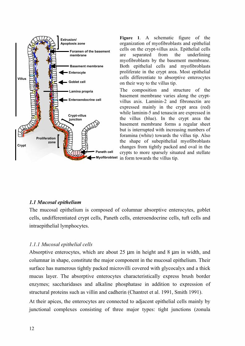
The epithelium is underlined with basement membrane with adjacent fibroblasts. Beneath this borderline is the lamina propria, composed of loose connective tissue with various immunocompetent cells including dendritic cells, macrophages and lymphocytes. All these together with the mucosal epithelial cells form a functional unit (Figure 1).
12
Figure 1. A schematic figure of the organization of myofibroblasts and epithelial cells on the crypt-villus axis. Epithelial cells are separated from the underlining myofibroblasts by the basement membrane. Both epithelial cells and myofibroblasts proliferate in the crypt area. Most epithelial cells differentiate to absorptive enterocytes on their way to the villus tip. The composition and structure of the basement membrane varies along the crypt-villus axis. Laminin-2 and fibronectin are expressed mainly in the crypt area (red) while laminin-5 and tenascin are expressed in the villus (blue). In the crypt area the basement membrane forms a regular sheet but is interrupted with increasing numbers of foramina (white) towards the villus tip. Also the shape of subepithelial myofibroblasts changes from tightly packed and oval in the crypts to more sparsely situated and stellate in form towards the villus tip.
1.1 Mucosal epithelium The mucosal epithelium is composed of columnar absorptive enterocytes, goblet cells, undifferentiated crypt cells, Paneth cells, enteroendocrine cells, tuft cells and intraepithelial lymphocytes.
1.1.1 Mucosal epithelial cells Absorptive enterocytes, which are about 25 µm in height and 8 µm in width, and columnar in shape, constitute the major component in the mucosal epithelium. Their surface has numerous tightly packed microvilli covered with glycocalyx and a thick mucus layer. The absorptive enterocytes characteristically express brush border enzymes; saccharidases and alkaline phosphatase in addition to expression of structural proteins such as villin and cadherin (Chantret et al. 1991, Smith 1991).
At their apices, the enterocytes are connected to adjacent epithelial cells mainly by junctional complexes consisting of three major types: tight junctions (zonula
Proliferationzone
Crypt-villus junction
Extrusion/ Apoptosis zone
Enterocyte
Goblet cell
Enteroendocrine cell
Lamina propria
Basement membrane
Myofibroblast
Villus
Crypt
Paneth cell
Foramen of the basement membrane
13
occlulens), adhesion junctions (zonula adherens), and desmosomes (macula adherens). The tight junction, which completely encircles the apical end of absorptive cells as a belt-like band, plays a role in separating the external and internal environments and functions as a selective barrier. It also divides the cell membrane into two different poles, the apical site situated above the junctional complex and the basolateral site beneath it. The molecules essential to the formation of the intercellular adhesion junctions (zonula adherens) in all solid tissues belong to the cadherin superfamily. E-cadherin is the predominant intercellular adhesion molecule expressed in intestinal epithelial cells (Takeichi 1991). At the cell surface E-cadherin-formed dimers interdigitate with other E-cadherin molecules on adjacent epithelial cells, resulting in the formation of cell adhesion ‘zippers’ at adherens junctions (Overduin et al. 1995, Shapiro et al. 1995). E-cadherin has been thought to be mainly involved in homotypic cell-cell interactions, but a role in the heterophilic interaction with αEβ7 integrin on the surface of intraepithelial lymphocytes has also been suggested (Cepek et al. 1994).
Goblet cells, present in both the small and large intestine, increase in number from the proximal to the distal portion of the intestine and are located on the villi and in the crypts. The main function of these cells is to produce and secrete mucus into the intestinal lumen.
Enteroendocrine cells are distributed throughout the gastrointestinal tract. Their main function is to release hormones into capillaries in the connective tissue in response to changes in the external environment.
Tuft cells, also called caveolated cells or fibrovesicular cells, have many caveolae or pits between the bases of microvilli which extend down to the level of the nucleus. The function of these pits is not known, but they are suspected to act as chemical sensors for the lumenal milieu.
1.1.2 Intraepithelial lymphocytes One of the functions of the intestinal mucosa is to defend the body against infectious pathogens; an immunological barrier is therefore created and maintained by the immune defense system. Part of this effector limb of the gut-associated lymphoid system comprises the intraepithelial lymphocytes (Cerf-Bensussan et al. 1997). These cells are located within the epithelial cell layer above the basement membrane, being separated from adjacent enterocytes by a 10 to 20 nm space. In
14
the normal adult jejunum the average number of intraepithelial T-cells/100 absorptive cells is 23 (95% CI 17-29) (Kaukinen et al.1998).
Like T cells in general, also intraepithelial lymphocytes can be divided into two classes based on the type of T-cell receptor they express. In the intestine αβ T cells prevail both in the intraepithelial compartment and in the lamina propria. From 2% to 10% of all intraepithelial T-cells are γδ positive, while less than 1% are found in the lamina propria (MacDonald 1992). Among all mucosal T-cells, irrespective of their T-cell receptor expression, 5 to 15% express CD4 (helper/inducer phenotype), the rest being positive for CD8 (cytotoxic/suppressor phenotype).
Although in general the functions of intraepithelial lymphocytes in the mucosal immune defence are unclear, possibilities include cytotoxicity, lymphokine secretion, regulation of renewal of the mucosal epithelium and tolerance.
1.2 The basement membrane
The basement membrane is a specialization of the extracellular matrix located at the epithelial-mesenchymal-stromal interface. This specialized 40-120-nm-thick extracellular structure forms a regular sheet in the crypt region and is interrupted toward the apical third of the villi, following changes in the morphology of subepithelial myofibroblasts (Toyada et al. 1997). Basement membrane molecules, like extracellular matrix molecules in general, are characterized by complex modular structures with binding domains, allowing them, particularly in the case of laminin and collagen IV, to form networks by self-assembly and to bind several other components, e.g. nidogen/entactin and decorin (Kedinger et al. 1999). The major basement membrane molecules, type IV collagen and laminin, belong to two distinct protein families. At present 16 different collagens and 11 laminins are known.
All collagens are triplehelical molecules composed of three collagen chains (α1, α2, α3). The triple-stranded helical structure is stabilized with intermolecular hydrogen bonds and additionally with other molecules such as decorin. Decorin binds noncovalently to the surface of fibrillar collagen, primarily type I, but also collagens II and IV, as well as fibronectin (Sini et al. 1997). The core protein of decorin is horseshoe-shaped and its concave surface is of appropriate size to bind one collagen triple helix (Weber et al. 1996). Binding of decorin to triplehelical collagen retards the rate and degree of collagen fibrillogenesis and, as has been shown with decorin-
15
deficient mice, participates in the maintenance of the structural integrity of the cutis and tendon sheets (Danielson et al. 1997, Sini et al. 1997). Collagen fibres are also stabilized by intermolecular cross-links.
The family of laminins comprises cross- and T-shaped heterotrimeric proteins composed of three genetically different chains, designated α (long arm), β and γ (short arm chains). Laminin self-assembles through terminal domain interactions to form a second polymer network, and this polymerization is regulated by heparin (Yurchenco et al. 1990). Entactin/nidogen, a dumbbell-shaped sulfated glycoprotein, binds laminin near its centre and interacts with type IV collagen, bridging the two (Yurchenco and Schittny 1990).
1.3 Subepithelial myofibroblasts
Intestinal subepithelial myofibroblasts and the interstitial cells of Cajal are the two types of myofibroblast identified in the intestine. The subepithelial type are located at the interface between epithelium and lamina propria adjacent to the basement membrane in most mucosal tissues. In the intestine these cells exhibit ultrastructural features of both smooth muscle cells and fibroblasts (Valentich et al. 1997).
Already 30 years ago it was shown that myofibroblasts, previously called subepithelial fibroblasts, proliferate in the near vicinity of crypts. In autoradiographic studies it was confirmed that in the intestine of rabbits (Kaye et al. 1968, Pascal et al. 1968) and mice (Marsh and Trier 1974) subepithelial fibroblasts migrate in synchrony with the proliferating and migrating epithelial cells. Although they were initially thought of as a sheath of fibroblasts, more dense in the region of crypts than at the surface of the colon or in the villi of the small intestine, it is now clear that they exist as a syncytium extending throughout the lamina propria of the gut, merging with the pericytes surrounding the blood vessels which course through the tissue (Joyce et al. 1987). In addition, subepithelial myofibroblasts show differences in their morphology along the crypt-villus axis, being tightly packed and oval in shape in the crypt area and more sparsely situated with stellate morphology in the small intestinal villi (Powell et al. 1999b).
These subepithelial myofibroblasts have an important role in organogenesis, inflammation and tissue repair through the secretion of cytokines, chemokines, and growth factors, as well as extracellular matrix proteins and proteases (Powell et al. 1999a).
16
2. Mesenchymal-epithelial cell interaction in the crypt-villus axis
Intestinal morphogenesis and maintenance of the steady state between cell proliferation and differentiation results from tightly controlled epithelial-mesenchymal cell interactions. Both epithelium and mesenchyme are necessary for proper development; however, the epithelium seems to have an instructive and the mesenchyme a permissive role in epithelial cell differentiation on the crypt villus axis. Cross-talk between these two cell types is coordinated by complex signaling mechanisms, and is thought to be mediated via basement membrane molecules, cell-cell contact, cytokines and growth factors.
2.1 The role of the basement membrane in mesenchymal-epithelial cell cross-talk
The composition of basement membrane molecules varies along the crypt villus axis. The differences in the spatial expressions of the various isoforms of each family of molecules determines, through integrins, the nature of the signals transmitted from extracellular matrix to cells (Bealieu 1999, Kedinger et al. 1999).
In general, both epithelial cells and myofibroblasts participate in the deposition of the basement membrane, as was shown in early studies with interspecies chick-rodent embryonic epithelial-mesenchymal tissue recombinants (Kedinger et al. 1986). Subsequent in vitro studies have shown that perlecan, a basement membrane proteoglycan, is deposited by epithelial cells, while type IV collagen and nidogen originate mostly from mesenchymal cells (Simon-Assmann et al. 1988, 1989, 1995). The division of labor in the production of different laminin isoforms between epithelial and mesenchymal cells is more complex; some of the individual α, β and γ chains are produced by both cell types and some (α2, β3) entirely by mesenchymal cells (Simon-Assmann et al. 1998).
Laminin-2 (α2β1γ1), localized only in the crypts, has been held to play a role in epithelial cell proliferation and Paneth cell differentiation (Simon-Assmann et al. 1994). Laminin-5 (α3β3γ2), which is present only in villi with an increasing gradient towards the villus tip, plays a crucial role in epithelium integrity by acting as an extracellular anchoring filament in the formation of hemidesmosomes (Uitto and Pulkkinen 1996). In addition, in vitro laminin-5 promotes the migration of intestinal epithelial carcinoma cell lines T84 and HT-29 (Lotz et al. 1997). Several
17
lines of evidence indicate that laminin-1 (α1β1γ1), present in the villi, is involved in triggering intestinal epithelial cell differentiation (Simo et al. 1992).
Also other extracellular matrix molecules, among them fibronectin and tenascin, are associated with the intestinal basement membrane. Fibronectin and tenascin exhibit a complementary expression pattern on the crypt-villus axis: fibronectin shows a decreasing gradient from the crypt basement membrane to the villus tip, whereas the expression of tenascin increases towards the villus tip (Beaulieu 1997, Simon-Assmann and Kedinger 1993). This has been held to be related to the antiadhesive role of tenascin, which at the villus tip could assist in epithelial cell shedding (Probstmeier et al. 1990).
In addition to the changes in the biochemical composition of the basement membrane, also the structure of the reticular fibrils along the crypt-villus axis varies. Studies with scanning electron microscopy have shown that the fibrils form a dense network in the crypt area with an increasing number of foramina towards the villus tip. In the villi these foramina are surrounded by end feet of the cytoplasmic processes of fibroblasts, often combined with dendritic cells or lamina propria lymphocytes. Thus, the foramina may participate in the communication between the intestinal epithelium and other cells, as also basal extensions of epithelial cells traverse through these foramina (Toyada et al. 1997).
Signals from the extracellular matrix are transmitted through integrins to cells. Integrins are a family of heterodimeric cell surface receptors for extracellular matrix proteins composed of noncovalently linked protein chains (α and β). The extracellular amino terminal portions of integrin molecules bind to extracellular ligands, and the intracellular carboxyl terminals are in contact with cytoplasmic cytoskeletal elements. Consequently, integrin cell surface molecules have multiple functions relating to cell anchoring, shape and migration (Tarone et al. 2000). Integrins are also linked to cell proliferation, and by virtue of cellular-extracellular matrix interactions may regulate the rate of extracellular matrix production (De Arcangelis and Georges-Labouesse 2000). In addition to anchoring cells to the extracellular matrix the integrins are important in intercellular adhesion, which often occurs between homotypic cells.
18
2.2 Matrix metalloproteinases (MMPs)
Extracellular matrix turnover is a critical step in tissue remodeling and it is now known that matrix metalloproteinases are the most important group of matrix-degrading enzymes involved in matrix turnover (Nagase and Woessner 1999). Proteolysis on the cell surface and in the extracellular matrix is essential for normal cellular functions during development and in the adult, but it may also have undesirable consequences such as promoting cancer and arthritis (Blobel 2000). The activity of these enzymes is regulated at three levels: gene transcription, activation of proenzyme, and inhibition of the active enzyme by natural inhibitors, the tissue inhibitors of matrix metalloproteinases (TIMPs) (Borden and Heller 1997, Murphy et al. 1999).
An example of how a protease can change the properties of a protein is the case of laminin-5 processing by matrix metalloproteinase-2 or membrane-type-1 matrix metalloproteinase. Although intact laminin-5 supports cell adhesion, laminin-5 whicht has been cleaved by matrix metalloproteinases promotes cell migration (Koshikawa et al. 2000).
In addition to matrix breakdown, these zinc atom-dependent endopeptidases function as regulatory proteins by processing cytokines, growth factors and adhesion molecules to generate fragments with either enhanced or reduced biological activity (Nelson et al. 2000).
2.3 Growth factors
Soluble mediators, growth factors, in part mediate the instructive interaction between myofibroblasts and epithelial cells. Secreted growth factors have in general three functions: 1) they initiate or increase cell mobility, 2) they induce proliferation, i.e., they are paracrine mitogens for epithelial or parenchymal cells and possibly autocrine mitogens for themselves, or 3) they induce terminal differentiation of these cells, even driving the cells to apoptosis. Some growth
factors seem to exert all three effects (Powell et al. 1999a).
By reason of the sensitivity of cells to growth factor molecules, the regulation of their action is controlled at many levels, including gene transcription and translation, receptor activation, and intracellular signaling. An additional mechanism controlling growth factor availability is their secretion in latent or
19
sequestered forms (Rifkin 1997). The most common proteolytic activation system for growth factor is the plasminogen activator-plasmin system (Rifkin et al. 1999).
2.3.1 Trefoil peptide
The name ‘trefoil peptides’ comes from their three-dimensional molecular structure resembling the "three leaf" trefoil shape (Chinery and Coffey 1996, Plaut 1997). The only trefoil factor present in the intestine is secreted principally by the goblet (mucus-secreting) cells of the epithelium and not by intestinal myofibroblasts (Powell et al. 1999a). Targeted gene disruption of this intestinal trefoil factor gives rise to abnormal epithelial cells and increased susceptibility to various injuries, resulting in a colitis-like picture (Kindon et al. 1995, Babyatsky et al. 1996).
2.3.2 Transforming growth factor -α (TGF-α) and epidermal growth factor (EGF)
TGF-α and EGF are produced as glycosylated, membrane-anchored precursors which are cleaved into soluble, mature proteins (Carpenter and Wahl 1990) before becoming ligands for epidermal growth factor tyrosine kinase receptors (Barnard et al. 1995). The TGF-α/EGF receptor appears to be upregulated in the mucosa of the injured intestine and other organs. TGF-α is expressed in epithelial cells, myofibroblasts and monocytes/macrophages, whereas EGF seems to be produced primarily by the epithelial cells of the salivary gland and Brunner's glands of the duodenum (Powell et al. 1999a). Additionally, a novel ulcer-associated epithelial stem cell-derived cell lineage has been described which secretes immunoreactive EGF into the intestinal lumen (Wright et al. 1990, Herminston and Gordon 1995) and stimulates epithelial proliferation and promotes ulcer healing (Wright et al. 1990). Soluble TGF-α is mitogenic for a number of cell lines in vitro and for intestinal epithelial cells also in vivo (Powell et al. 1999a).
2.3.3 Fibroblast growth factors (FGFs)
Fibroblast growth factors (FGFs) constitute a large family of at least 18 structurally related polypeptides, of which FGF-1 and FGF-2 (acidic and basic) are prototype members (Mason 1994). Many FGFs contain signal peptides for secretion and are then secreted into the extracellular environment, where they can bind to the heparan-like glycosaminoglycans of the extracellular matrix. From this reservoir,
20
FGFs may act directly on target cells, or they can be released through digestion of the extracellular matrix or the activity of a carrier protein, a secreted FGF binding protein. FGFs bind specific receptor tyrosine kinases in the presence of heparan-like glycosaminoglycans and this binding induces receptor dimerization and activation, ultimately resulting in the activation of various signal transduction cascades (Powers et al. 2000). FGFs play an important role in cell growth and differentiation, neurite outgrowth, embryogenesis, angiogenesis and wound healing (Basilico and Moscatelli 1992). In the intestine during cellular restitution or wound healing FGF has antiapoptotic effects (Gulati and Peluso 1997).
2.3.4 Insulin-like growth factors (IGFs)
The insulin-like growth factor (IGF) ligand-receptor system comprises three structurally-related peptide ligands (insulin, IGF-I, and IGF-II) and two high affinity, membrane-associated tyrosine kinase receptors (Powell et al. 1999a). IGF-I and IGF-II have various metabolic, proliferative and differentiating effects through endocrine, autocrine, and paracrine mechanisms (Jones and Clemmons 1995). IGFs are only weakly mitogenic for epithelial and parenchymal cells but powerfully mitogenic for myofibroblasts (Simmons et al. 1999) and smooth muscle cells (Wang et al. 1997). IGF is present in the circulation and is also secreted in a paracrine fashion by myofibroblasts adjacent to epithelial and parenchymal cells (Lund 1994). The IGF actions are regulated by the availability of free IGF, which in turn is modulated by the level of high-affinity IGF-binding proteins (IGFBPs). At present six of these binding proteins are known (Stewart and Rotwein 1996) These IGFBPs also express some biological activities themselves (Jones and Clemmons 1995).
2.3.5 Keratinocyte growth factor (KGF)
KGF is a member of the FGF family (FGF-7) (Housley et al. 1994, Rubin et al. 1995). This factor is unique in that, unlike other members of the FGF family, it does not appear to have activity on fibroblasts, endothelial cells or other nonepithelial targets. The transmembrane tyrosine kinase receptor for KGF is expressed only in epithelial cells. This receptor is also able to bind, in addition to KGF, other fibroblast growth factors, FGF-1 and FGF-2, although with different affinities. KGF has been shown to induce proliferation and differentiation of a host of epithelial and
21
parenchymal cells, including intestinal epithelial cells, type II pneumocytes, hepatocytes and keratinocytes of the skin. The expression and secretion of KGF is regulated by interleukin-1 (Chedid et al. 1994) and significantly upregulated in the lamina propria of the inflamed intestine (Finch et al. 1996).
2.3.6 Hepatocyte growth factor (HGF)
HGF is a glycosylated heparin-binding heterodimer related to plasminogen and consists of a heavy α chain and a light β chain held together by disulfide bonds. It is synthesized and secreted by fibroblasts/myofibroblasts and it induces epithelial cell proliferation (Goke et al. 1998), migration (Nusrat et al. 1994), apoptosis and angiogenesis (Bradbury 1998). Apart from mito- and motogenic functions HGF has been shown to act as a morphogen, since it has been observed to induce tubulus formation in murine kidney epithelial cells in a three-dimensional collagen gel culture (Montesano et al. 1991). The HGF tyrosine kinase receptor, expressed prominently on epithelial cells, is encoded by the protooncogene c-met (Bottaro et al. 1991). The activity of both HGF and c-met are regulated by proteases, and they are therefore active only when the right proteases are present (Powell et al. 1999a). HGF, like KGF, is a major mediator of epithelial-mesenchymal interactions and epithelial morphogenesis (Rubin et al. 1995).
2.3.7 Transforming growth factor-β (TGF-β)
In mammals, three closely related TGF-β isoforms exist: 1, 2 and 3. Each isoform is a 25 kDa homodimer derived from a precursor protein. TGF-β is usually released in a latent form from cells, e.g. in noncovalent association with latency-associated peptide (LAP). This small latent complex can form a large latent complex via LAP, which can disulfide-bond to latent TGF-β-binding proteins (LTBPs). This large complex can in turn be covalently incorporated in a tissue transglutaminase-catalyzed reaction to the extracellular matrix (Taipale et al. 1994, Nunes et al. 1997). Moreover, LTBP-1, either by itself or as part of the latent complex, associates specifically with matrix fibrils (Dallas et al. 1995) and can associate directly with fibronectin (Taipale et al. 1996). Human LTBP-1 and the β1 and β3 LAP isoforms contain the common integrin recognition sequence RGD, suggesting that the large latent complex is one of the numerous extracellular matrix proteins recognized by integrins (Munger et al. 1998).
22
The constitutive secretion of latent TGF-β by many cell types in culture suggests that there are extracellular mechanisms controlling the activation and thereby the activity of this potent cytokine (Munger et al. 1997). One important step might be the incorporation of the latent complex into the extracellular matrix in a transglutaminase-catalyzed reaction (Kojima et al. 1993) to make the complex susceptibile to proteolytic activation (Munger et al. 1997). The released TGF-β can interact with a number of soluble or matrix molecules, including α2-macroglobulin, decorin, betaglycan, heparin and fucoidan. These neutralizing interactions are thought to prevent the diffusing of TGF-β from loci of activation to inappropriate sites.
At present three different transforming growth factor receptors have been cloned (Massague et al. 1994). The receptor types I and II are serine-threonine kinase receptors (Franzen et al. 1993), while type III (betaglycan) is a heavily glycosylated large proteoglycan lacking an intracellular signaling domain (Lopez-Casillas et al. 1993). In addition, the type III receptor is believed to function as a ligand reservoir and to facilitate the binding of TGF-β to receptors I and II. The TGF-β mediated intracellular signaling occurs via phosphorylation of SMAD2 and SMAD3 following receptor activation, whereas SMAD6 and 7 antagonize this signaling (Heldin et al. 1997).
TGF-β isoforms are potent growth inhibitors in epithelial cells, including those of the gastrointestinal tract (Barnard et al. 1989). In epithelial cells proliferation arrest often induces simultaneous signaling for the terminal differentiation pathway (Kurokowa et al. 1987). In cultured intestinal epithelial cells, downregulation of cyclin D1 expression appears to be a key step in the G1 arrest induced by TGF-β (Ko et al. 1995). TGF-β has the opposite effect on mesenchymal cells; it stimulates fibroblast proliferation, possibly via increasing FGF-receptor expression at the cell surface (Thannickal et al. 1998).
In the intestine mRNA for TGF-β is expressed in crypt epithelial cells (Barnard et al. 1993), while the protein is present in villus epithelial cells (Lionnetti et al. 1999). LTBP is present in the basement membrane area along the crypt-villus axis (Mizoi et al. 1993)
Current knowledge supports the conception that TGF-β is the major autocrine and/or paracrine inhibitory regulator in the intestinal epithelium, as disruption of
23
TGF-β signaling allows cells to escape from normal growth constraints and leads to transformation (Markowitz and Roberts 1996).
TGF-β also regulates extracellular matrix turnover; it increases the synthesis of extracellular matrix proteins and proteinase inhibitors, reduces synthesis of matrix degrading activities, and modulates matrix receptors and binding proteins (Grande 1997). In addition to induction of extracellular matrix formation TGF-β has an effect on the immune system, and evinces proinflammatory and immunosuppressive activities (Litterio and Roberts 1997). The important immunomodulatory role of TGF-β was clearly demonstrated in TGF-β knock-out mice, which showed multifocal inflammatory disease with prominent inflammation in the intestine (Shull et al. 1992).
3. Mucosal IgA antibodies
The main function of antibodies present in mucosal secretion, particularly IgA, is to combine with microorganisms to reduce their motility and adhesive properties within the mucosal lumen and its surface. In addition, by forming immune complexes, mucosal antibodies prevent the absorption of food antigens from the large area of mucosal surfaces. The antibodies which predominate in human intestinal secretions are IgA class and are called secretory IgA (sIgA).
In the intestine M cells, the dome of Peyer’s patches, are responsible for luminal antigen sampling and transport to the underlining lymphoid tissue. At the mucosal surface the basolateral site of the M cell forms an intraepithelial pocket which contains B and T lymphocytes and occasionally macrophages. M cells may send cytoplasmic extensions to subepithelial tissues, where a network of dendritic cells, lymphocytes and macrophages is present (Neutra et al. 1987). For antigen-specific antibody formation to occur, processed antigen fragments are presented by antigen-presenting cells to helper T-cells, which in turn stimulate antibody production in B cells (Clark and Ledbetter 1994).
In addition to the aforementioned conventional antibody production a T-cell independent mechanism for IgA antibody production seems to operate at the mucosal surfaces, especially in the intestine (Macpherson et al. 2000). These polyreactive sIgA antibodies are present in humans, where they react with both self- and non-self antigens (Quan et al. 1997, Clemente et al. 2000). Furthermore, they
24
are locally produced and aimed to clear the lamina propria and epithelial cells of local pathogens and altered or denatured self-antigens (Kaetzel et al. 1991, Bouvet and Dighiero 1998). In mice these ‘natural’ antibodies are mainly produced at mucosal sites by T-cell independent B-1 cells (Macpherson et al. 2000).
4. Coeliac disease
Coeliac disease is a gluten-induced genetically linked disorder of the small intestine characterized morphologically by severe crypt hyperplasia with flattened villi, affecting 1:300 or even 1:100 persons among European populations (Mäki and Collin 1997). The loss of absorptive surface accompanying the flattening of the mucosa leads to the main clinical features, diarrhoea and malabsorption (Dicke 1950). The diagnostic criteria for coeliac disease were defined by the European Society for Paediatric Gastroenterology and Nutrition in 1969 and revised in 1990 (Walker-Smith et al. 1990). The revised ESPGHAN criteria include the finding of morphologically abnormal small-intestinal mucosa in a biopsy specimen, and clinical or histological remission on a gluten-free diet. However, in patients with silent coeliac disease a second biopsy specimen is always recommended to prove mucosal recovery on gluten-free diet.
Coeliac disease is triggered by ingested gluten, a single major environmental factor, when eating daily food made of wheat, rye or barley. The disease also has a particularly narrow highly specific HLA class-II association, DR3-DQ2 and to a lesser extent DR4-DQ8 (Sollid and Thorsby 1993); however, despite numerous studies no uniform susceptibility genes to coeliac disease have been identified. The same HLA types are also typical for many autoimmune diseases. In fact, common comorbidities include insulin-dependent diabetes mellitus, Sjögren’s syndrome and autoimmune thyroiditis (Collin et al. 1994). Furthermore, the prevalence of autoimmune disorders in coeliac disease is suggested to be related to the duration of exposure to gluten (Ventura et al. 1999). Untreated coeliac disease patients also run an increased risk of malignancies, especially for small-bowel lymphoma (Harris et al. 1967, Holmes et al. 1976, Swinson et al. 1983). Extraintestinal gluten-triggered manifestations are dermatitis herpetiformis, permanent-tooth enamel defects, osteopenia, infertility, liver involvement, ataxia and epilepsy with cerebral calcifications (Collin and Mäki 1994, Fasano and Catassi 2001). At present the only known medical treatment for coeliac disease is a strict life-long gluten-free diet.
25
4.1 Small-bowel mucosal morphology in coeliac disease
Three major dynamically interrelated patterns of mucosal change are present across the gluten sensitivity spectrum, which may be termed infiltrative (Type 1), hyperplastic (Type 2) and destructive (Type 3) lesions (Marsh 1992). The type 1 infiltrative lesion comprises normal mucosal architecture in which the villous epithelium carries a variable infiltrate of small, non-mitotic lymphocytes. The hyperplastic type 2 lesion is virtually identical to the infiltrative, but with the addition of hypertrophic crypts, whose epithelium may also be infiltrated by lymphocytes. These two types are common among dermatitis herpetiformis patients and in treated coeliac disease patients after short-term gluten challenge.
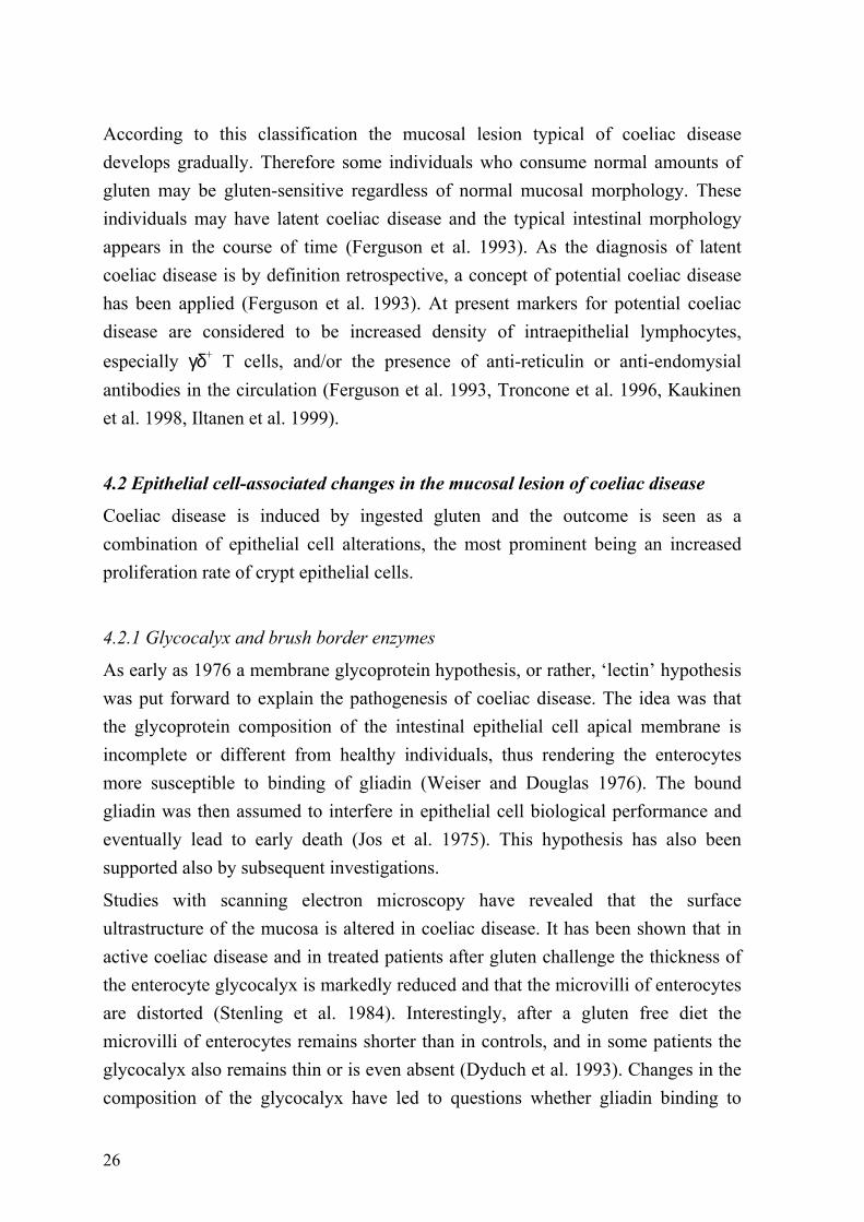
The destructive type 3 lesion is characterized by the established ‘flat’ mucosa of classic coeliac disease. Typical of type 3 is a fully developed cell-mediated mucosal immune response with hypertrophic crypts, lamina propria swelling and a flattened surface epithelium. However, similar morphological changes are also seen in tropical sprue, giardiasis, food sensitivity and immunoproliferative disease of the small intestine. In addition to these types 1-3 lesions classification also includes types 0 and 4, which represent the extremes. The pre-infiltrative type 0 lesion evinces no morphological changes, but anti-gliadin IgA or IgM antibodies may be present in intestinal secretions in the early state of developing disease (Marsh 1992). The hypoplastic/atrophic type 4 lesion represents the worst possible outcome of coeliac disease, showing patchy or diffuse jejunoileitis, intestinal ulceration or coeliac disease-associated lymphoma. These gradually appearing changes in the coeliac disease affected small-intestinal mucosa are presented, according to the Marsh-classification, in Figure 2.
Figure 2. The Marsh classification of developing lesion in coeliac disease.
Type 0 Type 1 Type 2 Type 3 Type 4
26
According to this classification the mucosal lesion typical of coeliac disease develops gradually. Therefore some individuals who consume normal amounts of gluten may be gluten-sensitive regardless of normal mucosal morphology. These individuals may have latent coeliac disease and the typical intestinal morphology appears in the course of time (Ferguson et al. 1993). As the diagnosis of latent coeliac disease is by definition retrospective, a concept of potential coeliac disease has been applied (Ferguson et al. 1993). At present markers for potential coeliac disease are considered to be increased density of intraepithelial lymphocytes, especially γδ+ T cells, and/or the presence of anti-reticulin or anti-endomysial antibodies in the circulation (Ferguson et al. 1993, Troncone et al. 1996, Kaukinen et al. 1998, Iltanen et al. 1999).
4.2 Epithelial cell-associated changes in the mucosal lesion of coeliac disease
Coeliac disease is induced by ingested gluten and the outcome is seen as a combination of epithelial cell alterations, the most prominent being an increased proliferation rate of crypt epithelial cells.
4.2.1 Glycocalyx and brush border enzymes
As early as 1976 a membrane glycoprotein hypothesis, or rather, ‘lectin’ hypothesis was put forward to explain the pathogenesis of coeliac disease. The idea was that the glycoprotein composition of the intestinal epithelial cell apical membrane is incomplete or different from healthy individuals, thus rendering the enterocytes more susceptible to binding of gliadin (Weiser and Douglas 1976). The bound gliadin was then assumed to interfere in epithelial cell biological performance and eventually lead to early death (Jos et al. 1975). This hypothesis has also been supported also by subsequent investigations.
Studies with scanning electron microscopy have revealed that the surface ultrastructure of the mucosa is altered in coeliac disease. It has been shown that in active coeliac disease and in treated patients after gluten challenge the thickness of the enterocyte glycocalyx is markedly reduced and that the microvilli of enterocytes are distorted (Stenling et al. 1984). Interestingly, after a gluten free diet the microvilli of enterocytes remains shorter than in controls, and in some patients the glycocalyx also remains thin or is even absent (Dyduch et al. 1993). Changes in the composition of the glycocalyx have led to questions whether gliadin binding to
27
intestinal epithelial cells is a potential key event in the pathogenesis of coeliac disease. In fact, interaction of different synthetic gliadin peptides with isolated brush border membranes from treated coeliac patients has evinced increased gliadin binding capacity when compared to controls (Bolte et al. 1999). However, these peptides were not the ones confined to coeliac-active peptides. Nonetheless parallel results have been obtained when the uptake of a pepsin trypsin digest of gliadin by epithelial cells has been followed; the uptake is increased in coeliac patients (Friis et al. 1992).
Typically in the coeliac disease-affected mucosa the expression of brush border enzymes, including disaccharidases and alkaline phosphatase, is also decreased. In addition, the expression of integrin α2 in epithelial cells is altered in coeliac lesion; it is also expressed in the villus when present (Patey et al. 1997). These changes presumably reflect changes in the degree of epithelial cell differentiation.
4.2.2 Proliferation and apoptosis
The most prominent feature of coeliac disease mucosal lesion is crypt cell hyperplasia. The number of mitotic figures is increased in the crypts and there is a greatly increased epithelial cell loss into the intestinal lumen (Trier and Browning 1970). More recent studies using a confocal microscope have confirmed previous finding showing on average a 2.8 times higher proliferation rate in coeliac disease lesion than in normal mucosa (Savidge et al. 1995). However, crypt hyperplasia seems to be a common tissue response to mucosal damage in food allergy and infection, although in coeliac disease disease-specific mechanisms are evident (Savidge et al. 1996).
The increased enterocyte apoptosis in the coeliac lesion is also well established and seems to balance the overproduction of epithelial cells almost to normal level; the rate of apoptosis is increased on average 2.3 times vs. normal mucosa (Moss et al. 1996). Recently it has been demonstrated that Fas-Fas ligand-mediated apoptosis is a major mechanism responsible for enterocyte death in coeliac disease (Ciccocioppo et al. 2001). Additionally, it has been shown in an organ culture model that gliadin induces this epithelial FAS engagement resulting in cell death (Maiuri et al. 2001a).
28
4.2.3 Changes in epithelial cell permeability
The permeability hypothesis, which suggests that in coeliac patients there is a primary defect in mucosal permeability, was launched in the 1980s (Bjarnason and Peters 1983, 1984), and is still under consideration.
In normal physiological circumstances the intestinal epithelium serves as the main barrier to macromolecules passing in the intestinal lumen. This barrier function is maintained by well-formed intercellular junctions such as tight junctions, adhesion junctions and desmosomes. In untreated coeliac disease, the epithelial barrier of the jejunum has been shown to be disturbed (Schulzke et al. 1995) by structural modifications of the tight junctions resulting in increased ionic permeability (Schulzke et al. 1998). The up-regulation of zonulin, a recently described intestinal peptide involved in tight junction regulation, seem to be responsible, at least in part, for the increased gut permeability characteristic of the early phase of coeliac disease (Fasano et al. 2000).
E-cadherin is the most common intercellular adhesion molecule in the small intestine typically used to anchor homotypic cells together. E-cadherin expression is significantly decreased, both at the mRNA and protein level, in the small intestinal epithelial cells of untreated coeliac disease patients (Perry et al. 1999). In addition to E-cadherin expression of the intracellular component of adherens junctions, catenin, is likewise decreased. These changes are reversible, as after a gluten-free diet the expression patterns appear to be normal (Perry et al. 1999). This is also supported by the previous finding of increased c-myc oncogene expression in active coeliac disease (Ciclitira et al. 1987), as c-myc negatively regulates E-cadherin expression on the cell surface (Pennisi 1998). E-cadherin-mediated changes in the integrity of the epithelial cell layer has been thought to underlie some of the changes typical for coeliac disease, such as increased mucosal permeability and epithelial cell proliferation.
4.3 T-cell-mediated changes in coeliac disease
The majority of studies carried out on coeliac disease pathogenesis are based on the immunological hypothesis, which rests basically upon the conception of a gluten-induced increase in the activation of lymphocytes, mostly T-cells, and the prominent HLA-DQ association in coeliac disease (Sollid 2000).
29
4.3.1 Intraepithelial lymphocytes
The density of αβ+ intraepithelial lymphocytes is markedly increased in untreated coeliac disease patients and decreases to normal during a gluten-free diet (Kutlu et al. 1993). Their suggested cytotoxic function has recently been partly evidenced, as it has been shown that the frequency of CD94+ intraepithelial lymphocytes (mostly αβ T cells) in the active phase of coeliac disease is conspicuously increased over controls; further, that IL-15, an interleukin secreted from epithelial cells, is able to induce the upregulation of CD94 in intraepithelial lymphocytes in vitro (Jabri et al. 2000). These findings suggest a scenario whereby after T cell receptor -mediated activation and exposure to the cytokines of the epithelial microenvironment, including IL-15, coeliac disease intraepithelial lymphocytes not only up-regulate CD94 expression, but also enhance their cytotoxic properties through the induction of perforin and granzyme B (Oberhuber et al. 1996, Jabri et al. 2000).
The importance of IL-15 in gliadin-triggered intestinal lesion formation has also been put forward in experiments using organ culture of treated/untreated coeliac disease biopsy specimens. In these studies gliadin was shown to induce epithelial cell proliferation marker Ki67, transferrin receptor and FAS expression as well as apoptosis in an IL-15-dependent manner (Maiuri et al. 2000). In addition, it was recently shown in this model that gliadin induces intraepithelial migration of CD94+ cells in an IL-15-dependent manner in coeliacs, but not in controls (Maiuri et al. 2001b).
The function of intraepithelial γδ-cells is not known (Born et al. 2000), but they are frequently found in untreated coeliac disease (Holm et al. 1992). They would seem nonetheless not to be a specific marker for the disease but rather for a hyperproliferative state (Spencer et al. 1991, Patey et al. 1997, Iltanen et al. 1999). It was previously thought that γδ-cells are involved in the pathogenesis of coeliac disease (Hayday et al. 1997) as activated γδ-cells produce KGF (Boismenu and Havran 1994), which is known to stimulate epithelial cell proliferation (Finch et al. 1996). However, the γδ-cells in the coeliac lesion do not produce KGF (Salvati et al. 2001) and therefore possibly contribute through other as yet unknown mechanisms in the formation of the mucosal lesion.
30
4.3.2 Activated lamina propria T-cells
Gluten induces infiltration and a non-proliferative activation of CD4+ lamina propria T cells (Halstensen et al. 1990, Halstensen and Brandtzaeg 1993). These activated T-cells produce increased amounts of interferon-γ and slightly increased amounts of IL-2, IL-4, IL-6 and TNF-α (Nielsen et al. 1998). The increased IFN-γ may cause the typical changes seen in enterocytes, or conceivably activation of macrophages, which in turn produce cytokines such as IL-10, IL-12, and TNF-α which induce the pathological changes in the intestine (Nielsen et al. 1998).
4.3.3 T-cell-induced matrix metalloproteinase activation and tissue destruction
In studies using an ex vivo foetal intestinal explant system it has been shown that mucosal destruction with villous atrophy and crypt hyperplasia is a consequence of T-cell activation (Pender et al. 1996, 1998) and further that especially the TNF-α produced by activated T-cells stimulates the production of MMPs (interstitial collagenase, gelatinase A and stromelysin-1) in intestinal myofibroblasts (Pender et al. 1997).
This kind of T-cell-mediated increase in the activation of MMPs has been held to cause the destruction of the villous structure also in coeliac disease (Schuppan 2000). Such a conception is supported by existing data showing an increased expression of MMP-1 and MMP-3 mRNAs in biopsy specimens obtained from coeliac disease patients (Daum et al. 1999). In addition, enhanced expression of the type 1 tissue inhibitor of matrix metalloproteinase (TIMP-1) mRNA level in coeliac lesion has been reported (Daum et al. 1999). Interestingly, TIMP-1 levels remained increased after a glutein-free diet whereas the mRNA levels of MMP-1 and MMP-3 decreased to the level seen in the intestinal mucosa of healthy individuals’ (Daum et al. 1999).
Elevated expression levels of MMP-1 and –3 also occur in dermatitis herpetiformis (Airola et al. 1995), a skin manifestation of mainly subclinical coeliac disease, after upregulation of urokinase plasminogen activator (Airola et al. 1997). In addition, it has recently been shown that MMP-12 mRNA levels are also enhanced in both the skin and intestinal biopsy samples of dermatitis herpetiformis (Salmela et al. 2001).
31
4.4 Antibodies in coeliac disease
4.4.1 Serum antibodies
One typical feature in coeliac disease patients, in addition to the characteristic changes in the mucosal architecture, is the appearance of certain disease-specific antibodies in the patient’s circulation after gluten ingestion.
Determination of gliadin antibodies from patient serum has been used for diagnostic and screening purposes. IgA-class antibodies against gliadin, were formerly considered specific for coeliac disease, especially in children (Savilahti et al. 1983). However, these antibodies are not disease-specific, as they are also found in patients with other gastrointestinal diseases (Scott et al. 1990) as well as in healthy individuals (Pettersson et al. 1993).
Serum reticulin antibody tests have been in use since 1971 (Seah et al. 1971, Mäki 1984). Reticulin antibodies are targeted against connective tissue fibres in rat liver, kidneys and stomach, where they follow the positive silver staining pattern in tissues when detected with immunofluorescence staining in cryostat tissue sections (Seah et al. 1971). The IgA-class antibody test has been shown to be highly specific for coeliac disease (Mäki 1995), but is also considered to be markedly laboratory-dependent (Mäki 2001). The reticulin antibody test has therefore often been replaced by the endomysial antibody test, also found to be highly specific and sensitive for coeliac disease (Chorzelski et al. 1983, Chorzelski et al. 1984, Stern et al. 2000). The original substratum, monkey oesophagus, used in endomysial antibody detection has in many laboratories since been replaced by human umbilical cord (Ladinser et al. 1994, Volta et al. 1995, Carroccio et al. 1996, Sulkanen et al. 1998a, b).
4.4.2 Mucosal antibodies
IgA-class antibodies have also been found deposited in the intestinal basement membrane area and on subepithelial fibroblasts in the mucosa of untreated coeliac disease patients’ as well as in cases where the mucosal architecture has been more or less normal (Karpati et al. 1988, Mäki et al. 1995b). It was in fact recently shown that coeliac disease-specific IgA-class tissue transglutaminase antibodies are produced solely in the intestinal mucosa (Marzari et al. 2001), where they find their way to the circulation, occasionally even prior to the formation of the mucosal
32
lesion and before γδ T-cells are driven to the intraepithelial compartment in morphologically normal mucosa (Mäki et al. 1995b, Iltanen et al. 1999). These IgA-class autoantibodies have hitherto been regarded as bystanders in the pathogenesis of coeliac disease, although their diagnostic value has been widely acknowledged (Braegger and MacDonald 1996).
4.5 The coeliac disease extracellular matrix autoantigen, tissue transglutaminase
The knowledge that disease-specific antibodies exist and that in humans the antigen is present in the extracellular matrix of most tissues (Seah et al. 1971, Hällström 1989, Karpati et al. 1991) stimulated an intensive search for the coeliac disease autoantigen.
Early studies demonstrated that IgA antibodies in sera from patients recognize a common antigen in an amorphous component associated with collagen fibres (Pras and Glynn 1973, Karpati et al. 1992). Subsequently it was shown that coeliac-disease-specific autoantibodies detect an extracellular matrix non-collagenous protein purified from human foetal lung tissue (Mäki et al. 1991) and further, that this antigen was produced by fibroblasts (Marttinen and Mäki 1993). This notwithstanding, this protein remained unidentified for years.
In 1997 tissue transglutaminase was immunoprecipitated with coeliac disease IgA from human fibrosarcoma HT 1080 cell lysates (Dieterich et al. 1997). This antigen reacted with all 25 coeliac disease serum samples tested but with none of the 25 control samples. The finding that this new autoantigen also almost completely abolished the endomysial staining pattern suggested that tissue transglutaminase could be the main autoantigen in coeliac disease. However, in order for a test to be useful in clinical practice it also needs to be tested with an extensive clinical material.
Tissue transglutaminase is widely distributed in human organs and belongs to a family of calcium-dependent protein cross-linking enzymes which polymerize proteins into high-molecular-weight aggregates via intramolecular ε(γ-glutamyl)lysine bonds. These enzymes are apparently involved in very disparate biological processes. Tissue transglutaminase (transglutaminase type II; EC 2.3.2.13), unlike the other transglutaminases, is a GTP-GDP binding enzyme (Bergamini and Signorini 1993).
33
An increasing body of evidence indicates that tissue transglutaminase has an important role at the cell surface and in the extracellular matrix, where the cross-linking of extracellular matrix proteins such as fibronectin, collagens and numerous other extracellular substrates is believed to be important in their deposition and stabilization (Aeschlimann and Thomazy 2000).
4.6 The proposed role of tissue transglutaminase in coeliac disease pathogenesis
The prevailing hypothesis is that tissue transglutaminase contributes to the development of the coeliac disease mucosal lesion by deamidating specific glutamine residues in gliadin. The reaction is believed to create an epitope which binds efficiently to both DQ2 and DQ8 and is recognized by gut-derived T-cells, thus enhancing the T-cell response to gliadin (Molberg et al. 1998, Van de Wal et al. 1998). Activated T-cells then, via tumour necrosis factor alpha, stimulate myofibroblasts to secrete matrix metalloproteinases, which ultimately cause mucosal destruction (Pender et al. 1997). In addition, new epitopes generated in transglutaminase-catalyzed reactions, e.g. gliadin-gliadin or gliadin-tissue transglutaminase complexes, have been held to stimulate T-cell directed autoantibody production by tissue transglutaminase-specific B-cells (Sollid 2000, Schuppan 2000).
Tissue transglutaminase is an enzyme, which has bioactivity both within and outside cells. It is known to have an important role in the regulation of different cellular processes in both compartments (Aeschlimann and Thomazy 2000). Although tissue transglutaminase seems to be the major coeliac disease autoantigen, the role of coeliac disease-specific autoantibodies targeted against tissue transglutaminase, in the pathogenesis of the disease has not been proposed.
34
PURPOSE OF THE PRESENT STUDY
The aims of the present study were:
1. to develop an in vitro model for the epithelial- mesenchymal cell interaction occurring on the crypt-villus axis.
2. to verify the sensitivity and specificity of transglutaminase autoantibodies in coeliac disease.
3. to characterize the possible effects of coeliac disease-specific autoantibodies in epithelial cell behaviour using the established three-dimensional epithelial cell-fibroblast co-culture model.
4. to characterize the possible effects of coeliac disease-specific autoantibodies on fibroblast biology.
35
MATERIALS AND METHODS
1. Cell lines (I, III, IV) The human intestinal epithelial cell line T84 (CCL 248) and the human embryonic lung fibroblast cell line IMR-90 (CCL 186) were purchased from the American Type Culture Collection (ATCC, Rockville, MD). The human intestinal epithelial cell line HT-29 was a generous kind gift from Dr. T.T. McDonald (The Medical College of St.Bartholomew´s Hospital, London, UK) and the primary human foreskin fibroblasts (FS) from the Department of Microbiology (Tampere University Hospital, Tampere, Finland). The passages used for T84 cells were 60-70, HT-29 160-170, IMR-90 12-19, and for FS 17-24. T84 epithelial cells were cultured in DMEM/F12 (1:1) medium, HT-29 epithelial cells in RPMI-1640; all fibroblasts were cultured in basal medium (Eagle), supplemented with 10% foetal bovine serum, and 2 mM glutamine (all from Gibco BRL, Paisley, Scotland).
2. Growth factors and antibodies (I, II, III, IV) Recombinant human growth factors, hepatocyte growth factor (HGF) and transforming growth factor β1 (TGF-β1) were purchased from R&D Systems (Oxon, UK). Goat anti-human HGF neutralizing antibodies, goat anti-human LAP (TGF-β1 latency-associated peptide) detection antibody, as well as rabbit pan-specific TGF-β neutralizing antibodies were likewise obtained from R&D Systems. Rabbit anti-human IgG antibodies (D 336) and negative control antibodies, Ig-subtype specific monoclonal mouse anti-rabbit immunoglobulins, were from Dako A/S (Copenhagen, Denmark). Mouse monoclonal antibodies against tissue transglutaminase type II, CUB7402 and TG-100 were purchased from NeoMarkers (Fremont, CA). Rabbit antiserum to laminin (Cat. No. 680-3019) was from Gibco BRL and rabbit antibodies against c-met (h-met, C-28) were obtained from Santa Cruz Biotechnology Inc. (Santa Cruz, CA). Mouse antibodies against human sucrase (HBB2/614/88) were a generous gift from Dr. H.P. Hauri (Biocenter of the University of Basle, Switzerland). Alkaline phosphatase-, peroxidase- and biotin-conjugated rabbit anti-human IgA antibodies, alkaline phosphatase-conjugated swine antibodies against rabbit IgG, and tetra-methyl-rhodamine isothiocyanate isomer R (TRITC)-conjugated rabbit anti-mouse IgG were purchased from Dako A/S. Fluorescein isothiocyanate (FITC)-conjugated goat anti-human IgA were obtained from Kallestadt Diagnostics (Chaska, MN). When the biotin-avidin system was used, the secondary antibodies and additional reagents were from ABC-Elite or the Vectastain Elite Kit obtained from Vector Laboratories (Burlingame, CA).
3. IgA purification Total serum IgA fractions from untreated coeliac disease patients with high endomysial antibody titres (serum dilution ≥ 1:1000) and from healthy individuals or disease control sera were purified using immobilized jacalin (Pierce Europe B.V., Oud Beijerland, Holland) or CNBr-activated Sepharose 4B (Pharmacia Upjohn, Uppsala, Sweden) coupled with rabbit anti-human IgA antibodies (a generous gift from Pharmacia Upjohn).
36
Purification with jacalin was performed according to the manufacturer’s instructions. Melibiose from the elution buffer was removed by dialysing the samples overnight against a 100X volume of HANKS balanced salt solution (Gibco BRL). In the case of affinity purified IgA, the glycine from the elution buffer was removed with PD-10 columns (Pharmacia Upjohn). The IgA concentrations from all samples were determined by enzyme-linked immunosorbent assay (ELISA) using human affinity purified IgA (Dako A/S) as standard and the IgA concentration of all total IgA fractions was adjusted to 100 µg/ml.
4. Recombinant IgA antibodies (IV) The cloned human antibodies were isolated from phage display antibody libraries as previously described (Marzari et al. 2001). In short: The complementary DNAs coding for variable regions of IgA obtained from coeliac disease patient intestinal lymphocytes were amplified using polymerase chain reaction and cloned into a phagemid vector. Phages expressing anti-tissue transglutaminase antibodies fused to a phage coat protein were isolated by recursive cycles of binding, elution and amplification on purified cloned human tissue transglutaminase. Antibody production in HB2151 E. coli cells transfected with the selected phages was induced with isopropyl-β-D-thiogalactopyranoside. The secreted antibodies were then collected from the supernatant and purified by affinity chromatography using Ni-NTA resin. The purified clones used were 2.18, 3.7 and 4.1. The clone G3, specific for gliadin served as control. These antibodies were added to cultures at 2 µl/25000 cells.
5. Collagen gel (I, III, IV) Type I native collagen was prepared by modification of an established procedure (Montesano et al. 1991) by dissolving type I collagen from dissected adult rat tail tendons in acetic acid. The collagen solution was cleared by centrifugation at 10 000 g and the concentration adjusted to 1.6 mg/ml or 2.0 mg/ml. The solution thus prepared was stored at +4 °C until use.
6. Three-dimensional cell cultures (I, III) Trypsinized T84 and HT-29 cells were suspended in ice-cold type I collagen solution (8 vol.) supplemented with 10 x concentrated RPMI-1640 (Gibco BRL) (1 vol.) and 7.5% NaHCO3 (1 vol.). The cell suspension was layered onto Nunclon 24-well plates (Nunc, Roskilde, Denmark) and allowed to gel. The fibroblasts were grown on the top as a monolayer separated from the epithelial cells by a cell-free collagen layer. In certain experiments the fibroblasts were replaced by fibroblast-conditioned medium, which was obtained from confluent fibroblast cultures, cleared by centrifugation and stored at - 20 °C until use. All cultures were incubated at 37 °C in a humidified 5% CO2 atmosphere, and cultured for 5-17 days.
37
7. Proliferation assay (I, III) T84 cells were cultured on porous polycarbonate membrane cell culture inserts (Corning Costar, Cambridge, MA) with (I) or without (III) fibroblasts. Different concentrations of growth factors or antibodies were added to the culture medium, when used. After three or four days in culture the proliferation rate of T84 cells was measured using CellTiter96TM AQueous Non-Radioactive Cell Proliferation Assay (Promega Corp., Madison, WI) according to the manufacturer’s instructions. The optical density was measured spectrophotometrically at 490 nm.
8. Adhesion (IV) The effects of antibodies agaist tissue transglutaminase on IMR-90 adhesion on type I collagen were studied using a previously described adhesion assay (Messent et al. 1998). In short: 96-well plates were coated with rat tail tendon-derived type I collagen (1.6 mg/ml) for 1 hour. IMR-90 cells in culture medium supplemented with bovine serum albumin (2 mg/ml) (Sigma Chemical Company, St.Louis, MO) were added to the collagen and allowed to attach for 30 or 60 minutes. The number of attached cells were determined using CellTiter96TM AQueous (Promega Corp.) according to the manufacturer’s instructions.
9. Cell migration (IV) To study the effects of antibodies against tissue transglutaminase on fibroblast migration, IMR-90 fibroblasts were plated on type I collagen-coated 24-well plates (Nunc). When the cultures had reached confluence they were wounded and the antibodies under study added to the culture medium. In addition, to distinguish migration from proliferation the cultures were treated with Mitomycin C (0.02 mg/ml, Sigma). After a 24-hour culture period the migration of IMR-90 cells was assessed under inverted microscope by numbering the cell nuclei observed across the wounded borders.
10. Collagen gel contraction (IV) To study the effects of anti-tissue transglutaminase antibodies on fibroblast-populated collagen gel contraction, detached IMR-90 cells were gently mixed into the neutralized type I collagen solution (1.6 mg/ml) before being transferred to 24-well plates. The formation of gels was initiated by incubating the plates at 37 °C for 30 min, whereafter the antibodies were added to the culture medium. The fibroblast-populated collagen gel contraction was allowed to proceed for three days, after which the cultures were stained with Coomassie blue and photographed and the surface areas of the gels measured from the prints.
11. Collagen degradation assay (IV) The capacity of the cells to degrade type I collagen was assessed as previously described (Holmbeck et al. 1999). In short, 24-well plates (Nunc) were coated with 200 µl of type I collagen (2 mg/ml). A pellet of IMR-90 cells in the growth medium was seeded in the centre of each well. The cells were allowed to attach for 2 hours and then incubated for 3 days with or without the presence of antibodies. Matrix metalloproteinase (MMP) activity was stimulated with 10-9 M interleukin 1β (IL-1β) (R&D Systems). After the culture
38
period the cells were removed and the residual collagen fibril films were stained with Coomassie blue. The unstained, collagen-free areas were measured from digital images using analySIS 3.0 software (Soft Imaging System GmbH, Münster, Germany). To determine collagen degradation by measuring protein concentration, the residual collagen film was solubilized with 10 M HCl, alkalized and again neutralized prior to protein determination with DC protein assay (Bio-Rad, Hercules, CA).
12. SDS-PAGE, immunoblotting (II, III) and zymography (IV) All proteins subjected to sodium dodecyl sulphate-polyacrylamide gel electrophoresis in 8-12 % polyacrylamide gel (SDS-PAGE) (Bio-Rad) were added to either non-reducing or reducing sample buffer. Kaleidoscope-prestained standards (cat.no.161-0324, Bio-Rad) were used as molecular weight standards. For immunoblotting the electrophoresed proteins were then transferred to Hybond-C extra nitrocellulose membranes (Amersham International plc., Buckinghamshire, England). The target antigens were detected in the nitrocellulose membranes with specific primary antibodies, and visualized using the avidin-biotin peroxidase method (ABC-Elite Kit, Vector Laboratories inc.). Guinea pig liver-derived tissue transglutaminase (Sigma T5398) was incubated with distilled water or 50 mM Tris-HCl buffered saline, pH 7.5, with 5 mM CaCl2 for 2 hours at 37 °C in a shaking water bath prior to running the SDS-PAGE (II). Extracellular matrices (III) from IMR-90 cell cultures were prepared as previously described (Hedman et al. 1979). In short, the confluent IMR-90 cell cultures were treated with buffered 0.5% sodium deoxycholate and the residues then collected from the air-dried plates into non-reducing SDS-PAGE sample buffer prior to the SDS-PAGE run. To measure MMP activity, the samples were subjected under non-reducing conditions to zymographic SDS-PAGE where the 8% polyacrylamide gels were impregnated with 2 mg/ml gelatin (Sigma) (IV). After the run, sodium dodecyl sulphate was removed from the gels by extensive rinsing prior to incubation overnight in the developing solution (50 mM Tris-HCl, 5 mM CaCl2, 1 µM ZnCl2, pH 7.6). Finally, the gels were stained with Coomassie blue (Sigma) and the areas of gelatinolytic activity detected as transparent bands.
13. Microscopy and histochemistry The three-dimensional collagen gel co-cultures (I, III) were studied daily under inverted microscope and the distribution of colony types recorded. For light microscopy, either paraffin or cryostat sections were used. To prepare the paraffin sections the formalin-fixed collagen gels were released from the wells and dehydrated by ascending ethanol series and xylene before being embedded in paraffin. To prepare cryostat sections the gels were embedded in Tissue Tek OCT (Miles Laboratories, Elkhart, IN) and frozen in liquid nitrogen. For transmission electron microscopy, the co-culture gels were fixed in neutral 2.5% glutaraldehyde in 0.1 M phosphate buffer, postfixed in 1% osmium tetroxide and dehydrated by an ascending acetone series and embedded in epon. The sections were stained with 1% uranyl acetate and 0.4% lead citrate. Sections were studied with a JEOL JEM 1200 EX transmission electron microscope (JEOL, Tokyo, Japan).
39
From the paraffin sections basement membranes were silver-stained by the Rio Hortega method (I, III); the brush border mucin profiles were stained with high iron diamine-alcian blue (I). The presence of alkaline phosphatase (I) was shown by incubating the sections with substrate, using 5-bromo-4-chloro-3-indolyl phosphate (Sigma) and Nitro Blue Tetrazolium (Sigma) as chromogen.
14. Immunohistochemistry (I) For immunohistochemistry the paraffin sections were treated with proteinase K (c-met staining) or microwaved (sucrase-isomaltase staining) to expose the antigenic determinants. Laminin staining was performed on acetone-fixed cryostat sections. After incubating the sections with appropriately diluted specific primary antibodies, suitable biotinylated secondary antibodies were applied. To detect the bound secondary antibodies the sections were incubated with biotin-avidin peroxidase reagent (Vectastain Elite Kit, Vector Lab.) and the colour developed with AEC-substrate solution (a mixture of 3-amino-9-ethylcarbazole and N,N-dimethyl-formamide in 0.1 M acetate buffer, pH 5.2, with 3% H2O2). The sections were counterstained with haematoxylin when necessary. Indirect immunofluorescence stainings were performed on Tissue Tek OCT (Miles Laboratories) embedded collagen gel co-culture or human umbilical cord cryostat sections. Bound serum IgA was detected with fluorescein isothiocyanate-conjugated secondary antibodies. In double immunostaining the sections were incubated, in addition to serum IgA, also with antibodies against tissue transglutaminase (CUB7402). Human IgA antibodies were visualized in green with FITC-conjugated anti-human IgA (Sanofi Diagnostics Pasteur, Inc. Chaska MN) and CUB7402 mouse antibodies were detected by TRITC-conjugated rabbit anti-mouse antibodies, resulting in red colour. To assess simultaneously both green and red emitted lights and the resulting yellow colour on overlapping tissue stainings, an Olympus BX50 fluorescent microscope with Pinkel-1 multi-band filter set (Chrome Technology Corporation, Vermont, NY) was used. Inhibition of endomysial staining was carried out as previously described (Dieterich et al. 1997). Coeliac disease patient sera and monoclonal CUB7402 antibodies were preincubated with tissue transglutaminase (Sigma T5389) for 1 hour at 25°C before being retested on human umbilical cord.
15. Enzyme-linked immunosorbent assay (ELISA) for TGF-β1 and tissue transglutaminase antibodies (II, III) Flat bottom microstrip 1x12 polystyrene wells (Labsystems OY, Helsinki, Finland) were coated with 1 µg/ml TGFβ-1 in carbonate buffer, pH 9.4. After blocking with 1% bovine serum albumine (Sigma A4503) the antibodies were diluted in phosphate-buffered saline pH 7.4. The primary antibodies were detected with alkaline phosphatase-conjugated secondary antibodies, the colour was developed with p-nitrophenyl phosphate disodium (Sigma 104) and the optical densities measured spectrophotometrically at 405 nm. To examine the purified IgA reactivity for tissue transglutaminase microtitre plates (Nunc Immuno Plate Maxisorb; Nunc A/S) were coated with 10 µg/ml of tissue transglutaminase in PBS as previously described (Dieterich et al. 1997).
40
To study the effects of the reactivity of IgA for Ca2+ -treated tissue transglutaminase the microtitre plates (Nunc A/S) were coated with 10 µg/ml of tissue transglutaminase (Sigma T5398) in 50 mM Tris-HCl buffered saline, pH 7.5, with 5 mM CaCl2. Purified IgA samples were diluted as mentioned above in 50 mM Tris-HCl buffered saline, pH 7.5, with 10 mM EDTA plus 0.1% Tween-20 (Sigma P7949) and applied for 1 hour. IgA was detected with peroxidase-conjugated anti-human IgA immunoglobulins and the colour developed with o-phenylenediamine dihydrocloride (DAKO A/S) and 0.06% H2O2 in 100 mM sodium citrate, pH 4.2, and the optical density at 450 nm was measured spectrophotometrically. Positive and negative control serum samples, positive reference serum provided by the European Medical Research Council (EMRC) Clinical Network for Gastroenterological Immunology and the ESPGHAN Working Group for Serological Screening of Coeliac Disease (Stern) and buffer blanks were included in each assay. The antibody concentrations were expressed as arbitrary units (AU), that is, as percentages of the positive reference serum.
16. Statistics In the tissue transglutaminase ELISA study with clinical material, statistical analysis was based on 95% confidence intervals. Computation was carried out using CIA software (Gardner and Altman 1989). Sensitivity was defined as the frequency of positive antibody results in patients with coeliac disease, and specificity as the frequency of negative antibody results in patients excluded for coeliac disease. All other numerical analyses of data consisting of the means of continuous variables were done by two-tailed Student’s t-test.
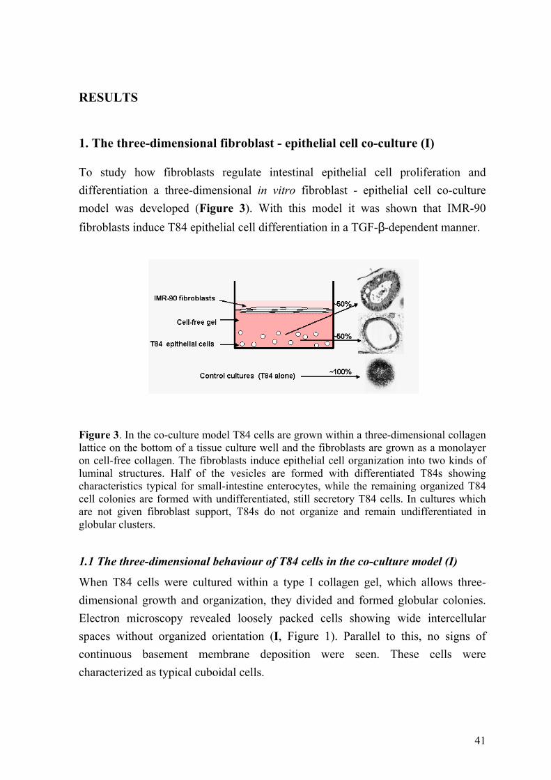
41
RESULTS
1. The three-dimensional fibroblast - epithelial cell co-culture (I)
To study how fibroblasts regulate intestinal epithelial cell proliferation and differentiation a three-dimensional in vitro fibroblast - epithelial cell co-culture model was developed (Figure 3). With this model it was shown that IMR-90 fibroblasts induce T84 epithelial cell differentiation in a TGF-β-dependent manner.
Figure 3. In the co-culture model T84 cells are grown within a three-dimensional collagen lattice on the bottom of a tissue culture well and the fibroblasts are grown as a monolayer on cell-free collagen. The fibroblasts induce epithelial cell organization into two kinds of luminal structures. Half of the vesicles are formed with differentiated T84s showing characteristics typical for small-intestine enterocytes, while the remaining organized T84 cell colonies are formed with undifferentiated, still secretory T84 cells. In cultures which are not given fibroblast support, T84s do not organize and remain undifferentiated in globular clusters.
1.1 The three-dimensional behaviour of T84 cells in the co-culture model (I)
When T84 cells were cultured within a type I collagen gel, which allows three-dimensional growth and organization, they divided and formed globular colonies. Electron microscopy revealed loosely packed cells showing wide intercellular spaces without organized orientation (I, Figure 1). Parallel to this, no signs of continuous basement membrane deposition were seen. These cells were characterized as typical cuboidal cells.
42
When fibroblasts were introduced to these cultures T84 cells responded by forming organized luminal structures. In cultures which were supported with primary foreskin fibroblasts a mean of 94% of all T84 cell colonies were organized (range 80-100 %), and with lung fibroblast cell line IMR-90 the number of organized colonies was on average 76% of all T84 epithelial cell colonies (range 55-95 %) (I, Figure 2). Vertical and horizontal cross sections of organized colonies from paraffin-embedded cultures revealed one epithelial cell layer surrounding the lumen. A well-deposited basement membrane was demonstrated around all luminal formations with silver impregnation staining, and by immune staining for laminin (I, Figure 7).
The luminal formations consisted of two types of colonies. A part of these organized colonies were formed with polarized undifferentiated cuboidal epithelial cells, showing irregular and distorted microvilli. The majority of organized luminal formations were composed of cells showing morphological features typical of absorptive enterocytes; columnar in shape with well-formed apical intercellular junctions and microvilli. Biochemical characterization further evidenced that these cells were differentiated, as the majority expressed brush border alkaline phosphatase (moderate/strong; I, Table 1) and sialomucins. Additionally, goblet cells were occasionally observed in both types of luminal formations.
1.2 Effects of soluble growth factors, HGF and TGF-β1 on the three-dimensional behaviour of T84 cells (I)
To establish whether the differentiation of crypt-like T84 cells was induced by soluble factors, IMR-90 fibroblasts were replaced by fibroblast-conditioned medium. The conditioned medium also induced epithelial cell organization, although not as effectively as fibroblasts.
HGF is known to be morphogenic for kidney epithelial cells (MDCK); it induces tubulus formation in similar culture conditions (Montesano et al. 1991). Based on this the ability of HGF to induce vesicle formation was tested. HGF failed to induce organization of T84 epithelial cells but increased epithelial cell proliferation in a dose-dependent fashion (I, Figure 9).
TGF-β1 has been reported to induce terminal differentiation in intestinal epithelial cells, and was therefore chosen as the next candidate. hTGF-β1 indeed increased dose-dependently the number of differentiated types of T84 cell colonies. The
43
optimal concentration, 20 ng/ml, induced organization in seven days with a mean of 65% (range 63-67%). When the hTGF-β1 concentration was further increased to 40 ng/ml the number of luminal formations decreased to the same level as with a concentration of 10 ng/ml, comprising on the average 57% of all T84 colonies (range 54-60%). It is noteworthy that nearly all luminal formations, on the average 93% of all T84 cell colonies, were formed with columnar, differentiated T84 cells showing positive alkaline phosphatase staining at the apical site.
To ensure that fibroblast-induced T84 epithelial cell differentiation was also due to TGF-β1, pan-specific neutralizing antibodies against TGF-β and antibodies against the latency-associated protein (LAP) were added to the three-dimensional cultures together with IMR-90 fibroblasts. The results clearly showed that the fibroblast-induced organization and differentiation T84 cells was TGF-β-mediated, as in cultures treated with neutralizing antibodies against TGF-β the numbers of differentiated-type of colonies were reduced from the accurate control level of 42% to 15% (p<0.001). The antibodies against LAP were even more effective, as they reduced the number of differentiated-type colonies down to 6% from the 42% seen in control cultures treated with irrelevant rabbit antibodies. In addition, the antibodies against TGF-β and LAP seemed to increase the proliferation of T84 cells, as the diameters of the unorganized cell clusters were significantly increased. This suggested that TGF-β inhibits proliferation, as was in fact further confirmed in the proliferation assay, which showed that hTGF-β1 slightly but dose-dependently reduced the proliferation rate of T84 cells. The results are summarized in Table 2 of I.
2. The effects of coeliac disease-specific IgA on epithelial cells (III)
The presence of a well-deposited basement membrane around the luminal formations in fibroblast-supported three-dimensional T84 epithelial cell cultures suggested that the coeliac disease autoantigen could be detectable in the three-dimensional co-culture. This was established in an indirect immunofluorescence test with coeliac disease patient sera IgA-class antibodies, which, as expected, revealed the presence of the coeliac disease autoantigen in the basement membrane area of organized luminal formations (III, Figure 1). The control sera (non-coeliac) showed no reaction with the substratum.
44
To ascertain whether coeliac disease-specific autoantibodies have any biological implications for epithelial cell biology purified total IgA fractions from both coeliac disease patient and healthy donor sera were added to the fibroblast-epithelial cell co-cultures. In control co-cultures, which were treated with the total IgA fraction from healthy donors or disease controls, the colony type distribution was as in untreated T84-IMR-90 co-cultures; 48% (range 39-55%) of all colonies were differentiated-type luminal formations. In co-cultures treated with purified IgA from coeliac disease patient sera, the number of differentiated-type colonies decreased significantly to 25% (range 19-32%) when compared to control cultures (p<0.001). The results are summarized in Figure 4.
In addition, IgA purified from coeliac disease patients’ sera on a gluten-free diet did not affect epithelial cell differentiation but total IgA obtained from the same patients after gluten challenge again reduced the number of differentiated-type colonies to the same level as with IgA from untreated coeliac disease patients (III, Figure 2).
When purified IgA from coeliac disease patient sera was added to cultures where epithelial cell differentiation was induced by TGF-β1, an interesting dose-dependent effect on the number of differentiated epithelial cell colonies was seen. At the optimal hTGF-β1 concentration (20 ng/ml) coeliac disease-specific IgA reduced the number of differentiated-type T84 cell colonies to 35% (range 31-40%) from the mean of 51% (range 46-56%) seen in control cultures treated with non-coeliac IgA (p<0.001). In cultures treated with the highest used hTGF-β1 concentration (40 ng/ml) the coeliac disease IgA significantly increased the number of differentiated-type colonies to 34% (range 28-40%) when compared to the 25% (range 21-30%) seen in control cultures treated with non-coeliac IgA. In all tested concentrations of hTGF-β1 non-coeliac disease-associated IgA showed no effect on T84 epithelial cell differentiation.
As increased crypt epithelial cell proliferation is well established in coeliac disease-affected small-intestinal mucosa, the effects of purified total coeliac disease-specific IgA on IMR-90-induced crypt-like T84 cell proliferation were now tested. As expected, the total IgA fraction from untreated coeliac disease patient sera increased T84 cell proliferation, but only slightly, to 105% (range 102-109%) from the corresponding control level of 100% (range 97-103%) when compared to cultures treated with control IgA (p=0.024).
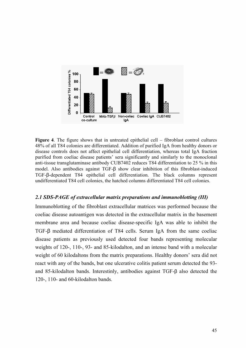
45
Figure 4. The figure shows that in untreated epithelial cell – fibroblast control cultures 48% of all T84 colonies are differentiated. Addition of purified IgA from healthy donors or disease controls does not affect epithelial cell differentiation, whereas total IgA fraction purified from coeliac disease patients’ sera significantly and similarly to the monoclonal anti-tissue transglutaminase antibody CUB7402 reduces T84 differentiation to 25 % in this model. Also antibodies against TGF-β show clear inhibition of this fibroblast-induced TGF-β-dependent T84 epithelial cell differentiation. The black columns represent undifferentiated T84 cell colonies, the hatched columns differentiated T84 cell colonies.
2.1 SDS-PAGE of extracellular matrix preparations and immunoblotting (III)
Immunoblotting of the fibroblast extracellular matrices was performed because the coeliac disease autoantigen was detected in the extracellular matrix in the basement membrane area and because coeliac disease-specific IgA was able to inhibit the TGF-β mediated differentiation of T84 cells. Serum IgA from the same coeliac disease patients as previously used detected four bands representing molecular weights of 120-, 110-, 93- and 85-kilodalton, and an intense band with a molecular weight of 60 kilodaltons from the matrix preparations. Healthy donors’ sera did not react with any of the bands, but one ulcerative colitis patient serum detected the 93- and 85-kilodalton bands. Interestinly, antibodies against TGF-β also detected the 120-, 110- and 60-kilodalton bands.
46
2.2 ELISA for hTGF-β1 antibodies (III)
The data obtained both from blocking studies and from immunoblotting suggested that coeliac disease-specific IgA could recognize similar proteins as neutralizing antibodies against TGF-β. This idea was tested with an ELISA for TGF-β. However, none of the IgA samples tested showed positive reaction for TGF-β1. The average optical density with coeliac disease-specific IgA was 0.098 (range 0.031-0.178) and 0.035 (range 0.010-0.064) with IgA from control individual sera, whereas the rabbit pan-specific TGF-β neutralizing antibodies, which served as a positive control, gave an optical density of 2.053.
3. Tissue transglutaminase as coeliac disease autoantigen (II)
While this study was in progress tissue transglutaminase was identified as one, if not sole, coeliac disease autoantigen (Dietrich et al. 1997). To study whether patient serum IgA class antibodies bind to tissue transglutaminase an ELISA was performed with guinea pig liver derived tissue transglutaminase as antigen. Serum samples from 136 untreated coeliac disease patients and 207 disease controls were tested.
When the ELISA was conducted as described in the original paper the mean optical density for the pilot group of 20 untreated coeliac disease patients tested was 0.455 and that for the first 24 disease control patients tested 0.132. However, when the unspecific sample background values were subtracted no differences in results were seen between coeliac disease patients and the controls.
Tissue transglutaminase is a Ca2+-dependent cross-linking enzyme. Calcium binding induces a conformational change in the enzyme needed for catalytic activity (Fesus and Laki, 1977). With this in mind the ELISA protocol was modified by adding Ca2+ to the coating buffer and EDTA to the dilution buffer to inhibit enzymatic activity. With these modifications the mean optical density was now, after sample background subtraction, 1.013 for the same first 20 untreated coeliac disease patients and 0.014 for the 24 control patients. When a large clinical material was tested with this ELISA, using calcium-activated guinea pig tissue transglutaminase as antigen, a sensitivity of 95% (129 out of 136) and a specificity of 94% (194 out of 207) with a cut-off level of 10 AU was obtained. In addition, this modified IgA-
47
class tissue transglutaminase autoantibody ELISA correlated well with the IgA-class endomysial antibody test (II, Table 1).
In indirect immunofluorescence staining on cryostat sections of human umbilical cord the commercial tissue transglutaminase monoclonal antibody, CUB7402, stained smooth muscle endomysial fibres around the vessels similarly to untreated coeliac disease patient serum IgA. A double immunofluorescence staining showed clear colocalization of the epitopes for CUB7402 and coeliac disease-specific antibodies (II, Figure 4). Pretreatment of coeliac disease patient sera with tissue transglutaminase completely abolished the endomysial staining pattern in human umbilical cord.
4. Antibodies against tissue transglutaminase in the co-culture model (III)
Once the tissue transglutaminase was identified as the coeliac disease autoantigen (Dieterich et al. 1997) the purified total IgA fractions from coeliac disease patient sera (used in the previous co-culture experiments) were tested with the aforementioned ELISA for tissue transglutaminase. Purified IgA from untreated coeliac disease patient sera and from patients on gluten challenge showed high optical densities (mean 2.803, range 2.630-2.927) whereas purified IgA from sera of patients on a gluten-free diet as well as purified IgA from non-coeliac sera were significantly lower (mean 0.174, range 0.146–0.231).
To study whether tissue transglutaminase antibodies show effects on epithelial cell differentiation similar to those of coeliac disease-specific IgA antibodies, commercial tissue transglutaminase antibodies were added to the three-dimensional fibroblast-epithelial cell co-cultures. The result showed that these commercial antibodies, CUB7402 and TG-100, reduced T84 epithelial cell differentiation to 23% of all T84 cell colonies (range 17-27%) from the 48% seen in control cultures, thus being similar to untreated coeliac disease-specific IgA (Figure 4). The irrelevant isotype-matched mouse control antibodies did not affect epithelial cell differentiation. In addition, monoclonal antibodies against tissue transglutaminase increased T84 epithelial cell proliferation to 130% from the control level of 100%.
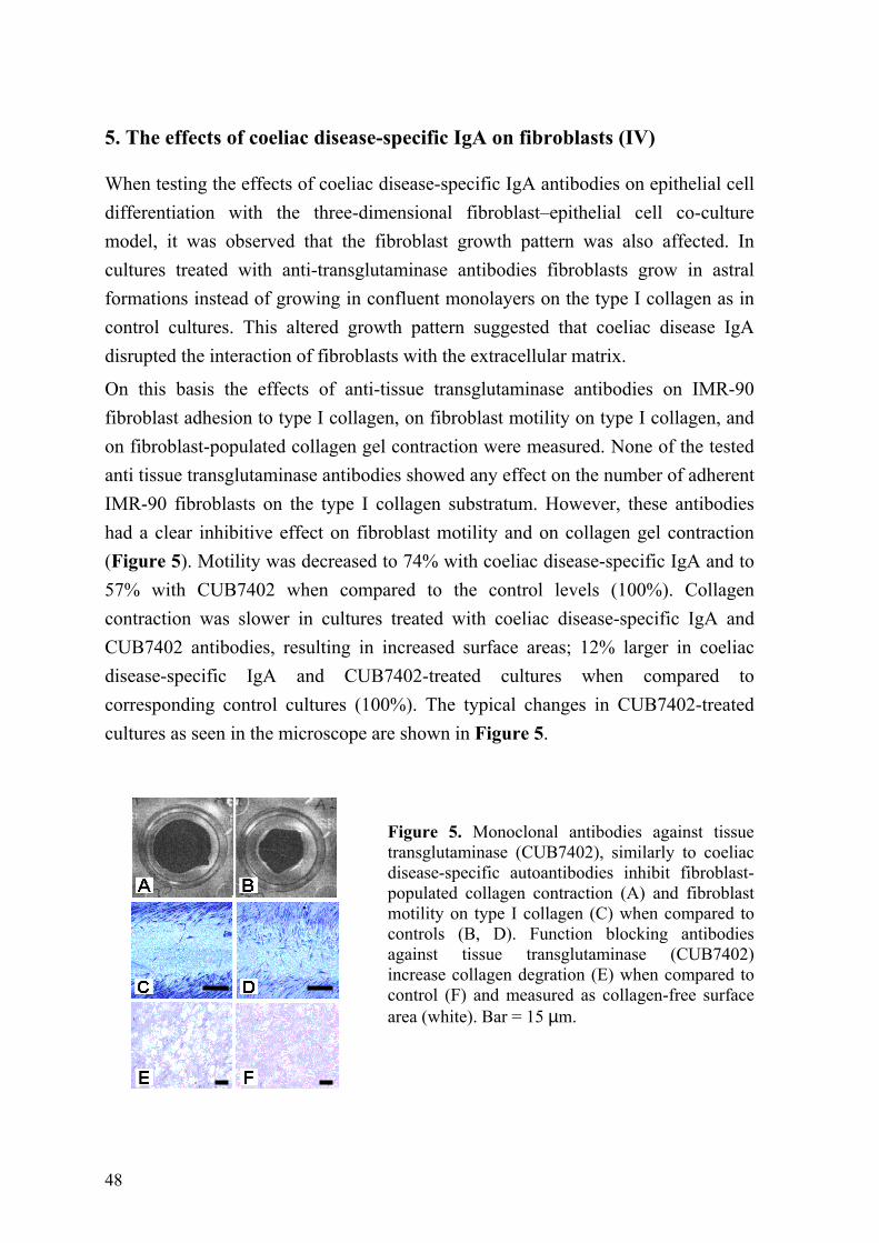
48
5. The effects of coeliac disease-specific IgA on fibroblasts (IV)
When testing the effects of coeliac disease-specific IgA antibodies on epithelial cell differentiation with the three-dimensional fibroblast–epithelial cell co-culture model, it was observed that the fibroblast growth pattern was also affected. In cultures treated with anti-transglutaminase antibodies fibroblasts grow in astral formations instead of growing in confluent monolayers on the type I collagen as in control cultures. This altered growth pattern suggested that coeliac disease IgA disrupted the interaction of fibroblasts with the extracellular matrix.
On this basis the effects of anti-tissue transglutaminase antibodies on IMR-90 fibroblast adhesion to type I collagen, on fibroblast motility on type I collagen, and on fibroblast-populated collagen gel contraction were measured. None of the tested anti tissue transglutaminase antibodies showed any effect on the number of adherent IMR-90 fibroblasts on the type I collagen substratum. However, these antibodies had a clear inhibitive effect on fibroblast motility and on collagen gel contraction (Figure 5). Motility was decreased to 74% with coeliac disease-specific IgA and to 57% with CUB7402 when compared to the control levels (100%). Collagen contraction was slower in cultures treated with coeliac disease-specific IgA and CUB7402 antibodies, resulting in increased surface areas; 12% larger in coeliac disease-specific IgA and CUB7402-treated cultures when compared to corresponding control cultures (100%). The typical changes in CUB7402-treated cultures as seen in the microscope are shown in Figure 5.
Figure 5. Monoclonal antibodies against tissue transglutaminase (CUB7402), similarly to coeliac disease-specific autoantibodies inhibit fibroblast-populated collagen contraction (A) and fibroblast motility on type I collagen (C) when compared to controls (B, D). Function blocking antibodies against tissue transglutaminase (CUB7402) increase collagen degration (E) when compared to control (F) and measured as collagen-free surface area (white). Bar = 15 µm.
49
The process which results in fibroblast-populated type I collagen contraction is dependent on integrin α2β1 and membrane type I matrix metalloproteinase activity (Broberg and Heino 1996, Deryugina et al. 1998). Because anti-transglutaminase antibodies did not affect IMR-90 fibroblast adhesion to type I collagen (integrin α2β1 –mediated) we chose to measure the effects of anti-tissue transglutaminase antibodies on type I collagen degradation. The assay used has been described as being solely dependent on membrane type I matrix metalloproteinase mediated collagen degradation (Holmbeck et al. 1999). The ability of IMR-90 cells to degrade type I collagen in IL-1β-stimulated cultures, measured as the average surface area of degraded collagen, was significantly increased in the presence of coeliac disease-specific IgA (138%) and CUB7402 (160%), when compared to the accurate controls (100%). The typical changes in CUB7402-treated cultures as seen in the microscope are shown in Figure 5. We also performed zymography from the culture medium obtained from the ‘degradation’ assays. No marked changes in metalloproteinase activity were seen regardless of the added antibodies.
In studies focusing on the effects of coeliac disease-specific IgA on fibroblast behaviour also monoclonal, recombinant coeliac disease-specific anti-transglutaminase IgA antibodies were used. The results obtained reflected the effects of the total IgA fraction purified from coeliac disease patients’ sera in experiments where similarly produced antibodies against gliadin served as control antibodies. Recombinant IgA antibodies reduced IMR-90 fibroblast motility to 74% of the control level. In the collagen gel contraction assay these antibodies inhibited contraction of the gels, resulting in a 20% higher surface area when compared to the corresponding control cultures. In the matrix degradation assay recombinant IgA antibodies against tissue transglutaminase antibodies increased degradation even more than CUB7402, resulting in a 167% larger collagen-free area when compared to cultures treated with recombinant antibodies against gliadin.
50
DISCUSSION
In the discussion the major points arising from the present results (I-IV) are discussed in the first three sections. In the last section the results are reconciled with the existing data and a new hypothesis of coeliac disease pathogenesis is proposed. 1. The three-dimensional fibroblast-epithelial cell co-culture model (I)
The human intestinal deep crypt-like T84 epithelial cell line was used in this study because they express only a limited set of markers typical for aggressive cancer cells. T84 cells do not form metastases when implanted in nude mice (Murakami and Masui 1980). They also exhibit well-formed tight junctions and desmosomes between adjacent cells when grown in monolayers and have maintained responsiveness to many peptide hormones and neurotransmitters (Dharmsathaphorn et al. 1984). These properties suggested that T84 cells might be able to differentiate if cultured in optimal conditions. The present study using a three-dimensional co-culture system showed for the first time that these deep crypt-like chloride-secreting transformed human intestinal T84 epithelial cells indeed organize and differentiate when grown within a collagen gel and when given fibroblast support. Furthermore, it was shown that this fibroblast-induced T84 differentiation was TGF-β -dependent.
In morphologically columnar T84 cell colonies differentiation was further characterized by the expression of alkaline phosphatase, which has been used as a differentiation marker for Caco-2 cells (Pinto et al. 1983, Matsumoto et al. 1990). Furthermore, a well-deposited basement membrane around organized luminal formations was detected (I). Although a colon cancer cell line, T84 cells responded to TGF-β, thus differing from other colon cancer cells, which typically evince genetic defects either in TGF-β receptors or in molecules involved in TGF-β signaling pathways (Blobe et al. 2000). Another characteristic untypical for cancer cells was found in differentiating T84 cells, as they downregulated the expression of the proto-oncogene product c-met, the HGF receptor (I). Its expression typically remains unaltered in cancer cells (Boros and Miller 1995). However, these features, together with increased E-cadherin expression (unpublished data), support the conception that T84 cells virtually undergo differentiation when grown within a
51
three-dimensional collagen lattice together with, but separated from, supporting fibroblasts.
The cell culture model described in this study offers a convenient means of investigating the role of mesenchymal cells in the regulation of epithelial cell proliferation and differentiation. A model which allows cross-talk between different cell types naturally reflects better the situation occurring in vivo, where instructive interactions between different cell populations form the bases of development and tissue maintenance. A good example of this is the growth factor pair TGF-β and HGF, which antagonize many of each other’s functions. TGF-β is the major autocrine and/or paracrine inhibitory regulator of epithelial cells, which induces terminal differentiation in epithelial cells (Kurokowa et al. 1987, Markowitz and Roberts 1996). HGF is a major mediator in epithelial-mesenchymal interaction and it stimulates epithelial cell proliferation and migration (Nusrat et al. 1994, Rubin et al. 1995, Goke et al. 1998). The antagonistic effect was clearly seen in the present study when neutralizing antibodies against TGF-β or HGF were added to fibroblast-supported three-dimensional T84 cell cultures. In the presence of anti-TGF-β antibodies differentiation of T84 cell colonies was reduced, with a simultaneous increase in the epithelial cell proliferation rate (I). The opposite effect was observed when the biological function of HGF was inhibited: proliferation of T84 cells decreased and the number of differentiated T84 cell colonies was slightly enhanced when compared to the control co-cultures (I). Thus the present model has advantages over previous models for intestinal epithelial cell differentiation, Caco-2 and HT-29 cells. Caco-2 cells differentiate spontaneously when grown to confluence (Pinto et al. 1983) and HT-29 cells need metabolic manipulation; the cells differentiate in the absence of glucose (Wice et al. 1985). In fact, HT-29 cells neither organized nor differentiated in the present model (I), although in monolayer co-cultures with IMR-90 cells they grow in lumen-shaped formations (data not shown).
Recently published data also support the observation that this co-culture model mimics the jejunal crypt villus axis biology where the chloride-secreting crypt cells proliferate and differentiate to absorptive villus tip enterocytes. With differential display polymerase chain reaction it has been shown that the expression patterns for CC3/TIP30 (a metastasis suppression gene product) and rab11 (a protein regulating
52
vesicle trafficking) in epithelial cells were similar in the co-culture model to those in the normal human jejunal crypt-villus axis (Lindfors et al. 2001).
2. Tissue transglutaminase as coeliac disease autoantigen (II)
Histological demonstration of an initial flat small-intestinal mucosa (villous atrophy and crypt hyperplasia), together with clinical and histological improvement when patients are on a gluten-free diet, continues to be the gold standard for coeliac disease diagnosis. Serological detection of anti-endomysial antibodies is used to support the diagnosis and to screen populations at risk; the sensitivity and specificity of the tests are 70-95% and 100%, respectively (Schuppan 2000).
Once tissue transglutaminase was identified as the major autoantigen in coeliac disease (Dieterich et al. 1997) the first question was whether tissue transglutaminase was responsible for the typical EmA staining pattern. This was shown in indirect immunofluorescence staining with commercial antibodies against tissue transglutaminase, where the typical coeliac disease-specific staining pattern was seen. In addition, when double staining with commercial monoclonal tissue transglutaminase and coeliac disease-specific IgA antibodies was performed, a complete overlapping was observed (II). Subsequent studies have confirmed these results (Lock et al. 1999a, Korponay-Szabo et al. 2000)
The next question was whether a simple, economical and rapid serum antibody ELISA test with the only available commercial tissue transglutaminase preparation, guinea pig tissue transglutaminase as antigen, could serve as a screening test for coeliac disease instead of laborious immunofluorescence testing. The results clearly showed that tissue transglutaminase autoantibody ELISA was highly sensitive (95%) and specific (94%) in detecting untreated coeliac disease. Most researchers today accept this view, as similar results have recently been reported by other groups (Dieterich et al. 1997, Lock et al. 1999b, Hansson et al. 2000, Leon et al. 2001). More recent studies using recombinant human tissue transglutaminase have been even better in terms of sensitivity and specificity in discriminating coeliac from non-coeliac individuals (Sardy et al. 1999, Bazzigaluppi et al. 1999, Sblattero et al. 2000, Martini et al. 2001). These results clearly indicate that tissue transglutaminase is the main, if not sole, autoantigen in coeliac disease. This assumption seems to hold true as, in immunofluorescence studies with coeliac
53
disease-specific IgA autoantibodies on tissue transglutaminase knock-out and wild-type mice tissues, the typical coeliac disease-specific staining pattern was seen only in wild-type tissues where the autoantigen, tissue transglutaminase, was present (Korponay-Szabo et al. 2001).
The results obtained in the present study showed the ultimate importance of the presence of Ca2+ for obtaining a specific antigen-antibody reaction, which would imply the involvement of conformational epitopes. This has been further evidenced in an elegant study using novel deletion mutants of tissue transglutaminase (Seissler et al. 2001). It was shown that coeliac disease-specific autoantibodies are indeed targeted against conformational epitopes and that these epitopes are located in distinct functional domains of tissue transglutaminase. 3. Biological function of coeliac disease-specific IgA (III, IV)
Tissue transglutaminase is a multifunctional cross-linking enzyme whose ultimate function is to stabilize extracellular matrix proteins (Aeschlimann and Thomazy 2000). It catalyzes the formation of epsilon(gamma-glutamyl)lysine isopeptide bonds, which are resistant to enzymatic, mechanical and chemical degradation (Haroon et al. 2001).
Our results show that coeliac disease-specific autoantibodies, which are targeted against tissue transglutaminase, possess biological activity exerting effects on both mesenchymal and epithelial cell biology (III, IV). These IgA autoantibodies inhibit fibroblast-induced TGF-β-mediated differentiation with a simultaneous increase in the proliferation rate of intestinal crypt-like T84 epithelial cells. Furthermore, purified total IgA fraction from coeliac disease patients’ sera significantly inhibited IMR-90 fibroblast motility and increased type I collagen degradation. The commercial monoclonal antibody against tissue transglutaminase, CUB7402, known to block the bioactivity of tissue transglutaminase, showed similar effects on both epithelial cells and mesenchymal cells. In fact, it has recently been shown that coeliac disease IgA is targeted against functional domains of tissue transglutaminase (Seissler et al. 2001) and that coeliac disease-specific IgA inhibits the enzymatic activity of tissue transglutaminase in vitro (Esposito et al. in press).
Tissue transglutaminase is involved in the activation of latent TGF-β (Nunes et al. 1997). This growth factor is considered to be the major autocrine/paracrine
54
inhibitory regulator in the intestinal epithelium (Markowitz and Roberts 1996). It induces proliferation arrest in epithelial cells often with simultaneous signaling for the terminal differentiation pathway (Kurokowa et al. 1987). By blocking the bioactivity of tissue transglutaminase, coeliac disease-specific IgA and monoclonal tissue transglutaminase antibodies presumably also block the tissue transglutaminase-dependent activation of TGF-β and could in that way inhibit epithelial cell differentiation, as was seen in our three-dimensional fibroblast-epithelial cell co-culture model (III).
The importance of tissue transglutaminase in extracellular matrix stabilization was more clearly seen in experiments with mesenchymal cells (IV). Function-blocking antibodies against tissue transglutaminase inhibited IMR-90 motility on type I collagen and collagen gel contraction, and increased type I collagen degradation. The observed increase in matrix degradation could well result from blocked transglutaminase cross-linking activity, as then the extracellular matrix remains more susceptible to matrix metalloproteinase degradation (Haroon et al. 2001).
It is noteworthy that the in normal situation the degradation of collagen triple helices releases collagen-bound decorin, a proteoglycan which binds to active TGF-β, thus neutralizing its bioactivity (Imai et al. 1997). Increased matrix degradation caused by autoantibody-mediated inhibition of tissue transglutaminase activity is hypothesized to enhance neutralization of TGF-β, as more decorin will be released. TGF-β also stimulates matrix production in mesenchymal cells in addition to its known ability to induce production of tissue inhibitors of matrix metalloproteinases, TIMPs. Thus blocking of the bioactivity of tissue transglutaminase also results, via neutralization of TGF-β, in reduced matrix deposition and increased matrix degradation, which further affects mesenchymal cell biology. In turn, keeping in mind the well-established instructive interaction between mesenchymal and epithelial cells, these changes in mesenchymal cell biology may be expected to also affect epithelial cell biological performance. The suggested biological functions of coeliac disease-specific autoantibodies are summarized in Figure 6.
Mechanisms similar to those seen in vitro might also be operative in vivo, especially as these locally produced (Marzari et al. 2001) IgA autoantibodies (replaced by IgM-class antibodies in IgA-deficient coeliac disease patients) are deposited on the myofibroblasts underlining the crypt epithelium already before the full-blown coeliac lesion is seen in the small bowel (Korponay-Szabo, personal
55
communication) and before the autoantibodies are present in the patient circulation (Mäki et al. 1995a, Iltanen et al. 1999). Interestingly, increased matrix degradation has been shown to dramatically affect tissue architecture in an organ culture model (Pender et al. 1997). In this model stimulation of T cells resulted in morphological changes typical of coeliac disease via increased matrix metalloproteinase activity of intestinal myofibroblasts. As the present results demonstrate, the disease-specific autoantibodies have parallel effects, which suggests that these autoantibodies have a role in the formation of the mucosal lesion typically seen in coeliac disease. 4. New perspectives on coeliac disease pathogenesis
Basically, in view of to the strong association of the disease with HLA class II molecules the prevailing and most commonly accepted theory of coeliac disease pathogenesis is the immunological hypothesis, which envisages the disease as T-cell-mediated. According to this hypothesis HLA DQ2 and DQ8 molecules present the disease-causing antigen, gliadin, to CD4+ T cells (Sollid and Thorsby 1993, McAdam and Sollid 2000). The presence of HLA-DQ-restricted T cells which respond to gliadin digests in the mucosa of coeliac disease patients indeed supports such a conception (Sollid 2000). Researchers were long puzzled because DQ2 preferentially binds to peptides with negatively charged amino acids, which are rarely found in gliadin. However, when certain glutamine residues of the gliadin molecule are deamidated to glutamic acid the binding of gliadin peptides to DQ2 is enhanced. It is of interest that tissue transglutaminase, whose expression in the coeliac disease-affected mucosa is increased (D'Argenio et al. 1989), can perform this task, as has recently been shown in vitro (Molberg et al. 2000). The gliadin peptides presented by DQ2 then activate CD4+ T-cells, which in turn produce increased amounts of IFN-γ and other typical Th1 or Th0 cytokines (Sollid 2000). These activated T cells are then believed to orchestrate the changes leading to the mucosal lesion via increased matrix degradation by myofibroblast-produced matrix metalloproteinases (MacDonald 1992).
The autoantibodies in coeliac disease are highly specific for the disease and mainly targeted against tissue transglutaminase. The ability of tissue transglutaminase to form complexes with gliadin has been held to drive the production of these autoantibodies. It has been proposed that tissue transglutaminase-specific B cells recognize the transglutaminase-gliadin complex, internalize it, and after processing
56
present the deamidated gliadin peptides to T-cells. These gliadin-specific T-cells then start to produce cytokines, which in turn stimulate the anti-transglutaminase antibody production in the antigen-presenting B cell (Sollid 2000, Schuppan 2000).
In other autoimmune diseases such as type I diabetes, the balance between Th1 (cytokines for cell-mediated immunity) / Th2 (cytokines for antibody production) expression has been shown to affect the manifestation of the disease (Shehadeh et al. 1993, Bach 2000). Similar mechanisms could also be operative in coeliac disease and explain the diversity in the natural history of gluten sensitivity in relation to the clinical manifestation of the disease.
However, based on the present results, together with recent findings, which show that coeliac disease-specific autoantibodies are produced in the intestine (Marzari et al. 2001) where they are already present before mucosal lesion is seen (Korponay-Szabo, personal communication) it is tempting to speculate that these autoantibodies could have a role in the pathogenesis of coeliac disease. The gliadin-triggered pathogenetic mechanism could be biphasic, the autoantibody-mediated events in phase I being followed by a T cell-driven immune mediated phase II where the mucosal lesion typical of coeliac disease finally appears (Figure 6).
The autoantibodies appear at the mucosal site at a very early stage of disease development and reduce the bioactivity of TGF-β, as the present results show (III). Although the coeliac disease-specific autoantibodies are able to cause some changes in intestinal biology (III, IV), the tissue seems to gain homeostasis in the course of IgA deposition and to tolerate the reduced availability of TGF-β. This is now regarded as phase I. This balance in the intestinal tissues, despite the presence of IgA, can be maintained for long periods, even years. However, in phase II an additional event (e.g. infection or IFN-α) is assumed to take place and cause a further decrease in the availability of active TGF-β or induce changes counter to TGF-β activity. This could finally lead to imbalanced biology on the crypt villus axis, which in turn is suggested to lead to the inflammation typically seen in coeliac disease.
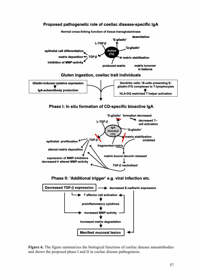
57
Figure 6. The figure summarizes the biological functions of coeliac disease autoantibodies and shows the proposed phase I and II in coeliac disease pathogenesis.
Gluten ingestion, coeliac trait individuals
Phase I: In situ formation of CD-specific bioactive IgA
’Q-gliadin’
’E-gliadin’ formation decreased
expression of MMP-inhibitors decreased altered MMP-activity
IgAblocked
tTG
L-TGF-β
TGF-β
fragmented matrix
matrix stabilization inhibitedepithelial proliferation
altered matrix deposition
matrix bound decorin released
TGF-β neutralized
decreased T–cell activation
ActivetTG
L-TGF-β
TGF-β
produced matrix
matrix stabilization
epithelial cell differentiation
matrix deposition
inhibition of MMP-activity
’Q-gliadin’
’E-gliadin’
matrix turnoverin balance
deamidation
Dendritic cells / B-cells presenting E-gliadin-tTG complexes to T-lymphocytes
HLA-DQ restricted T helper activation
Gliadin-induced cytokine expression
IgA-autoantibody production
Phase II: ’Additional trigger’ e.g. viral infection etc.
Proposed pathogenetic role of coeliac disease-specific IgA
Decreased TGF-β expression decreased E-cadherin expression
T effector cell activation
proinflammatory cytokines
increased MMP-activity
increased matrix degradation
Manifest mucosal lesion
Normal cross-linking function of tissue transglutaminase
Gluten ingestion, coeliac trait individuals
Phase I: In situ formation of CD-specific bioactive IgA
’Q-gliadin’
’E-gliadin’ formation decreased
expression of MMP-inhibitors decreased altered MMP-activity
IgAblocked
tTG
L-TGF-β
TGF-β
fragmented matrix
matrix stabilization inhibitedepithelial proliferation
altered matrix deposition
matrix bound decorin released
TGF-β neutralized
decreased T–cell activation
ActivetTG
L-TGF-β
TGF-β
produced matrix
matrix stabilization
epithelial cell differentiation
matrix deposition
inhibition of MMP-activity
’Q-gliadin’
’E-gliadin’
matrix turnoverin balance
deamidation
ActivetTG
L-TGF-β
TGF-β
produced matrix
matrix stabilization
epithelial cell differentiation
matrix deposition
inhibition of MMP-activity
’Q-gliadin’
’E-gliadin’
matrix turnoverin balance
deamidation
Dendritic cells / B-cells presenting E-gliadin-tTG complexes to T-lymphocytes
HLA-DQ restricted T helper activation
Gliadin-induced cytokine expression
IgA-autoantibody production
Gliadin-induced cytokine expression
IgA-autoantibody production
Phase II: ’Additional trigger’ e.g. viral infection etc.
Proposed pathogenetic role of coeliac disease-specific IgA
Decreased TGF-β expression decreased E-cadherin expression
T effector cell activation
proinflammatory cytokines
increased MMP-activity
increased matrix degradation
Manifest mucosal lesion
Normal cross-linking function of tissue transglutaminase
58
4.1 IgA autoantibody mediated events, phase I
The ability of coeliac disease-specific IgA to block the enzymatic activity of tissue transglutaminase suggest basically two possibilities for the autoantibodies to exert their role in coeliac disease pathogenesis. The first is based on the fact that tissue transglutaminase has been shown to be involved in the activation of TGF-β (Nunes et al. 1997) and the other relies on its ability to stabilize extracellular matrix structures (Aeschlimann and Thomazy 2000).
TGF-β is the major inhibitory regulator of the intestinal epithelium which arrests epithelial cells in the G1 phase of the cell cycle and often simultaneously signals for terminal differentiation. A part of this process is mediated by down-regulation of c-Myc, which is a critical event for growth inhibition by TGF-β (Yagi et al. 2002). In the normal situation, during epithelial cell differentiation, TGF-β acts as a potent inducer of the homotypic cell-cell adhesion by up-regulating the expression of E-cadherin and its undercoat-associated protein, β-catenin. TGF-β also increases the formation of the cadherin/catenin complex at the cell membrane and reduces the free cytoplasmic pool of β-catenin available for activation of c-myc transcription (Coffey et al. 1988, Stockinger et al. 2001, Wang and Chakrabarty 2001).
If we insert coeliac disease IgA autoantibodies into this process we presumably end up with decreased amounts of active TGF-β. The less TGF-β is available, the more c-Myc is expressed. Untimately this produces a situation similar to coeliac disease where c-myc expression is increased (Ciclitira et al. 1987) and E-cadherin expression decreased (Perry et al. 1999), resulting in increased epithelial cell proliferation.
Reduced bioactivity of TGF-β could also explain other features typical of coeliac disease, for example goblet cell hyperplasia (Marsh 1992). The circumstances where the availability of TGF-β is limited can be hypothesized to favour the maturation of goblet cells, as they are the only intestinal epithelial cell population whose maturation is not TGF-β dependent (Hafez et al. 1990).
The permeability of the epithelium in the coeliac disease-affected mucosa is increased. This has been shown to be related to structural modification in tight junctions between adjacent epithelial cells (Schulzke et al. 1995). It is noteworthy that intact cadherin-catenin complexes are important in the development of functional tight junctions (Rajasekaran et al. 1996), which are necessary for the
59
maintenance of the selective permeability and barrier function of the intestinal epithelium. As TGF-β stabilizes and induces the formation of the cadherin-catenin complex, the increased permeability seen in the coeliac disease-affected mucosa can be considered to be in part mediated by IgA autoantibodies, which reduce the bioactivity of TGF-β (III). In addition, also HGF, the TGF-β antagonist (I), has the capability to loosen intercellular tight junctions (Nusrat et al. 1994).
Increased matrix degradation has been held to play a major role in the formation of the intestinal lesion (Pender at el. 1997, Schuppan 2000). TGF-β is a negative regulator of matrix degradation; it stimulates the deposition of extracellular matrix proteins and upregulates the expression of matrix metalloproteinase inhibitors (Grande 1997). Following this, the diminished availability of active TGF-β could increase MMP activity, as fewer inhibitors are produced. In coeliac disease and dermatitis herpetiformis the expression of mRNA for MMP-1, MMP-3 and, surprisingly also TIMP-1, has been shown to be increased (Daum et al. 1999, Airola et al. 1997). In addition, the enzymatic activity of MMP-2 and MMP-9, which are not TIMP-1-inhibited, has been shown to be increased in the coeliac lesion (Lerner et al. 2000). It is of interest that MT1-MMP activates MMP-2 at the cell surface, which in turn is able to activate pro-MMP-9 (Deryugina et al. 1998). The present results show that IgA autoantibodies increase MT1-MMP-mediated matrix degradation in mesenchymal cells (IV), which could be responsible for the observed enhancement in the activity of MMP-2 and MMP-9.
Furthermore, MT1-MMP, whose expression is increased by HGF (Nabeshima et al. 2000) and negatively linked to E-cadherin expression (Ara et al. 2000), can degrade most basement membrane components; native collagens, fibronectin, tenascin, nidogen, laminin-5, aggrecan and perlecan (d’Ortho et al. 1997, Hotary et al. 2000, Koshikawa et al. 2000). When the cross-linking activity of tissue transglutaminase is also inhibited by coeliac disease autoantibodies (Esposito et al. 2000), the matrix presumably remains more susceptible to MMP degradation (Haroon et al. 2001). Following this, one would expect that the basement membrane in the coeliac lesion could be delinear and breached, as has in fact been reported (Verbeke et al. in press). However, contrasting results showing such changes only in 2/12 patients have also been reported (Korhonen et al. 2000). Interestingly, in patients with a certain form of intractable diarrhoea the laminin composition of the basement membrane is irregular, which results in morphological changes in the small intestine
60
similar to those seen in the coeliac disease-affected mucosa (Goulet et al. 1995). It is noteworthy that in these cases the mucosal changes, resembling those typically seen in coeliac disease, appeared without any sign of T-cell activation.
4.2 Lesion formation in phase II
Blocking the bioactivity of TGF-β by coeliac disease-specific IgA autoantibodies seems to be able to induce changes typical of coeliac disease. Although the antibodies might be present and deposited in the crypt basement membrane area for long periods, the process leading to crypt hyperplasia and villus atrophy may take years or even decades to develop in susceptible individuals. How can this be explained?
It should kept in mind that tissue transglutaminase is involved only in the plasmin-mediated activation of extracellular matrix-associated large latent TGF-β complex (Nunes et al. 1997). Other proteases such as mast cell chymase, leukocyte elastase and thrombin can also release LTBP from the extracellular matrix (Gleizes et al. 1997). It is therefore understandable that antibodies against tissue transglutaminase do not totally block TGF-β activation, but rather limit its availability (III), resulting in a “tolerable imbalance “.
Thus an additional factor which finally causes a breakdown in the barely sustained homeostasis is needed to mark the starting-point of phase II. It was previously suggested that adenoviral infections might be involved in the pathogenesis of coeliac disease (Kagnoff et al. 1987), and it has in fact been shown that adenoviral proteins such as E1A are more frequently found in the intestine of coeliac disease patients (Lawler et al. 1994). E1A binds to TGF-β1 promoter and causes decreased TGF-β expression (Datta and Bagchi 1994). Similarly, TGF-β1 expression is affected by changes in hormonal levels (Goddard et al. 2000) typically appearing in connection with pregnancy, which is also known to induce coeliac disease (Smecuol et al. 1996). Recently, a case of coeliac disease-like enteropathy was reported in a patient receiving INF-α for chronic myeloid leukaemia (Monteleone et al. 2001). Along the lines of such a conception in this certain case IFN-α could be the action causing the imbalance in the patient’s small intestine, as INF-α upregulates c-myc (Matikainen et al. 1999), thus resembling effects typically seen when TGF-β-mediated effects are blocked.
61
If we consider that the TGF-β level is further diminished from the already decreased level due to the presence of autoantibodies, we may deduce that nearly all TGF-β-mediated signaling, e.g. E-cadherin expression will fall to an extremely low level. In a study using chimeric mice it has been shown that expression of a dominant negative cadherin mutant in the crypt-villus axis results in disruption of intercellular and cell matrix contacts, with an increased cell migration rate up to the villus, loss of epithelial cell differentiation and polarization, and precocious apoptosis, confirming the importance of intact E-cadherin-mediated adhesion in regulating cell fate and maintenance of normal epithelial homeostasis (Herminston and Gordon 1995); all features typical of the coeliac disease-affected mucosa.
Furthermore, TGF-β is a potent regulator of immune and inflammatory systems and acts generally as an immunosuppressive agent. When mice homozygous for disrupted TGF-β1 gene were generated, the animals showed multifocal, mixed inflammatory cell response in many tissues. The histopathological changes resembled certain human autoimmune diseases such as myocarditis, polymyositis and Sjögren’s syndrome suggesting that the absence of TGF-β1 might reduce suppression by T cells and lead to autoreactivity in the affected animals. The autoimmune state may involve increased IFN-γ-mediated MHC class II expression which TGF-β1 downregulates when available. Consequently, the absence of TGF-β1 could lead to the presentation of self-antigens by inappropriate cells, thereby eliciting an autoimmune response (Shull et al. 1992). Thus the markedly decreased TGF-β seen at the beginning of phase II could favour the homing of a diverse lymphocyte population to the lamina propria.
The humoral responses in coeliac patients have been stated to be essentially secondary bystander events, and further, that these locally produced high-titre antibodies are totally unable to cause mucosal damage (Marsh 1992). However, the results presented in this thesis already demonstrated marked cellular effects caused by coeliac disease-specific-IgA in vitro. The future will show whether this deeply held conception of IgA as a bystander in the pathogenesis of coeliac disease will indeed turn out to be false.
62
SUMMARY AND CONCLUSIONS
In the present study a three-dimensional mesenchymal-epithelial cell co-culture model mimicking the epithelial cell biology on the crypt-villus axis was developed. With this model it was shown that coeliac disease-specific autoantibodies, targeted against tissue transglutaminase, do indeed have biological effects on epithelial and mesenchymal cells.
The present data show the calcium-activated tissue transglutaminase autoantibody ELISA to be highly sensitive and specific in detecting coeliac disease. In double immunofluorescence staining the colocalization of epitopes of monoclonal tissue transglutaminase antibodies with coeliac disease patients’ serum IgA was clearly demonstrated. Together these results confirm that tissue transglutaminase is the major autoantigen in coeliac disease. The need for calcium for a specific antibody-antigen reaction to occur suggests that coeliac disease autoantibodies detect conformational epitope(s) of tissue transglutaminase.
The transformed human intestinal chloride-secreting crypt-like T84 epithelial cells organized into two kinds of luminal structures when co-cultured with fibroblasts. Around all organized T84 cell colonies the basement membrane (silver staining) including laminin was well deposited. In half of all T84 cell colonies the epithelial cells were morphologically differentiated to columnar enterocyte-like cells with upregulated alkaline phosphatase brush border activity. This IMR-90 fibroblast induced differentiation was mediated via TGF-β.
The co-culture model developed here was applied to study whether coeliac disease-specific serum IgA interferes in reciprocal interactions between epithelium and fibroblasts and whether tissue transglutaminase is involved in this process. When antibodies against tissue transglutaminase, both commercial (CUB7402) and purified total IgA fraction from coeliac disease patients’ sera were added to the co-cultures, a significant inhibition in differentiation of the secretory crypt-like T84 cells to columnar, enterocyte-like T84 cells was observed. The percentage of differentiated epithelial cell colonies decreased significantly from the control level of 49% to 26% in CUB7402-treated co-cultures and to 25% in cultures treated with coeliac disease patients’ IgA. A simultaneous, slight but significant increase in epithelial cell proliferation was also observed.
63
In the co-cultures tissue transglutaminase antibodies induced an astral growth pattern in IMR-90 fibroblasts, suggesting disturbed interaction between the fibroblasts and the underlining extracellular matrix. In fact, antibodies against tissue transglutaminase significantly inhibited IMR-90 fibroblast motility and collagen gel contraction, while increasing type I collagen degradation.
In conclusion, the present study shows now, for the first time, that coeliac disease IgA-class autoantibodies targeted against tissue transglutaminase interfere in the mesenchymal cell-epithelial cell cross-talk in an in vitro model. The autoantibodies inhibited TGF-β-mediated epithelial cell differentiation and increased epithelial cell proliferation. Interferences in the regulation of epithelial cell differentiation and proliferation are also typical in vivo of the gluten-triggered coeliac small-bowel mucosal lesion (villous atrophy), where only the proliferative component seems to remain at the crypt-villus axis (crypt hyperplasia). Furthermore, these autoantibodies significantly affected mesenchymal cell biological performance by inhibiting motility and increasing matrix degradation. Provided that coeliac disease-specific autoantibodies, heavily deposited in the basement membrane area, exert the same effects in vivo as in vitro, it is highly likely that increased degradation of basement membrane components dramatically affects not only the biological functioning of intestinal myofibroblasts but equally of epithelial cells. Taken together, the presented data suggest that coeliac disease autoantibodies have an important role in the formation of the mucosal lesion typical of coeliac disease.
64
ACKNOWLEDGEMENTS
This study was carried out at the Medical School and at the Institute of Medical Technology, University of Tampere, and at the Department of Paediatrics, Tampere University Hospital.
First of all I want to thank my supervisor, Professor Markku Mäki, MD, for his patience while teaching me, time after time, the fundamentals of coeliac disease and scientific writing. His stimulating attitude and unbelievable enthusiasm for finding out all the secrets of coeliac disease has been more than encouraging. Furthermore, I want to thank him for the many inspiring scientific discussions (one of which is summarised on the covers of this thesis) we have had during these years.
I also wish to thank Professor Kai Krohn, M.D. the former Head of the Institute of Medical Technology, University of Tampere for providing me with the excellent working facilities. I also appreciate the constant interest he has shown to the progress of my thesis.
I wish to thank my external reviewers, Docent Markku Viander, MD, and Professor Riccardo Troncone, MD, for their valuable comments and advise which helped me to improve this thesis.
I am very much obliged to all my co-authors; Satu Sulkanen, MD, Professor Roberto Marzari, PhD, Danielle Sblattero, PhD, Docent Immo Rantala, PhD, Professor Heikki Kainulainen, PhD, Aulis Marttinen, PhD, Docent Pekka Collin, MD, Kaija-Leena Kolho, MD, Annikki Sarnesto, MD, and Professor Erkki Savilahti MD for pleasant collaboration. Especially I want to thank, in addition to valuable scientific contribution, Ilma Korponay-Szabo, MD, for showing me with tens of multicolour images the true beauty of the gut, and Kaija Laurila, MSc, for her friendliness and peaceful coexistence in our office.
My sincere thanks are due to all the people working in the former ‘clinical science’ lab, Coeliac Disease Study Group and service lab for the supportive and joyous teamwork. Especially I want to express my gratitude to Mr. Jokke Kulmala, for lifting my spirit with his excellent sense of situation comedy whenever needed as well as for his patience while implementing the experimental designs from the numerous napkin-notes. Mrs. Anna-Liisa Siponen is acknowledged for support and skilful assistance in addition to requesting for typed protocols. I also want to thank my former ‘room-mate’ and research colleague Katri Lindfors, MSc, not only for teaching me the advances in molecular biology, but also for adopting me as her
65
mother, thus giving me the opportunity to give motherly orders also in the IMT. However, regardless of my motherly position our intense discussion on life sciences, especially on coeliac disease, has been far from dull and boring. In addition, I want to thank my present 'next-door neighbor’-research colleagues, including Paula Huotari, MSc, for the numerous relaxing moments I’ve been allowed to hang around in their room during the last couple of months.
I wish to thank Robert MacGilleon, MA, for revising the language of this thesis and original papers.
The most heartfelt thanks I want to express to my parents and to all my dear friends for their support and interest in my work. Especially I want to thank my research colleague, Minna Laitinen, MD, for sharing the ups and downs both in business and pleasure, but most of all for teaching me the basics of goal setting while keeping me on the thesis-track when I occasionally wanted to give up. My warm thanks are due to Liisa Rihu, MA, and Onni Koivisto, DP, for their excellent company during the years and particularly for producing the ‘Hyhkyn hyvä’ naturally gluten-free wine for this special occasion. Merja Ritola, BA, and Jarmo Laakso, MSc (Eng), are gratefully acknowledged for the elementary last-minute-help.
Above all, I want to thank my dear and beloved husband Sakari for support and the never-ending trust to my scientific reasoning despite our numerous discussions on coeliac disease and life sciences in general, including the new theory of sludge thickening. I also want to express my deepest gratitude and love to Satu and Timo and thank you both for your patience. You three are the elementary source of my happiness.
This study was financially supported by the Päivikki and Sakari Sohlberg Foundation, the Foundation of the Friends of the University Children´s Hospitals in Finland, the Medical Research Fund of Tampere University Hospital, the Sigrid Juselius Foundation and the Academy of Finland Research Council for Health, funding decision number 73489. This study was also funded by the Commission of the European Communities, specific RTD programme “Quality of Life and Management of Living Resources”, QLRT-1999-00037, “Evaluation of the prevalence of coeliac disease and its genetic components in the European population”. It does not necessarily reflect its views and in no way anticipates the Commission’s future policy in this area.
Tampere11.3.2002
66
REFERENCES
Aeschlimann D and Thomazy V (2000): Protein crosslinking in assembly and remodelling of extracellular matrices: The role of tissue transglutaminase. Conn Tiss Res 41:1-27.
Airola K, Vaalamo M, Reunala T and Saarialho-Kere UK (1995): Enhanced expression of interstitial collagenase, stromelysin-1 and urokinase plasminogen activator in lesions of dermatitis herpetiformis. J Invest Dermatol 105:184-189.
Airola K, Reunala T, Salo S and Saarialho-Kere UK (1997): Expression of urokinase plasminogen activator precedes collagenase, stromelysin-1 and laminin-5 in lesions of dermatitis herpetiformis. J Invest Dermatol 108:7-11.
Ara T, Deyama Y, Yoshimura Y, Higashino F, Shindoh M, Matsumoto A and Fukuda H (2000): Membrane type 1-matrix metalloproteinase expression is regulated by E-cadherin through the suppression of mitogen-activated protein kinase cascade. Cancer Lett 157:115-121.
Babyatsky MW, deBeaumont M, Thim L and Podolsky DK (1996): Oral trefoil peptides protect against ethanol- and indomethacin-induced gastric injury in rats. Gastroenterology 110:489-497.
Bach JF (2000): New concepts of the etiopathogenesis and treatment of insulin dependent diabetes mellitus. Clin Rev Allergy Immunol 19:217-225.
Barnard JA, Beauchamp RD, Coffey RJ and Moses HL (1989): Regulation of intestinal epithelial cell growth by transforming growth factor type β. Proc Natl Acad Sci USA 86:1578-1582.
Barnard JA, Warwick GJ and Gold LI (1993): Localization of transforming growth factor β isoforms in the normal murine small intestine and colon. Gastroenterology 105:67-73.
Barnard JA, Beauchamp RD, Russell WE, DuBois RN and Coffey RJ (1995): Epidermal growth factor-related peptides and their relevance to gastrointestinal pathophysiology. Gastroenterology 108:564-580.
Basilico C and Moscatelli D (1992): The FGF family of growth factors and oncogenes. Adv Cancer Res 59:115-165.
Bazzigaluppi E, Lampasona V, Barera G, Venerando A, Bianchi C, Chiumello G, Bonifacio E and Bosi E (1999): Comparison of tissue transglutaminase-specific antibody assays with established antibody measurements for coeliac disease. J Autoimmun 12:51-56.
Beaulieu JF (1997): Extracellular matrix components and integrins in relation to human intestinal epithelial cell differentiation. Prog Histochem Cytochem 313:1-78.
Beaulieu JF (1999): Integrins and human intestinal cell functions. Front Biosci 4:D310-321
Bergamini CM and Signorini M (1993): Studies on tissue transglutaminases: interaction of erythrocyte type-2 transglutaminase with GTP. Biochem J 291:37-39
Bjarnason I and Peters TJ (1983): A persistent defect in small intestinal permeability in coeliac disease demonstrated by 51-Cr-labelled EDTA absorption test. Lancet 1:323-325.
67
Bjarnason I and Peters TJ (1984): In vitro determination of small intestinal permeability: demonstration of a persistent defect in patients with coeliac disease. Gut 25:145-150.
Bjerknes M and Cheng H (1999): Clonal analysis of mouse intestinal epithelial progenitors. Gastroenterology 116:7-14.
Blobe GC, Schiemann WP and Lodish HF (2000): Role of transforming growth factor β in human disease. New Eng J Med 342:1350-1358.
Blobel CP (2000): Remarkable roles of proteolysis on and beyond the cell surface. Curr Opin Cell Biol 12:606-612.
Boismenu R and Havran WL (1994): Modulation of epithelial growth by intraepithelial gamma/delta T cells. Science 266:1253-1255.
Bolte G, Seilmeir W, Wieser H, Holm K, Beuermann K and Newport H (1999): Enhanced peptide-binding capacities of small intestinal brush border membranes in coeliac disease. Pediatr Res 46:666-670.
Borden P and Heller RA (1997): Transcriptional control of matrix metalloproteinases and the tissue inhibitors of matrix metalloproteinases. Crit Rev Eukar Gene Exp 7:159-178.
Born WK, Lahn M, Takeda K, Kanehiro A, O'Brien RL and Gelfand EW (2000): Role of gammadelta T cells in protecting normal airway function. Respir Res 1:151-158
Boros P and Miller CM (1995): Hepacyte growth factor: a multifunctional cytokine. Lancet 345:293-295.
Bottaro DP, Rubin JS, Faletto DL, Chan AM-L, Kmiecik TE, Van de Woude GF and Aaronson SA (1991): Identification of the hepatocyte growth factor receptor as the c-met proto-oncogene product. Science 251:802-804.
Bouvet JP and Dighiero G (1998): From natural polyreactive autoantibodies to a´la carte monoreactive antibodies to infectious agents: Is it a small world after all? Infect Immun 66:1-4.
Bradbury J (1998): A two-pronged approach to the clinical use of HGF. Lancet 351: 272.
Braegger CP and MacDonald TT (1996): The immunologic basis for celiac disease and related disorders. Semin Gastrointest Dis 7:124-133.
Broberg A and Heino J (1996): Integrin α2β1-dependent contraction of floating collagen gels and induction of collagenase are inhibited by tyrosine kinase inhibitors. Exp Cell Res 228:29-35.
Carpenter G and Wahl MI (1990): The epidermal growth factor family. In: Handbook of Experimental Pharmacology Vol 95/1. Peptide growth factors and their receptors I, pp. 69-120. Eds. M Sporn and A Roberts, Springer-Verlag, Heidelberg.
Carroccio A, Cavataio F, Iacono G, Carroccio A, Cavataio F, Iacono G, Agate V, Ippolito S, Kazmierska I, Campagna P, Soresi M and Montalto G (1996): IgA antiendomysial antibodies on the umbilical cord in diagnosing celiac disease. Sensitivity, specificity, and comparative evaluation with the traditional kit. Scand J Gastroenterol 31:759-763.
Cepek KL, Shaw SK, Parker CM, Russell GJ, Morrow JS, Rimm DL and Brenner MB (1994): Adhesion between epithelial cells and T lymphocytes mediated by E-cadherin and alpha E beta 7 integrin. Nature 372:190-193.
68
Cerf-Bensussan N, Cuenod-Jabri B and Guy-Grand D (1997): Subsets of intraepithelial lymphocytes in normal intestine and in coeliac disease. In: Coeliac Disease, pp. 291-309. Eds. M Mäki, P Collin and J Visakorpi, University of Tampere, Tampere.
Chantret I, Barbat A, Dussaulx E, Brattain MG and Zweibaum A (1991): Epithelial polarity, villin expression, and enterocytic differentiation of cultured human colon carcinoma cells: A survey of twenty cell lines. Cancer Res 48:1936-1942.
Chedid M, Rubin JS, Csaky KG and Aaronson SA (1994): Regulation of keratinocyte growth factor gene expression by interleukin 1. J Biol Chem 269:10753-10757.
Chinery R and Coffey RJ (1996): Trefoil peptides: less clandestine in the intestine. Science 274:204-207.
Chorzelski TP, Sulej J, Tschorzewska H, Jablonska S, Beutner EH and Kumar V (1983): IgA class endomysial antibodies in dermatitis herpetiformis and celiac disease. Ann N Y Acad Sci 420:325-334.
Chorzelski TP, Beutner EH, Sulej J, Tchorzewska H, Jablonska S, Kumar V and Kapuscinska A (1984): IgA-antiendomysial antibody. A new immunological marker of dermatitis herpetiformis and celiac disease. Br J Dermatol 111:395-402.
Ciccocioppo R, Di Sabatino A, Parroni R, Muzi P, D'Alo S, Ventura T, Pistoia MA, Cifone MG and Corazza GR (2001): Increased enterocyte apoptosis and Fas-Fas ligand system in celiac disease. Am J Clin Pathol 115:494-503.
Ciclitira PJ, Stewart J, Evan G, Wight DGD and Sikora K (1987): Expression of C-myc oncogene in coeliac disease. J Clin Pathol 40:307-311.
Clark EA and Ledbetter JA (1994): How B and T cells talk to each other. Nature 367:425-428.
Clemente MG, Musu MP, Frau F, Brusco G, Sole G, Corazza GR and De Virgiliis S (2000): Immune reaction against the cytoskeleton in coeliac disease. Gut 47:520-526.
Coffey RJ, Bascom CC, Sipes NJ, Graves-Deal R, Weissman BE and Moses HL (1988): Selective inhibition of growth-related gene expression in murine keratinocytes by transforming growth factor β. Mol Cell Biol 8:3088-3093.
Collin P and Mäki M (1994): Associated disorders in coeliac disease: clinical aspects. Scand J Gastroenterol 29:769-775.
Collin P, Reunala T, Pukkala E, Laippala P, Keyriläinen O and Pasternack A (1994): Coeliac disease – associated diseases and survival. Gut 35:1215-1218.
Dallas SL, Miyazono K, Skerry TM, Mundy GR and Bonewald LF (1995): Dual role for the latent transforming growth factor-β binding protein in storage of latent TGF-β in the extracellular matrix and as a structural protein. J Cell Biol 131:539-549.
Danielson KG, Baribault H, Holmes DF, Graham H, Kadler KE and Iozzo RV (1997): Targeted disruption of decorin leads to abnormal collagen fibril formation and skin fragility. J Cell Biol 136:729-743.
Datta PK and Bagchi S (1994): Repression of transforming growth factor β1 promoter by the adenovirus oncogene E1A. J Biol Chem 269:25392-25399.
D'Argenio G, Sorrentini I, Ciacci C, Spagnuolo S, Ventriglia R, de Chiara A and Mazzacca G (1989): Human serum transglutaminase and coeliac disease: correlation between serum
69
and mucosal activity in an experimental model of rat small bowel enteropathy. Gut 30:950-954.
Daum S, Bauer U, Foss HD, Schuppan D, Stein H, Riecken EO and Ullrich R (1999): Increased expression of mRNA for matrix metalliproteinases-1 and -3 and tissue inhibitor of metalloproteinases-1 in intestinal biopsy specimens from patients with coeliac disease. Gut 44:17-25.
De Arcangelis A and Georges-Labouesse E (2000): Integrin and ECM functions: roles in vertebrate development. Trends Genet 16:389-395.
Deryugina EI, Bourdon MA, Reisfeld RA and Strongin A (1998): Remodeling of collagen matrix by human tumor cells requires activation and cell surface association of matrix metalloproteinase-2. Cancer Res 58:3743-3750.
Dharmsathaphorn K, McRoberts JA, Mandel KG, Tisdale LD and Masui H (1984): A human colonic tumor cell line that maintains vectorial electrolyte transport. Am J Physiol 246:G204-208.
Dicke WK (1950): Coeliac disease. Investigations of the harmful effects of certain types of cereal on patients with coeliac disease. Thesis, Utrecht.
Dieterich W, Ehnis T, Bauer M, Donner P, Volta U, Riecken EO and Schuppan D (1997): Identification of tissue transglutaminase as the autoantigen of coeliac disease. Nat Med 3:797-801.
d'Ortho MP, Will H, Atkinson S, Butler G, Messent A, Gavrilovic J, Smith B, Timpl R, Zardi L and Murphy G (1997): Membrane-type matrix metalloproteinases 1 and 2 exhibit broad-spectrum proteolytic capacities comparable to many matrix metalloproteinases. Eur J Biochem 250:751-757.
Dyduch A, Karczewska K, Grzybek H and Kaminski M (1993): Transmission electron microscopy of microvilli of intestinal epithelial cells in celiac disease in remission and transient gluten enteropathy in children after a gluten-free diet. J Ped Gastroenterol Nutr 16:269-272.
Esposito C, Paparo F, Caputo I, Rossi M, Maglio M, Sblattero D, Not T, Porta R, Auricchio S, Marzari R and Troncone R (In press): Serum immunoglobulins A and G, and monoclonal anti-tissue transglutaminase antibodies from coeliac patients inhibit the catalytic activity both in vitro and in situ. Gut.
Fasano A and Catassi C (2001): Current approaches to diagnosis and treatment of celiac disease. Gastroenterology 120:636-651.
Fasano A, Not T, Wang W, Uzzau S, Berti I, Tommasini A and Goldblum SE (2000): Zonulin, a newly discovered modulator of intestinal permeability, and its expression in coeliac disease. Lancet 358:1518-1519.
Ferguson A, Arranz E and O’Mahony S (1993): Clinical and pathological spectrum of coeliac disease – active, silent, latent, potential. Gut 34:150-161.
Fesus L and Laki K (1977): Two antigenic sites of tissue transglutaminase. Biochem 16:4061-4066.
Finch PW, Pricolo V, Wu A and Finkelstein SD (1996): Increased expression of keratinocyte growth factor messenger RNA associated with inflammatory bowel disease. Gastroenterology 110: 441-451.
70
Franzen P, ten Dijke P, Ichijo H, Yamashita H, Schulz P, Heldin CH and Miyazono K (1993): Cloning of a TGF type I receptor that forms a heteromeric complex with the TGF type II receptor. Cell 75:1-20.
Friis S, Dabelsteen E, Sjostrom H, Noren O and Jarnum S (1992): Gliadin uptake in human enterocytes. Differences between coeliac patients in remission and control individuals. Gut 33:1487-1492.
Fuller CE, Davies RP, Williams GT and Williams ED (1990): Crypt-restricted heterogeneity of goblet cell mucus glycoprotein in histologically normal human colonic mucosa; a potential marker for somatic mutation. Br J Cancer 61:382-384.
Gardner MJ and Altman DG (1989). Statistics with confidence. BMJ, London.
Gleizes PE, Munger JS, Nunes I, Harpel JG, Mazzieri R, Noguera I and Rifkin DB (1997): TGF-β latency: Biological significance and mechanisms of activation. Stem Cells 15:190-197.
Goddard I, Bouras M, Keramidas M, Hendrick JC, Feige JJ and Benahmed M (2000): Transforming growth factor-beta receptor types I and II in cultured porcine Leydig cells: expression and hormonal regulation. Endocrinol 141:2068-2073.
Goke M, Kanai M and Podolsky DK (1998): Intestinal fibroblasts regulate intestinal epithelial cell proliferation via hepatocyte growth factor. Am J Physiol 274:G809-G818.
Goulet O, Kedinger M, Brousse N, Cuenod B, Colomb V, Patey N, de Potter S, Mougenot JF, Canioni D, Cerf-Bensussan N and Ricour C (1995): Intractable diarrhea of infancy with epithelial and basement membrane abnormalities. J Pediatrics 127:212-219.
Grande JP (1997): Role of transforming growth factor-β in tissue injury and repair. Proc Soc Exp Biol Med 214:27-40.
Gulati R and Peluso JJ (1997): Opposing actions of hepatocyte growth factor and basic fibroblast growth factor on cell contact, intracellular free calcium levels, and rat ovarian surface epithelial cell viability. Endocrinol 138:1847-1856.
Hafez MM, Infante D, Winawer S and Friedman E (1990): Transforming growth factor beta 1 acts as an autocrine-negative growth regulator in colon enterocytic differentiation but not in goblet cell maturation. Cell Growth Differ 12:617-626.
Halstensen TS and Brandtzaeg P (1993): Activated T lymphocytes in the celiac lesion: non-proliferative activation (CD25) of CD4+ α/β cells in the lamina propria but no proliferation (Ki-67) of α/β and γ/δ cells in the epithelium. Eur J Immunol 23:505-510.
Halstensen TS, Farstad IN, Scott H, Fausa O and Brandtzaeg P (1990): Intraepithelial TcRα/β+ lymphocytes express CD45RO more often than the TcRγ/δ+ counterparts in coeliac disease. Immunol 71:460-466.
Hällström O (1989): Comparison of IgA-class reticulin and endomysium antibodies in coeliac disease and dermatitis herpetiformis. Gut 30:1225-1232.
Hansson T, Dahlbom I, Hall J, Holtz A, Elfman L, Dannaeus A and Klareskog L (2000): Antibody reactivity against human and guinea pig tissue transglutaminase in children with celiac disease. J Pediatr Gastroenterol Nutr 30:379-384.
71
Haroon ZA, Wannenburg T, Gupta M, Greenberg CS, Wallin R and Sane DC (2001): Localization of tissue transglutaminase in human carotid and coronary artery atherosclerosis: implications for plaque stability and progression. Lab Invest 81:83-93.
Harris OD, Cooke WT, Thompson H and Waterhouse JAH (1967): Malignancy in adult coeliac disease and idiopathic steatorrhea. Am J Med 42:899-912.
Hayday AC, Roberts S and Smith AL (1997): γδ cells in coeliac disease. In: Coeliac Disease, pp. 311-320. Eds. M Mäki, P Collin and J Visakorpi, University of Tampere, Tampere.
Hedman K, Kurkinen M, Alitalo K, Vaheri A, Johansson S and Höök M (1979): Isolation of the pericellular matrix of human fibroblast cultures. J Cell Biol 81:83-91.
Heldin CH, Miyazono K and ten Dijke P (1997): TGF signalling from cell membrane to the nucleus through SMAD proteins. Nature 390:465-471.
Herminston ML and Gordon JI (1995): Inflammatory bowel disease and adenomas in mice expressing a dominant negative N-cadherin. Science 270:1203-1207.
Holm K, Mäki M, Savilahti E, Lipsanen V, Laippala P and Koskimies S (1992): Intraepithelial γδ T-cell-receptor lymphocytes and genetic susceptibility to coeliac disease. Lancet 339:1500-1503.
Holmbeck K, Bianco P, Caterina J, Yamada S, Kromer M, Kuznetsov SA, Mankani M, Robey PG, Poole AR, Pidoux I, Ward JM and Birkedal-Hansen H (1999): MT1-MMP-deficient mice develop dwarfism, osteopenia, arthritis, and connective tissue disease due to inadequate collagen turnover. Cell 99:81-92.
Holmes GKT, Stokes PL, Sorahan TM, Prior P, Waterhouse JAH and Cooke WT (1976): Coeliac disease, gluten free diet and malignancy. Gut 17:612-619.
Hotary K, Allen E, Punturieri A, Yana I and Weiss SJ (2000): Regulation and morphogenesis in a three-dimensional type I collagen matrix by membrane-type matrix metalloproteinases 1, 2, and 3. J Cell Biol 149:1309-1322.
Housley RM, Morris CF, Boyle W, Ring B, Biltz R, Tarpley JE, Aukerman SL, Devine PL, Whitehead RH and Pierce GF (1994): Keratinocyte growth factor induces proliferation of hepatocytes and epithelial cells throughout the rat gastrointestinal tract. J Clin Invest 94:1764-1777.
Iltanen S, Holm K, Partanen J, Laippala P and Mäki M (1999): Increased density of jejunal γδ+ T cells in patients having normal mucosa - Marker of operative autoimmune mechanisms? Autoimmunity 29:179-187.
Imai K, Hiramatsu A, Fukushima D, Pierschbacher MD and Okada Y (1997): Degradation of decorin by matrix metalloproteinases: identification of the cleavage sites, kinetic analyses and transforming growth factor-beta1 release. Biochem J 322:809-814.
Jabri B, de Serre NP, Cellier C, Evans K, Gache C, Carvalho C, Mougenot JF, Allez M, Jian R, Desreumaux P, Colombel JF, Matuchansky C, Cugnenc H, Lopez-Botet M, Vivier E, Moretta A, Roberts AI, Ebert EC, Guy-Grand D, Brousse N, Schmitz J and Cerf-Bensussan N (2000): Selective expansion of intraepithelial lymphocytes expressing the HLA-E-specific natural killer receptor CD94 in celiac disease. Gastroenterology 118:867-879.
72
Jones JI and Clemmons DR (1995): Insulin-like growth factors and their binding proteins: Biological actions. Endocrin Rev 16:3-34.
Jos J, Lenoir G De Ritis G and Rey J (1975): In vitro pathogenetic studies of coeliac disease: effect of protein digests on coeliac intestinal biopsy specimens maintained in culture for 48 hours. Scan J Gastroenterol 10:121-128.
Joyce NC, Haire MF and Palade GE (1987): Morphologic and biochemical evidence for a contractile cell network within the rat intestinal mucosa. Gastroenterology 92:68-81.
Kaetzel CS, Robinson JK, Chantalacharuvu KR, Vaerman JP and Lamm ME (1991): The polymeric immunoglobulin receptor (secretory component) mediates transport of immune complexes across epithelial cells: a local defence function for IgA. PNAS 88:8796-8800.
Kagnoff ME, Pater YJ, Kumar PJ, Kasarda DD, Carbone FR Unsworth DJ and Austin RK (1987): Evidence for the role of a human intestinal adenovirus in the pathogenesis of coeliac disease. Gut 28:995-1001.
Karpati S, Kosnai I, Torok E and Kovacs JB (1988): Immunoglobulin A deposition in jejunal mucosa of children with dermatitis herpetiformis. J Invest Dermatol 91:336-339.
Karpati S, Meurer M, Stolz W, Burgin-Wolff A, Braun-Falco O and Krieg T (1992): Ultrastructural binding sites of endomysium antibodies from sera of patients with dermatitis herpetiformis and coeliac disease. Gut 33:191-193.
Kaukinen K, Collin P, Holm K, Karvonen AL, Pikkarainen P and Mäki M (1998): Small bowel mucosal inflammation in reticulin or gliadin antibody positive patients without villous atrophy. Scand J Gastroenterol 33:944-949.
Kaye GI, Lane N and Pascal RR (1968): Colonic pericryptal fibroblast sheath: replication, migration, and cytodifferentiation of a mesenchymal cell system in adult tissue. II. Fine structural aspects of normal rabbit and human colon. Gastroenterology 54:852-865.
Kedinger M, Simon-Assmann PM, Lacroix B, Marxer A, Hauri HP and Haffen K (1986): Fetal gut mesenchyme induces differentiation of cultured intestinal endodermal and crypt cells. Dev Biol 113:474-483.
Kedinger M, Freud JN, Launay JF and Simon-Assmann P (1999): Cell interactions through the basement membrane in intestinal development and differentiation. In: Development of the gastrointestinal tract, pp. 83-122. Eds. IR Sanderson and WA Walker, B.C.Decker Inc, Hamilton.
Kindon H, Pothoulakis C, Thim L, Lynch-Devaney K and Podolsky DK (1995): Trefoil peptide protection of intestinal epithelial barrier function: cooperative interaction with mucin glycoprotein. Gastroenterology 109:516-523.
Ko TC, Sheng HM, Reisman D, Thompson EA and Beauchamp RD (1995): Transforming growth factor beta inhibits cyclin D1 expression in intestinal epithelial cells. Oncogene 10:177-184.
Kojima S, Nara K and Rifkin DB (1993): Requirement for transglutaminase in the activation of latent transforming growth factor-β in bovine endothelial cells. J Cell Biol 121:439-448.
Korhonen M, Ornio M, Burgeson RE, Virtanen I and Savilahti E (2000): Unaltered distribution of laminins, fibronectin, and tenascin in celiac disease intestinal mucosa. J Histochem Cytochem 48:1011-1020.
73
Korponay-Szabo IR, Sulkanen S, Halttunen T, Maurano F, Rossi M, Mazzarella G, Laurila K, Troncone R and Mäki M (2000): Tissue transglutaminase is the target in both rodent and primate tissues for coeliac disease-specific autoantibodies. J Pediatr Gastr Nutr 31:520-527.
Korponay-Szabo IR, Szondy ZS, Laurila K, Halttunen T, Sarang ZS, Kovacs JB, Fesus L and Mäki M (2001): Coeliac autoantibody testing on tissue transglutaminase knockout mouse tissues. J Pediatr Gastroenterol Nutr 32:361
Koshikawa N, Gianelli G, Cirulli V, Miyazaki K and Quaranta V (2000): Role of cell surface metalloproteinase MT1-MMP in epithelial cell migration over laminin-5. J Cell Biol 277:615-624.
Kurokowa M, Lynch K and Podolsky K (1987): Effects of growth factors on an intestinal epithelial cell line: Transforming growth factor β inhibits proliferation and stimulates differentiation. Biochem Biophys Res Com 142:775-782.
Kutlu T, Brousse N, Rambaud C, LeDeist S, Schmitz J and Cerf-Bensussan N (1993): Numbers of T cell receptor (TCR) alpha beta+ but not TCR gamma delta+ intraepithelial lymphocytes correlate with the grade of villous atrophy in coeliac patients on a long term normal diet. Gut 34:208-214.
Ladinser B, Rossipal E and Pittschieler K (1994): Endomysium antibodies in coeliac disease: an improved method. Gut 35:776-778.
Lawler M, Humphries P, O’Farrelly C, Hoey H, Sheils O, Jeffers M, O’Brian DS and Kelleher D (1994): Adenovirus 12 E1A gene detection by polymerase chain reaction in both normal and coeliac duodenum. Gut 35:1226-1232.
Leon F, Camarero C, Pena R, Eiras P, Sanchez L, Baragano M, Lombardia M, Bootello A and Roy G (2001): Anti-transglutaminase IgA ELISA: clinical potential and drawbacks in celiac disease diagnosis. Scand J Gastroenterol 36:849-853.
Lerner A, Brecher A, Lahat N and Shapiro S (2000): Mucosal matrix metalloproteinases in active celiac disease. J Ped Gastr Nutr 31:30.
Lindfors K, Halttunen T, Kainulainen H and Mäki M (2001): Differentially expressed CC3/TIP30 and rab11 along in vivo and in vitro intestinal epithelial cell crypt-villus axis. Life Sci 69:1363-1372.
Lionnetti P, Pazzaglia A, Moriondo M, Azzari C, Resti M, Amorosi A and Vierucci A (1999): Differing patterns of transforming growth factor-β expression in normal intestinal mucosa and in active celiac disease. J Ped Gastrol Nutr 29:308-313.
Litterio JJ and Roberts AB (1997): TGF-β: a critical modulator of immune cell function. Clin Immunol Immunopathol 84:244-250.
Lock RJ, Gilmour JEM and Unsworth DJ (1999a): Anti-tissue transglutaminase, anti-endomysium and anti-R1-reticulin autoantibodies - the antibody trinity of coeliac disease. Clin Exp Immunol 116:258-262.
Lock RJ, Pitcher MC and Unsworth DJ (1999b): IgA anti-tissue transglutaminase as a diagnostic marker of gluten sensitive enteropathy. J Clin Pathol 52:274-277.
Lopez-Casillas F, Wrana JL and Massague J (1993): Betaglycan presents ligand to the TGF-β signaling receptor. Cell 73:1435-1444.
74
Lotz MM, Nusrat A, Madara JL, Ezzell R, Wewer UM and Mercurio AM (1997): Intestinal epithelial restitution. Involvement of specific laminin isoforms and integrin laminin receptors in wound closure of a transformed model epithelium. Am J Pathol 150:747-760.
Lund PK (1994): Insulin-like growth factors. In: Gut Peptides: Biochemistry and Physiology, pp. 587-613. Eds. G. Dockray and JH Walsh, Raven Press, New York.
MacDonald TT (1992): T-cell mediated intestinal injury. In: Coeliac disease, pp.283-304. Ed. MN Marsh, Blackwell Scientific Publications, Oxford.
Macpherson AJ, Gatto D, Sainsbury E, Harriman GR, Hengartner H and Zinkernagel RM (2000): A primitive T cell-independent mechanism of intestinal mucosal IgA responses to commensal bacteria. Science 288:2222-2226.
Maiuri L, Ciacci C, Auriccio S, Brown V, Quarantino S and Londei M (2000): Interleukin 15 mediates epithelial changes in celiac disease. Gastroenterology 119:996-1006.
Maiuri L, Ciacci C, Raia V, Vacca L, Ricciardelli I, Raimondi F, Auricchio S, Quaratino S and Londei M (2001a): FAS engagement drives apoptosis of enterocytes of coeliac patients. Gut 48:418-424.
Maiuri L, Ciacca C, Vacca L, Ricciardelli, Auricchio S, Quarantino S and Londei M (2001b): IL-15 drives the specific migration of CD94+ and TCR-gammadelta+ intraepithelial lymphocytes in organ cultures of treated celiac patients. Am J Gastroenterol 96:150-156.
Mäki M (1995): The humoral immune system in coeliac disease. Bailliers Clin Gastroenterol 9:231-249.
Mäki M (2001): Celiac disease. In: Gastrointestinal functions. Nestle nutrition workshop series, Pediatric program Vol 46, pp. 257-274. Eds. EE Delvin and MJ Lentze, Lippincott Williams & Wilkins, Philadelphia.
Mäki M and Collin P (1997): Coeliac disease. Lancet 349:1755-1759.
Mäki M, Hällström O, Vesikari T and Visakorpi JK (1984): Evaluation of a serum IgA-class reticulin antibody test for the detection of childhood celiac disease. J Pediatr 105:901-905.
Mäki M, Hällström O and Marttinen A (1991): Reaction of human non-collagenous polypeptides with coeliac disease autoantibodies. Lancet 338:724-725.
Mäki M, Hällström O, Sulkanen S and Marttinen A (1995a): Antibodies in coeliac disease. In: Mucosal immunity and the gut epithelium: Interactions in health and disease, pp. 115-126. Eds. S Auricchio, A Ferguson and R Troncone, Dyn Nutr Res, Karger, Basel.
Mäki M, Huupponen T, Holm K and Hällström O (1995b): Seroconversion of reticulin autoantibodies predicts coeliac disease in insulin dependent diabetes mellitus. Gut 36:239-242.
Markowitz SD and Roberts AB (1996): Tumor suppressor activity of the TGF-β pathway in human cancers. Cytokine Growth Factor Rev 7:93-102.
Marsh MN (1992): Mucosal pathology in gluten sensitivity. In: Coeliac disease, pp. 135-191. Ed. MN Marsh, Blackwell Scientific Publications, London.
75
Marsh MN and Trier JS (1974): Morphology and cell proliferation of subepithelial fibroblasts in adult mouse jejunum. II. Radioautographic studies. Gastroenterology 67:636-645.
Martini S, Mengozzi G, Aimo G, Pagni R and Sategna-Guidetti C (2001): Diagnostic accuracies for celiac disease of four tissue transglutaminase autoantibody tests using human antigen. Clin Chem 47:1722-1725.
Marttinen A and Mäki M (1993): Purification of fibroblast-derived autoantigen molecules. Ped Res 34:420-423.
Marzari R, Sblattero D, Florian F, Tongiorgi E, Not T, Tommasini A, Ventura A and Bradbury A (2001): Molecular dissection of the tissue transglutaminase autoantibody response in celiac disease. J Immunol 166:4170-4176.
Mason IJ (1994): The ins and outs of fibroblast growth factors. Cell 78:547-552.
Massague J, Attisano A and Wrana IL (1994): The TGF family and its composite receptors. Trends Cell Biol 4:172-178.
Matikainen S, Saraneva T, Ronni T, Lehtonen A, Koskinen PJ and Julkunen I (1999): Interferon-alpha activates multiple STAT proteins and upregulates proliferation-associated IL-2R alpha, c-myc, and pim-1 genes in human T cells. Blood 93:1980-1991.
Matsumoto H, Ericson RH, Gum JR, Yoshioka M, Gum E and Kim YS (1990): Biosynthesis of alkaline phosphatase during differentiation of the human colon cancer cell line Caco-2. Gastroenterology 98:1199-1207.
Messent AJ, Tuckwel DS, Knäuper V, Humphries MJ, Murphy G and Gavrilovic J (1998): Effects of collagenase-cleavage of type I collagen on α2β1 integrin-mediated cell adhesion. J Cell Sci 111:1127-1135.
Mizoi T, Ohtani H, Miyazono K, Miyazawa M, Matsuno S and Nagura H (1993): Immunoelectron microscopic localization of transforming growth factor β1 binding protein in human gastrointestinal carcinomas: Qualitative difference between cancer cells and stromal cells. Cancer Res 53:183-190.
Molberg Ø, MacAdam SN, Korner R, Quarsten H, Christiansen C, Madsen L, Fugged L, Scott H, Noren O, Roepstorff P, Lundin KEA, Sjöstrom H and Sollid L (1998): Tissue transglutaminase selectively modifies gliadin peptides that are recognized by gut-derived T cells in celiac disease. Nat Med 4:713-717.
Monteleone G, Pender SL, Alstead E, Hauer AC, Lionetti P and MacDonald TT (2001): Role of interferon alpha in promoting T helper cell type I responses in the small intestine in coeliac disease. Gut 48:425-429.
Montesano R, Matsumoto K and Nakamura T (1991): Identification of a fibroblast-derived epithelial morphogen as hepatocyte growth factor. Cell 67:901-908.
Moss SF, Attia L, Scholes JV, Walters JRF and Holt PR (1996): Increased small intestinal apoptosis in coeliac disease. Gut 39:811-817.
Munger JS, Harpel JG, Gleizes P-E, Mazzieri R, Nunes I and Rifkin DB (1997): Latent transforming factor-β: Structural features and mechanisms of activation. Kidney Int 51:1376-1382.
76
Munger SJ, Harpel JG, Giancotti FG and Rifkin DB (1998): Interactions between growth factors and integrins: Latent forms of transforming growth factor-β are ligands for the integrin αvβ1. Mol Biol Cell 9:2627-2638.
Murakami H and Masui H (1980): Hormonal control of human colon carcinoma cell growth in serum-free medium. PNAS 77:3464-3468.
Murphy G, Stanton H, Cowell S, Butler G, Knäuper V, Atkinson S and Gavrilovic J (1999): Mechanisms for pro matrix metalloproteinase activation. APMIS 107:38-44.
Nabeshima K, Inoue T, Shimao Y, Okada Y, Itoh Y, Seiki M and Koono M (2000): Front-cell-specific expression of membrane-type 1 matrix metalloproteinase and gelatinase A during cohort migration of colon carcinoma cells induced by hepatocyte growth factor/scatter factor. Cancer Res 60:3364-3369.
Nagase H and Woessner JF (1999): Matrix metalloproteinases. J Biol Chem 274:21491-21494.
Nelson AR, Fingleton B, Rothenberg ML and Matrisian ML (2000): Matrix metalloproteinases: biological activity and clinical implications. J Clin Oncol 18:1135-1149.
Neutra MR, Phillips TL, Mayer EL and Fishkind DJ (1987): Transport of membrane-bound macromolecules by M cells in follicle-associated epithelium of rabbit Peyer’s patch. Cell Tissue Res 247:537-546.
Nielsen EM, Jahnsen FL, Lundin KEA, Johansen FE, Fausa O, Sollid LM, Jahnsen J, Scott H and Brandtzaeg P (1998): Gluten induces an intestinal cytokine response strongly dominated by interferon gamma in patients with celiac disease. Gastroenterology 115:551-563.
Nunes I, Gleizes P-E, Metz CN and Rifkin DB (1997): Latent transforming growth factor-β binding domains involved in activation and transglutaminase-dependent cross-linking of latent transforming growth factor-β. J Cell Biol 136:1151-1163.
Nusrat A, Parkos CA, Bacarra AE, Godowski PJ, Delp-Archer C, Rosen EM and Madara JL (1994): Hepatocyte growth factor / scatter factor effects on epithelia. Regulation of intercellular junctions in transformed and non-transformed cell lines, basolateral polarization of c-met receptor in transformed and natural intestinal epithelia, and induction of rapid wound repair in a transformed model epithelium. J Clin Invest 93:2056-2065.
Oberhuber G, Vogelsang H, Stolte M, Muthenthaler S, Kummer AJ and Radaszkiewicz T (1996): Evidence that intestinal intraepithelial lymphocytes are activated cytotoxic T cells in celiac disease but not in giardiasis. Am J Pathol 148:1351-1357.
Overduin M, Harvey TS, Bagby S, Tong KI, Yau P, Takeichi M and Ikura M (1995): Solution structure of the epithelial cadherin domain responsible for selective cell adhesion. Science 267:386-390.
Pascal RR, Kaye GI and Lane N (1968): Colonic pericryptal fibroblast sheath: replication, migration, and cytodifferentiation of a mesenchymal cell system in adult tissue. I. Autoradiographic studies of normal rabbit colon. Gastroenterology 54:835-851.
Patey N, Scoazec JY, Cuenob-Jabri B, Canioni D, Kedinger M, Goulet O and Brousse N (1997): Distribution of cell adhesion molecules in infants with intestinal epithelial dysplasia (Tufting enteropathy). Gastroenterology 113:833-843.
77
Pender SLF, Lionetti P, Murch SH, Wathen N and MacDonald TT (1996): Proteolytic degradation of intestinal mucosal extracellular matrix after lamina propria T cell activation. Gut 39:284-290.
Pender SLF, Tickle SP, Docherty AJ, Howie D, Wathen NC and McDonald TT (1997): A major role of matrix metalloproteinases in T cell injury in the gut. J Immunol 158:1582-1590.
Pender SLF, Breese EJ, Gunther U, Howie D, Wathen NC, Schuppan D and MacDonald TT (1998): Suppression of T cell-mediated injury in human gut by interleukin 10: Role of matrix metalloproteinases. Gastroenterology 115:573-583.
Pennisi E (1998): How a growth control path takes wrong turn to cancer. Science 281:1438-1440.
Perry I, Tselepis C, Hoyland J, Iqbal T, Sanders DSA, Coober BT and Jankowski JAZ (1999): Reduced cadherin/catenin complex expression in celiac disease can be reproduced in vitro by cytokine stimulation. Lab Invest 79:1489-1499.
Pettersson A, Sjöberg K, Lernmark Å and Eriksson S (1993): HLA genotypes in coeliac disease and healthy individuals carrying gliadin antibodies. Eur J Gastroenterol Hepatol 5:445-450.
Pinto M, Robine–Leon S, Appay MD, Kedinger M, Triadou N, Dussaulx E, Lacroix B, Simon-Assmann P, Haffen K, Fogh J and Zweibaum A (1983): Enterocyte-like differentiation and polarization of the human colon carcinoma cell line Caco-2 in culture. Biol Cell 47:323-330.
Plaut AG (1997): Trefoil peptides in the defense of the gastrointestinal tract. N Engl J Med 336: 506-507.
Ponder BA, Schmidt GH, Wilkinson MM, Wood MJ, Monk M and Reid A (1985): Derivation of mouse intestinal crypts from a single progenitor cell. Nature 313:698-691.
Powell DW, Mifflin RC, Valentich JD, Crowe SE, Saada JI and West AB (1999a): Myofibroblasts. I. Paracrine cells, important in health and disease. Am J Physiol 277:C1-19.
Powell DW, Mifflin RC, Valentich JD, Crowe SE, Saada JI and West AB (1999b): Myofibroblasts. II. Intestinal subepithelial myofibroblasts. Am J Physiol 277:C183-201.
Powers CJ, McLeskey SW and Wellstein A (2000): Fibroblast growth factors, their receptors and signaling. Endocr Relat Cancer 7:165-197.
Pras M and Glynn LE (1973): Isolation of a non-collagenous reticulin component and its primary characterization. Brit J Exp Pathol 54:449-456.
Probstmeier R, Martini R and Schachner M (1990): Expression of J1/tenascin in the crypt-villus unit of adult mouse small intestine – implications for its role in epithelial cell shedding. Development 109:313-321.
Quan CP, Berneman A, Pires R, Avrameas S and Bouvet JP (1997): Natural polyreactive secretory immunoglobulin A autoantibodies as a possible barrier to infection in humans. Infect Immun 65:3997-4004.
Rajasekaran AK, Hoho M, Huima T and Rodriquez-Boulan E (1996): Catenins and zonula occludens-1 form a complex during early stages in the assembly of tight junctions. J Cell Biol 132:451-463.
78
Rifkin DB (1997): Cross-talk among proteases and matrix in the control of growth factor action. Fibrinol Proteol 11:3-9.
Rifkin DB, Mazzieri R, Munger JS, Noguera I and Sung J (1999): Proteolytic control of growth factor availability. APMIS 107:80-85.
Rubin JS, Bottaro DP, Chedid M, Miki T, Ron D, Cheon G, Taylor WG, Fortney E, Sakata H and Finch PW (1995): Keratinocyte growth factor. Cell Biol Int 19:399-411.
Salmela MT, Pender SLF, Reunala T, MacDonald T and Saarialho-Kere U (2001): Parallel expression of macrophage metalloelastase (MMP-12) in duodenal and skin lesions of patients with dermatitis herpetiformis. Gut 48:496-502.
Salvati VM, Bajaj-Elliott M, Poulson R, Mazzarella G, Lundin KE, Nielsen EM, Troncone R and MacDonald TT (2001): Keratinocyte growth factor and coeliac disease. Gut 49:176-81.
Sardy M, Odenthal U, Karpati S, Paulsson M and Smyth N (1999): Recombinant human tissue transglutaminase ELISA for the diagnosis of gluten-sensitive enteropathy. Clin Chem 45:2142-2149.
Savidge TC, Walker-Smith JA and Phillips AD (1995): Intestinal proliferation in coeliac disease: looking into the crypt. Gut 36:321-323
Savidge TC, Shmakov AN, Walker-Smith JA and Phillips AD (1996): Epithelial cell proliferation in childhood enteropathy. Gut 39:185-193.
Savilahti E, Viander M, Perkkio M, Vainio E, Kalimo K and Reunala T (1983): IgA antigliadin antibodies: a marker of mucosal damage in childhood coeliac disease. Lancet 12:320-322.
Sblattero D, Berti I, Trevisiol C, Marzari R, Tommasini A, Bradbury A, Fasano A, Ventura A and Not T (2000): Human recombinant tissue transglutaminase ELISA: an innovative diagnostic assay for celiac disease. Am J Gastroenterol 95:1253-1257.
Schulzke JD, Schulzke I, Fromm M and Riecken EO (1995): Epithelial barrier and ion transport in coeliac sprue: electrical measurements on intestinal aspiration biopsy specimens. Gut 37:777-782.
Schulzke JD, Bentzel CJ, Schulzke I, Riecken EO and Fromm M (1998): Epithelial tight junction structure in the jejunum of children with acute and treated celiac sprue. Pediatr Res 43:435-441.
Schuppan D (2000): Current concepts of celiac disease pathogenesis. Gastroenterology 119:234-242.
Scott H, Fausa O, Ek J, Valnes K, Blystad L and Brandtzaeg P (1990): Measurements of serum IgA and IgG activities to dietary antigens. A prospective study of the diagnostic usefulness in adult coeliac disease. Scand J Gastroenterol 25:287-292.
Seah PP, Fry LL, Rossiter MA, Hoffbrand AV and Holborow E (1971): Anti-reticulin antibodies in childhood coeliac disease. Lancet 1:834-836.
Seissler J, Wohlrab U, Wuensche C, Scherbaum WA and Boehm BO (2001): Autoantibodies from patients with coeliac disease recognize distinct functional domains of the autoantigen tissue transglutaminase. Clin Exp Immunol 125:216-221.
79
Shapiro L, Fannon AM, Kwong PD, Thompson A, Lehmann MS, Grubel G, Legrand JF, Als-Nielsen J, Colman DR and Hendrickson WA (1995): Structural basis of cell-cell adhesion by cadherins. Nature 372:190-193.
Shehadeh NN, LaRosa F and Lafferty KJ (1993): Altered cytokine activity in adjuvant inhibition of autoimmune diabetes. J Autoimmun 6:291-300.
Shull MM, Ormsby I, Kier AB, Pawlowski S, Diebold RJ, Yin M, Allen R, Sidman C, Proetzel G, Calvin D, Annunziata N and Doetschman T (1992): Targeted disruption of the mouse transforming growth factor-β1 gene results in multifocal inflammatory disease. Nature 359:693-699.
Simmons JG, Pucilowska JB and Lund PK (1999): Autocrine and paracrine actions of intestinal fibroblast-derived insulin-like growth factors. Am J Physiol 276:G817-G827.
Simo P, Simon-Assmann P, Arnold C and Kedinger M (1992): Mesenchyme-mediated effect of dexamethasone on laminin in cocultures of embryonic gut epithelial cells and mesenchyme-derived cells. J Cell Sci 101:161-171.
Simon-Assmann P and Kedinger M (1993): Heterotypic cellular cooperation in gut morphogenesis and differentiation. Semin Cell Biol 4:221-230.
Simon-Assmann P, Bouziges F, Arnold C and Kedinger M (1988): Epithelial-mesenchymal interactions in the production of basement membrane components in the gut. Development 102:339-347.
Simon-Assmann P, Bouziges F, Vigny M and Kedinger M (1989): Origin and deposition of basement membrane heparan sulfate proteoglycan in the developing intestine. J Cell Biol 109:1837-1848.
Simon-Assmann P, Duclos B, Orian-Rousseau V, Arnold C, Mathelin C, Engvall E and Kedinger M (1994): Differential expression of laminin isoforms and alpha 6-beta 4 integrin subunits in the developing human and mouse intestine. Dev Dyn 201:71-85.
Simon-Assmann P, Kedinger M, De Arcangelis A, Rousseau V and Simo P (1995): Extracellular matrix components in intestinal development. Expertia 51:883-900.
Simon-Assmann P, Lefebvre O, Bellissent-Waydelich A, Olsen J, Orian-Rousseau V and De Arcangelis A (1998): The laminins: role of intestinal morphogenesis and differentiation. Ann NY Acad Sci 859:46-64.
Sini P, Denti A, Tira ME and Balduini C (1997): Role of decorin on in vitro fibrillogenesis of type I collagen. Glycoconjug 14:871-871.
Smecuol E, Maurino E, Vazquez H, Pedreira S, Mazure R, Boerr L and Bai JC (1996): Gynaecological and obstetric disorders in coeliac disease: frequent clinical onset during pregnancy or the puerperium. Eur J Gastroenterol Hepatol 8:63-89.
Smith MV (1991): Cell biology and molecular genetics of enterocyte differentiation. Curr Topics in Membr 39:153-179.
Sollid LM (2000): Molecular basis of celiac disease. Annu Rev Immunol 18:53-81.
Sollid LM and Thorsby E (1993): HLA susceptibility genes in celiac disease: Genetic mapping and role in pathogenesis. Gastroenterology 105:910-922.
Spencer J, Isaacson PG, MacDonald TT, Thomas AJ and Walker-Smith JA (1991): Gamma/delta T cells and the diagnosis of coeliac disease. Clin Exp Immunol 85:109-113.
80
Stenling R, Fredrikzon B, Enberg S and Falkmer S (1984): Surface ultrastructure of the small intestinal mucosa in children with celiac disease. I. Untreated disease and effects of long-term gluten elimination and challenge. Ultrastruct Pathol 6:295-305.
Stern M, Working Group on Serologic Screening for Celiac Disease (2000): Comparative evaluation of serologic tests for celiac disease: a European initiative toward standardization. J Ped Gastroenterol Nutr 31:513-519.
Stewart CEH and Rotwein P (1996): Growth, differentiation and survival: multiple physiological functions for insulin growth factors. Physiol Rev 76:1005-10026.
Stockinger A, Eger A, Wolf J, Beug H and Foisner R (2001): E-cadherin regulates cell growth by modulating proliferation-dependent β-catenin transcriptional activity. J Cell Biol 154:1185-1196.
Sulkanen S, Collin P, Laurila K and Mäki M (1998a): IgA- and IgG-class antihuman umbilical cord antibody tests in adult coeliac disease. Scand J Gastroenterol 33:251-254.
Sulkanen S, Halttunen T, Marttinen A, Leivo EL, Laurila K and Mäki M (1998b): Autoantibodies in coeliac disease: importance of fibroblasts. J Pediatr Gastroenterol Nutr 27:206-213.
Swinson CM, Slavin G, Coles EC and Booth CC (1983): Coeliac disease and malignancy. Lancet 1:111-115.
Taipale J, Miyazono K, Heldin CH and Keski-Oja J (1994): Latent transforming growth factor-β1 associates to fibroblast extracellular matrix via latent TGF-β binding protein. J Cell Biol 124:171-181.
Taipale J, Saharinen J, Hedman K and Keski-Oja J (1996): Latent transforming growth factor-β1 and its binding protein are components of extracellular matrix microfibrils. J Histochem Cytochem 44:875-889.
Takeichi M (1991): Cadherin cell adhesion receptors as a morphogenetic regulator. Science 251:1451-1455.
Tarone G, Hirsch E, Brancaccio M, De Acetis M, Barberis L, Balzac F, Botta C, Altruda F and Silengo L (2000): Integrin function and regulation in development. Int J Dev Biol 44:725-731.
Thannickal VJ, Aldweib KDL, Rajan T and Fanburg BL (1998): Upregulated expression of fibroblast growth factor (FGF) receptors by transforming growth factor-β1 (TGF-β1) mediates enhanced mitogenic responses to FGFs in cultured human lung fibroblasts. Biochem Biophys Res Comm 251:437-441.
Toyada H, Ina K, Kitamura H, Tsuda A and Shimada T (1997): Organization of the lamina propria mucosae of rat intestinal mucosa, with special reference to the subepithelial connective tissue. Acta Anat 158:172-184.
Trier JS and Browning TD (1970): Epithelial cell renewal in cultured duodenal biopsies in coeliac disease. N Engl J Med 283:1245-1250.
Troncone R, Greco L, Mayer M, Paparo F, Caputo N, Micillo M, Mugione P and Auricchio S (1996): Latent and potential coeliac disease. Acta Paediatr Suppl 412:10-14.
Uitto J and Pulkkinen L (1996): Molecular complexity of the cutaneous basement membrane zone. Mol Biol Rep 23:35-46.
81
Valentich JD, Popov V, Saada JI and Powell DW (1997): Phenotypic characterization of an intestinal subepithelial myofibroblast cell line. Am J Physiol 272:C1513-C1524.
Van de Wal Y, Kooy Y, van Veelen P, Pena S, Mearin L, Papadopoulos G and Koning F (1998): Selective deamidation by tissue transglutaminase strongly enhances gliadin-specific T cell reactivity. J Immunol 161:1585-1588.
Ventura A, Magazzu G and Greco L (1999): Duration of exposure to gluten and risk for autoimmune disorders in patients with celiac disease. SIGEP Study Group for Autoimmune Disorders in Celiac Disease. Gastroenterology 117:297-303.
Verbeke S,. Gotteland M, Fernandez M, Bremer J, Rios G and Brusner O (in press): Basement membrane and connective tissue proteins in intestinal mucosa of coeliac patients. J Clin Pathol, in press.
Volta U, Molinaro N, de-Franceschi L, Fratangelo D and Bianchi FB (1995): IgA anti-endomysial antibodies on human umbilical cord tissue for celiac disease screening. Save both money and monkeys. Dig Dis Sci 40:1902-1905.
Wang H and Chakrabarty S (2001): Requirement of protein kinase Calpha, extracellular matrix remodeling, and cell-matrix interaction for transforming growth factor beta-regulated expression of E-cadherin and catenins. J Cell Physiol 187:188-195.
Wang J, Niu W, Nikiforov Y, Naito S, Chernausek S, Witte D, LeRoith D, Strauch A and Fagin JA (1997): Targeted overexpression of IGF-I evokes distinct patterns of organ remodeling in smooth muscle cell tissue beds of transgenic mice. J Clin Invest 100:1425-1439.
Walker-Smith JA, Guandalini S, Schmitz J, Shmerling DH and Visakorpi JK (1990): revised criteria for diagnosis of coeliac disease. Arch Dis Child 65:909-911.
Weber IT, Harrison RW and Iozzo RV (1996): Model structure of decorin and implications for collagen fibrillogenesis. J Biol Chem 271:31767-31770.
Weiser MM and Douglas AP (1976): An alternative mechanism for gluten toxicity in coeliac disease. Lancet 1:567-569.
Wice BM, Trugnan G, Pinto M, Rousset M, Chevalier G, Dussaulx E, Lacroix B and Zweibaum A (1985): The intracellular accumulation of UDP-N-acetylhexosamines is concomitant with the inability of human colon cancer cells to differentiate. J Biol Chem 260:139-146.
Wright NA, Pike C and Elia G (1990): Induction of a novel epidermal growth factor-secreting cell lineage by mucosal ulceration in human gastrointestinal stem cells. Nature 343: 82-85.
Yagi K, Furuhashi M, Aoki H, Goto D, Kuwano H, Sugamura K, Miyazono K and Kato M (2002): c-myc is a downstream target of Smad pathway. J Biol Chem 277:854-861.
Yurchenco PD and Schittny JC (1990): Molecular architecture of basement membranes. Faseb J 4:1577-1590.
Yurchenco PD, Cheng YS and Schittny JC (1990): Heparin modulation of laminin polymerization. J Biol Chem 265:3981-3991.

82

83
ORIGINAL PUBLICATIONS