Embed Size (px)
Citation preview
Bacterial DNA Signatures inArterial Inflammation
U N I V E R S I T Y O F T A M P E R E
ACADEMIC DISSERTATIONTo be presented, with the permission of
the Faculty of Medicine of the University of Tampere,for public discussion in the Small Auditorium of Building B,
Medical School of the University of Tampere,Medisiinarinkatu 3, Tampere, on March 29th, 2008, at 12 o’clock.
JAANA RENKO
DistributionBookshop TAJUP.O. Box 61733014 University of TampereFinland
Cover design byJuha Siro
Acta Universitatis Tamperensis 1297ISBN 978-951-44-7248-0 (print)ISSN 1455-1616
Tampereen Yliopistopaino Oy – Juvenes PrintTampere 2008
Tel. +358 3 3551 6055Fax +358 3 3551 [email protected]/tajuhttp://granum.uta.fi
Acta Electronica Universitatis Tamperensis 700ISBN 978-951-44-7249-7 (pdf )ISSN 1456-954Xhttp://acta.uta.fi
ACADEMIC DISSERTATIONUniversity of Tampere, Medical SchoolTampere University HospitalTampere Graduate School in Biomedicine and Biotechnology (TGSBB)Finland
Supervised byProfessor Seppo NikkariUniversity of Tampere
Reviewed byProfessor emeritus Pekka SaikkuUniversity of OuluDocent Jari JalavaUniversity of Turku

To my family

5
CONTENTS
LIST OF ORIGINAL COMMUNICATIONS............................................................7
ABBREVIATIONS ....................................................................................................8
ABSTRACT..............................................................................................................10
TIIVISTELMÄ .........................................................................................................11
INTRODUCTION ....................................................................................................12
REVIEW OF THE LITERATURE...........................................................................141. The structure of blood vessels ..........................................................................142. Classification of atherosclerotic lesions ...........................................................143. Infection, inflammation and atherosclerosis.....................................................16
3.1. The role of inflammation in atherosclerosis..............................................163.1.1 Creactive protein ...............................................................................173.1.2 Antibodies of oxidized LDL...............................................................173.1.3 Matrix metalloproteinase9 ................................................................18
3.2 The role of infection in atherosclerosis......................................................193.2.1 Chlamydia pneumoniae ......................................................................193.2.2 Helicobacter pylori .............................................................................203.2.3 Dental infections.................................................................................21
4. Molecular identification of bacteria .................................................................224.1 rRNA molecules ........................................................................................224.2 Bacterial taxonomy ....................................................................................224.3 Broadrange rRNA approach .....................................................................234.4 The rRNA approach on atherosclerotic diseases .......................................24
AIMS OF THE STUDY ...........................................................................................27
SUBJECTS, MATERIALS AND METHODS.........................................................281. Clinical specimens (IIII) .................................................................................282. Preparation of clinical tissue specimens (IIII).................................................28
2.1 DNA isolation from paraffin embedded temporal artery samples .............282.2 DNA isolation from coronary and abdominal aortic samples....................29
3. Broadrange bacterial PCR (IIII).....................................................................293.1 PCR............................................................................................................293.2 Control of DNA isolation...........................................................................303.3 Gel electrophoresis.....................................................................................303.4 Cloning of PCR products (II, III)...............................................................313.5 DNA Sequencing .......................................................................................313.6 Sequence analyses......................................................................................323.7 Arrangement of PCR laboratory and PCR work........................................32
4. Men with myocardial infarction from the 1997 FINRISK study (IV)..............335. Lipid analyses (IV) ...........................................................................................33
6
6. Measurement of serum CRP and C3complement (IV) ................................... 337. Determination of autoantibodies against oxLDL (IV) ..................................... 348. Determination of serum MMP9 concentration (IV)........................................ 349. Statistical analysis (IV) .................................................................................... 35
RESULTS................................................................................................................. 361. Morphological characteristics of study series (I III) ....................................... 362. Bacterial DNA in temporal arteritis (I) ............................................................ 373. Bacterial DNA in human coronary atheromas (II) ........................................... 384. Bacterial DNA in abdominal aorta atherosclerotic lesions (III)....................... 385. Inflammatory markers in men with a history of myocardial infarction (IV).... 39
DISCUSSION........................................................................................................... 401. Bacterial DNA in temporal arteritis (I) ............................................................ 402. Bacterial DNA in CAD (II) .............................................................................. 413. Bacterial DNA in abdominal aorta atherosclerotic lesions (III)....................... 424. Inflammatory markers in CHD (IV)................................................................. 445. A hypothesis of the role of infection in arterial inflammation ......................... 45
SUMMARY AND CONCLUSIONS ....................................................................... 47
ACKNOWLEDGEMENTS...................................................................................... 48
REFERENCES......................................................................................................... 50
ORIGINAL COMMUNICATIONS......................................................................... 72
7
LIST OF ORIGINAL COMMUNICATIONS
This thesis is based on the following original publications, referred to by their
Roman numerals in the text.
I Renko J, Kalela A, Karhunen PI, Helin H, Sillanaukee P, Nikkari S, Nikkari
ST (2003): Do temporal arteritis lesions contain bacterial DNA? Eur J Clin
Invest 33: 657 61.
II Lehtiniemi J, Karhunen PJ, Goebeler S, Nikkari S, Nikkari ST (2005):
Identification of different bacterial DNAs in human coronary arteries. Eur J
Clin Invest 35: 13 6.
III Renko J, Lepp PW, Oksala N, Nikkari S, Nikkari ST (2008): Bacterial
signatures in atherosclerotic lesions represent human commensals and
pathogens. Atherosclerosis (in press).
IV Renko J, Kalela A, Jaakkola O, Laine S, Höyhtyä M, Alho H, Nikkari ST
(2004): Serum matrix metalloproteinase9 is elevated in men with a history of
myocardial infarction. Scand J Clin Lab Invest 64: 255 61.
In addition, some unpublished data are presented. The original articles are
reproduced with the kind permission of Blackwell Publishing (I,II), Elsevier (III)
and Taylor & Francis Group (IV).
8
ABBREVIATIONS
AAA abdominal aortic aneurysm
ASO atherosclerosis obliterans
ATCC American Type Culture Collection
BHT butylated hydroxytoluene
BLAST basic local alignment search tool
BMI body mass index
bp base pair
BrPCR broadrange bacterial polymerase chain reaction
CAD coronary artery disease
CHD coronary heart disease
CRP Creactive protein
C3 C3complement
DNA deoxyribonucleic acid
dNTP deoxyribonucleotide triphosphate
EDTA ethylenediaminetetraacetic acid
ELISA enzymelinked immunosorbent assay
HDL high density lipoprotein
HSA human serum albumin
IHD ischemic heart disease
LAD left anterior descending coronary artery
LDL low density lipoprotein
LSU large subunit
MI myocardial infarction
MMP matrix metalloproteinase
natLDL native low density lipoprotein
OD optical density
oxLDL oxidized LDL cholesterol
PBS phosphate buffered saline
PCR polymerase chain reaction
rDNA gene encoding ribosomal RNA

9
rRNA ribosomal ribonucleic acid
SD standard deviation
SMC smooth muscle cell
SSU small subunit
TA temporal arteritis
TIMP(s) tissue inhibitor(s) of matrix metalloproteinases
UV ultra violet
VLDL very low density lipoprotein
10
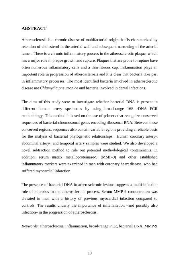
ABSTRACT
Atherosclerosis is a chronic disease of multifactorial origin that is characterized by
retention of cholesterol in the arterial wall and subsequent narrowing of the arterial
lumen. There is a chronic inflammatory process in the atherosclerotic plaque, which
has a major role in plaque growth and rupture. Plaques that are prone to rupture have
often numerous inflammatory cells and a thin fibrous cap. Inflammation plays an
important role in progression of atherosclerosis and it is clear that bacteria take part
in inflammatory processes. The most identified bacteria involved in atherosclerotic
disease are Chlamydia pneumoniae and bacteria involved in dental infections.
The aims of this study were to investigate whether bacterial DNA is present in
different human artery specimens by using broadrange 16S rDNA PCR
methodology. This method is based on the use of primers that recognize conserved
sequences of bacterial chromosomal genes encoding ribosomal RNA. Between these
concerved regions, sequences also contain variable regions providing a reliable basis
for the analysis of bacterial phylogenetic relationships. Human coronary artery,
abdominal artery, and temporal artery samples were studied. We also developed a
novel subtraction method to rule out potential methodological contaminants. In
addition, serum matrix metalloproteinase9 (MMP9) and other established
inflammatory markers were examined in men with coronary heart disease, who had
suffered myocardial infarction.
The presence of bacterial DNA in atherosclerotic lesions suggests a multiinfection
role of microbes in the atherosclerotic process. Serum MMP9 concentration was
elevated in men with a history of previous myocardial infaction compared to
controls. The results underly the importance of inflammation –and possibly also
infection– in the progression of atherosclerosis.
Keywords: atherosclerosis, inflammation, broadrange PCR, bacterial DNA, MMP9
11
TIIVISTELMÄ
Valtimonkovettumatauti eli ateroskleroosi on monien tekijöiden aiheuttama
krooninen tauti, jolle on ominaista kolesterolin kertyminen valtimon seinämään ja
valtimon ontelon kaventuminen. Ateroomaplakissa vallitsee krooninen tulehdus,
jolla on merkittävä rooli aterooman kasvamismekanismeissa ja repeämisessä.
Repeämälle alttiissa plakeissa on usein paljon tulehdussoluja ja ohut sidekudoskatto.
Inflammaatiolla on tärkeä rooli ateroskleroosin kehityksessä ja on ilmeistä, että
bakteerit osallistuvat inflammaation syntyyn. Ateroskleroosiin liittyvistä
bakteereista tunnetuimpia ovat Chlamydia pneumoniae ja hammasinfektioita
aiheuttavat bakteerit.
Tutkimuksessa selvitettiin broadrange 16S rDNA polymeraasiketjureaktion (PCR)
avulla, löytyykö bakteerien DNA:ta ihmisperäisistä valtimonäytteistä. Menetelmä
perustuu sellaisten alukkeiden käyttöön, jotka tunnistavat bakteerien
konservoituneita sekvenssejä ribosomaalista RNA:ta koodaavasta
kromosomaalisesta geenialueesta. Näiden konservoituneiden alueiden välissä on
variaabeleita alueita, joiden perusteella voidaan määrittää bakteerien fylogeneettisiä
sukulaisuussuhteita. Sepelvaltimo, vatsaaortta ja ohimovaltimonäytteet olivat
tutkimuksen kohteena. Kehitimme myös subtraktioanalyysin, jolla metologisia
kontaminantteja voidaan välttää. Lisäksi tutkimuksessa mitattiin seerumin matriksin
metalloproteinaasi9 (MMP9) pitoisuutta ja muita tulehdustekijöitä sellaisilta
sepelvaltimotautipotilailta, jotka olivat sairastaneet sydäninfarktin.
Ateroskleoottisissa plakeissa oli bakteerijälkiä taudinaiheuttajista, joka tukee
ateroskleroosin multiinfektioteoriaa. Seerumin MMP9 pitoisuus oli koholla
sydäninfarktin sairastaneilla potilailla verrattuna kontrolleihin. Tulokset osoittavat,
että valtimon seinämän tulehdus – ja mahdollisesti myös bakteeriinfektiot – ovat
yhteydessä ateroskleroosin kehitykseen.
Avainsanat: ateroskleroosi, inflammaatio, broadrange PCR, bakteeri DNA, MMP9
12
INTRODUCTION
Atherosclerosis is a disease of large and mediumsized arteries which leads to
intimal thickening and loss of arterial elasticity. The disease often begins in
childhood (Stary 1989, PDAY research group 1993), but clinical symptoms may not
appear until middle age or later when the arterial lesions precipitate organ injury.
The clinical complications of atherosclerosis, including myocardial infarction and
stroke, are the major cause of morbidity and mortality among men in the developed
world (Ross 1999b).
Inflammation has been shown to play an important role in the clinical complications
of atherosclerosis. The activated macrophages abundant in atheroma can produce
proteolytic enzymes, such as matrixdegrading metalloproteinases (MMPs), capable
of degrading the collagen that lends strength to the plaque’s protective fibrous cap,
rendering the cap thin, weak, and susceptible to rupture (Dollery et al. 1995, Galis &
Khatri 2002). Plaque disruption takes place often at the shoulder region between the
plaque and the adjacent vessel wall where the fibrous cap is the thinnest (Richardson
et al. 1989). The inflammation in atherosclerosis has been associated with infection
by various bacterial and viral pathogens (Epstein et al. 1999). Pathogens that have
been implicated include Chlamydia pneumoniae, cytomegalovirus, herpes simplex
virus, Helicobacter pylori, and periodontal infections (for reviews, see Epstein et al.
1999, Fong 2000, Leinonen & Saikku 2002). Other classical risk factors of
atherosclerosis are high serum cholesterol, hypertension, diabetes mellitus, and
smoking (Vartiainen et al. 1994, Burke et al. 1997, Wilson et al. 1998).
Ample evidence suggests that infectious agents may play a role in the pathogenesis
of atherosclerosis and in the clinical manifestations of vascular disease. However,
most of the studies have concentrated on the potential role of only one specific
microbe, such as C. pneumoniae (Campbell et al. 1995, Ramirez 1996) or H. pylori
(Ameriso et al. 2001, Kaplan et al. 2006). Less frequently, a broader multi
microbial presence in the etiology of the atherosclerotic plaque has been suggested
(Watt et al. 2003, Ott et al. 2006). This thesis aimed at studying which bacterial
signatures are present in human atherosclerotic arterial specimens as well as

13
demonstrating the presence of MMP9 and other inflammatory markers from sera of
subjects with complications of atherosclerosis.
14
REVIEW OF THE LITERATURE
1. The structure of blood vessels
The artery wall has three layers, the intima, the media, and the adventitia (Stary et
al. 1992). A single layer of endothelial cells covers the luminal side of the intima.
The intima consists of a narrow subendothelial space containing smooth muscle
cells (SMC), but also macrophages, Tlymphocytes, and mast cells may be present.
The intima is separated from the media by the internal elastic lamina, a fenestrated
sheet of collagen. The media is the thickest layer, responsible for the structural
integrity of the arterial wall. The outermost layer is the adventitia, which consists
mainly of fibroblasts and connective tissue together with nerves and small blood and
lymph vessels, termed vasa vasorum. The media and the adventitia are separated by
the external elastic lamina (Geer & Haust 1972, Stary et al. 1992).
2. Classification of atherosclerotic lesions
Atherosclerosis begins early in life with accumulation of low density lipoprotein
(LDL) cholesterol and monocytes in the intima of large arteries (McGill &
McMahan 1998). LDL is trapped in the intimal extracellular matrix, or it passes
through the internal elastic lamina and moves further into the media (Williams &
Tabas 1995, Williams & Tabas 1998, Kadar & Glasz 2001, Skålen et al. 2002). In
the intima, LDL may be taken up by monocytederived macrophages (Witztum &
Steinberg 1991) leading to foam cell formation. The earliest lesion, the fatty streak,
is primarily caused by the accumulation of lipid droplets in macrophages (Stary et
al. 1994). A part of the fatty streaks are disposed to progression and may proceed to
intermediate lesions and further to advanced lesions such as the atheroma,
fibroatheroma, and complicated fibroatheroma (Stary et al. 1994).
Since the 1990s, a specific classification of atherosclerotic lesions has been
established by the Committee on Vascular Lesions of the Counsil of Atherosclerosis,
American Heart Association (Stary et al. 1992, Stary et al. 1994, Stary et al. 1995).
15
This classification is based on histology and it divides the lesions into early and
advanced types.
The early lesions are classified as types I, II, and III. Type I lesions contain
macrophages with intracellular lipid droplets (foam cells) without other significant
histological changes (Stary et al. 1994). Type II lesions are those lesions that are
also known as fatty streaks. Fatty streaks may be seen on the inner surfaces of
arteries as relatively flat, yellow colored streaks or patches (Stary et al. 1994). They
consist mainly of macrophage foam cells, but intimal SMCs may contain lipid
droplets as well. Fatty streaks do not obstruct arterial blood flow. Type III lesions
form the bridge between minimal and advanced lesions as they contain extracellular
lipid droplets that accumulate into small pools lying below the macrophage layer
(Stary et al. 1994).
Advanced atherosclerotic lesions are referred to as type IV – VI. The type IV lesion
is the atheroma. It may not cause any symptoms, and these lesions are often not
visible by angiography. These lesions contain an accumulation of extracellular lipid
in the intima, known as the lipid core. The major cell types on top of the lipid core
are macrophages and SMCs with and without intracellular lipid (Stary et al. 1995).
There are also mast cells and lymphocytes in this region (Kovanen et al. 1995,
Kaartinen et al. 1998). These plaques have a thin cap of connective tissue above the
lipid core, which makes the plaque prone to rupture. Type V lesions have a
prominent fibrous cap of connective tissue between the lipid core and endothelium.
This type of lesion is referred to as a fibroatheroma (or type Va lesion). A type V
lesion, in which the lipid core or other parts of the lesion are calcified, is called a
calcified lesion (or type Vb lesion). A type V lesion in which the lipid core is absent
and lipid in general is minimal may be referred as type Vc. These lesions cause
various narrowing of arteries. The type V lesions are prone to progress to type VI
lesions by rupturing and subsequent formation of mural thrombi, causing further
narrowing of the arterial lumen. Type VI complicated lesions have a disrupted
structure including hematoma (type VIa), hemorrage (type VI b), or thrombosis (type
VIc). These lesions cause acute clinical manifestations of atherosclerosis (Stary et al.
1995).
16
The present study examined lesions ranging from fibroproliferative thickening
observed in temporal arteritis (TA) to complicated atherosclerotic lesions seen in
coronary artery disease and abdominal atherosclerosis. TA is a disease characterized
by acute inflammation in medium to large arteries in elderly people, especially
females (Bengtsson & Malmvall 1981, Nordborg et al. 2000, Salvarani et al. 2002a).
An environmental infective trigger for TA has long been supposed on the basis of the
clinical presentation as flulike symptoms are frequently seen at the onset of disease
and are followed by a marked acutephase response (Larson et al. 1984).
Abdominal aortic aneurysm (AAA) is a focal, balloonlike dilation of the terminal
aortic segment that affects the infrarenal portion of the abdominal aorta. AAA is
most likely related to systemic atherosclerosis since the diseases share risk factors
(Johnson et al. 1985, Reed et al. 1992, Van der Vliet & Boll 1997). Arteriosclerosis
obliterans (ASO) is another characteristic disorder of atherosclerosis, in which
arterial obstruction occurs in arteries supplying blood to lower extremities (Second
European Consensus Document on chronic critical leg ischemia 1991).
3. Infection, inflammation and atherosclerosis
3.1. The role of inflammation in atherosclerosis
Numerous inflammatory mediators play a role in early atherosclerotic lesion
formation. They take a part in the early events including attracting monocyte
leukocytes from circulating blood by vascular endothelial cells leading to their
migration into the intima, transitioning monocytes into macrophages and eventually
into lipidladen foam cells (Ross 1999a,b, Libby et al. 2002, Järvisalo et al. 2006).
Later in the process, inflammatory mediators can weaken the protective fibrous cap
of the atheroma possibly leading to thrombosis and the occurrence of acute
cardiovascular events such as unstable angina pectoris and myocardial infarction
(MI) (Ross 1999a,b, Libby et al. 2002).
17
3.1.1 Creactive protein
Creactive protein (CRP), an acute phase protein which is named for its capacity to
precipitate the somatic Cpolysaccharide of Streptococcus pneumoniae, is
considered to be the most valuable inflammatory marker to predict cardiovascular
events, including myocardial infarction, stroke, and vascular mortality (Haverkate et
al. 1997, Ridker et al. 1998a, Ferreiros et al. 1999, Lindahl et al. 2000, Rost et al.
2001, Ridker et al. 2002). It is synthesized mainly by hepatocytes under the control
of several cytokines, with interleukin6 being a primary stimulus (Pepys &
Hirschfield 2003). Although the principal source of CRP is the liver, recent data
have shown that vascular tissue itself can also produce CRP (Yasojima et al. 2001,
Ishikawa et al. 2004, Wilson et al. 2007). The major producers of CRP in
atherosclerotic lesions are smooth muscle cells and macrophages (Yasojima et al.
2001). In addition to predicting recurrent cardiovascular events in patients with
atherosclerotic disease, elevated levels of CRP are one of the strongest predictors of
progressive vascular disease (Van der Meer et al. 2002) and future cardiovascular
events in apparently healthy men and women (Ridker et al. 1997, Ridker et al.
1998b, Koenig et al. 1999).
3.1.2 Antibodies of oxidized LDL
Oxidized LDL cholesterol (OxLDL) has a significant role in the progression of
atherosclerosis (Steinbrecher & Pritchard 1989, Parthasarathy et al. 1990,
Parthasarathy & Rankin 1992). OxLDL accumulates in macrophages, accelerates the
development of foam cells and stimulates the monocyte adhesion to endothelium
(Witztum 1991, Witztum & Steinberg 1991, Jialal & Devaraj 1996). In addition,
oxLDL stimulates the synthesis of MMP9 in macrophages by increasing the
expression of mRNA and protein synthesis, influencing possibly the rupture of
atherosclerotic plaques (Xu et al.1999). The small, dense LDL particles are more
susceptible to oxidation, which mainly takes place in the arterial wall (Krauss 1995,
Galeano et al. 1998)
18
OxLDL is also immunogenic and it induces the formation of autoantibodies
(Palinski et al. 1989, Palinski et al. 1994, YläHerttuala et al. 1994, Palinski et al.
1996). The antibody titers of oxidized LDL have been shown to correlate with the
severity of atherosclerosis and it has been thought to predict myocardial infarction
(MI) (Salonen et al. 1992, Puurunen et al. 1994, Wu et al. 1997). However,
contradictory results have also been reported. A study by van de Vijver and her
colleagues (van de Vijver et al. 1996) did not discover any association between
oxLDL autoantibodies and the extent of coronary stenosis. In addition, lower
oxLDL autoantibody levels in elderly patients with ischemic stroke (Cherubini et al.
1997) and in acute MI compared to controls have been reported (Schumacher et al.
1995).
3.1.3 Matrix metalloproteinase9
Activated cells in inflamed atherosclerotic plaques, such as macrophages, smooth
muscle cells, and endothelial cells, secrete matrixdegrading proteases including
matrix metalloproteinases (MMPs). Elevated levels of MMP1, MMP2, MMP3,
MMP7, MMP9, and MMP12 have been observed in atherosclerotic plaques
(Henney et al. 1991, Galis et al. 1994a, Brown et al. 1995, Nikkari et al. 1995,
Halpert et al. 1996, Li et al. 1996). Vascular cells also produce tissue inhibitors of
matrix metalloproteinases (TIMPs) that inhibit MMP activity (Galis et al. 1995).
MMP9, also known as gelatinase B or 92 kDa gelatinase, is associated with arterial
inflammation. It has been indicated to be highly expressed in the disruptionprone
regions of atherosclerosic plaques (Galis et al. 1994a, Brown et al. 1995, Zaltsman
& Newby 1997) whereas nonatherosclerotic arteries have not been shown to
contain detectable amounts of MMP9 (Brown et al. 1995). Macrophages are the
major source of MMP9 although a variety of other cell types also express this
protease (Galis et al. 1994b, Brown et al. 1995, Kaartinen et al. 1998, Nelimarkka et
al. 1998). Increased serum levels of MMP9 have been detected both in stable and
unstable CAD (Kai et al. 1998, Inokubo et al. 2001, Noji et al. 2001).
19
3.2 The role of infection in atherosclerosis
Microorganisms may play a role in different stages of atherogenesis by numerous
mechanisms of action. They may infect vascular endothelium directly, thus initiating
the inflammatory response to induce atherogenesis (Libby et al. 1997, Nieto 1998,
Albert 2000, Fong 2000, O’Connor et al. 2001, Fong 2002). In addition, although
the induction or initial injury to the endothelial wall would be caused by another
agent or factor such as hypercholesterolemia or hypertension, microorganisms
enhance atherogenesis through number of mechanisms. These include recruitment
and stimulation of proinflammatory cytokines and tissue growth factors in the
arterial wall (Coombes & Mahony 1999, Summersgill et al. 2000), as well as
enhancement of LDL accumulation through stimulation of macrophage scavenger
or LDLreceptors (Zhou et al. 1996, Kalayoglu & Byrne 1998, Epstein et al. 1999,
Fong 2002). Microorganisms could also indirectly influence the development of
atherosclerosis by systemic changes through initiation of immunological responses
to microbial antigens, which crossreact with normal human antigens (Libby et al.
1997, Muhlestein 1998, Nieto 1998, Albert 2000). The most frequently studied
bacteria for their possible role in the process of atherosclerosis are Chlamydia
pneumoniae, Helicobacter pylori and bacteria involved in dental infections (for
reviews, see Fong 2000, Leinonen & Saikku 2002, Muhlestein & Anderson 2003).
3.2.1 Chlamydia pneumoniae
Chlamydia pneumoniae, a gramnegative obligate intracellular bacterium, is a
widespread respiratory pathogen. After the first report by Saikku and colleagues in
1988 (Saikku et al. 1988), several seroepidemiological and other studies have
demonstrated both positive and negative associations between chronic C.
pneumoniae infection and atherosclerosis. Strong associations have been verified by:
(a) seroepidemiological surveys showing that patients with cardiovascular disease
have higher titres of antiC. pneumoniae antibodies compared with control patients
(Saikku et al. 1992, Romano Carratelli et al. 2006); (b) detecting the microbe within
atheroma but not in adjacent normal tissue by electron microscopy,
immunocytochemistry, and polymerase chain reaction (PCR) (Kuo et al. 1993,
20
Grayston et al. 1995, Ramirez 1996, Jackson et al. 1997) and by culturing the
microbe from atherosclerotic tissue (Ramirez 1996, Maass et al. 1998); and (c)
showing that C. pneumoniae can either initiate development of atherosclerosis or
generate exacerbation of lesions in rabbit and mouse animal models, respectively
(Fong et al. 1997, Laitinen et al. 1997, May et al. 2003). Numerous studies have
also failed to: (i) confirm the association between antibodies to C. pneumoniae and
cardiovascular disease (Altman et al. 1999, Nobel et al. 1999, Chandra et al. 2001,
Kähler et al. 2001) (ii) detect C. pneumoniae in atherosclerotic lesions by PCR
(Weiss et al. 1996, Ong et al. 2001, Apfalter et al. 2004, Maraha et al. 2004); (iii)
detect increased lesion development after C. pneumoniae infection in ApoE
deficient mice (AaltoSetälä et al. 2001, Caligiuri et al. 2001). In addition, some
metaanalyses of prospective studies have not provided strong evidence of an
association between atherosclerosis and C. pneumoniae (Danesh 1999, Danesh et al.
2000).
3.2.2 Helicobacter pylori
Helicobacter pylori is a gramnegative bacterium whose only natural niche is in the
human stomach, where it resides in the mucus layer and on the gastric epithelium
(Dunn et al.1997). The association between H. pylori and atherosclerosis was first
suggested by Mendall and his colleagues in 1994 (Mendall et al. 1994). Since that, a
number of seroepidemiological studies have suggested that patients with ischemic
heart disease (IHD) have higher titers of antiH. pylori antibodies compared to
controls (Pasceri et al. 1998, Danesh et al. 1999a, Pellicano et al. 1999, Alkout et al.
2000) although previous published studies are inconsistent (Folsom et al. 1998,
Strachan et al. 1998, Zhu et al. 2002). In addition, molecular studies investigating
the presence of H. pylori in atherosclerotic plaques by PCR are contradictory.
Several studies have not been able to detect H. pylori from atherosclerotic samples
(Blasi et al. 1996, Malnick et al. 1999, Danesh et al. 1999b, Dore et al. 2003, Weiss
et al. 2006) whereas others have reported its presence (Farsak et al. 2000, Ameriso
et al. 2001, Kaplan et al. 2006).
21
3.2.3 Dental infections
Gramnegative bacteria, including Porphyromonas gingivalis, Actinobacillus
actinomycetemcomitans, and Prevotella intermedia, are related with gingivitis and
periodontitis (Williams 1990) whereas grampositive bacteria, particularly
Streptococcus mutans, are related with etiology of caries (Loesche 1986). Mattila
and his colleaques (Mattila et al. 1995) were the first to show that dental infections
evaluated by the pantomography index were significantly more common in patients
with advanced coronary atherosclerosis. After that, DNA of periodontitisrelated
bacteria has been identified in different atheromatous samples in several recent
studies (Haraszthy et al. 2000, Okuda et al. 2001, Stelzel et al. 2002, Taylor
Robinson et al. 2002, Kurihara et al. 2004, Fiehn et al. 2005). S. mutans has been
detected in atheromatous plaque specimens as well (Kozarov et al. 2006, Nakano et
al. 2006). Furthermore, a recently published study by Kozarov et al. (2005)
demonstrated signs of viable Actinobacillus actinomycetemcomitans and
Porphyromonas gingivalis in human atherosclerotic plaque material by using cell
culture invasion assays and immunofluorescent microscopy. The association
between periodontal microbes and atherosclerosis is speculated by the fact that the
oral cavity is well vascularized, and the occurrence of infection in this site provides
an instant and direct route to the bloodstream. Professional dental manipulations and
daily oral care practices, such as tooth brushing and flossing, lead to seeding of the
oral flora to the bloodstream (Silver et al. 1977, Heimdahl et al. 1990, Roberts et al.
1997, Seymour et al. 2000, Bhanji et al. 2002, Rajasuo et al. 2004, Forner et al.
2006). However, even though many epidemiological studies have indicated
significant relations between periodontal diseases and atherosclerosis (Mattila et al.
1995, Beck et al. 1996, Joshipura et al. 1996, Morrison et al. 1999), there are some
epidemiological studies that have not been able to show such relations (Hujoel et al.
2000, Howell et al. 2001).
22
4. Molecular identification of bacteria
4.1 rRNA molecules
Ribosomes, which are large complex ribonucleotide particles, are composed of
several different ribosomal RNA (rRNA) molecules and over 50 proteins, organized
into a large (LSU) and small (SSU) subunits. Homologous SSU and LSU major
rRNA molecules can be found in all bacteria, where they play a role in protein
synthesis (Noller 1984, Gutell et al. 1994, Lodish et al. 2000, Lewin 2004).
Prokaryotic SSU has a sedimentation value of approximately 30S consisting one
major RNA molecule and 21 proteins. Prokaryotic LSU has a sedimentation rate of
about 50S consisting one major and one minor RNA molecule and 31 different
protein molecules (Lodish et al. 2000, Berg et al. 2002, Lewin 2004). rRNA
molecules are named according to their sedimentation rate. In prokaryotes, the
rRNA molecule of SSU is named as 16S rRNA; the LSU containing two rRNA
molecules are named 23S rRNA and 5S rRNA. 16S and 23S rRNA molecules are of
large size: about 1540 and 2900 bases, respectively (Lodish et al. 2000, Lewin
2004).
4.2 Bacterial taxonomy
Bacterial taxonomy, or systematics, may be defined as a scientific study of the
diversity of organisms and their relationships (Rademaker & Savelkoul 2004).
Nomenclature, classification, and identification are its three parts. The purpose of
classification is to define the characteristics of a taxon and its position in the
hierarchical system. This information is used when a new isolate is defined before it
can be identified as a member of a known taxon. Nomenclature governs the
taxonomic names to the taxa e.g. according to the International Code of
Nomeclature of Bacteria (Sneath 1992). In identification, members of different
taxonomic unit are named on the basis of characteristics or properties of the
organism that distinguishes them from others (Rademaker & Savelkoul 2004).
23
Traditionally, classification and identification of bacteria in the clinical laboratory
have been based on isolation of bacterial strains as pure culture and analysis of the
isolate´s phenotypic characters such as certain biochemical properties (Kloos &
Schleifer 1975, Schleifer & Kloos 1975). No phylogenybased classification scheme
of bacteria existed until the rRNA sequencebased methods were introduced (Woese
1987).
4.3 Broadrange rRNA approach
The phylogenetic classification of microorganisms based on rRNA molecule
sequences offers a distinct framework which can be used to develop molecular tools
for detection and identification of microbes (Stahl & Amann 1991, Stahl & Kane
1992). The rRNA genes have well conserved domains which are suitable as sites for
PCR primers that recognize large groups of organisms, and variable domains that
provide signatures for more precise identification at phylogenetic levels below the
level originally targeted by the primers (Gutell et al. 1985, Lane et al. 1985, Woese
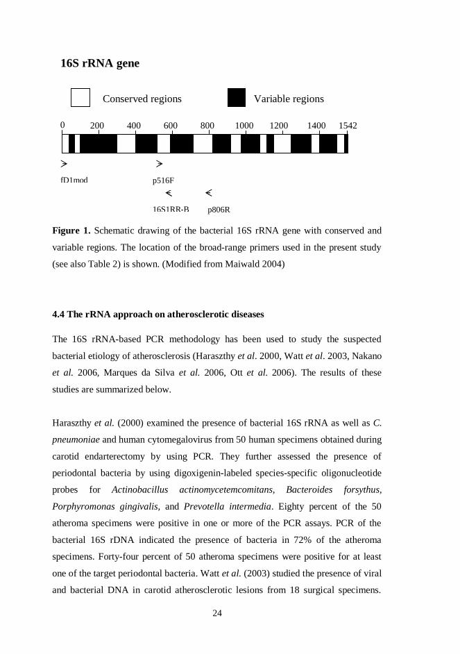
1987, Maiwald 2004). An overview of both conserved and variable regions in the
bacterial 16S rRNA gene is given in Figure 1. Thus, the power of broadrange rRNA
PCR lies in the relative absence of selectivity, so that –at least in theory– any kind of
bacterium present in a sample can be detected and identified without prior
cultivation or any knowledge of its nature (Relman et al. 1990, Kroes et al. 1999,
Maiwald 2004).
24
Figure 1. Schematic drawing of the bacterial 16S rRNA gene with conserved and
variable regions. The location of the broadrange primers used in the present study
(see also Table 2) is shown. (Modified from Maiwald 2004)
4.4 The rRNA approach on atherosclerotic diseases
The 16S rRNAbased PCR methodology has been used to study the suspected
bacterial etiology of atherosclerosis (Haraszthy et al. 2000, Watt et al. 2003, Nakano
et al. 2006, Marques da Silva et al. 2006, Ott et al. 2006). The results of these
studies are summarized below.
Haraszthy et al. (2000) examined the presence of bacterial 16S rRNA as well as C.
pneumoniae and human cytomegalovirus from 50 human specimens obtained during
carotid endarterectomy by using PCR. They further assessed the presence of
periodontal bacteria by using digoxigeninlabeled speciesspecific oligonucleotide
probes for Actinobacillus actinomycetemcomitans, Bacteroides forsythus,
Porphyromonas gingivalis, and Prevotella intermedia. Eighty percent of the 50
atheroma specimens were positive in one or more of the PCR assays. PCR of the
bacterial 16S rDNA indicated the presence of bacteria in 72% of the atheroma
specimens. Fortyfour percent of 50 atheroma specimens were positive for at least
one of the target periodontal bacteria. Watt et al. (2003) studied the presence of viral
and bacterial DNA in carotid atherosclerotic lesions from 18 surgical specimens.
200 400 600 800 1000 1200 1400 15420
Conserved regions Variable regions
16S rRNA gene
p806R16S1RRB
fD1mod p516F
25
Specific primers for C. pneumoniae, cytomegalovirus, herpes simplex virus 1 and 2,
and bacterial 16S rDNA were used in PCR assays. Only herpes simplex 1 DNA (3/
18 specimens) and bacterial 16S rDNA (8/18 specimens) were detected from carotid
atherosclerotic lesions. Detected bacterial DNA was shown to belong to the human
flora (oral, genital, or faecal) or the environment. Nakano et al. (2006) analyzed the
presence of streptococcal species in diseased heart valve (n=35) and atherosclerotic
plaque (n=27) specimens, as well as in dental plaque samples (n=32) from the same
subjects by using oral streptococcal species specific primer sets for the
glucosyltransferase gene as well as broad range bacterial 16S rDNA PCR with direct
sequencing. Streptococcal species were detected by both PCR methods. Especially
S. mutans was significantly prevalent. Ott et al. (2006) studied 16S rDNA signatures
in atherosclerotic tissue obtained through catheterbased atherectomy of 38 patients
with CHD, control material from postmortem patients (n=15), and heartbeating
organ donors (n=11) using clone libraries, denaturating gradient gel analysis, and
fluorescence in situ hybridization. Bacterial DNA was found in all CHD patients by
conserved PCR but not in control material or in any of the normal/unaffected
coronary arteries. Presence of bacteria in atherosclerotic lesions was confirmed by
fluorescence in situ hybridization. Over 50 different species were demonstrated in
>1500 clones from a combined library and confirmed by denaturating gradient gel
analysis. Several of the species detected in human coronaries in this study
represented commensal human flora (skin, respiratory, and oral cavity). Marques da
Silva et al. (2006) studied the presence of bacterial DNA in 10 specimens from
arterial wall of aortic aneurysms. 27 different species were identified among 83
clones sequenced. These bacteria included oral as well as environmental bacteria.
The results of these studies are summarized in Table 1.
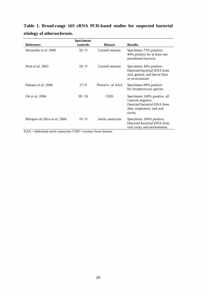
26
Table 1. Broadrange 16S rRNA PCRbased studies for suspected bacterial
etiology of atherosclerosis.
ReferenceSpecimens/
controls Disease ResultsHaraszthy et al. 2000 50 / 0 Carotid stenosis Specimens 72% positive;
44% positive for at least oneperiodontal bacteria
Watt et al. 2003 18 / 0 Carotid stenosis Specimens 44% positive.Detected bacterial DNA fromoral, genital, and faecal floraor environment
Nakano et al. 2006 27/ 0 Thoracic or AAA Specimens 89% positivefor Streptococcus species
Ott et al. 2006 38 / 26 CHD Specimens 100% positive, allcontrols negative.Detected bacterial DNA fromskin, respiratory, and oralcavity
Marquez da Silva et al. 2006 10 / 0 Aortic aneurysm Specimens 100% positive.Detected bacterial DNA fromoral cavity and environment
AAA = abdominal aortic aneurysm; CHD= coronary heart disease.
27
AIMS OF THE STUDY
Inflammation is an important defense mechanism against infection. Chronic or
recurrent infections may cause sustained inflammation that has been linked to
increased risk of atherosclerosis. Several epidemiological, biological, experimental,
and clinical studies have shown that infections may play a role in atherosclerosis and
related diseases.
The aim of this study was to investigate whether bacterial DNA is present in arterial
walls of different clinical specimen by using BrPCR methodology. In addition,
serum inflammatory markers in male subjects with a history of previous MI were
compared to controls.
The spesific aims were:
1. to study whether bacterial DNA are present in temporal artery tissue of
patients with temporal arteritis (TA).
2. to elucidate whether bacterial DNA is present in coronary specimens
obtained from left anterior descending coronary arteries of subjects with
sudden deaths of cardiovascular and other causes, as verified by autopsy.
3. to define bacterial DNA signatures in surgically removed sterile abdominal
aorta samples of patients with aortic atherosclerosis.
4. to examine whether serum MMP9 concentration may reflect inflammatory
pathologic processes that are related to progression of atherosclerosis in
subjects with a history of MI.
28
SUBJECTS, MATERIALS AND METHODS
1. Clinical specimens (IIII)
Temporal artery specimens (I) were collected from 27 patients with temporal
arteritis (20 women, 7 men; mean age 75 years; range 61 to 99 years). Five
uninvolved temporal arteries were also included (4 women, 1 man; mean age 64
years; range 54 to 80 years). A lung sample obtained from a 91 year old man, who
died of sepsis and bacterial pneumonia following gastrointestinal surgery, acted as a
positive control. Coronary specimens (II) were obtained from the proximal part of
the main trunk of the left anterior descending coronary artery (LAD) of five subjects
who died of sudden coronary causes (4 men, 1 woman; mean age 69 years; range 56
to 79 years) and five controls (4 men, 1 woman; mean age 34 years; range 26 to 48
years) within 3 days after death. At autopsy, the proximal part of the LAD was
removed and immediately frozen at –70 °C. Abdominal aortic specimens (III) were
obtained in abdominal aortic surgeries of 20 patients (18 men and 2 women; mean
age 65 years; range 45 to 81 years) who had a medical history of atherosclerosis
obliterans (n= 4) or who had suffered from abdominal aortic aneurysm (n= 16).
None of the patients were known to have any clinical signs of infection in the days
before the surgery. After surgical removal, the abdominal aortic tissue was placed in
a sterile container, kept at +4 °C, and transported within the same day to the
laboratory on ice. All patients considered for the study gave written informed
consent before surgery. The study protocols were approved by the Ethical
Committee of Tampere University Hospital.
2. Preparation of clinical tissue specimens (IIII)
2.1 DNA isolation from paraffin embedded temporal artery samples
Paraffinembedded transverse sections of temporal arteries and lung sample were
deparaffinized with xylene and ethanol. Tissues were digested with proteinase K
(final concentration 0.1 mg/ ml) at 56 °C for 3 hours. Proteinase K was heat
29
inactivated (8 minutes at 95 °C) and the reaction mixture was centrifuged (10
minutes at maximum speed) and the supernatant was used for PCR analysis.
2.2 DNA isolation from coronary and abdominal aortic samples
Clinical specimens adjacent to the cryostat sections were digested with proteinase K
(final concentration 0.1 mg/ ml) at 56 °C for 37 hours. After proteinase K
treatment, the samples were heated 8 minutes at 95 °C to inactivate proteinase K,
and then the reaction mixture was centrifuged (15 minutes at maximum speed) and
the supernatant was used for PCR analysis.
3. Broadrange bacterial PCR (IIII)
3.1 PCR
Exact procedures for DNA amplifications are described in original communications
(IIII). A location of used broadrange primers for 16S rDNA is shown in Figure 1.
The total reaction volume was 50 µl, containing either 0.5 µl or 5µl of the
supernatant from DNA extraction, 10 to 100 pmol/ µl of each primer (Table 2), and
standard amounts of HotStarTaq Master Mixreagent containing 2.5 units
HotStarTaq DNA Polymerase, 15mM MgCl2, and 200µM of each dNTP (Qiagen
GmbH, Hilden, Germany). PCR reactions were performed in sterilized 500 µl thin
wall tubes. Thermal cycling conditions are given in the original communications (I
III). PCR was performed using Eppendorf Mastercycler Gradient (Eppendorf,
Hamburg, Germany) (I) and TechGene (Techne, Burlington, USA) (IIIII) thermal
cyclers described in the original communications (IIII). Twenty eight to 50 thermal
cycles were used.
30
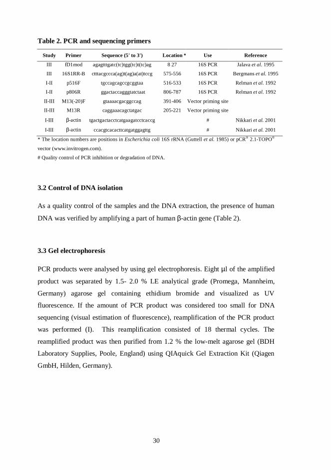
Table 2. PCR and sequencing primers
Study Primer Sequence (5' to 3') Location * Use Reference
III fD1mod agagtttgatc(tc)tgg(tc)t(tc)ag 8 27 16S PCR Jalava et al. 1995
III 16S1RRB ctttacgccca(ag)t(ag)a(at)tccg 575556 16S PCR Bergmans et al. 1995
III p516F tgccagcagccgcggtaa 516533 16S PCR Relman et al. 1992
III p806R ggactaccagggtatctaat 806787 16S PCR Relman et al. 1992
IIIII M13(20)F gtaaaacgacggccag 391406 Vector priming site
IIIII M13R caggaaacagctatgac 205221 Vector priming site
IIII βactin tgactgactacctcatgaagatcctcaccg # Nikkari et al. 2001
IIII βactin ccacgtcacacttcatgatggagttg # Nikkari et al. 2001
* The location numbers are positions in Escherichia coli 16S rRNA (Guttell et al. 1985) or pCR® 2.1TOPO®
vector (www.invitrogen.com).
# Quality control of PCR inhibition or degradation of DNA.
3.2 Control of DNA isolation
As a quality control of the samples and the DNA extraction, the presence of human
DNA was verified by amplifying a part of human βactin gene (Table 2).
3.3 Gel electrophoresis
PCR products were analysed by using gel electrophoresis. Eight µl of the amplified
product was separated by 1.5 2.0 % LE analytical grade (Promega, Mannheim,
Germany) agarose gel containing ethidium bromide and visualized as UV
fluorescence. If the amount of PCR product was considered too small for DNA
sequencing (visual estimation of fluorescence), reamplification of the PCR product
was performed (I). This reamplification consisted of 18 thermal cycles. The
reamplified product was then purified from 1.2 % the lowmelt agarose gel (BDH
Laboratory Supplies, Poole, England) using QIAquick Gel Extraction Kit (Qiagen
GmbH, Hilden, Germany).
31
3.4 Cloning of PCR products (II, III)
Amplified PCR products were ligated into the pCR2.1TOPO vector and
transformed into E. coli cells by using the TOPO TA cloning system (Invitrogen,
Carlsbad, CA, USA). Two separate clone libraries were created from each PCR
product amplified from tissue digest supernatant. Transformants were analyzed by
picking colonies and resuspending them individually in 50 µl of PCR cocktail
consisting 10 pmol/µl of M13 Forward (20) and M13 Reverse primers (Table 2),
and standard amounts of HotStarTaq Master Mixreagent containing 2.5 units
HotStarTaq DNA Polymerase, 15mM MgCl2, and 200µM of each dNTP (Qiagen).
The PCR amplification procedures are described in original communications II and
III. Success in ligation of inserts of the expected size was verified by electrophoresis
in a 2.0 % LE analytical grade (Promega, Mannheim, Germany) agarose gel
containing ethidium bromide and visualized as UV fluorescence. Positive
transformants were purified by using QiaQuick PCR Purification Kit (Qiagen
GmbH, Hilden, Germany).
3.5 DNA Sequencing
Semiautomated sequencing method was used for sequencing of PCR products. The
sequencing primers are shown in Table 2. Taq DyeDeoxy Terminator Cycle
Sequencing kit (Applied Biosystems, Foster City, CA, USA) (IIII) and BigDye
Terminator Cycle sequencing chemistry (Applied Biosystems) were used for
sequencing reactions. Cyclic sequencing steps (96 °C for 30 seconds, 50 °C for 15
seconds, 60 °C for 4 minutes) were repeated 25 times using a TechGene (Techne,
Burlington, USA) (IIIII) thermal cycler. Unincorporated dye terminators were then
removed by using a DyeEx 2.0 Spin kit (Qiagen GmbH, Hilden, Germany).
Sequencing products were resolved using the automated ABI PRISM 310 Genetic
Analyzer (Applied Biosystems).
32
3.6 Sequence analyses
Both DNA strands were analysed and baseediting was performed together with
manual review of the electropherograms (IIII). The nucleotide sequences were
edited and aligned using the Chromas 2.31 (Technelysium) and ClustalW sequence
analysis software (III), and compared with those in the GenBank (Benson et al.
2007) database by using the BLAST search tool (Altschul et al. 1990) available at
the National Center for Biotechnology Information (http://www.ncbi.nlm.nih.gov).
Initial alignment of amplified sequences in original communication III was
performed using the automated 16S rRNA sequence aligner of the ARB software
(Ludwig et al. 2004) against a database of 102, 134 complete and partial rRNA
sequences. Ambiguously and incorrectly aligned positions were aligned manually on
the basis of conserved primary sequence and secondary structure. The phylogenetic
associations were determined from 495 masked positions using a maximum
likelihood algorithm (Felsenstein 1981, Olsen et al. 1994). These associations were
confirmed using a leastsquare fit (De Soete 1983) of JukesCantor corrected
evolutionary distances and maximum parsimony algorithms.
3.7 Arrangement of PCR laboratory and PCR work
To avoid contamination, all steps of PCR analysis (DNA extraction, preparation of
PCR mixtures, amplification, cloning, and analysis of PCR products) were
performed in separated places. PCR reaction mixtures were prepared in a sterile
laminar flow, used only for this purpose. All the PCR reagents and DNA samples
were added to the reaction mixture using pipettes with filtered tips. A sample
preparation control with sterile water that went through the same DNA extraction
steps as the clinical samples served as a negative control. As a positive control,
DNA from Escherichia coli strain B (ATCC 11303) (Sigma, St.Louis, USA) was
used.
33
4. Men with myocardial infarction from the 1997 FINRISK study
(IV)
Every five years since 1972 the National Public Health Institute of Finland has
performed large, crosssectional population surveys related to the risk factors of
coronary heart disease in five geographic areas. The present population originated
from the 1997 FINRISK study, which had a total sample size of 11 500 subjects:
6000 men, 5500 women. The survey included a selfadministered questionnaire that
was sent to the subjects in advance including 165 questions about previous and
existing diseases, which participants returned to the survey site. Subsequently, the
participants’ height, weight, and blood pressure were measured using standard
procedures, and a venous blood specimen was taken. Body mass index (BMI) was
calculated as the ratio of weight (kg) to height squared (m2). Subjects with a history
of previous MI were defined on the basis of the answers. In order to be included in
the MI group, the participants needed to have had a MI that was diagnosed by a
physician. A group of 120 men with a history of diagnosed MI and 250 agematched
controls were selected from the total sample population evenly from different
regions. The study was conducted according to the Helsinki Declaration of 1975 on
Human Experimentation and was approved by the Ethical Committee of Primary
Health Care Clinics in Finland.
5. Lipid analyses (IV)
Total cholesterol and triglycerides were determined from fresh serum samples by an
enzymatic method (Roche Diagnostics, F. HoffmannLa Roche Ltd.). HDL
cholesterol was measured with the same enzymatic method after precipitation of
LDL and VLDL with polyethylene glycol.
6. Measurement of serum CRP and C3complement (IV)
Serum CRP and C3 were determined by using N High Sensitivity CRP and N
Antisera to Human Complement Factor C3c assays as recommended by the
manufacturer (Dade Behring, Marburg, Germany). Immunonephelometry was
34
performed using BNTM Systems (Dade Behring, Marburg, Germany). The
concentrations of the samples were determined versus dilutions of standards of
known concentrations.
7. Determination of autoantibodies against oxLDL (IV)
Antigens for this assay included native LDL (natLDL) prepared from the pooled
plasma of three donors and protected against oxidation by 0.27 mM
ethylenediaminetetraacetic acid (EDTA) and 20 µM butylated hydroxytoluene
(BHT) in phosphate buffered saline (PBS), and oxLDL obtained after 24h oxidation
of the natLDL with 2 µM CuSO4. An enzymelinked immunosorbent assay
(ELISA) was used to determine autoantibodies against oxLDL. Exact procedure is
described in original communication (I). Shortly, conditions of the assay were as
follows: coating concentration 5 g/mL of natLDL and oxLDL on a microtiter plate,
blocking with 2% human serum albumin (HSA) in PBS containing 20 µM BHT and
0.27 mM EDTA, sample dilution 1:30, and conjugate dilution 1:4000 in 0.27 mM
EDTA, 20 µM BHT, 1 % HSA, 0.05 % Tween in PBS. The optical density (OD)
was measured at 492 nm using a microplate reader (Wallac 1420 Victor, Wallac
Oy, Turku, Finland).
8. Determination of serum MMP9 concentration (IV)
For the accurate assessment of serum MMP9, blood samples were centrifuged
within an hour after venipuncture. Aliquots of sera were removed and stored at 70
°C until analysis in a freezer that was not in daily use. Quantitation of
immunoreactive MMP9 was carried out by ELISA (Diabor Ltd, Oulu, Finland).
Recombinant MMP9 was used as standard. The microtiter plate was coated with the
monoclonal antibody (code GE213). The bound proteins from serum and standards
were detected with a secondary polyclonal antibody produced in chicken against
MMP9. A peroxidaselabeled antichickenIgG (Chemicon, USA) was used for
detection of the bound secondary antibody. Ophenylenediamine (OPD) was used to
visualize the peroxidase label. The color formation was measured at 450 nm (Anhos
35
2000 microplate reader) and calculations were done using a Multicalc program
(Wallac, Finland). The monoclonal antibody recognizes both the free MMP9 and
that bound to its inhibitor, tissue inhibitor of metalloproteinases1 (TIMP1).
9. Statistical analysis (IV)
The statistical evaluation was done with a microcomputer using Statistica for
Windows version 5.1 (Statsoft, Inc., Tulsa, OK, USA). Values of the MI group and
controls were compared by Mann Whitney Utest. Logistic regression analysis was
used to evaluate the factors predicting coronary infarction. All results are expressed
as mean (±SD). Pvalues less than 0.05 were considered as being statistically
significant.
36
RESULTS
1. Morphological characteristics of study series (I III)
In the temporal artery sample series (I), all temporal arteritis specimens showed
strong medial and adventitial inflammation characterized by numerous macrophages
and lymphocytes, whereas normal temporal artery samples showed normal
morphology with no inflammation. Nineteen of the 27 temporal arteritis specimens
had characteristic infiltrates of giant cells. No bacterial structures were visualized.
The lung sample, obtained from the autopsy of a man who died of sepsis, exhibited
strong infiltration of polymorphonuclear leukocytes, consistent with an acute
infection, but no bacterial structures. See Figure 1AC in original communication I.
All coronary artery specimens showed different types of atherosclerotic lesions
regardless of cause of death (II). Four of the five cases, whose cause of death was
coronary artery disease (CAD), had type Va or Vb lesions. In addition, all five
control coronary specimens had different types of atherosclerotic lesions from fatty
streaks to small atheroma. The abdominal aorta samples exhibited type Va or Vb
atherosclerotic lesions (III). In addition, all abdominal aorta specimens exhibited
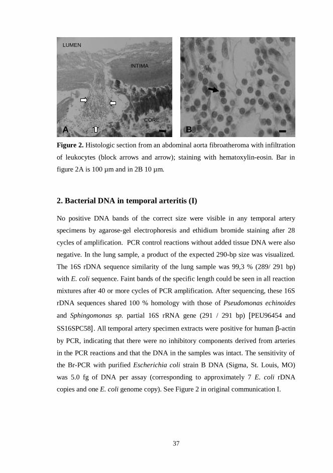
inflammation with variable amouts of leukocyte infiltration. Figures 2A and 2B
show an example from an abdominal aorta (unpublished data).
37
Figure 2. Histologic section from an abdominal aorta fibroatheroma with infiltration
of leukocytes (block arrows and arrow); staining with hematoxylineosin. Bar in
figure 2A is 100 µm and in 2B 10 µm.
2. Bacterial DNA in temporal arteritis (I)
No positive DNA bands of the correct size were visible in any temporal artery
specimens by agarosegel electrophoresis and ethidium bromide staining after 28
cycles of amplification. PCR control reactions without added tissue DNA were also
negative. In the lung sample, a product of the expected 290bp size was visualized.
The 16S rDNA sequence similarity of the lung sample was 99,3 % (289/ 291 bp)
with E. coli sequence. Faint bands of the specific length could be seen in all reaction
mixtures after 40 or more cycles of PCR amplification. After sequencing, these 16S
rDNA sequences shared 100 % homology with those of Pseudomonas echinoides
and Sphingomonas sp. partial 16S rRNA gene (291 / 291 bp) [PEU96454 and
SS16SPC58]. All temporal artery specimen extracts were positive for human βactin
by PCR, indicating that there were no inhibitory components derived from arteries
in the PCR reactions and that the DNA in the samples was intact. The sensitivity of
the BrPCR with purified Escherichia coli strain B DNA (Sigma, St. Louis, MO)
was 5.0 fg of DNA per assay (corresponding to approximately 7 E. coli rDNA
copies and one E. coli genome copy). See Figure 2 in original communication I.
LUMEN
INTIMA
CORE
A B
38
3. Bacterial DNA in human coronary atheromas (II)
Ten coronary arteries from 10 autopsy cases were studied. All of the samples were
positive by βactin PCR. The sequence comparison revealed that all coronary
specimens contained DNA sequences of oral microflora. The bacterial species
identified after cloning and sequencing are summarized in Table 1 of original
communication II. The finding of Escherichia coli sequences that were detected
both from two coronary arteries and extraction control reagents was omitted as their
potential origin as methodological contaminants possibly from Taq DNA
polymerase (Nikkari et al. 2001) could not be ruled out.
4. Bacterial DNA in abdominal aorta atherosclerotic lesions (III)
Of all 20 abdominal aorta samples analyzed, 11 (55%) were positive by the 16S
rDNA PCR done with 38 cycles. All the five negative control reactions were
negative when analyzed as the clinical samples. In order to rule out methodological
contaminants, 5 control samples without tissue extract were given 12 more PCR
cycles than the aorta samples (total of 50 cycles) with subsequent cloning and
sequencing. A bacterial diversity of over 45 different species was demonstrated in
160 sequences from 11 patient samples (subjects AK) and the 5 control samples.
Mean bacterial diversity in atheromas was then 5.5 ± 1.3. The sequences of positive
clinical samples and the sensitized negative controls were included in the systematic
phylogenetic analysis. In order to clean out possible background sequencetypes
deriving from the DNA purification and PCR amplification reagents, the patient
bacterial sequencetypes that showed over 99% similarity to those from sensitized
controls were omitted. After this subtraction procedure, all sequencetypes from two
initially positive patient samples (subjects G and J) were dropped out from the final
results as their potential origin as methodological contaminants could not be ruled
out. Finally, the mean number of bacterial sequencetypes showing diversity of 99%
or less within each group from the remaining 9 patient samples was 2.2 ±1.2, mean
length of sequences being 506.6 ± 22.2 bp.
39
Phylogenetic relationships among the atherosclerosis associated bacterial 16S rDNA
sequences, the relatedness of the consensus sequence of each abdominal aorta
sequencetype with those available at GenBank, and a total of 29 potential non
relevant sequencetypes representing 26 species that were present in the libraries
from both abdominal aorta and negative control PCR products are summarized in
original communication III (Figure 1, Table 1, and Table 2, respectively).
5. Inflammatory markers in men with a history of myocardial
infarction (IV)
The MI group had lower serum total cholesterol concentrations than controls, but
when subjects with cholesterollowering medication were excluded, there was no
variation in total cholesterol between the groups, even though concentrations of
HDL cholesterol remained lower and triglycerides higher in the MI group.
Serum MMP9 concentration was higher in the MI group than in control subjects
(p= 0.037, Mann Whitney Utest). Levels of the inflammatory markers CRP and C3
were also higher in the MI group compared to controls (p= 0.004, and p= 0.006,
respectively). When oxLDL titers were determined, no difference was found
between the MI group and control subjects as measured by oxLDLnatLDL (the
binding of autoantibodies to natLDL is subtracted from that to oxLDL).
When independent variables (BMI, HDL cholesterol, triglycerides, CRP, C3, and
MMP9) predicting MI were analyzed by stepwise multiple logistic regression, the
significant predictors for MI were HDL cholesterol (p=0.002) and MMP9
(p=0.015).
40
DISCUSSION
1. Bacterial DNA in temporal arteritis (I)
Molecular studies of TA have suggested a possible link between infection and TA, in
particular Chlamydia pneumoniae (Rimenti et al. 2000, Wagner et al. 2000) and
parvovirus B19 (Gabriel et al. 1999, Salvarani et al. 2002b). In the first study, we
chose to investigate temporal artery biopsies for evidence of bacterial involvement
in TA by using broadrange 16S rDNA PCR.
We found bacterial DNA neither in 27 temporal arteritis samples nor in five normal
temporal artery samples. Our findings are in concordance with study reports where
DNA of parvovirus B19, Chlamydia pneumoniae, or human herpes virus could not
be detected from temporal arteritis specimens (Haugeberg et al. 2000, Helweg
Larsen et al. 2002, Regan et al. 2002, RodriguezPla et al. 2004). Inhibitors of the
PCR reaction cannot explain our negative results since βactin could be amplified
from all clinical specimens.
Greer and coworkers (Greer et al. 1991) have suggested that freshfrozen tissue
would be the preferable specimen for broadrange PCR because formalin fixation
can damage nucleic acids at particular positions causing higher polymerase error
rates. Yet, we were able to identify E. coli in a formalin fixed paraffinembedded
lung sample from a patient with sepsis and bacterial pneumonia indicating that our
DNA extraction procedure was adequate. Low amounts of bacteria could have been
detected in the temporal arteritis specimens because the sensitivity of our PCR assay
was 5.0 fg of DNA. However, the lack of detectable amounts of bacterial rDNA
sequences do not rule out the possibility for bacterial degradation products as the
inflammatory agents, or of bacterial involvement in triggering an autoimmune host
defense mechanism.
41
2. Bacterial DNA in CAD (II)
Various studies have suggested that diverse bacterial colonization in the atheroma
may be more important than a single pathogen in the link between infection and
atherosclerosis. Additionally, there is at present ample evidence to suggest that oral
(Pussinen et al. 2003, Pussinen et al. 2005) and other respiratory tract bacteria, such
as Chlamydia pneumoniae (Linnanmäki et al. 1993, Miyashita et al. 1998), are
strongly associated with coronary disease. Broadrange 16S rDNA PCR was used to
evaluate the bacterial flora of coronary arteries from five subjects with sudden
coronary death and five controls.
The roles played by the organisms that were detected in coronary artery specimens
are open to speculation. First, there is Streptococcus pyogenes that was the only
bacterial species detected in 67yearold man coronary artery. The known
association of this organism with rheumatic heart disease is wellestablished
(Carapetis et al. 2005). Molecular mimicry between pathogen and human proteins
has been proposed as a triggering factor leading to autoimmunity in this disease.
Evidence suggests that S. pyogenes contains proteins that exhibit some degree of
homology to host proteins that they can stimulate existing B and T lymphocytes to
respond to self proteins (Cunningham 2003). This is known to lead to autoimmune
poststreptococcal rheumatic carditis involving the myocardium and valves. Second,
there are the oral microbes which have been suspected to enter the blood stream
during transient bacteremias. These microbes (See Table 1 in original
communication II) are typical bacteria in normal oral flora. They are weak
pathogens and and do not generally cause infections in healthy individuals.
However, it is possible that they have no role in atherogenesis and act more as
“innocent bystanders”.
We observed a wide palette of oral bacterial signatures in all coronary specimens.
Our findings are in line with a recent study conducted on coronary atherectomy
samples, where direct universal bacterial 16S rDNA PCRs gave high overall
diversity of bacterial signatures originating from skin, respiratory, or oral flora (Ott
et al. 2006). We suggest that atheromas may act as mechanical sieves collecting
42
bacteria from the circulation. Actually, bacterial DNA has been previously reported
to be present in blood samples of healthy individuals (Nikkari et al. 2001), and it is
likely that the oral cavity and other mucosal surfaces, e.g. the gut, represent the most
common origin of transient bacteremia. The presence of such pathogens in
atherosclerotic lesions may, in certain individuals, contribute to the development of
clinically significant CAD.
3. Bacterial DNA in abdominal aorta atherosclerotic lesions (III)
In the third study, we defined bacterial DNA in surgically removed sterile abdominal
aorta samples of patients with aortic atherosclerosis by using a novel subtraction Br
PCR approach. Our histochemical investigation showed distinct localization of
leukocytes in atherosclerotic lesions obtained from both AAA and ASO patients
indicating clear inflammatory process on site (unpublished data). Ample evidence
suggests that infectious agents may play a role in the pathogenesis of atherosclerosis
and in the clinical manifestations of vascular disease. However, as outlined above,
most of the studies have concentrated on the potential role of only one specific
microbe, such as C. pneumoniae or H. pylori. Less frequently, a broader multi
microbial presence in the etiology of the atherosclerotic plaque has been suggested
(Watt et al. 2003, Ott et al. 2006). From abdominal aortas, we were able to detect a
wide range of different bacterial DNA signatures. Our results are in concordance
with a recently published study by Marques da Silva et al. (2006) who hypothesized
that bacteria might play role in the inflammatory reaction leading to aneurysm
formation. We found bacterial rDNAs also from the aorta wall of patients with ASO,
which is a disease consisting of focal or diffuse narrowing or occlusion of peripheral
arteries due to atherosclerotic deposition. Atherosclerosis is considered a common
underlying etiologic factor for both ASO and AAA (Reed et al. 1992).
Our study underlines the necessity that when low DNA template concentrations are
used in BrPCR, elimination of background sequencetypes should be performed
from the final results. Of note is that bacterial rDNA sequences were not detected
from the negative controls of this study when they were prepared and analysed as
the clinical samples. However, the reagent controls, when analysed with 12
43
additional PCR cycles, gave a multitude of lowlevel bacterial signals that were
mostly associated with water and soilassociated bacterial species. With the purpose
of removing a multitude lowlevel background bacterial signals that was potentially
affecting our analysis and interpretation of the sequencetypes deriving from the
clinical samples, those bacterial sequences from clinical samples showing over 99%
similarity sequences from optimized controls were omitted from our analysis.
Similar contaminant bacterial sequencetypes that were present in our negative
control PCR products were reported by Marques da Silva and his colleagues (2006)
to be present in aortic aneurysm samples. These included Bradyrhizobium sp. and
Comamonas testosteroni.
The roles played by the bacterial sequencetypes that we identified from abdominal
aortas in AAA and ASO are at present unknown, but there are previous reports
indicating that ten of the identified 16 bacterial species (63%) represent human
pathogenic bacteria (See original article III). These included Staphylococcus hominis
(Sunbul et al. 2006, Kessler et al. 1998), Micrococcus luteus (Albertson et al. 1978,
Seifert et al. 1995) Corynebacterium sp. (Khamis et al. 2005), Corynebacterium
sundsvallense (Collins et al. 1999), Corynebacterium tuberculostearicum (Vedel et
al. 2006), Acidovorax sp. (Shetty et al. 2005), Brevundimonas diminuta (Paster et al.
2002, Han et al. 2005, Marques da Silva et al. 2006), Comamonas sp. (Horowitz et
al. 1990, Stonecipher et al. 1991), Sphingomonas sp. (Martino et al. 1996, Hsueh et
al. 1998), and “freshwater bacterium" sharing 98.1 % similarity to a beta
proteobacterium sp. [AY005031] sequence detected from a patient with
periodontitis (Paster et al. 2001).
Four sequencetypes were related with bacteria without any published clinical
association. The remaining two sequencetypes from the clinical samples were not
closely related (less than 97% sequence similarity to known sequences) to any
published bacterial 16S rDNA sequences submitted to GenBank, suggesting these
were potential new species.
44
The difficulty in detection of C. pneumoniae or other more widely suspected
atherogenic species by universal bacterial DNA amplification does not necessarily
mean that other bacterial species are more common visitors of atherosclerotic
lesions, but may be explained by low sensitivity of this assay to these pathogens
(Olmez et al. 2001). Several studies performed with speciesspecific primers have
reported the presence of C. pneumoniae in samples of atherosclerotic arteries
(Campbell et al. 1995, Ramirez 1996). This indicates that when using specific DNA
primers the sensitivity of the PCR may be higher. However, the presence of both
commensal and pathogenic bacteria in atherosclerotic lesions suggests that it is
unlikely that a single microbe could act as the causative agent behind atherogenesis
or plaque rupture.
4. Inflammatory markers in CHD (IV)
A great amount of data indicates that inflammation plays an important role in all
stages of atherosclerotic process (Ross 1999a,b). This recognition has inspired the
evaluation of diverse inflammatory markers as potential predictors of cardiovascular
risk. The purpose of study IV was to compare serum MMP9, CRP, C3complement
(C3), and autoantibodies against oxLDL in male subjects with a history of previous
MI to controls from a crosssectional population survey, the FINRISK study.
In original communication IV, raised serum concentrations of MMP9 were found in
the MI group. This is in line with our previous observations and those of others that
this protease is increased in sera of subjects with severe and complicated coronary
heart disease (CHD) (Kalela et al. 2002, Fukuda et al. 2006). Therefore, we suggest
that elevated serum levels of MMP9 in MI may be associated with arterial
inflammation that is related to progression of atherosclerosis. In fact, increased
concentrations of MMP9 have also been reported in asthma (Kelly et al. 2000) and
rheumatoid arthritis (Gruber et al. 1996), which support this association.
To date, CRP has been reported to be increased in CHD by various studies (Biasucci
et al. 1999, Fichtlscherer et al. 2000, Blackburn et al. 2001, Burke et al. 2002),
including our present findings. Components of the complement participate in the
45
inflammatory process, and CRP modifies and attenuates this response (Ablij et al.
2002). Thus, it is not surprising that the most important member of the complement,
C3, was elevated in subjects with a history of MI compared to controls. Other
studies support these findings (Muscari et al. 1995, Muscari et al. 1998).
Statins have been shown to have many antiinflammatory effects (Ablij et al. 2002).
These include decreasing serum CRP levels in subject with CHD and serum MMP9
concentration in clinically healthy men which may reflect reduction of chronic
arterial inflammation (Kalela et al. 2001). Most of the men with a history of MI (38
%) were on statin treatment compared to controls (9 %). The difference in statin
treatment might have attenuated some of the differences in serum inflammatory
parameters observed in the present study. Additionally, the serum antibody levels
against oxLDL were not elevated in subjects with history of MI.
5. A hypothesis of the role of infection in arterial inflammation
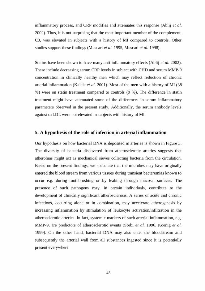
Our hypothesis on how bacterial DNA is deposited in arteries is shown in Figure 3.
The diversity of bacteria discovered from atherosclerotic arteries suggests that
atheromas might act as mechanical sieves collecting bacteria from the circulation.
Based on the present findings, we speculate that the microbes may have originally
entered the blood stream from various tissues during transient bacteremias known to
occur e.g. during toothbrushing or by leaking through mucosal surfaces. The
presence of such pathogens may, in certain individuals, contribute to the
development of clinically significant atherosclerosis. A series of acute and chronic
infections, occurring alone or in combination, may accelerate atherogenesis by
increasing inflammation by stimulation of leukocyte activation/infiltration in the
atherosclerotic arteries. In fact, systemic markers of such arterial inflammation, e.g.
MMP9, are predictors of atherosclerotic events (Sorbi et al. 1996, Koenig et al.
1999). On the other hand, bacterial DNA may also enter the bloodstream and
subsequently the arterial wall from all substances ingested since it is potentially
present everywhere.
46
Figure 3. Hypothetical mechanisms of infection in arterial inflammation.
MMP9
ARTERYLUMEN
ARTERYWALL
TISSUES
ATHEROMA
Chronic arterial inflammationmacrophages
Gut Oral cavity
TRANSIENTBACTEREMIA
Bacterial DNA in food andwater?
Bacterial DNA
Acute arterial inflammationleukocytes
Bacteria
Urinary tract
Macrophage
Respiratory tract
47
SUMMARY AND CONCLUSIONS
Inflammation has been shown to play an important role in the clinical complications
of atherosclerosis. This is mediated partly through upregulation of proteolytic
enzymes by leukocytes. Bacterial infections are potential factors that may induce
and promote inflammatory process. The present study investigated whether bacterial
DNA is present in arterial walls of different clinical specimens by using broadrange
16S rDNA PCR methodology. In addition, serum MMP9 and other inflammatory
markers in male subjects with a history of previous MI were compared to controls.
The findings and conclusions are:
1. The lack of detectable amounts of bacterial DNA from temporal arteritis
samples suggests that viable bacteria do not have a role in chronic stages of this
disease. However, these findings do not exclude the possibility of bacterial
degradation products as stimulants of chronic inflammation, or of viable
microbes as triggering factors of acute temporal arteritis.
2. The high overall diversity of bacterial DNA in atherosclerotic coronary and
abdominal artery specimens suggests a multiinfection role of microbes in the
pathogenesis of atherosclerosis.
3. Elimination of background sequencetypes should be performed from the final
results of BrPCR. Therefore, we created a novel subtraction analysis to exclude
background signal.
4. Elevated serum MMP9 concentration in subjects with a history of MI may
reflect inflammatory pathologic processes that are related to progression of
atherosclerosis.
Generally, it can be concluded that atheromas may act as mechanical sieves
collecting bacteria from the circulation. The subsequent arterial infection may
enhance arterial inflammation and thus the progression of atherosclerosis.
48
ACKNOWLEDGEMENTS
This study was carried out in the Department of Medical Biochemistry, Medical
School, University of Tampere and Tampere University Hospital, during the years
20022007.
I express my gratitude to my supervisor, Professor Seppo Nikkari, for
admitting me to his research group and giving me this interesting and challenging
research project to work on. His guidance and support over the years have been a
great value. It has been a privilege and joy to work with him.
I wish to thank my thesis committee member and coauthor, Docent Simo
Nikkari, whose expertice has been a great asset to me during these years. I am also
greatful to my other thesis committee member, Professor Markku PeltoHuikko, for
the thesis followup.
My reviewers, Professor Pekka Saikku from University of Oulu, and
Docent Jari Jalava from University of Turku, are warmly thanked for their
constructive criticism and careful evaluation that helped me to improve my
manuscript.
I feel gratitude to my coauthors and collaborators for their professional
help and for all their contributions throughout the final steps of the original
communications of this dissertation.
I wish to express my sincere thanks to present and past members of the
Department of Medical Biochemistry Tuija Aarnio, Kati Huuskonen, Virpi Juopperi,
Anne Kalela, Tarja Kunnas, Sari Ojala, Mirka Pietiläinen, Seija Rautiainen, Ulla
Saarijoki, and Tiina Solakivi. They and many other memorable persons in our coffee
room (Anja, Maria, Patu, and others) will be warmly membered. We have shared
many absolutely fabulous moments during the coffee breaks. Especially Mirka and
Kati are acknowledged as excellent listeners and friends in and outside work. Leena
Viiri, a colleague from Forensic Department, and Reetta Hinttala, a friend and a
colleague from Oulu, also deserve to be mentioned as wonderful travel companions
during congress trips.
My godsons and my brother´s children are heartily thanked for reminding
me all the small pleasures in life. Your smiles always make my day! My friends and

49
relatives here in Tampere area and spread around Finland also deserve my warm
thanks. I have enjoyed sharing my thoughts and feelings with you, through good and
bad times. My special thanks and a big, warm hug goes to Pekka.
Finally, I owe my deepest gratidute to my parents, Riitta and Jaakko, and to
my brothers, JaniPetteri, JariPekka, and JuhoMatti, for their neverending support,
patience and belief in me. You gave me the strength to complete this study.
This study was financially supported by grants of the Medical Research Fund of
Tampere University Hospital, the Pirkanmaa Regional Fund of the Finnish Cultural
Foundation, and the Scientific Foundation of the City of Tampere.
Tampere, February 2008
Jaana Renko
50
REFERENCES
Anonymous (1991): Second European Consensus Document on chronic critical legischemia. Circulation 84:IV126.
Anonymous (1993): Natural history of aortic and coronary atherosclerotic lesions inyouth. Findings from the PDAY Study. Pathobiological Determinants ofAtherosclerosis in Youth (PDAY) Research Group. Arterioscler.Thromb.13:12911298.
AaltoSetälä K, Laitinen K, Erkkilä L, Leinonen M, Jauhiainen M, Ehnholm C,Tamminen M, Puolakkainen M, Penttila I and Saikku P (2001): Chlamydiapneumoniae does not increase atherosclerosis in the aortic root ofapolipoprotein Edeficient mice. Arterioscler.Thromb.Vasc.Biol. 21:578584.
Ablij H and Meinders A (2002): Creactive protein: history and revival.Eur.J.Intern.Med. 13:412.
Albert NM (2000): Inflammation and infection in acute coronary syndrome.J.Cardiovasc.Nurs. 15:1326.
Albertson D, Natsios GA and Gleckman R (1978): Septic shock with Micrococcusluteus. Arch.Intern.Med. 138:487488.
Alkout AM, Ramsay EJ, Mackenzie DA, Weir DM, Bentley AJ, Elton RA, Sutherland S,Busuttil A and Blackwell CC (2000): Quantitative assessment of IgG antibodiesto Helicobacter pylori and outcome of ischaemic heart disease. FEMSImmunol.Med.Microbiol. 29:271274.
Altman R, Rouvier J, Scazziota A, Absi RS and Gonzalez C (1999): Lack of associationbetween prior infection with Chlamydia pneumoniae and acute or chroniccoronary artery disease. Clin.Cardiol. 22:8590.
Altschul SF, Gish W, Miller W, Myers EW and Lipman DJ (1990): Basic localalignment search tool. J.Mol.Biol. 215:403410.
Ameriso SF, Fridman EA, Leiguarda RC and Sevlever GE (2001): Detection ofHelicobacter pylori in human carotid atherosclerotic plaques. Stroke 32:385391.
51
Apfalter P, Barousch W, Nehr M, Willinger B, Rotter M and Hirschl AM (2004): Noevidence of involvement of Chlamydia pneumoniae in severe cerebrovascularatherosclerosis by means of quantitative realtime polymerase chain reaction.Stroke. 35:20242028.
Beck J, Garcia R, Heiss G, Vokonas PS and Offenbacher S (1996): Periodontal diseaseand cardiovascular disease. J.Periodontol. 67:11231137.
Bengtsson BA and Malmvall BE (1981): The epidemiology of giant cell arteritisincluding temporal arteritis and polymyalgia rheumatica. Incidences of differentclinical presentations and eye complications. Arthritis Rheum. 24:899904.
Benson DA, KarschMizrachi I, Lipman DJ, Ostell J and Wheeler DL (2007): GenBank.Nucleic Acids Res. 35:D215.
Berg JM, Tymoczko JL, Stryer L (2002): Biochemistry. New York, USA; W. H.Freeman and Company.
Bergmans AM, Groothedde JW, Schellekens JF, van Embden JD, Ossewaarde JM andSchouls LM (1995): Etiology of cat scratch disease: comparison of polymerasechain reaction detection of Bartonella (formerly Rochalimaea) and Afipia felisDNA with serology and skin tests. J.Infect.Dis. 171:916923.
Bhanji S, Williams B, Sheller B, Elwood T and Mancl L (2002): Transient bacteremiainduced by toothbrushing a comparison of the Sonicare toothbrush with aconventional toothbrush. Pediatr.Dent. 24:295299.
Biasucci LM, Liuzzo G, Grillo RL, Caligiuri G, Rebuzzi AG, Buffon A, Summaria F,Ginnetti F, Fadda G and Maseri A (1999): Elevated levels of Creactive proteinat discharge in patients with unstable angina predict recurrent instability.Circulation 99:855860.
Blackburn R, Giral P, Bruckert E, Andre JM, Gonbert S, Bernard M, Chapman MJ andTurpin G (2001): Elevated Creactive protein constitutes an independentpredictor of advanced carotid plaques in dyslipidemic subjects.Arterioscler.Thromb.Vasc.Biol. 21:19621968.
Blasi F, Denti F, Erba M, Cosentini R, Raccanelli R, Rinaldi A, Fagetti L, Esposito G,Ruberti U and Allegra L (1996): Detection of Chlamydia pneumoniae but notHelicobacter pylori in atherosclerotic plaques of aortic aneurysms.J.Clin.Microbiol. 34:27662769.
52
Brown DL, Hibbs MS, Kearney M, Loushin C and Isner JM (1995): Identification of 92kD gelatinase in human coronary atherosclerotic lesions. Association of activeenzyme synthesis with unstable angina. Circulation 91:21252131.
Burke AP, Farb A, Malcom GT, Liang YH, Smialek J and Virmani R (1997): Coronaryrisk factors and plaque morphology in men with coronary disease who diedsuddenly. N.Engl.J.Med. 336:12761282.
Burke AP, Tracy RP, Kolodgie F, Malcom GT, Zieske A, Kutys R, Pestaner J, Smialek Jand Virmani R (2002): Elevated Creactive protein values and atherosclerosis insudden coronary death: association with different pathologies. Circulation105:20192023.
Caligiuri G, Rottenberg M, Nicoletti A, Wigzell H and Hansson GK (2001): Chlamydiapneumoniae infection does not induce or modify atherosclerosis in mice.Circulation 103:28342838.
Campbell LA, O'Brien ER, Cappuccio AL, Kuo CC, Wang SP, Stewart D, Patton DL,Cummings PK and Grayston JT (1995): Detection of Chlamydia pneumoniaeTWAR in human coronary atherectomy tissues. J.Infect.Dis. 172:585588.
Carapetis JR, Steer AC, Mulholland EK, Weber M (2005): The global burden of groupA streptococcal diseases. Lancet Infect.Dis. 5:685694.
Chandra HR, Choudhary N, O'Neill C, Boura J, Timmis GC and O'Neill WW (2001):Chlamydia pneumoniae exposure and inflammatory markers in acute coronarysyndrome (CIMACS). Am.J.Cardiol. 88:214218.
Cherubini A, Mecocci P, Senin U, Lowenthal DT, Barnert G and Chionne F (1997):Autoantibodies against oxidized lowdensity lipoproteins in older strokepatients. J.Am.Geriatr.Soc. 45:125.
Collins MD, Bernard KA, Hutson RA, Sjoden B, Nyberg A and Falsen E (1999):Corynebacterium sundsvallense sp. nov., from human clinical specimens.Int.J.Syst.Bacteriol. 49 Pt 2:361366.
Coombes BK and Mahony JB (1999): Chlamydia pneumoniae infection of humanendothelial cells induces proliferation of smooth muscle cells via an endothelialcellderived soluble factor(s). Infect.Immun. 67:29092915.
Cunningham MW (2003): Autoimmunity and molecular mimicry in the pathogenesis ofpoststreptococcal heart disease. Front.Biosci. 8: s533543.
53
Danesh J (1999): Coronary heart disease, Helicobacter pylori, dental disease, Chlamydiapneumoniae, and cytomegalovirus: metaanalyses of prospective studies.Am.Heart J. 138:S4347.
Danesh J, Youngman L, Clark S, Parish S, Peto R and Collins R (1999a): Helicobacterpylori infection and early onset myocardial infarction: casecontrol and siblingpairs study. BMJ 319:11571162.
Danesh J, Koreth J, Youngman L, Collins R, Arnold JR, Balarajan Y, McGee J andRoskell D (1999b): Is Helicobacter pylori a factor in coronary atherosclerosis?J.Clin.Microbiol. 37:1651.
Danesh J, Whincup P, Walker M, Lennon L, Thomson A, Appleby P, Wong Y,BernardesSilva M and Ward M (2000): Chlamydia pneumoniae IgG titres andcoronary heart disease: prospective study and metaanalysis. BMJ 321:208213.
De Soete G (1983): A least squares algorithm for fitting additive trees to proximity data.Psychometrika 48:621626.
Dollery CM, McEwan JR and Henney AM (1995): Matrix metalloproteinases andcardiovascular disease. Circ.Res. 77:863868.
Dore MP, Sepulveda AR, Bacciu PP, Blasi F, Simula L, Marras L, Piccolo D, CherchiGB, Graham DY and Realdi G (2003): Detection of Chlamydiae pneumoniaebut not Helicobacter pylori DNA in atherosclerosis plaques. Dig.Dis.Sci.48:945951.
Dunn BE, Cohen H and Blaser MJ (1997): Helicobacter pylori. Clin.Microbiol.Rev.10:720741.
Epstein SE, Zhou YF and Zhu J (1999): Infection and atherosclerosis: emergingmechanistic paradigms. Circulation 100:e208.
Farsak B, Yildirir A, Akyon Y, Pinar A, Oc M, Boke E, Kes S and Tokgozoglu L(2000): Detection of Chlamydia pneumoniae and Helicobacter pylori DNA inhuman atherosclerotic plaques by PCR. J.Clin.Microbiol. 38:44084411.
Felsenstein J (1981): Evolutionary trees from DNA sequences: a maximum likelihoodapproach. J.Mol.Evol. 17:368376.
Ferreiros ER, Boissonnet CP, Pizarro R, Merletti PF, Corrado G, Cagide A and BazzinoOO (1999): Independent prognostic value of elevated Creactive protein inunstable angina. Circulation 100:19581963.
54
Fichtlscherer S, Rosenberger G, Walter DH, Breuer S, Dimmeler S and Zeiher AM(2000): Elevated Creactive protein levels and impaired endothelialvasoreactivity in patients with coronary artery disease. Circulation 102:10001006.
Fiehn NE, Larsen T, Christiansen N, Holmstrup P and Schroeder TV (2005):Identification of periodontal pathogens in atherosclerotic vessels. J.Periodontol.76:731736.
Folsom AR, Nieto FJ, Sorlie P, Chambless LE and Graham DY (1998): Helicobacterpylori seropositivity and coronary heart disease incidence. Atherosclerosis RiskIn Communities (ARIC) Study Investigators. Circulation 98:845850.
Fong IW (2000): Emerging relations between infectious diseases and coronary arterydisease and atherosclerosis. CMAJ 163:4956.
Fong IW (2002): Infections and their role in atherosclerotic vascular disease.J.Am.Dent.Assoc. 133 Suppl:7S13S.
Fong IW, Chiu B, Viira E, Fong MW, Jang D and Mahony J (1997): Rabbit model forChlamydia pneumoniae infection. J.Clin.Microbiol. 35:4852.
Forner L, Larsen T, Kilian M and Holmstrup P (2006): Incidence of bacteremia afterchewing, tooth brushing and scaling in individuals with periodontalinflammation. J.Clin.Periodontol. 33:401407.
Fukuda D, Shimada K, Tanaka A, Kusuyama T, Yamashita H, Ehara S, Nakamura Y,Kawarabayashi T, Iida H, Yoshiyama M and Yoshikawa J (2006): Comparisonof levels of serum matrix metalloproteinase9 in patients with acute myocardialinfarction versus unstable angina pectoris versus stable angina pectoris.Am.J.Cardiol. 97:175180.
Gabriel SE, Espy M, Erdman DD, Bjornsson J, Smith TF and Hunder GG (1999): Therole of parvovirus B19 in the pathogenesis of giant cell arteritis: a preliminaryevaluation. Arthritis Rheum. 42:12551258.
Galeano NF, AlHaideri M, Keyserman F, Rumsey SC and Deckelbaum RJ (1998):Small dense low density lipoprotein has increased affinity for LDL receptorindependent cell surface binding sites: a potential mechanism for increasedatherogenicity. J.Lipid Res. 39:12631273.
Galis ZS and Khatri JJ (2002): Matrix metalloproteinases in vascular remodeling andatherogenesis: the good, the bad, and the ugly. Circ.Res. 90:251262.

55
Galis ZS, Sukhova GK, Lark MW and Libby P (1994a): Increased expression of matrixmetalloproteinases and matrix degrading activity in vulnerable regions ofhuman atherosclerotic plaques. J.Clin.Invest. 94:24932503.
Galis ZS, Muszynski M, Sukhova GK, SimonMorrissey E and Libby P (1995):Enhanced expression of vascular matrix metalloproteinases induced in vitro bycytokines and in regions of human atherosclerotic lesions. Ann.N.Y.Acad.Sci.748:501507.
Galis ZS, Muszynski M, Sukhova GK, SimonMorrissey E, Unemori EN, Lark MW,Amento E and Libby P (1994b): Cytokinestimulated human vascular smoothmuscle cells synthesize a complement of enzymes required for extracellularmatrix digestion. Circ.Res. 75:181189.
Geer JC and Haust MD (1972): Smooth muscle cells in atherosclerosis.Monogr.Atheroscler. 2:1140.
Grayston JT, Kuo CC, Coulson AS, Campbell LA, Lawrence RD, Lee MJ, StrandnessED and Wang SP (1995): Chlamydia pneumoniae (TWAR) in atherosclerosisof the carotid artery. Circulation 92:33973400.
Greer CE, Lund JK and Manos MM (1991): PCR amplification from paraffinembeddedtissues: recommendations on fixatives for longterm storage and prospectivestudies. PCR Methods Appl. 1:4650.
Gruber BL, Sorbi D, French DL, Marchese MJ, Nuovo GJ, Kew RR and Arbeit LA(1996): Markedly elevated serum MMP9 (gelatinase B) levels in rheumatoidarthritis: a potentially useful laboratory marker. Clin.Immunol.Immunopathol.78:161171.
Gutell RR, Larsen N and Woese CR (1994): Lessons from an evolving rRNA: 16S and23S rRNA structures from a comparative perspective. Microbiol.Rev. 58:1026.
Gutell RR, Weiser B, Woese CR and Noller HF (1985): Comparative anatomy of 16Slike ribosomal RNA. Prog.Nucleic Acid Res.Mol.Biol. 32:155216.
Halpert I, Sires UI, Roby JD, PotterPerigo S, Wight TN, Shapiro SD, Welgus HG,Wickline SA and Parks WC (1996): Matrilysin is expressed by lipidladenmacrophages at sites of potential rupture in atherosclerotic lesions and localizesto areas of versican deposition, a proteoglycan substrate for the enzyme.Proc.Natl.Acad.Sci.U.S.A. 93:97489753.
56
Han XY and Andrade RA (2005): Brevundimonas diminuta infections and its resistanceto fluoroquinolones. J.Antimicrob.Chemother. 55:853859.
Haraszthy VI, Zambon JJ, Trevisan M, Zeid M and Genco RJ (2000): Identification ofperiodontal pathogens in atheromatous plaques. J.Periodontol. 71:15541560.
Haugeberg G, Bie R and Nordbo SA (2000): Chlamydia pneumoniae not detected intemporal artery biopsies from patients with temporal arteritis.Scand.J.Rheumatol. 29:127128.
Haverkate F, Thompson SG, Pyke SD, Gallimore JR and Pepys MB (1997): Productionof Creactive protein and risk of coronary events in stable and unstable angina.European Concerted Action on Thrombosis and Disabilities Angina PectorisStudy Group. Lancet 349:462466.
Heimdahl A, Hall G, Hedberg M, Sandberg H, Soder PO, Tuner K and Nord CE (1990):Detection and quantitation by lysisfiltration of bacteremia after different oralsurgical procedures. J.Clin.Microbiol. 28:22052209.
HelwegLarsen J, Tarp B, Obel N and Baslund B (2002): No evidence of parvovirusB19, Chlamydia pneumoniae or human herpes virus infection in temporal arterybiopsies in patients with giant cell arteritis. Rheumatology (Oxford) 41:445449.
Henney AM, Wakeley PR, Davies MJ, Foster K, Hembry R, Murphy G and HumphriesS (1991): Localization of stromelysin gene expression in atheroscleroticplaques by in situ hybridization. Proc.Natl.Acad.Sci.U.S.A. 88:81548158.
Horowitz H, Gilroy S, Feinstein S and Gilardi G (1990): Endocarditis associated withComamonas acidovorans. J.Clin.Microbiol. 28:143145.
Howell TH, Ridker PM, Ajani UA, Hennekens CH and Christen WG (2001):Periodontal disease and risk of subsequent cardiovascular disease in U.S. malephysicians. J.Am.Coll.Cardiol. 37:445450.
Hsueh PR, Teng LJ, Yang PC, Chen YC, Pan HJ, Ho SW and Luh KT (1998):Nosocomial infections caused by Sphingomonas paucimobilis: clinical featuresand microbiological characteristics. Clin.Infect.Dis. 26:676681.
Hujoel PP, Drangsholt M, Spiekerman C and DeRouen TA (2000): Periodontal diseaseand coronary heart disease risk. JAMA 284:14061410.
57
Inokubo Y, Hanada H, Ishizaka H, Fukushi T, Kamada T and Okumura K (2001):Plasma levels of matrix metalloproteinase9 and tissue inhibitor ofmetalloproteinase1 are increased in the coronary circulation in patients withacute coronary syndrome. Am.Heart J. 141:211217.
Ishikawa T, Imamura T, Hatakeyama K, Date H, Nagoshi T, Kawamoto R, MatsuyamaA, Asada Y, Eto T (2004): Possible contribution of Creactive protein withincoronary plaque to increasing its own plasma levels across coronary circulation.Am.J.Cardiol. 93:611614.
Jackson LA, Campbell LA, Schmidt RA, Kuo CC, Cappuccio AL, Lee MJ and GraystonJT (1997): Specificity of detection of Chlamydia pneumoniae in cardiovascularatheroma: evaluation of the innocent bystander hypothesis. Am.J.Pathol.150:17851790.
Jalava J, Kotilainen P, Nikkari S, Skurnik M, Vanttinen E, Lehtonen OP, Eerola E andToivanen P (1995): Use of the polymerase chain reaction and DNA sequencingfor detection of Bartonella quintana in the aortic valve of a patient with culturenegative infective endocarditis. Clin.Infect.Dis. 21:891896.
Jialal I and Devaraj S (1996): The role of oxidized low density lipoprotein inatherogenesis. J.Nutr. 126:1053S7S.
Johnson G,Jr, Avery A, McDougal EG, Burnham SJ and Keagy BA (1985): Aneurysmsof the abdominal aorta. Incidence in blacks and whites in North Carolina.Arch.Surg. 120:11381140.
Joshipura KJ, Rimm EB, Douglass CW, Trichopoulos D, Ascherio A and Willett WC(1996): Poor oral health and coronary heart disease. J.Dent.Res. 75:16311636.
Järvisalo MJ, Juonala M and Raitakari OT (2006): Assessment of inflammatory markersand endothelial function. Curr.Opin.Clin.Nutr.Metab.Care 9:547552.
Kaartinen M, van der Wal AC, van der Loos CM, Piek JJ, Koch KT, Becker AE andKovanen PT (1998): Mast cell infiltration in acute coronary syndromes:implications for plaque rupture. J.Am.Coll.Cardiol. 32:606612.
Kadar A and Glasz T (2001): Development of atherosclerosis and plaque biology.Cardiovasc.Surg. 9:109121.
Kai H, Ikeda H, Yasukawa H, Kai M, Seki Y, Kuwahara F, Ueno T, Sugi K andImaizumi T (1998): Peripheral blood levels of matrix metalloproteases2 and 9
58
are elevated in patients with acute coronary syndromes. J.Am.Coll.Cardiol.32:368372.
Kalayoglu MV and Byrne GI (1998): Induction of macrophage foam cell formation byChlamydia pneumoniae. J.Infect.Dis. 177:725729.
Kalela A, Koivu TA, Sisto T, Kanervisto J, Höyhtyä M, Sillanaukee P, Lehtimäki T andNikkari ST (2002): Serum matrix metalloproteinase9 concentration inangiographically assessed coronary artery disease. Scand.J.Clin.Lab.Invest.62:337342.
Kalela A, Laaksonen R, Lehtimäki T, Koivu TA, Höyhtyä M, Janatuinen T, Pöllänen P,Vesalainen R, Saikku P, Knuuti J and Nikkari ST (2001): Effect of pravastatinin mildly hypercholesterolemic young men on serum matrix metalloproteinases.Am.J.Cardiol. 88:1735, A6.
Kaplan M, Yavuz SS, Cinar B, Koksal V, Kut MS, Yapici F, Gercekoglu H andDemirtas MM (2006): Detection of Chlamydia pneumoniae and Helicobacterpylori in atherosclerotic plaques of carotid artery by polymerase chain reaction.Int.J.Infect.Dis. 10:116123.
Kelly EA, Busse WW and Jarjour NN (2000): Increased matrix metalloproteinase9 inthe airway after allergen challenge. Am.J.Respir.Crit.Care Med. 162:11571161.
Kessler RB, Kimbrough RC,3rd and Jones SR (1998): Infective endocarditis caused byStaphylococcus hominis after vasectomy. Clin.Infect.Dis. 27:216217.
Khamis A, Raoult D and La Scola B (2005): Comparison between rpoB and 16S rRNAgene sequencing for molecular identification of 168 clinical isolates ofCorynebacterium. J.Clin.Microbiol. 43:19341936.
Kloos WE and Schleifer KH (1975): Simplified scheme for routine identification ofhuman Staphylococcus species. J Clin Microbiol. 1:8288.
Koenig W, Sund M, Frohlich M, Fischer HG, Lowel H, Doring A, Hutchinson WL andPepys MB (1999): CReactive protein, a sensitive marker of inflammation,predicts future risk of coronary heart disease in initially healthy middleagedmen: results from the MONICA (Monitoring Trends and Determinants inCardiovascular Disease) Augsburg Cohort Study, 1984 to 1992. Circulation99:237242.
59
Kovanen PT, Kaartinen M and Paavonen T (1995): Infiltrates of activated mast cells atthe site of coronary atheromatous erosion or rupture in myocardial infarction.Circulation 92:10841088.
Kozarov E, Sweier D, Shelburne C, ProgulskeFox A and Lopatin D (2006): Detectionof bacterial DNA in atheromatous plaques by quantitative PCR. MicrobesInfect. 8:687693.
Kozarov EV, Dorn BR, Shelburne CE, Dunn WA,Jr and ProgulskeFox A (2005):Human atherosclerotic plaque contains viable invasive Actinobacillusactinomycetemcomitans and Porphyromonas gingivalis.Arterioscler.Thromb.Vasc.Biol. 25:e178.
Krauss RM (1995): Dense low density lipoproteins and coronary artery disease.Am.J.Cardiol. 75:53B57B.
Kroes I, Lepp PW and Relman DA (1999): Bacterial diversity within the humansubgingival crevice. Proc.Natl.Acad.Sci.U.S.A. 96:1454714552.
Kuo CC, Shor A, Campbell LA, Fukushi H, Patton DL and Grayston JT (1993):Demonstration of Chlamydia pneumoniae in atherosclerotic lesions of coronaryarteries. J.Infect.Dis. 167:841849.
Kurihara N, Inoue Y, Iwai T, Umeda M, Huang Y and Ishikawa I (2004): Detection andlocalization of periodontopathic bacteria in abdominal aortic aneurysms.Eur.J.Vasc.Endovasc.Surg. 28:553558.
Kähler J, Gerth S, Schafer P, Boersma E, Köster R, Terres W, Simoons ML, Berger J,Meinertz T and Hamm CW (2001): Antibodies to chlamydiallipopolysaccharides in unstable angina pectoris. Am.J.Cardiol. 87:11501153.
Laitinen K, Laurila A, Pyhälä L, Leinonen M and Saikku P (1997): Chlamydiapneumoniae infection induces inflammatory changes in the aortas of rabbits.Infect.Immun. 65:48324835.
Lane DJ, Pace B, Olsen GJ, Stahl DA, Sogin ML and Pace NR (1985): Rapiddetermination of 16S ribosomal RNA sequences for phylogenetic analyses.Proc.Natl.Acad.Sci.U.S.A. 82:69556959.
Larson TS, Hall S, Hepper NG and Hunder GG (1984): Respiratory tract symptoms as aclue to giant cell arteritis. Ann.Intern.Med. 101:594597.
60
Leinonen M and Saikku P (2002): Evidence for infectious agents in cardiovasculardisease and atherosclerosis. Lancet Infect.Dis. 2:1117.
Lewin B (ed.) (2004): Genes VIII. Upper Saddle River, NJ; Pearson Prentice Hall.
Li Z, Li L, Zielke HR, Cheng L, Xiao R, Crow MT, StetlerStevenson WG, Froehlich Jand Lakatta EG (1996): Increased expression of 72kd type IV collagenase(MMP2) in human aortic atherosclerotic lesions. Am.J.Pathol. 148:121128.
Libby P, Egan D and Skarlatos S (1997): Roles of infectious agents in atherosclerosisand restenosis: an assessment of the evidence and need for future research.Circulation 96:40954103.
Libby P, Ridker PM and Maseri A (2002): Inflammation and atherosclerosis. Circulation105:11351143.
Lindahl B, Toss H, Siegbahn A, Venge P and Wallentin L (2000): Markers ofmyocardial damage and inflammation in relation to longterm mortality inunstable coronary artery disease. FRISC Study Group. Fragmin duringInstability in Coronary Artery Disease. N.Engl.J.Med. 343:11391147.
Linnanmäki E, Leinonen M, Mattila K, Nieminen MS, Valtonen V and Saikku P (1993):Chlamydia pneumoniaespecific circulating immune complexes in patients withchronic coronary heart disease. Circulation 87:11301134.
Lodish H, Berk A, Zipursky SL, Matsudaira P, Baltimore D, Darnell JE (eds.) (2000):Molecular cell biology. New York, USA; W. H. Freeman and Company.
Loesche WJ (1986): Role of Streptococcus mutans in human dental decay.Microbiol.Rev. 50:353380.
Ludwig W, Strunk O, Westram R, Richter L, Meier H, Yadhukumar, Buchner A, Lai T,Steppi S, Jobb G, Forster W, Brettske I, Gerber S, Ginhart AW, Gross O,Grumann S, Hermann S, Jost R, Konig A, Liss T, Lussmann R, May M,Nonhoff B, Reichel B, Strehlow R, Stamatakis A, Stuckmann N, Vilbig A,Lenke M, Ludwig T, Bode A and Schleifer KH (2004): ARB: a softwareenvironment for sequence data. Nucleic Acids Res. 32:13631371.
Maass M, Bartels C, Engel PM, Mamat U and Sievers HH (1998): Endovascularpresence of viable Chlamydia pneumoniae is a common phenomenon incoronary artery disease. J.Am.Coll.Cardiol. 31:827832.
61
Maiwald M (2004): Broadrange PCR for detection and identification of bacteria. InMolecular microbiology. Diagnostic principles and practice. Edited by PersingDH. Herndon, USA; ASM Press.
Malnick SD, Goland S, Kaftoury A, Schwarz H, Pasik S, Mashiach A and Sthoeger Z(1999): Evaluation of carotid arterial plaques after endarterectomy forHelicobacter pylori infection. Am.J.Cardiol. 83:15867, A8.
Maraha B, Berg H, Kerver M, Kranendonk S, Hamming J, Kluytmans J, Peeters M andvan der Zee A (2004): Is the perceived association between Chlamydiapneumoniae and vascular diseases biased by methodology? J.Clin.Microbiol.42:39373941.
Marques da Silva R, Caugant DA, Eribe ER, Aas JA, Lingaas PS, Geiran O, Tronstad Land Olsen I (2006): Bacterial diversity in aortic aneurysms determined by 16Sribosomal RNA gene analysis. J.Vasc.Surg. 44:10551060.
Martino R, Martinez C, Pericas R, Salazar R, Sola C, Brunet S, Sureda A and DomingoAlbos A (1996): Bacteremia due to glucose nonfermenting gramnegativebacilli in patients with hematological neoplasias and solid tumors.Eur.J.Clin.Microbiol.Infect.Dis. 15:610615.
Mattila KJ, Valtonen VV, Nieminen M and Huttunen JK (1995): Dental infection andthe risk of new coronary events: prospective study of patients with documentedcoronary artery disease. Clin.Infect.Dis. 20:588592.
May AE, Redecke V, Gruner S, Schmidt R, Massberg S, Miethke T, Ryba B, Prazeres daCosta C, Schomig A and Neumann FJ (2003): Recruitment of Chlamydiapneumoniaeinfected macrophages to the carotid artery wall in noninfected,nonatherosclerotic mice. Arterioscler.Thromb.Vasc.Biol. 23:789794.
McGill HC,Jr and McMahan CA (1998): Determinants of atherosclerosis in the young.Pathobiological Determinants of Atherosclerosis in Youth (PDAY) ResearchGroup. Am.J.Cardiol. 82:30T36T.
Mendall MA, Goggin PM, Molineaux N, Levy J, Toosy T, Strachan D, Camm AJ andNorthfield TC (1994): Relation of Helicobacter pylori infection and coronaryheart disease. Br.Heart J. 71:437439.
Miyashita N, Toyota E, Sawayama T, Matsumoto A, Mikami Y, Kawai N, Takada K,Niki Y and Matsushima T (1998): Association of chronic infection ofChlamydia pneumoniae and coronary heart disease in the Japanese. Intern.Med.37:913916.
62
Morrison HI, Ellison LF and Taylor GW (1999): Periodontal disease and risk of fatalcoronary heart and cerebrovascular diseases. J.Cardiovasc.Risk 6:711.
Muhlestein JB (1998): Bacterial infections and atherosclerosis. J.Investig.Med. 46:396402.
Muhlestein JB and Anderson JL (2003): Chronic infection and coronary artery disease.Cardiol.Clin. 21:333362.
Muscari A, Bozzoli C, Puddu GM, Sangiorgi Z, Dormi A, Rovinetti C, Descovich GCand Puddu P (1995): Association of serum C3 levels with the risk of myocardialinfarction. Am.J.Med. 98:357364.
Muscari A, Massarelli G, Bastagli L, Poggiopollini G, Tomassetti V, Volta U, PudduGM and Puddu P (1998): Relationship between serum C3 levels and traditionalrisk factors for myocardial infarction. Acta Cardiol. 53:345354.
Nakano K, Inaba H, Nomura R, Nemoto H, Takeda M, Yoshioka H, Matsue H,Takahashi T, Taniguchi K, Amano A and Ooshima T (2006): Detection ofcariogenic Streptococcus mutans in extirpated heart valve and atheromatousplaque specimens. J.Clin.Microbiol. 44:33133317.
Nelimarkka LO, Nikkari ST, Ravanti LS, Kahari VM and Järveläinen HT (1998):Collagenase1, stromelysin1 and 92 kDa gelatinase are associated with tumornecrosis factoralpha induced morphological change of human endothelial cellsin vitro. Matrix Biol. 17:293304.
Nieto FJ (1998): Infections and atherosclerosis: new clues from an old hypothesis?Am.J.Epidemiol. 148: 937948.
Nikkari S, McLaughlin IJ, Bi W, Dodge DE and Relman DA (2001): Does blood ofhealthy subjects contain bacterial ribosomal DNA? J.Clin.Microbiol. 39:19561959.
Nikkari ST, O'Brien KD, Ferguson M, Hatsukami T, Welgus HG, Alpers CE andClowes AW (1995): Interstitial collagenase (MMP1) expression in humancarotid atherosclerosis. Circulation 92:13931398.
Nobel M, De Torrente A, Peter O and Genne D (1999): No serological evidence ofassociation between chlamydia pneumonia infection and acute coronary heartdisease. Scand.J.Infect.Dis. 31:261264.
63
Noji Y, Kajinami K, Kawashiri MA, Todo Y, Horita T, Nohara A, Higashikata T, InazuA, Koizumi J, Takegoshi T and Mabuchi H (2001): Circulating matrixmetalloproteinases and their inhibitors in premature coronary atherosclerosis.Clin.Chem.Lab.Med. 39:380384.
Noller HF (1984): Structure of ribosomal RNA. Annu.Rev.Biochem. 53:119162.
Nordborg C, Nordborg E and Petursdottir V (2000): Giant cell arteritis. Epidemiology,etiology and pathogenesis. APMIS 108:713724.
O'Connor S, Taylor C, Campbell LA, Epstein S and Libby P (2001): Potential infectiousetiologies of atherosclerosis: a multifactorial perspective. Emerg.Infect.Dis.7:780788.
Okuda K, Ishihara K, Nakagawa T, Hirayama A, Inayama Y and Okuda K (2001):Detection of Treponema denticola in atherosclerotic lesions. J.Clin.Microbiol.39:11141117.
Olmez N, Wang GF, Li Y, Zhang H, Schumacher HR (2001): Chlamydial nucleic acidsin synovium in osteoarthritis: what are the implications? J. Rheum. 28:18741880.
Olsen GJ, Matsuda H, Hagstrom R and Overbeek R (1994): fastDNAmL: a tool forconstruction of phylogenetic trees of DNA sequences using maximumlikelihood. Comput.Appl.Biosci. 10:4148.
Ong GM, Coyle PV, Barros D'Sa AA, McCluggage WG, Duprex WP, O'Neill HJ, WyattDE, Bamford KB, O'Loughlin B and McCaughey C (2001): Nondetection ofChlamydia species in carotid atheroma using generic primers by nested PCR ina population with a high prevalence of Chlamydia pneumoniae antibody. BMCInfect.Dis. 1:12.
Ott SJ, El Mokhtari NE, Musfeldt M, Hellmig S, Freitag S, Rehman A, Kuhbacher T,Nikolaus S, Namsolleck P, Blaut M, Hampe J, Sahly H, Reinecke A, Haake N,Gunther R, Kruger D, Lins M, Herrmann G, Folsch UR, Simon R and SchreiberS (2006): Detection of diverse bacterial signatures in atherosclerotic lesions ofpatients with coronary heart disease. Circulation 113:929937.
Palinski W, Ord VA, Plump AS, Breslow JL, Steinberg D and Witztum JL (1994):ApoEdeficient mice are a model of lipoprotein oxidation in atherogenesis.Demonstration of oxidationspecific epitopes in lesions and high titers ofautoantibodies to malondialdehydelysine in serum. Arterioscler.Thromb.14:605616.
64
Palinski W, Hörkkö S, Miller E, Steinbrecher UP, Powell HC, Curtiss LK and WitztumJL (1996): Cloning of monoclonal autoantibodies to epitopes of oxidizedlipoproteins from apolipoprotein Edeficient mice. Demonstration of epitopesof oxidized low density lipoprotein in human plasma. J.Clin.Invest. 98:800814.
Palinski W, Rosenfeld ME, YlaHerttuala S, Gurtner GC, Socher SS, Butler SW,Parthasarathy S, Carew TE, Steinberg D and Witztum JL (1989): Low densitylipoprotein undergoes oxidative modification in vivo.Proc.Natl.Acad.Sci.U.S.A. 86:13721376.
Parthasarathy S and Rankin SM (1992): Role of oxidized low density lipoprotein inatherogenesis. Prog.Lipid Res. 31:127143.
Parthasarathy S, Fong LG, Quinn MT and Steinberg D (1990): Oxidative modification ofLDL: comparison between cellmediated and coppermediated modification.Eur.Heart J. 11 Suppl E:8387.
Pasceri V, Cammarota G, Patti G, Cuoco L, Gasbarrini A, Grillo RL, Fedeli G,Gasbarrini G and Maseri A (1998): Association of virulent Helicobacter pyloristrains with ischemic heart disease. Circulation 97:16751679.
Paster BJ, Boches SK, Galvin JL, Ericson RE, Lau CN, Levanos VA, Sahasrabudhe Aand Dewhirst FE (2001): Bacterial diversity in human subgingival plaque.J.Bacteriol. 183:37703783.
Paster BJ, Falkler Jr WA,Jr, Enwonwu CO, Idigbe EO, Savage KO, Levanos VA, TamerMA, Ericson RL, Lau CN and Dewhirst FE (2002): Prevalent bacterial speciesand novel phylotypes in advanced noma lesions. J.Clin.Microbiol. 40:21872191.
Pellicano R, Mazzarello MG, Morelloni S, Allegri M, Arena V, Ferrari M, Rizzetto Mand Ponzetto A (1999): Acute myocardial infarction and Helicobacter pyloriseropositivity. Int.J.Clin.Lab.Res. 29:141144.
Pepys MB and Hirschfield GM (2003): Creactive protein: a critical update.J.Clin.Invest. 111:18051812.
Pussinen PJ, Jousilahti P, Alfthan G, Palosuo T, Asikainen S and Salomaa V (2003):Antibodies to periodontal pathogens are associated with coronary heart disease.Arterioscler.Thromb.Vasc.Biol. 23:12501254.
Pussinen PJ, Nyyssönen K, Alfthan G, Salonen R, Laukkanen JA and Salonen JT(2005): Serum antibody levels to Actinobacillus actinomycetemcomitans

65
predict the risk for coronary heart disease. Arterioscler.Thromb.Vasc.Biol.25:833838.
Puurunen M, Mänttäri M, Manninen V, Tenkanen L, Alfthan G, Ehnholm C, Vaarala O,Aho K and Palosuo T (1994): Antibody against oxidized lowdensitylipoprotein predicting myocardial infarction. Arch.Intern.Med. 154:26052609.
Rademaker JLV and Savelkoul P (2004): PCR Ampilificationbased Microbial Typing.In Molecular microbiology. Diagnostic principles and practice. Edited byPersing DH. Herndon, USA; ASM Press.
Rajasuo A, Nyfors S, Kanervo A, JousimiesSomer H, Lindqvist C and Suuronen R(2004): Bacteremia after plate removal and tooth extraction. Int.J.OralMaxillofac.Surg. 33:356360.
Ramirez JA (1996): Isolation of Chlamydia pneumoniae from the coronary artery of apatient with coronary atherosclerosis. The Chlamydiapneumoniae/Atherosclerosis Study Group. Ann.Intern.Med. 125:979982.
Reed D, Reed C, Stemmermann G and Hayashi T (1992): Are aortic aneurysms causedby atherosclerosis? Circulation 85:205211.
Regan MJ, Wood BJ, Hsieh YH, Theodore ML, Quinn TC, Hellmann DB, Green WR,Gaydos CA and Stone JH (2002): Temporal arteritis and Chlamydiapneumoniae: failure to detect the organism by polymerase chain reaction inninety cases and ninety controls. Arthritis Rheum. 46:10561060.
Relman DA, Schmidt TM, MacDermott RP and Falkow S (1992): Identification of theuncultured bacillus of Whipple's disease. N.Engl.J.Med. 327:293301.
Relman DA, Loutit JS, Schmidt TM, Falkow S and Tompkins LS (1990): The agent ofbacillary angiomatosis. An approach to the identification of unculturedpathogens. N.Engl.J.Med. 323:15731580.
Richardson PD, Davies MJ and Born GV (1989): Influence of plaque configuration andstress distribution on fissuring of coronary atherosclerotic plaques. Lancet2:941944.
Ridker PM, Glynn RJ and Hennekens CH (1998a): Creactive protein adds to thepredictive value of total and HDL cholesterol in determining risk of firstmyocardial infarction. Circulation 97:20072011.

66
Ridker PM, Cushman M, Stampfer MJ, Tracy RP and Hennekens CH (1997):Inflammation, aspirin, and the risk of cardiovascular disease in apparentlyhealthy men. N.Engl.J.Med. 336:973979.
Ridker PM, Buring JE, Shih J, Matias M and Hennekens CH (1998b): Prospective studyof Creactive protein and the risk of future cardiovascular events amongapparently healthy women. Circulation 98:731733.
Ridker PM, Rifai N, Rose L, Buring JE and Cook NR (2002): Comparison of Creactiveprotein and lowdensity lipoprotein cholesterol levels in the prediction of firstcardiovascular events. N.Engl.J.Med. 347:15571565.
Rimenti G, Blasi F, Cosentini R, Moling O, Pristera R, Tarsia P, Vedovelli C and MianP (2000): Temporal arteritis associated with Chlamydia pneumoniae DNAdetected in an artery specimen. J.Rheumatol. 27:27182720.
Roberts GJ, Holzel HS, Sury MR, Simmons NA, Gardner P and Longhurst P (1997):Dental bacteremia in children. Pediatr.Cardiol. 18:2427.
RodriguezPla A, BoschGil JA, EchevarriaMayo JE, RosselloUrgell J, SolansLaqueR, HuguetRedecilla P, Stone JH and VilardellTarres M (2004): No detectionof parvovirus B19 or herpesvirus DNA in giant cell arteritis. J.Clin.Virol.31:1115.
Romano Carratelli C, Nuzzo I, Cozzolino D, Bentivoglio C, Paolillo R and Rizzo A(2006): Relationship between Chlamydia pneumoniae infection, inflammatorymarkers, and coronary heart diseases. Int.Immunopharmacol. 6:848853.
Ross R (1999a): Atherosclerosis is an inflammatory disease. Am.Heart J. 138:S41920.
Ross R (1999b): Atherosclerosisan inflammatory disease. N.Engl.J.Med. 340:115126.
Rost NS, Wolf PA, Kase CS, KellyHayes M, Silbershatz H, Massaro JM, D'AgostinoRB, Franzblau C and Wilson PW (2001): Plasma concentration of Creactiveprotein and risk of ischemic stroke and transient ischemic attack: theFramingham study. Stroke 32:25752579.
Saikku P, Leinonen M, Mattila K, Ekman MR, Nieminen MS, Mäkelä PH, Huttunen JKand Valtonen V (1988): Serological evidence of an association of a novelChlamydia, TWAR, with chronic coronary heart disease and acute myocardialinfarction. Lancet 332:983986.
67
Saikku P, Leinonen M, Tenkanen L, Linnanmäki E, Ekman MR, Manninen V, MänttäriM, Frick MH and Huttunen JK (1992): Chronic Chlamydia pneumoniaeinfection as a risk factor for coronary heart disease in the Helsinki Heart Study.Ann.Intern.Med. 116:273278.
Salonen JT, YläHerttuala S, Yamamoto R, Butler S, Korpela H, Salonen R, NyyssönenK, Palinski W and Witztum JL (1992): Autoantibody against oxidised LDL andprogression of carotid atherosclerosis. Lancet 339:883887.
Salvarani C, Cantini F, Boiardi L and Hunder GG (2002a): Polymyalgia rheumatica andgiantcell arteritis. N.Engl.J.Med. 347:261271.
Salvarani C, Farnetti E, Casali B, Nicoli D, Wenlan L, Bajocchi G, Macchioni P, LoScocco G, Grazia Catanoso M, Boiardi L and Cantini F (2002b): Detection ofparvovirus B19 DNA by polymerase chain reaction in giant cell arteritis: acasecontrol study. Arthritis Rheum. 46:30993101.
Schleifer KH and Kloos WE (1975): A simple test system for the separation ofstaphylococci from micrococci. J Clin Microbiol. 1:337338.
Schumacher M, Eber B, Tatzber F, Kaufmann P, Halwachs G, Fruhwald FM, Zweiker R,Esterbauer H and Klein W (1995): Transient reduction of autoantibodies againstoxidized LDL in patients with acute myocardial infarction. FreeRadic.Biol.Med. 18:10871091.
Seifert H, Kaltheuner M and PerdreauRemington F (1995): Micrococcus luteusendocarditis: case report and review of the literature. Zentralbl.Bakteriol.282:431435.
Seymour RA, Lowry R, Whitworth JM and Martin MV (2000): Infective endocarditis,dentistry and antibiotic prophylaxis; time for a rethink? Br.Dent.J. 189:610616.
Shetty A, Barnes RA, Healy B and Groves P (2005): A case of sepsis caused byAcidovorax. J.Infect. 51:e1712.
Silver JG, Martin AW and McBride BC (1977): Experimental transient bacteraemias inhuman subjects with varying degrees of plaque accumulation and gingivalinflammation. J.Clin.Periodontol. 4:9299.
Skålen K, Gustafsson M, Rydberg EK, Hulten LM, Wiklund O, Innerarity TL and BorenJ (2002): Subendothelial retention of atherogenic lipoproteins in earlyatherosclerosis. Nature 417:750754.
68
Sneath PHA (ed.) (1992): The International Code of Nomenclature of Bacteria, 1990revision. Washington, DC; American Society For Microbiology.
Sorbi D, French DL, Nuovo GJ, Kew RR, Arbeit LA and Gruber BL (1996): Elevatedlevels of 92kd type IV collagenase (matrix metalloproteinase 9) in giant cellarteritis. Arthritis Rheum. 39:17471753.
Stahl DA, Amann R (1991): Development and application of nucleic acid probes. InNucleic acid techniques in bacterial systematics. Edited by Stackebrant E,Goodfellow M. Chihester, England; John Wiley & Sons Ltd.
Stahl DA, Kane MD (1992): Methods of microbial identification, tracking andmonitoring of function. Current Opinion in Biotechnology 3:244252.
Stary HC (1989): Evolution and progression of atherosclerotic lesions in coronaryarteries of children and young adults. Arteriosclerosis 9:I1932.
Stary HC, Blankenhorn DH, Chandler AB, Glagov S, Insull W,Jr, Richardson M,Rosenfeld ME, Schaffer SA, Schwartz CJ and Wagner WD (1992): A definitionof the intima of human arteries and of its atherosclerosisprone regions. Areport from the Committee on Vascular Lesions of the Council onArteriosclerosis, American Heart Association. Circulation 85:391405.
Stary HC, Chandler AB, Glagov S, Guyton JR, Insull W,Jr, Rosenfeld ME, Schaffer SA,Schwartz CJ, Wagner WD and Wissler RW (1994): A definition of initial, fattystreak, and intermediate lesions of atherosclerosis. A report from the Committeeon Vascular Lesions of the Council on Arteriosclerosis, American HeartAssociation. Circulation 89:24622478.
Stary HC, Chandler AB, Dinsmore RE, Fuster V, Glagov S, Insull W,Jr, Rosenfeld ME,Schwartz CJ, Wagner WD and Wissler RW (1995): A definition of advancedtypes of atherosclerotic lesions and a histological classification ofatherosclerosis. A report from the Committee on Vascular Lesions of theCouncil on Arteriosclerosis, American Heart Association. Circulation 92:13551374.
Steinbrecher UP and Pritchard PH (1989): Hydrolysis of phosphatidylcholine duringLDL oxidation is mediated by plateletactivating factor acetylhydrolase. J.LipidRes. 30:305315.
Stelzel M, Conrads G, Pankuweit S, Maisch B, Vogt S, Moosdorf R and FloresdeJacoby L (2002): Detection of Porphyromonas gingivalis DNA in aortic tissueby PCR. J.Periodontol. 73:868870.

69
Stonecipher KG, Jensen HG, Kastl PR, Faulkner A and Rowsey JJ (1991): Ocularinfections associated with Comamonas acidovorans. Am.J.Ophthalmol. 112:4649.
Strachan DP, Mendall MA, Carrington D, Butland BK, Yarnell JW, Sweetnam PM andElwood PC (1998): Relation of Helicobacter pylori infection to 13yearmortality and incident ischemic heart disease in the caerphilly prospective heartdisease study. Circulation 98:12861290.
Summersgill JT, Molestina RE, Miller RD and Ramirez JA (2000): Interactions ofChlamydia pneumoniae with human endothelial cells. J.Infect.Dis. 181 Suppl3:S47982.
Sunbul M, Demirag MK, Yilmaz O, Yilmaz H, Ozturk R and Leblebicioglu H (2006):Pacemaker lead endocarditis caused by Staphylococcus hominis. PacingClin.Electrophysiol. 29:543545.
TaylorRobinson D, AduseOpoku J, Sayed P, Slaney JM, Thomas BJ and Curtis MA(2002): Orodental bacteria in various atherosclerotic arteries.Eur.J.Clin.Microbiol.Infect.Dis. 21:755757.
van de Vijver LP, Steyger R, van Poppel G, Boer JM, Kruijssen DA, Seidell JC andPrincen HM (1996): Autoantibodies against MDALDL in subjects with severeand minor atherosclerosis and healthy population controls. Atherosclerosis122:245253.
Van Der Meer IM, De Maat MP, Hak AE, Kiliaan AJ, Del Sol AI, Van Der Kuip DA,Nijhuis RL, Hofman A and Witteman JC (2002): Creactive protein predictsprogression of atherosclerosis measured at various sites in the arterial tree: theRotterdam Study. Stroke 33:27502755.
van der Vliet JA and Boll AP (1997): Abdominal aortic aneurysm. Lancet 349:863866.
Vartiainen E, Puska P, Pekkanen J, Tuomilehto J and Jousilahti P (1994): Changes inrisk factors explain changes in mortality from ischaemic heart disease inFinland. BMJ 309:2327.
Vedel G, Toussaint G, Riegel P, Fouilladieu JL, Billöet A and Poyart C (2006):Corynebacterium pseudogenitalium urinary tract infection. Emerg.Infect.Dis.12:355356.
Wagner AD, Gerard HC, Fresemann T, Schmidt WA, GromnicaIhle E, Hudson AP andZeidler H (2000): Detection of Chlamydia pneumoniae in giant cell vasculitis
70
and correlation with the topographic arrangement of tissueinfiltrating dendriticcells. Arthritis Rheum. 43:15431551.
Watt S, Aesch B, Lanotte P, Tranquart F and Quentin R (2003): Viral and bacterial DNAin carotid atherosclerotic lesions. Eur.J.Clin.Microbiol.Infect.Dis. 22:99105.
Weiss SM, Roblin PM, Gaydos CA, Cummings P, Patton DL, Schulhoff N, Shani J,Frankel R, Penney K, Quinn TC, Hammerschlag MR and Schachter J (1996):Failure to detect Chlamydia pneumoniae in coronary atheromas of patientsundergoing atherectomy. J.Infect.Dis. 173:957962.
Weiss TW, Kvakan H, Kaun C, Prager M, Speidl WS, Zorn G, Pfaffenberger S, Huk I,Maurer G, Huber K and Wojta J (2006): No evidence for a direct role ofHelicobacter pylori and Mycoplasma pneumoniae in carotid arteryatherosclerosis. J.Clin.Pathol. 59:11861190.
Williams KJ and Tabas I (1995): The responsetoretention hypothesis of earlyatherogenesis. Arterioscler.Thromb.Vasc.Biol. 15:551561.
Williams KJ and Tabas I (1998): The responsetoretention hypothesis of atherogenesisreinforced. Curr.Opin.Lipidol. 9:471474.
Williams RC (1990): Periodontal disease. N.Engl.J.Med. 322:373382.
Wilson AM, Swan JD, Ding H, Zhang Y, Whitbourn RJ, Gurry J, Yii M, Wilson AC,Hill M, Triggle C, Best JD, Jenkins AJ (2007): Widespread vascular productionof Creactive protein (CRP) and a relationship between serum CRP, plaqueCRP and intimal hypertrophy. Atherosclerosis 191:175180.
Wilson PW, D'Agostino RB, Levy D, Belanger AM, Silbershatz H and Kannel WB(1998): Prediction of coronary heart disease using risk factor categories.Circulation 97:18371847.
Witztum JL (1991): The role of oxidized LDL in atherosclerosis. Adv.Exp.Med.Biol.285:353365.
Witztum JL and Steinberg D (1991): Role of oxidized low density lipoprotein inatherogenesis. J.Clin.Invest. 88:17851792.
Woese CR (1987): Bacterial evolution. Microbiol.Rev. 51:221271.
71
Wu R, Nityanand S, Berglund L, Lithell H, Holm G and Lefvert AK (1997): Antibodiesagainst cardiolipin and oxidatively modified LDL in 50yearold men predictmyocardial infarction. Arterioscler.Thromb.Vasc.Biol. 17:31593163.
Xu XP, Meisel SR, Ong JM, Kaul S, Cercek B, Rajavashisth TB, Sharifi B and Shah PK(1999): Oxidized lowdensity lipoprotein regulates matrix metalloproteinase9and its tissue inhibitor in human monocytederived macrophages. Circulation99:993998.
Yasojima K, Schwab C, McGeer EG and McGeer PL (2001): Generation of Creactiveprotein and complement components in atherosclerotic plaques. Am.J.Pathol.158:10391051.
YläHerttuala S, Palinski W, Butler SW, Picard S, Steinberg D and Witztum JL (1994):Rabbit and human atherosclerotic lesions contain IgG that recognizes epitopesof oxidized LDL. Arterioscler.Thromb. 14:3240.
Zaltsman AB and Newby AC (1997): Increased secretion of gelatinases A and B fromthe aortas of cholesterol fed rabbits: relationship to lesion severity.Atherosclerosis 130:6170.
Zhou YF, Guetta E, Yu ZX, Finkel T and Epstein SE (1996): Human cytomegalovirusincreases modified low density lipoprotein uptake and scavenger receptormRNA expression in vascular smooth muscle cells. J.Clin.Invest. 98:21292138.
Zhu J, Quyyumi AA, Muhlestein JB, Nieto FJ, Horne BD, ZallesGanley A, AndersonJL and Epstein SE (2002): Lack of association of Helicobacter pylori infectionwith coronary artery disease and frequency of acute myocardial infarction ordeath. Am.J.Cardiol. 89:155158.

72
ORIGINAL COMMUNICATIONS
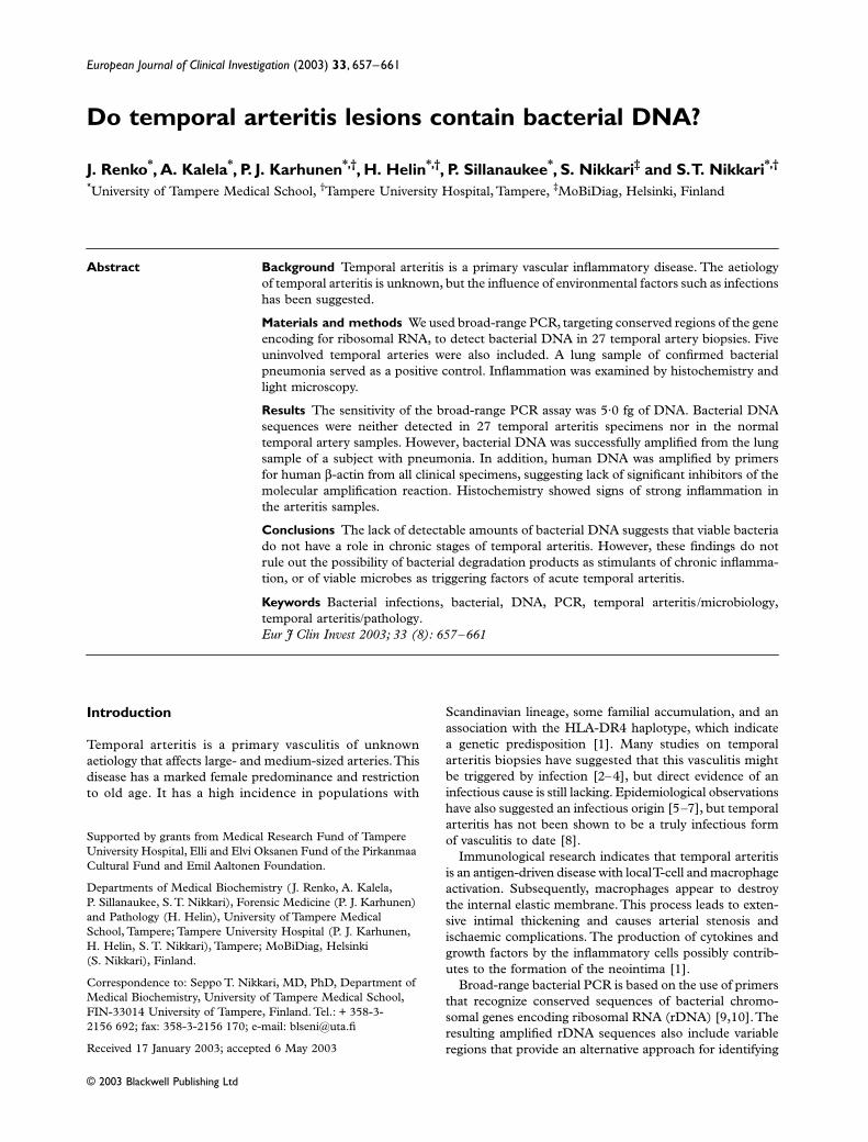
European Journal of Clinical Investigation
(2003)
33
, 657–661
© 2003 Blackwell Publishing Ltd
Blackwell Publishing Ltd.
Do temporal arteritis lesions contain bacterial DNA?
J. Renko
*
, A. Kalela
*
, P. J. Karhunen
*,†
, H. Helin
*,†
, P. Sillanaukee
*
, S. Nikkari
‡
and S. T. Nikkari
*,†
*
University of Tampere Medical School,
†
Tampere University Hospital, Tampere,
‡
MoBiDiag, Helsinki, Finland
Abstract Background
Temporal arteritis is a primary vascular inflammatory disease. The aetiologyof temporal arteritis is unknown, but the influence of environmental factors such as infectionshas been suggested.
Materials and methods
We used broad-range PCR, targeting conserved regions of the geneencoding for ribosomal RNA, to detect bacterial DNA in 27 temporal artery biopsies. Fiveuninvolved temporal arteries were also included. A lung sample of confirmed bacterialpneumonia served as a positive control. Inflammation was examined by histochemistry andlight microscopy.
Results
The sensitivity of the broad-range PCR assay was 5·0 fg of DNA. Bacterial DNAsequences were neither detected in 27 temporal arteritis specimens nor in the normaltemporal artery samples. However, bacterial DNA was successfully amplified from the lungsample of a subject with pneumonia. In addition, human DNA was amplified by primersfor human
β
-actin from all clinical specimens, suggesting lack of significant inhibitors of themolecular amplification reaction. Histochemistry showed signs of strong inflammation inthe arteritis samples.
Conclusions
The lack of detectable amounts of bacterial DNA suggests that viable bacteriado not have a role in chronic stages of temporal arteritis. However, these findings do notrule out the possibility of bacterial degradation products as stimulants of chronic inflamma-tion, or of viable microbes as triggering factors of acute temporal arteritis.
Keywords
Bacterial infections, bacterial, DNA, PCR, temporal arteritis /microbiology,temporal arteritis/pathology.
Eur J Clin Invest 2003; 33 (8): 657–661
Introduction
Temporal arteritis is a primary vasculitis of unknownaetiology that affects large- and medium-sized arteries. Thisdisease has a marked female predominance and restrictionto old age. It has a high incidence in populations with
Scandinavian lineage, some familial accumulation, and anassociation with the HLA-DR4 haplotype, which indicatea genetic predisposition [1]. Many studies on temporalarteritis biopsies have suggested that this vasculitis mightbe triggered by infection [2–4], but direct evidence of aninfectious cause is still lacking. Epidemiological observationshave also suggested an infectious origin [5–7], but temporalarteritis has not been shown to be a truly infectious formof vasculitis to date [8].
Immunological research indicates that temporal arteritisis an antigen-driven disease with local T-cell and macrophageactivation. Subsequently, macrophages appear to destroythe internal elastic membrane. This process leads to exten-sive intimal thickening and causes arterial stenosis andischaemic complications. The production of cytokines andgrowth factors by the inflammatory cells possibly contrib-utes to the formation of the neointima [1].
Broad-range bacterial PCR is based on the use of primersthat recognize conserved sequences of bacterial chromo-somal genes encoding ribosomal RNA (rDNA) [9,10]. Theresulting amplified rDNA sequences also include variableregions that provide an alternative approach for identifying
Supported by grants from Medical Research Fund of Tampere University Hospital, Elli and Elvi Oksanen Fund of the Pirkanmaa Cultural Fund and Emil Aaltonen Foundation.
Departments of Medical Biochemistry (J. Renko, A. Kalela, P. Sillanaukee, S. T. Nikkari), Forensic Medicine (P. J. Karhunen) and Pathology (H. Helin), University of Tampere Medical School, Tampere; Tampere University Hospital (P. J. Karhunen, H. Helin, S. T. Nikkari), Tampere; MoBiDiag, Helsinki (S. Nikkari), Finland.
Correspondence to: Seppo T. Nikkari, MD, PhD, Department of Medical Biochemistry, University of Tampere Medical School, FIN-33014 University of Tampere, Finland. Tel.: + 358-3-2156 692; fax: 358-3-2156 170; e-mail: [email protected]
Received 17 January 2003; accepted 6 May 2003
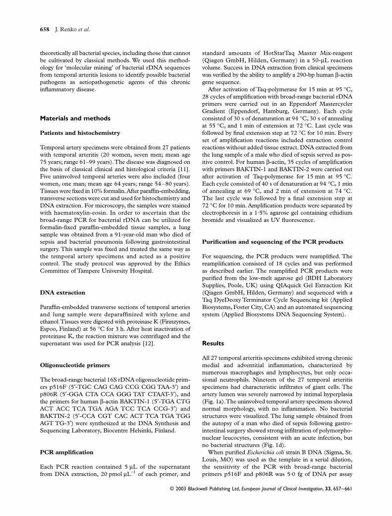
658
J. Renko
et al.
© 2003 Blackwell Publishing Ltd,
European Journal of Clinical Investigation
,
33
, 657–661
theoretically all bacterial species, including those that cannotbe cultivated by classical methods. We used this method-ology for ‘molecular mining’ of bacterial rDNA sequencesfrom temporal arteritis lesions to identify possible bacterialpathogens as aetiopathogenetic agents of this chronicinflammatory disease.
Materials and methods
Patients and histochemistry
Temporal artery specimens were obtained from 27 patientswith temporal arteritis (20 women, seven men; mean age75 years; range 61–99 years). The disease was diagnosed onthe basis of classical clinical and histological criteria [11].Five uninvolved temporal arteries were also included (fourwomen, one man; mean age 64 years; range 54–80 years).Tissues were fixed in 10% formalin. After paraffin-embedding,transverse sections were cut and used for histochemistry andDNA extraction. For microscopy, the samples were stainedwith haematoxylin-eosin. In order to ascertain that thebroad-range PCR for bacterial rDNA can be utilized forformalin-fixed paraffin-embedded tissue samples, a lungsample was obtained from a 91-year-old man who died ofsepsis and bacterial pneumonia following gastrointestinalsurgery. This sample was fixed and treated the same way asthe temporal artery specimens and acted as a positivecontrol. The study protocol was approved by the EthicsCommittee of Tampere University Hospital.
DNA extraction
Paraffin-embedded transverse sections of temporal arteriesand lung sample were deparaffinized with xylene andethanol. Tissues were digested with proteinase K (Finnzymes,Espoo, Finland) at 56
°
C for 3 h. After heat inactivation ofproteinase K, the reaction mixture was centrifuged and thesupernatant was used for PCR analysis [12].
Oligonucleotide primers
The broad-range bacterial 16S rDNA oligonucleotide prim-ers p516F (5
′
-TGC CAG CAG CCG CGG TAA-3
′
) andp806R (5
′
-GGA CTA CCA GGG TAT CTAAT-3
′
), andthe primers for human
β
-actin BAKTIN-1 (5
′
-TGA CTGACT ACC TCA TGA AGA TCC TCA CCG-3
′
) andBAKTIN-2 (5
′
-CCA CGT CAC ACT TCA TGA TGGAGT TG-3
′
) were synthesized at the DNA Synthesis andSequencing Laboratory, Biocentre Helsinki, Finland.
PCR amplification
Each PCR reaction contained 5
µ
L of the supernatantfrom DNA extraction, 20 pmol
µ
L
−
1
of each primer, and
standard amounts of HotStarTaq Master Mix-reagent(Qiagen GmbH, Hilden, Germany) in a 50-
µ
L reactionvolume. Success in DNA extraction from clinical specimenswas verified by the ability to amplify a 290-bp human
β
-actingene sequence.
After activation of Taq-polymerase for 15 min at 95
°
C,28 cycles of amplification with broad-range bacterial rDNAprimers were carried out in an Eppendorf MastercyclerGradient (Eppendorf, Hamburg, Germany). Each cycleconsisted of 30 s of denaturation at 94
°
C, 30 s of annealingat 55
°
C, and 1 min of extension at 72
°
C. Last cycle wasfollowed by final extension step at 72
°
C for 10 min. Everyset of amplification reactions included extraction controlreactions without added tissue extract. DNA extracted fromthe lung sample of a male who died of sepsis served as pos-itive control. For human
β
-actin, 35 cycles of amplificationwith primers BAKTIN-1 and BAKTIN-2 were carried outafter activation of Taq-polymerase for 15 min at 95
°
C.Each cycle consisted of 40 s of denaturation at 94
°
C, 1 minof annealing at 69
°
C, and 2 min of extension at 74
°
C.The last cycle was followed by a final extension step at72
°
C for 10 min. Amplification products were separated byelectrophoresis in a 1·5% agarose gel containing ethidiumbromide and visualized as UV fluorescence.
Purification and sequencing of the PCR products
For sequencing, the PCR products were reamplified. Thereamplification consisted of 18 cycles and was performedas described earlier. The reamplified PCR products werepurified from the low-melt agarose gel (BDH LaboratorySupplies, Poole, UK) using QIAquick Gel Extraction Kit(Qiagen GmbH, Hilden, Germany) and sequenced with aTaq DyeDeoxy Terminator Cycle Sequencing kit (AppliedBiosystems, Foster City, CA) and an automated sequencingsystem (Applied Biosystems DNA Sequencing System).
Results
All 27 temporal arteritis specimens exhibited strong chronicmedial and adventitial inflammation, characterized bynumerous macrophages and lymphocytes, but only occa-sional neutrophils. Nineteen of the 27 temporal arteritisspecimens had characteristic infiltrates of giant cells. Theartery lumen was severely narrowed by intimal hyperplasia(Fig. 1a). The uninvolved temporal artery specimens showednormal morphology, with no inflammation. No bacterialstructures were visualized. The lung sample obtained fromthe autopsy of a man who died of sepsis following gastro-intestinal surgery showed strong infiltration of polymorpho-nuclear leucocytes, consistent with an acute infection, butno bacterial structures (Fig. 1d).
When purified
Escherichia coli
strain B DNA (Sigma, St.Louis, MO) was used as the template in a serial dilution,the sensitivity of the PCR with broad-range bacterialprimers p516F and p806R was 5·0 fg of DNA per assay
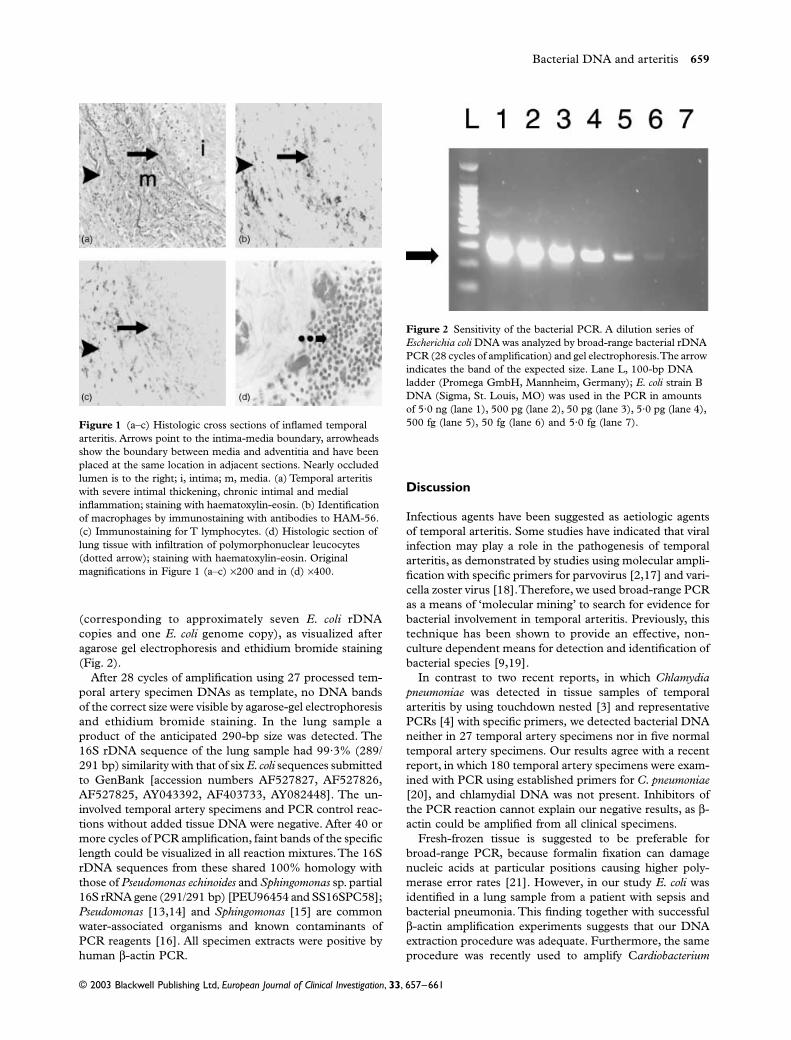
Bacterial DNA and arteritis
659
© 2003 Blackwell Publishing Ltd,
European Journal of Clinical Investigation
,
33
, 657–661
(corresponding to approximately seven
E. coli
rDNAcopies and one
E. coli
genome copy), as visualized afteragarose gel electrophoresis and ethidium bromide staining(Fig. 2).
After 28 cycles of amplification using 27 processed tem-poral artery specimen DNAs as template, no DNA bandsof the correct size were visible by agarose-gel electrophoresisand ethidium bromide staining. In the lung sample aproduct of the anticipated 290-bp size was detected. The16S rDNA sequence of the lung sample had 99·3% (289/291 bp) similarity with that of six
E. coli
sequences submittedto GenBank [accession numbers AF527827, AF527826,AF527825, AY043392, AF403733, AY082448]. The un-involved temporal artery specimens and PCR control reac-tions without added tissue DNA were negative. After 40 ormore cycles of PCR amplification, faint bands of the specificlength could be visualized in all reaction mixtures. The 16SrDNA sequences from these shared 100% homology withthose of
Pseudomonas echinoides
and
Sphingomonas
sp. partial16S rRNA gene (291/291 bp) [PEU96454 and SS16SPC58];
Pseudomonas
[13,14] and
Sphingomonas
[15] are commonwater-associated organisms and known contaminants ofPCR reagents [16]. All specimen extracts were positive byhuman
β
-actin PCR.
Discussion
Infectious agents have been suggested as aetiologic agentsof temporal arteritis. Some studies have indicated that viralinfection may play a role in the pathogenesis of temporalarteritis, as demonstrated by studies using molecular ampli-fication with specific primers for parvovirus [2,17] and vari-cella zoster virus [18]. Therefore, we used broad-range PCRas a means of ‘molecular mining’ to search for evidence forbacterial involvement in temporal arteritis. Previously, thistechnique has been shown to provide an effective, non-culture dependent means for detection and identification ofbacterial species [9,19].
In contrast to two recent reports, in which
Chlamydiapneumoniae
was detected in tissue samples of temporalarteritis by using touchdown nested [3] and representativePCRs [4] with specific primers
,
we detected bacterial DNAneither in 27 temporal artery specimens nor in five normaltemporal artery specimens. Our results agree with a recentreport, in which 180 temporal artery specimens were exam-ined with PCR using established primers for
C. pneumoniae
[20], and chlamydial DNA was not present. Inhibitors ofthe PCR reaction cannot explain our negative results, as
β
-actin could be amplified from all clinical specimens.
Fresh-frozen tissue is suggested to be preferable forbroad-range PCR, because formalin fixation can damagenucleic acids at particular positions causing higher poly-merase error rates [21]. However, in our study
E. coli
wasidentified in a lung sample from a patient with sepsis andbacterial pneumonia. This finding together with successful
β
-actin amplification experiments suggests that our DNAextraction procedure was adequate. Furthermore, the sameprocedure was recently used to amplify C
ardiobacterium
Figure 1 (a–c) Histologic cross sections of inflamed temporal arteritis. Arrows point to the intima-media boundary, arrowheads show the boundary between media and adventitia and have been placed at the same location in adjacent sections. Nearly occluded lumen is to the right; i, intima; m, media. (a) Temporal arteritis with severe intimal thickening, chronic intimal and medial inflammation; staining with haematoxylin-eosin. (b) Identification of macrophages by immunostaining with antibodies to HAM-56. (c) Immunostaining for T lymphocytes. (d) Histologic section of lung tissue with infiltration of polymorphonuclear leucocytes (dotted arrow); staining with haematoxylin-eosin. Original magnifications in Figure 1 (a–c) ×200 and in (d) ×400.
Figure 2 Sensitivity of the bacterial PCR. A dilution series of Escherichia coli DNA was analyzed by broad-range bacterial rDNA PCR (28 cycles of amplification) and gel electrophoresis. The arrow indicates the band of the expected size. Lane L, 100-bp DNA ladder (Promega GmbH, Mannheim, Germany); E. coli strain B DNA (Sigma, St. Louis, MO) was used in the PCR in amounts of 5·0 ng (lane 1), 500 pg (lane 2), 50 pg (lane 3), 5·0 pg (lane 4), 500 fg (lane 5), 50 fg (lane 6) and 5·0 fg (lane 7).
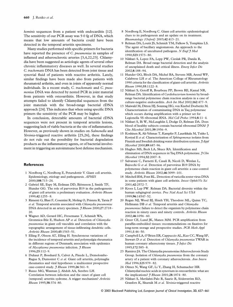
660
J. Renko
et al.
© 2003 Blackwell Publishing Ltd,
European Journal of Clinical Investigation
,
33
, 657–661
hominis
sequences from a patient with endocarditis [12].The sensitivity of our PCR assay was 5·0 fg of DNA, whichmeans that low amounts of bacteria could have beendetected in the temporal arteritis specimens.
Many studies performed with specific primers for bacteriahave reported the presence of
C. pneumoniae
in samples ofinflamed and atherosclerotic arteries [3,4,22,23]. Chlamy-dia have been suggested as aetiologic agents of several otherchronic inflammatory diseases as well. In several studies
C. trachomatis
DNA has been detected from joint tissue andsynovial fluid of patients with reactive arthritis. Lately,similar findings have been made also from patients withrheumatoid arthritis, and even in joints of apparently normalindividuals. In a recent study,
C. trachomatis
and
C. pneu-moniae
DNA was detected by nested PCR in joint materialfrom patients with osteoarthitis. However, in that studyattempts failed to identify Chlamydial sequences from thejoint materials with the broad-range bacterial rDNAapproach [24]. This indicates that when using specific DNAprimers the sensitivity of the PCR may be higher.
In conclusion, detectable amounts of bacterial rDNAsequences were not present in temporal arteritis lesions,suggesting lack of viable bacteria at the site of inflammation.However, as previously shown in studies on
Salmonella
and
Yersinia
-triggered reactive arthritis [25,26], these findingsdo not rule out the possibility for bacterial degradationproducts as the inflammatory agents, or of bacterial involve-ment in triggering an autoimmune host defense mechanism.
References
1 Nordborg C, Nordborg E, Petursdottir V. Giant cell arteritis. Epidemiology, etiology and pathogenesis.
APMIS
2000;
108
:713–24.2 Gabriel SE, Espy M, Erdman DD, Björnsson J, Smith TF,
Hunder GG. The role of parvovirus B19 in the pathogenesis of giant cell arteritis: a preliminary evaluation.
Arthritis Rheum
1999;
42
:1255–8.3 Rimenti G, Blasi F, Cosentini R, Moling O, Pristera R, Tarsia P
et al.
Temporal arteritis associated with
Chlamydia pneumoniae
DNA detected in an artery specimen.
J Rheum
2000;
27
:2718–20.
4 Wagner AD, Gerard HC, Fresemann T, Schmidt WA, Gromnica-Ihle E, Hudson AP
et al.
Detection of
Chlamydia pneumoniae
in giant cell vasculitis and correlation with the topographic arrangement of tissue-infiltrating dendritic cells.
Arthritis Rheum
2000;
43
:1543–51.5 Elling P, Olsson AT, Elling H. Synchronous variations of
the incidence of temporal arteritis and polymyalgia rheumatica in different regions of Denmark; association with epidemics of
Mycoplasma pneumoniae
infection.
J Rheum
1996;
23
:112–9.6 Duhaut P, Bosshard S, Calvet A, Pinede L, Demolombe-
Rague S, Dumontet C
et al.
Giant cell arteritis, polymaglia rheumatica and viral hypotheses: a multicenter prospective case-control study.
J Rheum
1999;
26
:361–9.7 Russo MG, Waxman J, Abdoh AA, Serebro LH.
Correlation between infection and the onset of giant cell (temporal) arteritis sydrome. A trigger mechanism?
Arthritis Rheum
1995;
38
:374–80.
8 Nordborg E, Nordborg C. Giant cell arteritis: epidemiological clues to its pathogenesis and an update on its treatment.
Rheumatology (Oxford)
2003;
42
:413–21.9 Relman DA, Loutit JS, Schmidt TM, Falkow S, Tompkins LS.
The agent of bacillary angiomatosis. An approach to the identification of uncultured pathogens.
N Engl J Med
1990;
323
:1573–80.10 Nikkari S, Lopez FA, Lepp PW, Cieslak PR, Danila R,
Relman DA. Broad range bacterial detection and the analysis of unexplained death and critical illness.
Emerg Infect Dis
2002;
8
:188–94.11 Hunder GG, Bloch DA, Michel BA, Stevens MB, Arend WP,
Calabrese LH
et al.
The American College of Rheumatology 1990 criteria for the classification of giant-cell arteritis.
Arthritis Rheum
1990;
33:1122–8.12 Nikkari S, Gotoff R, Bourbeau PP, Brown RE, Kamal NR,
Relman DA. Identification of Cardiobacterium hominis by broad-range bacterial polymerase chain reaction analysis in a case of culture-negative endocarditis. Arch Int Med 2002;162:477–9.
13 Maiwald M, Ditton HJ, Sonntag HG, von Knebel Doeberitz M. Characterization of contaminating DNA in Taq polymerase which occurs during amplification with a primer set for Legionella 5S ribosomal RNA. Mol Cell Probes 1994;8:11–4.
14 Nikkari S, Bi W, McLaughlin I, Dodge D, Relman DA. Does blood of healthy subjects contain bacterial ribosomal DNA? J Clin Microbiol 2001;39:1956–9.
15 Koskinen R, Ali-Vehmas T, Kampfer P, Laurikkala M, Tsitko I, Kostyal E et al. Characterization of Sphingomonas isolates from Finnish and Swedish drinking water distribution systems. J Appl Microbiol 2000;89:687–96.
16 Hughes MS, Beck LA, Skuce RA. Identification and elimination of DNA sequences in Taq DNA polymerase. J Clin Microbiol 1994;32:2007–8.
17 Salvarani C, Farnetti E, Casali B, Nicoli D, Wenlan L, Bajocchi G et al. Detection of parvovirus B19 DNA by polymerase chain reaction in giant cell arteritis: a case-control study. Arthritis Rheum 2002;46:3099–101.
18 Mitchell BM, Font RL. Detection of varicella zoster virus DNA in some patients with giant cell arthritis. Invest Ophth Vis Sci 2001;42:2572–7.
19 Kroes I, Lep PW. Relman DA. Bacterial diversity within the human subgingival crevice. Proc Natl Acad Sci USA 1999;96:14547–52.
20 Regan MJ, Wood BJ, Hsieh YH, Theodore ML, Quinn TC, Hellmann DB et al. Temporal arteritis and Chlamydia pneumoniae: failure to detect the organism by polymerase chain reaction in ninety cases and ninety controls. Arthritis Rheum 2002;46:1056–60.
21 Greer CE, Lund JK, Manos MM. PCR amplification from paraffin-embedded tissues: recommendations on fixatives for long-term storage and prospective studies. PCR Meth Appl 1991;1:46–50.
22 Campbell LA, O’Brien ER, Cappuccio AL, Kuo CC, Wang SP, Stewart D et al. Detection of Chlamydia pneumoniae TWAR in human coronary atherectomy tissues. J Infect Dis 1995;172:585–8.
23 Ramirez JA. The Chlamydia pneumoniae Atherosclerosis Study Group. Isolation of Chlamydia pneumoniae from the coronary artery of a patient with coronary atherosclerosis. Ann Intern Med 1996;125:979–82.
24 Olmez N, Wang GF, Li Y, Zhang H, Schumacher HR. Chlamydial nucleic acids in synovium in osteoarthritis: what are the implications? J Rheum 2001;28:1874–80.
25 Nikkari S, Merilahti-Palo R, Saario R, Söderström KO, Granfors K, Skurnik M et al. Yersinia-triggered reactive

Bacterial DNA and arteritis 661
© 2003 Blackwell Publishing Ltd, European Journal of Clinical Investigation, 33, 657–661
arthritis. Use of polymerase chain reaction and immunocytochemical staining in the detection of bacterial components from synovial specimens. Arthritis Rheum 1992;35:682–7.
26 Nikkari S, Rantakokko K, Ekman P, Mottonen T,
Leirisalo-Repo MM, Virtala M et al. Salmonella-triggered reactive arthritis: use of polymerase chain reaction, immunocytochemical staining, and gas chromatography mass spectrometry in the detection of bacterial components from syanovial fluid. Arthritis Rheum 1999;42:84–9.
European Journal of Clinical Investigation
(2005)
35
, 13–16
© 2005 Blackwell Publishing Ltd
.Blackwell Publishing, Ltd.
Identification of different bacterial DNAs in human coronary arteries
J. Lehtiniemi
*
, P. J. Karhunen
*†
, S. Goebeler
*†
, S. Nikkari
‡
and S. T. Nikkari
*†
*
University of Tampere Medical School, Tampere,
†
Tampere University Hospital, Tampere,
‡
Institute of Military Medicine,
Helsinki, Finland
Abstract Background
Various studies have suggested a link between infection, atherosclerosis andcoronary artery disease. We studied whether bacterial DNA is present in coronary specimensobtained from left anterior descending coronary arteries of subjects having sudden deathsof cardiovascular and other causes, as verified by an autopsy.
Materials and methods
Coronary specimens were obtained from five subjects who diedof sudden coronary causes and five controls. Broad-range 16-s rDNA PCR (Br-PCR)amplification, cloning and sequencing were used to detect bacterial rDNA.
Results
Bacterial rDNA sequences of oral pathogens were detected from the coronarysamples in all cases regardless of the cause of death.
Conclusions
Br-PCR is a powerful method to detect bacterial rDNA. By this method wewere able to detect wide palette of oral bacteria from coronary tissues. Our findings suggestthat atheromas may act as mechanical sieves collecting bacteria from the circulation.
Keywords
Bacterial infections, bacterial, coronary artery disease, DNA, PCR.
Eur J Clin Invest 2005; 35 (1): 13–16
Introduction
Inflammation is an important defense mechanism againstinfection. Chronic or recurrent infections may cause sus-tained inflammation, which has been linked to increasedrisk of coronary artery disease (CAD) [1,2]. Several studieshave suggested a link between bacterial infection and CAD.
We used a comprehensive PCR methodology to studywhether more than one bacterial species might be found in
coronary artery tissue material from subjects with CAD.The broad-range bacterial PCR methodology is based onthe use of primers that recognize conserved sequences ofbacterial chromosomal genes encoding ribosomal RNA(rDNA) [3,4]. The resulting amplified rDNA sequencesinclude variable regions that provide sequence informationfor phylogenetic identifying of theoretically all bacterialspecies, including those that cannot be cultivated by clas-sical methods. We used this methodology for ‘molecularmining’ of bacterial rDNA sequences from coronary lesionsobtained from rapid medicolegal autopsies to identify poten-tial aetiopathogenetic agents of this chronic inflammatorydisease.
Materials and methods
Patients and histochemistry
Coronary specimens were obtained from the proximal partof the main trunk of the left anterior descending coronaryartery (LAD) of 10 cases within 3 days after death. Atautopsy, the proximal part of the LAD was removed andimmediately frozen at
−
70
°
C. For histochemistry, thetissues were fixed in 10% formalin, embedded in paraffin,cut and stained with Hematoxyline-Eosine.
This study was supported by the Yrjö Jahnsson Foundation, the Elli and Elvi Oksanen Fund of the Pirkanmaa Fund under the auspices of the Finnish Cultural Foundation, the Medical Research Fund of Tampere University Hospital, and the Finnish Foundation for Cardiovascular Research.
Departments of Medical Biochemistry ( J. Lehtiniemi, S. T. Nikkari) and Forensic Medicine (P. J. Karhunen, S. Goebeler), University of Tampere Medical School, Tampere; Department of Clinical Chemistry (P. J. Karhunen, S. Goebeler, S. T. Nikkari), Tampere University Hospital, Tampere; Institute of Military Medicine, Helsinki (S. Nikkari), Finland.
Correspondence to: Seppo T. Nikkari, MD, PhD, Department of Medical Biochemistry, University of Tampere Medical School, FIN-33014, Finland. Tel.: +358 3 2156 692; fax: 358 3 2156 170; e-mail: [email protected]
Received 18 August 2004; accepted 9 November 2004
14
J. Lehtiniemi
et al.
© 2005 Blackwell Publishing Ltd,
European Journal of Clinical Investigation
,
35
, 13–16
This study was approved by Ethics Committee of theNational Authority for Medicolegal Affairs, Helsinki, Finland.
DNA extraction
Coronary artery tissues adjacent to the cryostat sectionswere digested with proteinase K (Finnzymes, Espoo, Fin-land) at 56
°
C for 3 h. After heat inactivation of proteinaseK, the reaction mixture was centrifuged and the supernatantwas used for PCR analysis [5]. Control extractions of waterwere also used as a negative control.
PCR amplification
Each PCR reaction contained 5
µ
L of the supernatant fromthe DNA extraction, 20 pmol
µ
L
−
1
of each primer, andstandard amounts of HotStarTaq Master Mix-reagent (Qia-gen GmbH, Hilden, Germany) in a 50-
µ
L reaction volume.Success in DNA extraction from clinical specimens wasverified by the ability to amplify a 290-bp human
β
-actingene sequence.
Following activation of Taq-polymerase for 15 min at95
°
C, 38 cycles of amplification (94
°
C for 30 s, 55
°
C for30 s and 72
°
C for 1 min) with broad-range bacterial 16-srDNA primers p516F and p806R [4] were carried out inEppendorf Mastercycler Gradient (Eppendorf, Hamburg,Germany). The last cycle was followed by a final extensionstep at 72
°
C for 10 min. Every set of amplification reactionsincluded extraction control reactions without an added tissueextract.
Escherichia coli
strain B DNA (Sigma, St. Louis, MO)served as a positive control. For human
β
-actin, 40 cyclesof amplification (94
°
C for 35 s, 60
°
C for 2 min and 72
°
Cfor 2 min) with primers BAKTIN-1 (5
′
-TGA CTG ACTACC TCA TGA AGA TCC TCA CCG-3
′
) and BAKTIN-2 (5
′
-CCA CGT CAC ACT TCA TGA TGG AGT TG-3
′
)were carried out after activation of Taq-polymerase for15 min at 95
°
C. Amplification products were separated byelectrophoresis in a 2% agarose gel containing ethidiumbromide, and visualized as UV fluorescence.
Cloning and sequencing of the PCR products
Amplified PCR products were ligated into the pCR2·1-TOPO vector and transformed into
E. coli
cells by using theTOPO TA cloning system (Invitrogen, Carlsbad, CA). Twoseparate clone libraries were created from each PCR prod-uct amplified from the tissue digest supernatant. M13 for-ward (
−
20) and M13 reverse primers were used to analyzetransformants by picking colonies and resuspending themindividually in 50
µ
L of PCR cocktail consisting of 10 pmol
µ
L
−
1
of each primer and standard amounts of HotStarTaqMaster Mix-reagent. After activation of Taq-polymerase for15 min at 95
°
C, 40 cycles of amplification (95
°
C for 1 min,55
°
C for 1 min and 72
°
C for 1 min) with M13 primerswere carried out in an Eppendorf Mastercycler Gradient.The last cycle was followed by final extension step at 72
°
C
for 10 min. Success in ligation of inserts of the expected sizewas verified by electrophoresis in a 2% agarose gel contain-ing ethidium bromide and visualized as UV fluorescence.Positive transformants were purified by using QiaQuickPCR Purification Kit (Qiagen GmbH, Hilden, Germany).
After cloning, the automated ABI PRISM 310 GeneticAnalyser (Applied Biosystems, Foster City, CA) and BigDyeTerminator Cycle sequencing chemistry (Applied Biosystems)were used to determine the DNA sequences. The consensussequence from both DNA strands was compared with thosein GenBank by the BLAST search tool [6].
Results
Background characteristics of the subjects’ coronary artery,morphological findings and PCR results are summarized inTable 1. The bacterial 16S rDNA sequences were 99–100%similar to those submitted to GenBank, including bacterialspecies belonging to the oral microflora marked in bold. Thefinding of
E. coli
sequences from two coronary arteries wasomitted, as they were owing to Taq DNA polymerase con-tamination with
E. coli
DNA detected from extraction controlreagents [7]. These contaminants become well-representedin recombinant bacterial 16S rDNA clone libraries whenstudying low concentrations of bacterial DNA specimens[8,9]. All specimen extracts were positive by human
β
-actinPCR, indicating that the DNA extraction procedure wasadequate and that inhibitors of the PCR reaction were notpresent.
It is noteworthy that all coronary specimens containedDNA sequences of oral microflora. These findings includeda common pathogen
Streptococcus pyogenes
and species suchas
Actinomyces odontolyticus
,
Haemophilus parahaemolyticus
,
Lactobacillus crispatus
,
Lactobacillus delbrueckii
and
Lacto-bacillus fermentum
. Additionally, other oral bacteria sequence-types could be identified to the genus level, such as
Lactobacillus
sp.,
Neisseria
sp.,
Prevotella
sp.,
Streptococcus mitis
group,
Streptococcus salivarius
group and
Micrococcus
sp. Thespecies
Lactococcus lactis
sp. and
Enterococcus
sp. aregenerally regarded as belonging to the gastrointestinal tractflora.
Discussion
Infectious agents have been suggested as aetiologic agentsof complicated CAD. We used broad-range PCR as a meansof ‘molecular mining’ to search for the presence of bacterialnucleic acids in autopsy samples of coronary arteries fromsubjects with sudden death. Use of this technique to identifypost mortem coronary artery material has not been reportedto date. Previously, this technique has been shown to providean effective, nonculture-dependent means for detection andidentification of bacterial species [3,10].
The roles played by the organisms corresponding to thesequence-types identified in the coronary artery specimens
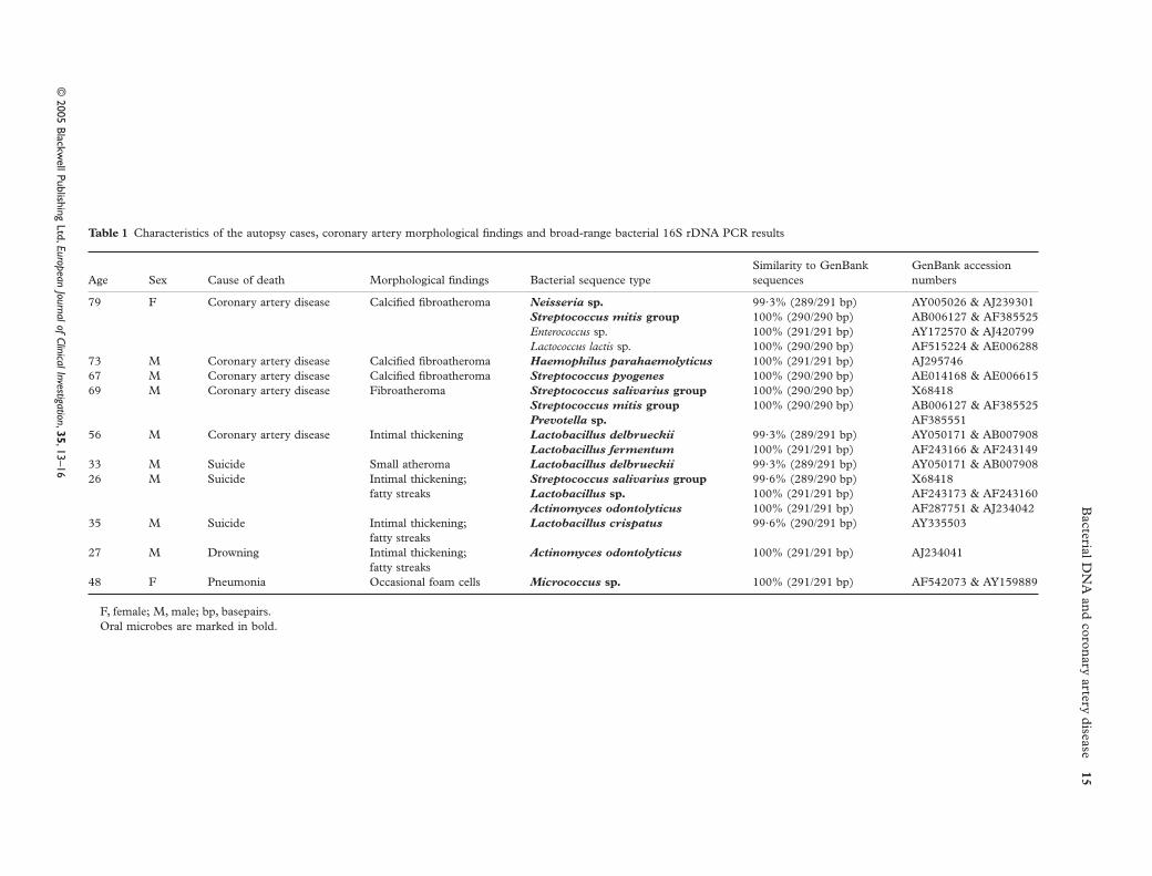
Bacterial D
NA
and coronary artery disease
15
© 2005 Blackw
ell Publishing Ltd,
European Journal of Clinical Investigation
,
35
, 13–16
Table 1
Characteristics of the autopsy cases, coronary artery morphological findings and broad-range bacterial 16S rDNA PCR results
Age Sex Cause of death Morphological findings Bacterial sequence typeSimilarity to GenBank sequences
GenBank accession numbers
79 F Coronary artery disease Calcified fibroatheroma
Neisseria
sp.
99·3% (289/291 bp) AY005026 & AJ239301
Streptococcus mitis
group
100% (290/290 bp) AB006127 & AF385525
Enterococcus
sp. 100% (291/291 bp) AY172570 & AJ420799
Lactococcus lactis
sp. 100% (290/290 bp) AF515224 & AE00628873 M Coronary artery disease Calcified fibroatheroma
Haemophilus parahaemolyticus
100% (291/291 bp) AJ29574667 M Coronary artery disease Calcified fibroatheroma
Streptococcus pyogenes
100% (290/290 bp) AE014168 & AE00661569 M Coronary artery disease Fibroatheroma
Streptococcus salivarius
group
100% (290/290 bp) X68418
Streptococcus mitis
group
100% (290/290 bp) AB006127 & AF385525
Prevotella
sp.
AF38555156 M Coronary artery disease Intimal thickening
Lactobacillus delbrueckii
99·3% (289/291 bp) AY050171 & AB007908
Lactobacillus fermentum
100% (291/291 bp) AF243166 & AF24314933 M Suicide Small atheroma
Lactobacillus delbrueckii
99·3% (289/291 bp) AY050171 & AB00790826 M Suicide Intimal thickening;
fatty streaks
Streptococcus salivarius
group
99·6% (289/290 bp) X68418
Lactobacillus
sp.
100% (291/291 bp) AF243173 & AF243160
Actinomyces odontolyticus
100% (291/291 bp) AF287751 & AJ23404235 M Suicide Intimal thickening;
fatty streaks
Lactobacillus crispatus
99·6% (290/291 bp) AY335503
27 M Drowning Intimal thickening;fatty streaks
Actinomyces odontolyticus
100% (291/291 bp) AJ234041
48 F Pneumonia Occasional foam cells
Micrococcus
sp.
100% (291/291 bp) AF542073 & AY159889
F, female; M, male; bp, basepairs.Oral microbes are marked in bold.
16
J. Lehtiniemi
et al.
© 2005 Blackwell Publishing Ltd,
European Journal of Clinical Investigation
,
35
, 13–16
is not currently known. We speculate that the microbes mayhave entered the blood stream during transient bacteraemiasknown to occur, e.g. during toothbrushing or by leakingthrough mucosal surfaces. Except for
Streptococcus pyogenes
,most have low virulence and they do not generally causeinfections in healthy individuals. However, the microbesmay also act as ‘innocent bystanders’ entering the atheroma.
At present there is ample evidence to suggest that oral[11] and other respiratory tract bacteria such as
Chlamydiapneumoniae
[12] are strongly associated with coronary dis-ease. Br-PCR is powerful method to detect bacterial DNA.Our findings suggest that atheromas may act as mechanicalsieves collecting bacteria from the circulation. In fact,bacterial DNA has been previously reported to be presentin blood samples of healthy individuals [7], and it is likelythat the oral cavity and other mucosal surfaces, e.g. the gut,represent the most common origin of transient bacteraemia.The presence of such pathogens may, in certain individuals,contribute to the development of clinically significant CAD.
References
1 Zhu J, Quyyumi AA, Norman JE, Csako G, Waclawiw MA, Shearer GM
et al.
Effects of total pathogen burden on coronary artery disease risk and C-reactive protein levels.
Am J Cardiol
2000;
85
:140–6.2 Kol A, Libby P. The mechanisms by which infectious agents
may contribute to atherosclerosis and its clinical manifestations.
Trends Cardiovasc Med
1998;
8
:191–9.3 Relman DA, Loutit JS, Schmidt TM, Falkow S, Tompkins LS.
The agent of bacillary angiomatosis. An approach to the
identification of uncultured pathogens.
N Engl J Med
1990;
323
:1573–80.4 Nikkari S, Lopez FA, Lepp PW, Cieslak PR, Danila R,
Relman DA. Broad range bacterial detection and the analysis of unexplained death and critical illness.
Emerg Infect Dis
2002;
8
:188–94.5 Nikkari S, Gotoff R, Bourbeau PP, Brown RE, Kamal NR,
Relman DA. Identification of Cardiobacterium hominis by broad-range bacterial polymerase chain reaction analysis in a case of culture-negative endocarditis.
Arch Int Med
2002;
162
:477–9.6 Altschul SF, Gish W, Miller W, Myers EW, Lipman DJ.
Basic local alignment search tool.
J Mol Biol 1990;215:403–10.7 Nikkari S, McLaughlin IJ, Bi W, Dodge DE, Relman DA. Does
blood of healthy subjects contain bacterial ribosomal DNA? J Clin Microbiol 2001;39:1956–9.
8 Tanner MA, Goebel BM, Dojka MA, Pace NR. Specific ribosomal DNA sequences from diverse environmental setting correlate with experimental contaminants. Appl Environ Microbiol 1998;64:3110–3.
9 Lawyer FC, Stoffel S, Saiki RK, Myambo K, Drummond R, Gelfand DH. Isolation, characterisation, and expression in E. coli of the DNA polymerase gene from the extreme thermophile, Thermus aquaticus. J Biol Chem 1989;264:6427–37.
10 Kroes I, Lep PW, Relman DA. Bacterial diversity within the human subgingival crevice. Proc Natl Acad Sci USA 1999;96:14547–52.
11 Pussinen PJ, Jousilahti P, Alfthan G, Palosuo T, Asikainen S, Salomaa V. Antibodies to periodontal pathogens are associated with coronary heart disease. Arterioscler Thromb Vasc Biol 2003;23:1250–4.
12 Linnanmäki E, Leinonen M, Mattila K, Nieminen MS, Valtonen V, Saikku P. Chlamydia pneumoniae-specific circulating immune complexes in patients with chronic coronary heart disease. Circulation 1993;87:1130–4.