Embed Size (px)
Citation preview
Out-of-Hospital Cardiac Arrest
Studies on aetiology, treatment and outcome
U N I V E R S I T Y O F T A M P E R E
ACADEMIC DISSERTATIONTo be presented, with the permission of
the Faculty of Medicine of the University of Tampere,for public discussion in the Small Auditorium of Building K,
Medical School of the University of Tampere,Teiskontie 35, Tampere, on June 6th, 2008, at 12 o’clock.
ILKKA VIRKKUNEN
DistributionBookshop TAJUP.O. Box 61733014 University of TampereFinland
Cover design byJuha Siro
Acta Universitatis Tamperensis 1321ISBN 978-951-44-7345-6 (print)ISSN 1455-1616
Tampereen Yliopistopaino Oy – Juvenes PrintTampere 2008
Tel. +358 3 3551 6055Fax +358 3 3551 [email protected]/tajuhttp://granum.uta.fi
Acta Electronica Universitatis Tamperensis 732ISBN 978-951-44-7346-3 (pdf )ISSN 1456-954Xhttp://acta.uta.fi
ACADEMIC DISSERTATIONUniversity of Tampere, Medical SchoolTampere University Hospital, Department of Surgery and AnaesthesiologyHelsinki University Hospital, Department of Anaesthesiology and Intensive Care MedicineUniversity of Turku, Faculty of MedicineFinland
Supervised byProfessor Arvi Yli-HankalaUniversity of TampereDocent Tom SilfvastUniversity of Helsinki
Reviewed byDocent Ilkka ParviainenUniversity of KuopioDocent Tarja RandellUniversity of Helsinki

To my wife Tarja and our children Salla, Tuomas and Aapo.

4
5
Abstract
The survival rate after outofhospital cardiac arrest (OHCA) has not improvedmuch over the last decade. The aim of this thesis was to study treatment andoutcome of OHCA in the emergency medical service (EMS) systems in TampereDistrict EMS and the physicianstaffed helicopter emergency medical service(HEMS) in the Helsinki and Turku areas in Southern Finland.
Gastric regurgitation and pulmonary aspiration are serious adverse events inOHCA. The objective was to determine whether there is an association betweenbystander mouthtomouth ventilation and regurgitation in prehospital cardiacarrest patients. In this prospective material, the study population consisted of 529consecutive prehospital cardiac arrest patients with attempted resuscitation.Exclusion criteria were cardiac arrest due to trauma or drug overdose. The EMSpersonnel documented if regurgitation was present in OHCA patients on thescene. Regurgitation occurred in a fourth of patients. Bystander cardiopulmonaryresuscitation (CPR) with mouthtomouth ventilation was associated with asignificantly increased risk of regurgitation compared with no CPR and CPRwithout ventilation. The mode and role of bystander CPR in cardiac arrest needsto be further evaluated.
The association between clinical signs of regurgitation and radiologicalfindings consistent with aspiration in resuscitated OHCA patients admitted tohospital were studied. The incidence of regurgitation was studied in 182successfully resuscitated OHCA patients. The inclusion criterion was therestoration of spontaneous circulation (ROSC) after OHCA not caused by traumaor drug overdose. The incidence of regurgitation was 20 %. Regurgitation wasassociated with radiological findings consistent with aspiration with highspecificity (81 %) and low sensitivity (46 %). Although there was a strongassociation between clinical regurgitation and radiological findings consistentwith aspiration, our data suggest that regurgitation is not invariably followed byradiological findings compatible with aspiration. Radiological findingsconsistent with aspiration appeared to be relatively infrequent without precedingsigns of regurgitation in resuscitated patients.
The objective in Study III was the cause of death (COD) in patients who diedafter an unsuccessful attempt at outofhospital resuscitation when the initialcardiac rhythm had been pulseless electrical activity (PEA). The aim was todetermine whether there is a difference in the distribution of CODs betweenthose who underwent autopsy and those whose COD was estimated based onclinical and previous medical history. Data were collected from 91 patientstreated by the emergency medical service systems. An autopsy was performed on
6
59 patients, while the COD was determined without autopsy in 32 patients. There were significantly more diagnoses of acute myocardial infarction (AMI) and less pulmonary embolism (PE), aortic dissection and rupture among those without autopsy compared with those who underwent autopsy. The conclusion was that in unsuccessful resuscitation from OHCA with PEA as initial rhythm, an autopsy should be performed to determine the exact cause of death.
Mild therapeutic hypothermia improves neurological outcome after cardiac arrest. The cooling and haemodynamic effects of prehospital infusion of ice-cold Ringer’s solution were studied in 13 adult patients after successful resuscitation from non-traumatic cardiac arrest. After haemodynamic stabilisation, 30 ml/kg of Ringer’s solution was infused at a rate of 100 ml/min into the antecubital vein. Arterial blood pressure and blood gases, pulse rate, end-tidal CO2 and oesophageal temperature (Tesof) were monitored closely. The mean core temperature decreased significantly from 35.8 ± 0.9 °C at the start of infusion to 34.0 ± 1.2 °C on arrival at hospital. No serious adverse haemodynamic effects occurred. It was concluded that the induction of therapeutic hypothermia using this technique in the prehospital setting is feasible.
7
Tiivistelmä
Sairaalan ulkopuolella tapahtuneen sydänpysähdyksen ennuste ei ole parantunut paljoa viime vuosikymmenen aikana. Tämän väitöskirjan tarkoituksena oli tutkia hoitoa ja selviytymistä sairaalan ulkopuolella tapahtuneesta sydänpysähdyksestä Tampereen aluepelastuslaitoksen, Medi-Heli 01:n ja Medi-Heli 02:n alueilla Etelä-Suomessa.
Mahansisällön regurgitaatio ja aspiroituminen keuhkoihin on vakava komplikaatio elvytyksen aikana. Ensimmäisen osatyön tavoitteena oli selvittää maallikkoelvytyksen vaikutusta aspiraatioriskiin ja tarkentaa elottomuuteen liittyviä riskitekijöitä. Tässä prospektiivisessa tutkimuksessa tutkittiin 529 oletettavasti sydänperäisen elvytyspotilaan tietoja. Ensihoitohenkilöstö rekisteröi regurgitaation ilmaantuvuuden ja ajankohdan. Noin neljännes potilaista regurgitoi. Maallikon suorittama painelu-puhalluselvytys tai pelkkä puhalluselvytys lisäsi merkitsevästi regurgitaatiota elvytyksen aikana verrattuna pelkkään paineluelvytykseen. Maallikkoelvytyksen rooli ja laatu tulee selvittää tulevissa tutkimuksissa.
Toisessa osatyössä tutkittiin kentällä havaitun elvytyksen aikaisen regurgitaation vaikutusta sairaalassa tehtyihin radiologisiin löydöksiin. Tutkimukseen otettiin mukaan 182 potilasta, jotka oli onnistuneesti elvytetty sairaalan ulkopuolella ja joiden elottomuuden syy ei ollut trauma tai myrkytys. Näillä potilailla regurgitaatio ilmeni 20 %. Kentällä todetun regurgitaation yhteys radiologisiin löydöksiin todettiin korkealla spesifiteetillä (81 %) ja matalalla sensitiviteetillä (46 %). Vaikka todetun regurgitaation ja aspiraation sopivien radiologisten löydösten välillä oli vahva riippuvuus, niin tulostemme mukaan aspiraatiosta ei aina seuraa radiologisesti todennettavia muutoksia. Aspiraatioon viittaavat radiologiset löydökset ovat suhteellisen harvinaisia elvytetyillä potilailla, ellei elvytyksen aikana kentällä ole tehty kliinistä havaintoa regurgitaatiosta.
Sairaalan ulkopuolella epäonnistuneesti PEA-alkurytmistä elvytettyjen sydänpysähdyspotilaiden kuolinsyyt tutkittiin. Tarkoituksena oli selvittää, poikkeavatko ruumiinavauksen perusteella määritettyjen kuolinsyiden jakauma potilaan lääketieteellisen historian ja elvytyksen kulun perusteella kliinisesti määritettyjen kuolinsyiden jakaumasta. Tutkimukseen otettiin mukaan 91 epäonnistuneeseen elvytykseen päättynyttä sydänpysähdyspotilasta PEA-alkurytmillä, joista ruumiinavaus suoritettiin 59 vainajalle. 32 vainajan kuolinsyyt määritettiin edellä mainituin kliinisin perustein. Sydäninfarktien osuus kuolinsyistä oli merkitsevästi yliedustettuna ja keuhkoveritulpat ja aortan repeämät tai dissekaatiot aliedustettuina niillä vainajilla, joiden kuolinsyy oli
8
määritetty kliinisin perustein. Voidaankin todeta, että PEA-alkurytmillä alkaneeseen sydänpysähdykseen menehtyneen vainajan kuolinsyy tulee määrittää ruumiinavauksella todellisen kuolinsyyn selvittämiseksi.
Hypotermiahoito sydänpysähdyksen jälkeen parantaa ennustetta. Ensimmäisessä osatyössä selvitettiin jääkylmällä Ringerin nesteellä toteutetun hypotermiahoidon toteutumista ja verenkierrollisia vaikutuksia 13 potilaalla sairaalan ulkopuolisen sydänperäisen elottomuuden jälkeen. Verenkierron vakauttamisen jälkeen, 30ml/kg Ringerin nestettä 100ml/min infusoitiin kyynärtaipeen laskimoon. Verikaasuja, verenpainetta, pulssia, hengitysilman ulostulevaa hiilidioksidia ja ruokatorven lämpötilaa mitattiin tarkasti. Potilaan ydinlämpö laski merkitsevästi 35.8 ± 0.9 °C:sta 34.0 ± 1.2 °C:een. Vakavia verenkierron häiriöitä ei havaittu. Todettiin, että sairaalan ulkopuolella indusoitu hypotermiahoito on tällä menetelmällä toteuttamiskelpoinen.
9
Contents
ABSTRACT.................................................................................................. 5
TIIVISTELMÄ ............................................................................................. 7
CONTENTS.................................................................................................. 9
ABBREVIATIONS .................................................................................... 11
LIST OF ORIGINAL PUBLICATIONS.................................................... 13
INTRODUCTION ...................................................................................... 15
REVIEW OF THE LITERATURE ............................................................ 17
1. Historical perspective of resuscitation ...............................................................17
2. Out-of-Hospital Cardiac arrest...........................................................................20
3. Regurgitation and aspiration in cardiac arrest ...................................................22
4. Pulseless electrical activity ................................................................................25
5. Post-resuscitation disease...................................................................................26
6. Hypothermia after cardiac arrest........................................................................26
7. Techniques for the induction of hypothermia after cardiac arrest .....................29
AIMS OF THE STUDY ............................................................................. 30
MATERIAL AND METHODS.................................................................. 31
Patients and methods..............................................................................................31
RESULTS ................................................................................................... 37
Regurgitation and bystander CPR..........................................................................37
10
Aspiration and radiological findings in pre-hospital cardiac arrest .......................38
Cause of death in resuscitation with PEA..............................................................38
Induction of therapeutic hypothermia after OHCA ...............................................40
DISCUSSION..............................................................................................41
LIMITATIONS OF THE STUDY ..............................................................48
CONCLUSIONS .........................................................................................49
ACKNOWLEDGEMENTS.........................................................................50
REFERENCES ............................................................................................52
ORIGINAL PUBLICATIONS ....................................................................59
11
Abbreviations
ALS Advanced life support AMI Acute myocardial infarction ARDS Acute respiratory distress syndrome ASY Asystole ATP Adenosine triphosphate BLS Basic life support °C Symbol for degree Celsius CA Cardiac arrest COD Cause of death CNS Central nervous system CO2 Carbon dioxide COPD Chronic obstructive pulmonary disease CPC Cerebral performance category CPP Coronary perfusion pressure CPR Cardiopulmonary resuscitation ECG Electrocardiogram EMD Electromechanical dissociation EMS Emergency medical service ERC European Resuscitation Council EVO Competitive research funding of the Pirkanmaa Hospital District GCS Glasgow coma score H+ Hydrogen ion
HACA Hypothermia after Cardiac Arrest Study Group HEMS Helicopter emergency medical service H2O Water ICH Intracranial haemorrhage ICU Intensive Care Unit ILCOR International Liaison Committee on Resuscitation LES Lower oesophageal sphincter min minute ml millilitre NNT Number needed to treat NPV Negative predictive value OHCA Out-of-Hospital Cardiac arrest OPC Overall performance category OR Odds Ratio PCO2 Partial pressure of carbon dioxide
12
PE Pulmonary embolism PEA Pulseless electrical activity PPV Positive predictive value ROSC Restoration of spontaneous circulation SBP Systolic blood pressure SCA Sudden cardiac arrest SCD Sudden cardiac death SD Standard deviation SpO2 Saturation of peripheral blood oxygen ΔT Change in temperature Tesof Oesophageal temperature VF Ventricular fibrillation VT Ventricular tachycardia X-ray Roentgen ray; here radiological imaging study
13
List of original publications
This thesis is based on the following original publications referred to in the text by Roman numerals I-IV:
I. Virkkunen I, Kujala S, Ryynänen S, Vuori A, Pettilä V, Yli-Hankala
A, Silfvast T. Bystander mouth-to-mouth ventilation and regurgitation during cardiopulmonary resuscitation. J Intern Med 2006; 260:39–42.
II. I. Virkkunen, S. Ryynänen, S. Kujala, A. Vuori, A. Piilonen, J-P.
Kääriä, V. Kähärä, V. Pettilä, A. Yli-Hankala, T. Silfvast. Incidence of regurgitation and pulmonary aspiration of gastric contents in survivors from out-of-hospital-cardiac arrest. Acta Anaesthesiol Scand. 2007; 51:202–205.
III. Virkkunen I, Paasio L, Ryynänen S, Vuori A, Sajantila A, Yli-
Hankala A, Silfvast T. Pulseless electrical activity and unsuccessful out-of-hospital resuscitation – What is the cause of death? Resuscitation 2008; 77:207-210.
IV. Virkkunen I. Yli-Hankala A. Silfvast T. Induction of therapeutic
hypothermia after cardiac arrest in prehospital patients using ice-cold Ringer's solution: a pilot study. Resuscitation 2004; 62:299–302.
The original publications are reprinted with the permission of the copyright holders.

14
15
Introduction
Ischaemic heart disease is a leading cause of death in the industrial world and sudden cardiac arrest (SCA) is the cause of death in 60 % of adult deaths from coronary disease (Zheng et al. 2001). Based on data from Finland, the annual incidence of resuscitation for out-of-hospital cardiac arrest (OHCA) of cardiac aetiology is 53 per 100,000 population (Kuisma et al. 1996). Mortality among these patients admitted to hospital after OHCA remains high.
Gastric regurgitation and pulmonary aspiration are serious adverse events in OHCA. Assisted ventilation without a secured airway is often associated with regurgitation, leading to increased morbidity and mortality (Pepe 1996). It is not known how often documented gastric regurgitation during treatment of cardiac arrest leads to radiographic findings compatible with aspiration.
A recent study has suggested that ventilation may not be needed for several minutes after onset of cardiac arrest since outcome after CPR with chest compressions only has been shown to be similar to that with conventional CPR including mouth-to-mouth ventilation (Hallstrom et al. 2000). Also, compression only CPR has been shown to be better than no CPR at all (Bossaert et al. 1989, Van Hoeyweghen et al. 1993).
The distribution of primary cardiac rhythm in cardiac arrest is changing. Although ventricular fibrillation (VF) has been considered the most common initial rhythm (50-83 %) ( Weaver et al. 1986, Bayes de Luna et al. 1989) in OHCA, a major decline (50 %) in its incidence has occurred in recent decades. Concomitantly, the number of patients with pulseless electrical activity (PEA) as initial cardiac rhythm has increased (Herlitz et al. 2000, Kuisma et al. 2001, Cobb et al. 2002). Recent studies have shown the incidence of primary PEA to be 22-27 % in OHCA (Engdahl et al. 2001), and as high as 32 % in in-hospital cardiac arrest . The aetiology behind PEA is not very well known and needs further investigation.
Therapeutic induced hypothermia is reported to improve survival and neurological outcome in patients with VF (Bernard et al. 2002; Hypothermia after Cardiac Arrest Study Group 2002). Hypothermia should be induced as soon as possible after return of spontaneous circulation (ROSC) (Safar et al. 2002). Ideally, the technique should already be available in the prehospital setting and should also be easily managed by non-physician prehospital care providers. Medical experience of induced hypothermia after cardiac arrest is based on in-hospital studies and needs to be studied in the field for wider utilisation of this promising technique.

16
The purpose of the study was to determine whether there is an association between bystander mouth-to-mouth ventilation and regurgitation in prehospital cardiac arrest and to asses the association between prehospital regurgitation and subsequent radiological findings of resuscitated patients in the hospital. In addition, the aetiology behind PEA in unsuccessful resuscitation was studied. A feasibility trial of induced hypothermia soon after ROSC was undertaken.
17
Review of the literature
Cardiac arrest is the sudden, abrupt loss of heart function. It has been estimated that incidence of sudden cardiac death (SCD) is 1 per 1,000 inhabitants annually in USA and Europe (Myerburg et al. 1992, Priori et al. 2001). The incidence of OHCA in Helsinki is 80/100,000 inhabitants/year (Kuisma et al. 1996). Resuscitation is attempted in 50-66/100,000 inhabitants/year (Herlitz et al. 1999). SCA is responsible for more than 60 % of adult deaths from coronary disease (Zheng et al. 2001). The purpose of CPR is to reverse sudden unexpected cardiac arrest from a potentially reversible cause and to restore prearrest life.
1. Historical perspective of resuscitation
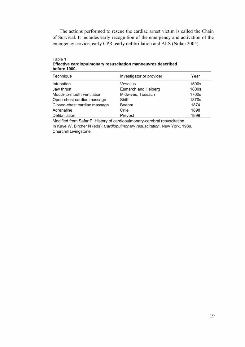
Possibly the earliest record of mouth-to-mouth resuscitation can be found in Old Testament, in the Septuagint (LXX) version of Kings, where reads in 21 verse: “Then he blew air into the boy three times” (Paraskos 1992). On October 4, 1858, János Balassa reported an 18 year old woman suffering cardiac arrest due to airway obstruction from ulcerated laryngitis. Tracheotomy was made immediately and Balassa “exerted bellows-like rhythmic pressure to the front of her chest imitating breathing. Air entered the lungs with a sharp whistling sound.” After 6 minutes of resuscitation she begun to breathe and after 15 minutes she regained consciousness (Robicsek et al. 2004). Although all effective therapies were described before the year 1900 (Table 1), it took many decades to integrate these techniques into modern CPR. Zoll reported the first successful defibrillation of human VF with external paddles in 1956 (Zoll et al. 1956). Safar and Elam described mouth-to-mouth ventilation and effective airway techniques in 1958 (Elam et al. 1958 , Safar et al. 1958). Kouwenhoven rediscovered closed chest cardiac massage in 1960 (Kouwenhoven et al. 1960). It took six years of synthesis to introduce the first recommendation on CPR (Anonymous 1966). Since 1973 the American Heart Association (AHA) has published “Standards for Cardiopulmonary resuscitation and Emergency Cardiac Care”. Guidelines were updated with publications in 1980, 1986 and 1992. In 1993 on the basis of worldwide co-operation the International Liaison Committee on Resuscitation (ILCOR) was formed to identify and review international science and knowledge relevant to CPR, and to offer consensus on treatment recommendations. In 2005 the European
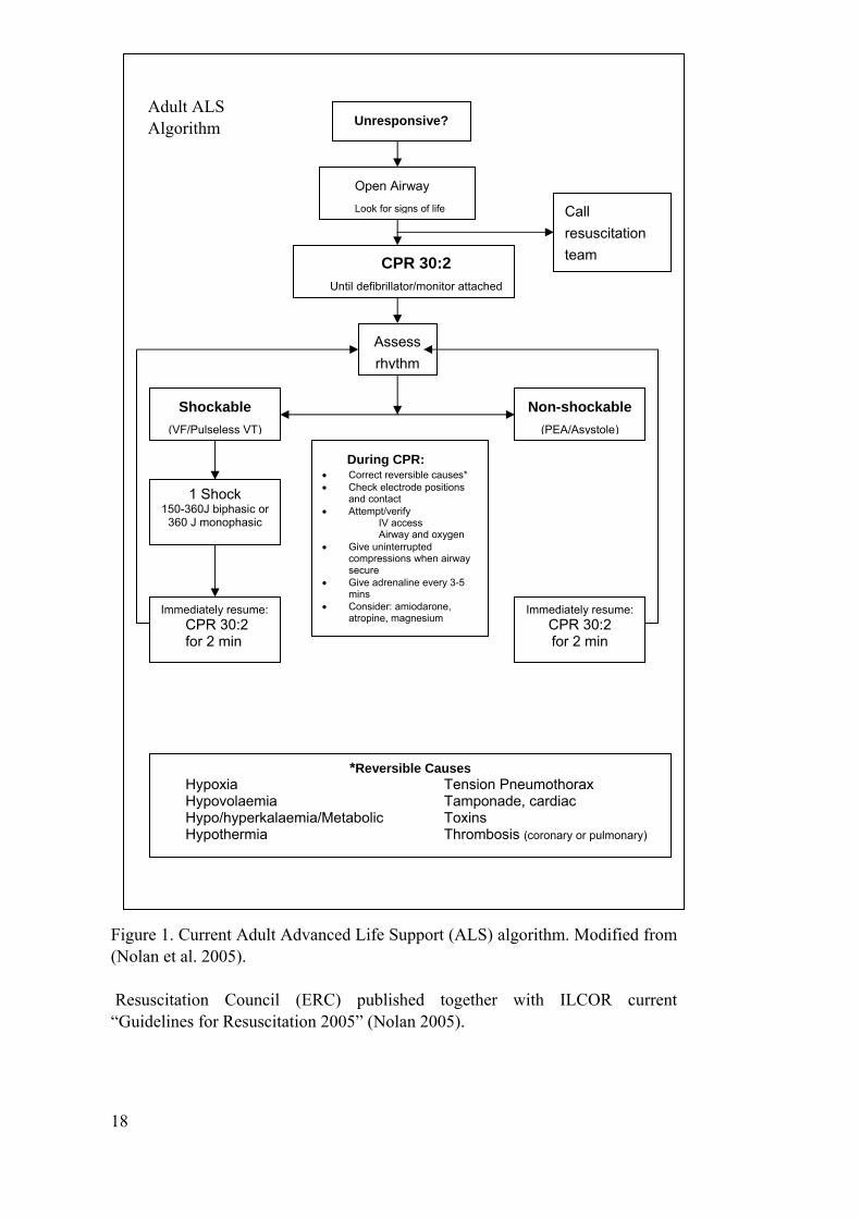
18
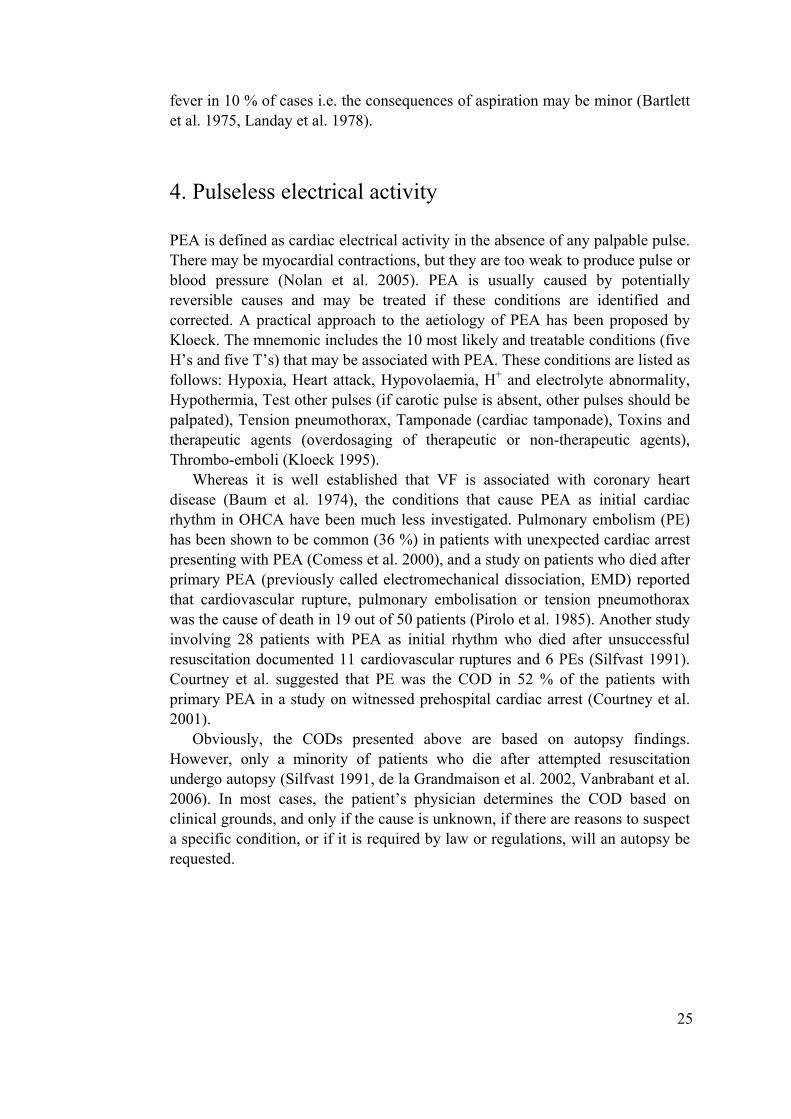
Adult ALS Algorithm Unresponsive?
Open Airway
Figure 1. Current Adult Advanced Life Support (ALS) algorithm. Modified from (Nolan et al. 2005). Resuscitation Council (ERC) published together with ILCOR current “Guidelines for Resuscitation 2005” (Nolan 2005).
Look for signs of life
CPR 30:2 Until defibrillator/monitor attached
Call resuscitation team
Assess rhythm
Shockable Non-shockable (VF/Pulseless VT) (PEA/Asystole)
1 Shock 150-360J biphasic or
360 J monophasic
Immediately resume: CPR 30:2 for 2 min
*Reversible Causes Hypoxia Tension Pneumothorax Hypovolaemia Tamponade, cardiac Hypo/hyperkalaemia/Metabolic Toxins Hypothermia Thrombosis (coronary or pulmonary)
Immediately resume: CPR 30:2 for 2 min
During CPR: • Correct reversible causes* • Check electrode positions
and contact • Attempt/verify
IV access Airway and oxygen
• Give uninterrupted compressions when airway secure
• Give adrenaline every 3-5 mins
• Consider: amiodarone, atropine, magnesium
The actions performed to rescue the cardiac arrest victim is called the Chain of Survival. It includes early recognition of the emergency and activation of the emergency service, early CPR, early defibrillation and ALS (Nolan 2005).
Table 1 Effective cardiopulmonary resuscitation manoeuvres described before 1900.
Technique Investigator or provider Year
Intubation Vesalius 1500s Jaw thrust Esmarch and Heiberg 1800s Mouth-to-mouth ventilation Midwives, Tossach 1700s Open-chest cardiac massage Shiff 1870s Closed-chest cardiac massage Boehm 1874 Adrenaline Crile 1898 Defibrillation Prevost 1899 Modified from Safar P: History of cardiopulmonary-cerebral resuscitation. In Kaye W, Bircher N (eds): Cardiopulmonary resuscitation, New York, 1989, Churchill Livingstone.
19
20
2. Out-of-Hospital Cardiac arrest
SCA is one of the most frequent CODs in industrialised countries (Herlitz et al. 1999). Despite CPR after OHCA and post-resuscitation care mortality remains high at 44-66 % (Langhelle et al. 2003). Survival from cardiac arrest depends on a sequence of interventions, all of which have to be optimised to maximize survival (Cummins et al. 1991). The chain of survival is early recognition and call for help, early CPR, early defibrillation and post resuscitation care (Nolan et al. 2006).
Dispatching centres handle all emergency calls in Finland and dispatch units according to medical risk analysis protocols. Basic Life Support (BLS) units are capable of performing CPR and using an automated external defibrillator. ALS units are capable of securing the airway with intubation tube or supraglottic airway and they can administer intravenous drugs. An emergency physician staffed ambulance unit or helicopter emergency medical service (HEMS) unit is capable of initiating intensive care level procedures on the patient. The dispatching centres have instructions to dispatch the nearest possible unit as a first responding unit in suspected cardiac arrest. Simultaneously a BLS unit and ALS unit are dispatched. In areas where an emergency physician manned ambulance unit or HEMS unit is available, it will also be dispatched (Väisänen et al. 2006).
The risk factors affecting survival from cardiac arrest are several. The most important factors are short time intervals in the treatment of cardiac arrest. Bystander CPR has a beneficial effect on surviving from OHCA (Van Hoeyweghen et al. 1993) and the unfavourable effect of delay in initiation of resuscitation is well documented, especially in initiation of ALS. The chance of survival decreases with each passing minute without defibrillation, CPR or ALS. In cardiac arrest patients with witnessed VF, survival decreased 3 % with each minute until CPR was started and 4 % with each minute to first shock after initiation of CPR (Weaver et al. 1986).
Initial cardiac rhythm is an important predictor of outcome. VF with early defibrillation as a primary initial rhythm is associated with more favourable outcome than other rhythms (Silfvast 1990, Cummins et al. 1991). Patients with ventricular tachycardia (VT) or VF have several times better outcome than with PEA or asystole (ASY) (Herlitz et al. 1999). Aetiology of cardiac arrest does play a role in outcome. Cardiac arrest due to presumed cardiac origin is associated with over three times better outcome compared to non-cardiac origin cardiac arrest (Pell et al. 2003). The rescuer performance has an impact on survival after OHCA. In a retrospective observational study conducted in the United Kingdom, the experience of the ambulance crew and the level of their training influenced outcome after OHCA (Soo et al. 1999). High socioeconomic status is associated with a 1.6 fold increase in survival rate after VF, after
21
adjustment for other factors (e.g. age, time from call to paramedic arrival, activity, location, witnessed collapse, bystander CPR, and chronic morbidity) (Hallstrom et al. 1993). In the last part of the chain of survival is in-hospital care of patients resuscitated from OHCA. In Sweden and Norway the outcome after OHCA varies between different hospitals. Optimised in-hospital factors are associated with improved outcome after OHCA (Engdahl et al. 2000, Langhelle et al. 2003).
VF has the best prognosis, but the incidence of VF has been declining 50 % from 50-83 % incidence (Weaver et al. 1986, Bayes de Luna et al. 1989) in OHCA during the last two decades. At the same time, there has been an increase in the number of patients with PEA as initial cardiac rhythm. The proportion of these patients increased by more than 60 % during the 1980s and 1990s (Herlitz et al. 2000, Kuisma et al. 2001, Cobb et al. 2002) and the factors behind this phenomenon are unclear.
22
3. Regurgitation and aspiration in cardiac arrest
After the genesis of modern CPR in the early 1960s, the first reports of complications were reported in the mid-1960s. Attention was directed to multiple rib fractures, haemothorax, pneumothorax, contusion and laceration of lungs, and fat and bone-marrow embolism. In the early years of resuscitation outside the operating theatre, vomitus and subsequent aspiration was often seen because of lack of reflexes protecting the airway during resuscitation (Greenberg 1967).
Regurgitation is defined as a passive flow of gastric contents to the pharynx. Aspiration is defined as aspiration of gastric contents into the lungs, as a subsequent phenomenon to the regurgitation. This complication has been reported several times during the last three decades during resuscitation in OHCA patients. A study where rescue units detected and treated a VF during resuscitation was reported to have an 11% incidence of aspiration (Liberthson et al. 1974). A prospective autopsy study of 705 cases concerning complications after unsuccessful resuscitation reported an incidence of oropharyngeal vomitus in 10.1% and tracheal vomitus in 9.4% (Krischer et al. 1987). A group of patients who were resuscitated after OHCA and who died within 24h after admission were studied retrospectively to determine the incidence of pulmonary aspiration. The incidence of aspiration of gastric contents or blood in autopsy material after CPR was found to be 29 %. No details are available on the mode or duration of ventilation prior to the intubation in this study. The aetiology of cardiac arrest within these patients included cardiac and non-cardiac causes. The authors stated that the incidence of pulmonary aspiration (29 %) may underestimate the problem, because 46 % of the patients studied had full stomachs (Lawes et al. 1987). In Vienna, a prospective observational study was conducted to discover whether bystander CPR increases mouth-to-mouth ventilation related complications and adverse effects caused by chest compressions. The chest radiographs on admission were studied from patients surviving cardiac arrest, and no difference was found between bystander CPR group and no bystander CPR group. A 17–18% incidence of severe gastric insufflation after CPR was revealed (Oschatz et al. 2001). In a Swedish study the experiences of bystanders were studied shortly after performing CPR. The rescuers most frequently had problems regarding the patient with mouth-to-mouth ventilation (20 %) and vomiting (18%) (Axelsson et al. 1996). An in-hospital study compared the incidence of gastric regurgitation between the bag valve mask and laryngeal mask airway (Stone et al. 1998). The details of gastric regurgitation were prospectively recorded from 797 patients. Regurgitation occurred at some stage of resuscitation in 180 (23 %) patients.
23
3.1 The role of ventilation in cardiopulmonary resuscitation
Since the beginning of the modern CPR in the 1960’s, mouth-to-mouth ventilation and subsequent assisted ventilation and intubation of the trachea have been the cornerstones of CPR.
The benefits of ventilation during respiratory arrest were already demonstrated in the 1950’s (Elam et al. 1958 , Safar et al. 1958). Elam and Safar showed the feasibility of direct mouth-to-mouth ventilation by a layperson on curarised patients and that exhaled air is a resuscitative gas. The “victims” of respiratory arrest were healthy volunteers with normal haemodynamics.
The international guidelines on the role of ventilation remained virtually unchanged from 1966 to 1986. However, as cumulating data showed an increased likelihood of gastric inflation and subsequent pulmonary aspiration, new recommendations for ventilation in CPR were introduced (Melker 1985).
The assisted ventilation manoeuvres without a secured airway are also often linked with regurgitation and increased morbidity and mortality (Pepe 1996). In the delivery of artificial ventilation with “bag and mask” the pressure in the hypopharynx may exceed 25 H2O cm, a pressure causing the opening of the gastro-oesophageal sphincter in most patients (Ruben et al. 1961). Gastric insufflation and subsequent regurgitation of gastric contents with aspiration usually follows (McIntyre et al. 1978). The role of the lower oesophageal sphincter (LES) in regurgitation is crucial but there is little evidence on its pressure and function during resuscitation. In a laboratory trial using domestic swine, a rapid and severe decrease in LES tone was demonstrated during prolonged cardiac arrest. The LES tone decreased from mean baseline 21 cm H2O to mean 3.3 cm H2O during seven minutes of cardiac arrest. When ROSC occurred after defibrillation the LES tone was then measured for a further seven minutes. It increased rapidly but only to half of the prearrest baseline. Unfortunately the impact of CPR on LES tone was not studied during CPR (Bowman et al. 1995).
The need for initial mouth-to-mouth ventilation and subsequent assisted ventilation has been challenged in CPR (Berg et al. 1993, Van Hoeyweghen et al. 1993, Hallstrom et al. 2000, Berg et al. 2001). CPR performed with 15 chest compressions (at a rate of 100/min) and 2 rescue breathings compared to continuous chest compressions at the same rate showed a compromised effect on haemodynamics in ventilated swine (Berg et al. 2001). Since ventilation has been considered an essential part of CPR, the impact on the survival of the cardiac arrest patient should be positive. In a clinical study, the survival was better when bystanders performed chest compression only i.e. cardiac-only resuscitation, instead of conventional CPR (Hallstrom et al. 2000). The existing guidelines indicate that chest compression only CPR should be performed only if bystander is unwilling or unable to give mouth-to-mouth ventilation (Handley et al. 2005).
There are several reasons why bystander mouth-to-mouth ventilation may not be conductive to survival. The first obstacle in survival from OHCA is lack of
24
bystander CPR. The need for mouth-to-mouth ventilation greatly reduces the willingness to initiate bystander CPR (Ewy 2005) and in a swine model of bystander resuscitation a prompt initiation of chest compressions alone was as effective as chest compressions plus ventilation (Berg et al. 1993). When mouth-to-mouth ventilation cannot be applied, chest compression only CPR is better than no CPR at all with respect to outcome (Van Hoeyweghen et al. 1993). The coronary perfusion pressure is the difference between the aortic diastolic pressure and the right atrial diastolic pressure. In a clinical observation study professional rescuers were shown to ventilate OHCA patients excessively during resuscitation. In a subsequent animal study aortic, right atrial and thoracic pressure were measured during CPR. Three different ventilation rates were studied (12, 20 and 30 breaths per minute) and the ventilation was initiated during the decompression phase. These results showed that excessive ventilation rates significantly decreased coronary perfusion and survival rates. Furthermore, the venous return was shown to be reduced to the right heart in this setting. (Aufderheide et al. 2004a, Aufderheide et al. 2004b). This situation is exacerbated, if powerful ventilation is given during chest compressions, because of a further increase in intrathoracic pressure (Aufderheide et al. 2004a). There is also evidence that air on the alveolar level is equivalent to the room air when the airway is open; therefore blood in the arterial system is already oxygenated without artificial ventilation (Mithoefer et al. 1967, Meursing 1983) and enables chest compression to circulate oxygenated blood (Meursing et al. 2005).
3.2. Radiological findings of gastric aspiration
Predisposing conditions to pulmonary aspiration of gastric contents are reduced levels of consciousness, which is evident during OHCA and CPR (Adnet et al. 1996, Bartlett et al. 1975). However, it is not known how often documented gastric regurgitation during the treatment of cardiac arrest leads to radiographic findings compatible with aspiration. The consequence of the aspiration of gastric contents is known as Mendelson’s syndrome: Initially there is abrupt onset of acute respiratory distress. Bronchospasm is a characteristic feature in all patients. Chest X-ray film changes consisting of soft, irregular, mottled densities in the right lower lobe or both lower lobes, are associated with frothy nonpurulent sputum (Mendelson 1946). Hypoxia, together with normal to lowered PCO2, indicates ventilation-perfusion disturbances (Bartlett et al. 1975).
Specific studies concerning the role of documented aspiration during resuscitation and survival after OHCA have not been conducted. In a study on the acute aspiration of gastric contents, altered state of consciousness played a role in aspiration, but not cardiac arrest. On the day of aspiration 54 out of 60 patients had abnormalities in the first chest X-ray. In the appropriate clinical setting, any radiographic infiltrates should raise the suspicion of aspiration (Landay et al. 1978). Even in well documented gastric aspiration into the lungs with tachypnea, couch, cyanosis, and wheezing, the only clinical sign may be
25
fever in 10 % of cases i.e. the consequences of aspiration may be minor (Bartlett et al. 1975, Landay et al. 1978).
4. Pulseless electrical activity
PEA is defined as cardiac electrical activity in the absence of any palpable pulse. There may be myocardial contractions, but they are too weak to produce pulse or blood pressure (Nolan et al. 2005). PEA is usually caused by potentially reversible causes and may be treated if these conditions are identified and corrected. A practical approach to the aetiology of PEA has been proposed by Kloeck. The mnemonic includes the 10 most likely and treatable conditions (five H’s and five T’s) that may be associated with PEA. These conditions are listed as follows: Hypoxia, Heart attack, Hypovolaemia, H+ and electrolyte abnormality, Hypothermia, Test other pulses (if carotic pulse is absent, other pulses should be palpated), Tension pneumothorax, Tamponade (cardiac tamponade), Toxins and therapeutic agents (overdosaging of therapeutic or non-therapeutic agents), Thrombo-emboli (Kloeck 1995).
Whereas it is well established that VF is associated with coronary heart disease (Baum et al. 1974), the conditions that cause PEA as initial cardiac rhythm in OHCA have been much less investigated. Pulmonary embolism (PE) has been shown to be common (36 %) in patients with unexpected cardiac arrest presenting with PEA (Comess et al. 2000), and a study on patients who died after primary PEA (previously called electromechanical dissociation, EMD) reported that cardiovascular rupture, pulmonary embolisation or tension pneumothorax was the cause of death in 19 out of 50 patients (Pirolo et al. 1985). Another study involving 28 patients with PEA as initial rhythm who died after unsuccessful resuscitation documented 11 cardiovascular ruptures and 6 PEs (Silfvast 1991). Courtney et al. suggested that PE was the COD in 52 % of the patients with primary PEA in a study on witnessed prehospital cardiac arrest (Courtney et al. 2001).
Obviously, the CODs presented above are based on autopsy findings. However, only a minority of patients who die after attempted resuscitation undergo autopsy (Silfvast 1991, de la Grandmaison et al. 2002, Vanbrabant et al. 2006). In most cases, the patient’s physician determines the COD based on clinical grounds, and only if the cause is unknown, if there are reasons to suspect a specific condition, or if it is required by law or regulations, will an autopsy be requested.
26
5. Post-resuscitation disease
Although in OHCA the primary resuscitation is often successful, the major obstacle to good neurological survival is post-resuscitation disease. Negovsky described post-resuscitation disease as a specific multiorgan pathophysiological state of the resuscitated cardiac arrest patient. These post-resuscitation processes do not involve only the CNS system, but also the rest of the body, and may lead to severe disability or even death after otherwise successful resuscitation. This clinical syndrome affects the cardiovascular, neurological, pulmonary, renal and metabolic systems. Such disorders in these systems are caused by marked endotoxemia washed out from ischaemia affected organs and tissues, altered haemodynamics after resuscitation, and changes in neuroendocrine profile and rheological characteristics of the blood (Negovsky 1988). There is a notable inter-relationship between the pathological processes developing in the brain and the extracerebral system. The pathological changes in the brain after successful resuscitation are discussed further in the next chapter. Survival from cardiac arrest is dependent on how rapidly CPR, defibrillation, and ALS have been initiated (Cummins et al. 1991). The quality of CPR has also been on focus in the literature. Chest compressions appear to be the most important factor in resuscitation of a human being (Van Hoeyweghen et al. 1993). The coronary perfusion pressure (CPP) is the difference between the aortic diastolic pressure and the right atrial diastolic pressure. The importance of chest compressions has been studied and an investigation established that interrupting chest compression for rescue breathing causes a 7 mmHg drop in mean CCP during CPR (Berg et al. 2001). Recently, quality of out-of-hospital CPR has been studied and the main finding was that during CPR chest compression were not delivered for half of the time. Furthermore, most compressions were too shallow (Wik et al. 2005). Skrifvars et al. demonstrated that multiple factors affect 6-month outcome following resuscitation from cardiac arrest. Strict glucose control in the Intensive Care Unit (ICU), serum potassium level and beta-blocking agents were independently associated with survival (Skrifvars et al. 2003). There are cumulating data showing that in-hospital factors are associated with outcome after OHCA (Engdahl et al. 2000, Langhelle et al. 2003).
6. Hypothermia after cardiac arrest
Interventions to mitigate neuronal injury after cardiac arrest have been studied with different approaches, but only therapeutic hypothermia has been shown to reduce mortality and morbidity. Induced hypothermia has been in use since the 1950s to protect the brain against global ischaemia during open-heart surgery. A case report of the successful use of hypothermia after cardiac arrest outside the operating theatre was published at the end of the 1950’s. Two children and two
27
adults were treated, and three of them recovered completely and one with moderate neurological impairment (Williams et al. 1958). One year later a study with a control group was published, where nineteen patients (including those previously reported two children and two adults) were resuscitated after cardiac arrest with resultant neurological damage. These patients were divided into a normothermia group and a hypothermia group. Survival was 14% and 50% respectively (Benson et al. 1959). The method was subsequently abandoned due to uncertain benefit and difficulties with its use. Interest in induced hypothermia after return of spontaneous circulation rose again in the 1990s and has been associated with improved functional recovery and reduced cerebral histological defects in this setting (Sterz et al. 1991). The timing of induction of therapeutic hypothermia has been shown to be critical. In a canine study with induced mild hypothermia after normothermic cardiac arrest, hypothermia improved cerebral functional and morphologic outcome. However, if the induction of cooling was delayed for 15 min after ROSC, it did not improve functional outcome, although it may have mitigated histological tissue damage (Kuboyama et al. 1993)
The exact mechanism of induced therapeutic hypothermia is not clear. A reduction of cerebral oxygen consumption has been proposed (Hegnauer et al. 1954) and other multifactorial physical and chemical mechanisms during and after low-flow induced ischaemia have also been postulated (Hypothermia after Cardiac Arrest Study Group 2002). These include reduction of intracellular acidosis (Chopp et al. 1989), reducing cerebral oedema and protection of lipoprotein membranes (Dempsey et al. 1987), inhibition of biosynthesis and release of excitatory neurotransmitters (Busto et al. 1989). ATP concentration in brain tissue has been shown to most consistently reflect biochemical activity among available biochemical indicators. Profound hypothermia has been shown to result in a three to fourfold increase in survival of cerebral ATP during circulatory arrest (Kramer et al. 1968).
Promising preliminary human data accumulated at the turn of the century. Surface cooling after OHCA maintained for 12 hours in the ICU significantly improved outcome compared to retrospective controls (Bernard et al. 1997). The use of mild hypothermia after OHCA yielded better outcome, but also more pneumonias after 48-hour hypothermia and very slow re-warming at a rate no greater than 1 °C per day (Yanagawa et al. 1998). In a pilot study of the HACA-Study group, external cooling of the head and trunk after ROSC in the emergency department was feasible and safe (Zeiner et al. 2000). Felberg et al. reported a feasibility trial where external cooling was feasible and safe. However, external cooling was slow and imprecise and efforts to speed up the start of cooling and to improve the cooling process are needed (Felberg et al. 2001).
A first out-of-hospital prospective randomized trial was conducted by Hachimi-Idrissi et al., where patients with cardiac arrest due to ASY or PEA were enrolled and randomized to a normothermic and a hypothermic group. Hypothermia was induced using a helmet device containing an aqueous glycerol solution and was found feasible, easy to use, inexpensive and effective with no
28
additional complications (Hachimi-Idrissi et al. 2001). Callaway et al., however, reported that application of ice to the head and neck during ongoing CPR failed to produce a significant cooling effect on cerebral or core temperatures. Furthermore, it was found to be moderately cumbersome and necessitated additional personnel in the field. The authors proposed the use of cold intravenous fluids in further studies (Callaway et al. 2002).
In February 2002 two randomised clinical trials, one in Europe (Hypothermia after Cardiac Arrest Study Group 2002) and another in Australia (Bernard et al. 2002), were reported. These studies showed a fundamental improvement in both neurological outcome and reduction of mortality in OHCA with VF as a primary rhythm. These two studies yielded similar results, thus making the important conclusions more convincing. In the HACA study the number needed to treat (NNT) for favourable neurological outcome (good recovery or moderate disability) was 6, while NNT to prevent a death was 7. In the study by Bernard et al., NNT for normal or minimal disability at the time of discharge from the hospital was 4, and NNT for avoidance of death at the same time was 6. In both studies the complication rates did not differ significantly between the hypothermia and normothermia groups. Based on these two studies, a strong recommendation was made: “Although we await further studies with great interest, we recommend the use of mild hypothermia in survivors of cardiac arrest − as early as possible and for at least 12 hours (Safar et al. 2002).” ILCOR made the following recommendations in October 2002 (Nolan et al. 2003): “Unconscious adult patients with spontaneous circulation after OHCA should be cooled to 32-34 °C for 12-24 h when the initial rhythm is VF. Such cooling may also be beneficial for other rhythms or in-hospital cardiac arrest.” A recent meta-analysis yielded a statement that one patient would leave the hospital with favourable neurological recovery by treating 4 to 13 OHCA patients with mild hypothermia (Holzer et al. 2005).
Bernard et al. conducted a pilot study using a rapid infusion of large volume (30 ml/kg), ice-cold (4 °C) intravenous fluid. The method was found to be a safe, rapid and inexpensive technique to induce mild hypothermia in OHCA patients. Regardless of quite a large and fast volume load, no patient developed pulmonary oedema (Bernard et al. 2003). The effect of large volume, ice-cold fluid intravenous infusion for the induction of moderate hypothermia on younger and older healthy volunteers has also been studied. A volume load of 40ml/kg was infused at a rate of 70-100ml/min and no pulmonary oedema was reported among patients (Frank et al. 2000, Rajek et al. 2000).
The most optimal timing of the induction of hypothermia remains uncertain. It is surprising that the clinical benefits associated with hypothermia occurred despite long delays in attaining target body temperature in the above mentioned studies (Bernard et al. 2002, Hypothermia after Cardiac Arrest Study Group 2002).
29
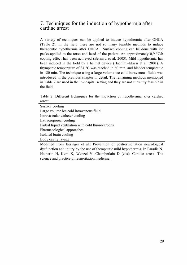
7. Techniques for the induction of hypothermia after cardiac arrest
A variety of techniques can be applied to induce hypothermia after OHCA (Table 2). In the field there are not so many feasible methods to induce therapeutic hypothermia after OHCA. Surface cooling can be done with ice packs applied to the torso and head of the patient. An approximately 0,9 °C/h cooling effect has been achieved (Bernard et al. 2003). Mild hypothermia has been induced in the field by a helmet device (Hachimi-Idrissi et al. 2001). A thympanic temperature of 34 °C was reached in 60 min. and bladder temperatue in 180 min. The technique using a large volume ice-cold intravenous fluids was introduced in the previous chapter in detail. The remaining methods mentioned in Table 2 are used in the in-hospital setting and they are not currently feasible in the field.
Table 2. Different techniques for the induction of hypothermia after cardiac arrest. Surface cooling Large volume ice cold intravenous fluid Intravascular catheter cooling Extracorporeal cooling Partial liquid ventilation with cold fluorocarbons Pharmacological approaches Isolated brain cooling Body cavity lavage Modified from Beringer et al.: Prevention of postresuscitation neurological dysfunction and injury by the use of therapeutic mild hypothermia. In Paradis N, Halperin H, Kern K, Wenzel V, Chamberlain D (eds): Cardiac arrest. The science and practice of resuscitation medicine.
30
Aims of the study
The aim of this thesis was to study aetiology, treatment and outcome in OHCA by the emergency medical service (EMS) systems in Tampere District EMS and the physician-staffed Helicopter EMSs in the Helsinki and Turku areas in Southern Finland. The specific aims were the following:
1. To determine whether there is an association between bystander mouth-
to-mouth ventilation and regurgitation in prehospital cardiac arrest patients (I) and to assess the association between clinical signs of prehospital regurgitation and radiological findings in resuscitated patients. (II)
2. To study the causes of death after witnessed cardiac arrest followed by
pulseless electrical activity and unsuccessful out-of-hospital resuscitation; and to detect any differences between causes of death determined at autopsy and those inferred from clinical history. (III)
3. To evaluate the haemodynamic and cooling effects of infusing ice-cold
Ringer’s solution in the field immediately after return of spontaneous circulation. (IV)
31
Material and methods
Patients and methods
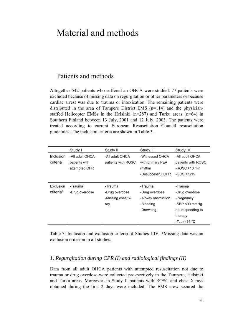
Altogether 542 patients who suffered an OHCA were studied. 77 patients were excluded because of missing data on regurgitation or other parameters or because cardiac arrest was due to trauma or intoxication. The remaining patients were distributed in the area of Tampere District EMS (n=114) and the physician-staffed Helicopter EMSs in the Helsinki (n=287) and Turku areas (n=64) in Southern Finland between 13 July, 2001 and 12 July, 2003. The patients were treated according to current European Resuscitation Council resuscitation guidelines. The inclusion criteria are shown in Table 3.
Study I Study II Study III Study IV Inclusion criteria
-All adult OHCA
patients with
attempted CPR
-All adult OHCA
patients with ROSC
-Witnessed OHCA
with primary PEA
rhythm
-Unsuccessful CPR
-All adult OHCA
patients with ROSC
-ROSC ≥10 min
-GCS ≤ 5/15
Exclusion
criteria* -Trauma
-Drug overdose
-Trauma
-Drug overdose
-Missing chest x-
ray
-Trauma
-Drug overdose
-Airway obstruction
-Bleeding
-Drowning
-Trauma
-Drug overdose
-Pregnancy
-SBP <90 mmHg
not responding to
therapy
-Tesof <34 °C
Table 3. Inclusion and exclusion criteria of Studies I-IV. *Missing data was an exclusion criterion in all studies.
1. Regurgitation during CPR (I) and radiological findings (II)
Data from all adult OHCA patients with attempted resuscitation not due to trauma or drug overdose were collected prospectively in the Tampere, Helsinki and Turku areas. Moreover, in Study II patients with ROSC and chest X-rays obtained during the first 2 days were included. The EMS crew secured the
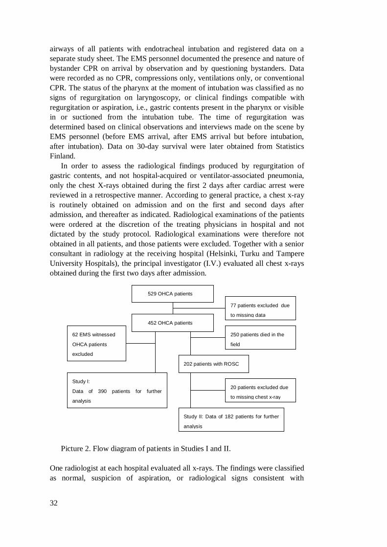
32
airways of all patients with endotracheal intubation and registered data on aseparate study sheet. The EMS personnel documented the presence and nature ofbystander CPR on arrival by observation and by questioning bystanders. Datawere recorded as no CPR, compressions only, ventilations only, or conventionalCPR. The status of the pharynx at the moment of intubation was classified as nosigns of regurgitation on laryngoscopy, or clinical findings compatible withregurgitation or aspiration, i.e., gastric contents present in the pharynx or visiblein or suctioned from the intubation tube. The time of regurgitation wasdetermined based on clinical observations and interviews made on the scene byEMS personnel (before EMS arrival, after EMS arrival but before intubation,after intubation). Data on 30day survival were later obtained from StatisticsFinland.
In order to assess the radiological findings produced by regurgitation ofgastric contents, and not hospitalacquired or ventilatorassociated pneumonia,only the chest Xrays obtained during the first 2 days after cardiac arrest werereviewed in a retrospective manner. According to general practice, a chest xrayis routinely obtained on admission and on the first and second days afteradmission, and thereafter as indicated. Radiological examinations of the patientswere ordered at the discretion of the treating physicians in hospital and notdictated by the study protocol. Radiological examinations were therefore notobtained in all patients, and those patients were excluded. Together with a seniorconsultant in radiology at the receiving hospital (Helsinki, Turku and TampereUniversity Hospitals), the principal investigator (I.V.) evaluated all chest xraysobtained during the first two days after admission.
Picture 2. Flow diagram of patients in Studies I and II.
One radiologist at each hospital evaluated all xrays. The findings were classifiedas normal, suspicion of aspiration, or radiological signs consistent with
529 OHCA patients
452 OHCA patients
77 patients excluded due
to missing data
Study I:
Data of 390 patients for further
analysis
Study II: Data of 182 patients for further
analysis
62 EMS witnessed
OHCA patients
excluded
250 patients died in the
field
202 patients with ROSC
20 patients excluded due
to missing chest xray
aspiration. The radiologist and the principal investigator were blinded to the clinical findings of regurgitation at the time of the radiological evaluation.
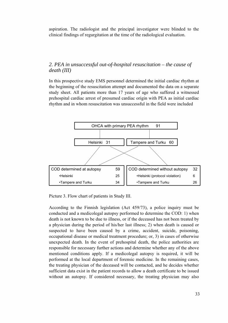
2. PEA in unsuccessful out-of-hospital resuscitation – the cause of death (III)
In this prospective study EMS personnel determined the initial cardiac rhythm at the beginning of the resuscitation attempt and documented the data on a separate study sheet. All patients more than 17 years of age who suffered a witnessed prehospital cardiac arrest of presumed cardiac origin with PEA as initial cardiac rhythm and in whom resuscitation was unsuccessful in the field were included
OHCA with primary PEA rhythm 91
Helsinki 31 Tampere and Turku 60
COD determined at autopsy 59•Helsinki 25
•Tampere and Turku 34
COD determined without autopsy 32•Helsinki (protocol violation) 6
•Tampere and Turku 26
Picture 3. Flow chart of patients in Study III. According to the Finnish legislation (Act 459/73), a police inquiry must be conducted and a medicolegal autopsy performed to determine the COD: 1) when death is not known to be due to illness, or if the deceased has not been treated by a physician during the period of his/her last illness; 2) when death is caused or suspected to have been caused by a crime, accident, suicide, poisoning, occupational disease or medical treatment procedure; or, 3) in cases of otherwise unexpected death. In the event of prehospital death, the police authorities are responsible for necessary further actions and determine whether any of the above mentioned conditions apply. If a medicolegal autopsy is required, it will be performed at the local department of forensic medicine. In the remaining cases, the treating physician of the deceased will be contacted, and he decides whether sufficient data exist in the patient records to allow a death certificate to be issued without an autopsy. If considered necessary, the treating physician may also
33
34
request a routine autopsy to determine the COD before issuing the death certificate. In this study, two strategies were designed. In the Helsinki area, the EMS physician on the scene made a request for an autopsy on the EMS run sheet in all cases where resuscitation was terminated as unsuccessful when the first recorded rhythm had been PEA, irrespective of the suspected COD. In the Tampere and Turku areas the EMS crew made no requests for autopsy on the EMS run sheets, and the COD was determined according to general practice. In these areas, unless a medicolegal autopsy was required by law, the police contacted the deceased’s treating physician. The treating physician decided whether the death certificate could be issued on the basis of previous history and clinical notes of the cardiac arrest. If not, he ordered a medical autopsy. The EMS run sheets of all patients in the study areas were collected and evaluated. Special attention was focused on any notes regarding the treating EMS crews’ observations on or suspicion of the cause of arrest. The autopsy referrals made by the treating physicians to the pathologists were also retrieved and reviewed for purposes of requesting an autopsy. Data on the COD stated on the death certificates of the patients who did not undergo an autopsy were obtained from Statistics Finland. The corresponding data of those who were autopsied were retrieved from the autopsy protocols.
3 Induction of therapeutic hypothermia (IV)
This prospective study was conducted in the Helsinki Area HEMS between 23rd April 2002 and 12th July 2003. The inclusion criteria were OHCA not due to trauma or drug overdose, age over 18 years and ROSC later than 10 minutes from the onset of cardiac arrest, and Glasgow Coma Score (GCS) ≤ 5. Exclusion criteria were pregnancy, systolic blood pressure < 90 mmHg not responding to volume or inotropes, or oesophageal temperature (Tesof) < 34.0 °C.
After ROSC patients’ lungs were manually ventilated and end-tidal CO2 (Life-Cap; Medtronic PhysioControl, Redmond, Washington, USA) was monitored continuously to achieve normocapnoea. An arterial blood gas measurement was undertaken using the i-STAT (i-STAT Corporation, Windsor, New Jersey, USA) portable blood gas analyser with the EC6+ cartridge to obtain electrolyte values, pH, and blood gases, and to find out the possible difference between ETCO2 and arterial CO2. A Tesof probe was inserted and connected to the monitor. When the patient was stabilised and found eligible, informed consent was obtained from relatives.
Mild hypothermia was induced with ice-cold Ringer’s acetate. The fluids were stored in an insulated box with ice cubes to maintain + 4 °C temperature. Pressure bags were used to infuse the target volume of 30 ml/kg at a rate of 100 ml/min. Tesof was monitored continuously and the infusion stopped if the core temperature of 33 °C was reached or adverse haemodynamic events (i.e.
35
arrhythmias or hypotension) occurred before the calculated volume had been infused. Blood pressure, heart rate, SpO2, ECG, and end tidal CO2 were closely monitored and data was collected every five min. The haemodynamic effects were defined to be rhythm observation (especially in the case of new arrhythmia e.g. VT, VF or other rearrest) and changes in arterial blood pressure.
At the end of infusion, arterial blood gas analysis was repeated. After that, the patient was carried to the ambulance and transported to hospital with all monitoring in place. On arrival at hospital, arterial blood gases were analysed, the last temperature was recorded and the study ended. Further care in hospital was at the discretion of the treating physicians.
4. Ethical aspects
The study protocols were approved by the institutional review board of Helsinki University Hospital. The need for informed consent from relatives was waived due to the observational nature of the studies I-III. The HEMS physician explained the study protocol to the relatives of the patients and written informed consent was obtained before induction of hypothermia in study IV.
5. Statistical methods
Statistical calculations were made using the SPSS versions 9, 11, 12 or 15 (SPSS Inc, Chicago, IL, USA). In Study I the association between CPR and clinical findings of regurgitation was analysed with the Pearson Chi square test, where a p-value <0.05 was considered significant. Also, Odds Ratios (OR) for achieving ROSC and 30-day survival if the patient regurgitated were calculated with a confidence interval of 95 %. In Study II the association between clinical signs of regurgitation of gastric contents or pulmonary aspiration documented at the time of intubation and radiological signs consistent with pulmonary aspiration was analysed with the Chi-square test. The null hypothesis was that no such association exists. Inter-group differences in demographics, rhythm and ROSC data were analysed with the Kruskal-Wallis test, followed by the Mann-Whitney independent sample test. In addition, the sensitivity, specificity and positive (PPV) and negative (NPV) predictive values of clinical regurgitation to predict radiological signs consistent with pulmonary aspiration were calculated. In Study III the association between determination the COD in the clinical history group and the autopsied group was analysed with the Pearson Chi-square test and Fisher’s exact test, where appropriate. The null hypothesis was that no such association exists. Analysis of variance and t-tests were used in Study IV where appropriate.

36
Due to the nature of the Studies I-IV, no preliminary sample size calculations were performed.
A p-value <0.05 was considered significant.
37
Results
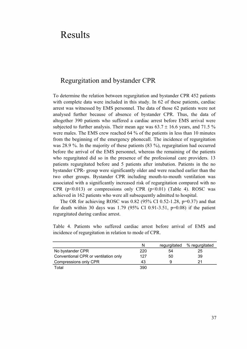
Regurgitation and bystander CPR
To determine the relation between regurgitation and bystander CPR 452 patients with complete data were included in this study. In 62 of these patients, cardiac arrest was witnessed by EMS personnel. The data of those 62 patients were not analysed further because of absence of bystander CPR. Thus, the data of altogether 390 patients who suffered a cardiac arrest before EMS arrival were subjected to further analysis. Their mean age was 63.7 ± 16.6 years, and 71.5 % were males. The EMS crew reached 64 % of the patients in less than 10 minutes from the beginning of the emergency phonecall. The incidence of regurgitation was 28.9 %. In the majority of these patients (83 %), regurgitation had occurred before the arrival of the EMS personnel, whereas the remaining of the patients who regurgitated did so in the presence of the professional care providers. 13 patients regurgitated before and 5 patients after intubation. Patients in the no bystander CPR- group were significantly older and were reached earlier than the two other groups. Bystander CPR including mouth-to-mouth ventilation was associated with a significantly increased risk of regurgitation compared with no CPR (p<0.013) or compressions only CPR (p<0.01) (Table 4). ROSC was achieved in 162 patients who were all subsequently admitted to hospital.
The OR for achieving ROSC was 0.82 (95% CI 0.52-1.28, p=0.37) and that for death within 30 days was 1.79 (95% CI 0.91-3.51, p=0.08) if the patient regurgitated during cardiac arrest. Table 4. Patients who suffered cardiac arrest before arrival of EMS and incidence of regurgitation in relation to mode of CPR.
N regurgitated % regurgitated No bystander CPR 220 54 25 Conventional CPR or ventilation only 127 50 39 Compressions only CPR 43 9 21 Total 390
38
Aspiration and radiological findings in pre-hospital cardiac arrest
Radiological findings after out-of-hospital regurgitation during CPR were studied. Resuscitation was successful in 202 patients, and they were admitted to hospital. No radiological examinations were performed on 20 patients, leaving 182 patients for further analysis. Their mean age was 62.4 ± 15.5 (mean ± Standard Deviation (SD)) years and 74 % were males. ROSC had been achieved within 21.6 ± 12.8 (mean ± SD) min. Altogether 256 chest x-rays were available for further analysis, one for 108 patients and two for 74 patients. The timing of the first chest x-ray was the day of admission in 36%, the day after admission in 60 % and the second day after admission in 4 % of the patients.
In 20 % of the patients (n=37), EMS personnel documented signs of regurgitation on the scene. In hospital, the chest x-ray showed suspicion of or findings consistent with aspiration in 24 % of patients (n=44). The chest x-ray revealed findings compatible with pulmonary aspiration in 46 % of patients with clinical signs of regurgitation on the scene compared with 19 % of patients without such findings. Thus, clinical signs of regurgitation in the prehospital phase resulted in radiological signs consistent with pulmonary aspiration with 81 % specificity and 46 % sensitivity. The PPV was thus 0.39 and NPV 0.86.
Cause of death in resuscitation with PEA
To determine the COD in patients who died after an unsuccessful attempt at out-of-hospital resuscitation with PEA as a primary rhythm, 91 patients were included during the study period, 31 in the Helsinki area and 60 in the Tampere and Turku areas. The mean age (± SD) of the patients was 73.5 ± 11.9 years, and 62 % were males. Cardiac arrest occurred in the presence of the EMS crew in 24 % of the patients; in the remaining patients the arrest was witnessed by a bystander. An autopsy was performed on 59 patients (65 %), in 81 % of those in the Helsinki area and in 57% of those in the Tampere and Turku areas.
Non-cardiac CODs were diagnosed almost entirely in autopsy and there were significant difference between diagnoses in the cardiac COD and non-cardiac COD groups (Table 5). The distribution of diagnoses was significantly different between the patients whose cause of death was determined by autopsy compared with those whose cause of death was determined on clinical grounds (Table 6).
When the COD was determined based on the clinical course of the resuscitation and previous clinical history of the deceased, there were significantly more AMIs and significantly fewer PEs and aortic dissections or ruptures compared with those who underwent autopsy. There were no differences between these two groups in intracranial haemorrhage (ICH), ischaemic coronary disease or in the other COD group.
39
There was a suspicion of a specific cause for the arrest mentioned on the EMS run sheet or on the referral from the treating physician in only 6 of the deceased patients. In five of these six, the clinical suspicions appeared to be correct. Due to protocol violation, the treating physician determined the CODs of six patients in Helsinki and their CODs were determined based on medical history and the course of the resuscitation attempt. These CODs (in the treating physicians’ opinion) were AMI in 4 patients and chronic obstructive pulmonary disease (COPD) in 2 patients.
Table 5. Cardiac and non-cardiac causes of death in relation to mode of determination of death. Cause of death Autopsy
Yes
Autopsy
No
Total p-value*
Cardiac 23 (48) 25 (52) 48 <0.001
Non-cardiac 36 (84) 7 (16) 43 <0.001
Total 59 (65) 32 (35) 91
* A p-value less than 0.05 was considered significant with Chi-square test and Fisher's exact test
Table 6. Cause of death in relation to mode of determination of death.
Autopsy Total p-value*
Cause of death Yes No Acute myocardial infarction 11 19 30 <0.001 Aortic dissection or rupture 17 1 18 0.002 Pulmonary embolism 15 0 15 0.001 Coronary heart disease 8 4 12 1.000 Other cardiac cause 4 2 6 1.000 Intracranial haemorrhage 2 1 3 1.000 Other cause 2 5 7 0.092 Total 59 32 91
Other cardiac causes include two congestive heart failure, undefined pericardial disease, rheumatic valvular disease (mitral and aortic valve), undefined pulmonary hypertension, aortic valve stenosis. Other causes include two chronic obstructive pulmonary disease, motoneuron disease, cancer, pneumonia and unexplained sudden death. * A p-value less than 0.05 was considered significant with Chi-square test and Fisher's exact test.
40
Induction of therapeutic hypothermia after OHCA
The course and efficiency of hypothermia induction, and the demographics of the subjects are shown in Table 7. The mean age of patients was 60.8±12.5 years. The gender distribution was 62% males vs. 38% females. ROSC was achieved at 26±10 min. Initial cardiac rhythm was VF (53.8%), ASY (30.8%) or PEA (15.4%). Mean core temperature decreased from 35.8 ± 0.9 °C at the start of infusion to 34.0 ± 1.2 °C on arrival at hospital. The mean infused volume was 2188 ± 754 ml. Infusion started at 27 ± 12 min after ROSC and mean duration of infusion was 25 ± 11 min. One patient (Patient 2) experienced a transient drop in blood pressure. It responded well to inotrope therapy and there was no obvious cause for it. No other adverse changes in haemodynamics were observed.
Therapeutic hypothermia was continued in 5 patients for 24 hours in the ICU according to the same protocol as in the HACA study.
Table 7. Effect of hypothermia induction and demographics.
Patient Sex Initial rhythm
Age (years)
Permanent ROSC (min)
T before induction
T on admission
ΔT °C
Infused amount (ml)
1 male Asystole 63 22 35.1 33 -2.1 2500 2 female VF 72 21 35.9 33,4 -2.5 2450 3 female Asystole 53 23 35.8 33,1 -2.7 1800 4 male VF 55 27 35.0 34,7 -0.3 500 5 male VF 79 32 35.1 33,2 -1.9 2000 6 female PEA 79 42 35.8 32,7 -3.1 2200 7 male Asystole 64 22 37.2 35,3 -1.9 3000 8 male VF 41 46 36.1 33 -3.1 3000 9 female Asystole 52 19 34.2 32,7 -1.5 1000 10 male VF 65 13 36.0 36,4 0.4 2000 11 male VF 53 15 35.9 34,3 -1.6 2500 12 male VF 43 26 36.9 35,3 -1.6 2500 13 female PEA 71 29 36.7 34,3 -2.4 3000 VF = ventricular fibrillation, PEA = pulseless electrical activity, ROSC = return of spontaneous circulation T = oesophageal temperature, ΔT °C = change of oesophageal temperature
41
Discussion
It was found in Study I that more than a fourth of patients who suffered prehospital cardiac arrest expressed clinical signs of regurgitation at some point during the prehospital phase. This concurs with previous findings (Stone et al. 1998). Although the bystanders were not asked at which point after cardiac arrest regurgitation occurred, it seemed that bystander CPR including mouth-to-mouth ventilation increased the risk of aspiration of gastric contents. In most patients, regurgitation had occurred already before the arrival of the EMS crew, soon after cardiac arrest. This finding was confirmed later in a recent study, where paramedics determined the presence and timing of emesis in the field. The incidence of regurgitation was 32 %, and in a majority of these patients (66 %), regurgitation had occurred before the arrival of EMS personnel (Simons et al. 2007). In the same study the patients who received bystander CPR expressed emesis more frequently than those who did not receive bystander CPR. Of special interest in this context is whether the victim received bystander ventilation during CPR, because it seems to be associated with increased risk of regurgitating gastric contents during resuscitation. Unfortunately Simons et al. did not report detailed data on the mode of bystander CPR (ventilation only, chest compression only or conventional CPR) which would have shed more light on the impact of bystander ventilation on regurgitation of gastric contents during resuscitation.
Thirteen regurgitated before intubation when EMS personnel were present and five patients regurgitated after intubation. In the former situation there is a possibility to prevent the regurgitation and subsequent aspiration into the lungs with the use of the Sellick manoeuvre in the intubation of the patient. In the latter situation the regurgitation should be quite well tolerated if the cuff of the intubation tube is correctly inflated and the intubation tube is correctly placed into the trachea.
A possible explanation for increased incidence of regurgitation is that regurgitation may have been provoked by air being ventilated into the stomach in the patients who received conventional bystander CPR or ventilation only, causing gastric distension. The risk of regurgitation may be further increased due to loss of tone of the lower oesophageal sphincter. This has been found to occur in experimental cardiac arrest, where a rapid and marked decrease in lower oesophageal sphincter (LES) tone was shown to occur within a few minutes after onset of cardiac arrest (Bowman et al. 1995). This could also explain why the patients who suffered an EMS crew witnessed cardiac arrest regurgitated less
42
frequently because of rapid response to cardiac arrest and securing the airway before the loss of tone of LES.
The current resuscitation guidelines discuss the risk of gastric distension and possible regurgitation during mouth-to-mouth ventilation in cardiac arrest, but little information has been provided on the incidence of regurgitation or advice on how to act in case of regurgitation. So far, conventional CPR including compressions and ventilations has been the general recommendation in resuscitation guidelines (Anonymous 2000). However, compressions-only CPR has been shown to be associated with similar outcome to that of conventional CPR in patients with a short delay from the onset of cardiac arrest to the arrival of the EMS crew (Hallstrom et al. 2000). Although compressions-only CPR currently is recommended only in dispatcher assisted CPR or if the lay rescuer is unwilling to perform mouth-to-mouth ventilation (Anonymous 2000), it seems that more consideration should be given to this option if the dispatcher estimates that qualified help will arrive within minutes.
New data on the mode of bystander CPR have been published in Japan, where a study reported that cardiac-only resuscitation performed by a bystander is the preferable approach to the resuscitation of adult patients (SOS-Kanto study group 2007). The technical reasons support cardiac-only resuscitation because of interruptions to chest compressions during ventilation and subsequent interruption of cerebral blood flow (Assar et al. 2000, Ewy 2007). Unfortunately this study was also lacking the information about the regurgitation of gastric contents and bystander performed ventilation. An editorial was published in the same journal, where an opinion that the current guidelines regarding bystander mouth-to-mouth ventilation should be changed without delay in light of new evidence published (Ewy 2007), but the European Resuscitation Council immediately responded by retaining current guidelines. The study by the SOS-Kanto study group was undertaken in 2002-2003, that is, under the previous version of the international resuscitation guidelines (Zideman et al. 2007). The guidelines will be revised after an international review of resuscitation science in 2010.
Study II showed an association between clinical regurgitation documented
during OHCA and early radiological findings consistent with aspiration. The incidence of clinically recognised regurgitation of gastric contents among the patients admitted to hospital after ROSC in this study (20 %) was close to the incidence of aspiration reported in an autopsy material (Lawes et al. 1987) showing findings of blood or gastric contents in the airways in 29 % of subjects. Lawes et al. studied the incidence of pulmonary aspiration in patients admitted to hospital after resuscitation from OHCA. The aetiology of OHCA included both cardiac and non-cardiac causes. The authors stated that the incidence of pulmonary aspiration (29 %) may underestimate the problem, because 46 % of patients studied had full stomachs. It seems that not all patients with full stomach always regurgitate during CPR.
43
Oschatz et al. found in a prospective observational study that bystander CPR did not increase the mouth-to-mouth ventilation related complications and adverse effects caused by chest compressions. A 17–18% incidence of severe gastric insufflation after CPR was revealed (Oschatz et al. 2001). In a Swedish study the experiences of bystanders were studied shortly after performing CPR. The rescuers most frequently had problems with patient’s vomiting (18%) and mouth-to-mouth ventilation (20 %) (Axelsson et al. 1996)
Despite the strong association between clinical signs of regurgitation and the subsequent development of radiological findings of aspiration, the sensitivity was relatively poor, 46 %. When regurgitation was detected on the scene there was a 39 % (PPV) chance of finding radiological signs consistent with pulmonary aspiration. When there were no signs of regurgitation there was an 86 % chance that no radiological signs consistent with pulmonary aspiration would develop (NPV). To what extent the early development of radiological signs of aspiration should be interpreted as infection has not been studied. Previous work reporting infectious complications after cardiac arrest has documented that radiological signs of pneumonia develop after 7 days (Bartlett et al. 1975), and that progressive deterioration of radiographic infiltrates after 3 days suggests secondary bacterial pneumonia, acute respiratory distress syndrome (ARDS) or PE (Landay et al. 1978), but the role of aspiration was not specifically addressed in these studies. Because the subsequent development of infectious complications was not the aim of this study, we only evaluated x-rays obtained on the first and second days after admission.
No radiological signs of aspiration appeared in 54 % of those who regurgitated. One explanation evinced has been the pH of the aspirate, since it has been shown that only when the pH of the aspirated matter is less than 2.4 does it cause acidic pneumonitis, manifesting as radiological signs of aspiration (Rello et al. 1995). The nature of the regurgitated matter was not characterised. Therefore, we do not know whether it was liquid or food, an issue that may affect the clinical consequences.
Also, in other documented cases of aspiration of gastric contents, 8-10 % of the patients developed no other symptoms than fever, and the radiographic findings were extremely variable (Landay et al. 1978). Besides the pH, another factor influencing the development of radiological signs of aspiration is probably the volume of the aspirate. Thus, the overall incidence of radiological findings consistent with aspiration in this study is close to the 18 % reported by Oschatz et al. in their survey (Oschatz et al. 2001).
In the majority of patients, only one chest X-ray was obtained during the first 2 days after cardiac arrest. This was because the study protocol did not dictate the timing of radiological examinations, and there were no strict protocols to guide such examinations after cardiac arrest in the participating hospitals. Considering that all hospitals were university teaching hospitals, the small number of radiological examinations was surprising. One explanation for the lack of X-ray examinations may be that the physicians in charge have a particularly pessimistic view with regard to the prognosis of resuscitated
44
patients. Because two chest X-rays were not obtained in all patients, the true incidence of radiological findings may be greater than documented here. As we evaluated only the chest X-rays obtained during the first 2 days, it is possible that regurgitation-related alterations may have manifested later.
It is well known from earlier studies that VF as an initial rhythm is associated
with coronary heart disease (Baum et al. 1974). This has also been documented with an autopsy study (Soo et al. 2001). It has also been shown that PEA is associated with primary non-coronary causes of cardiac arrest (Hess et al. 2007), but systematic efforts to document the causes behind primary PEA in SCA have not been undertaken. In a former Finnish study, the COD was verified by autopsy in 39 % of patients (Silfvast 1991). At autopsy, coronary heart disease was considered to be the COD in 78 % of patients with VF. The COD in patients presenting with PEA was massive PE in 21 % of patients, and cardiovascular rupture in 39 % of patients who underwent autopsy. Recent studies have shown the incidence of primary PEA to be rising in OHCA (Engdahl et al. 2001, Wik et al. 2005, Hallstrom et al. 2006). Incidence of primary PEA has been as high as 32 – 52 % in in-hospital cardiac arrests (Gajic et al. 2004, Nadkarni et al. 2006).
Study III revealed a striking difference between the causes of death determined by autopsy compared with those based on the opinion of the treating physician in patients who died after unsuccessful prehospital resuscitation after witnessed prehospital cardiac arrest with PEA as an initial rhythm. This difference does not seem to be due to selection bias for autopsy. Determination of the correct cause of death is essential for reliable mortality statistics, to guide research and, in the future, treatment efforts, and potentially also guidelines in preventing deep venous thrombosis. Patients who suffer a SCA in the prehospital setting are a very challenging group to diagnose. That is because frequently the final event is rapid and very limited information is available in the scene. The tool for reliable determination of the cause of death is autopsy.
However, the number of autopsies performed in hospitalized patients has been decreasing in recent decades (Marwick 1995, Hull et al. 2007), and the number of autopsies performed on patients who die outside hospital has been reported to be 26-39 % (Silfvast 1991, Vanbrabant et al. 2006). In a large number of patients, the COD is a clinical estimation and true CODs may be missed.
In light of the present findings, it seems that an autopsy should be performed on patients who die after unsuccessful resuscitation from primary PEA. If not, large number of patients with aortic rupture (dissection or rupture of an aortic aneurysm) and PE will probably be misdiagnosed to have died from coronary heart disease (Table 3). Greater awareness of the association between primary PEA and PE may also have clinical implications, because thrombolysis during CPR has resulted in survival and good neurological outcome (Bottiger et al. 2001). There are data on thrombolysis during CPR due to massive PE and good neurological survival when prolonged resuscitation is combined with therapeutic hypothermia as post-resuscitation treatment (Bartels et al. 2007).
45
The environment is demanding in the prehospital HEMS system for keeping
a fluid at + 4 °C 24 hours per day, when there is no continuous electricity available to run a refrigerator. In summer, the temperature may easily rise to + 40 °C in a grounded helicopter or an emergency vehicle. Both vehicles need their own cool storage. Ringer’s acetate has to be cold (+ 4 °C) to induce hypothermia when cold fluids are infused intravenously. Unfortunately there is no simple and inexpensive method to achieve this goal. We ended up using common modified insulated transport boxes with ice cubes made by an ice cube machine. The fluids were placed in the box with ice cubes, so when there were any ice and water left the box the temperature of the Ringer’s acetate would be 0 °C. This method is simple, inexpensive and reliable, but needs daily maintaining i.e. removing melted water and adding new ice cubes to the box.
A strong recommendation to initiate therapeutic hypothermia as soon as possible after successful resuscitation after OHCA was made at the same time when two large randomised controlled studies was published (Hypothermia after Cardiac Arrest Study Group 2002, Safar et al. 2002, Bernard et al. 2003). The results of Study IV suggest that induction of therapeutic hypothermia with rapid infusion of ice-cold Ringer’s solution soon after ROSC is well tolerated and already feasible in the prehospital setting. An unconscious adult patient after ROSC with OHCA should be cooled to 32-34 °C. Induction of hypothermia should be initiated as soon as possible after ROSC according to the international guidelines on resuscitation. The international resuscitation guidelines recommend the use of 30mg kg-1 of 4 °C-saline to initiate cooling after cardiac arrest (Nolan et al. 2005).
Later 2 litres of 4°C normal saline was used to induce hypothermia in hospitalised patients and it was also found safe and effective in lowering body temperature with hospitalised patients after OHCA (Kim et al. 2005). A larger prospective interventional study showed that induction of hypothermia with ice-cold intravenous fluids combined with ice-water cooling blanket was safe, efficacious and quick. In this study the patients had various neurological injuries, postanoxic encephalopathy, subarachnoid haemorrhage, or traumatic brain injury (Polderman et al. 2005). After these studies therapeutic hypothermia after cardiac arrest was recommended as a standard for care (Bernard 2006). In 2007 Kim et al. published a prehospital study of 125 patients who were randomised to receive standard care with or without intravenous cooling (Kim et al. 2007). Sixty three patients were randomised to cooling, and their temperature decreased 1.24 ± 1 °C while 62 patients in the normothermia group had no alteration in their temperature. The study was unfortunately only prehospital and the protocol was not continued in the hospital. In the outcome there was only a trend to better outcome in the patients resuscitated from VF.
In a recent article induced hypothermia was shown to be underused after resuscitation from OHCA (Abella et al. 2005). In that study, 87 % of physicians practising with patients resuscitated from OHCA with VF in United States had never used hypothermia as a postresuscitation treatment. Merchant et al. found
46
that hypothermia was used as a postresuscitation treatment after OHCA with VF by only 26-36 % of physicians practising in units treating resuscitated cardiac arrest patients in the UK, Finland and the United States (Merchant et al. 2006).
The rather low survival rate in Study IV, although not the study endpoint, may be explained by the large number of patients with known factors for poor outcome (Kuisma et al. 1997). There were four patients with non-coronary causes of arrest, and ROSC times were long. Therefore, the infusion of cold saline was unlikely to contribute to the number of non-survivors (Hallstrom et al. 2000). On the other hand in study IV, 57 % of patients survived from OHCA with VF, which is well in line with a recent study where standardised treatment protocols for post resuscitation care after OHCA were implemented (Sunde et al. 2007). The current resuscitation guidelines also suggest that OHCA patients with non-shockable rhythm may also benefit from induced hypothermia after cardiac arrest (Nolan et al. 2005). Post-resuscitation care has been proposed to be the missing link in the chain of the survival of cardiac arrest patient (Peberdy et al. 2005). A fundamental effect of the implementation of standardised post-resuscitation care protocols to guide post-resuscitation care and strong commitment to the protocols has shown a significantly improved outcome after OHCA (Sunde et al. 2007).
We used an oesophageal thermal probe as a fast and simple method for continuous measurement of core temperature. This method has been shown to reliably reflect core temperature with intact circulation (Lenhardt 2003, Insler 2006). There are, however, weaknesses in its use, which may explain the findings in two of our patients. The probe may come into close contact with the superior vena cava and thus register falsely low “core” temperatures as blood mixed with cold saline passes by. This may explain why the temperature of patient # 4 decreased rapidly after only 500 ml of fluid, and why the temperature rose again shortly after the infusion was stopped. The probe may also be inadvertently advanced too deep, eventually to enter the stomach, thus not reflecting correct values (e.g. patient # 10, whose temperature remained unchanged).
There are also other methods to induce hypothermia. Cold infusions are a feasible and safe way to initiate therapeutic hypothermia, but for maintenance additional cooling systems are needed (Kliegel et al. 2007). External cooling methods, ice packs and cold-air blankets, ice-water blankets and closed loop intravenous cooling catheters have been used to maintain therapeutic hypothermia (Merchant et al. 2006). Cool packs are a simple and inexpensive method to induce hypothermia, but were very unpopular with medical and nursing staff (Bernard 2006). In a Norwegian study, prehospital cooling was initiated in the field with sport ice-packs and continued with ice-water soaked towels over the torso in ICU. This method is also inexpensive and easily implemented in any system (Busch et al. 2006).
Now ice-cold intravenous fluids should be applied to induce hypothermia in survivors of OHCA (Mayer 2005). Clinical trials that compare out-of-hospital

47
paramedic cooling with hospital cooling are under way in Melbourne, Australia and in Seattle, WA (Bernard 2006).
48
Limitations of the study
The main limitation of Study I is that it is an observational one and therefore cannot produce the same level of evidence as a randomised trial. We did not include patients in whom an EMS crew-initiated attempt of CPR was considered inappropriate. Therefore we do not know the exact number of patients who actually regurgitated. Also, the percentage of patients with incomplete or missing data was 14.6 %, which was rather high. However, data collection in prehospital studies is a challenging task, and it is difficult to achieve the designed level of completeness.
The retrospective collection and variable number of chest X-rays were major weaknesses in Study II. The study protocol did not dictate the timing of radiological examinations, and there were no strict protocols to guide such examinations after cardiac arrest in the participating hospitals. It is also possible that patients regurgitated and subsequently aspirated gastric contents on the hospital ward.
Study III was a prehospital study, and the causes of death may be different in hospitalised patients. In the Helsinki area, a protocol violation resulted in the loss of 6 cases. Their death certificates were issued on the basis of clinical judgment, and their true CODs remain open. In the Turku and Tampere areas, the police authorities and physicians were unaware of the study, and their actions in dealing with these cases were supposed to reflect common practice. A selection bias is nevertheless possible. On the other hand, the striking difference in the distribution of diagnoses between those who underwent autopsy and those who did not strongly suggests that the correct diagnosis remained undetermined in a significant number of patients whose cause of death was estimated without autopsy.
The small number of patients in Study IV increases the risk of a beta error, but the lack of observed adverse effects of this treatment renders further studies warranted. Second, we did not have a control group. In a study with a control group using the same cooling method the temperature in the control group did not change (Kim et al. 2007). Third, the measurement of Tesof is prone to errors, but the decrease in temperature was continuous and relevant in the majority of patients.
49
Conclusions
In conclusion, it was found that bystander CPR including mouth-to-mouth ventilation was associated with a significantly increased risk of regurgitation. In view of these findings the role of bystander ventilation during CPR in cardiac arrest needs to be further evaluated.
Regurgitation was associated with subsequent development of radiological findings consistent with aspiration; with moderate sensitivity and high specificity. PPV was 0.39 and NPV was 0.86. It seems that clinical signs of regurgitation of gastric contents and subsequent aspiration into the lungs do not always lead to the development of radiological signs consistent with pulmonary aspiration.
Aortic dissection or rupture, pulmonary embolism and acute myocardial infarction account for 73% of causes of deaths after witnessed cardiac arrest followed by pulseless electrical activity in an out-of-hospital setting. In the death certificates issued, a higher portion of diagnoses of AMI, and fewer PEs, aortic dissections, or ruptures were present among those without autopsy than in those who underwent autopsy. Therefore, in unsuccessful resuscitation from OHCA with PEA as initial rhythm an autopsy should be performed to determine the exact cause of death.
Induction of therapeutic hypothermia with peripheral infusion of ice-cold Ringer’s solution seems feasible soon after ROSC in the prehospital setting. No serious adverse haemodynamic effects occurred, and the mean core temperature decreased significantly.
50
Acknowledgements
These studies were carried out at the Department of Surgery and Anaesthesiology, Tampere University Hospital and at the Medical School, Helsinki Area Emergency Medical Air Service (Medi-Heli 01), Vantaa, Turku Area Emergency Medical Air Service (Medi-Heli 02) and Tampere District Emergency Medical Service.
I am deeply indebted my supervisors Professor Arvi Yli-Hankala and
Adjunct Professor Tom Silfvast, for their guidance, friendliness and patience through these years. Professor Arvi Yli-Hankala has taught me the philosophy of life, clinical research and scientific writing. These studies would have never been accomplished without Adjunct Professor Tom Silfvast’s comprehensive knowledge, brilliant ideas and support concerning the field of emergency medicine and intensive care.
I sincerely thank the reviewers of this thesis, Adjunct Professor Tarja Randell
and Adjunct Professor Ilkka Parviainen for reviewing the manuscript and providing their expert opinions and constructive criticism, which helped to further improve the manuscript. I am grateful to Virgina Mattila, MA for skilful and educational revision of this thesis.
I wish to express my sincere gratitude to the unique personnel of Medi-Heli
01 for their valuable help in enrolling the study patients. It has been a privilege to work as a member of this crew. My warm thanks go also to the Tampere District Emergency Medical Service and to the crew of Medi-Heli 02 for enrolling the study patients.
I am particularly grateful to Professor Leena Lindgren for all kind of support
during the study. I would also like to thank my co-authors. Adjunct Professor Ville Pettilä did
the statistical analyses in Studies I and II and advised me with statistical problems. I am indebted to Professor Antti Sajantila for his support and co-operation in the field of forensic medicine. My warm thanks go to Adjunct Professor Anneli Piilonen, Juha-Pekka Kääriä, MD and Adjunct Professor Veikko Kähärä for their expert opinions and skilful teaching in interpreting chest X-rays. I am most thankful to Adjunct Professor Arno Vuori for organising and setting up the data collection in Turku. I wish to thank Sanna Kujala, MD, Laura
Paasio, MD and Sanna Ryynänen, MD for their collaboration during the study period.
I wish to thank my mother for her encouraging attitude and belief in me, who,
to my sorrow, never saw this dissertation completed and to my father for the support which made this dissertation possible.
My most loving thanks go to my dear wife Tarja for her love and endless
support throughout all these years of too much work. I am grateful to our lovely children Salla, Tuomas and Aapo, who have brought us so much joy and taught the meaning of life.
This study was supported by competitive research funding of the Pirkanmaa Hospital District and Emergency Medicine Support Foundation.
Pirkkala, May 2008
51
52
References
Abella BS, Rhee JW, Huang K-N, Vanden Hoek TL and Becker LB (2005): Induced hypothermia is underused after resuscitation from cardiac arrest: a current practice survey. Resuscitation 64: 181-6.
Adnet F and Baud F (1996): Relation between Glasgow Coma Scale and aspiration pneumonia. Lancet 348: 123-4.
Anonymous (1966): Cardiopulmonary resuscitation. JAMA 198: 372-9. Anonymous (2000): Part 3: adult basic life support. European Resuscitation
Council. Resuscitation 46: 29-71. Assar D, Chamberlain D, Colquhoun M, Donnelly P, Handley AJ, Leaves S and
Kern KB (2000): Randomised controlled trials of staged teaching for basic life support. 1. Skill acquisition at bronze stage. Resuscitation 45: 7-15.
Aufderheide TP and Lurie KG (2004a): Death by hyperventilation: a common and life-threatening problem during cardiopulmonary resuscitation. Critical Care Medicine 32: S345-51.
Aufderheide TP, Sigurdsson G, Pirrallo RG, Yannopoulos D, McKnite S, von Briesen C, Sparks CW, Conrad CJ, Provo TA and Lurie KG (2004b): Hyperventilation-Induced Hypotension During Cardiopulmonary Resuscitation. Circulation 109: 1960-1965.
Axelsson A, Herlitz J, Ekstrom L and Holmberg S (1996): Bystander-initiated cardiopulmonary resuscitation out-of-hospital. A first description of the bystanders and their experiences. Resuscitation 33: 3-11.
Bartels M, Tjan DHT, Reussen EM and van Zanten ARH (2007): Therapeutic hypothermia after prolonged cardiopulmonary resuscitation for pulseless electrical activity. Netherlands Journal of Medicine 65: 38-41.
Bartlett JG and Gorbach SL (1975): The triple threat of aspiration pneumonia. Chest 68: 560-6.
Baum R, Alvarez Hr and Cobb L (1974): Survival after resuscitation from out-of-hospital ventricular fibrillation. Circulation 50: 1231-5.
Bayes de Luna A, Coumel P and Leclercq J (1989): Ambulatory sudden cardiac death: mechanisms of production of fatal arrhythmia on the basis of data from 157 cases. Am Heart J 117: 151-9.
Benson D, Williams GJ, Spencer F and Yates A (1959): The use of hypothermia after cardiac arrest. Anesthesia & Analgesia 38: 423-8.
Berg RA, Kern KB, Sanders AB, Otto CW, Hilwig RW and Ewy GA (1993): Bystander cardiopulmonary resuscitation. Is ventilation necessary? Circulation 88: 1907-15.
Berg R, Sanders A, Kern K, Hilwig R, Heidenreich J, Porter M and Ewy G (2001): Adverse hemodynamic effects of interrupting chest compressions for rescue breathing during cardiopulmonary resuscitation for ventricular fibrillation cardiac arrest. Circulation 104: 2465-70.
Bernard S (2006): Therapeutic hypothermia after cardiac arrest: now a standard of care. Critical Care Medicine 34: 923-4.
Bernard S, Buist M, Monteiro O and Smith K (2003): Induced hypothermia using large volume, ice-cold intravenous fluid in comatose survivors of
53
out-of-hospital cardiac arrest: a preliminary report. Resuscitation 56: 9-13.
Bernard SA, Gray TW, Buist MD, Jones BM, Silvester W, Gutteridge G and Smith K (2002): Treatment of comatose survivors of out-of-hospital cardiac arrest with induced hypothermia. New England Journal of Medicine 346: 557-63.
Bernard SA, Jones BM and Horne MK (1997): Clinical trial of induced hypothermia in comatose survivors of out-of-hospital cardiac arrest. Annals of Emergency Medicine 30: 146-53.
Bossaert L and Van Hoeyweghen R (1989): Bystander cardiopulmonary resuscitation (CPR) in out-of-hospital cardiac arrest. The Cerebral Resuscitation Study Group. Resuscitation 17: S55-69.
Bottiger BW and Martin E (2001): Thrombolytic therapy during cardiopulmonary resuscitation and the role of coagulation activation after cardiac arrest. Current Opinion in Critical Care 7: 176-83.
Bowman FP, Menegazzi JJ, Check BD and Duckett TM (1995): Lower esophageal sphincter pressure during prolonged cardiac arrest and resuscitation. Annals of Emergency Medicine 26: 216-9.
Busch M, Soreide E, Lossius HM, Lexow K and Dickstein K (2006): Rapid implementation of therapeutic hypothermia in comatose out-of-hospital cardiac arrest survivors. Acta Anaesthesiol Scand 50: 1277-83.
Busto R, Globus MY, Dietrich WD, Martinez E, Valdes I and Ginsberg MD (1989): Effect of mild hypothermia on ischemia-induced release of neurotransmitters and free fatty acids in rat brain. Stroke 20: 904-10.
Callaway CW, Tadler SC, Katz LM, Lipinski CL and Brader E (2002): Feasibility of external cranial cooling during out-of-hospital cardiac arrest. Resuscitation 52: 159-65.
Chopp M, Knight R, Tidwell CD, Helpern JA, Brown E and Welch KM (1989): The metabolic effects of mild hypothermia on global cerebral ischemia and recirculation in the cat: comparison to normothermia and hyperthermia. Journal of Cerebral Blood Flow & Metabolism 9: 141-8.
Cobb L, Fahrenbruch C, Olsufka M and Copass M (2002): Changing incidence of out-of-hospital ventricular fibrillation, 1980-2000. JAMA 288: 3008-13.
Comess K, DeRook F, Russell M, Tognazzi-Evans T and Beach K (2000): The incidence of pulmonary embolism in unexplained sudden cardiac arrest with pulseless electrical activity. Am J Med 109: 351-6.
Courtney D, Sasser H, Pincus C and Kline J (2001): Pulseless electrical activity with witnessed arrest as a predictor of sudden death from massive pulmonary embolism in outpatients. Resuscitation 49: 265-72.
Cummins RO, Ornato JP, Thies WH and Pepe PE (1991): Improving survival from sudden cardiac arrest: the "chain of survival" concept. A statement for health professionals from the Advanced Cardiac Life Support Subcommittee and the Emergency Cardiac Care Committee, American Heart Association. Circulation 83: 1832-47.
de la Grandmaison G and Durigon M (2002): Sudden adult death: a medico-legal series of 77 cases between 1995 and 2000. Med Sci Law 42: 225-32.
Dempsey RJ, Combs DJ, Maley ME, Cowen DE, Roy MW and Donaldson DL (1987): Moderate hypothermia reduces postischemic edema development and leukotriene production. Neurosurgery 21: 177-81.
Elam JO, Greene DG, Brown ES and Clements JA (1958 ): Oxygen and carbon dioxide exchange and energy cost of expired air resuscitation. JAMA 167: 328-34.
Engdahl J, Abrahamsson P, Bang A, Lindqvist J, Karlsson T and Herlitz J (2000): Is hospital care of major importance for outcome after out-of-hospital cardiac arrest? Experience acquired from patients with out-of-
54
hospital cardiac arrest resuscitated by the same Emergency Medical Service and admitted to one of two hospitals over a 16-year period in the municipality of Goteborg. Resuscitation 43: 201-11.
Engdahl J, Bang A, Lindqvist J and Herlitz J (2001): Factors affecting short- and long-term prognosis among 1069 patients with out-of-hospital cardiac arrest and pulseless electrical activity. Resuscitation 51: 17-25.
Ewy GA (2005): Cardiocerebral resuscitation: the new cardiopulmonary resuscitation. Circulation 111: 2134-42.
Ewy GA (2007): Cardiac arrest--guideline changes urgently needed. Lancet 369: 882-4.
Felberg RA, Krieger DW, Chuang R, Persse DE, Burgin WS, Hickenbottom SL, Morgenstern LB, Rosales O and Grotta JC (2001): Hypothermia after cardiac arrest: feasibility and safety of an external cooling protocol. Circulation 104: 1799-804.
Frank SM, Raja SN, Bulcao C and Goldstein DS (2000): Age-related thermoregulatory differences during core cooling in humans. Am J Physiol Regulatory Integrative Comp Physiol 279: 349-54.
Gajic O, Festic E and Afessa B (2004): Infectious complications in survivors of cardiac arrest admitted to the medical intensive care unit. Resuscitation 60: 65-9.
Greenberg HB (1967): Aspiration pneumonia after cardiac arrest and resuscitation. Journal of the American Geriatrics Society 15: 148-52.
Hachimi-Idrissi S, Corne L, Ebinger G, Michotte Y and Huyghens L (2001): Mild hypothermia induced by a helmet device: a clinical feasibility study. Resuscitation 51: 275-81.
Hallstrom A, Boutin P, Cobb L and Johnson E (1993): Socioeconomic status and prediction of ventricular fibrillation survival. Am J Public Health 83: 245-8.
Hallstrom A, Cobb L, Johnson E and Copass M (2000): Cardiopulmonary resuscitation by chest compression alone or with mouth-to-mouth ventilation. New England Journal of Medicine 342: 1546-53, 2000 May 25.
Hallstrom A, Rea T, Sayre M, Christenson J, Anton A, Mosesso VJ, Van Ottingham L, Olsufka M, Pennington S, White L, Yahn S, Husar J, Morris M and Cobb L (2006): Manual chest compression vs use of an automated chest compression device during resuscitation following out-of-hospital cardiac arrest: a randomized trial. JAMA 295: 2620-8.
Handley AJ, Koster R, Monsieurs K, Perkins GD, Davies S, Bossaert L and European Resuscitation C (2005): European Resuscitation Council guidelines for resuscitation 2005. Section 2. Adult basic life support and use of automated external defibrillators.[erratum appears in Resuscitation. 2006 May;69(2):351]. Resuscitation 67: S7-23.
Hegnauer AH and D'Amato HE (1954): Oxygen consumption and cardiac output in the hypothermic dog. American Journal of Physiology 178: 138-42.
Herlitz J, Andersson E, Bang A, Engdahl J, Holmberg M, lindqvist J, Karlson B and Waagstein L (2000): Experiences from treatment of out-of-hospital cardiac arrest during 17 years in Goteborg. Eur Heart J 21: 1251-8.
Herlitz J, Bahr J, Fischer M, Kuisma M, Lexow K and Thorgeirsson G (1999): Resuscitation in Europe: a tale of five European regions. Resuscitation 41: 121-31.
Hess EP, Campbell RL and White RD (2007): Epidemiology, trends, and outcome of out-of-hospital cardiac arrest of non-cardiac origin. Resuscitation 72: 200-6.
Holzer M, Bernard SA, Hachimi-Idrissi S, Roine RO, Sterz F, Mullner M and on behalf of the Collaborative Group on Induced Hypothermia for Neuroprotection After Cardiac A (2005): Hypothermia for
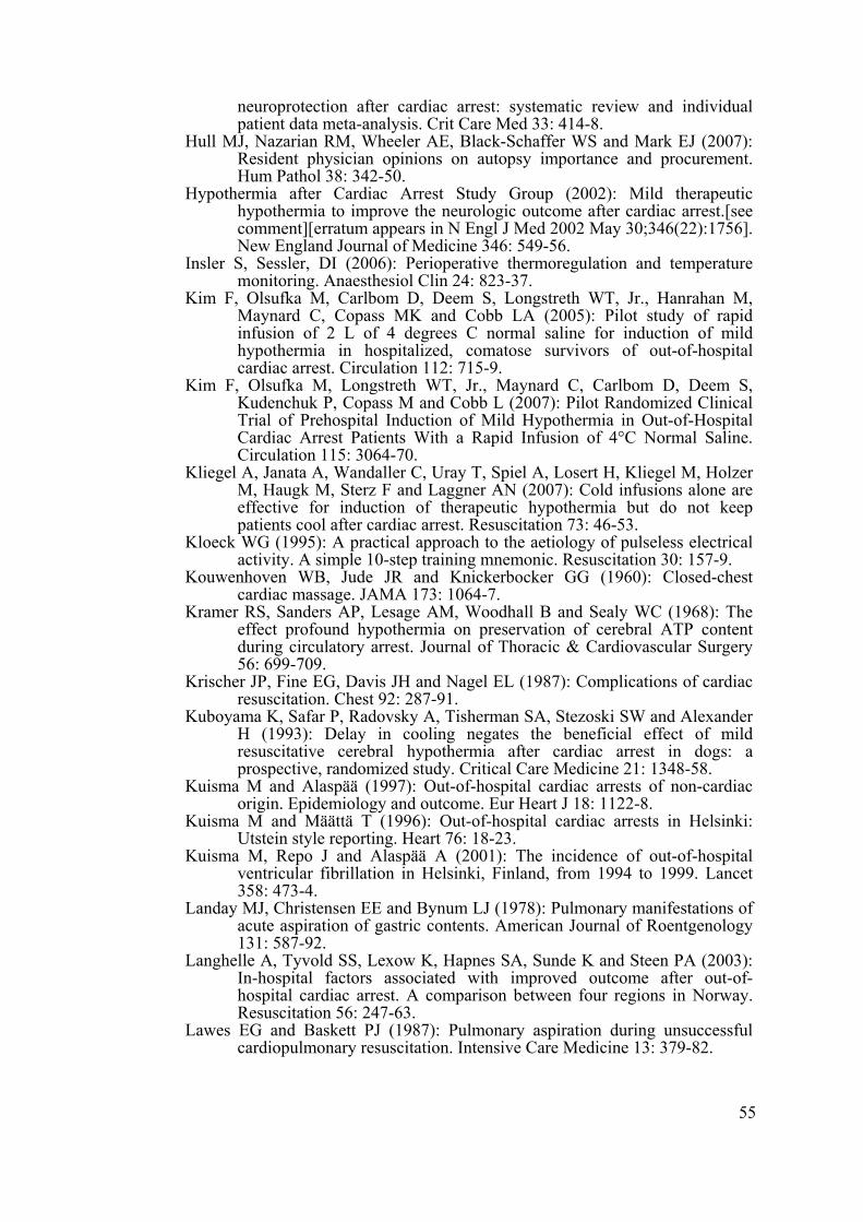
55
neuroprotection after cardiac arrest: systematic review and individual patient data meta-analysis. Crit Care Med 33: 414-8.
Hull MJ, Nazarian RM, Wheeler AE, Black-Schaffer WS and Mark EJ (2007): Resident physician opinions on autopsy importance and procurement. Hum Pathol 38: 342-50.
Hypothermia after Cardiac Arrest Study Group (2002): Mild therapeutic hypothermia to improve the neurologic outcome after cardiac arrest.[see comment][erratum appears in N Engl J Med 2002 May 30;346(22):1756]. New England Journal of Medicine 346: 549-56.
Insler S, Sessler, DI (2006): Perioperative thermoregulation and temperature monitoring. Anaesthesiol Clin 24: 823-37.
Kim F, Olsufka M, Carlbom D, Deem S, Longstreth WT, Jr., Hanrahan M, Maynard C, Copass MK and Cobb LA (2005): Pilot study of rapid infusion of 2 L of 4 degrees C normal saline for induction of mild hypothermia in hospitalized, comatose survivors of out-of-hospital cardiac arrest. Circulation 112: 715-9.
Kim F, Olsufka M, Longstreth WT, Jr., Maynard C, Carlbom D, Deem S, Kudenchuk P, Copass M and Cobb L (2007): Pilot Randomized Clinical Trial of Prehospital Induction of Mild Hypothermia in Out-of-Hospital Cardiac Arrest Patients With a Rapid Infusion of 4°C Normal Saline. Circulation 115: 3064-70.
Kliegel A, Janata A, Wandaller C, Uray T, Spiel A, Losert H, Kliegel M, Holzer M, Haugk M, Sterz F and Laggner AN (2007): Cold infusions alone are effective for induction of therapeutic hypothermia but do not keep patients cool after cardiac arrest. Resuscitation 73: 46-53.
Kloeck WG (1995): A practical approach to the aetiology of pulseless electrical activity. A simple 10-step training mnemonic. Resuscitation 30: 157-9.
Kouwenhoven WB, Jude JR and Knickerbocker GG (1960): Closed-chest cardiac massage. JAMA 173: 1064-7.
Kramer RS, Sanders AP, Lesage AM, Woodhall B and Sealy WC (1968): The effect profound hypothermia on preservation of cerebral ATP content during circulatory arrest. Journal of Thoracic & Cardiovascular Surgery 56: 699-709.
Krischer JP, Fine EG, Davis JH and Nagel EL (1987): Complications of cardiac resuscitation. Chest 92: 287-91.
Kuboyama K, Safar P, Radovsky A, Tisherman SA, Stezoski SW and Alexander H (1993): Delay in cooling negates the beneficial effect of mild resuscitative cerebral hypothermia after cardiac arrest in dogs: a prospective, randomized study. Critical Care Medicine 21: 1348-58.
Kuisma M and Alaspää (1997): Out-of-hospital cardiac arrests of non-cardiac origin. Epidemiology and outcome. Eur Heart J 18: 1122-8.
Kuisma M and Määttä T (1996): Out-of-hospital cardiac arrests in Helsinki: Utstein style reporting. Heart 76: 18-23.
Kuisma M, Repo J and Alaspää A (2001): The incidence of out-of-hospital ventricular fibrillation in Helsinki, Finland, from 1994 to 1999. Lancet 358: 473-4.
Landay MJ, Christensen EE and Bynum LJ (1978): Pulmonary manifestations of acute aspiration of gastric contents. American Journal of Roentgenology 131: 587-92.
Langhelle A, Tyvold SS, Lexow K, Hapnes SA, Sunde K and Steen PA (2003): In-hospital factors associated with improved outcome after out-of-hospital cardiac arrest. A comparison between four regions in Norway. Resuscitation 56: 247-63.
Lawes EG and Baskett PJ (1987): Pulmonary aspiration during unsuccessful cardiopulmonary resuscitation. Intensive Care Medicine 13: 379-82.
56
Lenhardt R (2003): Monitoring and thermal management. Best Pract Res Clin Anaesthesiol 17: 569-81.
Liberthson RR, Nagel EL, Hirschman JC and Nussenfeld SR (1974): Prehospital ventricular defibrillation. Prognosis and follow-up course. New England Journal of Medicine 291: 317-21.
Marwick C (1995): Pathologists request autopsy revival. JAMA 273: 1889. Mayer SA (2005): Refrigerated intravenous fluids: kick-starting the cooling
process.[comment]. Critical Care Medicine 33: 2844-5. McIntyre KM, Parisi AF, Benfari R, Goldberg AH and Dalen JE (1978):
Pathophysiologic syndromes of cardiopulmonary resuscitation. Archives of Internal Medicine 138: 1130-3.
Melker RJ (1985): Recommendations for ventilation during cardiopulmonary resuscitation: time for change? Critical Care Medicine 13: 882-3.
Mendelson C (1946): The aspiration of stomach contents into the lungs during obstetric anesthesia. American Journal of Obstetrics and Gynecology 52: 191-205.
Merchant RM, Soar J, Skrifvars MB, Silfvast T, Edelson DP, Ahmad F, Huang K-N, Khan M, Vanden Hoek TL, Becker LB and Abella BS (2006): Therapeutic hypothermia utilization among physicians after resuscitation from cardiac arrest. Critical Care Medicine 34: 1935-40.
Meursing BTJ, Wulterkens DW and van Kesteren RG (2005): The ABC of resuscitation and the Dutch (re)treat. Resuscitation 64: 279-86.
Meursing B, Zimmerman, AN, van Heyst, AN (1983): Experimental evidence in favor of a reversed sequence in cardiopulmonary resuscitation. J Am Coll Cardiol 1: 610.
Mithoefer JC, Mead G, Hughes JM, Iliff LD and Campbell EJ (1967): A method of distinguishing death due to cardiac arrest from asphyxia. Lancet 2(7517): 654-6.
Myerburg RJ, Kessler KM and Castellanos A (1992): Sudden cardiac death. Structure, function, and time-dependence of risk. Circulation 85: I2-10.
Nadkarni VM, Larkin GL, Peberdy MA, Carey SM, Kaye W, Mancini ME, Nichol G, Lane-Truitt T, Potts J, Ornato JP, Berg RA and National Registry of Cardiopulmonary Resuscitation I (2006): First documented rhythm and clinical outcome from in-hospital cardiac arrest among children and adults.[see comment]. JAMA 295: 50-7.
Negovsky V (1988): Postresuscitation disease. Crit Care Med 16: 942-6. Nolan J (2005): European Resuscitation Council guidelines for resuscitation
2005. Section 1. Introduction. Resuscitation 67: S3-6. Nolan JP, Deakin CD, Soar J, Bottiger BW, Smith G and European Resuscitation
C (2005): European Resuscitation Council guidelines for resuscitation 2005. Section 4. Adult advanced life support. Resuscitation 67: S39-86.
Nolan JP, Morley PT, Hoek TLV, Hickey RW and Advancement Life support Task Force of the International Liaison committee on R (2003): Therapeutic hypothermia after cardiac arrest. An advisory statement by the Advancement Life support Task Force of the International Liaison committee on Resuscitation. Resuscitation 57: 231-5.
Nolan J, Soar J and Eikeland H (2006): The chain of survival. Resuscitation 71: 270-1.
Oschatz E, Wunderbaldinger P, Sterz F, Holzer M, Kofler J, Slatin H, Janata K, Eisenburger P, Bankier AA and Laggner AN (2001): Cardiopulmonary resuscitation performed by bystanders does not increase adverse effects as assessed by chest radiography.[see comment]. Anesthesia & Analgesia 93: 128-33.
Paraskos J (1992): Biblical accounts of resuscitation. J Hist Med All Sci 47: 310-21.
57
Peberdy MA and Ornato JP (2005): Post-resuscitation care: is it the missing link in the Chain of Survival? Resuscitation 64: 135-7.
Pell JP, Sirel JM, Marsden AK, Ford I, Walker NL and Cobbe SM (2003): Presentation, management, and outcome of out of hospital cardiopulmonary arrest: comparison by underlying aetiology. Heart 89: 839-42.
Pepe P (1996): Acute respiratory insufficiency. In: Harwood-Nuss A, Linden CH, Luten RC, Shepherd SM, Wolson AB eds.l. In: The Clinical practise of Emergency Medicine, 636-40. Eds, JB Lippincott, Philadelphia.
Pirolo J, Hutchins G and Moore G (1985): Electromechanical dissociation: pathologic explanations in 50 patients. Hum Pathol 16: 485-7.
Polderman KH, Rijnsburger ER, Peerdeman SM and Girbes ARJ (2005): Induction of hypothermia in patients with various types of neurologic injury with use of large volumes of ice-cold intravenous fluid.[see comment]. Critical Care Medicine 33: 2744-51.
Priori SG, Aliot E, Blomstrom-Lundqvist C, Bossaert L, Breithardt G, Brugada P, Camm AJ, Cappato R, Cobbe SM, Di Mario C, Maron BJ, McKenna WJ, Pedersen AK, Ravens U, Schwartz PJ, Trusz-Gluza M, Vardas P, Wellens HJ and Zipes DP (2001): Task Force on Sudden Cardiac Death of the European Society of Cardiology. European Heart Journal 22: 1374-450.
Rajek A, Greif R, Sessler DI, Baumgardner J, Laciny S and Bastanmehr H (2000): Core cooling by central venous infusion of ice-cold (4 degrees C and 20 degrees C) fluid: isolation of core and peripheral thermal compartments. Anesthesiology 93: 629-37.
Rello J, Valles J, Jubert P, Ferrer A, Domingo C, Mariscal D, Fontanals D and Artigas A (1995): Lower respiratory tract infections following cardiac arrest and cardiopulmonary resuscitation. Clinical Infectious Diseases 21: 310-4.
Robicsek SA and Robicsek F (2004): Janos Balassa and the first CPR. American Journal of Emergency Medicine 22: 622-3.
Ruben H, Knudsen EJ and Carugati G (1961): Gastric inflation in relation to airway pressure. Acta Anaesthesiol Scand 5: 107-14.
Safar PJ and Kochanek PM (2002): Therapeutic hypothermia after cardiac arrest. New England Journal of Medicine 346: 612-3.
Safar P, Escarraga L and Elam J (1958): A comparison of the mouth-to-mouth and mouth-to-airway methods of artificial respiration with the chest-pressure arm-lift methods. N Engl J Med 258: 671-7.
Silfvast T (1990): Prehospital resuscitation in Helsinki, Finland. American Journal of Emergency Medicine 8: 359-64.
Silfvast T (1991): Cause of death in unsuccessful prehospital resuscitation. J Intern Med 229: 331-5.
Simons R, Rea T, Becker L and Eisenberg M (2007): The incidence and significance of emesis associated with out-of-hospital cardiac arrest. Resuscitation doi:10.1016/j.resuscitation.2007.01.038:
Skrifvars M, Pettilä V, Rosenberg P and Castren M (2003): A multiple logistic regression analysis of in-hospital factors related to survival at six months in patients resuscitated from out-of-hospital ventricular fibrillation. Resuscitation 59: 319-28.
Soo LH, Gray D and Hampton JR (2001): Pathological features of witnessed out-of-hospital cardiac arrest presenting with ventricular fibrillation. Resuscitation 51: 257-64.
Soo LH, Gray D, Young T, Skene A and Hampton JR (1999): Influence of ambulance crew's length of experience on the outcome of out-of-hospital cardiac arrest. Eur Heart J 20: 535-40.
58
SOS-Kanto study group (2007): Cardiopulmonary resuscitation by bystanders with chest compression only (SOS-KANTO): an observational study. Lancet 369: 920-6.
Sterz F, Safar P, Tisherman S, Radovsky A, Kuboyama K and Oku K (1991): Mild hypothermic cardiopulmonary resuscitation improves outcome after prolonged cardiac arrest in dogs. Critical Care Medicine 19: 379-89.
Stone BJ, Chantler PJ and Baskett PJ (1998): The incidence of regurgitation during cardiopulmonary resuscitation: a comparison between the bag valve mask and laryngeal mask airway. Resuscitation 38: 3-6.
Sunde K, Pytte M, Jacobsen D, Mangschau A, Jensen LP, Smedsrud C, Draegni T and Steen PA (2007): Implementation of a standardised treatment protocol for post resuscitation care after out-of-hospital cardiac arrest. Resuscitation 73: 29-39.
Van Hoeyweghen RJ, Bossaert LL, Mullie A, Calle P, Martens P, Buylaert WA and Delooz H (1993): Quality and efficiency of bystander CPR. Belgian Cerebral Resuscitation Study Group. Resuscitation 26: 47-52.
Vanbrabant P, Dhondt E, Billen P and Sabbe M (2006): Aetiology of unsuccessful prehospital witnessed cardiac arrest of unclear origin. Eur J Emerg Med 13: 144-7.
Weaver W, Cobb L, Hallstrom A, Copass M, Ray R, Emery M and Fahrenbruch C (1986): Considerations for improving survival from out-of-hospital cardiac arrest. Ann Emerg Med 15: 1181-6.
Wik L, Kramer-Johansen J, Myklebust H, Sorebo H, Svensson L, Fellows B and Steen PA (2005): Quality of cardiopulmonary resuscitation during out-of-hospital cardiac arrest. JAMA 293: 299-304.
Williams GR, Jr. and Spencer FC (1958): The clinical use of hypothermia following cardiac arrest. Annals of Surgery 148: 462-8.
Väisänen O, Mäkijärvi M, Pietilä K and Silfvast T (2006): Influence of medical direction on the management of prehospital myocardial infarction. Resuscitation 70: 207-14.
Yanagawa Y, Ishihara S, Norio H, Takino M, Kawakami M, Takasu A, Okamoto K, Kaneko N, Terai C and Okada Y (1998): Preliminary clinical outcome study of mild resuscitative hypothermia after out-of-hospital cardiopulmonary arrest. Resuscitation 39: 61-6.
Zeiner A, Holzer M, Sterz F, Behringer W, Schorkhuber W, Mullner M, Frass M, Siostrzonek P, Ratheiser K, Kaff A and Laggner AN (2000): Mild resuscitative hypothermia to improve neurological outcome after cardiac arrest. A clinical feasibility trial. Hypothermia After Cardiac Arrest (HACA) Study Group. Stroke 31: 86-94.
Zheng ZJ, Croft JB, Giles WH and Mensah GA (2001): Sudden cardiac death in the United States, 1989 to 1998. Circulation 104: 2158-63.
Zideman D and Koster R (2007). Comments on CPR only in Lancet. 2007. http://www.erc.edu/index.php/doclibrary/en/86/1/ (assessed 17.04.2008)
Zoll P, Linenthal A, Gibson W, Paul M and Norman L (1956): Termination of ventricular fibrillation in man by externally applied electric countershock. N Engl J Med 254: 727-32.

59
Original publications
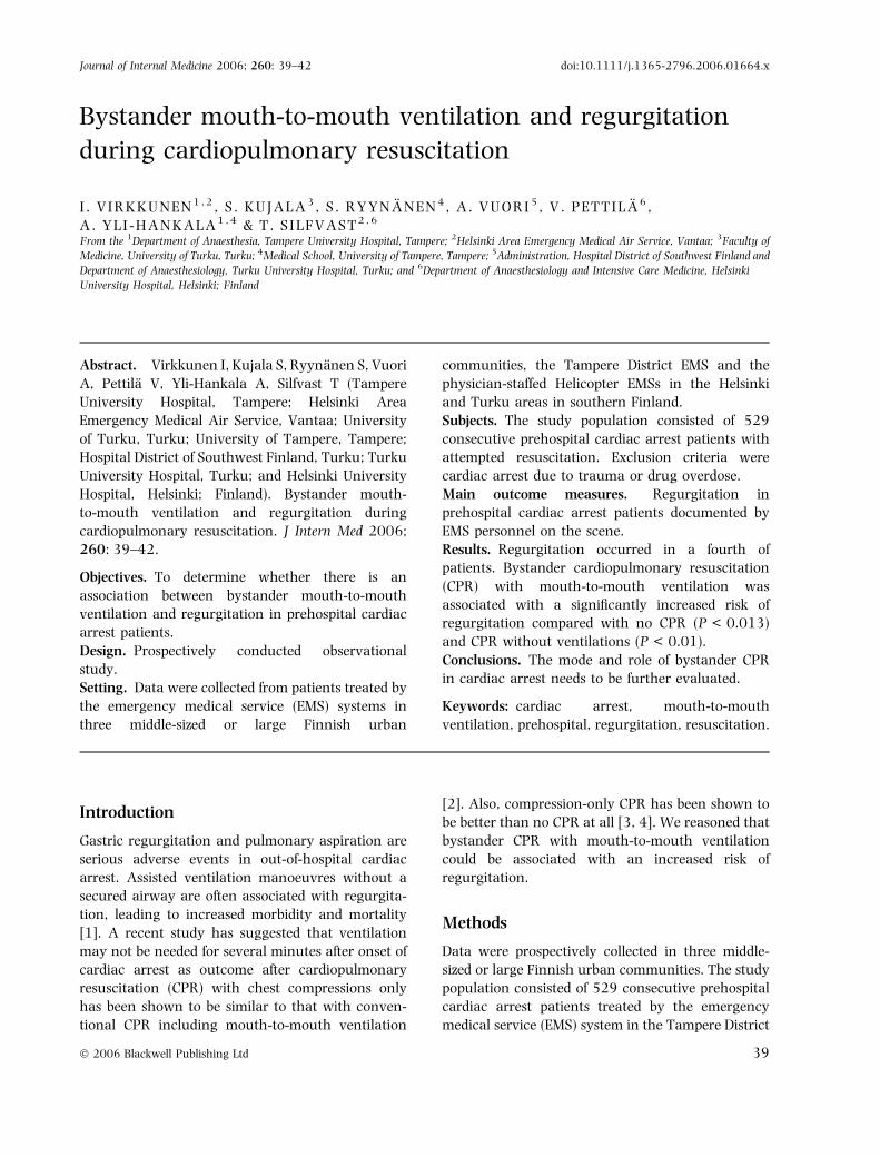
Bystander mouth-to-mouth ventilation and regurgitation
during cardiopulmonary resuscitation
I . V I R K K U N E N 1 , 2 , S . K U J A L A 3 , S . R Y Y N A N E N 4 , A . V U O R I 5 , V . P E T T I L A 6 ,
A . Y L I - H A N K A L A 1 , 4 & T . S I L F V A S T 2 , 6
From the 1Department of Anaesthesia, Tampere University Hospital, Tampere; 2Helsinki Area Emergency Medical Air Service, Vantaa; 3Faculty of
Medicine, University of Turku, Turku; 4Medical School, University of Tampere, Tampere; 5Administration, Hospital District of Southwest Finland and
Department of Anaesthesiology, Turku University Hospital, Turku; and 6Department of Anaesthesiology and Intensive Care Medicine, Helsinki
University Hospital, Helsinki; Finland
Abstract. Virkkunen I, Kujala S, Ryynanen S, Vuori
A, Pettila V, Yli-Hankala A, Silfvast T (Tampere
University Hospital, Tampere; Helsinki Area
Emergency Medical Air Service, Vantaa; University
of Turku, Turku; University of Tampere, Tampere;
Hospital District of Southwest Finland, Turku; Turku
University Hospital, Turku; and Helsinki University
Hospital, Helsinki; Finland). Bystander mouth-
to-mouth ventilation and regurgitation during
cardiopulmonary resuscitation. J Intern Med 2006;
260: 39–42.
Objectives. To determine whether there is an
association between bystander mouth-to-mouth
ventilation and regurgitation in prehospital cardiac
arrest patients.
Design. Prospectively conducted observational
study.
Setting. Data were collected from patients treated by
the emergency medical service (EMS) systems in
three middle-sized or large Finnish urban
communities, the Tampere District EMS and the
physician-staffed Helicopter EMSs in the Helsinki
and Turku areas in southern Finland.
Subjects. The study population consisted of 529
consecutive prehospital cardiac arrest patients with
attempted resuscitation. Exclusion criteria were
cardiac arrest due to trauma or drug overdose.
Main outcome measures. Regurgitation in
prehospital cardiac arrest patients documented by
EMS personnel on the scene.
Results. Regurgitation occurred in a fourth of
patients. Bystander cardiopulmonary resuscitation
(CPR) with mouth-to-mouth ventilation was
associated with a significantly increased risk of
regurgitation compared with no CPR (P < 0.013)
and CPR without ventilations (P < 0.01).
Conclusions. The mode and role of bystander CPR
in cardiac arrest needs to be further evaluated.
Keywords: cardiac arrest, mouth-to-mouth
ventilation, prehospital, regurgitation, resuscitation.
Introduction
Gastric regurgitation and pulmonary aspiration are
serious adverse events in out-of-hospital cardiac
arrest. Assisted ventilation manoeuvres without a
secured airway are often associated with regurgita-
tion, leading to increased morbidity and mortality
[1]. A recent study has suggested that ventilation
may not be needed for several minutes after onset of
cardiac arrest as outcome after cardiopulmonary
resuscitation (CPR) with chest compressions only
has been shown to be similar to that with conven-
tional CPR including mouth-to-mouth ventilation
[2]. Also, compression-only CPR has been shown to
be better than no CPR at all [3, 4]. We reasoned that
bystander CPR with mouth-to-mouth ventilation
could be associated with an increased risk of
regurgitation.
Methods
Data were prospectively collected in three middle-
sized or large Finnish urban communities. The study
population consisted of 529 consecutive prehospital
cardiac arrest patients treated by the emergency
medical service (EMS) system in the Tampere District
Journal of Internal Medicine 2006; 260: 39–42 doi:10.1111/j.1365-2796.2006.01664.x
� 2006 Blackwell Publishing Ltd 39
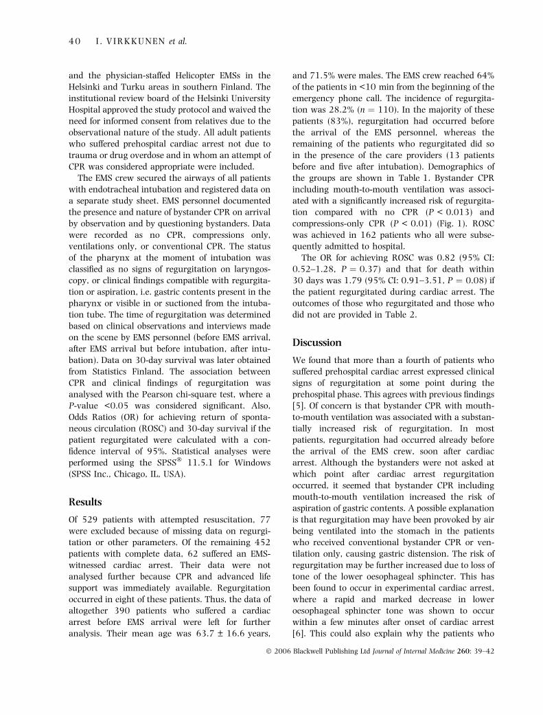
and the physician-staffed Helicopter EMSs in the
Helsinki and Turku areas in southern Finland. The
institutional review board of the Helsinki University
Hospital approved the study protocol and waived the
need for informed consent from relatives due to the
observational nature of the study. All adult patients
who suffered prehospital cardiac arrest not due to
trauma or drug overdose and in whom an attempt of
CPR was considered appropriate were included.
The EMS crew secured the airways of all patients
with endotracheal intubation and registered data on
a separate study sheet. EMS personnel documented
the presence and nature of bystander CPR on arrival
by observation and by questioning bystanders. Data
were recorded as no CPR, compressions only,
ventilations only, or conventional CPR. The status
of the pharynx at the moment of intubation was
classified as no signs of regurgitation on laryngos-
copy, or clinical findings compatible with regurgita-
tion or aspiration, i.e. gastric contents present in the
pharynx or visible in or suctioned from the intuba-
tion tube. The time of regurgitation was determined
based on clinical observations and interviews made
on the scene by EMS personnel (before EMS arrival,
after EMS arrival but before intubation, after intu-
bation). Data on 30-day survival was later obtained
from Statistics Finland. The association between
CPR and clinical findings of regurgitation was
analysed with the Pearson chi-square test, where a
P-value <0.05 was considered significant. Also,
Odds Ratios (OR) for achieving return of sponta-
neous circulation (ROSC) and 30-day survival if the
patient regurgitated were calculated with a con-
fidence interval of 95%. Statistical analyses were
performed using the SPSS� 11.5.1 for Windows
(SPSS Inc., Chicago, IL, USA).
Results
Of 529 patients with attempted resuscitation, 77
were excluded because of missing data on regurgi-
tation or other parameters. Of the remaining 452
patients with complete data, 62 suffered an EMS-
witnessed cardiac arrest. Their data were not
analysed further because CPR and advanced life
support was immediately available. Regurgitation
occurred in eight of these patients. Thus, the data of
altogether 390 patients who suffered a cardiac
arrest before EMS arrival were left for further
analysis. Their mean age was 63.7 ± 16.6 years,
and 71.5% were males. The EMS crew reached 64%
of the patients in <10 min from the beginning of the
emergency phone call. The incidence of regurgita-
tion was 28.2% (n ¼ 110). In the majority of these
patients (83%), regurgitation had occurred before
the arrival of the EMS personnel, whereas the
remaining of the patients who regurgitated did so
in the presence of the care providers (13 patients
before and five after intubation). Demographics of
the groups are shown in Table 1. Bystander CPR
including mouth-to-mouth ventilation was associ-
ated with a significantly increased risk of regurgita-
tion compared with no CPR (P < 0.013) and
compressions-only CPR (P < 0.01) (Fig. 1). ROSC
was achieved in 162 patients who all were subse-
quently admitted to hospital.
The OR for achieving ROSC was 0.82 (95% CI:
0.52–1.28, P ¼ 0.37) and that for death within
30 days was 1.79 (95% CI: 0.91–3.51, P ¼ 0.08) if
the patient regurgitated during cardiac arrest. The
outcomes of those who regurgitated and those who
did not are provided in Table 2.
Discussion
We found that more than a fourth of patients who
suffered prehospital cardiac arrest expressed clinical
signs of regurgitation at some point during the
prehospital phase. This agrees with previous findings
[5]. Of concern is that bystander CPR with mouth-
to-mouth ventilation was associated with a substan-
tially increased risk of regurgitation. In most
patients, regurgitation had occurred already before
the arrival of the EMS crew, soon after cardiac
arrest. Although the bystanders were not asked at
which point after cardiac arrest regurgitation
occurred, it seemed that bystander CPR including
mouth-to-mouth ventilation increased the risk of
aspiration of gastric contents. A possible explanation
is that regurgitation may have been provoked by air
being ventilated into the stomach in the patients
who received conventional bystander CPR or ven-
tilation only, causing gastric distension. The risk of
regurgitation may be further increased due to loss of
tone of the lower oesophageal sphincter. This has
been found to occur in experimental cardiac arrest,
where a rapid and marked decrease in lower
oesophageal sphincter tone was shown to occur
within a few minutes after onset of cardiac arrest
[6]. This could also explain why the patients who
� 2006 Blackwell Publishing Ltd Journal of Internal Medicine 260: 39–42
4 0 I . V I R K K U N E N et al.
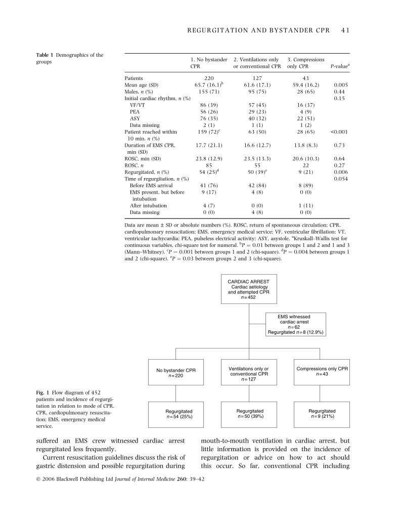
suffered an EMS crew witnessed cardiac arrest
regurgitated less frequently.
Current resuscitation guidelines discuss the risk of
gastric distension and possible regurgitation during
mouth-to-mouth ventilation in cardiac arrest, but
little information is provided on the incidence of
regurgitation or advice on how to act should
this occur. So far, conventional CPR including
CARDIAC ARRESTCardiac aetiology
and attempted CPRn = 452
Compressions only CPR n = 43
No bystander CPRn = 220
Ventilations only orconventional CPR
n = 127
Regurgitated n = 54 (25%)
Regurgitatedn = 50 (39%)
Regurgitated n = 9 (21%)
EMS witnessedcardiac arrest
n = 62Regurgitated n = 8 (12.9%)
Fig. 1 Flow diagram of 452
patients and incidence of regurgi-
tation in relation to mode of CPR.
CPR, cardiopulmonary resuscita-
tion; EMS, emergency medical
service.
Table 1 Demographics of the
groups 1. No bystander
CPR
2. Ventilations only
or conventional CPR
3. Compressions
only CPR P-valuea
Patients 220 127 43
Mean age (SD) 65.7 (16.1)b 61.6 (17.1) 59.4 (16.2) 0.005
Males, n (%) 155 (71) 95 (75) 28 (65) 0.44
Initial cardiac rhythm, n (%) 0.15
VF/VT 86 (39) 57 (45) 16 (37)
PEA 56 (26) 29 (23) 4 (9)
ASY 76 (35) 40 (32) 22 (51)
Data missing 2 (1) 1 (1) 1 (2)
Patient reached within
10 min, n (%)
159 (72)c 63 (50) 28 (65) <0.001
Duration of EMS CPR,
min (SD)
17.7 (21.1) 16.6 (12.7) 13.8 (8.3) 0.73
ROSC, min (SD) 23.8 (12.9) 23.5 (13.3) 20.6 (10.3) 0.64
ROSC, n 85 55 22 0.27
Regurgitated, n (%) 54 (25)d 50 (39)e 9 (21) 0.006
Time of regurgitation, n (%) 0.054
Before EMS arrival 41 (76) 42 (84) 8 (89)
EMS present, but before
intubation
9 (17) 4 (8) 0 (0)
After intubation 4 (7) 0 (0) 1 (11)
Data missing 0 (0) 4 (8) 0 (0)
Data are mean ± SD or absolute numbers (%). ROSC, return of spontaneous circulation; CPR,
cardiopulmonary resuscitation; EMS, emergency medical service; VF, ventricular fibrillation; VT,
ventricular tachycardia; PEA, pulseless electrical activity; ASY, asystole. aKruskall–Wallis test for
continuous variables, chi-square test for numeral. bP ¼ 0.01 between groups 1 and 2 and 1 and 3
(Mann–Whitney). cP ¼ 0.001 between groups 1 and 2 (chi-square). dP ¼ 0.004 between groups 1
and 2 (chi-square). eP ¼ 0.03 between groups 2 and 3 (chi-square).
� 2006 Blackwell Publishing Ltd Journal of Internal Medicine 260: 39–42
R E G U R G I T A T I O N A N D B Y S T A N D E R C P R 4 1
compressions and ventilations has been the general
recommendation in resuscitation guidelines [7].
However, compressions-only CPR has been shown
to be associated with similar outcome as conven-
tional CPR in patients with a short delay from the
onset of cardiac arrest to the arrival of the EMS crew
[2]. Although compressions-only CPR currently is
recommended only in dispatcher-assisted CPR or if
the lay rescuer is unwilling to perform mouth-
to-mouth ventilation [7], it seems that stronger
consideration should be given to this option if the
dispatcher estimates that qualified help will arrive
within minutes.
The main limitation of this study is that it is an
observational one and therefore cannot produce the
same level of evidence as a randomized trial. We did
not include patients in whom an EMS crew-initiated
attempt of CPR was considered inappropriate.
Therefore we do not know the exact number of
patients who actually regurgitated. Also, the per-
centage of patients with incomplete or missing data
was 14.6%, which was rather high. However, data
collection in prehospital studies is a challenging
task, and it is difficult to achieve the level of
completeness that is aimed at.
In conclusion, we found that bystander CPR
including mouth-to-mouth ventilation was associ-
ated with a significantly increased risk of regurgita-
tion. In the view of these findings and previous data
[2], the mode and role of bystander CPR in cardiac
arrest needs to be further evaluated.
Conflict of interest statement
No conflict of interest was declared.
Acknowledgements
We would like to thank the crews of the Helsinki and
Turku Area HEMS and Tampere District Emergency
Medical Services for their valuable help in enrolling
the study patients. This study was supported by
institutional EVO grant no. 9B102 from Special
State Allocation via Tampere University Hospital.
Funding sources had no role in the design and
conduct of the study; collection, management,
analysis, and interpretation of the data; and prepar-
ation, review, or approval of the manuscript. We
also thank Statistics Finland for providing data on
30-day survival.
References
1 Pepe P. Acute respiratory insufficiency. In: Harwood-Nuss A,
Linden CH, Luten RC, Shepherd SM, Wolson AB, eds. The
Clinical Practise of Emergency Medicine. Philadelphia, PA: JB
Lippincott, 1996: 636–40.
2 Hallstrom A, Cobb L, Johnson E, Copass M. Cardiopulmonary
resuscitation by chest compression alone or with mouth-
to-mouth ventilation. N Engl J Med 2000; 342: 1546–53.
3 Van Hoeyweghen RJ, Bossaert LL, Mullie A et al. Quality and
efficiency of bystander CPR. Belgian Cerebral Resuscitation
Study Group. Resuscitation 1993; 26: 47–52.
4 Bossaert L, Van Hoeyweghen R. Belgian Cerebral Resuscitation
Study Group, Bystanders cardiopulmonary resuscitation (CPR)
in out-of-hospital cardiac arrest. Resuscitation 1989; 17: S55–
69.
5 Stone BJ, Chantler PJ, Baskett PJ. The incidence of regurgitation
during cardiopulmonary resuscitation: a comparison between
the bag valve mask and laryngeal mask airway. Resuscitation
1998; 38: 3–6.
6 Bowman FP, Menegazzi JJ, Check BD, Duckett TM. The lower
esophageal sphincter pressure during prolonged cardiac arrest
and resuscitation. Ann Emerg Med 1995; 26: 216–9.
7 Anonymous. Part 3: adult basic life support. European Resus-
citation Council. Resuscitation 2000; 46: 29–71.
Correspondence: Ilkka Virkkunen MD, Department of Anaesthesia,
Tampere University Hospital, POB 2000, FIN-33521 Tampere,
Finland.
(fax: +358 3 31164363; e-mail: [email protected]).
Table 2 Relationship between regurgitation and achieving
ROSC and regurgitation and death 30-days (data missing in one
patient)
Regurgitation Yes No Total
ROSC
Yes 43 70 113
No 119 158 277
Total 162 228 390
Death
Yes 100 12 112
No 228 49 277
Total 328 61 389
� 2006 Blackwell Publishing Ltd Journal of Internal Medicine 260: 39–42
4 2 I . V I R K K U N E N et al.

Erratum Publication I, page 40, right column, fourth line should be: “tion was 29.0% (n=113). In the majority of these”
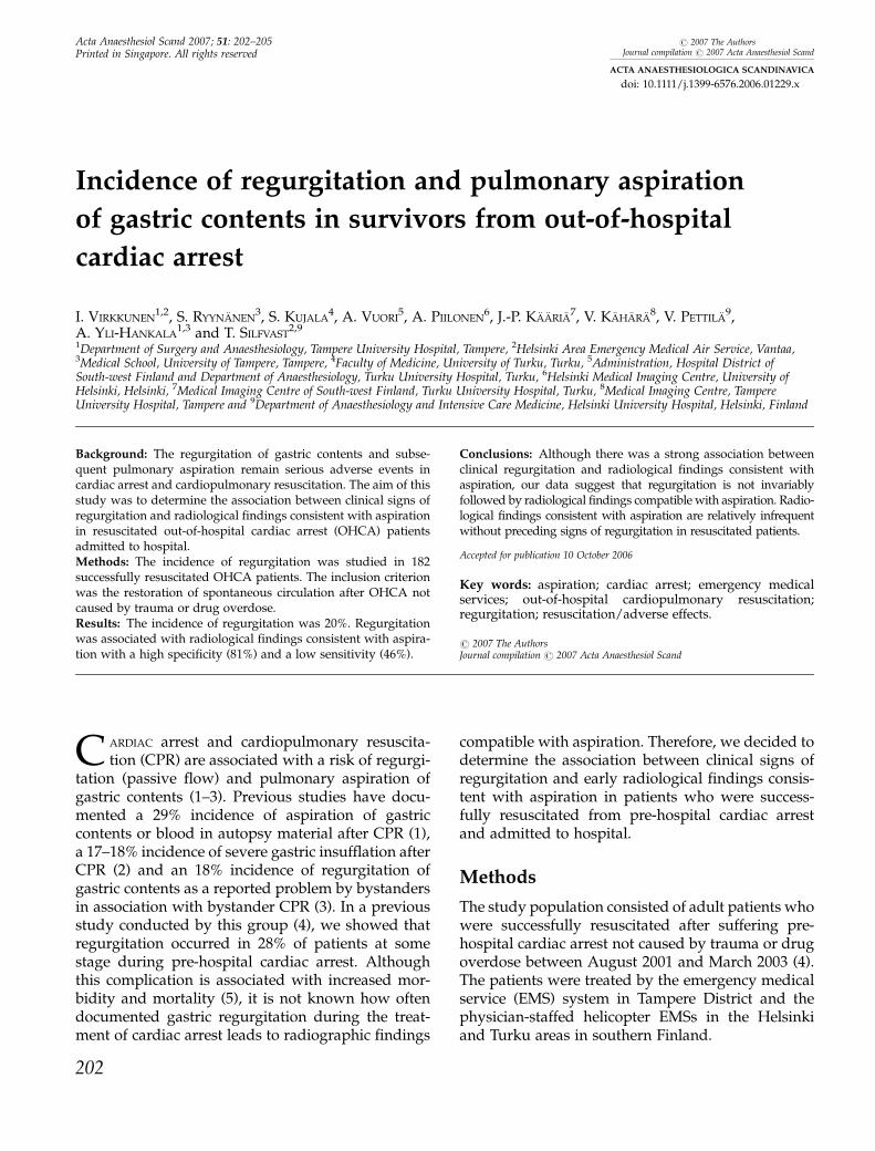
Acta Anaesthesiol Scand 2007; 51: 202–205Printed in Singapore. All rights reserved
# 2007 The AuthorsJournal compilation # 2007 Acta Anaesthesiol Scand
ACTA ANAESTHESIOLOGICA SCANDINAVICA
doi: 10.1111/j.1399-6576.2006.01229.x
Incidence of regurgitation and pulmonary aspiration
of gastric contents in survivors from out-of-hospital
cardiac arrest
I. VIRKKUNEN1,2, S. RYYNANEN
3, S. KUJALA4, A. VUORI
5, A. PIILONEN6, J.-P. KAARIA
7, V. KAHARA8, V. PETTILA
9,A. YLI-HANKALA
1,3 and T. SILFVAST2,9
1Department of Surgery and Anaesthesiology, Tampere University Hospital, Tampere, 2Helsinki Area Emergency Medical Air Service, Vantaa,3Medical School, University of Tampere, Tampere, 4Faculty of Medicine, University of Turku, Turku, 5Administration, Hospital District ofSouth-west Finland and Department of Anaesthesiology, Turku University Hospital, Turku, 6Helsinki Medical Imaging Centre, University ofHelsinki, Helsinki, 7Medical Imaging Centre of South-west Finland, Turku University Hospital, Turku, 8Medical Imaging Centre, TampereUniversity Hospital, Tampere and 9Department of Anaesthesiology and Intensive Care Medicine, Helsinki University Hospital, Helsinki, Finland
Background: The regurgitation of gastric contents and subse-quent pulmonary aspiration remain serious adverse events incardiac arrest and cardiopulmonary resuscitation. The aim of thisstudy was to determine the association between clinical signs ofregurgitation and radiological findings consistent with aspirationin resuscitated out-of-hospital cardiac arrest (OHCA) patientsadmitted to hospital.Methods: The incidence of regurgitation was studied in 182successfully resuscitated OHCA patients. The inclusion criterionwas the restoration of spontaneous circulation after OHCA notcaused by trauma or drug overdose.Results: The incidence of regurgitation was 20%. Regurgitationwas associated with radiological findings consistent with aspira-tion with a high specificity (81%) and a low sensitivity (46%).
Conclusions: Although there was a strong association betweenclinical regurgitation and radiological findings consistent withaspiration, our data suggest that regurgitation is not invariablyfollowed by radiological findings compatible with aspiration. Radio-logical findings consistent with aspiration are relatively infrequentwithout preceding signs of regurgitation in resuscitated patients.
Accepted for publication 10 October 2006
Key words: aspiration; cardiac arrest; emergency medicalservices; out-of-hospital cardiopulmonary resuscitation;regurgitation; resuscitation/adverse effects.
# 2007 The AuthorsJournal compilation # 2007 Acta Anaesthesiol Scand
CARDIAC arrest and cardiopulmonary resuscita-tion (CPR) are associated with a risk of regurgi-
tation (passive flow) and pulmonary aspiration ofgastric contents (1–3). Previous studies have docu-mented a 29% incidence of aspiration of gastriccontents or blood in autopsy material after CPR (1),a 17–18% incidence of severe gastric insufflation afterCPR (2) and an 18% incidence of regurgitation ofgastric contents as a reported problem by bystandersin association with bystander CPR (3). In a previousstudy conducted by this group (4), we showed thatregurgitation occurred in 28% of patients at somestage during pre-hospital cardiac arrest. Althoughthis complication is associated with increased mor-bidity and mortality (5), it is not known how oftendocumented gastric regurgitation during the treat-ment of cardiac arrest leads to radiographic findings
compatible with aspiration. Therefore, we decided todetermine the association between clinical signs ofregurgitation and early radiological findings consis-tent with aspiration in patients who were success-fully resuscitated from pre-hospital cardiac arrestand admitted to hospital.
Methods
The study population consisted of adult patients whowere successfully resuscitated after suffering pre-hospital cardiac arrest not caused by trauma or drugoverdose between August 2001 and March 2003 (4).The patients were treated by the emergency medicalservice (EMS) system in Tampere District and thephysician-staffed helicopter EMSs in the Helsinkiand Turku areas in southern Finland.
202
The institutional review board of the HelsinkiUniversity Hospital approved the study protocol.EMS personnel intubated all patients in the fieldand documented prospectively the status of thepharynx at the moment of intubation on a separatestudy sheet. Findings were classified as follows: nosigns of regurgitation on laryngoscopy; gastric con-tents present in the pharynx; or gastric contentsvisible in or suctioned from the intubation tube. Afterthe restoration of spontaneous circulation (ROSC)and stabilization in the field, the patients were admit-ted to hospital.In order to assess the radiological findings pro-
duced by the regurgitation of gastric contents, andnot hospital-acquired or ventilator-associated pneu-monia, only the chest X-rays obtained during the first2 days after cardiac arrest were reviewed. Radiolog-ical examinations of the patients were ordered at thediscretion of the treating physicians in hospital andnot dictated by the study protocol. For that reason,radiological examinations were not performed in allpatients, and patients who did not undergo radio-logical examination were excluded. According togeneral practice, a chest X-ray is routinely obtainedon admission and thereafter as indicated. Togetherwith a senior consultant in radiology at the receivinghospital (Helsinki, Turku and Tampere UniversityHospitals), the principal investigator (I.V.) evaluatedall chest X-rays obtained during the first 2 days afteradmission. One radiologist evaluated all X-rays ateach hospital. The findings were classified as follows:normal; suspicion of aspiration; or radiological signsconsistent with aspiration. The radiologist and prin-cipal investigator were blind to the clinical find-ings of regurgitation at the time of the radiologicalevaluation.The association between clinical signs of re-
gurgitation of gastric contents or pulmonary aspi-ration documented at the time of intubation andradiological signs consistent with pulmonary aspi-ration was analysed using the chi-squared test. Thenull hypothesis was that no such association exists.Inter-group differences in demographics, rhythmand ROSC data were analysed using the Kruskal–Wallis test, followed by the Mann–Whitney inde-pendent sample test. P < 0.05 was considered to besignificant. In addition, the sensitivity, specificityand positive (PPV) and negative (NPV) predictivevalues of clinical regurgitation to predict radio-logical signs consistent with pulmonary aspirationwere calculated. Statistical analyses were performedusing SPSS� 11.5.1 forWindows (SPSS Inc., Chicago,IL).
Results
During the study period, resuscitation was successfulin 202 patients, and they were admitted to hospital.No radiological examinations were performed in 20patients, leaving 182 patients for further analysis.Their mean age was 62.4 � 15.5 years [mean �standard deviation (SD)] and 74% were males. ROSChad been achieved within 21.6 � 12.8 min (mean �SD). Altogether, 256 chest X-rays were available forfurther analysis, one in 108 patients and two in 74patients. The timing of the first chest X-ray was theday of admission in 36%, the day after admission in60% and the second day after admission in 4% ofpatients.In 37 patients (20%), EMS personnel documented
signs of regurgitation on the scene. In hospital, thechest X-ray showed a suspicion of or findings com-patible with aspiration in 44 patients (24%). Chest X-ray revealed findings compatible with pulmonaryaspiration in 17 of the 37 patients (46%) with clinicalsigns of regurgitation on the scene, compared with 27of the 145 patients (19%) without such findings (P <0.001 by chi-squared test) (Table 1). Thus, clinicalsigns of regurgitation in the pre-hospital phaseresulted in radiological signs consistent with pulmo-nary aspiration with an 81% specificity (118 of 145)and a 46% sensitivity (17 of 37). PPV was 0.39 andNPV was 0.86.
Discussion
This study revealed a strong association between clin-ical regurgitation documented during out-of-hospitalcardiac arrest (OHCA) and early radiological find-ings consistent with aspiration. The incidence of clini-cally recognized regurgitation of gastric contents inthe patients admitted to hospital after ROSC in this
Table 1
Relationship between clinical observations on regurgitation ofgastric contents and radiological findings compatible with theaspiration of gastric contents (P < 0.001). Sensitivity, 0.46;specificity, 0.81; positive predictive value (PPV), 0.39; negativepredictive value (NPV), 0.86.
Regurgitation on scene
Yes No Total
Radiological findingscompatible with aspiration
Yes 17 27 44
No 20 118 138Total 37 145 182
203
Aspiration in pre-hospital cardiac arrest
study (20%) was close to the incidence of aspirationreported in autopsy material (1), which showedfindings of blood or gastric contents in the airwaysin 29% of subjects. Other studies have shown severegastric insufflation and regurgitation of gastric con-tents in 17–18% of patients who received bystanderCPR after suffering OHCA (2, 3).Despite the strong association between clinical
signs of regurgitation and the subsequent develop-ment of radiological findings of aspiration, the sen-sitivity was relatively poor: 46%. When regurgitationwas detected on the scene, there was a 39% (PPV)chance of finding radiological signs consistent withpulmonary aspiration. When there were no signsof regurgitation, there was an 86% chance that noradiological signs consistent with pulmonary aspira-tion would develop (NPV). To what extent the earlydevelopment of radiological signs of aspirationshould be interpreted as infection has not been studied.A previous investigation reporting infectious com-plications after cardiac arrest has documented thatradiological signs of pneumonia develop after 7� 6.2days (6), and that progressive worsening of radio-graphic infiltrates after 3 days suggests secondarybacterial pneumonia, acute respiratory distress syn-drome (ARDS) or pulmonary embolism (7), but therole of aspiration was not specifically addressed inthese studies. Because the later development ofinfectious complications was not the aim of thisstudy, we only evaluated X-rays obtained on the firstand second days after admission.No radiological signs of aspiration appeared in
26% of those who regurgitated. One explanationoffered is the pH of the aspirate, as it has been shownthat only when the aspirated matter has pH < 2.4does it cause acidic pneumonitis, manifesting asradiological signs of aspiration (8). The nature ofthe regurgitated matter was not characterized. There-fore, we do not know whether it was liquid or food,an issue that may have an influence on the clinicalconsequences.Moreover, in other well-documented cases of the
aspiration of gastric contents, 8–10% of patientsdeveloped no symptoms other than fever, and theradiographic findings were extremely variable (7). Inaddition to the pH, another probable factor influenc-ing the development of radiological signs of aspira-tion is the volume of the aspirate. Thus, the overallincidence of radiological findings consistent withaspiration in this study is close to the 18% reportedby Oschatz et al. (2) in their survey.There are some limitations in this study. The
retrospective collection and variable number of chest
X-rays were major weaknesses. In the majority ofpatients, only one chest X-ray was obtained duringthe first 2 days after cardiac arrest. This was becausethe study protocol did not dictate the timing ofradiological examinations, and there were no strictprotocols to guide such examinations after cardiacarrest in the participating hospitals. Considering thatall hospitals were university teaching hospitals, thisfinding was surprising. One explanation for the lackof X-ray examinations may be that the physicians incharge have a particularly pessimistic view withregard to the prognosis of resuscitated patients.Because two chest X-rays were not obtained in allpatients, the true incidence of radiological findingsmay be greater than documented here. As we eval-uated only the chest X-rays obtained during the first2 days, it is possible that regurgitation-related alter-ations may have manifested later.In conclusion, the incidence of regurgitation during
pre-hospital cardiac arrest was 20% in patientsadmitted to hospital after ROSC in this study. Regur-gitation was associated with the subsequent devel-opment of radiological findings consistent withaspiration with moderate sensitivity and high speci-ficity. It seems that clinical signs of regurgitationof gastric contents and subsequent aspiration intothe lungs do not always lead to the developmentof radiological signs consistent with pulmonaryaspiration.
Acknowledgements
We would like to thank the crews of the Helsinki and Turku areashelicopter EMSs and the Tampere District EMS for their valuablehelp in enrolling the study patients.
References
1. Lawes EG, Baskett PJ. Pulmonary aspiration during unsuccess-ful cardiopulmonary resuscitation. J Intensive Care Med 1987;13: 379–82.
2. Oschatz E, Wunderbaldinger P, Sterz F et al. Cardiopulmonaryresuscitation performed by bystanders does not increaseadverse effects as assessed by chest radiography. Anesth Analg2001; 93: 128–33.
3. Axelsson A, Herlitz J, Ekstrom L Holmberg S. Bystander-initiated cardiopulmonary resuscitation out-of-hospital. A firstdescription of the bystanders and their experiences. Resuscita-tion 1996; 33: 3–11.
4. Virkkunen I, Kujala S, Ryynanen S et al. Bystander mouth-to-mouth ventilation and regurgitation during cardiopulmo-nary resuscitation. J Intern Med 2006; 260: 39–42.
5. Bellingan GJ. Aspiration and inhalation. In: Webb, AR,Shapiro, MJ, Singer, M, Suter, PM, eds. Oxford Textbook ofCritical Care. New York: Oxford University Press, 1999: 79–85.
204
I. Virkkunen et al.

6. Rello J, Valles J, Jubert P et al. Lower respiratory tract infec-tions following cardiac arrest and cardiopulmonary resuscita-tion. Clin Infect Dis 1995; 21: 310–4.
7. Landay MJ, Christensen EE, Bynum LJ. Pulmonary manifes-tations of acute aspiration of gastric contents. Am J Roentgenol1978; 131: 587–92.
8. Bartlett JG, Gorbach SL. The triple threat of aspiration pneu-monia. Chest 1975; 68: 560–6.
Address:Dr Ilkka VirkkunenDepartment of Surgery and AnaesthesiologyTampere University HospitalPO Box 2000FIN-33521 TampereFinlande-mail: [email protected]
205
Aspiration in pre-hospital cardiac arrest
Author's personal copy
Resuscitation (2008) 77, 207—210
avai lab le at www.sc iencedi rec t .com
journa l homepage: www.e lsev ier .com/ locate / resusc i ta t ion
CLINICAL PAPER
Pulseless electrical activity and unsuccessfulout-of-hospital resuscitation: What is the causeof death?�
Ilkka Virkkunena,∗, Laura Paasiob, Sanna Ryynanenc,Arno Vuorid, Antti Sajantilae, Arvi Yli-Hankalaa,c, Tom Silfvast f
a Tampere University Hospital, Department of Surgery and Anaesthesiology, PO Box 2000, FIN-33521 Tampere, Finlandb University of Turku, Faculty of Medicine, Kiinanmyllynkatu 13, FIN-20520 Turku, Finlandc University of Tampere, Medical School, FIN-33014 Tampere, Finlandd Administration, Hospital District of Southwest Finland and Department of Anaesthesiology,Turku University Hospital, PO Box 52, FIN-20521 Turku, Finlande University of Helsinki, Department of Forensic Medicine, PO Box 40, FIN-00014 Helsinki, Finlandf Department of Anaesthesiology and Intensive Care Medicine, Helsinki University Hospital, PO Box 340, FIN-00029 HUS, Finland
Received 3 September 2007; received in revised form 20 November 2007; accepted 18 December 2007
KEYWORDSOut-of-hospital;Cardiac arrest;Cause of death;Autopsy;Cardiopulmonaryresuscitation;Pulseless electricalactivity
SummaryAims: To study the cause of deaths after witnessed cardiac arrest followed by pulseless elec-trical activity and unsuccessful of out-of-hospital resuscitation; and to detect any differencesbetween causes of death determined at autopsy and those inferred from clinical history.Methods: In this prospective observational study, data were collected from 91 individuals trea-ted by the emergency medical services in three urban communities in southern Finland.Results: Cause of death was determined at autopsy in 59 cases and without autopsy in 32 cases.There were significantly more diagnoses of acute myocardial infarction and fewer of pulmonaryembolism and aortic dissection and rupture among cases without autopsy compared with thosefollowed by autopsy.Conclusion: In unsuccessful resuscitation from out-of-hospital cardiac arrest with pulseless elec-trical activity as initial rhythm, an autopsy should be performed to determine the correct causeof death.© 2007 Elsevier Ireland Ltd. All rights reserved.
� A Spanish translated version of the summary of this articleappears as Appendix in the final online version at doi:10.1016/j.resuscitation.2007.12.006.
∗ Corresponding author. Tel.: +358 331165081;fax: +358 331164363.
E-mail address: [email protected] (I. Virkkunen).
Introduction
Although ventricular fibrillation (VF) has been consideredthe most common initial rhythm (37—83%) in out-of-hospitalcardiac arrest (OHCA), a major decline (50%) in its incidencehas occurred during recent decades.1—3 Concomitantly,the number of cases of pulseless electrical activity (PEA)
0300-9572/$ — see front matter © 2007 Elsevier Ireland Ltd. All rights reserved.doi:10.1016/j.resuscitation.2007.12.006
Author's personal copy
208 I. Virkkunen et al.
as initial cardiac rhythm has increased.4—6 Recent stu-dies have shown the incidence of primary PEA to be22—30% in OHCA,7—11 and even 32—52% in in-hospital cardiacarrest.12,13
Whereas VF is strongly associated with coronary heartdisease,14 the conditions that cause PEA as initial cardiacrhythm in OHCA often reflect a non-coronary aetiology. Pul-monary embolism (PE) has been shown to be common (36%)in cases of unexpected cardiac arrest presenting with PEA,15
and a study of people who died after primary PEA (previouslycalled electromechanical dissociation) reported that cardio-vascular rupture, PE or tension pneumothorax was the causeof death (COD) in 19 of 50 cases.16 Another study invol-ving 28 people with PEA as initial rhythm, who died afterunsuccessful resuscitation, documented 11 cardiovascularruptures and 6 PEs.17 Courtney et al.18 found that PE was theCOD in 52% of cases of primary PEA in a study of witnessedprehospital cardiac arrests.
Obviously, the CODs presented above were based onautopsy findings. However, only a minority (26—39%) ofpersons who die after attempted resuscitation undergoautopsy.17,19,20 In most cases, the patient’s physician deter-mines the COD according to clinical history. Only if the causeis unknown, if there are reasons to suspect a specific condi-tion or there are legal requirements is an autopsy requested.We hypothesised that if the COD was determined on clinicalgrounds, based on previous medical history and the courseof OHCA, this estimated COD would not be correct in consi-derable number of cases where the initial cardiac rhythmwas PEA. Therefore, we conducted a prospective study of allcases of OHCA where the first recorded rhythm had been PEAand resuscitation was terminated as unsuccessful, and com-pared CODs determined after autopsy with CODs estimatedwithout autopsy.
Materials and methods
The study included patients in the emergency medical ser-vice (EMS) systems in the city of Tampere and the Turkuand Helsinki urban areas in southern Finland. The organi-sation of these EMS systems has been described in recentpapers.21,22 In the Tampere EMS a person with cardiac arrestis reached by a basic life support unit within a mean 7.5 min,and the distribution of initial rhythms is ventricular fibrilla-tion/tachycardia (VF/VT) 30%, PEA 34% and asystole (ASY)36%.11 The corresponding data are not available for the Turkuand Helsinki areas because they consist of several indivi-dual EMSs covered by a helicopter unit. All patients morethan 17 years of age, who suffered a witnessed prehospi-tal cardiac arrest of presumed cardiac origin with PEA asinitial cardiac rhythm and for whom resuscitation was unsuc-cessful in the field, were prospectively included. The causeof arrest was presumed to be cardiac if no obvious othercause was evident, e.g. trauma, intoxication, airway obs-truction, bleeding or drowning. The patients were treatedaccording to current European Resuscitation Council resus-citation guidelines.23
Finnish legislation states that a police inquiry must beconducted and a medicolegal autopsy performed to deter-mine the cause of death:
when death is not known to be due to illness, or if thedeceased has not been treated by a physician during theperiod of his/her last illness;when death is caused or suspected to have been caused bya crime, accident, suicide, poisoning, occupational diseaseor medical treatment procedure;in cases of otherwise unexpected death. In the event ofprehospital death, the police are responsible for neces-sary further actions and they determine whether any ofthe above-mentioned conditions apply.
In this study, in the Helsinki area an autopsy was reques-ted for all persons with primary PEA when resuscitation hadbeen unsuccessful. The EMS physician on the scene recor-ded a recommendation for medicolegal autopsy because ofa possible non-coronary cause for the arrest. In the two otherstudy areas, the COD was determined according to currentgeneral practice, and no efforts to influence this practicewere made. If considered by the police to be required by law,a medicolegal autopsy was performed, and in the remainingcases the treating physician of the deceased decided whe-ther sufficient data existed to allow for a death certificate tobe issued without an autopsy. The treating physician couldrequest an autopsy to determine the COD before issuing thedeath certificate if considered necessary. Data on the CODas stated on the death certificates of individuals who did notundergo an autopsy were obtained from Statistics Finland.The corresponding data of those who were autopsied wereretrieved from the autopsy records. The institutional reviewboard of the Helsinki University Hospital approved the studyprotocol.
The association between CODs and mode of determiningCOD (autopsy or no autopsy) was analysed with the PearsonChi-squared test and Fisher’s exact test, where appropriate.The null hypothesis was that no such association exists. Sta-tistical analyses were performed using SPSS® for Windows;p-values <0.05 were considered significant.
Results
A total of 91 patients were included, 31 in the Helsinki areaand 60 in the control areas. Their mean age (±S.D.) was73.5 ± 11.9 years, and 62% were men. In the Helsinki areathe mean age (±S.D.) was 73.3 ± 7.7 years, and 68% weremen, whereas the corresponding figures in the combinedcontrol areas were 73.6 ± 13.7 years and 58%, respectively.An autopsy was performed in 59 cases (65%), i.e. 81% ofthose in the Helsinki area and in 57% of those in the Tam-pere and Turku areas. The distribution of diagnoses betweenthose who were autopsied and those who were not differed(Table 1). There were more diagnoses of acute myocardialinfarction (AMI) and less PEs, aortic dissections and rupturesamong cases without autopsy compared with those includingautopsy. A suspicion of a specific COD was stated on thereferral sheets for autopsy in six cases in the control areas.Five of these suspected diagnoses proved to be correct: twoAMIs, two PEs and one ruptured acute abdominal aneurysmwhich was incorrectly suspected to be AMI. The clinical diag-noses in the six cases in the Helsinki area without autopsy(because of protocol violation) were AMI in four and chronicobstructive pulmonary disease in two.
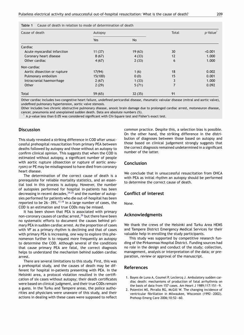
Author's personal copy
Pulseless electrical activity and unsuccessful out-of-hospital resuscitation: What is the cause of death? 209
Table 1 Cause of death in relation to mode of determination of death
Cause of death Autopsy Total p-Value*
Yes No
CardiacAcute myocardial infarction 11 (37) 19 (63) 30 <0.001Coronary heart disease 8 (67) 4 (33) 12 1.000Other cardiac 4 (67) 2 (33) 6 1.000
Non-cardiacAortic dissection or rupture 17(94) 1 (6) 18 0.002Pulmonary embolism 15(100) 0 (0) 15 0.001Intracranial haemorrhage 2 (67) 1 (33) 3 1.000Other 2 (29) 5 (71) 7 0.092
Total 59 (65) 32 (35) 91
Other cardiac includes two congestive heart failure, undefined pericardial disease, rheumatic valvular disease (mitral and aortic valve),undefined pulmonary hypertension, aortic valve stenosis.Other includes two chronic obstructive pulmonary disease, anoxic brain damage due to prolonged cardiac arrest, motoneuron disease,cancer, pneumonia and unexplained sudden death. Data are absolute numbers (%).
* A p-value less than 0.05 was considered significant with Chi-Square test and Fisher’s exact test.
Discussion
This study revealed a striking difference in COD after unsuc-cessful prehospital resuscitation from primary PEA betweendeaths followed by autopsy and those without an autopsy toconfirm clinical opinion. This suggests that when the COD isestimated without autopsy, a significant number of peoplewith aortic rupture (dissection or rupture of aortic aneu-rysm) or PE may be misdiagnosed to have died from coronaryheart disease.
The determination of the correct cause of death is aprerequisite for reliable mortality statistics, and an essen-tial tool in this process is autopsy. However, the numberof autopsies performed for hospital in-patients has beendecreasing in recent decades,24,25 and the number of autop-sies performed for patients who die out-of-hospital has beenreported to be 26—39%.17,19 In a large number of cases, theCOD is an estimation and true CODs may be missed.
It has been shown that PEA is associated with primarynon-coronary causes of cardiac arrest,26 but there have beenno systematic efforts to document the causes behind pri-mary PEA in sudden cardiac arrest. As the proportion of caseswith VF as a primary rhythm is declining and that of caseswith primary PEA is increasing, one way to explore this phe-nomenon further is to request more frequently an autopsyto determine the COD. Although several of the conditionsthat cause primary PEA are fatal, the correct diagnosishelps to understand the mechanism behind sudden cardiacarrest.
There are several limitations to this study. First, this wasa prehospital study, and the causes of death may be dif-ferent for hospital in-patients presenting with PEA. In theHelsinki area, a protocol violation resulted in the certifi-cation of six cases without autopsy; their death certificateswere based on clinical judgment, and their true CODs remaina guess. In the Turku and Tampere areas, the police autho-rities and physicians were unaware of this study, and theiractions in dealing with these cases were supposed to reflect
common practice. Despite this, a selection bias is possible.On the other hand, the striking difference in the distri-bution of diagnoses between those based on autopsy andthose based on clinical judgement strongly suggests thatthe correct diagnosis remained undetermined in a significantnumber of the latter.
Conclusion
We conclude that in unsuccessful resuscitation from OHCAwith PEA as initial rhythm an autopsy should be performedto determine the correct cause of death.
Conflict of interest
None.
Acknowledgments
We thank the crews of the Helsinki and Turku Area HEMSand Tampere District Emergency Medical Services for theirvaluable help in enrolling the study participants.
This study was supported by competitive research fun-ding of the Pirkanmaa Hospital District. Funding sources hadno role in the design and conduct of the study; collection,management, analysis or interpretation of the data; or pre-paration, review or approval of the manuscript.
References
1. Bayes de Luna A, Coumel P, Leclercq J. Ambulatory sudden car-diac death: mechanisms of production of fatal arrhythmia onthe basis of data from 157 cases. Am Heart J 1989;117:151—9.
2. Polentini MS, Pirrallo RG, McGill W. The changing incidence ofventricular fibrillation in Milwaukee, Wisconsin (1992—2002).Prehosp Emerg Care 2006;10:52—60.
Author's personal copy
210 I. Virkkunen et al.
3. Weaver W, Cobb L, Hallstrom A, et al. Considerations for impro-ving survival from out-of-hospital cardiac arrest. Ann Emerg Med1986;15:1181—6.
4. Cobb L, Fahrenbruch C, Olsufka M, Copass M. Changing inci-dence of out-of-hospital ventricular fibrillation, 1980—2000.JAMA 2002;288:3008—13.
5. Herlitz J, Andersson E, Bang A, et al. Experiences from treat-ment of out-of-hospital cardiac arrest during 17 years inGoteborg. Eur Heart J 2000;21:1251—8.
6. Kuisma M, Repo J, Alaspaa A. The incidence of out-of-hospitalventricular fibrillation in Helsinki, Finland, from 1994 to 1999.Lancet 2001;358:473—4.
7. Engdahl J, Bang A, Lindqvist J, Herlitz J. Factors affectingshort- and long-term prognosis among 1069 patients with out-of-hospital cardiac arrest and pulseless electrical activity.Resuscitation 2001;51:17—25.
8. Hallstrom A, Rea T, Sayre M, et al. Manual chest compressionvs use of an automated chest compression device during resus-citation following out-of-hospital cardiac arrest: a randomizedtrial. JAMA 2006;295:2620—8.
9. Kette F, Pordenone G. Cardiac Arrest Cooperative Study. Increa-sed survival despite a reduction in out-of-hospital ventricularfibrillation in north-east Italy. Resuscitation 2007;72:52—8.
10. Wik L, Kramer-Johansen J, Myklebust H, et al. Quality of cardio-pulmonary resuscitation during out-of-hospital cardiac arrest.JAMA 2005;293:299—304.
11. Virkkunen I, Kujala S, Ryynanen S, et al. Bystander mouth-to-mouth ventilation and regurgitation during cardiopulmonaryresuscitation. J Intern Med 2006;260:39—42.
12. Gajic O, Festic E, Afessa B. Infectious complications in survivorsof cardiac arrest admitted to the medical intensive care unit.Resuscitation 2004;60:65—9.
13. Nadkarni VM, Larkin GL, Peberdy MA, et al. First documentedrhythm and clinical outcome from in-hospital cardiac arrestamong children and adults. JAMA 2006;295:50—7 [see com-ment].
14. Baum R, Alvarez Hr, Cobb L. Survival after resuscita-tion from out-of-hospital ventricular fibrillation. Circulation1974;50:1231—5.
15. Comess K, DeRook F, Russell M, Tognazzi-Evans T, Beach K.The incidence of pulmonary embolism in unexplained sudden
cardiac arrest with pulseless electrical activity. Am J Med2000;109:351—6.
16. Pirolo J, Hutchins G, Moore G. Electromechanical dissocia-tion: pathologic explanations in 50 patients. Hum Pathol1985;16:485—7.
17. Silfvast T. Cause of death in unsuccessful prehospital resuscita-tion. J Intern Med 1991;229:331—5.
18. Courtney D, Sasser H, Pincus C, Kline J. Pulseless electricalactivity with witnessed arrest as a predictor of sudden deathfrom massive pulmonary embolism in outpatients. Resuscitation2001;49:265—72.
19. Vanbrabant P, Dhondt E, Billen P, Sabbe M. Aetiology of unsuc-cessful prehospital witnessed cardiac arrest of unclear origin.Eur J Emerg Med 2006;13:144—7.
20. de la Grandmaison G, Durigon M. Sudden adult death: a medico-legal series of 77 cases between 1995 and 2000. Med Sci Law2002;42:225—32.
21. Kamarainen A, Virkkunen I, Yli-Hankala A, Silfvast T. Presumedfutility in paramedic-treated out-of-hospital cardiac arrest:an Utstein style analysis in Tampere, Finland. Resuscitation2007;75:235—43.
22. Vaisanen O, Makijarvi M, Lund V, Silfvast T. Arrhrythmiasand haemodynamic effects associated with early versus lateprehospital thrombolysis for acute myocardial infarction. Resus-citation 2004;62:175—80.
23. de Latorre F, Nolan J, Robertson C, Chamberlain D, BaskettP. European Resuscitation Council Guidelines 2000 for AdultAdvanced Life Support. A statement from the Advanced LifeSupport Working Group (1) and approved by the Executive Com-mittee of the European Resuscitation Council. Resuscitation2001;48:211—21.
24. Hull M, Nazarian R, Wheeler A, Black-Schaffer W, Mark E.Resident physician opinions on autopsy importance and procu-rement. Hum Pathol 2007;38:342—50.
25. Marwick C. Pathologists request autopsy revival. JAMA1995;273:1889.
26. Hess E, Campbell R, White R. Epidemiology, trends, and out-come of out-of-hospital cardiac arrest of non-cardiac origin.Resuscitation 2007;72:200—6.
Resuscitation 62 (2004) 299–302
Induction of therapeutic hypothermia after cardiac arrest in prehospitalpatients using ice-cold Ringer’s solution: a pilot study
Ilkka Virkkunena,b,∗, Arvi Yli-Hankalab,c, Tom Silfvasta,d
a Helsinki Area Emergency Medical Air Service, Vantaa, Finlandb Department of Anaesthesia, Tampere University Hospital, POB 2000, FIN-33521 Tampere, Finland
c Department of Anaesthesia, Medical School, University of Tampere, Tampere, Finlandd Department of Anaesthesia and Intensive Care, Helsinki University Hospital, Helsinki, Finland
Received 28 November 2003; received in revised form 29 March 2004; accepted 8 April 2004
Abstract
The cooling and haemodynamic effects of prehospital infusion of ice-cold Ringer’s solution were studied in 13 adult patients after successfulresuscitation from non-traumatic cardiac arrest. After haemodynamics stabilisation, 30 ml/kg of Ringer’s solution was infused at a rate of100 ml/min into the antecubital vein. Arterial blood pressure and blood gases, pulse rate, end-tidal CO2 and oesophageal temperature weremonitored closely. The mean core temperature decreased from 35.8± 0.9◦C at the start of infusion to 34.0± 1.2◦C on arrival at hospital(P < 0.0001). No serious adverse haemodynamic effects occurred. It is concluded that the induction of therapeutic hypothermia using thistechnique in the prehospital setting is feasible.© 2004 Elsevier Ireland Ltd. All rights reserved.
Keywords: Out-of-hospital CPR; Cardiac arrest; Hypothermia; Haemodynamics; Emergency medical services; Fluid therapy
Resumo
Foram estudados os efeitos hemodinamicos e o arrefecimento induzidos pela perfusão pré hospitalar de uma solução de Ringer geladaperfundida a 100 ml/min na veia antecubital. Monitorizaram-se apertadamente a pressão arterial e os gases sangue, a frequencia de pulso,o CO2 no final da expiração e a temperatura esofágica. A temperatura média central diminuiu de 35.8± 0.9◦C no inıcio da perfusão para34.0± 1.2◦C na chegada ao hospital (P < 0.0001). Não ocorreram efeitos hemodinamicos adversos severos. Concluımos que é exequıvel aindução de terapeutica hipotérmica com esta técnica no ambiente pré-hospitalar.© 2004 Elsevier Ireland Ltd. All rights reserved.
Palavras chave: CPR extra-hospitalar; Paragem cardıaca; Hipotermia; Hemodinamica; Serviços de Emergencia Médica; Fluidoterapia
Resumen
Se estudiaron los efectos hemodinámicos y el enfriamiento producidos por la infusión prehospitalaria de solución muy frıa de Ringer, en 13pacientes adultos después de la resucitación exitosa de paro cardıaco no traumático. Después de estabilizar la hemodinamia, se infundieron30 ml/kg de solución de Ringer a una velocidad de 100 ml/min en la vena antecubital. Se monitorearon estrechamente presión arterial y gasesen sangre, frecuencia de pulso, CO2 espiratorio y temperatura esofágica. La temperatura central promedio bajo de 35.8± 0.9◦C al inicio dela infusión a 34.0± 1.2◦C a la llegada al hospital (P < 0.0001). No ocurrieron efectos hemodinámicas adversos severos. Se concluye que esfactible la inducción de hipotermia terapéutica en el ambiente prehospitalario usando esta técnica.© 2004 Elsevier Ireland Ltd. All rights reserved.
Palabras clave: RCP extrahospitalaria; Paro cardıaco; Hipotermia; Hemodinamia; Servicios de emergencias médicas; Terapia de fluidos
∗ Corresponding author. Tel.:+358 3 311611; fax:+358 3 31164363.E-mail address: [email protected] (I. Virkkunen).
1. Introduction
Mild therapeutic hypothermia improves neurological out-come after cardiac arrest[1,2]. It has been suggested that hy-pothermia should be induced as soon as possible after return
0300-9572/$ – see front matter © 2004 Elsevier Ireland Ltd. All rights reserved.doi:10.1016/j.resuscitation.2004.04.003
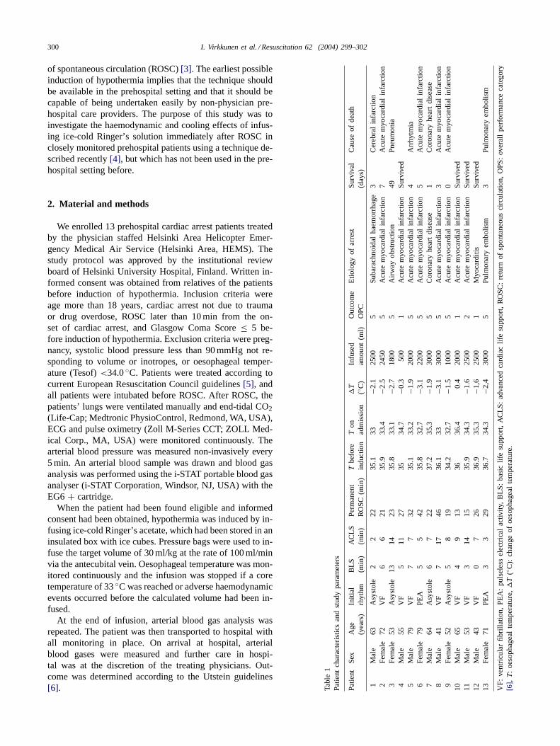
300 I. Virkkunen et al. / Resuscitation 62 (2004) 299–302
of spontaneous circulation (ROSC)[3]. The earliest possibleinduction of hypothermia implies that the technique shouldbe available in the prehospital setting and that it should becapable of being undertaken easily by non-physician pre-hospital care providers. The purpose of this study was toinvestigate the haemodynamic and cooling effects of infus-ing ice-cold Ringer’s solution immediately after ROSC inclosely monitored prehospital patients using a technique de-scribed recently[4], but which has not been used in the pre-hospital setting before.
2. Material and methods
We enrolled 13 prehospital cardiac arrest patients treatedby the physician staffed Helsinki Area Helicopter Emer-gency Medical Air Service (Helsinki Area, HEMS). Thestudy protocol was approved by the institutional reviewboard of Helsinki University Hospital, Finland. Written in-formed consent was obtained from relatives of the patientsbefore induction of hypothermia. Inclusion criteria wereage more than 18 years, cardiac arrest not due to traumaor drug overdose, ROSC later than 10 min from the on-set of cardiac arrest, and Glasgow Coma Score≤ 5 be-fore induction of hypothermia. Exclusion criteria were preg-nancy, systolic blood pressure less than 90 mmHg not re-sponding to volume or inotropes, or oesophageal temper-ature (Tesof)<34.0◦C. Patients were treated according tocurrent European Resuscitation Council guidelines[5], andall patients were intubated before ROSC. After ROSC, thepatients’ lungs were ventilated manually and end-tidal CO2(Life-Cap; Medtronic PhysioControl, Redmond, WA, USA),ECG and pulse oximetry (Zoll M-Series CCT; ZOLL Med-ical Corp., MA, USA) were monitored continuously. Thearterial blood pressure was measured non-invasively every5 min. An arterial blood sample was drawn and blood gasanalysis was performed using the i-STAT portable blood gasanalyser (i-STAT Corporation, Windsor, NJ, USA) with theEG6+ cartridge.
When the patient had been found eligible and informedconsent had been obtained, hypothermia was induced by in-fusing ice-cold Ringer’s acetate, which had been stored in aninsulated box with ice cubes. Pressure bags were used to in-fuse the target volume of 30 ml/kg at the rate of 100 ml/minvia the antecubital vein. Oesophageal temperature was mon-itored continuously and the infusion was stopped if a coretemperature of 33◦C was reached or adverse haemodynamicevents occurred before the calculated volume had been in-fused.
At the end of infusion, arterial blood gas analysis wasrepeated. The patient was then transported to hospital withall monitoring in place. On arrival at hospital, arterialblood gases were measured and further care in hospi-tal was at the discretion of the treating physicians. Out-come was determined according to the Utstein guidelines[6]. Ta
ble
1P
atie
ntch
arac
teris
tics
and
stud
ypa
ram
eter
s
Pat
ient
Sex
Age
(yea
rs)
Initi
alrh
ythm
BLS
(min
)A
CLS
(min
)P
erm
anen
tR
OS
C(m
in)
Tbe
fore
indu
ctio
nT
onad
mis
sion
�T
(◦C
)In
fuse
dam
ount
(ml)
Out
com
eO
PC
Etio
logy
ofar
rest
Sur
viva
l(d
ays)
Cau
seof
deat
h
1M
ale
63A
syst
ole
22
2235
.133
−2.1
2500
5S
ubar
achn
oida
lha
emor
rhag
e3
Cer
ebra
lin
farc
tion
2F
emal
e72
VF
66
2135
.933
.4−2
.524
505
Acu
tem
yoca
rdia
lin
farc
tion
7A
cute
myo
card
ial
infa
rctio
n3
Fem
ale
53A
syst
ole
1314
2335
.833
.1−2
.718
005
Airw
ayob
stru
ctio
n49
Pne
umon
ia4
Mal
e55
VF
511
2735
34.7
−0.3
500
1A
cute
myo
card
ial
infa
rctio
nS
urvi
ved
5M
ale
79V
F7
732
35.1
33.2
−1.9
2000
5A
cute
myo
card
ial
infa
rctio
n4
Arr
hytm
ia6
Fem
ale
79P
EA
55
4235
.832
.7−3
.122
005
Acu
tem
yoca
rdia
lin
farc
tion
5A
cute
myo
card
ial
infa
rctio
n7
Mal
e64
Asy
stol
e6
722
37.2
35.3
−1.9
3000
5C
oron
ary
hear
tdi
seas
e1
Cor
onar
yhe
art
dise
ase
8M
ale
41V
F7
1746
36.1
33−3
.130
005
Acu
tem
yoca
rdia
lin
farc
tion
3A
cute
myo
card
ial
infa
rctio
n9
Fem
ale
52A
syst
ole
58
1934
.232
.7−1
.510
005
Acu
tem
yoca
rdia
lin
farc
tion
0A
cute
myo
card
ial
infa
rctio
n10
Mal
e65
VF
49
1336
36.4
0.4
2000
1A
cute
myo
card
ial
infa
rctio
nS
urvi
ved
11M
ale
53V
F3
1415
35.9
34.3
−1.6
2500
2A
cute
myo
card
ial
infa
rctio
nS
urvi
ved
12M
ale
43V
F0
726
36.9
35.3
−1,6
2500
1M
yoca
rditi
sS
urvi
ved
13F
emal
e71
PE
A3
329
36.7
34.3
−2,4
3000
5P
ulm
onar
yem
bolis
m3
Pul
mon
ary
embo
lism
VF
:ve
ntric
ular
fibril
latio
n,P
EA
:pu
lsel
ess
elec
tric
alac
tivity
,B
LS:
basi
clif
esu
ppor
t,A
CLS
:ad
vanc
edca
rdia
clif
esu
ppor
t,R
OS
C:
retu
rnof
spo
ntan
eous
circ
ulat
ion,
OP
S:
over
all
perf
orm
ance
cate
gory
[6],
T:
oeso
phag
eal
tem
pera
ture
,�
T(◦
C):
chan
geof
oeso
phag
eal
tem
pera
ture
.
I. Virkkunen et al. / Resuscitation 62 (2004) 299–302 301
Fig. 1. The data of resuscitated patients before, during and after ice-coldinfusion of Ringer’s acetate, and on arrival at hospital. Symbols andbars = mean (S.D.). (�) Systolic blood pressure; (�) heart rate; (�)diastolic blood pressure; (�); temperature.∗P < 0.0001, as compared tothe pre-treatment value.
The SPSS 9.01 for PC was used for statistical analyses ofthe data. Analysis of variance andt-tests were used, whereappropriate. AP-value less than 0.05 was considered sig-nificant.
3. Results
Research data and demographics are shown inTable 1.The mean age of eight male and five female patients was60.8 ± 12.5 years. ROSC was achieved at 26± 10 min.Initial cardiac rhythm was ventricular fibrillation (53.8%),asystole (30.8%) or pulseless electrical activity (15.4%).The oesophageal temperature decreased significantly dur-ing treatment (Fig. 1). The mean infused volume was 2188± 754 ml. Infusion started at 27± 12 min after ROSC andmean duration of infusion was 25± 11 min. One patient(patient 2) experienced a transient decrease in blood pres-sure, but no other haemodynamic side effects were observed(Fig. 1). Therapeutic hypothermia was continued in pa-tients 3, 4, 8, 10 and 11 for 24 h after admission to hospitalaccording to the same protocol as in the HACA study[1].
4. Discussion
The results of this pilot study suggest that induction oftherapeutic hypothermia with rapid infusion of ice-coldRinger’s solution soon after ROSC is well tolerated andfeasible in the prehospital setting. This treatment has beendocumented in hospitalised patients starting, on average,
73 min after ROSC[4]. However, the cardiovascular situ-ation might be different immediately after ROSC, and thehaemodynamic effects of induction of cooling by infusionat this stage have not been reported, although recommended[3]. This feasibility study suggests that further evaluationsof this technique seem justified.
We used an oesophageal thermal probe as a fast and sim-ple method for continuous measurement of core tempera-ture. There are, however, weaknesses of its use, which mayexplain the findings in two of our patients. The probe maycome in close contact with the superior vena cava and thusregister falsely low “core” temperatures when blood mixedwith cold saline passes by. This may explain why the tem-perature of patient 4 decreased rapidly after only 500 ml offluid, and why the temperature rose again shortly after theinfusion was stopped. The probe can also inadvertently beadvanced too far, eventually entering the stomach, thus notreflecting correct values (e.g. patient 10 whose temperatureremained unchanged).
The rather low survival rate, although not the study end-point, may be explained by the large number of patientswith known factors for poor outcome[7]. There were fourpatients with non-coronary causes for arrest, and times toROSC were long. Therefore, the infusion of cold saline isunlikely to have contributed to the number of non-survivors.
There are some obvious limitations in our study. First, thesmall number of patients increases the risk of a beta error,but the lack of observed adverse effects of this treatmentrenders further studies warranted. Second, we did not have acontrol group. Third, the measurement of oesophageal tem-perature is prone to errors, but the decrease in temperaturewas continuous and relevant in the majority of patients.
5. Conclusion
We conclude that induction of therapeutic hypothermiawith peripheral infusion of ice-cold Ringer’s solution seemsfeasible soon after ROSC in the prehospital setting. Becausethis technique is also readily available to non-physician careproviders, the induction of therapeutic hypothermia can bestarted earlier if found safe and well tolerated in larger trials.
Acknowledgements
We would like to thank the crew of the Helsinki AreaHEMS for the valuable help in enrolling the study patients.This study was supported by institutional EVO grant #9B102from Special State Allocation via Tampere University Hos-pital.
References
[1] The Hypothermia after Cardiac Arrest Study G. Mild therapeutichypothermia to improve the neurologic outcome after cardiac arrest.N Engl J Med 2002;346:549–56.

302 I. Virkkunen et al. / Resuscitation 62 (2004) 299–302
[2] Bernard SA, Gray TW, Buist MD, et al. Treatment of comatosesurvivors of out-of-hospital cardiac arrest with induced hypothermia.N Engl J Med 2002;346:557–63.
[3] Safar PJ, Kochanek PM. Therapeutic hypothermia after cardiac arrest.N Engl J Med 2002;346:612–3.
[4] Bernard S, Buist M, Monteiro O, Smith K. Induced hypothermiausing large volume, ice-cold intravenous fluid in comatose survivorsof out-of-hospital cardiac arrest: a preliminary report. Resuscitation2003;56:9–13.
[5] de Latorre F, Nolan J, Robertson C, Chamberlain D, Baskett P. Euro-pean resuscitation C. European Resuscitation Council Guidelines 2000
for adult advanced life support. A statement from the Advanced LifeSupport Working Group (1) and approved by the Executive Committeeof the European Resuscitation Council. Resuscitation 2001;48:211–21.
[6] Recommended guidelines for uniform reporting of data fromout-of-hospital cardiac arrest: the ‘Utstein style’. Prepared by aTask Force of Representatives from the European ResuscitationCouncil, American Heart Association, Heart and Stroke Founda-tion of Canada, Australian Resuscitation Council. Resuscitation 1991;22:1–26.
[7] Kuisma M, Alaspää A. Out-of-hospital cardiac arrest of non-cardiacorigin. Epidemiology and outcome. Eur Heart J 1997;18:1122–8.