Embed Size (px)
Citation preview

The Human Body:An Orientation

Anatomy: “to cut apart”; the study of the structure and shape of the body and body parts and their relationships to one another◦ Gross anatomy: study of large, easily observable
structures; ex: heart, bones◦ Microscopic anatomy: study of microscopic or very
small structures in the body; ex: cells, tissues Physiology: “to study the nature of”; the study
of how the body and its parts work or function◦ Usually subdivided: neuro-physiology or cardiac
physiology
ALWAYS RELATED!
An Overview

Atoms – building blocks of matter Molecules – water, sugar, proteins, etc. Cells – smallest unit of all living things Tissues – group of similar cells that have a
common function◦ Epithelial, connective, muscular, and neural
Organ – structure that is composed of two or more tissue types; ex: heart, kidney
Organ system – group of organs that cooperate to accomplish a common purpose; ex: digestive system
Organism – living body of organ systems
Levels of Structural Organization
Organ System Overview

External covering of the body, or the skin Functions:
◦ Waterproofs◦ Cushions◦ Protects from injury◦ Excretes salts and urea as perspiration◦ Helps regulate body temperature◦ Sensory receptor
Integumentary System
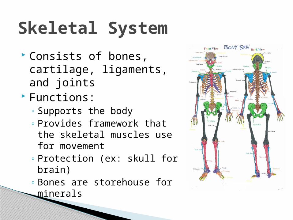
Consists of bones, cartilage, ligaments, and joints
Functions:◦ Supports the body◦ Provides framework that the
skeletal muscles use for movement
◦ Protection (ex: skull for brain)◦ Bones are storehouse for
minerals
Skeletal System

One function: CONTRACTION, or shorten
Skeletal muscles: large, fleshy muscles attached to bones (make up the muscular system)◦ Enable standing, walking,
grasping, etc. Distinct from the muscles of
the heart and other hollow organs, which move fluids and other substances along definite pathways within the body.
Muscular System

The body’s fast-acting control center Consists of the brain, spinal cord, nerves,
and sensory receptors Responds to stimuli or irritants
◦ Outside: light, sound, changes in temperature◦ Inside: decrease in oxygen or stretching of
tissue Send messages, via electrical signals
called nerve impulses, to the central nervous system
Message is interpreted and assessed to activate the muscles or glands for response
Nervous System

Controls body activities through hormones
Glands include the pituitary, thyroid, parathyroids, adrenals, thymus, pancreas, pineal, ovaries (in female) and testes ( in male)
Secretes hormones which regulate other structures
Functions controlled include growth, reproduction, metabolism or nutrient use
Endocrine System

Also called circulatory system Consists of heart and blood
vessels Blood is transporting fluid Carries oxygen, nutrients,
hormones, etc. Exchanges made in tissue cells White blood cells and
chemicals in blood help protect body from foreign invaders
Heart acts as pump
Cardiovascular System

Includes lymphatic vessels, lymph nodes, and other lymphoid organs such as spleen and tonsils
Vessels return fluid leaked from the blood to the blood vessels
Lymph nodes and others cleanse the blood and house the cells involved in immunity
Lymphatic System

Keeps the body constantly supplied with oxygen and removes carbon dioxide
Consists of nasal passages, pharynx, larynx, trachea, bronchi, and lungs
Air sacs in lungs are the site of gas exchange with the blood
Respiratory System

Tube running through body from mouth to anus
Consists of mouth, esophagus, stomach, small and large intestines, and rectum
Functions:◦ Break down food◦ Deliver nutrients to blood for
transport Undigested food leaves as
feces through anus Liver and pancreas are also
considered digestive organs
Digestive System

Removes the nitrogen-containing wastes from the blood and flushes them out as urine
Composed of the kidneys, ureters, bladder, and urethra
Also maintains the body’s water and salt (electrolyte) balance and regulates acid-base balance of blood
Urinary or Excretory System

Primary function is to produce offspring
Sperm are produced in testes of male
Other male reproductive structures: scrotum, penis, accessory glands and the duct system
Eggs (ova) produced in ovaries of female
Other female reproductive structures: uterine tubes, uterus, and vagina
Uterus is site of development for fetus once fertilization has occured
Reproductive System

Organ systems do not work in isolation; instead, they work together to promote the well-being of the entire body.
Maintaining Life

Each contributes to the 8 necessary life functions:1) Maintaining boundaries: “inside” remains distinct
from “outside”; membranes, skin2) Movement: all the activities promoted by muscular
system3) Responsiveness: also called irritability; ability to
sense changes in environment; nervous system4) Digestion: process of breaking down ingested food
into simple molecules; digestive system and cardiovascular system
Maintaining Life

5) Metabolism: all chemical reactions that occur within the body cells; digestive, respiratory, cardiovascular, and endocrine systems
6) Excretion: removing excreta, or wastes, from the body; digestive, urinary, and lymphatic systems
7) Reproduction: production of offspring, and can happen at the cellular level or organismal level; reproductive system and endocrine system
8) Growth: increase in size or number of cells; reproductive and endocrine systems
Maintaining Life contd.

In order for life to be maintained several factors must be available1. Nutrients: obtained through diet; contain the chemicals
used for energy and cell building; ex: carbohydrates, proteins, fats, minerals and vitamins
2. Oxygen: chemical reactions that release energy from food require oxygen; 20% of air we breathe is oxygen
3. Water: 60 – 80% of body weight; single most abundant chemical substance in the body; provides fluid base for all secretions and excretions
4. Body Temperature: must be maintained at 37ºC (98ºF); chemical reactions depend on it
5. Atmospheric Pressure: force exerted on the surface of the body by the weight of air
Survival Needs

The body’s ability to maintain relatively stable internal conditions even though the outside world is continuously changing
A dynamic state of equilibrium, or a balance in which internal conditions change and vary but always within relatively narrow limits
Vital for proper body system function All communication for homeostasis done
through nervous and endocrine systems
Homeostasis

Three components:◦ Receptor: type of sensor that monitors and
responds to changes in the environment, called stimuli, and sends info to control center
◦ Control center: analyzes incoming data from receptor and determines appropriate response or course of action
◦ Effector: receives response from control center and provides response to stimulus
Two pathways:◦ Afferent: receptor to control center (approaching)◦ Efferent: control center to effector (exiting)
Control Mechanisms

Results of response then feedback to influence the stimulus, either by depressing it (negative feedback) or enhancing it (positive feedback)
Most are negative Negative feedback mechanisms are meant to
shut off original stimulus or reduce intensity; ex: home heating system w/thermostat◦ Body temp. regulation, heart rate, blood pressure,
breathing rate, blood levels of glucose, oxygen, carbon dioxide, and minerals
Feedback Systems

Positive feedback systems tend to increase the original disturbance (stimulus) and to push the variable farther from its original value
Rare in the body Control infrequent events that occur
explosively and do not require continuous adjustments
Ex: blood clotting and the birth of a baby
Feedback Systems

The Language of Anatomy
Directional terms

Initial reference point Body erect with feet parallel and the arms
hanging at the sides with the palms facing forward
Anatomical Position

Anatomical Position
Body erect Feet slightly apart Palms facing forward Thumbs point away
from body
Figure 1.7a

Directional Terms Superior and inferior – toward and away
from the head, respectively Anterior and posterior – toward the front
and back of the body Medial, lateral, and intermediate –
toward the midline, away from the midline, and between a more medial and lateral structure
Directional Terms
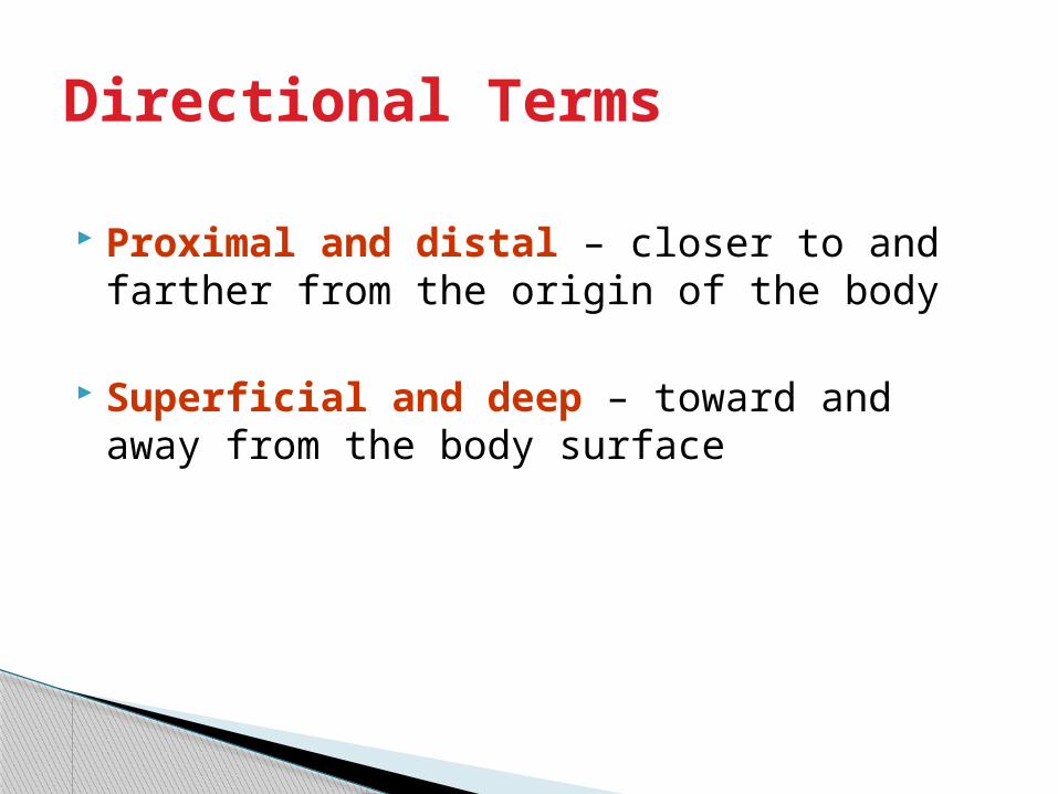
Proximal and distal – closer to and farther from the origin of the body
Superficial and deep – toward and away from the body surface

Directional Terms Table 1.1

Directional Terms Table 1.1
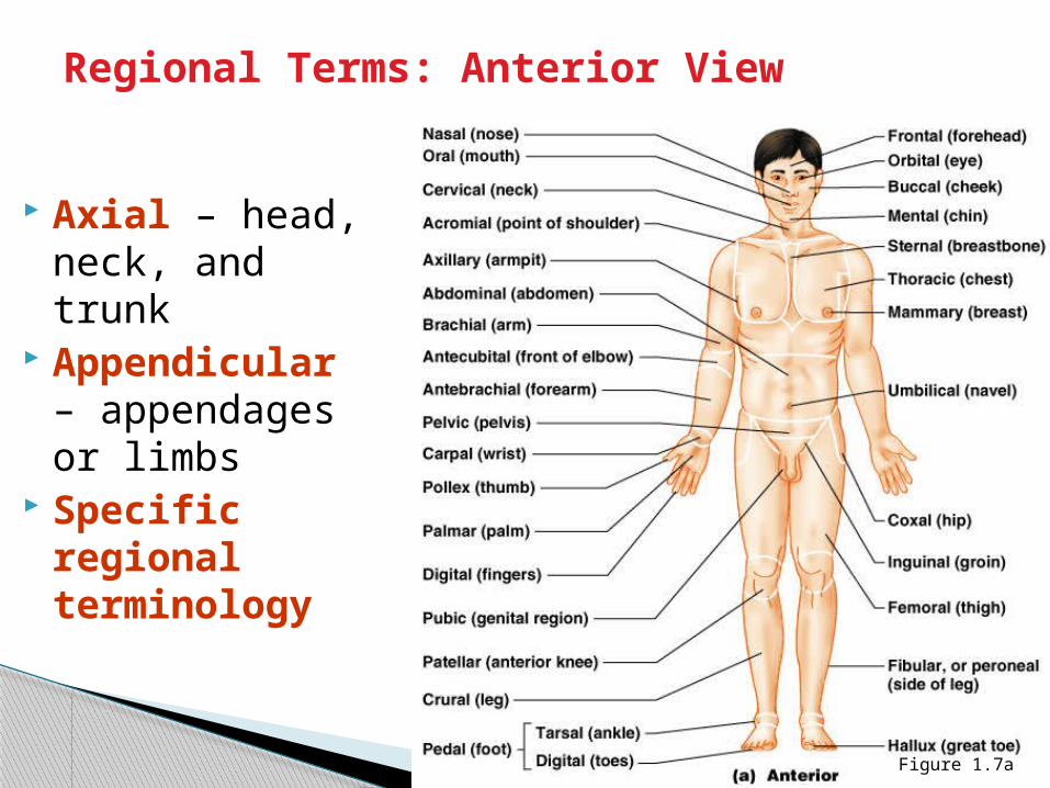
Regional Terms: Anterior View
Axial – head, neck, and trunk
Appendicular – appendages or limbs
Specific regional terminology
Figure 1.7a

Regional Terms: Posterior View
Figure 1.7b
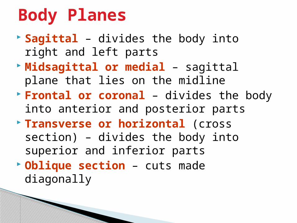
Body Planes Sagittal – divides the body into right and
left parts Midsagittal or medial – sagittal plane that
lies on the midline Frontal or coronal – divides the body into
anterior and posterior parts Transverse or horizontal (cross section) –
divides the body into superior and inferior parts
Oblique section – cuts made diagonally

Body Planes Figure 1.8

Body Cavities Dorsal cavity protects the nervous system,
and is divided into two subdivisions◦Cranial cavity is within the skull and encases the brain
◦Vertebral cavity runs within the vertebral column and encases the spinal cord
Ventral cavity houses the internal organs (viscera), and is divided into two subdivisions: - Thoracic and Abdominopelvic cavities

Body Cavities
Figure 1.9a

Body CavitiesFigure 1.9b
Body Cavities
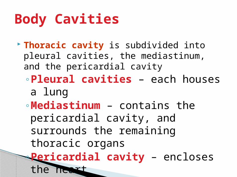
Thoracic cavity is subdivided into pleural cavities, the mediastinum, and the pericardial cavity◦Pleural cavities – each houses a lung
◦Mediastinum – contains the pericardial cavity, and surrounds the remaining thoracic organs
◦Pericardial cavity – encloses the heart

Body Cavities The abdominopelvic cavity is separated
from the superior thoracic cavity by the dome-shaped diaphragm
It is composed of two subdivisions◦Abdominal cavity – contains the stomach, intestines, spleen, liver, and other organs
◦Pelvic cavity – lies within the pelvis and contains the bladder, reproductive organs, and rectum

Ventral Body Cavity Membranes Parietal serosa lines internal body walls
Visceral serosa covers the internal organs
Serous fluid separates the serosae

Ventral Body Cavity Membranes
Figure 1.10a

Ventral Body Cavity Membranes
Figure 1.10b

Other Body Cavities Oral and digestive – mouth and cavities of
the digestive organs Nasal –located within and posterior to the
nose Orbital – house the eyes Middle ear – contain bones (ossicles) that
transmit sound vibrations Synovial – joint cavities

Abdominopelvic Regions
Umbilical Epigastric Hypogastric Right and left iliac
or inguinal Right and left
lumbar Right and left
hypochondriac
Figure 1.11a

Organs of the Abdominopelvic Regions
Figure 1.11b

Abdominopelvic Quadrants
Right upper (RUQ) Left upper (LUQ) Right lower (RLQ) Left lower (LLQ)
Figure 1.12