Embed Size (px)
Citation preview
DEEP VEIN THROMBOSIS
Definition
Deep vein thrombosis is a condition by which blood changes from liquid to solid state and produces a blood clot (thrombus) within the deep venous system typically in the lower limbs
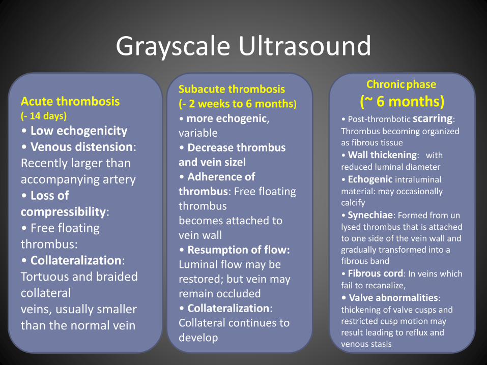
Grayscale Ultrasound
Acute thrombosis (- 14 days)
• Low echogenicity• Venous distension: Recently larger than accompanying artery• Loss of compressibility:• Free floating thrombus:• Collateralization: Tortuous and braided collateralveins, usually smaller than the normal vein
Subacute thrombosis (- 2 weeks to 6 months)
• more echogenic, variable• Decrease thrombus and vein sizel• Adherence of thrombus: Free floating thrombusbecomes attached to vein wall• Resumption of flow: Luminal flow may berestored; but vein may remain occluded• Collateralization: Collateral continues to develop
Chronic phase
(~ 6 months)• Post-thrombotic scarring:
Thrombus becoming organized as fibrous tissue
• Wall thickening: with reduced luminal diameter
• Echogenic intraluminalmaterial: may occasionally calcify
• Synechiae: Formed from un lysed thrombus that is attached to one side of the vein wall and gradually transformed into a fibrous band
• Fibrous cord: In veins which fail to recanalize,
• Valve abnormalities: thickening of valve cusps and restricted cusp motion may result leading to reflux and venous stasis
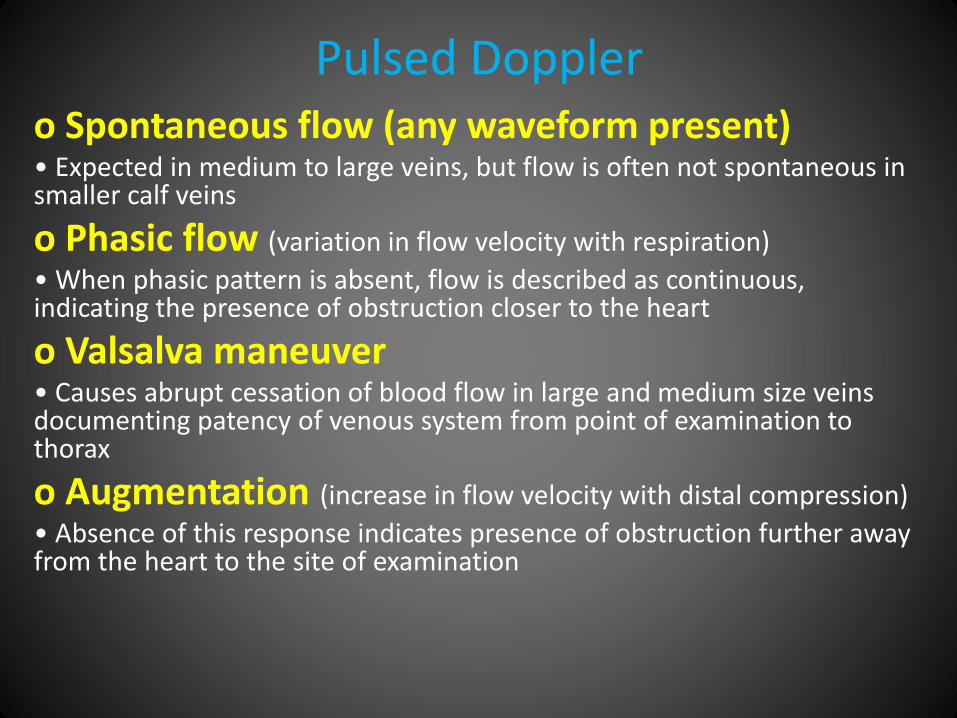
Pulsed Dopplero Spontaneous flow (any waveform present)• Expected in medium to large veins, but flow is often not spontaneous in smaller calf veins
o Phasic flow (variation in flow velocity with respiration)
• When phasic pattern is absent, flow is described as continuous, indicating the presence of obstruction closer to the heart
o Valsalva maneuver• Causes abrupt cessation of blood flow in large and medium size veins documenting patency of venous system from point of examination to thorax
o Augmentation (increase in flow velocity with distal compression)
• Absence of this response indicates presence of obstruction further away from the heart to the site of examination
Color Doppler
- Useful to detect low echo or anechoic thrombus which may be missed on grayscale US
- Demonstration of recanalized lumen in the thrombus and collateralization
- Demonstration of reflux in valvular incompetence
• Power Doppler: Particularly useful in the
demonstration of slow flow through recanalized
lumen and collaterals
Imaging Recommendations
• Best imaging toolo Duplex Doppler ultrasound is first line imaging investigation with sensitivity and specificity for acute symptomatic DVT between 90-100%
o CECT and MR/MR venography are good non-invasive imaging tools for assessment of pelvic veins and IVC and for exclusion of pelvic and abdominal causes of DVT
o Conventional venography has a false negative rate of 11% and should be reserved for use as problem solving aid
DIFFERENTIAL DIAGNOSIS
Interpretation Errors
• Baker cyst, artifactual "echocontrast" from slow flow,
thickened valve mistaken for thrombus in chronic
venous obstruction, failure to identify duplicated vein
Technical Errors
• Inadequate compression, improper use of color flow
image, poor venous distension, misidentification of
deep vs. superficial veins
CLINICAL ISSUES• Most common signs/symptoms
o Acute DVT: Swollen and tender lower limb (extent of swelling depends on site of DVT), increased temperature
o Post thrombotic syndrome: Sequelae of DVT resulting from chronic venous obstruction and/or acquired incompetence of valves
o Chronic leg swelling, ankle pigmentation, and ulceration in the lower calf and ankle (gaiter zone)
• Other signs/symptoms: Signs and symptoms from pulmonary embolism: Shortness of breath, pleuriticchest pain, tachycardia, hypoxia, hypotension
Image Interpretation Pearls
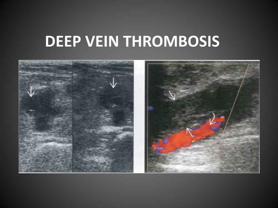
• Thrombus is excluded if the vein is completely compressed
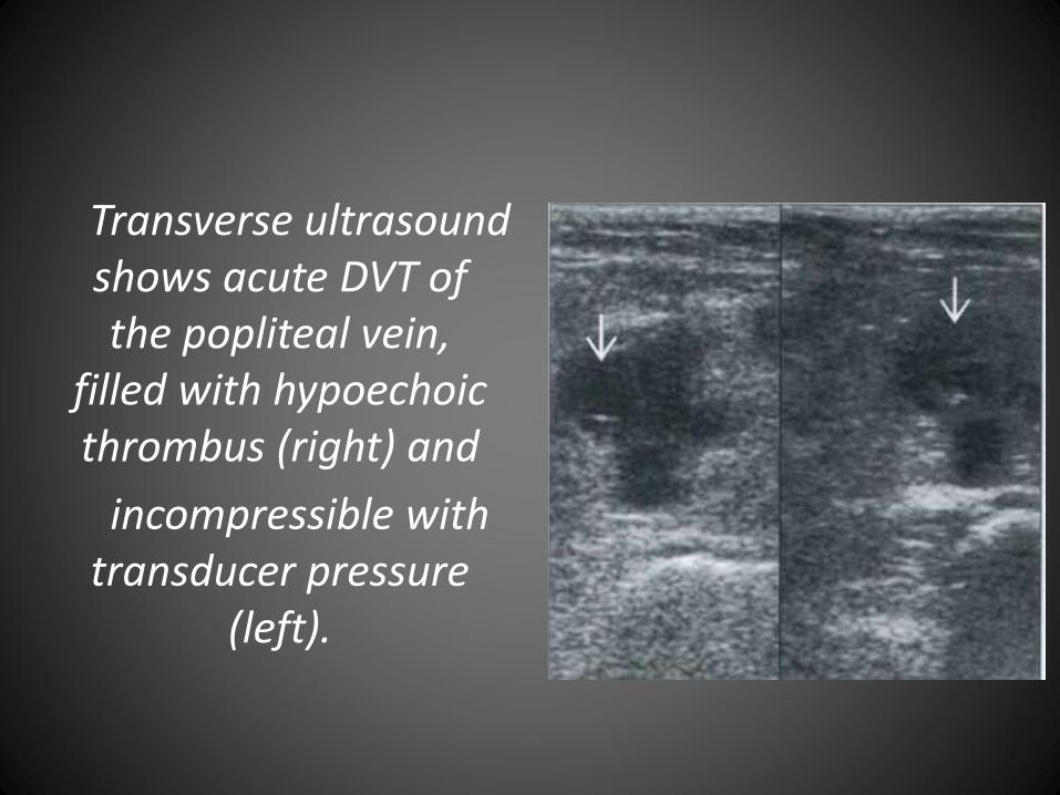
Transverse ultrasound shows acute DVT of the popliteal vein,
filled with hypoechoicthrombus (right) and
incompressible with transducer pressure
(left).
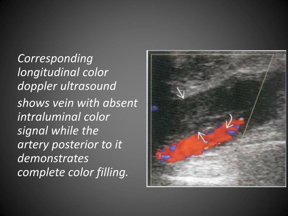
Corresponding longitudinal color doppler ultrasound
shows vein with absent intraluminal color signal while the artery posterior to it demonstrates complete color filling.
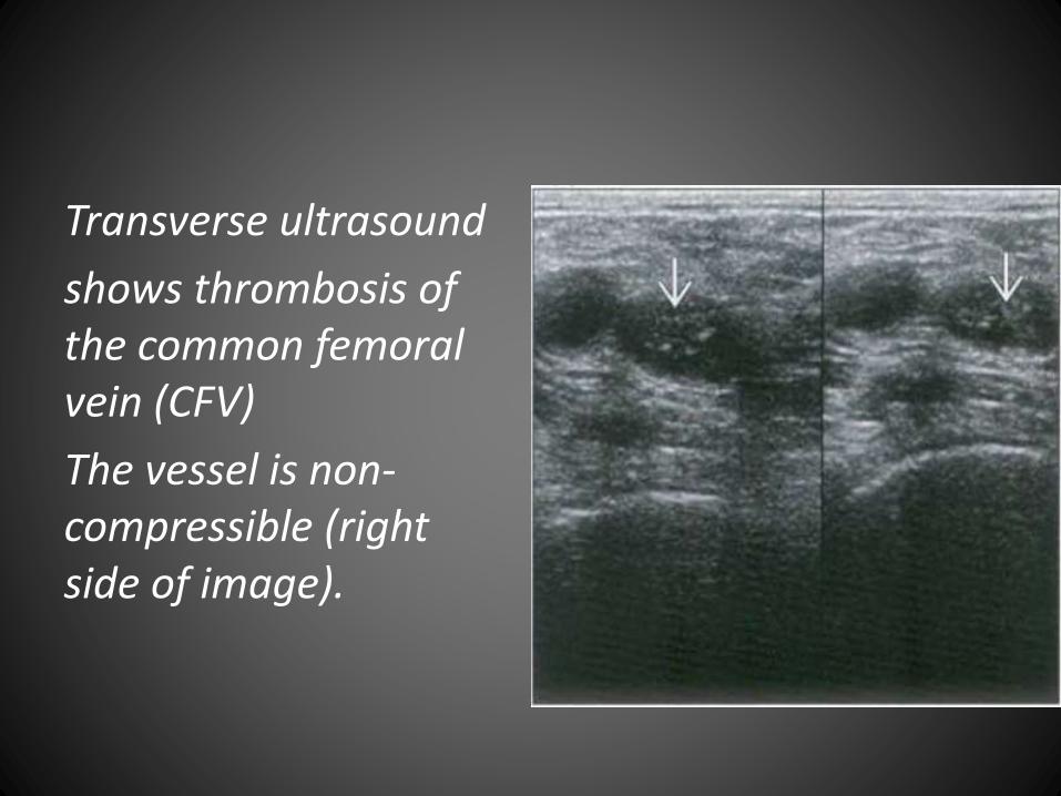
Transverse ultrasound
shows thrombosis of the common femoral vein (CFV)
The vessel is non-compressible (right side of image).
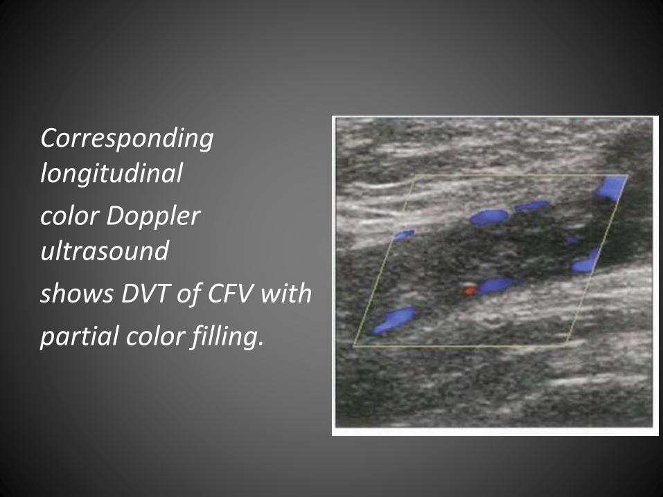
Corresponding longitudinal
color Doppler ultrasound
shows DVT of CFV with
partial color filling.
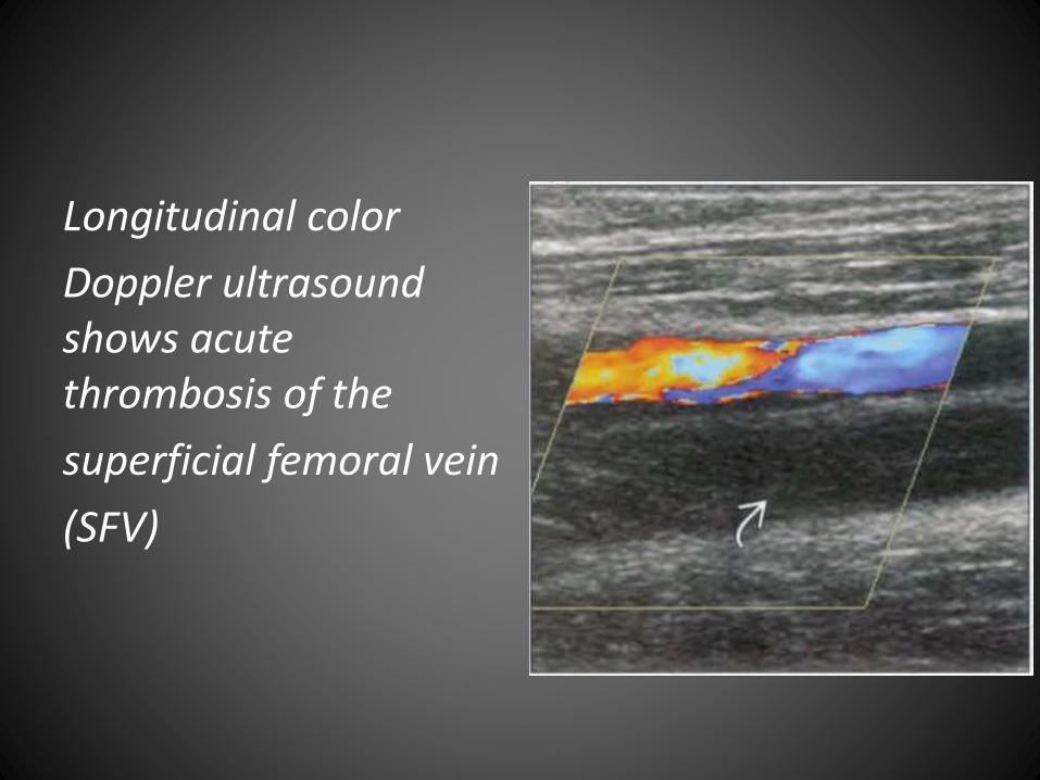
Longitudinal color
Doppler ultrasound shows acute thrombosis of the
superficial femoral vein
(SFV)
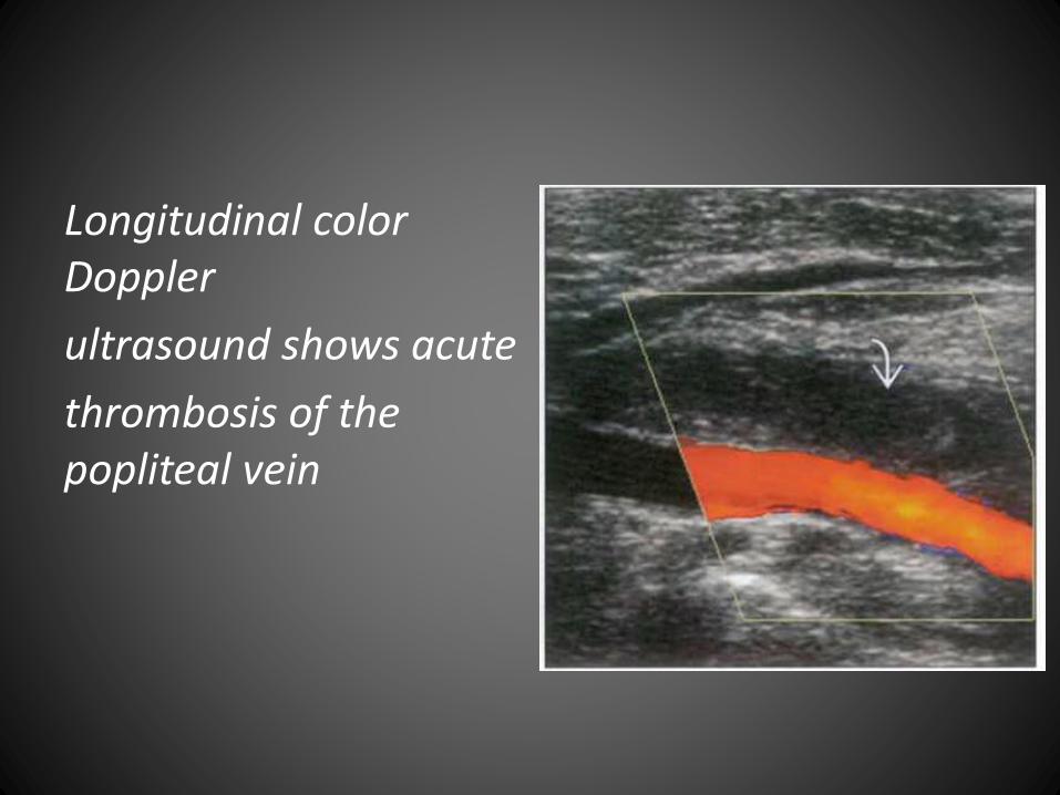
Longitudinal color Doppler
ultrasound shows acute
thrombosis of the popliteal vein
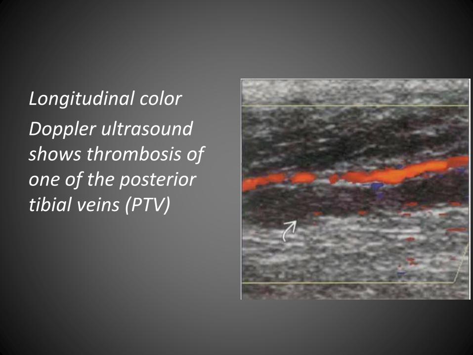
Longitudinal color
Doppler ultrasound shows thrombosis of one of the posterior tibial veins (PTV)
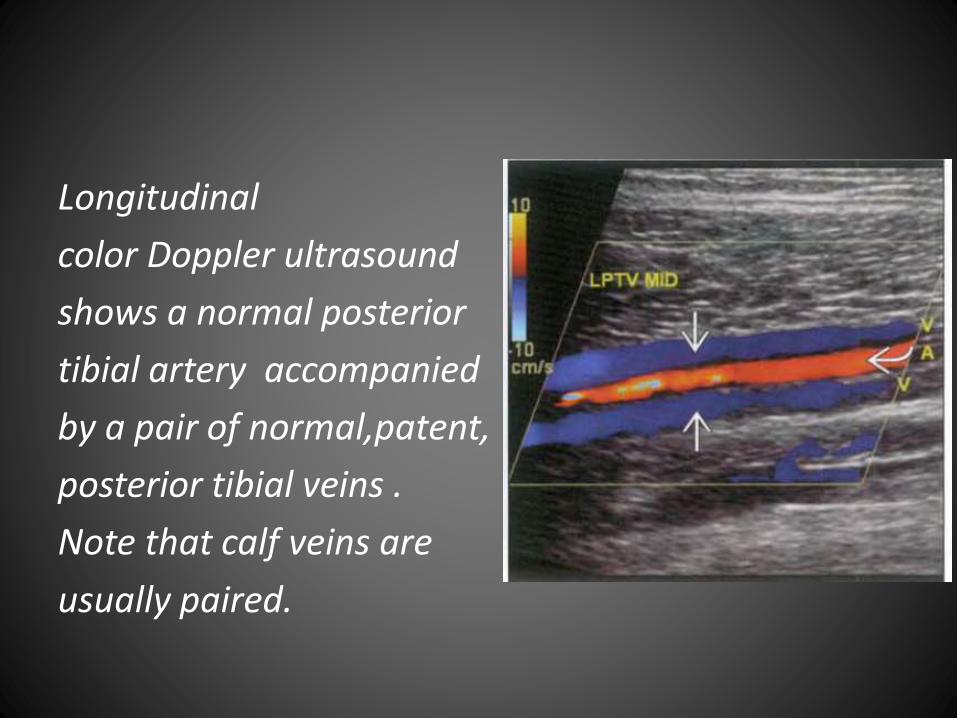
Longitudinal
color Doppler ultrasound
shows a normal posterior
tibial artery accompanied
by a pair of normal,patent,
posterior tibial veins .
Note that calf veins are
usually paired.
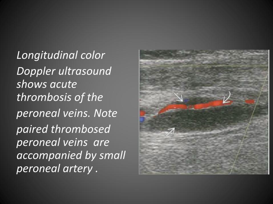
Longitudinal color
Doppler ultrasound shows acute thrombosis of the
peroneal veins. Note
paired thrombosedperoneal veins are accompanied by small peroneal artery .
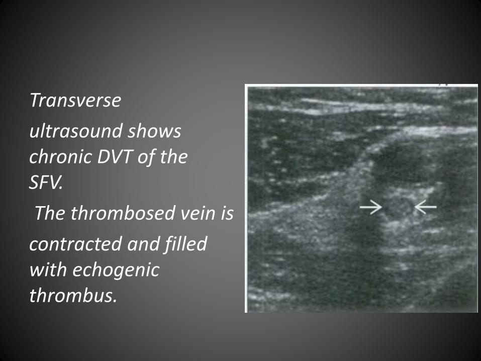
Transverse
ultrasound shows chronic DVT of the SFV.
The thrombosed vein is
contracted and filled with echogenicthrombus.
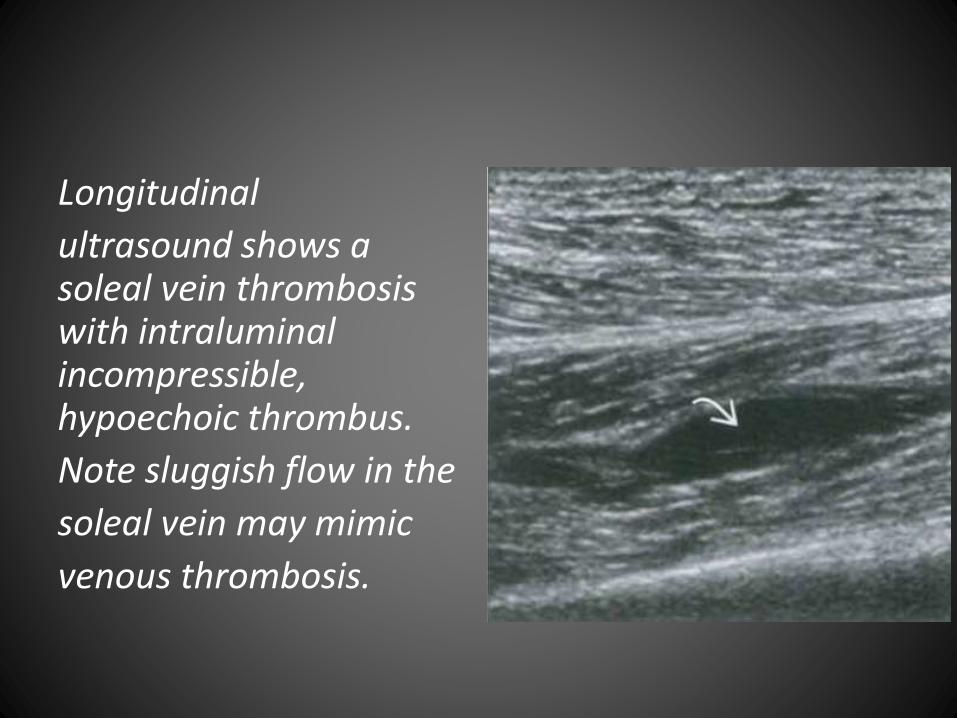
Longitudinal
ultrasound shows a soleal vein thrombosis with intraluminalincompressible, hypoechoic thrombus.
Note sluggish flow in the
soleal vein may mimic
venous thrombosis.
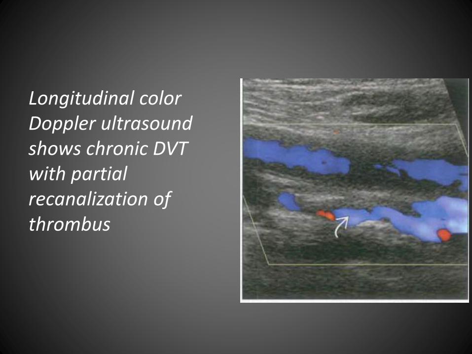
Longitudinal color Doppler ultrasound shows chronic DVT with partial recanalization of thrombus
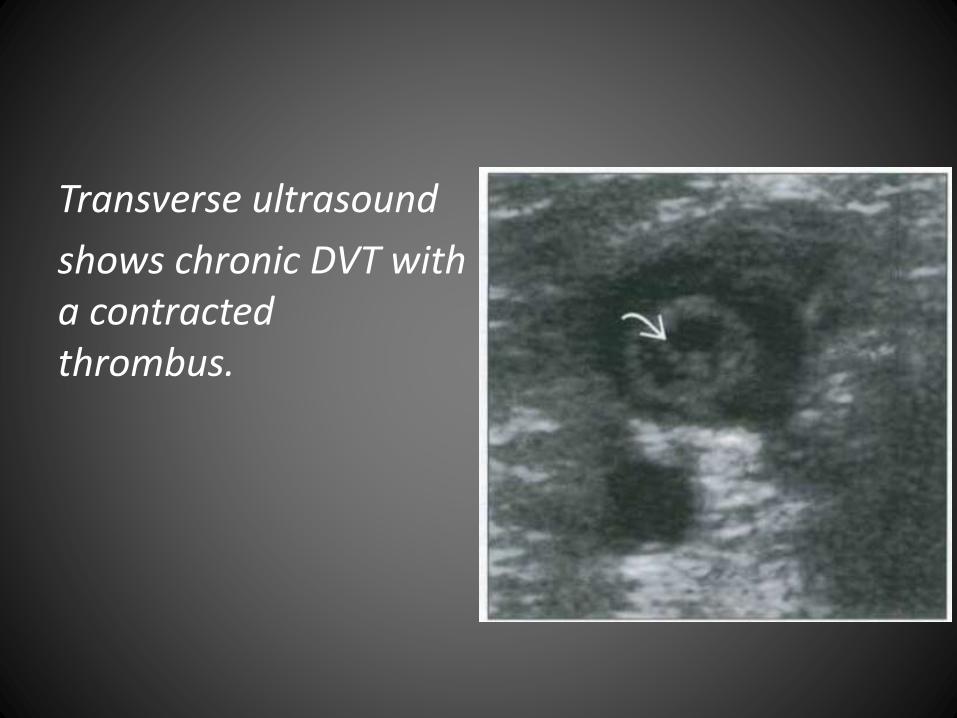
Transverse ultrasound
shows chronic DVT with a contracted thrombus.
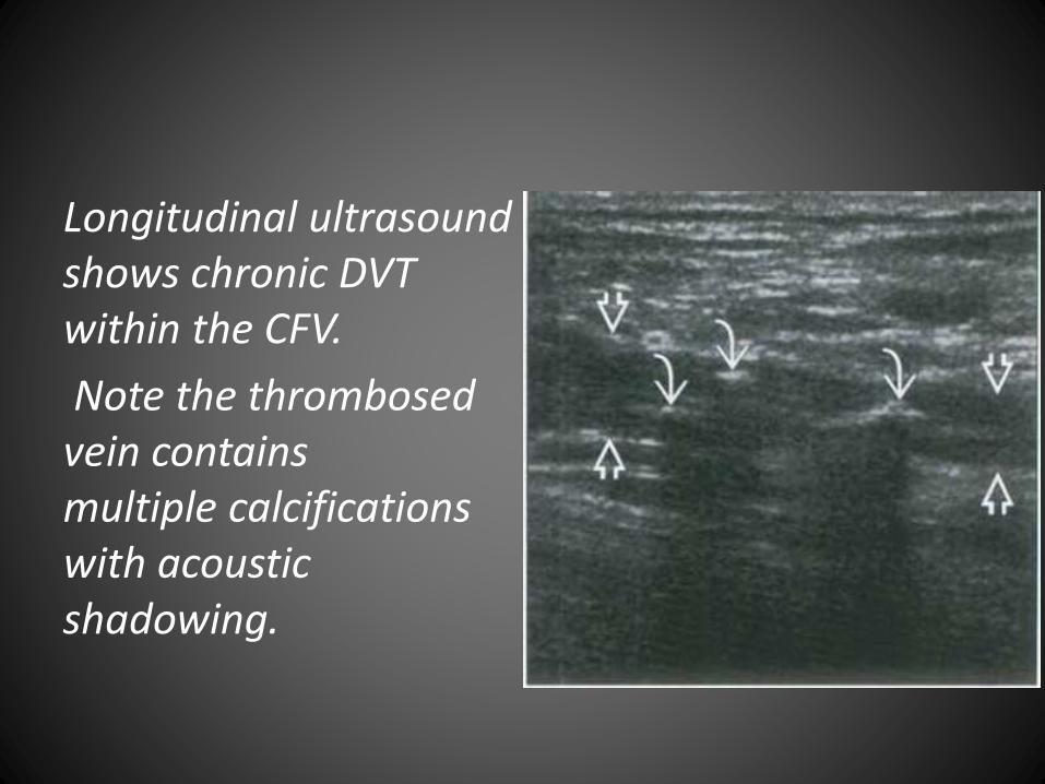
Longitudinal ultrasound shows chronic DVT within the CFV.
Note the thrombosedvein contains multiple calcifications with acoustic shadowing.
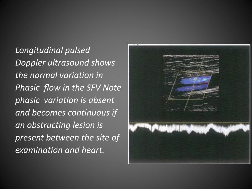
Longitudinal pulsed
Doppler ultrasound shows
the normal variation in
Phasic flow in the SFV Note
phasic variation is absent
and becomes continuous if
an obstructing lesion is
present between the site of
examination and heart.
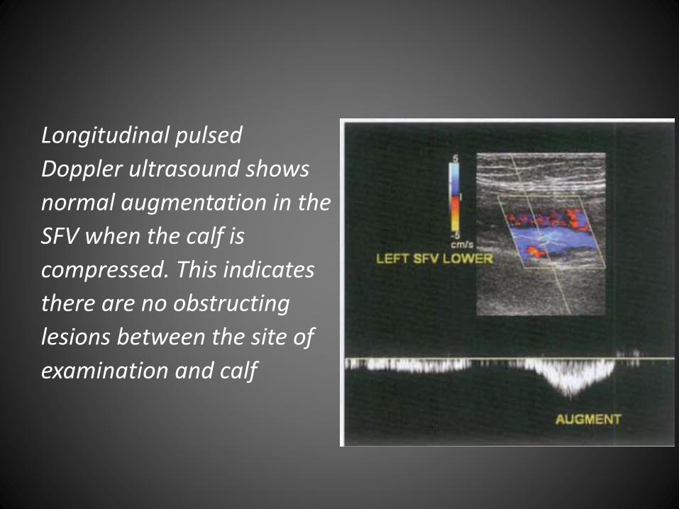
Longitudinal pulsed
Doppler ultrasound shows
normal augmentation in the
SFV when the calf is
compressed. This indicates
there are no obstructing
lesions between the site of
examination and calf
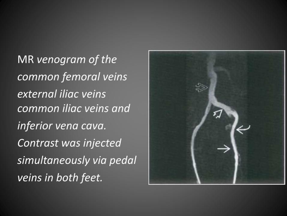
MR venogram of the
common femoral veins
external iliac veins common iliac veins and
inferior vena cava.
Contrast was injected
simultaneously via pedal
veins in both feet.
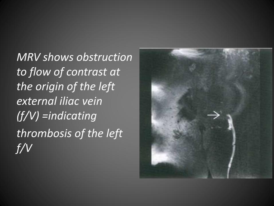
MRV shows obstruction to flow of contrast at the origin of the left external iliac vein (f/V) =indicating
thrombosis of the left f/V
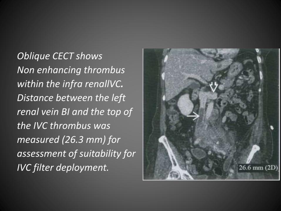
Oblique CECT shows
Non enhancing thrombus
within the infra renallVC.
Distance between the left
renal vein BI and the top of
the IVC thrombus was
measured (26.3 mm) for
assessment of suitability for
IVC filter deployment.
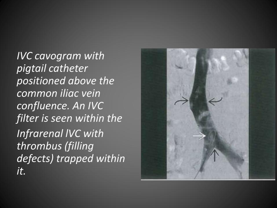
IVC cavogram with pigtail catheter positioned above the common iliac vein confluence. An IVC filter is seen within the
Infrarenal lVC with thrombus (filling defects) trapped within it.