Embed Size (px)
Citation preview
Guidance for the treatment of deep vein thrombosisand pulmonary embolism
Michael B. Streiff1 • Giancarlo Agnelli2 • Jean M. Connors3 • Mark Crowther4 •
Sabine Eichinger5 • Renato Lopes6 • Robert D. McBane7 • Stephan Moll8 •
Jack Ansell9
Published online: 16 January 2016
� The Author(s) 2016. This article is published with open access at Springerlink.com
Abstract This guidance document focuses on the diag-
nosis and treatment of venous thromboembolism (VTE).
Efficient, cost effective diagnosis of VTE is facilitated by
combining medical history and physical examination with
pre-test probability models, D dimer testing and selective
use of confirmatory imaging. Clinical prediction rules,
biomarkers and imaging can be used to tailor therapy to
disease severity. Anticoagulation options for acute VTE
include unfractionated heparin, low molecular weight
heparin, fondaparinux and the direct oral anticoagulants
(DOACs). DOACs are as effective as conventional therapy
with LMWH and vitamin K antagonists. Thrombolytic
therapy is reserved for massive pulmonary embolism (PE)
or extensive deep vein thrombosis (DVT). Inferior vena
cava filters are reserved for patients with acute VTE and
contraindications to anticoagulation. Retrievable filters are
strongly preferred. The possibility of thoracic outlet syn-
drome and May-Thurner syndrome should be considered in
patients with subclavian/axillary and left common iliac
vein DVT, respectively in absence of identifiable triggers.
The optimal duration of therapy is dictated by the presence
of modifiable thrombotic risk factors. Long term antico-
agulation should be considered in patients with unprovoked
VTE as well as persistent prothrombotic risk factors such
as cancer. Short-term therapy is sufficient for most patients
with VTE associated with transient situational triggers such
as major surgery. Biomarkers such as D dimer and risk
assessment models such the Vienna risk prediction model
offer the potential to customize VTE therapy for the indi-
vidual patient. Insufficient data exist to support the inte-
gration of bleeding risk models into duration of therapy
planning.
Keywords Anticoagulant therapy � Venousthromboembolism � Deep vein thrombosis � Pulmonary
embolism � NOACs � DOACs
Introduction
Venous thromboembolism (VTE) which consists princi-
pally of deep vein thrombosis (DVT) and pulmonary
embolism (PE) is a common cause of morbidity and mor-
tality. Consequently, health care providers in all clinical
settings will be faced with managing patients with this
illness. Numerous evidence-based guidelines are available
& Michael B. Streiff
1 Division of Hematology, Department of Medicine and
Pathology, The Johns Hopkins University School of
Medicine, Baltimore, MD, USA
2 Stroke Unit, Department of Internal Medicine, University of
Perugia, Perugia, Italy
3 Hematology Division, Brigham and Women’s Hospital, Dana
Farber Cancer Institute, Harvard Medical School, Boston,
MA, USA
4 Departments of Medicine and Pathology and Molecular
Medicine, McMaster University, Hamilton, Canada
5 Department of Medicine, Medical University of Vienna,
Vienna, Austria
6 Division of Cardiology, Department of Medicine, Duke
University Medical Center, Durham, NC, USA
7 Cardiovascular Division, Department of Medicine, Mayo
Clinic, Rochester, MN, USA
8 Department of Medicine, University of North Carolina
School of Medicine, Chapel Hill, NC, USA
9 Department of Medicine, Hofstra North Shore/LIJ School of
Medicine, Hempstead, NY, USA
123
J Thromb Thrombolysis (2016) 41:32–67
DOI 10.1007/s11239-015-1317-0
to assist providers in clinical decision-making. However,
there are many clinical scenarios where a paucity of data
exist. The purpose of this guidance document is to provide
advice to providers on all aspects of the treatment of VTE
based upon the best available information including situa-
tions where evidence is limited.
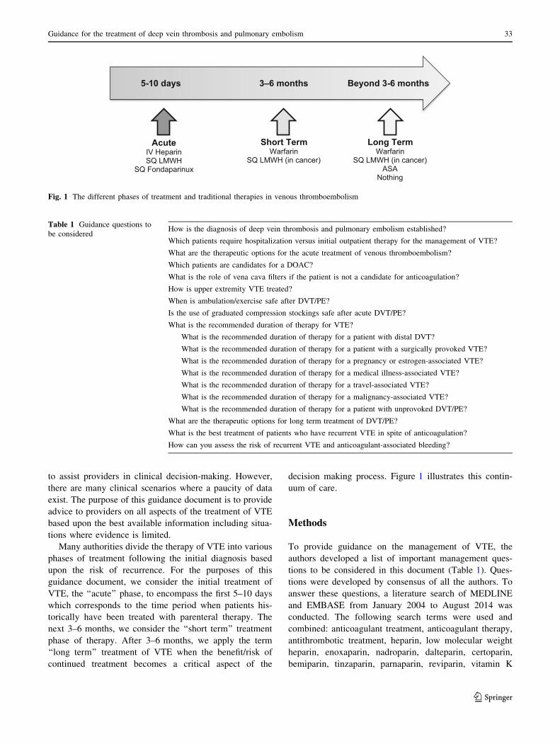
Many authorities divide the therapy of VTE into various
phases of treatment following the initial diagnosis based
upon the risk of recurrence. For the purposes of this
guidance document, we consider the initial treatment of
VTE, the ‘‘acute’’ phase, to encompass the first 5–10 days
which corresponds to the time period when patients his-
torically have been treated with parenteral therapy. The
next 3–6 months, we consider the ‘‘short term’’ treatment
phase of therapy. After 3–6 months, we apply the term
‘‘long term’’ treatment of VTE when the benefit/risk of
continued treatment becomes a critical aspect of the
decision making process. Figure 1 illustrates this contin-
uum of care.
Methods
To provide guidance on the management of VTE, the
authors developed a list of important management ques-
tions to be considered in this document (Table 1). Ques-
tions were developed by consensus of all the authors. To
answer these questions, a literature search of MEDLINE
and EMBASE from January 2004 to August 2014 was
conducted. The following search terms were used and
combined: anticoagulant treatment, anticoagulant therapy,
antithrombotic treatment, heparin, low molecular weight
heparin, enoxaparin, nadroparin, dalteparin, certoparin,
bemiparin, tinzaparin, parnaparin, reviparin, vitamin K
AcuteIV HeparinSQ LMWH
SQ Fondaparinux
Short Term Warfarin
SQ LMWH (in cancer)
5-10 days 3–6 months Beyond 3-6 months
Long TermWarfarin
SQ LMWH (in cancer)ASA
Nothing
Fig. 1 The different phases of treatment and traditional therapies in venous thromboembolism
Table 1 Guidance questions to
be consideredHow is the diagnosis of deep vein thrombosis and pulmonary embolism established?
Which patients require hospitalization versus initial outpatient therapy for the management of VTE?
What are the therapeutic options for the acute treatment of venous thromboembolism?
Which patients are candidates for a DOAC?
What is the role of vena cava filters if the patient is not a candidate for anticoagulation?
How is upper extremity VTE treated?
When is ambulation/exercise safe after DVT/PE?
Is the use of graduated compression stockings safe after acute DVT/PE?
What is the recommended duration of therapy for VTE?
What is the recommended duration of therapy for a patient with distal DVT?
What is the recommended duration of therapy for a patient with a surgically provoked VTE?
What is the recommended duration of therapy for a pregnancy or estrogen-associated VTE?
What is the recommended duration of therapy for a medical illness-associated VTE?
What is the recommended duration of therapy for a travel-associated VTE?
What is the recommended duration of therapy for a malignancy-associated VTE?
What is the recommended duration of therapy for a patient with unprovoked DVT/PE?
What are the therapeutic options for long term treatment of DVT/PE?
What is the best treatment of patients who have recurrent VTE in spite of anticoagulation?
How can you assess the risk of recurrent VTE and anticoagulant-associated bleeding?
Guidance for the treatment of deep vein thrombosis and pulmonary embolism 33
123
antagonists, warfarin, acenocoumarol, phenprocoumon,
thrombolysis, thrombolytic treatment, fibrinolytic agent,
fibrinolysis, urokinase, tenecteplase, alteplase, rtPA, tPA;
aspirin, ticlopidine, clopidogrel; venous thromboembolism,
venous thrombosis, deep venous thrombosis, deep vein
thrombosis, superficial venous thrombosis, superficial
venous thrombophlebitis; diagnosis. The search strategy
was restricted to papers published in English. Detailed
information on the results of the literature search is avail-
able upon request.
For papers published before 2004, we only considered
the most important studies that were likely to influence our
responses to the questions. These studies were selected and
suggested by the authors of this guidance document.
Guidance
(1) How is the diagnosis of deep vein thrombosis and
pulmonary embolism established?
Deep vein thrombosis should be suspected in any patient
who presents with unexplained extremity swelling, pain,
warmth or erythema. Pain associated with DVT is often
described as being a cramp or ache in the calf or thigh.
Pulmonary embolism is often heralded by development of
dyspnea and pleuritic chest or back pain. Pulmonary
embolism can also cause progressive fatigue, dyspnea on
exertion, syncope or pre-syncope or sudden death. Since
these symptoms can be caused by many diseases, the
likelihood of VTE can be estimated by assessing a patient’s
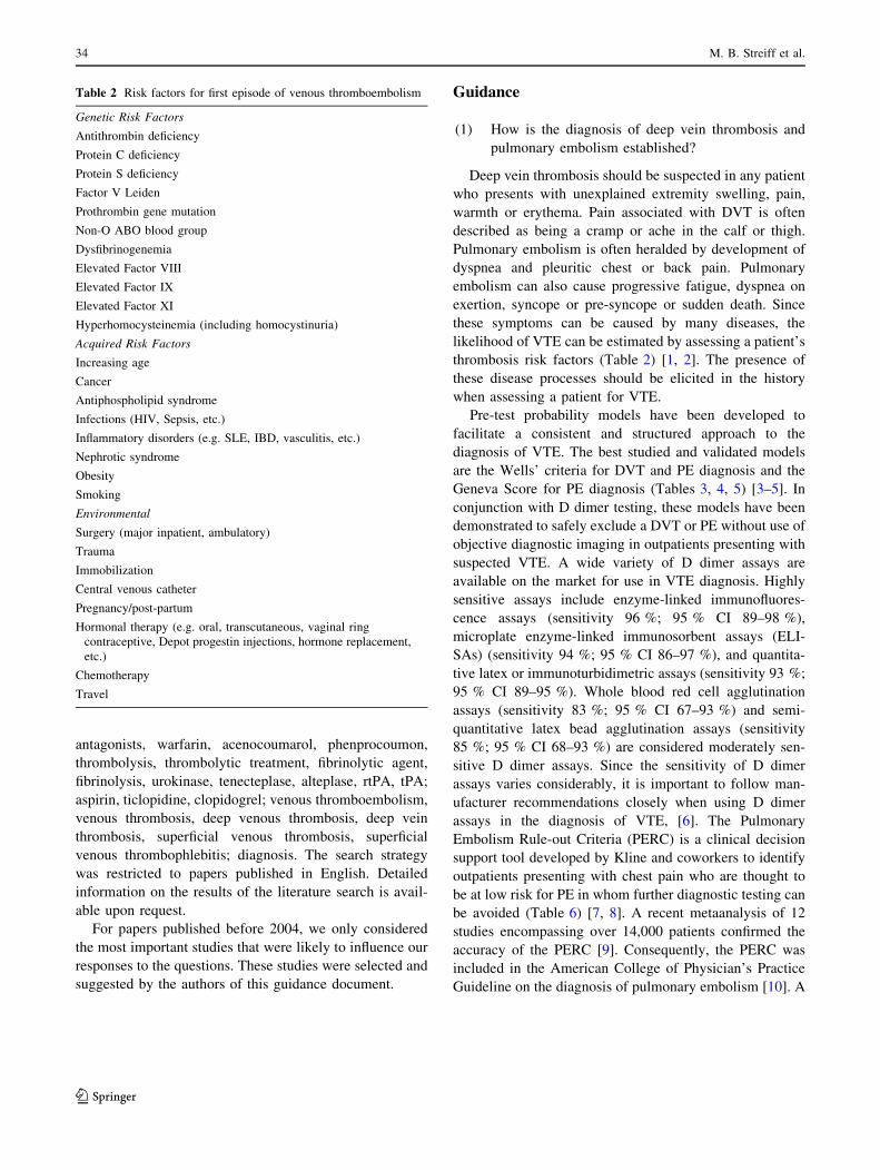
thrombosis risk factors (Table 2) [1, 2]. The presence of
these disease processes should be elicited in the history
when assessing a patient for VTE.
Pre-test probability models have been developed to
facilitate a consistent and structured approach to the
diagnosis of VTE. The best studied and validated models
are the Wells’ criteria for DVT and PE diagnosis and the
Geneva Score for PE diagnosis (Tables 3, 4, 5) [3–5]. In
conjunction with D dimer testing, these models have been
demonstrated to safely exclude a DVT or PE without use of
objective diagnostic imaging in outpatients presenting with
suspected VTE. A wide variety of D dimer assays are
available on the market for use in VTE diagnosis. Highly
sensitive assays include enzyme-linked immunofluores-
cence assays (sensitivity 96 %; 95 % CI 89–98 %),
microplate enzyme-linked immunosorbent assays (ELI-
SAs) (sensitivity 94 %; 95 % CI 86–97 %), and quantita-
tive latex or immunoturbidimetric assays (sensitivity 93 %;
95 % CI 89–95 %). Whole blood red cell agglutination
assays (sensitivity 83 %; 95 % CI 67–93 %) and semi-
quantitative latex bead agglutination assays (sensitivity
85 %; 95 % CI 68–93 %) are considered moderately sen-
sitive D dimer assays. Since the sensitivity of D dimer
assays varies considerably, it is important to follow man-
ufacturer recommendations closely when using D dimer
assays in the diagnosis of VTE, [6]. The Pulmonary
Embolism Rule-out Criteria (PERC) is a clinical decision
support tool developed by Kline and coworkers to identify
outpatients presenting with chest pain who are thought to
be at low risk for PE in whom further diagnostic testing can
be avoided (Table 6) [7, 8]. A recent metaanalysis of 12
studies encompassing over 14,000 patients confirmed the
accuracy of the PERC [9]. Consequently, the PERC was
included in the American College of Physician’s Practice
Guideline on the diagnosis of pulmonary embolism [10]. A
Table 2 Risk factors for first episode of venous thromboembolism
Genetic Risk Factors
Antithrombin deficiency
Protein C deficiency
Protein S deficiency
Factor V Leiden
Prothrombin gene mutation
Non-O ABO blood group
Dysfibrinogenemia
Elevated Factor VIII
Elevated Factor IX
Elevated Factor XI
Hyperhomocysteinemia (including homocystinuria)
Acquired Risk Factors
Increasing age
Cancer
Antiphospholipid syndrome
Infections (HIV, Sepsis, etc.)
Inflammatory disorders (e.g. SLE, IBD, vasculitis, etc.)
Nephrotic syndrome
Obesity
Smoking
Environmental
Surgery (major inpatient, ambulatory)
Trauma
Immobilization
Central venous catheter
Pregnancy/post-partum
Hormonal therapy (e.g. oral, transcutaneous, vaginal ring
contraceptive, Depot progestin injections, hormone replacement,
etc.)
Chemotherapy
Travel
34 M. B. Streiff et al.
123
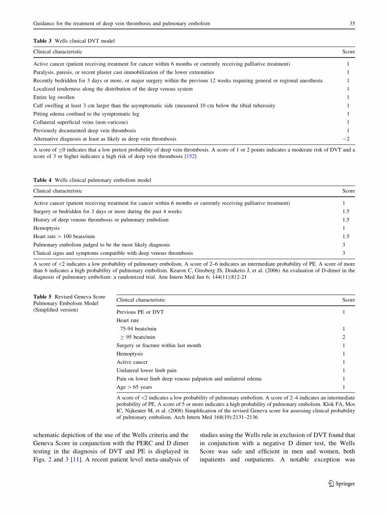
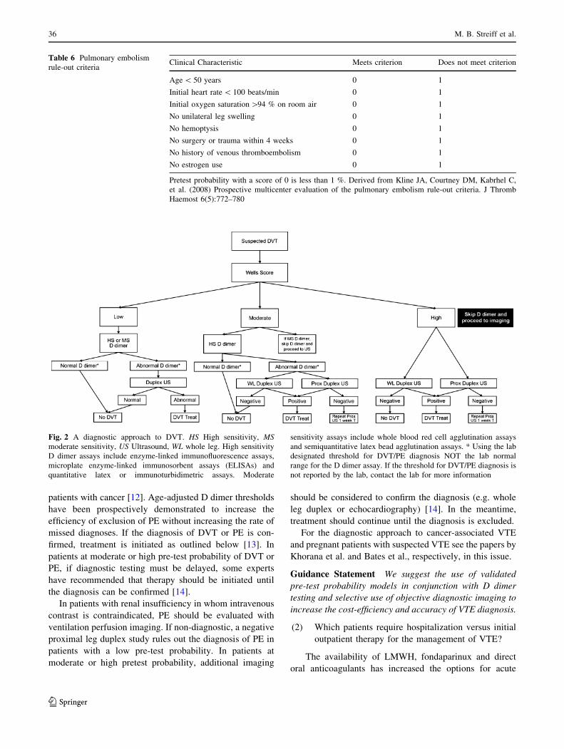
schematic depiction of the use of the Wells criteria and the
Geneva Score in conjunction with the PERC and D dimer
testing in the diagnosis of DVT and PE is displayed in
Figs. 2 and 3 [11]. A recent patient level meta-analysis of
studies using the Wells rule in exclusion of DVT found that
in conjunction with a negative D dimer test, the Wells
Score was safe and efficient in men and women, both
inpatients and outpatients. A notable exception was
Table 3 Wells clinical DVT model
Clinical characteristic Score
Active cancer (patient receiving treatment for cancer within 6 months or currently receiving palliative treatment) 1
Paralysis, paresis, or recent plaster cast immobilization of the lower extremities 1
Recently bedridden for 3 days or more, or major surgery within the previous 12 weeks requiring general or regional anesthesia 1
Localized tenderness along the distribution of the deep venous system 1
Entire leg swollen 1
Calf swelling at least 3 cm larger than the asymptomatic side (measured 10 cm below the tibial tuberosity 1
Pitting edema confined to the symptomatic leg 1
Collateral superficial veins (non-varicose) 1
Previously documented deep vein thrombosis 1
Alternative diagnosis at least as likely as deep vein thrombosis -2
A score of B0 indicates that a low pretest probability of deep vein thrombosis. A score of 1 or 2 points indicates a moderate risk of DVT and a
score of 3 or higher indicates a high risk of deep vein thrombosis [152]
Table 4 Wells clinical pulmonary embolism model
Clinical characteristic Score
Active cancer (patient receiving treatment for cancer within 6 months or currently receiving palliative treatment) 1
Surgery or bedridden for 3 days or more during the past 4 weeks 1.5
History of deep venous thrombosis or pulmonary embolism 1.5
Hemoptysis 1
Heart rate[ 100 beats/min 1.5
Pulmonary embolism judged to be the most likely diagnosis 3
Clinical signs and symptoms compatible with deep venous thrombosis 3
A score of\2 indicates a low probability of pulmonary embolism. A score of 2–6 indicates an intermediate probability of PE. A score of more
than 6 indicates a high probability of pulmonary embolism. Kearon C, Ginsberg JS, Douketis J, et al. (2006) An evaluation of D-dimer in the
diagnosis of pulmonary embolism: a randomized trial. Ann Intern Med Jun 6; 144(11):812-21
Table 5 Revised Geneva Score
Pulmonary Embolism Model
(Simplified version)
Clinical characteristic Score
Previous PE or DVT 1
Heart rate
75-94 beats/min 1
C 95 beats/min 2
Surgery or fracture within last month 1
Hemoptysis 1
Active cancer 1
Unilateral lower limb pain 1
Pain on lower limb deep venous palpation and unilateral edema 1
Age[ 65 years 1
A score of\2 indicates a low probability of pulmonary embolism. A score of 2–4 indicates an intermediate
probability of PE. A score of 5 or more indicates a high probability of pulmonary embolism. Klok FA, Mos
IC, Nijkeuter M, et al. (2008) Simplification of the revised Geneva score for assessing clinical probability
of pulmonary embolism. Arch Intern Med 168(19):2131–2136
Guidance for the treatment of deep vein thrombosis and pulmonary embolism 35
123
patients with cancer [12]. Age-adjusted D dimer thresholds
have been prospectively demonstrated to increase the
efficiency of exclusion of PE without increasing the rate of
missed diagnoses. If the diagnosis of DVT or PE is con-
firmed, treatment is initiated as outlined below [13]. In
patients at moderate or high pre-test probability of DVT or
PE, if diagnostic testing must be delayed, some experts
have recommended that therapy should be initiated until
the diagnosis can be confirmed [14].
In patients with renal insufficiency in whom intravenous
contrast is contraindicated, PE should be evaluated with
ventilation perfusion imaging. If non-diagnostic, a negative
proximal leg duplex study rules out the diagnosis of PE in
patients with a low pre-test probability. In patients at
moderate or high pretest probability, additional imaging
should be considered to confirm the diagnosis (e.g. whole
leg duplex or echocardiography) [14]. In the meantime,
treatment should continue until the diagnosis is excluded.
For the diagnostic approach to cancer-associated VTE
and pregnant patients with suspected VTE see the papers by
Khorana et al. and Bates et al., respectively, in this issue.
Guidance Statement We suggest the use of validated
pre-test probability models in conjunction with D dimer
testing and selective use of objective diagnostic imaging to
increase the cost-efficiency and accuracy of VTE diagnosis.
(2) Which patients require hospitalization versus initial
outpatient therapy for the management of VTE?
The availability of LMWH, fondaparinux and direct
oral anticoagulants has increased the options for acute
Table 6 Pulmonary embolism
rule-out criteriaClinical Characteristic Meets criterion Does not meet criterion
Age\ 50 years 0 1
Initial heart rate\ 100 beats/min 0 1
Initial oxygen saturation[94 % on room air 0 1
No unilateral leg swelling 0 1
No hemoptysis 0 1
No surgery or trauma within 4 weeks 0 1
No history of venous thromboembolism 0 1
No estrogen use 0 1
Pretest probability with a score of 0 is less than 1 %. Derived from Kline JA, Courtney DM, Kabrhel C,
et al. (2008) Prospective multicenter evaluation of the pulmonary embolism rule-out criteria. J Thromb
Haemost 6(5):772–780
Fig. 2 A diagnostic approach to DVT. HS High sensitivity, MS
moderate sensitivity, US Ultrasound, WL whole leg. High sensitivity
D dimer assays include enzyme-linked immunofluorescence assays,
microplate enzyme-linked immunosorbent assays (ELISAs) and
quantitative latex or immunoturbidimetric assays. Moderate
sensitivity assays include whole blood red cell agglutination assays
and semiquantitative latex bead agglutination assays. * Using the lab
designated threshold for DVT/PE diagnosis NOT the lab normal
range for the D dimer assay. If the threshold for DVT/PE diagnosis is
not reported by the lab, contact the lab for more information
36 M. B. Streiff et al.
123
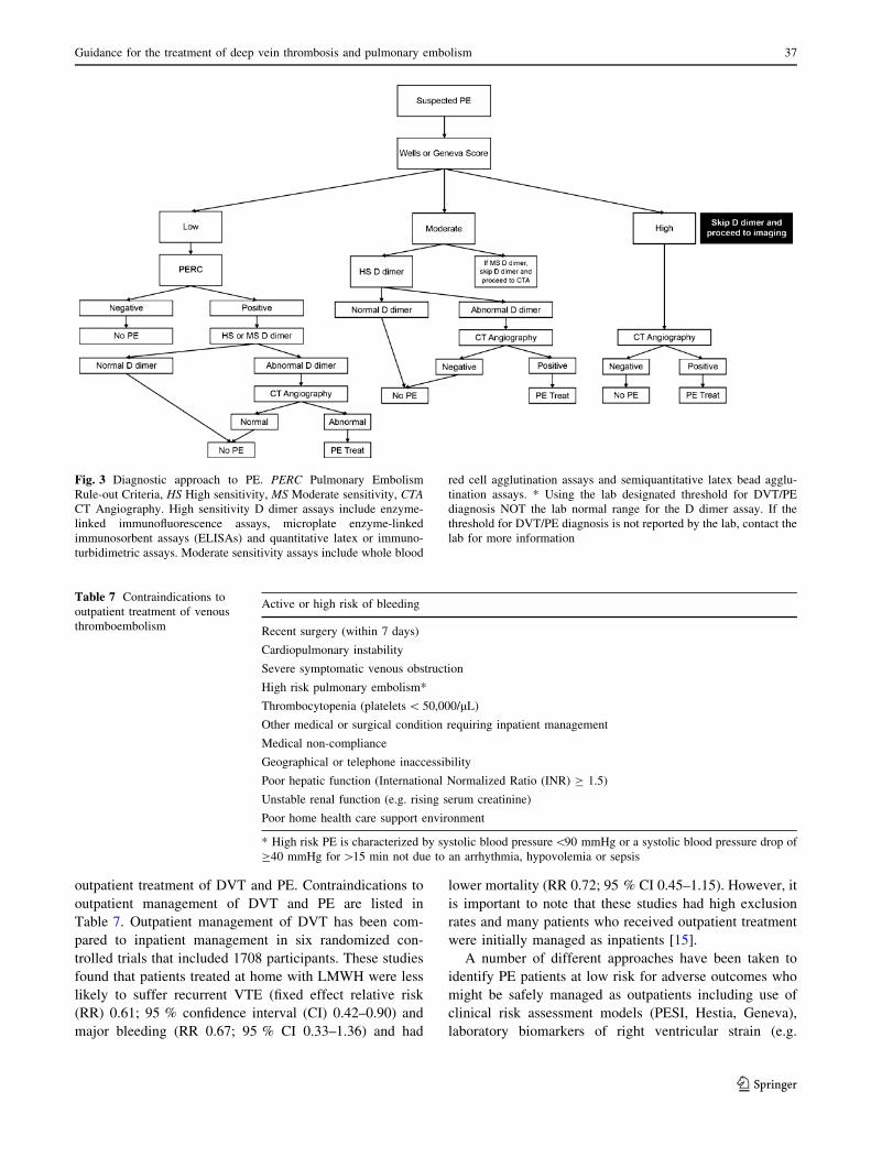
outpatient treatment of DVT and PE. Contraindications to
outpatient management of DVT and PE are listed in
Table 7. Outpatient management of DVT has been com-
pared to inpatient management in six randomized con-
trolled trials that included 1708 participants. These studies
found that patients treated at home with LMWH were less
likely to suffer recurrent VTE (fixed effect relative risk
(RR) 0.61; 95 % confidence interval (CI) 0.42–0.90) and
major bleeding (RR 0.67; 95 % CI 0.33–1.36) and had
lower mortality (RR 0.72; 95 % CI 0.45–1.15). However, it
is important to note that these studies had high exclusion
rates and many patients who received outpatient treatment
were initially managed as inpatients [15].
A number of different approaches have been taken to
identify PE patients at low risk for adverse outcomes who
might be safely managed as outpatients including use of
clinical risk assessment models (PESI, Hestia, Geneva),
laboratory biomarkers of right ventricular strain (e.g.
Fig. 3 Diagnostic approach to PE. PERC Pulmonary Embolism
Rule-out Criteria, HS High sensitivity, MS Moderate sensitivity, CTA
CT Angiography. High sensitivity D dimer assays include enzyme-
linked immunofluorescence assays, microplate enzyme-linked
immunosorbent assays (ELISAs) and quantitative latex or immuno-
turbidimetric assays. Moderate sensitivity assays include whole blood
red cell agglutination assays and semiquantitative latex bead agglu-
tination assays. * Using the lab designated threshold for DVT/PE
diagnosis NOT the lab normal range for the D dimer assay. If the
threshold for DVT/PE diagnosis is not reported by the lab, contact the
lab for more information
Table 7 Contraindications to
outpatient treatment of venous
thromboembolism
Active or high risk of bleeding
Recent surgery (within 7 days)
Cardiopulmonary instability
Severe symptomatic venous obstruction
High risk pulmonary embolism*
Thrombocytopenia (platelets\ 50,000/lL)
Other medical or surgical condition requiring inpatient management
Medical non-compliance
Geographical or telephone inaccessibility
Poor hepatic function (International Normalized Ratio (INR) C 1.5)
Unstable renal function (e.g. rising serum creatinine)
Poor home health care support environment
* High risk PE is characterized by systolic blood pressure\90 mmHg or a systolic blood pressure drop of
C40 mmHg for[15 min not due to an arrhythmia, hypovolemia or sepsis
Guidance for the treatment of deep vein thrombosis and pulmonary embolism 37
123
troponin, NT pro-BNP) and imaging studies (CT or
echocardiogram assessment of right ventricular overload)
[16]. The four chamber cardiac view on chest CT can be
used to identify right ventricular pressure overload. In a
retrospective study of 431 patients with PE, RV enlarge-
ment on CT was an independent predictor of 30 day
mortality (hazard ratio: 5.17;95 % CI 1.63–16.35) [17].
However, a meta-analysis of 10 studies of normotensive PE
patients determined that although CT RVD was associated
with an overall increased risk of death (OR 1.8 95 % CI
1.3–2.6), with death resulting from PE(OR 7.4; 95 % CI
1.4–39.5), and with PE-related complications (OR 2.4;
95 % CI 1.2–4.7), CT only demonstrated modest utility in
assessing risk for adverse outcomes and thus should not be
used in isolation for determining management [18].
Echocardiographic evidence of RV dysfunction has
been identified as an independent predictor of adverse
outcomes. However, a meta-analysis noted that echocar-
diography had an unsatisfactory negative likelihood ratio
for early all-cause mortality (0.62; 95 % CI 0.41–0.92) and
PE-related mortality (0.36; 95 % CI 0.20–0.80). This result
may be due to the lack of standardized echocardiographic
criteria for RV dysfunction and the difficulty inherent in
attempting to differentiate between acute and chronic RV
overload [19]. Therefore, it is currently premature to rely
upon echocardiography to identify low risk patients with
PE.
Several clinical prediction models have been developed
to determine the outcome of patients with acute PE
including the Pulmonary Embolism Severity Index (PESI)
score, the Geneva score and the Hestia criteria (Tables 8, 9,
10). Of these, the PESI score and a simplified version,
sPESI, have been the most extensively validated. In a
multicenter prospective open randomized clinical trial of
inpatient versus outpatient management of low risk PE
patients as determined by the PESI score, Aujesky et al.
found that there was no difference between outpatients and
inpatients in recurrent VTE (1 of 171, 0.6 % vs. 0 of 168;
95 % upper CI limit 2.7 %), major bleeding (3 of 171,
1.8 % vs. 0 of 168, 0 %, 95 % upper CI limit 4.5 %) and
90 day mortality (1 of 171, 0.6 % vs. 1 of 168, 0.6 %;
95 % upper CI limit 2.1 %). These data indicate that out-
patient management of low risk PE patients (as identified
by the PESI score) is feasible and associated with excellent
outcomes [20]. The HESTIA criteria have also been
demonstrated to be useful in identifying patients for out-
patient management [21].
Cardiac biomarkers that are released from myocytes
during right ventricular strain have also proven useful for
identification of PE at risk for adverse outcomes. In a
multicenter prospective study of cardiac biomarkers for
risk stratification of PE, Vuilleumier and colleagues found
that a NT-pro-BNP level\ 300 pg/mL had a negative
predictive value of 100 % (95 % CI 91–100) for adverse
outcomes at 3 months. Troponins have also been identified
as useful biomarkers for risk stratification in PE [22]. High
sensitivity assays for troponin I and T have also been useful
in identification of low risk patients with PE. In a
prospective validation study of 526 normotensive patients
with PE, Lankeit et al. noted that only 4 of 214 (1.9 %)
patients with a high sensitive troponin T\ 14 pg/mL had
adverse outcomes at 30 days. When combined with a
simplified Pulmonary Embolism Severity Index (sPESI)
score of zero, none of 127 patients with this combination
had adverse outcomes [23]. A combination of clinical and
laboratory biomarkers may represent the ideal strategy for
identification of normotensive patients at low risk for
adverse outcomes. Jimenez et al. conducted a multicenter
cohort study of normotensive PE patients to identify a
multi-marker prognostic score for risk stratification. The
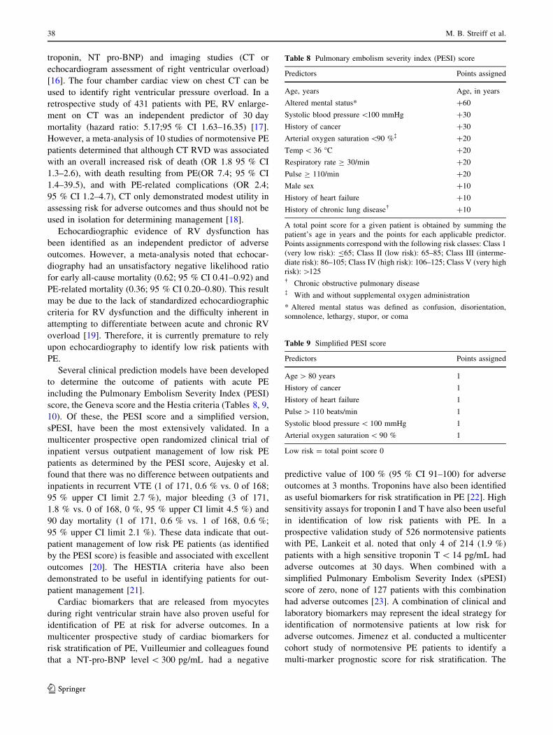
Table 8 Pulmonary embolism severity index (PESI) score
Predictors Points assigned
Age, years Age, in years
Altered mental status* ?60
Systolic blood pressure\100 mmHg ?30
History of cancer ?30
Arterial oxygen saturation\90 %� ?20
Temp\ 36 �C ?20
Respiratory rate C 30/min ?20
Pulse C 110/min ?20
Male sex ?10
History of heart failure ?10
History of chronic lung disease� ?10
A total point score for a given patient is obtained by summing the
patient’s age in years and the points for each applicable predictor.
Points assignments correspond with the following risk classes: Class 1
(very low risk): B65; Class II (low risk): 65–85; Class III (interme-
diate risk): 86–105; Class IV (high risk): 106–125; Class V (very high
risk):[125� Chronic obstructive pulmonary disease� With and without supplemental oxygen administration
* Altered mental status was defined as confusion, disorientation,
somnolence, lethargy, stupor, or coma
Table 9 Simplified PESI score
Predictors Points assigned
Age[ 80 years 1
History of cancer 1
History of heart failure 1
Pulse[ 110 beats/min 1
Systolic blood pressure\ 100 mmHg 1
Arterial oxygen saturation\ 90 % 1
Low risk = total point score 0
38 M. B. Streiff et al.
123
combination of a sPESI and a BNP level\100 pg/mL was
associated with a negative predictive value of 99 and
100 % in the derivation and validation cohorts [24].
A recent systematic review of outpatient treatment of PE
including 11 studies and 1258 patients noted that the rates
of recurrent VTE (1.47 %; 95 % CI 0.47–3.0 %), fatal PE
(0.47 %; 95 % CI 0.16–1.0 %), major bleeding (0.81 %;
95 % CI 0.37–1.42 %) and mortality (1.58 %; 95 % CI
0.71–2.80 %) were low, similar to the rates identified in
inpatient treatment studies. Furthermore, the authors found
that both ‘‘clinical gestalt’’ and standardized risk assess-
ment models appeared to be equally useful in identifying
low risk patients appropriate for outpatient management.
However, they recommended that future studies comparing
formal risk stratification models and ‘‘clinical gestalt’’
should be conducted since there was more heterogeneity in
the studies on clinical gestalt [25].
Management of patients with PE should be guided by an
assessment of their risk for adverse outcomes (Table 11).
Normotensive patients in PESI Class I or II or simplified
PESI Class 0 do not need further risk stratification with
imaging (e.g. echocardiography) and can be considered for
outpatient management. Normotensive patients in PESI
Class C II or simplified PESI C 1 should undergo
additional imaging and laboratory risk assessment and
warrant initial inpatientmanagement until the results of these
studies are complete. Patients in this group who have no sign
of right ventricular dysfunction on echocardiography or
abnormal cardiac biomarkers are considered at low inter-
mediate risk for adverse outcomes. This group of patients can
be considered for early discharge from the hospital. Patients
with abnormal echocardiography or cardiac biomarkers are
consider intermediate-low risk patients and are often man-
aged in the hospital. Patients with abnormal echocardiogra-
phy and cardiac biomarkers are considered at intermediate
high risk of adverse outcomes and are generally managed as
inpatients. Intermediate high risk PE patients are considered
for thrombolytic therapy on a case-by-case basis. PE patients
with hypotension are at high risk for adverse outcomes. They
routinely undergo echocardiography and are strongly con-
sidered for thrombolytic therapy [14]. Further discussion of
PE management can be found in the accompanying paper by
Vedantham et al.
Guidance Statement We suggest that most patients with
DVT and many patients with PE can be managed as out-
patients. PE patients should be risk stratified to determine
appropriate management. A variety of laboratory tests and
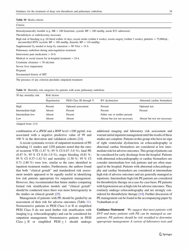
Table 10 Hestia criteria
Criteria
Hemodynamically instable (e.g. HR[ 100 beats/min, systolic BP\ 100 mmHg, needs ICU admission)
Thrombolysis or embolectomy necessary
High risk of bleeding (e.g. GI bleed within 14 days, recent stroke (within 4 weeks), recent surgery (within 2 weeks), platelets\ 75,000/lL,uncontrolled HTN (systolic BP[ 180 mmHg, diastolic BP[ 110 mmHg)
Supplemental O2 needed to keep O2 saturation[ 90 %for[ 24 h
Pulmonary embolism during anticoagulation treatment
Intravenous pain medication[ 24 h
Medical or social reason for in-hospital treatment[ 24 h
Creatinine clearance\ 30 mL/min
Severe liver impairment
Pregnant
Documented history of HIT
The presence of any criterion precludes outpatient treatment
Table 11 Mortality risk categories for patients with acute pulmonary embolism
30 day mortality risk Risk factors
Hypotension PESI Class III through V RV dysfunction Abnormal cardiac biomarkers
High Present Optional assessment Present Optional test
Intermediate-high Absent Present Present Present
Intermediate-low Absent Present Either one or neither present
Low Absent Absent Absent but test not necessary Absent but test not necessary
Adapted from: [14]
Guidance for the treatment of deep vein thrombosis and pulmonary embolism 39
123
imaging modalities as well as clinical risk prediction
models are available to identify PE patients who are
suitable for outpatient management. Further research is
needed to identify the optimal approach to risk stratifica-
tion of PE patients.
(3) What are the therapeutic options for the acute
treatment of venous thromboembolism?
Anticoagulation (AC) is the primary approach to ther-
apy during all three phases of VTE treatment (acute, short
term and long term). For those with life- or limb-threat-
ening thrombosis or in patients with significant thrombus
burden, systemic (for PE) or catheter-directed thrombolysis
in conjunction with mechanical thrombectomy can be
considered in the acute phase of treatment. Application of
these therapies in the short term treatment phase of therapy
is associated with a less favorable benefit:risk ratio as the
thrombus becomes better organized and correspondingly
less amenable to lysis/fragmentation.
In patients with contraindications to anticoagulation,
placement of a vena cava filter can be considered in
patients at risk for PE. In patients with distal ‘‘calf’’ DVT,
serial duplex studies can be considered to determine if clot
extension occurs that would place the patient at risk for PE
warranting filter placement if AC is still contraindicated.
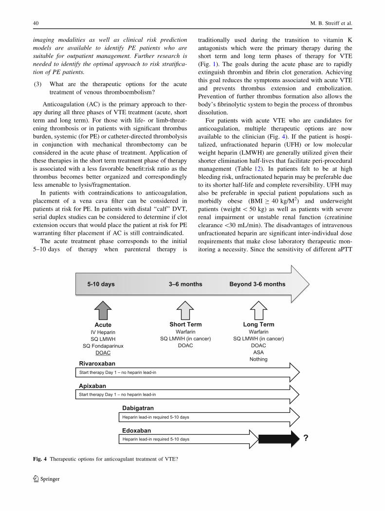
The acute treatment phase corresponds to the initial
5–10 days of therapy when parenteral therapy is
traditionally used during the transition to vitamin K
antagonists which were the primary therapy during the
short term and long term phases of therapy for VTE
(Fig. 1). The goals during the acute phase are to rapidly
extinguish thrombin and fibrin clot generation. Achieving
this goal reduces the symptoms associated with acute VTE
and prevents thrombus extension and embolization.
Prevention of further thrombus formation also allows the
body’s fibrinolytic system to begin the process of thrombus
dissolution.
For patients with acute VTE who are candidates for
anticoagulation, multiple therapeutic options are now
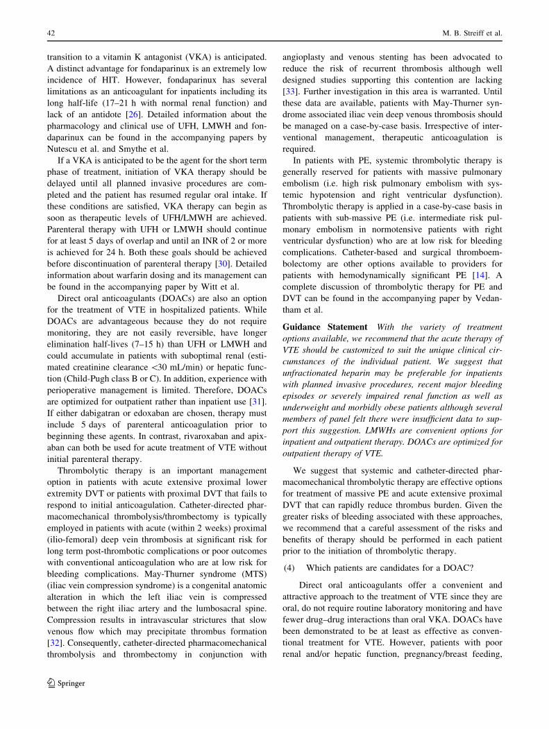
available to the clinician (Fig. 4). If the patient is hospi-
talized, unfractionated heparin (UFH) or low molecular
weight heparin (LMWH) are generally utilized given their
shorter elimination half-lives that facilitate peri-procedural
management (Table 12). In patients felt to be at high
bleeding risk, unfractionated heparin may be preferable due
to its shorter half-life and complete reversibility. UFH may
also be preferable in special patient populations such as
morbidly obese (BMI C 40 kg/M2) and underweight
patients (weight\ 50 kg) as well as patients with severe
renal impairment or unstable renal function (creatinine
clearance\30 mL/min). The disadvantages of intravenous
unfractionated heparin are significant inter-individual dose
requirements that make close laboratory therapeutic mon-
itoring a necessity. Since the sensitivity of different aPTT
Start therapy Day 1 – no heparin lead-in
Start therapy Day 1 – no heparin lead-in
Heparin lead-in required 5-10 days
Heparin lead-in required 5-10 days
Rivaroxaban
Apixaban
Dabigatran
Edoxaban
AcuteIV HeparinSQ LMWH
SQ FondaparinuxDOAC
Short Term Warfarin
SQ LMWH (in cancer)DOAC
5-10 days 3–6 months Beyond 3-6 months
Long TermWarfarin
SQ LMWH (in cancer)DOACASA
Nothing
?
Fig. 4 Therapeutic options for anticoagulant treatment of VTE?
40 M. B. Streiff et al.
123
reagents to UFH varies substantially, it is important for
each laboratory to establish its own therapeutic range based
upon UFH levels as measured by protamine titration or
chromogenic anti-Xa levels [26]. Observational studies
have demonstrated that optimal management of UFH is
difficult to achieve in routine clinical practice [27]. In
addition, UFH poses an 8-10-fold higher risk for heparin-
induced thrombocytopenia (HIT) than LMWH [28, 29].
Given the disadvantages associated with UFH, LMWH
is often preferred outside of special hospitalized patient
populations. Fondaparinux can also be employed as a
parenteral agent for hospitalized patients in whom
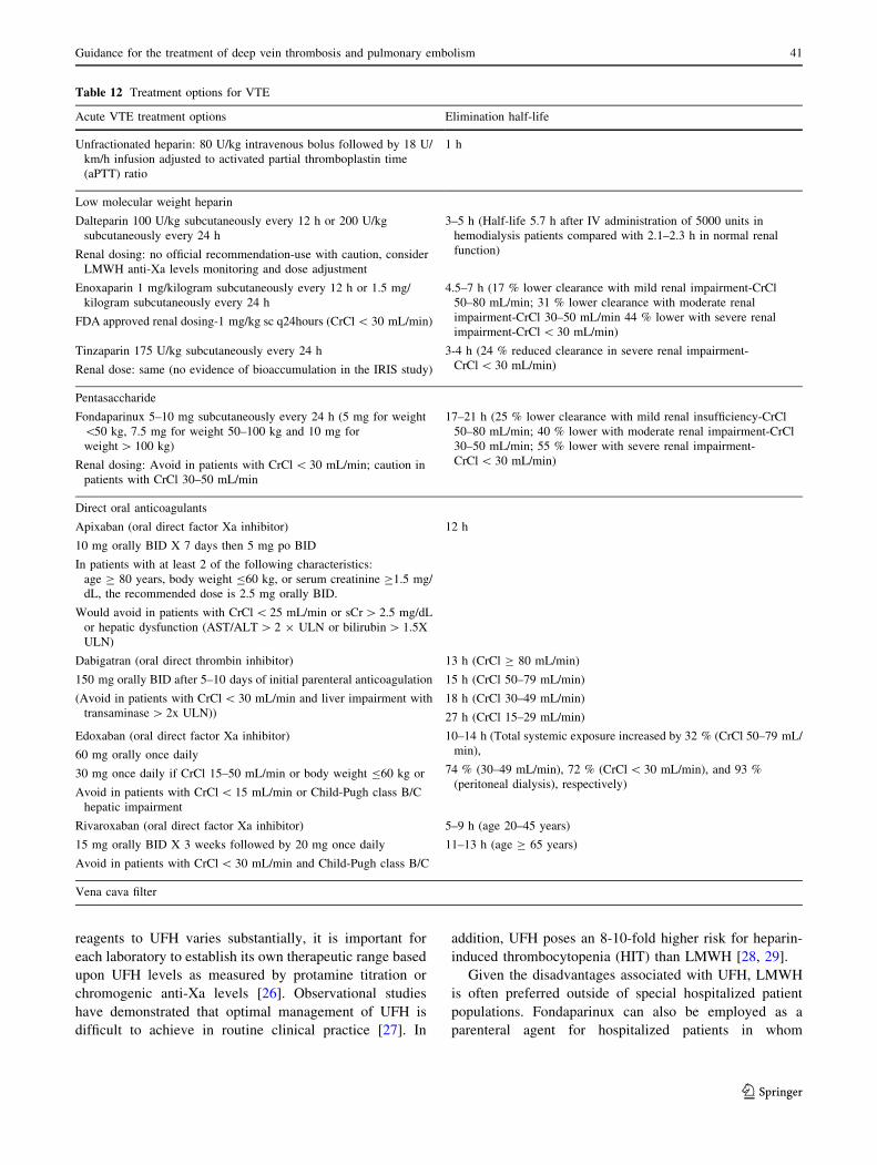
Table 12 Treatment options for VTE
Acute VTE treatment options Elimination half-life
Unfractionated heparin: 80 U/kg intravenous bolus followed by 18 U/
km/h infusion adjusted to activated partial thromboplastin time
(aPTT) ratio
1 h
Low molecular weight heparin
Dalteparin 100 U/kg subcutaneously every 12 h or 200 U/kg
subcutaneously every 24 h
Renal dosing: no official recommendation-use with caution, consider
LMWH anti-Xa levels monitoring and dose adjustment
3–5 h (Half-life 5.7 h after IV administration of 5000 units in
hemodialysis patients compared with 2.1–2.3 h in normal renal
function)
Enoxaparin 1 mg/kilogram subcutaneously every 12 h or 1.5 mg/
kilogram subcutaneously every 24 h
FDA approved renal dosing-1 mg/kg sc q24hours (CrCl\ 30 mL/min)
4.5–7 h (17 % lower clearance with mild renal impairment-CrCl
50–80 mL/min; 31 % lower clearance with moderate renal
impairment-CrCl 30–50 mL/min 44 % lower with severe renal
impairment-CrCl\ 30 mL/min)
Tinzaparin 175 U/kg subcutaneously every 24 h
Renal dose: same (no evidence of bioaccumulation in the IRIS study)
3-4 h (24 % reduced clearance in severe renal impairment-
CrCl\ 30 mL/min)
Pentasaccharide
Fondaparinux 5–10 mg subcutaneously every 24 h (5 mg for weight
\50 kg, 7.5 mg for weight 50–100 kg and 10 mg for
weight[ 100 kg)
Renal dosing: Avoid in patients with CrCl\ 30 mL/min; caution in
patients with CrCl 30–50 mL/min
17–21 h (25 % lower clearance with mild renal insufficiency-CrCl
50–80 mL/min; 40 % lower with moderate renal impairment-CrCl
30–50 mL/min; 55 % lower with severe renal impairment-
CrCl\ 30 mL/min)
Direct oral anticoagulants
Apixaban (oral direct factor Xa inhibitor)
10 mg orally BID X 7 days then 5 mg po BID
In patients with at least 2 of the following characteristics:
age C 80 years, body weight B60 kg, or serum creatinine C1.5 mg/
dL, the recommended dose is 2.5 mg orally BID.
Would avoid in patients with CrCl\ 25 mL/min or sCr[ 2.5 mg/dL
or hepatic dysfunction (AST/ALT[ 2 9 ULN or bilirubin[ 1.5X
ULN)
12 h
Dabigatran (oral direct thrombin inhibitor)
150 mg orally BID after 5–10 days of initial parenteral anticoagulation
(Avoid in patients with CrCl\ 30 mL/min and liver impairment with
transaminase[ 2x ULN))
13 h (CrCl C 80 mL/min)
15 h (CrCl 50–79 mL/min)
18 h (CrCl 30–49 mL/min)
27 h (CrCl 15–29 mL/min)
Edoxaban (oral direct factor Xa inhibitor)
60 mg orally once daily
30 mg once daily if CrCl 15–50 mL/min or body weight B60 kg or
Avoid in patients with CrCl\ 15 mL/min or Child-Pugh class B/C
hepatic impairment
10–14 h (Total systemic exposure increased by 32 % (CrCl 50–79 mL/
min),
74 % (30–49 mL/min), 72 % (CrCl\ 30 mL/min), and 93 %
(peritoneal dialysis), respectively)
Rivaroxaban (oral direct factor Xa inhibitor)
15 mg orally BID X 3 weeks followed by 20 mg once daily
Avoid in patients with CrCl\ 30 mL/min and Child-Pugh class B/C
5–9 h (age 20–45 years)
11–13 h (age C 65 years)
Vena cava filter
Guidance for the treatment of deep vein thrombosis and pulmonary embolism 41
123
transition to a vitamin K antagonist (VKA) is anticipated.
A distinct advantage for fondaparinux is an extremely low
incidence of HIT. However, fondaparinux has several
limitations as an anticoagulant for inpatients including its
long half-life (17–21 h with normal renal function) and
lack of an antidote [26]. Detailed information about the
pharmacology and clinical use of UFH, LMWH and fon-
daparinux can be found in the accompanying papers by
Nutescu et al. and Smythe et al.
If a VKA is anticipated to be the agent for the short term
phase of treatment, initiation of VKA therapy should be
delayed until all planned invasive procedures are com-
pleted and the patient has resumed regular oral intake. If
these conditions are satisfied, VKA therapy can begin as
soon as therapeutic levels of UFH/LMWH are achieved.
Parenteral therapy with UFH or LMWH should continue
for at least 5 days of overlap and until an INR of 2 or more
is achieved for 24 h. Both these goals should be achieved
before discontinuation of parenteral therapy [30]. Detailed
information about warfarin dosing and its management can
be found in the accompanying paper by Witt et al.
Direct oral anticoagulants (DOACs) are also an option
for the treatment of VTE in hospitalized patients. While
DOACs are advantageous because they do not require
monitoring, they are not easily reversible, have longer
elimination half-lives (7–15 h) than UFH or LMWH and
could accumulate in patients with suboptimal renal (esti-
mated creatinine clearance\30 mL/min) or hepatic func-
tion (Child-Pugh class B or C). In addition, experience with
perioperative management is limited. Therefore, DOACs
are optimized for outpatient rather than inpatient use [31].
If either dabigatran or edoxaban are chosen, therapy must
include 5 days of parenteral anticoagulation prior to
beginning these agents. In contrast, rivaroxaban and apix-
aban can both be used for acute treatment of VTE without
initial parenteral therapy.
Thrombolytic therapy is an important management
option in patients with acute extensive proximal lower
extremity DVT or patients with proximal DVT that fails to
respond to initial anticoagulation. Catheter-directed phar-
macomechanical thrombolysis/thrombectomy is typically
employed in patients with acute (within 2 weeks) proximal
(ilio-femoral) deep vein thrombosis at significant risk for
long term post-thrombotic complications or poor outcomes
with conventional anticoagulation who are at low risk for
bleeding complications. May-Thurner syndrome (MTS)
(iliac vein compression syndrome) is a congenital anatomic
alteration in which the left iliac vein is compressed
between the right iliac artery and the lumbosacral spine.
Compression results in intravascular strictures that slow
venous flow which may precipitate thrombus formation
[32]. Consequently, catheter-directed pharmacomechanical
thrombolysis and thrombectomy in conjunction with
angioplasty and venous stenting has been advocated to
reduce the risk of recurrent thrombosis although well
designed studies supporting this contention are lacking
[33]. Further investigation in this area is warranted. Until
these data are available, patients with May-Thurner syn-
drome associated iliac vein deep venous thrombosis should
be managed on a case-by-case basis. Irrespective of inter-
ventional management, therapeutic anticoagulation is
required.
In patients with PE, systemic thrombolytic therapy is
generally reserved for patients with massive pulmonary
embolism (i.e. high risk pulmonary embolism with sys-
temic hypotension and right ventricular dysfunction).
Thrombolytic therapy is applied in a case-by-case basis in
patients with sub-massive PE (i.e. intermediate risk pul-
monary embolism in normotensive patients with right
ventricular dysfunction) who are at low risk for bleeding
complications. Catheter-based and surgical thromboem-
bolectomy are other options available to providers for
patients with hemodynamically significant PE [14]. A
complete discussion of thrombolytic therapy for PE and
DVT can be found in the accompanying paper by Vedan-
tham et al.
Guidance Statement With the variety of treatment
options available, we recommend that the acute therapy of
VTE should be customized to suit the unique clinical cir-
cumstances of the individual patient. We suggest that
unfractionated heparin may be preferable for inpatients
with planned invasive procedures, recent major bleeding
episodes or severely impaired renal function as well as
underweight and morbidly obese patients although several
members of panel felt there were insufficient data to sup-
port this suggestion. LMWHs are convenient options for
inpatient and outpatient therapy. DOACs are optimized for
outpatient therapy of VTE.
We suggest that systemic and catheter-directed phar-
macomechanical thrombolytic therapy are effective options
for treatment of massive PE and acute extensive proximal
DVT that can rapidly reduce thrombus burden. Given the
greater risks of bleeding associated with these approaches,
we recommend that a careful assessment of the risks and
benefits of therapy should be performed in each patient
prior to the initiation of thrombolytic therapy.
(4) Which patients are candidates for a DOAC?
Direct oral anticoagulants offer a convenient and
attractive approach to the treatment of VTE since they are
oral, do not require routine laboratory monitoring and have
fewer drug–drug interactions than oral VKA. DOACs have
been demonstrated to be at least as effective as conven-
tional treatment for VTE. However, patients with poor
renal and/or hepatic function, pregnancy/breast feeding,
42 M. B. Streiff et al.
123
thrombocytopenia, high bleeding risk and potent drug–drug
interactions were excluded from participation in the phase
3 VTE studies. In addition, certain patient populations were
not well represented in these studies such as patients with
active cancer. Therefore, it is important to consider the
inclusion and exclusion criteria and the enrolled popula-
tions in the published studies when considering a DOAC
for treatment of VTE. In addition, 2 of the DOACs
(dabigatran and edoxaban) were studied using acute treat-
ment with a parenteral agent (dabigatran median duration
9 days; edoxaban median duration 7 days). Therefore,
these agents should be used only after an initial period of
parenteral therapy for acute VTE (Fig. 4).
Dabigatran is an oral direct thrombin inhibitor that has
been compared to warfarin in the short term treatment and
warfarin and placebo in long term treatment of VTE in 3
double blind randomized controlled trials, the RECOVER,
REMEDY and RESONATE studies. In the RE-COVER
study, 2564 patients with acute symptomatic objectively
documented proximal lower extremity DVT or PE were
randomized to either dabigatran 150 mg twice daily or
adjusted-dose warfarin (INR range 2–3) after acute treat-
ment with unfractionated or low molecular weight heparin
(median parenteral treatment duration = 9 days). Seven
patients in the dabigatran group and 18 in the warfarin
group did not receive study medication leaving a total of
1274 dabigatran patients and 1265 warfarin patients in the
population for efficacy analysis. In the warfarin group, the
time in therapeutic range over the duration of the study was
60 % (53 % month 1, 66 % in the last month). Thirty of
1274 patients on dabigatran (2.4 %) and 27 of 1265 war-
farin recipients (2.1 %) suffered recurrent VTE (0.4 %
absolute risk difference; 95 % CI for non-inferiority -0.8
to 1.5). The hazard ratio (HR) with dabigatran was 1.10
(95 % CI 0.65–1.84). Major bleeding occurred in 20
patients assigned to dabigatran (1.6 %) and in 24 patients
taking warfarin (1.9 %) for a hazard ratio with dabigatran
of 0.82 (95 % CI 0.42–1.48) (Table 13). There was no
difference in mortality, acute coronary events or abnormal
liver function tests [34].
These results were confirmed in RECOVER II, a ran-
domized double-blind double dummy study that compared
dabigatran 150 mg twice daily with warfarin (INR 2–3)
after median of 9 days of parenteral therapy. Recurrent
symptomatic objectively confirmed VTE occurred in 30 of
1279 dabigatran patients (2.3 %) and 28 of 1289 warfarin
patients (2.2 %) (HR 1.08; 95 % CI 0.64–1.80). Major
bleeding occurred in 15 dabigatran patients (1.2 %) and 22
warfarin patients (1.7 %) (HR 0.69; 95 % CI 0.36–1.32)
(Table 13). Pooled analysis with the RECOVER and
RECOVER II studies produced a hazard ratio for recurrent
VTE of 1.09 (95 % CI 0.76–1.57), major bleeding of 0.73
(95 % CI 0.48–1.11) and for any bleeding of 0.70 (95 % CI
0.61–0.79) [35]. These studies demonstrate that dabigatran
is at least as effective as warfarin for short term treatment
of VTE. Compared with warfarin, dabigatran was associ-
ated with an increased risk of myocardial infarction or
acute coronary syndrome in a meta-analysis of the ran-
domized clinical trials (RCT) leading to its approval
(dabigatran, 237 of 20,000 [1.19 %] vs. control, 83 of
10,514 [0.79 %]; OR 1.33; 95 % CI 1.03–1.71; P = 0.03)
[36]. However, no difference was seen in a recent large
new user cohort of 134,414 propensity-matched elderly US
Medicare patients (Hazard ratio 0.92 (95 % CI 0.78–1.08)
perhaps due to clinical differences in the two study popu-
lations [37]. Until this issue is further clarified, prescribers
should use caution when prescribing dabigatran in elderly
patients at risk for acute coronary syndrome.
GI bleeding also appears to be more common in higher
risk patients treated with dabigatran compared with
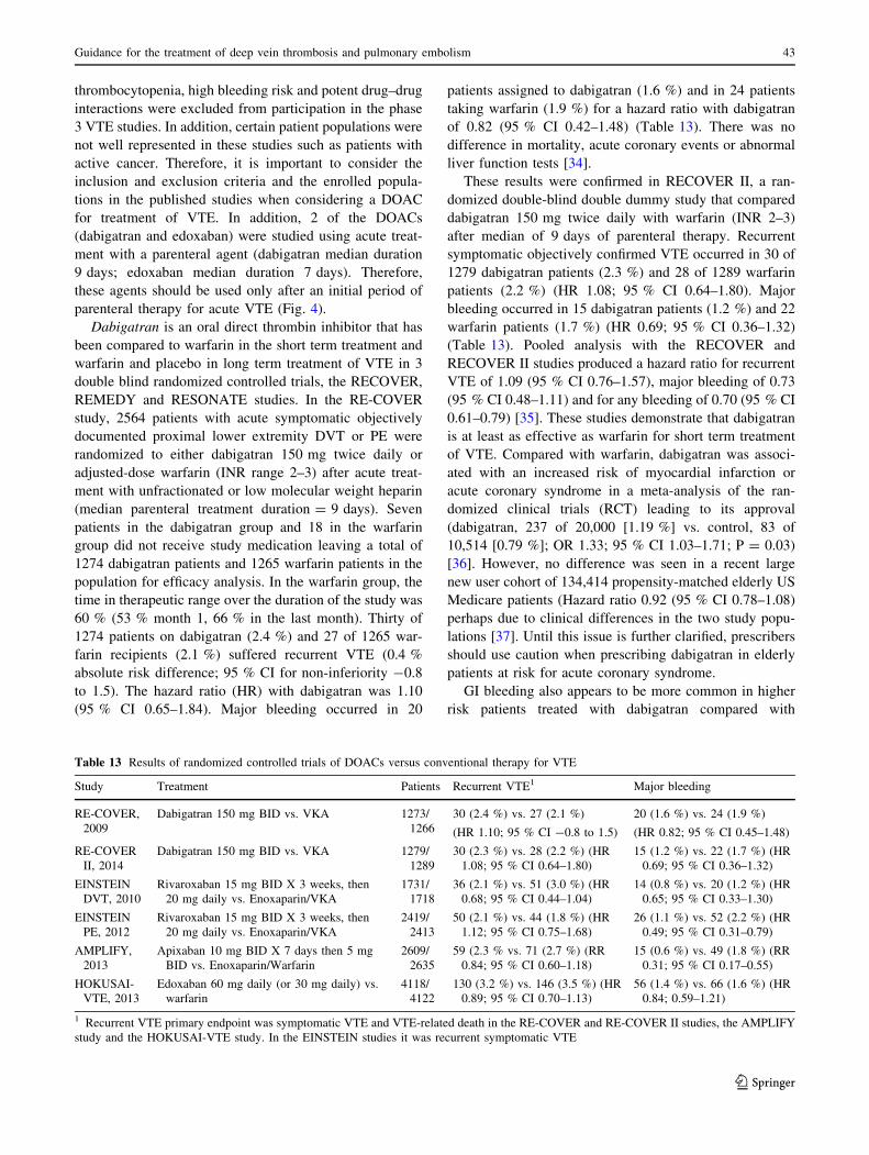
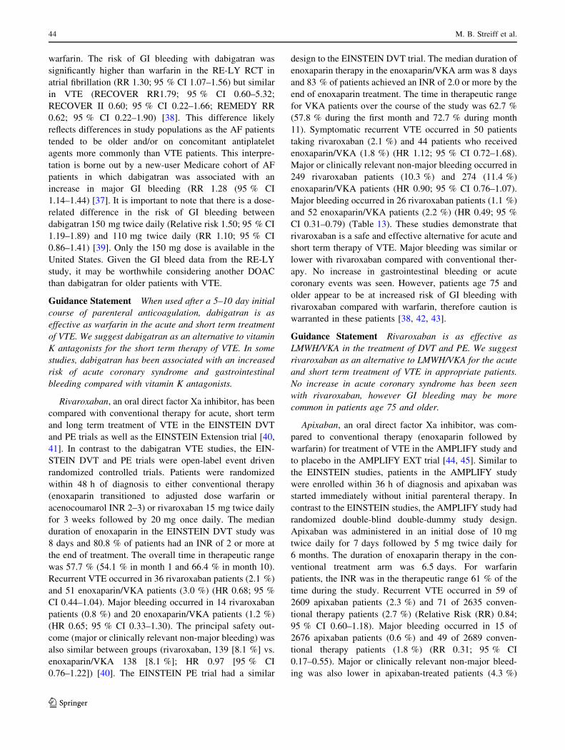
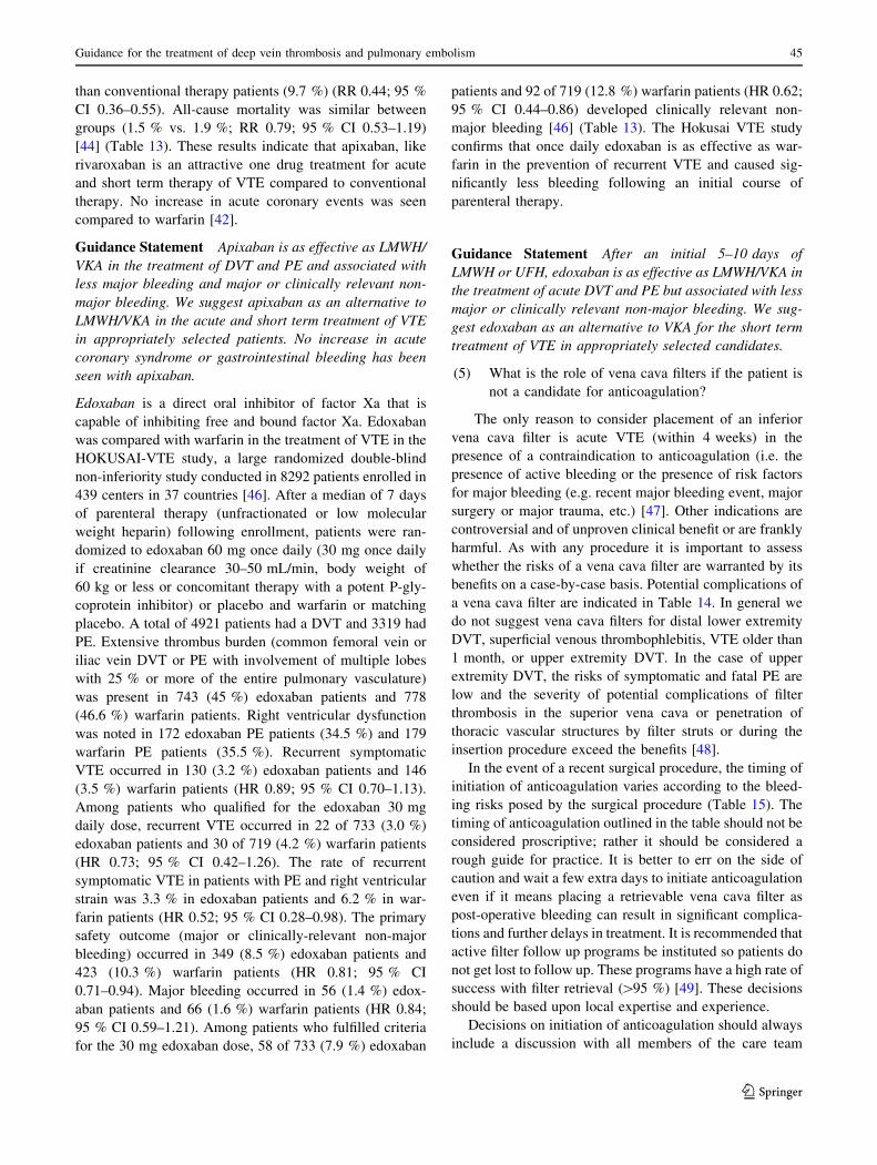
Table 13 Results of randomized controlled trials of DOACs versus conventional therapy for VTE
Study Treatment Patients Recurrent VTE1 Major bleeding
RE-COVER,
2009
Dabigatran 150 mg BID vs. VKA 1273/
1266
30 (2.4 %) vs. 27 (2.1 %)
(HR 1.10; 95 % CI -0.8 to 1.5)
20 (1.6 %) vs. 24 (1.9 %)
(HR 0.82; 95 % CI 0.45–1.48)
RE-COVER
II, 2014
Dabigatran 150 mg BID vs. VKA 1279/
1289
30 (2.3 %) vs. 28 (2.2 %) (HR
1.08; 95 % CI 0.64–1.80)
15 (1.2 %) vs. 22 (1.7 %) (HR
0.69; 95 % CI 0.36–1.32)
EINSTEIN
DVT, 2010
Rivaroxaban 15 mg BID X 3 weeks, then
20 mg daily vs. Enoxaparin/VKA
1731/
1718
36 (2.1 %) vs. 51 (3.0 %) (HR
0.68; 95 % CI 0.44–1.04)
14 (0.8 %) vs. 20 (1.2 %) (HR
0.65; 95 % CI 0.33–1.30)
EINSTEIN
PE, 2012
Rivaroxaban 15 mg BID X 3 weeks, then
20 mg daily vs. Enoxaparin/VKA
2419/
2413
50 (2.1 %) vs. 44 (1.8 %) (HR
1.12; 95 % CI 0.75–1.68)
26 (1.1 %) vs. 52 (2.2 %) (HR
0.49; 95 % CI 0.31–0.79)
AMPLIFY,
2013
Apixaban 10 mg BID X 7 days then 5 mg
BID vs. Enoxaparin/Warfarin
2609/
2635
59 (2.3 % vs. 71 (2.7 %) (RR
0.84; 95 % CI 0.60–1.18)
15 (0.6 %) vs. 49 (1.8 %) (RR
0.31; 95 % CI 0.17–0.55)
HOKUSAI-
VTE, 2013
Edoxaban 60 mg daily (or 30 mg daily) vs.
warfarin
4118/
4122
130 (3.2 %) vs. 146 (3.5 %) (HR
0.89; 95 % CI 0.70–1.13)
56 (1.4 %) vs. 66 (1.6 %) (HR
0.84; 0.59–1.21)
1 Recurrent VTE primary endpoint was symptomatic VTE and VTE-related death in the RE-COVER and RE-COVER II studies, the AMPLIFY
study and the HOKUSAI-VTE study. In the EINSTEIN studies it was recurrent symptomatic VTE
Guidance for the treatment of deep vein thrombosis and pulmonary embolism 43
123
warfarin. The risk of GI bleeding with dabigatran was
significantly higher than warfarin in the RE-LY RCT in
atrial fibrillation (RR 1.30; 95 % CI 1.07–1.56) but similar
in VTE (RECOVER RR1.79; 95 % CI 0.60–5.32;
RECOVER II 0.60; 95 % CI 0.22–1.66; REMEDY RR
0.62; 95 % CI 0.22–1.90) [38]. This difference likely
reflects differences in study populations as the AF patients
tended to be older and/or on concomitant antiplatelet
agents more commonly than VTE patients. This interpre-
tation is borne out by a new-user Medicare cohort of AF
patients in which dabigatran was associated with an
increase in major GI bleeding (RR 1.28 (95 % CI
1.14–1.44) [37]. It is important to note that there is a dose-
related difference in the risk of GI bleeding between
dabigatran 150 mg twice daily (Relative risk 1.50; 95 % CI
1.19–1.89) and 110 mg twice daily (RR 1.10; 95 % CI
0.86–1.41) [39]. Only the 150 mg dose is available in the
United States. Given the GI bleed data from the RE-LY
study, it may be worthwhile considering another DOAC
than dabigatran for older patients with VTE.
Guidance Statement When used after a 5–10 day initial
course of parenteral anticoagulation, dabigatran is as
effective as warfarin in the acute and short term treatment
of VTE. We suggest dabigatran as an alternative to vitamin
K antagonists for the short term therapy of VTE. In some
studies, dabigatran has been associated with an increased
risk of acute coronary syndrome and gastrointestinal
bleeding compared with vitamin K antagonists.
Rivaroxaban, an oral direct factor Xa inhibitor, has been
compared with conventional therapy for acute, short term
and long term treatment of VTE in the EINSTEIN DVT
and PE trials as well as the EINSTEIN Extension trial [40,
41]. In contrast to the dabigatran VTE studies, the EIN-
STEIN DVT and PE trials were open-label event driven
randomized controlled trials. Patients were randomized
within 48 h of diagnosis to either conventional therapy
(enoxaparin transitioned to adjusted dose warfarin or
acenocoumarol INR 2–3) or rivaroxaban 15 mg twice daily
for 3 weeks followed by 20 mg once daily. The median
duration of enoxaparin in the EINSTEIN DVT study was
8 days and 80.8 % of patients had an INR of 2 or more at
the end of treatment. The overall time in therapeutic range
was 57.7 % (54.1 % in month 1 and 66.4 % in month 10).
Recurrent VTE occurred in 36 rivaroxaban patients (2.1 %)
and 51 enoxaparin/VKA patients (3.0 %) (HR 0.68; 95 %
CI 0.44–1.04). Major bleeding occurred in 14 rivaroxaban
patients (0.8 %) and 20 enoxaparin/VKA patients (1.2 %)
(HR 0.65; 95 % CI 0.33–1.30). The principal safety out-
come (major or clinically relevant non-major bleeding) was
also similar between groups (rivaroxaban, 139 [8.1 %] vs.
enoxaparin/VKA 138 [8.1 %]; HR 0.97 [95 % CI
0.76–1.22]) [40]. The EINSTEIN PE trial had a similar
design to the EINSTEIN DVT trial. The median duration of
enoxaparin therapy in the enoxaparin/VKA arm was 8 days
and 83 % of patients achieved an INR of 2.0 or more by the
end of enoxaparin treatment. The time in therapeutic range
for VKA patients over the course of the study was 62.7 %
(57.8 % during the first month and 72.7 % during month
11). Symptomatic recurrent VTE occurred in 50 patients
taking rivaroxaban (2.1 %) and 44 patients who received
enoxaparin/VKA (1.8 %) (HR 1.12; 95 % CI 0.72–1.68).
Major or clinically relevant non-major bleeding occurred in
249 rivaroxaban patients (10.3 %) and 274 (11.4 %)
enoxaparin/VKA patients (HR 0.90; 95 % CI 0.76–1.07).
Major bleeding occurred in 26 rivaroxaban patients (1.1 %)
and 52 enoxaparin/VKA patients (2.2 %) (HR 0.49; 95 %
CI 0.31–0.79) (Table 13). These studies demonstrate that
rivaroxaban is a safe and effective alternative for acute and
short term therapy of VTE. Major bleeding was similar or
lower with rivaroxaban compared with conventional ther-
apy. No increase in gastrointestinal bleeding or acute
coronary events was seen. However, patients age 75 and
older appear to be at increased risk of GI bleeding with
rivaroxaban compared with warfarin, therefore caution is
warranted in these patients [38, 42, 43].
Guidance Statement Rivaroxaban is as effective as
LMWH/VKA in the treatment of DVT and PE. We suggest
rivaroxaban as an alternative to LMWH/VKA for the acute
and short term treatment of VTE in appropriate patients.
No increase in acute coronary syndrome has been seen
with rivaroxaban, however GI bleeding may be more
common in patients age 75 and older.
Apixaban, an oral direct factor Xa inhibitor, was com-
pared to conventional therapy (enoxaparin followed by
warfarin) for treatment of VTE in the AMPLIFY study and
to placebo in the AMPLIFY EXT trial [44, 45]. Similar to
the EINSTEIN studies, patients in the AMPLIFY study
were enrolled within 36 h of diagnosis and apixaban was
started immediately without initial parenteral therapy. In
contrast to the EINSTEIN studies, the AMPLIFY study had
randomized double-blind double-dummy study design.
Apixaban was administered in an initial dose of 10 mg
twice daily for 7 days followed by 5 mg twice daily for
6 months. The duration of enoxaparin therapy in the con-
ventional treatment arm was 6.5 days. For warfarin
patients, the INR was in the therapeutic range 61 % of the
time during the study. Recurrent VTE occurred in 59 of
2609 apixaban patients (2.3 %) and 71 of 2635 conven-
tional therapy patients (2.7 %) (Relative Risk (RR) 0.84;
95 % CI 0.60–1.18). Major bleeding occurred in 15 of
2676 apixaban patients (0.6 %) and 49 of 2689 conven-
tional therapy patients (1.8 %) (RR 0.31; 95 % CI
0.17–0.55). Major or clinically relevant non-major bleed-
ing was also lower in apixaban-treated patients (4.3 %)
44 M. B. Streiff et al.
123
than conventional therapy patients (9.7 %) (RR 0.44; 95 %
CI 0.36–0.55). All-cause mortality was similar between
groups (1.5 % vs. 1.9 %; RR 0.79; 95 % CI 0.53–1.19)
[44] (Table 13). These results indicate that apixaban, like
rivaroxaban is an attractive one drug treatment for acute
and short term therapy of VTE compared to conventional
therapy. No increase in acute coronary events was seen
compared to warfarin [42].
Guidance Statement Apixaban is as effective as LMWH/
VKA in the treatment of DVT and PE and associated with
less major bleeding and major or clinically relevant non-
major bleeding. We suggest apixaban as an alternative to
LMWH/VKA in the acute and short term treatment of VTE
in appropriately selected patients. No increase in acute
coronary syndrome or gastrointestinal bleeding has been
seen with apixaban.
Edoxaban is a direct oral inhibitor of factor Xa that is
capable of inhibiting free and bound factor Xa. Edoxaban
was compared with warfarin in the treatment of VTE in the
HOKUSAI-VTE study, a large randomized double-blind
non-inferiority study conducted in 8292 patients enrolled in
439 centers in 37 countries [46]. After a median of 7 days
of parenteral therapy (unfractionated or low molecular
weight heparin) following enrollment, patients were ran-
domized to edoxaban 60 mg once daily (30 mg once daily
if creatinine clearance 30–50 mL/min, body weight of
60 kg or less or concomitant therapy with a potent P-gly-
coprotein inhibitor) or placebo and warfarin or matching
placebo. A total of 4921 patients had a DVT and 3319 had
PE. Extensive thrombus burden (common femoral vein or
iliac vein DVT or PE with involvement of multiple lobes
with 25 % or more of the entire pulmonary vasculature)
was present in 743 (45 %) edoxaban patients and 778
(46.6 %) warfarin patients. Right ventricular dysfunction
was noted in 172 edoxaban PE patients (34.5 %) and 179
warfarin PE patients (35.5 %). Recurrent symptomatic
VTE occurred in 130 (3.2 %) edoxaban patients and 146
(3.5 %) warfarin patients (HR 0.89; 95 % CI 0.70–1.13).
Among patients who qualified for the edoxaban 30 mg
daily dose, recurrent VTE occurred in 22 of 733 (3.0 %)
edoxaban patients and 30 of 719 (4.2 %) warfarin patients
(HR 0.73; 95 % CI 0.42–1.26). The rate of recurrent
symptomatic VTE in patients with PE and right ventricular
strain was 3.3 % in edoxaban patients and 6.2 % in war-
farin patients (HR 0.52; 95 % CI 0.28–0.98). The primary
safety outcome (major or clinically-relevant non-major
bleeding) occurred in 349 (8.5 %) edoxaban patients and
423 (10.3 %) warfarin patients (HR 0.81; 95 % CI
0.71–0.94). Major bleeding occurred in 56 (1.4 %) edox-
aban patients and 66 (1.6 %) warfarin patients (HR 0.84;
95 % CI 0.59–1.21). Among patients who fulfilled criteria
for the 30 mg edoxaban dose, 58 of 733 (7.9 %) edoxaban
patients and 92 of 719 (12.8 %) warfarin patients (HR 0.62;
95 % CI 0.44–0.86) developed clinically relevant non-
major bleeding [46] (Table 13). The Hokusai VTE study
confirms that once daily edoxaban is as effective as war-
farin in the prevention of recurrent VTE and caused sig-
nificantly less bleeding following an initial course of
parenteral therapy.
Guidance Statement After an initial 5–10 days of
LMWH or UFH, edoxaban is as effective as LMWH/VKA in
the treatment of acute DVT and PE but associated with less
major or clinically relevant non-major bleeding. We sug-
gest edoxaban as an alternative to VKA for the short term
treatment of VTE in appropriately selected candidates.
(5) What is the role of vena cava filters if the patient is
not a candidate for anticoagulation?
The only reason to consider placement of an inferior
vena cava filter is acute VTE (within 4 weeks) in the
presence of a contraindication to anticoagulation (i.e. the
presence of active bleeding or the presence of risk factors
for major bleeding (e.g. recent major bleeding event, major
surgery or major trauma, etc.) [47]. Other indications are
controversial and of unproven clinical benefit or are frankly
harmful. As with any procedure it is important to assess
whether the risks of a vena cava filter are warranted by its
benefits on a case-by-case basis. Potential complications of
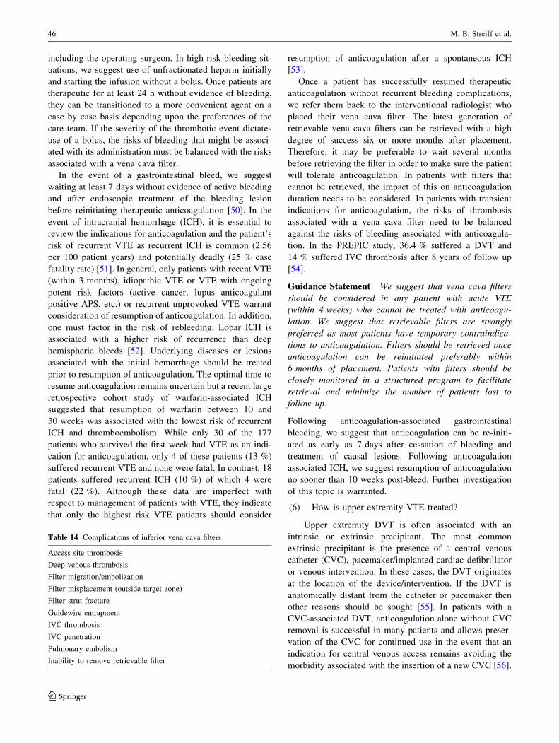
a vena cava filter are indicated in Table 14. In general we
do not suggest vena cava filters for distal lower extremity
DVT, superficial venous thrombophlebitis, VTE older than
1 month, or upper extremity DVT. In the case of upper
extremity DVT, the risks of symptomatic and fatal PE are
low and the severity of potential complications of filter
thrombosis in the superior vena cava or penetration of
thoracic vascular structures by filter struts or during the
insertion procedure exceed the benefits [48].
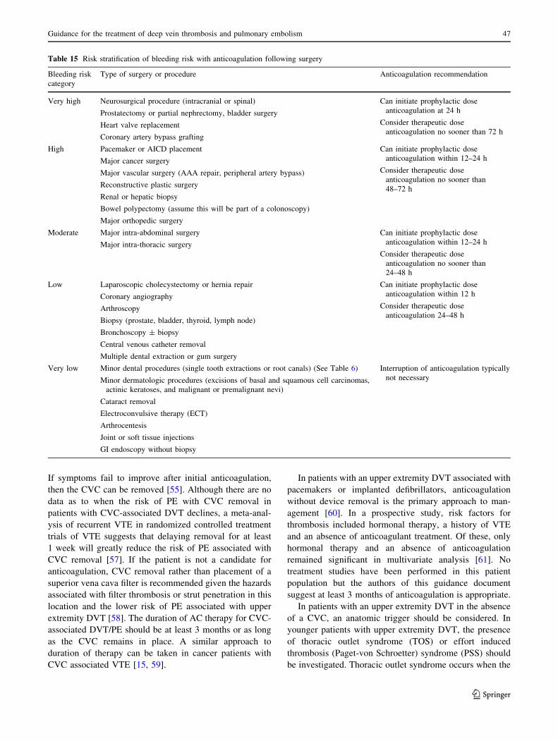
In the event of a recent surgical procedure, the timing of
initiation of anticoagulation varies according to the bleed-
ing risks posed by the surgical procedure (Table 15). The
timing of anticoagulation outlined in the table should not be
considered proscriptive; rather it should be considered a
rough guide for practice. It is better to err on the side of
caution and wait a few extra days to initiate anticoagulation
even if it means placing a retrievable vena cava filter as
post-operative bleeding can result in significant complica-
tions and further delays in treatment. It is recommended that
active filter follow up programs be instituted so patients do
not get lost to follow up. These programs have a high rate of
success with filter retrieval ([95 %) [49]. These decisions
should be based upon local expertise and experience.
Decisions on initiation of anticoagulation should always
include a discussion with all members of the care team
Guidance for the treatment of deep vein thrombosis and pulmonary embolism 45
123
including the operating surgeon. In high risk bleeding sit-
uations, we suggest use of unfractionated heparin initially
and starting the infusion without a bolus. Once patients are
therapeutic for at least 24 h without evidence of bleeding,
they can be transitioned to a more convenient agent on a
case by case basis depending upon the preferences of the
care team. If the severity of the thrombotic event dictates
use of a bolus, the risks of bleeding that might be associ-
ated with its administration must be balanced with the risks
associated with a vena cava filter.
In the event of a gastrointestinal bleed, we suggest
waiting at least 7 days without evidence of active bleeding
and after endoscopic treatment of the bleeding lesion
before reinitiating therapeutic anticoagulation [50]. In the
event of intracranial hemorrhage (ICH), it is essential to
review the indications for anticoagulation and the patient’s
risk of recurrent VTE as recurrent ICH is common (2.56
per 100 patient years) and potentially deadly (25 % case
fatality rate) [51]. In general, only patients with recent VTE
(within 3 months), idiopathic VTE or VTE with ongoing
potent risk factors (active cancer, lupus anticoagulant
positive APS, etc.) or recurrent unprovoked VTE warrant
consideration of resumption of anticoagulation. In addition,
one must factor in the risk of rebleeding. Lobar ICH is
associated with a higher risk of recurrence than deep
hemispheric bleeds [52]. Underlying diseases or lesions
associated with the initial hemorrhage should be treated
prior to resumption of anticoagulation. The optimal time to
resume anticoagulation remains uncertain but a recent large
retrospective cohort study of warfarin-associated ICH
suggested that resumption of warfarin between 10 and
30 weeks was associated with the lowest risk of recurrent
ICH and thromboembolism. While only 30 of the 177
patients who survived the first week had VTE as an indi-
cation for anticoagulation, only 4 of these patients (13 %)
suffered recurrent VTE and none were fatal. In contrast, 18
patients suffered recurrent ICH (10 %) of which 4 were
fatal (22 %). Although these data are imperfect with
respect to management of patients with VTE, they indicate
that only the highest risk VTE patients should consider
resumption of anticoagulation after a spontaneous ICH
[53].
Once a patient has successfully resumed therapeutic
anticoagulation without recurrent bleeding complications,
we refer them back to the interventional radiologist who
placed their vena cava filter. The latest generation of
retrievable vena cava filters can be retrieved with a high
degree of success six or more months after placement.
Therefore, it may be preferable to wait several months
before retrieving the filter in order to make sure the patient
will tolerate anticoagulation. In patients with filters that
cannot be retrieved, the impact of this on anticoagulation
duration needs to be considered. In patients with transient
indications for anticoagulation, the risks of thrombosis
associated with a vena cava filter need to be balanced
against the risks of bleeding associated with anticoagula-
tion. In the PREPIC study, 36.4 % suffered a DVT and
14 % suffered IVC thrombosis after 8 years of follow up
[54].
Guidance Statement We suggest that vena cava filters
should be considered in any patient with acute VTE
(within 4 weeks) who cannot be treated with anticoagu-
lation. We suggest that retrievable filters are strongly
preferred as most patients have temporary contraindica-
tions to anticoagulation. Filters should be retrieved once
anticoagulation can be reinitiated preferably within
6 months of placement. Patients with filters should be
closely monitored in a structured program to facilitate
retrieval and minimize the number of patients lost to
follow up.
Following anticoagulation-associated gastrointestinal
bleeding, we suggest that anticoagulation can be re-initi-
ated as early as 7 days after cessation of bleeding and
treatment of causal lesions. Following anticoagulation
associated ICH, we suggest resumption of anticoagulation
no sooner than 10 weeks post-bleed. Further investigation
of this topic is warranted.
(6) How is upper extremity VTE treated?
Upper extremity DVT is often associated with an
intrinsic or extrinsic precipitant. The most common
extrinsic precipitant is the presence of a central venous
catheter (CVC), pacemaker/implanted cardiac defibrillator
or venous intervention. In these cases, the DVT originates
at the location of the device/intervention. If the DVT is
anatomically distant from the catheter or pacemaker then
other reasons should be sought [55]. In patients with a
CVC-associated DVT, anticoagulation alone without CVC
removal is successful in many patients and allows preser-
vation of the CVC for continued use in the event that an
indication for central venous access remains avoiding the
morbidity associated with the insertion of a new CVC [56].
Table 14 Complications of inferior vena cava filters
Access site thrombosis
Deep venous thrombosis
Filter migration/embolization
Filter misplacement (outside target zone)
Filter strut fracture
Guidewire entrapment
IVC thrombosis
IVC penetration
Pulmonary embolism
Inability to remove retrievable filter
46 M. B. Streiff et al.
123
If symptoms fail to improve after initial anticoagulation,
then the CVC can be removed [55]. Although there are no
data as to when the risk of PE with CVC removal in
patients with CVC-associated DVT declines, a meta-anal-
ysis of recurrent VTE in randomized controlled treatment
trials of VTE suggests that delaying removal for at least
1 week will greatly reduce the risk of PE associated with
CVC removal [57]. If the patient is not a candidate for
anticoagulation, CVC removal rather than placement of a
superior vena cava filter is recommended given the hazards
associated with filter thrombosis or strut penetration in this
location and the lower risk of PE associated with upper
extremity DVT [58]. The duration of AC therapy for CVC-
associated DVT/PE should be at least 3 months or as long
as the CVC remains in place. A similar approach to
duration of therapy can be taken in cancer patients with
CVC associated VTE [15, 59].
In patients with an upper extremity DVT associated with
pacemakers or implanted defibrillators, anticoagulation
without device removal is the primary approach to man-
agement [60]. In a prospective study, risk factors for
thrombosis included hormonal therapy, a history of VTE
and an absence of anticoagulant treatment. Of these, only
hormonal therapy and an absence of anticoagulation
remained significant in multivariate analysis [61]. No
treatment studies have been performed in this patient
population but the authors of this guidance document
suggest at least 3 months of anticoagulation is appropriate.
In patients with an upper extremity DVT in the absence
of a CVC, an anatomic trigger should be considered. In
younger patients with upper extremity DVT, the presence
of thoracic outlet syndrome (TOS) or effort induced
thrombosis (Paget-von Schroetter) syndrome (PSS) should
be investigated. Thoracic outlet syndrome occurs when the
Table 15 Risk stratification of bleeding risk with anticoagulation following surgery
Bleeding risk
category
Type of surgery or procedure Anticoagulation recommendation
Very high Neurosurgical procedure (intracranial or spinal)
Prostatectomy or partial nephrectomy, bladder surgery
Heart valve replacement
Coronary artery bypass grafting
Can initiate prophylactic dose
anticoagulation at 24 h
Consider therapeutic dose
anticoagulation no sooner than 72 h
High Pacemaker or AICD placement
Major cancer surgery
Major vascular surgery (AAA repair, peripheral artery bypass)
Reconstructive plastic surgery
Renal or hepatic biopsy
Bowel polypectomy (assume this will be part of a colonoscopy)
Major orthopedic surgery
Can initiate prophylactic dose
anticoagulation within 12–24 h
Consider therapeutic dose
anticoagulation no sooner than
48–72 h
Moderate Major intra-abdominal surgery
Major intra-thoracic surgery
Can initiate prophylactic dose
anticoagulation within 12–24 h
Consider therapeutic dose
anticoagulation no sooner than
24–48 h
Low Laparoscopic cholecystectomy or hernia repair
Coronary angiography
Arthroscopy
Biopsy (prostate, bladder, thyroid, lymph node)
Bronchoscopy ± biopsy
Central venous catheter removal
Multiple dental extraction or gum surgery
Can initiate prophylactic dose
anticoagulation within 12 h
Consider therapeutic dose
anticoagulation 24–48 h
Very low Minor dental procedures (single tooth extractions or root canals) (See Table 6)
Minor dermatologic procedures (excisions of basal and squamous cell carcinomas,
actinic keratoses, and malignant or premalignant nevi)
Cataract removal
Electroconvulsive therapy (ECT)
Arthrocentesis
Joint or soft tissue injections
GI endoscopy without biopsy
Interruption of anticoagulation typically
not necessary
Guidance for the treatment of deep vein thrombosis and pulmonary embolism 47
123
nerve, artery and/or vein traversing the thoracic outlet are
compressed by the surrounding anatomic structures. This
compression can cause venous, arterial or neurologic
compromise. In the venous form of this syndrome, the
compression causes endothelial damage and stasis leading
to local anatomic clot formation. In PSS repetitive upper
extremity exercise usually in the context of a tight thoracic
outlet can lead to vascular damage, stasis and subsequent
thrombus formation. A history of upper extremity throm-
bosis in the absence of CVC or upper extremity or thoracic
or neck vein intravenous access procedures should prompt
consideration of TOS. A recent history of upper extremity
exertion or exercise should also raise suspicions of TOS/
PSS. Patients often complain of aching and swelling in the
upper extremity and demonstrate venous distention and
bluish discoloration in the affected arm. Physical exam
findings that suggest the presence of TOS include Adson’s
test (ipsilateral rotation and extension of the neck during
deep inspiration result in a diminution of the radial pulse)
and Wright’s test (hyperextension of the arm diminishes
the radial pulse). Nevertheless, imaging studies are essen-
tial to demonstrate TOS. Venous and arterial duplex
ultrasound with the patient’s arm in stress positions are the
most sensitive study for assessing the presence of TOS [15,
59].
For TOS/PSS, thrombolytic therapy followed by surgi-
cal repair (thoracic rib resection and/or scalenectomy) has
been advocated as an important component of successful
therapy in addition to anticoagulation. Thrombolysis fol-
lowed by endovascular stenting does not appear to be as
beneficial as surgery [62]. However, the benefits of surgical
therapy remain to be demonstrated in a rigorous fashion
[63]. Consequently, the writing committee of this guidance
document was divided as to the value of surgical repair of
thoracic outlet syndrome. Until well-designed studies are
conducted examining the risks and benefits of surgical
therapy, we suggest providers consider surgical repair in
addition to thrombolysis and anticoagulation versus
thrombolysis/anticoagulation alone on a case-by-case
basis. We suggest that patients with upper extremity deep
vein thrombosis receive at least 3 months of anticoagula-
tion with or without surgical therapy.
Other important causes of upper extremity DVT include
intra-thoracic or cervical tumors or nodal masses or
infections that can result in vascular wall inflammation and
compression. Diagnosis can generally be established with
duplex ultrasound or contrast CT venographic imaging.
Identification and treatment of the underlying disease
process (cancer, infection, etc.) and anticoagulation are
both likely to be important factors in successful treatment.
We suggest that anticoagulation should be continued for at
least 3 months or until precipitating factors have been
eliminated (vascular compression by tumor), whichever is
longer [64].
Guidance Statement Identification and elimination of
trigger factors when feasible is important to reduce the
incidence of recurrent upper extremity DVT. For CVC-
associated DVT, we suggest that anticoagulation without
CVC removal is the treatment of choice. If symptoms fail to
resolve, CVC removal can be considered. We suggest that
anticoagulation should be continued for at least 3 months
or the duration of the CVC whichever is longer. At least
3 months of anticoagulation is appropriate for pacemaker
wire-associated VTE.
The committee was divided as to the optimal approach
to treatment of TOS/PSS-associated upper extremity DVT.
The benefits of rib resection/scalenectomy following
thrombolysis and anticoagulation remain to be rigorously
demonstrated. Therefore, providers should consider ther-
apy for TOS/PSS on a case-by-case basis until higher
quality data are available. We suggest that TOS/PSS-as-
sociated upper extremity DVT warrants anticoagulation for
at least 3 months. Treatment of upper extremity DVT
associated with extrinsic compression due to cancer or
infection should include treatment of the underlying dis-
ease in addition to anticoagulation.
(7) When is ambulation/exercise safe after DVT/PE?
Four randomized controlled trials have examined the
question of whether early ambulation with or without early
compression therapy is associated with an increased risk of
pulmonary embolism. Patients began to walk on the day of
diagnosis (3 studies) or after 2 days of leg elevation. No
difference on symptomatic pulmonary embolism was noted
(risk ratio 1.16 [95 % CI 0.66–2.05]). Early ambulation was
associated with a reduction in acute limb pain and an
improvement in quality of life due to DVT in one study
(p\ 0.01 and p\ 0.05) whereas it had no effect in two other
studies. Early ambulation did not increase the risk of
thrombus progression (RR 0.38; 95 % CI 0.13–1.15) [65].
Although the number of patients is small, the existing liter-
ature suggest that ambulation in patients with acute DVT/PE
is safe as soon as therapeutic anticoagulation is achieved.
Guidance Statement We suggest that ambulation is safe
in patients with acute DVT ± PE after achievement of
therapeutic anticoagulation.
(8) Is the use of graduated compression stockings safe
after acute DVT/PE?
In the 4 ambulation studies mentioned above, 2 studies
prescribed compression therapy to both groups and 2
studies applied compression therapy only in the ambulation
48 M. B. Streiff et al.
123
arm so we cannot use these studies to determine the safety
of GCS during the acute treatment of VTE. Brandjes et al.
randomized 194 patients to knee high GCS or no stockings
for prevention of post-thrombotic syndrome within
2–3 weeks of a first episode of DVT. No difference in the
rate of recurrent VTE was noted between the groups (14 of
96 patients [14�6 %] in the GCS group vs. 13 of 98 patients
in no GCS group [13�3 %) [66]. Similarly, Prandoni et al.
found no difference in recurrent VTE in their open ran-
domized study of GCS for prevention of PTS (GCS group
12/90 [13.3 %] vs. control group 13/90 [14.4 %]). The
average time of enrollment was 7 days (range 5–10 days)
after diagnosis [67]. Finally the SOX trial, a double blind
placebo controlled randomized controlled trial of GCS in
the prevention of PTS also noted no difference in recurrent
VTE between active GCS patients (33 patients [8�1 %]; 45
events [36 DVT, 9 pulmonary embolism]) and placebo
stocking patients (38 patients [9�6 %]; 44 events [32 DVT,
12 pulmonary embolism]. The median time to enrollment
was less than 5 days [68]. This study also showed that GCS
did not significantly reduce leg pain associated with DVT
[69]. These studies indicate that application of GCS during
the acute treatment of VTE is not associated with an
increased risk of recurrent VTE but does not appear to
reduce pain associated with acute DVT. For further dis-
cussion of graduated compression stockings, see the
accompanying paper by Kahn et al.
Guidance Statement We suggest that GCS do not
increase the risk of recurrent thromboembolism in patients
with acute VTE. We suggest that GCS do not have any
beneficial effect on leg discomfort associated with acute
DVT.
(9) What is the recommended duration of therapy for
VTE?
Long term therapy corresponds to anticoagulation
beyond 3–6 months when the primary goals of therapy are
to continue to suppress thrombin generation in order to
prevent recurrent VTE. The primary long term treatment of
VTE is anticoagulation. In the past, oral VKA were the
mainstay of long term therapy except for in cancer patients
in whom LMWH has been preferred. DOACs also repre-
sent an attractive option for long term therapy of VTE in
appropriate candidates. In patients with contraindications
to initial anticoagulation, vena cava filters are employed. In
these patients, it is important to routinely reassess patients
for the continued presence of contraindications to antico-
agulation on an ongoing basis as VCF are associated with
an increased risk of recurrent DVT and IVC thrombosis.
Since the majority of patients have temporary contraindi-
cations to anticoagulation, retrievable filters with a broad
window of retrievability should be used. In patients on long
term anticoagulation it is important to reassess the risks and
benefits of continued anticoagulation on a routine basis
given the changing medical circumstances of patients over
time.
(a) What is the recommended duration of therapy for a
patient with distal (calf vein) DVT?
Distal (calf vein) DVT represents venous thrombosis that
occurs in the distal deep veins of the lower extremity which
include the paired anterior and posterior tibial veins and the
peroneal veins as well as the muscular veins of the calf
(gastrocnemius and soleus). The consequences of distal
DVT are less than those of proximal DVT and PE so dif-
ferent management options exist. The risks of distal DVT
include extension to the proximal deep veins, PE and post-
thrombotic syndrome. Early studies of distal DVT noted
that 20 % of calf vein DVT extend into the proximal deep
vein system, primarily within 1 week of diagnosis [70, 71].
However, more recent studies have found lower rates of
extension. MacDonald et al. found that only 3 % of patients
with distal DVT experience proximal extension of throm-
bosis [72]. PE also appears to occur less frequently with calf
vein DVT. In the CALTHRO study, only one of 64 patients
with calf DVT suffered PE in 3 months of follow up [73]. In
contrast, in the population-based observational Worchester
VTE study, the rate of recurrent VTE (30 days: 7.6 vs.
4.1 %; 6 months: 11.0 vs. 8.7 %; 1 year: 11.0 vs. 11.5 %;
P = 0.47) and PE (30 days: 1.9 vs. 1.0 %, 6 months: 2.6 vs.
1.8 %;1 year 3.3 vs. 2.4 %; P = 0.72) was similar among
patients with distal and proximal DVT although use of
anticoagulation or vena cava filters was less common (No
anticoagulation or IVC filter; 15.9 vs. 7.7 %; p = 0.01)
[74]. In the RIETE multicenter prospective registry, recur-
rent DVT (24/1921, 1.3 % vs. 135/9165, 1.5 %, Odds ratio
(OR) 0.85 [0.55–1.31], p 0.45) and PE (14/1921, 0.7 % vs.
114/9165, 1.2 %; OR 0.58 [0.33–1.02], p = 0.06) were
similar between distal and proximal DVT, although recur-
rent PE approached significance. Use of anticoagulation for
10 or more days was similar (96.8 vs. 97.3 %, p = 0.24)
between distal and proximal DVT although patients with
distal DVT were less likely to be treated for 3 months
(89.1 vs. 91.8 %, p\ 0.001) or receive an IVC filter
(0.7 vs. 1.8 %, p\ 0.001) [75]. In the OPTIMEV study
cohort, patients with distal and proximal DVT suffered
recurrent VTE (17 of 787, 2.2 % vs. 15 of 598, 2.5 %) at
similar rates (OR 0.8 [0.4–1.8]) [76]. In a prospective open
randomized clinical trial, Lagerstedt et al. found that
patients with symptomatic distal DVT that received 5 days
of heparin only had a much higher rate of progressive/re-
current thrombosis than patients who received heparin fol-
lowed by 3 months of warfarin (8/28 vs. 0/23, p\ 0.01).
Seven of the 8 patients had symptomatic recurrence. This
Guidance for the treatment of deep vein thrombosis and pulmonary embolism 49
123
study has been criticized for its small size and open label
design and the use of an antiquated surveillance strategy,99mtechnetium labeled plasmin scanning [77]. The open
label randomized pilot study, the Anticoagulation of Calf
Thrombosis (ACT), also found suggestive evidence of a
difference in outcomes with anticoagulation versus symp-
tomatic treatment with non-steroidal anti-inflammatory
medications and acetaminophen. No patient in the antico-
agulation arm (N = 35) suffered progressive thrombosis
compared with 4 of 35 patients (11.4 %, 95 % CI -1.5 to
26.7 %, p = 0.11) in the no anticoagulation arm [78].
Although clearly more data are needed, we favor anti-
coagulation for the acute treatment of symptomatic isolated
calf vein DVT. In patients deemed to be at high risk for
bleeding with anticoagulation, we favor repeat duplex
studies in 1 week for evidence of proximal extension over
placement of a vena cava filter. Risk factors for thrombus
extension include an elevated D dimer, extensive throm-
bosis (e.g. length[ 5 cm, maximal diameter [7 mm,
multiple veins involved) or thrombus closed to the proxi-
mal deep veins, unprovoked thrombosis, active cancer, a
history of VTE and inpatient status. Conversely, throm-
bosis involving only the calf muscles (e.g. soleus, gas-
trocnemius) appear to be at lower risk of progression [72].
There is limited information on the duration of therapy
for patients with isolated distal DVT. Pinede et al. con-
ducted an open randomized trial of 6 versus 12 weeks of
anticoagulation. The risk of recurrent VTE (2 of 105, 2 %
vs. 3 of 92, 3.4 %; Relative risk (RR) 0.58 [0.10–3.36]) and
major bleeding (1 of 105, 1 % vs. 3 of 92, 3.4 %; RR 0.29
[0.03–2.72]) were similar between the 6 week and 12 week
groups [79]. Although this result would favor shorter
duration treatment for distal DVT, it should be considered
preliminary since this study was terminated early after less
than half the target subject population had been recruited
due to slow accrual. Therefore, we would suggest 3 months
of therapy for patients with distal DVT.
Guidance Statement We suggest treatment of distal (calf
vein) DVT with anticoagulation versus observation. We
suggest a duration of therapy of 3 months. In patients with
contraindications to anticoagulation, we favor repeat
duplex surveillance in 1 week rather than vena cava filter
insertion.
(b) What is the recommended duration of therapy for a
patient with a surgically-provoked VTE?
Major surgery and trauma are major situational triggers
for VTE. In the Million Women’s Study, major inpatient
surgery was associated with a 70-fold relative risk (95 %
CI 63–76) of VTE compared to the general population
without surgery during the first 6 post-operative weeks.
Ambulatory surgery is associated with a relative risk of
VTE of 9.6 (8.0–11.5) in the first 6 weeks post-operation.
The relative risks remain substantial for the first 12 weeks
and do not return to baseline until 12 months post-opera-
tion [80]. Given the potency of these situational thrombotic
risk factors, the duration of therapy for anticoagulation is
limited to 3 months. In a meta-analysis of randomized
controlled trials and prospective observational studies of at
least 3 months of anticoagulation for VTE, the risk of
recurrence after discontinuation of AC was only 0.7 % per
patient-year, which is less than the risk of major bleeding
associated with VKA as well as direct oral anticoagulants
[81]. Therefore, longer durations of anticoagulation are
likely to be associated with net harm. Although previous
studies have focused primarily on inpatient surgical pro-
cedures, the risk associated with ambulatory surgery is still
substantial and transient such that a limited duration ther-
apy is still appropriate. Further studies looking specifically
at this population are warranted.
Guidance Statement We suggest that 3 months of anti-
coagulation is adequate for surgical risk factor-associated
VTE unless risk factors for recurrence persist.
(c) What is the appropriate duration of therapy for a
pregnancy or estrogen-associated VTE?
Pregnancy is associated with a 4–5 fold increased risk of
VTE. The risk of VTE is highest in the early post-partum
period but the risk remains elevated up to 6–12 weeks post-
partum [82, 83]. In the absence of thromboprophylaxis,
women with a previous history of pregnancy-associated
VTE have a 2–10 % chance of suffering a recurrent event
during pregnancy (OR 24.8; 95 % CI 17.1–36.0) [84–86].
Women with pregnancy-associated VTE have a lower risk
of recurrence than women with unprovoked VTE (5.8 vs.
10.4 %, HR 0.6; [95 % CI 0.4–0.9]; p = 0.02). However,
women with pregnancy-associated VTE have a higher risk
of recurrence during pregnancy than women with a previ-
ous unprovoked VTE (4.5 vs. 2.7 %, RR = 1.7, 95 % CI
1.0–2.8) [87]. Therefore, patients with pregnancy-associ-
ated VTE should be treated with anticoagulation for at least
3 months and for the duration of the pregnancy and post-
partum period (up to 12 weeks post-partum), whichever is
longer. During subsequent pregnancies, we recommend
that patients should be strongly considered for thrombo-
prophylaxis. The appropriate intensity of thromboprophy-
laxis remains to be determined, however, recurrent events
have occurred in some patients treated with prophylactic
doses of LMWH [88].
Hormonal contraceptives and hormone replacement
therapy increase the risk of VTE to a varying degree (2–4
fold) depending upon the dose of estrogen, the type of pro-
gestin in combined estrogen/progestin tablets and, in the case
of hormone replacement therapy, the route of administration
50 M. B. Streiff et al.
123
[89]. The risk of VTE appears to be higher for oral contra-
ceptive preparations containing gestodene, desogestrel,
drospirenone, and cyproterone than those containing levo-
norgestrel or norgestimate [90]. In the prospective United
Kingdom National Health Service population-based Million
Women Study, transdermal estrogen only hormone
replacement therapy was not associated with an increased
risk of VTE (Relative risk 0.82; 95 % C 0.64–1.06) com-
pared to oral estrogen-progestin (RR 2.07; 95 % CI
1.86–2.31] and oral estrogen-only therapy (RR 1.42; 95 %
CI 1.21–1.66) [91]. In contrast, in comparison with non-
users, users of transdermal combined estrogen-progestin
contraceptive patches (RR 7.9; 95 % CI 3.5–17.7) and
vaginal rings (RR6.5; 95 %CI 4.7–8.9)were associatedwith
an increased risk of VTE compared with women who used
progestin-only etonogestrel only subcutaneous implants (RR
1.4; 95 % CI 0.6–3.4) and the levonorgestrel intrauterine
system (RR 0.6; 95 % CI 0.4–0.8) [92]. In patients with
hormone-associated VTE, the risk of recurrence is lower
among patients who discontinue hormonal therapy. In the
MEGA study the risk of recurrence was 9.7 per 1000 patient-
years (95 % CI 4.3–21.5) among patients who completed at
least 3 months of anticoagulation and discontinued hor-
monal therapy. Recurrence rates were higher among patients
who continued on hormonal therapy (27.3 per 1000 patient-
years [95 % CI 14.7–50.7]), particularly if one focused only
on the time period of hormone administration (55.3 per 1000
pt.-years (95 % CI 29.8–102.9). The investigators did not
note a difference in recurrence rates between women with or
without hormonal therapy exposure prior to their initial VTE
(9.7 per 1000 patient-years [4.3–21.5] vs. 16.2 per 1000
patient-years (8.7–30.2)). [93] In the PREVENT study,
women with a hormone associated event had a 46 % lower
recurrence risk (HR 0.54; 95 % CI 0.19–1.54) [94]. In a
prospective cohort study of 660 women with VTE, Eischer
et al. noted an adjusted relative risk of 0.4 (95 % CI 0.2–0.8)
among estrogen-containing contraceptive users compared
with non-users [95]. These findings are corroborated in a
patient level meta-analysis by Douketis et al. who found that
womenwith hormone-associated VTE had a 50 % lower risk
of recurrence than women without hormone-associated VTE
(HR 0.5; 95 % CI 0.3–0.8). When the type of hormonal
therapy was specified, women with oral contraceptive-as-
sociated VTE had a lower risk of recurrence than non-hor-
mone users (HR 0.39, 95 % CI 0.16–0.91). The risk of
recurrence among users of hormone replacement therapy
was slightly less although not significant (HR 0.76, 95 % CI
0.39–1.49) [96]. Use of hormones at the time of the index
VTE was also associated with a significant reduction in
recurrence in the DASH cohort [97]. In patients with a hor-
mone-associatedVTE and two negative D dimer results (one
on therapy and the second 1 month after discontinuing
anticoagulation), the risk of recurrent VTE was very low
(0 %, 95 % CI 0.0–3.0 %) [98]. Therefore, if hormonal
therapy is medically necessary, anticoagulation should be
continued as this strategy has been shown to be effective in
preventing recurrent VTE in patients on hormonal therapy
[99].
For an in depth discussion of pregnancy-associated VTE
see the accompanying paper by Bates et al.
Guidance Statement We suggest that patients with
pregnancy-associated VTE should be treated for the
duration of the pregnancy and the post-partum period (up
to 12 weeks post-partum) or as long as dictated by the
thrombotic event, whichever is longer. Patients with
pregnancy-associated VTE are at high risk for recurrent
VTE with subsequent pregnancies, therefore we suggest
that thromboprophylaxis for the duration of the pregnancy
and post-partum period should be strongly considered.
Patients with hormone-associated VTE appear to be at
lower risk for recurrent VTE particularly if their D dimer is
negative at the end of therapy and 1 month after discon-
tinuing anticoagulation. Therefore, we suggest that long
term anticoagulation beyond 3–6 months may not be
associated with a favorable risk: benefit balance in patients
with hormone-associated VTE if hormonal therapy has
been discontinued. If hormonal therapy is medically nec-
essary, we suggest that anticoagulation should be continued
as these patients are at high risk for recurrent VTE.
(d) What is the recommended duration of therapy for a
medical illness-associated VTE?
The presence of an activemedical illness at the time of the
index VTE is associated with an intermediate risk of recur-
rent VTEonce a course ofAChas been completed (4.2 %per
patient-year) [81]. Consequently, it is appropriate to consider
continuation of anticoagulation for as long as the medical
illness is active (i.e. inflammatory bowel disease, nephrotic
syndrome, etc.) or at least 3 months, whichever is longer.
The identifiable risk factors present at the time of the event
should be eliminated prior to discontinuation of anticoagu-
lation to reduce the risk of recurrence. Further research in this
area is needed to refine the approach to duration of therapy in
this patient population.
Guidance Statement We suggest that patients with
medical-illness associated VTE should be treated for at
least 3 months or as long as the medical risk factors for
VTE remain present.
(e) What is the recommended duration of therapy for a
travel-associated VTE?
Travel is a highly publicized risk factor for VTE that is
associated with a variable risk of thrombosis. Travel by
airplane, car, bus and train all increase the risk of VTE
Guidance for the treatment of deep vein thrombosis and pulmonary embolism 51
123
[100]. The duration of travel is the most important risk
factor. Regarding air travel, duration of 4–8 h and 8–12 h
increase in the incidence of VTE by 2 fold while travel of
12–16 h and [16 h are associated with incidence rate
ratios of 5.3 (95 % CI 2.3–12.4) and 5.7 (95 % CI
2.0–16.5), respectively. The risk for VTE associated with
travel remains significantly elevated only for 4 weeks post-
travel [101]. Therefore, VTE should only be ascribed to air
travel if it occurs within this period. Events occurring later
are probably better considered as being unprovoked for the
purposes of determining the duration of therapy. The risk
of air travel-associated VTE is increased by the presence of
other concomitant risk factors [102] (see accompanying
paper by Heit et al. Therefore, it is important to eliminate
any removable risk factors if possible to reduce the chances
of recurrent VTE after discontinuation of anticoagulation.
Patients with air travel-associated VTE should be treated
for at least 3 months. In patients who have suffered air
travel-associated VTE, it is reasonable to consider travel
prophylaxis. Low molecular weight heparin has been used
for this purpose [103]. The most appropriate agent and dose
for thromboprophylaxis is unknown. However DOACs are
an attractive alternative as they are convenient, easy to
administer and effective for prevention of thrombosis in
high risk patients and are not associated with the com-
plexities of travelling with syringes that LMWH requires.
Guidance Statement Travel ([4 h duration) is a modest
and transient risk factor for VTE. Therefore, VTE should
only be ascribed to air travel if it presents within 4 weeks
of travel and is not associated with other concomitant
triggers. In the absence of other precipitants, we suggest
that travel-associated VTE should be treated for at least
3 months. We suggest that travel thromboprophylaxis be
considered for future travel in these patients.
(f) What is the recommended duration of therapy for
malignancy-associated VTE?
Active cancer is associated with a 4–6 fold increased
risk of VTE [104, 105]. This risk is modified by the pri-
mary site, type and extent of cancer and its treatment and
concomitant pre-existing risk factors for VTE (e.g.
thrombophilia). Patients with active cancer are at high risk
for recurrent VTE as long as the cancer is present or under
active treatment. Therefore, long term anticoagulation is
indicated for as long as the cancer is present or under
treatment. A risk model for assessing the risk of recurrent
VTE in cancer patients has recently been developed [59].
These topics are covered in more detail in the accompa-
nying paper by Khorana et al.
Guidance Statement Active cancer is a potent risk factor
for VTE that varies with the type and extent of cancer and its
treatment. Therefore, we suggest anticoagulationbe continued
as long as the underlying cancer is active or under treatment.
(g) What is the recommended duration of therapy for a
patient with an unprovoked DVT/PE?
Patients with unprovoked VTE represent the subpopu-
lation of patients at highest risk for recurrent thromboem-
bolism. To qualify for this designation, a patient cannot
have another identifiable trigger that contributed to the
thrombotic event (e.g. surgery, trauma, medical illness,
exogenous hormones, etc.). The presence of unprovoked
VTE signifies that this patient is thrombophilic regardless of
the identification of a defined thrombophilic state (i.e. factor
V Leiden) on laboratory testing. Numerous randomized
controlled clinical trials of duration of therapy for VTE
have demonstrated that protection against recurrent VTE in
patients with unprovoked VTE afforded by anticoagulation
only lasts as long as anticoagulation therapy continues.
Once anticoagulation is discontinued, the risk of recurrent
VTE returns [106–109]. The meta-analysis of Iorio and
colleagues estimated that the risk of recurrent VTE among
patients with unprovoked VTE is 7.4 % per patient-year,
which exceeds the risk of major bleeding posed by antico-
agulation in most patients [81]. Therefore, extended anti-
coagulation is often recommended for patients with
unprovoked VTE as intermediate durations of anticoagu-
lation (6 months, 12 months, etc.) have not been associated
with lasting reductions in recurrent VTE [15, 110]. Long
term therapy trials with DOACs indicate that these medi-
cations will be very effective for long term therapy of VTE
in patients with unprovoked VTE [40, 45, 111]. In patients
with unprovoked VTE who are considering discontinuation
of anticoagulation, D dimer testing and multicomponent
risk stratification models can be useful to identify patients
who are at higher risk for recurrence (see section below).
Guidance Statement Patients with unprovoked VTE are
at high risk for recurrence so we suggest long term anti-
coagulation. As there is limited information on the risks
and benefits of anticoagulation beyond 2 years, we suggest
that providers reassess patients on long term anticoagu-
lation on an annual basis.
(10) What are the therapeutic options for long term
treatment of DVT/PE?
(a) Vitamin K antagonists
Data from randomized controlled clinical trials of dif-
ferent durations or intensities of anticoagulation support
the efficacy and safety of VKA for the long term treatment
of VTE. In patients with unprovoked VTE, standard
intensity (INR 2–3) anticoagulation is associated with an
88 % relative risk reduction (RR 0.12; 95 % CI 0.05–0.25)
52 M. B. Streiff et al.
123
of recurrent VTE compared with no treatment. Low
intensity VKA therapy (INR 1.5–2) is associated with a
64 % reduction in the risk of recurrent VTE (95 % CI
23–83 %). Anticoagulation with VKA is associated with a
2.6 fold increase in the risk of major bleeding (95 % CI
1.02–6.78) [112] (Table 16). In a double blind randomized
controlled trial, standard intensity (INR 2–3) and low
intensity (1.5–2) were associated with a similar risk of
major bleeding (0.9 per 100 patient-years vs. 1.1 per 100
person-years) [113]. Therefore, it is important to balance
the risks and benefits of therapy when considering long
term anticoagulation. The case fatality rate for a recurrent
episode of VTE has been estimated at 3.6 % while a major
bleed is associated with a case fatality rate of 11.3 %. It is
important to note that the estimates for the case fatality rate
for major bleeding are based upon the initial course of
anticoagulation when bleeding tends to be more frequent
[114]. These limitations underscore that it is essential to
discuss the risk and benefits of therapy with patients to
accommodate individual patient preferences.
Guidance Statement Adjusted dose vitamin K antago-
nists (INR 2–3) reduce the relative risk of recurrent VTE by
88 %, but they are associated with 2.6 fold increase in
major bleeding compared with placebo. Consequently, it is
important to assess the risks and benefits of long term
anticoagulation on a case-by-case basis. Since low inten-
sity (INR 1.5–2) anticoagulation is associated with a sim-
ilar risk of major bleeding, we prefer standard intensity
anticoagulation for long term therapy of VTE.
(b) LMWH/Fondaparinux
LMWH has been compared with VKA in 15 randomized
controlled trials reviewed by Andras et al. in a recent
Cochrane review. Recurrent VTE occurred during active
therapy in 86 of 1652 patients receiving VKA (5.2 %) and
69 of 1545 patients receiving LMWH (4.5 %) resulting in a
non-significant difference between the two treatments
(odds ratio (OR) symptomatic recurrent VTE 0.82 [95 %
CI 0.59–1.39). There was no evidence of heterogeneity. In
pooled analysis the incidence of major bleeding during
therapy was 49 of 1652 patients taking VKA (3.0 %) and
24 of 1545 patients taking LMWH (1.6 %) which was
associated with a statistically significant difference in favor
of LMWH (OR 0.50; 95 % CI 0.30–0.79). No hetero-
geneity was identified. Mortality was similar between
patients taking VKA (59 of 1652, 3.6 %) and LMWH (61
of 1545, 3.9 %) (OR 1.06; 95 % CI 0.74–1.54) [115].
These data demonstrate that LMWH is equivalent to VKA
for prevention of recurrent VTE but is associated with
fewer major bleeding episodes. No data exist comparing
LMWH with placebo for VTE treatment. Although osteo-
porotic fractures have been seen with UFH therapy in
pregnant women [116], no decrease in bone mineral den-
sity or osteoporotic fractures have been noted in small
studies of pregnant women receiving LMWH [117, 118].
Other disadvantages of LMWH compared to VKA include
the necessity for once or twice daily injections, higher costs
and a small risk of heparin-induced thrombocytopenia.
Fondaparinux has not been extensively studied for the short
term or long term treatment of VTE. Shetty et al. conducted a
single arm observational cohort study of fondaparinux in
patientswith intoleranceofVKAtherapy.Twenty-sixpatients
completed a 90 day treatment regimen. Sixteen (53 %) had a
history of recurrent VTE, eleven (37 %) had idiopathic VTE
and 3 (10 %) had cancer. No episodes of recurrent VTE or
major bleeding occurred [119]. Pesavento and colleagues
reviewed the experience of the RIETE investigators with sub-
acute fondaparinux therapy for VTE. Of 47,378 patients in
RIETE, 263 were treated for at least 3 months with fonda-
parinux. Seventy-eight of these patients had cancer. After
propensity score matching, there was no difference in recur-
rent DVT or PE in patients taking fondaparinux and VKA or
LMWH.Major bleeding was similar between cancer patients
taking fondaparinux (1, 1.2 %) and LMWH (2, 0.65 %),
however major bleeding was more common among patients
without cancer taking fondaparinux (6, 3.24 %) compared to
patients taking VKA (7, 0.95 %) [120]. In conclusion, there
are limited data to assess the outcome of patients taking sub-
acute or extended fondaparinux for treatment of VTE. Its use
in patientswithout cancermay be associatedwith a higher risk
of bleeding complications. In vitro studies suggest that fon-
daparinux is likely to be associated with a low risk of osteo-
porosis [121, 122].
Guidance statement Evidence indicates that LMWH is
as effective as VKA in the reduction of recurrent VTE but
associated with a reduced risk of major bleeding. Limited
experience with fondaparinux in the long term treatment of
VTE suggests that it is as effective as LMWH in the pre-
vention of recurrent VTE. Fondaparinux may cause more
bleeding than VKA in patients without cancer. We suggest
that LMWH and fondaparinux are acceptable alternatives
to VKA for treatment of VTE.
(c) Direct Oral Anticoagulants
Dabigatran was compared in 2 double-blind random-
ized clinical trials with warfarin and placebo, respectively
for long term treatment of VTE. In the RE-MEDY study,
dabigatran 150 mg twice daily was compared with war-
farin (INR 2–3) for long term treatment of VTE in patients
who had completed at least 3 months of therapy. The
median time in therapeutic range for the warfarin group
was 65.3 %. Recurrent VTE occurred in 26 of 1830
patients (1.8 %) in the dabigatran group and 18 of 1426
patients (1.3 %) in the warfarin group (Hazard ratio (HR)
Guidance for the treatment of deep vein thrombosis and pulmonary embolism 53
123
with dabigatran 1.44; 95 % CI 0.78–2.64). Major bleeding
occurred in 13 dabigatran patients (0.9 %) and 25 warfarin
patients (1.8 %) (HR 0.52; 95 % CI 0.27–1.02) Acute
coronary syndrome occurred in 13 dabigatran patients
(0.9 %) and 3 warfarin patients (0.2 %) (p = 0.02). No
difference in mortality or liver toxicity was seen.
Dyspepsia was noted in 3 % of dabigatran patients. In the
placebo-controlled RE-SONATE study, recurrent VTE
occurred in 3 of 681 dabigatran patients (0.4 %) compared
with 37 of 662 placebo patients (5.6 %) (HR 0.08; 95 % CI
0.02–0.25). Major or clinically relevant non-major bleed-
ing occurred in 36 dabigatran patients (5.3 %) and 12
placebo patients (1.8 %) (HR 2.92; 95 % CI 1.52–5.6)
(Table 16). Acute coronary syndrome occurred in 1 patient
in both groups [111].
Guidance Statement In long term therapy of VTE,
dabigatran was as effective as warfarin and superior to
placebo in prevention of recurrent thromboembolism.
There was a trend toward reduced major bleeding with
dabigatran compared with warfarin but major bleeding
was 3-fold higher with dabigatran than placebo. We sug-
gest that these data establish dabigatran as a viable option
to vitamin K antagonists for long term therapy of VTE.
Rivaroxaban was investigated in long term treatment
of VTE in the double-blind, double dummy EINSTEIN
extension study. In this study which compared rivaroxa-
ban 20 mg daily to placebo, 8 rivaroxaban recipients
(1.3 %) and 42 placebo recipients (7.1 %) suffered
recurrent VTE (HR 0.18; 95 % CI 0.09–0.39). Major
bleeding occurred in 4 rivaroxaban patients (0.7 %) and
no placebo recipients. Major or clinically relevant non-
major bleeding occurred in 36 rivaroxaban patients
(6.0 %) and 7 placebo patients (1.2 %) (HR 5.19; 95 %
CI 2.3–11.7) (Table 16) [40].
Guidance Statement In long term treatment of VTE
(after 3–6 months of therapy), rivaroxaban was more
effective than placebo but associated with 5 fold increase
in major or clinically relevant non-major bleeding. We
suggest that these data support the use of rivaroxaban in
the long term treatment of VTE in candidates suitable for
anticoagulation.
Apixaban
In the AMPLIFY-EXT study, patients with VTE who had
completed 6–12 months of therapy were randomized in a
double blind fashion to either 2.5 or 5 mg of apixaban twice
daily or placebo for 12 months [45]. Recurrent VTE occur-
red in 73 of 829 placebo recipients (8.8 %), 14 of 840
patients receiving 2.5 mg of apixaban (1.7 %; RR vs. pla-
cebo 0.19; 95 % CI 0.11–0.33) and 14 of 813 patients
receiving 5 mg of apixaban twice daily (1.7 %; RR vs.
placebo 0.20; 95 % CI 0.11–0.34). Non-VTE related car-
diovascular death, myocardial infarction or stroke were
similar between treatment groups. Major bleeding occurred
in 4 patients on placebo (0.5 %), 2 patients on apixaban
2.5 mg twice daily (0.2 %; RR vs. placebo 0.49; 95 % CI
0.09–2.64) and 1 patient on apixaban 5 mg twice daily
(0.1 %; RR vs. placebo 0.25; 95 % CI 0.03–2.24). Major or
clinically relevant non-major bleeding occurred in 22 pla-
cebo recipients (2.7 %), 27 patients taking apixaban 2.5 mg
twice daily (3.2 %; RR vs. placebo 1.20; 95 %CI 0.69–2.10)
and 35 patients taking apixaban 5 mg twice daily (4.3 %;RR
vs. placebo 1.62; 95 % CI 0.96–2.73) (Table 16) [45].
Guidance Statement In long term treatment of VTE
(after 6 months of therapy), apixaban was more effective
than placebo and associated with a similar risk of major or
clinically relevant non-major bleeding. We suggest that
these data support the use of apixaban for the long term
treatment of VTE in patients who are appropriate candi-
dates. The option of a reduced dose for long term sec-
ondary prevention may be attractive for some patients.
(d) Aspirin has been compared to placebo in 2 double-
blind randomized controlled trials for prevention of
VTE in patients with unprovoked VTE who were not
considered candidates for long term anticoagulation
therapy [123, 124]. The WARFASA study random-
ized 402 patients with a first episode of unprovoked
VTE who had completed 6–18 months of oral anti-
coagulation to aspirin 100 mg daily or placebo.
During a median treatment period of 24.6 months,
recurrent VTE occurred in 28 of 205 aspirin recipi-
ents and 43 of 197 placebo recipients (6.6 % vs.
11.2 % per year; HR 0.58; 95 % CI 0.36–0.93). One
patient in each group had major bleeding [123]. The
ASPIRE study randomly assigned 822 patients with
a first episode of unprovoked VTE to aspirin or
placebo. During a median follow up duration of
37.2 months, 57 of 411 aspirin recipients and 73 of
411 placebo recipients suffered recurrent VTE
(4.8 % per year vs. 6.5 % per year; HR 0.74; 95 %
CI 0.52–1.05). Aspirin reduced the rate of the pre-
specified composite outcome of VTE, MI, stroke and
cardiovascular death by 34 % (5.2 % per year vs.
8.0 % per year, HR 0.66; 95 % CI 0.48–0.92). Major
or clinically relevant non-major bleeding was similar
(aspirin 1.1 % per year vs. placebo 0.6 % per year,
p = 0.22) [124] (Table 16). An individual patient
level data analysis of both trials found that aspirin
therapy reduced recurrent VTE (5.1 % per year vs.
7.5 % per year; HR 0.68; 95 % CI 0.51–0.90,
p = 0.008) including DVT (HR 0.66; 95 % CI
0.41–0.92, p = 0.01) and PE (HR 0.66; 95 % CI
0.41–1.06, p = 0.08). Major bleeding was low
54 M. B. Streiff et al.
123
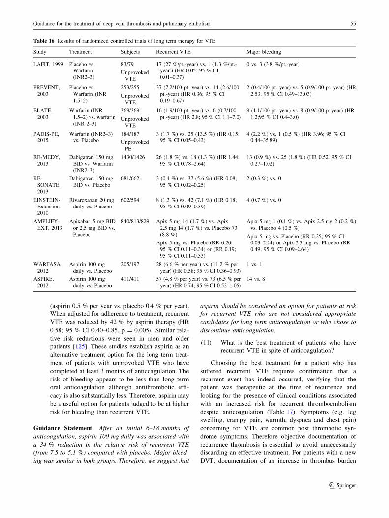
(aspirin 0.5 % per year vs. placebo 0.4 % per year).
When adjusted for adherence to treatment, recurrent
VTE was reduced by 42 % by aspirin therapy (HR
0.58; 95 % CI 0.40–0.85, p = 0.005). Similar rela-
tive risk reductions were seen in men and older
patients [125]. These studies establish aspirin as an
alternative treatment option for the long term treat-
ment of patients with unprovoked VTE who have
completed at least 3 months of anticoagulation. The
risk of bleeding appears to be less than long term
oral anticoagulation although antithrombotic effi-
cacy is also substantially less. Therefore, aspirin may
be a useful option for patients judged to be at higher
risk for bleeding than recurrent VTE.
Guidance Statement After an initial 6–18 months of
anticoagulation, aspirin 100 mg daily was associated with
a 34 % reduction in the relative risk of recurrent VTE
(from 7.5 to 5.1 %) compared with placebo. Major bleed-
ing was similar in both groups. Therefore, we suggest that
aspirin should be considered an option for patients at risk
for recurrent VTE who are not considered appropriate
candidates for long term anticoagulation or who chose to
discontinue anticoagulation.
(11) What is the best treatment of patients who have
recurrent VTE in spite of anticoagulation?
Choosing the best treatment for a patient who has
suffered recurrent VTE requires confirmation that a
recurrent event has indeed occurred, verifying that the
patient was therapeutic at the time of recurrence and
looking for the presence of clinical conditions associated
with an increased risk for recurrent thromboembolism
despite anticoagulation (Table 17). Symptoms (e.g. leg
swelling, crampy pain, warmth, dyspnea and chest pain)
concerning for VTE are common post thrombotic syn-
drome symptoms. Therefore objective documentation of
recurrence thrombosis is essential to avoid unnecessarily
discarding an effective treatment. For patients with a new
DVT, documentation of an increase in thrombus burden
Table 16 Results of randomized controlled trials of long term therapy for VTE
Study Treatment Subjects Recurrent VTE Major bleeding
LAFIT, 1999 Placebo vs.
Warfarin
(INR2–3)
83/79
Unprovoked
VTE
17 (27 %/pt.-year) vs. 1 (1.3 %/pt.-
year.) (HR 0.05; 95 % CI
0.01–0.37)
0 vs. 3 (3.8 %/pt.-year)
PREVENT,
2003
Placebo vs.
Warfarin (INR
1.5–2)
253/255
Unprovoked
VTE
37 (7.2/100 pt.-year) vs. 14 (2.6/100
pt.-year) (HR 0.36; 95 % CI
0.19–0.67)
2 (0.4/100 pt.-year) vs. 5 (0.9/100 pt.-year) (HR
2.53; 95 % CI 0.49–13.03)
ELATE,
2003
Warfarin (INR
1.5–2) vs. warfarin
(INR 2–3)
369/369
Unprovoked
VTE
16 (1.9/100 pt.-year) vs. 6 (0.7/100
pt.-year) (HR 2.8; 95 % CI 1.1–7.0)
9 (1.1/100 pt.-year) vs. 8 (0.9/100 pt.year) (HR
1.2;95 % CI 0.4–3.0)
PADIS-PE,
2015
Warfarin (INR2–3)
vs. Placebo
184/187
Unprovoked
PE
3 (1.7 %) vs. 25 (13.5 %) (HR 0.15;
95 % CI 0.05–0.43)
4 (2.2 %) vs. 1 (0.5 %) (HR 3.96; 95 % CI
0.44–35.89)
RE-MEDY,
2013
Dabigatran 150 mg
BID vs. Warfarin
(INR2–3)
1430/1426 26 (1.8 %) vs. 18 (1.3 %) (HR 1.44;
95 % CI 0.78–2.64)
13 (0.9 %) vs. 25 (1.8 %) (HR 0.52; 95 % CI
0.27–1.02)
RE-
SONATE,
2013
Dabigatran 150 mg
BID vs. Placebo
681/662 3 (0.4 %) vs. 37 (5.6 %) (HR 0.08;
95 % CI 0.02–0.25)
2 (0.3 %) vs. 0
EINSTEIN-
Extension,
2010
Rivaroxaban 20 mg
daily vs. Placebo
602/594 8 (1.3 %) vs. 42 (7.1 %) (HR 0.18;
95 % CI 0.09–0.39)
4 (0.7 %) vs. 0
AMPLIFY-
EXT, 2013
Apixaban 5 mg BID
or 2.5 mg BID vs.
Placebo
840/813/829 Apix 5 mg 14 (1.7 %) vs. Apix
2.5 mg 14 (1.7 %) vs. Placebo 73
(8.8 %)
Apix 5 mg vs. Placebo (RR 0.20;
95 % CI 0.11–0.34) or (RR 0.19;
95 % CI 0.11–0.33)
Apix 5 mg 1 (0.1 %) vs. Apix 2.5 mg 2 (0.2 %)
vs. Placebo 4 (0.5 %)
Apix 5 mg vs. Placebo (RR 0.25; 95 % CI
0.03–2.24) or Apix 2.5 mg vs. Placebo (RR
0.49; 95 % CI 0.09–2.64)
WARFASA,
2012
Aspirin 100 mg
daily vs. Placebo
205/197 28 (6.6 % per year) vs. (11.2 % per
year) (HR 0.58; 95 % CI 0.36–0.93)
1 vs. 1
ASPIRE,
2012
Aspirin 100 mg
daily vs. Placebo
411/411 57 (4.8 % per year) vs. 73 (6.5 % per
year) (HR 0.74; 95 % CI 0.52–1.05)
14 vs. 8
Guidance for the treatment of deep vein thrombosis and pulmonary embolism 55
123
(involvement of previously uninvolved vascular territories
or an increase in thrombus diameter of 4 mm or more). In
the event of an increase in thrombus diameter of
1–3.9 mm, repeat imaging in 1 week or venography is
warranted [126]. Recurrent PE is diagnosed if CT
angiography documents a new filling defect in a segmental
or larger artery or new segmental mismatched defect is
identified on ventilation perfusion scanning [127].
If a new event is confirmed it is important to determine
whether subtherapeutic anticoagulation was a contributing
factor. For patients on a VKA, review of the recent INR
values is essential. If subtherapeutic values are identified
redoubled efforts to maintain therapeutic INR values are
appropriate or perhaps a slight adjustment of the INR range
upward (increase from INR 2–3 to 2.5–3.5) to reduce the
chances of subtherapeutic INR values in the future. In
patients on a VKA it is also important to determine whether
they have one of a selected group of thrombophilic disor-
ders that predisposes to recurrent VTE despite therapeutic
anticoagulation. Considerations include cancer, antiphos-
pholipid syndrome, dysfibrinogenemia and delayed heparin
induced thrombocytopenia as well as anatomic reasons for
recurrent DVT. If antiphospholipid syndrome or dysfib-
rinogenemia is present, then the validity of the INR in the
patient should be confirmed using a test that is not influ-
enced by the coagulopathy, such as the chromogenic factor
X activity assay. For patients with cancer, therapeutic dose
LMWH should be used for therapy. For patients with
delayed HIT, use of a direct thrombin inhibitor or fonda-
parinux with later transition to warfarin following HIT
resolution should be considered. Consideration of surgical
correction of anatomic vascular compression is warranted.
For patients on a VKA who suffer recurrent VTE without
an identifiable cause, there are limited data for guidance but
potential options include use of a higher INR target range
(INR 2.5–3.5 or 3–4 depending upon the INR at the time of
the thrombotic event) or switching to a LMWH or fonda-
parinux [119, 128–130]. SinceDOACswere demonstrated to
be equivalent to VKA in the treatment of VTE, additional
data are needed to support their use in the setting of recurrent
VTE during anticoagulation with VKA. For patients on
LMWH, providers should make sure they have been taking
the appropriate dose and refilling their prescriptions at
appropriate intervals. LMWH (anti-Xa) levels are not usu-
ally practical as they are rarely obtained in a timely manner
when the results would be useful. If patients are taking the
appropriate dose then increasing the dose by 25 % is a
common strategy used in cancer patients [131]. In patients
taking enoxaparin, one can switch to 1 mg/kg twice daily
dosing if 1.5 mg/kg once daily dosing had been used.
Alternatively, one could switch to fondaparinux which has a
longer half-life than LMWH. Rarely, cancer patients with
Trousseau’s syndrome are not responsive to LMWH or
fondaparinux in therapeutic doses. In these instances one
might use IV UFH to assess the dose necessary for control of
the coagulopathy and then transition to subcutaneous UFH
adjusted to aPTT levels. Whenever recurrence occurs it is
important to identify risk factors for thrombosis and then
eliminate any removable risk factors (i.e. vascular com-
pression causing stasis) [59]. For patients on a DOAC who
experience recurrent VTE, providers should ask patients
about missed doses prior to the event. Since DOACs have a
short half-life, measuring anti-Xa levels (factor Xa inhibi-
tors) or anti-IIa assays (Hemoclot� thrombin inhibitor assay
or ecarin clotting time) is not a practical strategy to assess
adherence. If there are no identifiable reasons (i.e. cancer,
anatomic vascular compression) for recurrence in a patient
taking a DOAC for VTE, switching the patient to a VKA
where adherence can be monitored may be preferable.
Guidance Statement In patients with recurrent VTE
despite anticoagulation, we suggest that it is important for
providers to assess adherence to therapy and identify clinical
conditions associated with anticoagulation failure including
cancer, antiphospholipid syndrome, heparin-induced throm-
bocytopenia and vascular compression syndromes (May-
Thurner syndrome, thoracic outlet syndrome). We suggest
that higher-intensity anticoagulation (VKA INR 2.5–3.5 or
3–4 or based upon chromogenic factor X activity or escalated
dose [125 % dose] LMWH), alternative forms of parenteral
anticoagulation and therapies directed at restoring adequate
blood flow are effective strategies to consider.
(12) How can you assess the risk of recurrent VTE and
anticoagulant-associated bleeding?
Recurrent VTE Risk factors for recurrent VTE include
unprovoked VTE, certain hereditary thrombophilia,
antiphospholipid syndrome, cancer, male gender, elevated
D dimer on and off anticoagulation and the presence of
residual venous thrombosis on duplex ultrasound [132]. As
outlined above in the duration of therapy section of this
chapter, the most important risk factor for recurrence is the
presence or absence of situational prothrombotic triggers at
the time of the VTE. Patients with unprovoked VTE are
intrinsically hypercoagulable and remain at elevated risk
for recurrent VTE if anticoagulation is discontinued. In
contrast, patients who suffer VTE in the presence of potent
situational triggers such as major surgery or trauma are at
low risk of recurrent VTE [81]. If a patient still has active
risk factors for VTE at the conclusion of therapy (relative
immobility, persistent infections or hospitalization associ-
ated with surgical complications) then prolongation of the
course of anticoagulation should be considered on a case-
by-case basis. Patients with non-surgical risk factors for
VTE are at intermediate risk for recurrence (4.2 % per
year) [81]. In these patients, continuation of therapy is
56 M. B. Streiff et al.
123
appropriate if the identifiable risk factors (active inflam-
matory bowel disease, etc.) are still present [132].
A prime example of this patient subgroup are cancer
patients. Cancer patients with VTE are 3-fold higher risk of
recurrence than patients without cancer [133]. Therefore,
cancer patients with VTE should remain on anticoagulation
until they are in remission and no longer on cancer therapy.
Not surprisingly, the risk of recurrence varies with the type
and extent of cancer. Patients with stage 1 or 2 cancer have
a similar risk of recurrence as patients without cancer while
patients with stage 3 or 4 disease are at significantly higher
risk of recurrence. Patients with lung, gastrointestinal and
genitourinary cancers were at greater risk for recurrence
than other cancer sites [133]. Chee et al. also noted cancer
stage and primary site were risk factors for recurrence in
their population-based cohort study [134]. The under-
standing that not all cancer patients are at equal risk of
recurrence has spurred the development of risk assessment
models to identify patients at greater risk of recurrent VTE
such as the Ottawa Risk Prediction Model [135]. More
detailed discussion of treatment of VTE in cancer patients
can be found in accompanying paper by Khorana et al.
As described above and the accompanying paper by
Stevens et al., inherited thrombophilic disorders are asso-
ciated with a risk of recurrent VTE which varies by the
genetic defect. Hormonal therapy and pregnancy as well as
the post-partum period represent risk factors for initial and
recurrent VTE (see duration of therapy section above and
the accompanying paper by Stevens et al.
Obesity has been associated with a 1.6 fold increased
risk of recurrent VTE [136]. Therefore, weight loss should
be included in VTE risk modification strategies for patients
with VTE.
Residual vein obstruction (RVO) refers to the presence
of residual thrombus at the site of an initial DVT after a
defined period of anticoagulation. In a patient-level meta-
analysis, RVO was associated with a 1.3 fold increased risk
of recurrent VTE (95 % CI 1.06–1.65) among patients with
unprovoked DVT. RVO measured at 3 months was asso-
ciated with a higher risk of recurrent VTE (HR 2.17; 95 %
CI 1.11–4.25) while RVO detected beyond six months was
not a significant predictor of recurrence risk (HR 1.19;
95 % CI 0.87–1.61) [137]. Aside from its utility as a
prognostic tool, duplex ultrasound can be used to establish
a baseline at the end of therapy in case a patient reports
symptoms potentially attributable to a new DVT [126,
127]. Since the symptoms of DVT and post-thrombotic
syndrome can be difficult to differentiate, we obtain
baseline duplex studies at the end of therapy in patients at
high risk for recurrence who are discontinuing therapy
[138].
An abnormal D dimer is associated with a 2.6 fold
(95 % CI 1.90–3.52) increased risk of recurrent VTE
among patients with unprovoked events regardless of age
or cut point (500 vs. 250 lg/L) [139]. In a prospective
management study of patients with unprovoked VTE who
discontinued anticoagulation, Cosmi et al. found that
patients with persistently abnormal D dimer studies at and
beyond 90 days post-discontinuation of AC had a 9 fold
higher risk of recurrence than patients with normal D
dimers (Recurrent VTE 27 per 100 patient years [95 % CI
12–48] vs. 2.9 per 100 patient years [95 % CI 1–7]; HR
9.38, p = 0.0004) [140]. Palareti et al. also found that D
dimer testing identified patients with an initial unprovoked
VTE or one associated with minimal risk factors who were
at risk for recurrent VTE (8.8 % per patient-year vs. 3.0 %
per patient-year; HR 2.92; 95 % CI 1.87–9.72;
P = 0.0006) [141]. Using serial qualitative D dimer testing
in patients with an unprovoked VTE or one provoked by
estrogen therapy, Kearon and colleagues noted that women
with a negative D dimer who had an estrogen associated
VTE were at low risk for recurrence (0 %,95 % CI
0.0–3.0 %). Men (Recurrent VTE 9.7 % per patient year;
95 % CI 6.7–13.7 %) and women with unprovoked VTE
(5.4 % per patient year; 95 % CI 2.5–10.2 %) remained at
substantial risk of recurrence despite two negative D
dimers [98]. In patients with provoked VTE, Cosmi et al.
found that an abnormal D dimer at the time of discontin-
uation of AC (Day 0) (11.1 VTE per 100 patient-years
[95 % CI 4–24] vs. 2.2 VTE per 100 pt.-years [95 % CI
1–4]; adj. HR 4.2 [1.2–14.2]) and 30 days after discon-
tinuation of AC (Day 30) (6.7 VTE per 100 patient-years
[95 % CI 3–12] vs. 1.5 VTE per 100 pt.-years [95 % CI
0–3]; adj. HR 3.8[1.2–12.1]) were both associated with an
increased risk of recurrence [142].
Other global measures of activated coagulation also
have been associated with recurrence risk. In a prospective
study of 914 patients with spontaneous VTE, Hron et al.
found that thrombin generation (TG) less than 300 nM
(nM) (RR 0.37 [95 % CI 0.20–0.67]) and 300–400 nM (RR
0.45 [95 % CI 0.28–0.73] were associated with a reduced
risk of recurrent VTE compared to patients with TG greater
than 400 nM [143]. In a prospective study of 188 patients
with unprovoked VTE or VTE associated with a non-sur-
gical trigger, Besser et al. also noted that endogenous
thrombin potential (ETP) greater than the 50th percentile
was associated with an almost 3 fold greater risk of
recurrence (HR 2.9, 95 % CI 1.3–6.6, cumulative recur-
rence at 4 years 27 vs. 11 %). A high ETP remained a
significant predictor of recurrent VTE even after adjust-
ment for D dimer level, presence of thrombophilia, sex and
whether or not the first event was unprovoked (HR 2.6,
95 % CI 1.2–6.0) [144]. Tripodi et al. found that patients
with unprovoked VTE who had an ETP of greater than
960 nM times minutes or peak TG greater than 193 nM
measured in the presence of thrombomodulin were at
Guidance for the treatment of deep vein thrombosis and pulmonary embolism 57
123
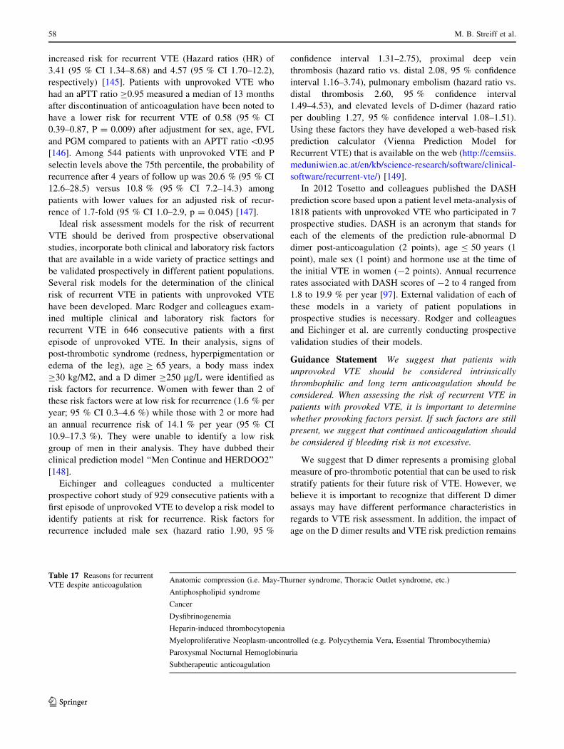
increased risk for recurrent VTE (Hazard ratios (HR) of
3.41 (95 % CI 1.34–8.68) and 4.57 (95 % CI 1.70–12.2),
respectively) [145]. Patients with unprovoked VTE who
had an aPTT ratio C0.95 measured a median of 13 months
after discontinuation of anticoagulation have been noted to
have a lower risk for recurrent VTE of 0.58 (95 % CI
0.39–0.87, P = 0.009) after adjustment for sex, age, FVL
and PGM compared to patients with an APTT ratio\0.95
[146]. Among 544 patients with unprovoked VTE and P
selectin levels above the 75th percentile, the probability of
recurrence after 4 years of follow up was 20.6 % (95 % CI
12.6–28.5) versus 10.8 % (95 % CI 7.2–14.3) among
patients with lower values for an adjusted risk of recur-
rence of 1.7-fold (95 % CI 1.0–2.9, p = 0.045) [147].
Ideal risk assessment models for the risk of recurrent
VTE should be derived from prospective observational
studies, incorporate both clinical and laboratory risk factors
that are available in a wide variety of practice settings and
be validated prospectively in different patient populations.
Several risk models for the determination of the clinical
risk of recurrent VTE in patients with unprovoked VTE
have been developed. Marc Rodger and colleagues exam-
ined multiple clinical and laboratory risk factors for
recurrent VTE in 646 consecutive patients with a first
episode of unprovoked VTE. In their analysis, signs of
post-thrombotic syndrome (redness, hyperpigmentation or
edema of the leg), age C 65 years, a body mass index
C30 kg/M2, and a D dimer C250 lg/L were identified as
risk factors for recurrence. Women with fewer than 2 of
these risk factors were at low risk for recurrence (1.6 % per
year; 95 % CI 0.3–4.6 %) while those with 2 or more had
an annual recurrence risk of 14.1 % per year (95 % CI
10.9–17.3 %). They were unable to identify a low risk
group of men in their analysis. They have dubbed their
clinical prediction model ‘‘Men Continue and HERDOO2’’
[148].
Eichinger and colleagues conducted a multicenter
prospective cohort study of 929 consecutive patients with a
first episode of unprovoked VTE to develop a risk model to
identify patients at risk for recurrence. Risk factors for
recurrence included male sex (hazard ratio 1.90, 95 %
confidence interval 1.31–2.75), proximal deep vein
thrombosis (hazard ratio vs. distal 2.08, 95 % confidence
interval 1.16–3.74), pulmonary embolism (hazard ratio vs.
distal thrombosis 2.60, 95 % confidence interval
1.49–4.53), and elevated levels of D-dimer (hazard ratio
per doubling 1.27, 95 % confidence interval 1.08–1.51).
Using these factors they have developed a web-based risk
prediction calculator (Vienna Prediction Model for
Recurrent VTE) that is available on the web (http://cemsiis.
meduniwien.ac.at/en/kb/science-research/software/clinical-
software/recurrent-vte/) [149].
In 2012 Tosetto and colleagues published the DASH
prediction score based upon a patient level meta-analysis of
1818 patients with unprovoked VTE who participated in 7
prospective studies. DASH is an acronym that stands for
each of the elements of the prediction rule-abnormal D
dimer post-anticoagulation (2 points), age B 50 years (1
point), male sex (1 point) and hormone use at the time of
the initial VTE in women (-2 points). Annual recurrence
rates associated with DASH scores of -2 to 4 ranged from
1.8 to 19.9 % per year [97]. External validation of each of
these models in a variety of patient populations in
prospective studies is necessary. Rodger and colleagues
and Eichinger et al. are currently conducting prospective
validation studies of their models.
Guidance Statement We suggest that patients with
unprovoked VTE should be considered intrinsically
thrombophilic and long term anticoagulation should be
considered. When assessing the risk of recurrent VTE in
patients with provoked VTE, it is important to determine
whether provoking factors persist. If such factors are still
present, we suggest that continued anticoagulation should
be considered if bleeding risk is not excessive.
We suggest that D dimer represents a promising global
measure of pro-thrombotic potential that can be used to risk
stratify patients for their future risk of VTE. However, we
believe it is important to recognize that different D dimer
assays may have different performance characteristics in
regards to VTE risk assessment. In addition, the impact of
age on the D dimer results and VTE risk prediction remains
Table 17 Reasons for recurrent
VTE despite anticoagulationAnatomic compression (i.e. May-Thurner syndrome, Thoracic Outlet syndrome, etc.)
Antiphospholipid syndrome
Cancer
Dysfibrinogenemia
Heparin-induced thrombocytopenia
Myeloproliferative Neoplasm-uncontrolled (e.g. Polycythemia Vera, Essential Thrombocythemia)
Paroxysmal Nocturnal Hemoglobinuria
Subtherapeutic anticoagulation
58 M. B. Streiff et al.
123
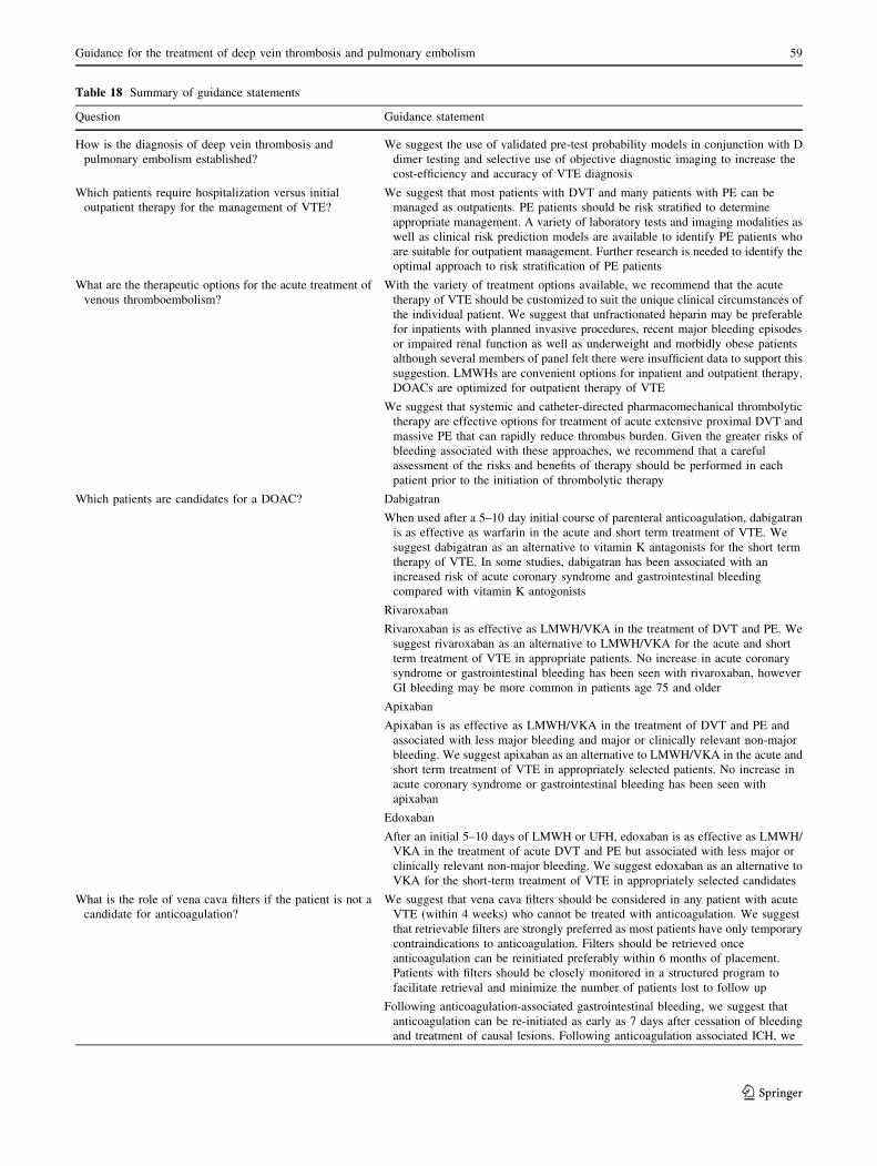
Table 18 Summary of guidance statements
Question Guidance statement
How is the diagnosis of deep vein thrombosis and
pulmonary embolism established?
We suggest the use of validated pre-test probability models in conjunction with D
dimer testing and selective use of objective diagnostic imaging to increase the
cost-efficiency and accuracy of VTE diagnosis
Which patients require hospitalization versus initial
outpatient therapy for the management of VTE?
We suggest that most patients with DVT and many patients with PE can be
managed as outpatients. PE patients should be risk stratified to determine
appropriate management. A variety of laboratory tests and imaging modalities as
well as clinical risk prediction models are available to identify PE patients who
are suitable for outpatient management. Further research is needed to identify the
optimal approach to risk stratification of PE patients
What are the therapeutic options for the acute treatment of
venous thromboembolism?
With the variety of treatment options available, we recommend that the acute
therapy of VTE should be customized to suit the unique clinical circumstances of
the individual patient. We suggest that unfractionated heparin may be preferable
for inpatients with planned invasive procedures, recent major bleeding episodes
or impaired renal function as well as underweight and morbidly obese patients
although several members of panel felt there were insufficient data to support this
suggestion. LMWHs are convenient options for inpatient and outpatient therapy.
DOACs are optimized for outpatient therapy of VTE
We suggest that systemic and catheter-directed pharmacomechanical thrombolytic
therapy are effective options for treatment of acute extensive proximal DVT and
massive PE that can rapidly reduce thrombus burden. Given the greater risks of
bleeding associated with these approaches, we recommend that a careful
assessment of the risks and benefits of therapy should be performed in each
patient prior to the initiation of thrombolytic therapy
Which patients are candidates for a DOAC? Dabigatran
When used after a 5–10 day initial course of parenteral anticoagulation, dabigatran
is as effective as warfarin in the acute and short term treatment of VTE. We
suggest dabigatran as an alternative to vitamin K antagonists for the short term
therapy of VTE. In some studies, dabigatran has been associated with an
increased risk of acute coronary syndrome and gastrointestinal bleeding
compared with vitamin K antogonists
Rivaroxaban
Rivaroxaban is as effective as LMWH/VKA in the treatment of DVT and PE. We
suggest rivaroxaban as an alternative to LMWH/VKA for the acute and short
term treatment of VTE in appropriate patients. No increase in acute coronary
syndrome or gastrointestinal bleeding has been seen with rivaroxaban, however
GI bleeding may be more common in patients age 75 and older
Apixaban
Apixaban is as effective as LMWH/VKA in the treatment of DVT and PE and
associated with less major bleeding and major or clinically relevant non-major
bleeding. We suggest apixaban as an alternative to LMWH/VKA in the acute and
short term treatment of VTE in appropriately selected patients. No increase in
acute coronary syndrome or gastrointestinal bleeding has been seen with
apixaban
Edoxaban
After an initial 5–10 days of LMWH or UFH, edoxaban is as effective as LMWH/
VKA in the treatment of acute DVT and PE but associated with less major or
clinically relevant non-major bleeding. We suggest edoxaban as an alternative to
VKA for the short-term treatment of VTE in appropriately selected candidates
What is the role of vena cava filters if the patient is not a
candidate for anticoagulation?
We suggest that vena cava filters should be considered in any patient with acute
VTE (within 4 weeks) who cannot be treated with anticoagulation. We suggest
that retrievable filters are strongly preferred as most patients have only temporary
contraindications to anticoagulation. Filters should be retrieved once
anticoagulation can be reinitiated preferably within 6 months of placement.
Patients with filters should be closely monitored in a structured program to
facilitate retrieval and minimize the number of patients lost to follow up
Following anticoagulation-associated gastrointestinal bleeding, we suggest that
anticoagulation can be re-initiated as early as 7 days after cessation of bleeding
and treatment of causal lesions. Following anticoagulation associated ICH, we
Guidance for the treatment of deep vein thrombosis and pulmonary embolism 59
123
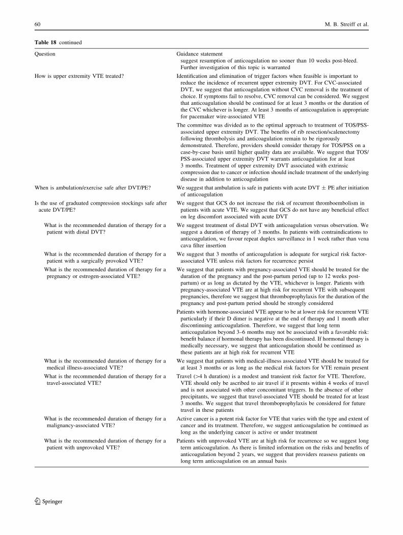
Table 18 continued
Question Guidance statement
suggest resumption of anticoagulation no sooner than 10 weeks post-bleed.
Further investigation of this topic is warranted
How is upper extremity VTE treated? Identification and elimination of trigger factors when feasible is important to
reduce the incidence of recurrent upper extremity DVT. For CVC-associated
DVT, we suggest that anticoagulation without CVC removal is the treatment of
choice. If symptoms fail to resolve, CVC removal can be considered. We suggest
that anticoagulation should be continued for at least 3 months or the duration of
the CVC whichever is longer. At least 3 months of anticoagulation is appropriate
for pacemaker wire-associated VTE
The committee was divided as to the optimal approach to treatment of TOS/PSS-
associated upper extremity DVT. The benefits of rib resection/scalenectomy
following thrombolysis and anticoagulation remain to be rigorously
demonstrated. Therefore, providers should consider therapy for TOS/PSS on a
case-by-case basis until higher quality data are available. We suggest that TOS/
PSS-associated upper extremity DVT warrants anticoagulation for at least
3 months. Treatment of upper extremity DVT associated with extrinsic
compression due to cancer or infection should include treatment of the underlying
disease in addition to anticoagulation
When is ambulation/exercise safe after DVT/PE? We suggest that ambulation is safe in patients with acute DVT ± PE after initiation
of anticoagulation
Is the use of graduated compression stockings safe after
acute DVT/PE?
We suggest that GCS do not increase the risk of recurrent thromboembolism in
patients with acute VTE. We suggest that GCS do not have any beneficial effect
on leg discomfort associated with acute DVT
What is the recommended duration of therapy for a
patient with distal DVT?
We suggest treatment of distal DVT with anticoagulation versus observation. We
suggest a duration of therapy of 3 months. In patients with contraindications to
anticoagulation, we favour repeat duplex surveillance in 1 week rather than vena
cava filter insertion
What is the recommended duration of therapy for a
patient with a surgically provoked VTE?
We suggest that 3 months of anticoagulation is adequate for surgical risk factor-
associated VTE unless risk factors for recurrence persist
What is the recommended duration of therapy for a
pregnancy or estrogen-associated VTE?
We suggest that patients with pregnancy-associated VTE should be treated for the
duration of the pregnancy and the post-partum period (up to 12 weeks post-
partum) or as long as dictated by the VTE, whichever is longer. Patients with
pregnancy-associated VTE are at high risk for recurrent VTE with subsequent
pregnancies, therefore we suggest that thromboprophylaxis for the duration of the
pregnancy and post-partum period should be strongly considered
Patients with hormone-associated VTE appear to be at lower risk for recurrent VTE
particularly if their D dimer is negative at the end of therapy and 1 month after
discontinuing anticoagulation. Therefore, we suggest that long term
anticoagulation beyond 3–6 months may not be associated with a favorable risk:
benefit balance if hormonal therapy has been discontinued. If hormonal therapy is
medically necessary, we suggest that anticoagulation should be continued as
these patients are at high risk for recurrent VTE
What is the recommended duration of therapy for a
medical illness-associated VTE?
We suggest that patients with medical-illness associated VTE should be treated for
at least 3 months or as long as the medical risk factors for VTE remain present
What is the recommended duration of therapy for a
travel-associated VTE?
Travel ([4 h duration) is a modest and transient risk factor for VTE. Therefore,
VTE should only be ascribed to air travel if it presents within 4 weeks of travel
and is not associated with other concomitant triggers. In the absence of other
precipitants, we suggest that travel-associated VTE should be treated for at least
3 months. We suggest that travel thromboprophylaxis be considered for future
travel in these patients
What is the recommended duration of therapy for a
malignancy-associated VTE?
Active cancer is a potent risk factor for VTE that varies with the type and extent of
cancer and its treatment. Therefore, we suggest anticoagulation be continued as
long as the underlying cancer is active or under treatment
What is the recommended duration of therapy for a
patient with unprovoked VTE?
Patients with unprovoked VTE are at high risk for recurrence so we suggest long
term anticoagulation. As there is limited information on the risks and benefits of
anticoagulation beyond 2 years, we suggest that providers reassess patients on
long term anticoagulation on an annual basis
60 M. B. Streiff et al.
123
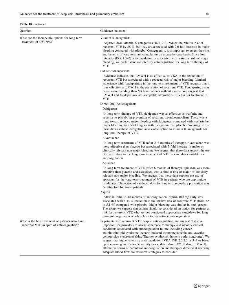
Table 18 continued
Question Guidance statement
What are the therapeutic options for long term
treatment of DVT/PE?
Vitamin K antagonists
Adjusted dose vitamin K antagonists (INR 2–3) reduce the relative risk of
recurrent VTE by 88 %, but they are associated with 2.6 fold increase in major
bleeding compared with placebo. Consequently, it is important to assess the risks
and benefits of long term anticoagulation on a case-by-case basis. Since low
intensity (INR 1.5–2) anticoagulation is associated with a similar risk of major
bleeding, we prefer standard intensity anticoagulation for long term therapy of
VTE
LMWH/Fondaparinux
Evidence indicates that LMWH is as effective as VKA in the reduction of
recurrent VTE but associated with a reduced risk of major bleeding. Limited
experience with fondaparinux in the long term treatment of VTE suggests that it
is as effective as LMWH in the prevention of recurrent VTE. Fondaparinux may
cause more bleeding than VKA in patients without cancer. We suggest that
LMWH and fondaparinux are acceptable alternatives to VKA for treatment of
VTE
Direct Oral Anticoagulants
Dabigatran
In long term therapy of VTE, dabigatran was as effective as warfarin and
superior to placebo in prevention of recurrent thromboembolism. There was a
trend toward reduced major bleeding with dabigatran compared with warfarin but
major bleeding was 3-fold higher with dabigatran than placebo. We suggest that
these data establish dabigatran as a viable option to vitamin K antagonists for
long term therapy of VTE
Rivaroxaban
In long term treatment of VTE (after 3–6 months of therapy), rivaroxaban was
more effective than placebo but associated with 5 fold increase in major or
clinically relevant non-major bleeding. We suggest that these data support the use
of rivaroxaban in the long term treatment of VTE in candidates suitable for
anticoagulation
Apixaban
In long term treatment of VTE (after 6 months of therapy), apixaban was more
effective than placebo and associated with a similar risk of major or clinically
relevant non-major bleeding. We suggest that these data support the use of
apixaban for the long term treatment of VTE in patients who are appropriate
candidates. The option of a reduced dose for long term secondary prevention may
be attractive for some patients
Aspirin
After an initial 6–18 months of anticoagulation, aspirin 100 mg daily was
associated with a 34 % reduction in the relative risk of recurrent VTE (from 7.5
to 5.1 %) compared with placebo. Major bleeding was similar in both groups.
Therefore, we suggest that aspirin should be considered an option for patients at
risk for recurrent VTE who are not considered appropriate candidates for long
term anticoagulation or who chose to discontinue anticoagulation
What is the best treatment of patients who have
recurrent VTE in spite of anticoagulation?
In patients with recurrent VTE despite anticoagulation, we suggest that it is
important for providers to assess adherence to therapy and identify clinical
conditions associated with anticoagulation failure including cancer,
antiphospholipid syndrome, heparin-induced thrombocytopenia and vascular
compression syndromes (May-Thurner syndrome, thoracic outlet syndrome). We
suggest that higher-intensity anticoagulation (VKA INR 2.5-3.5 or 3–4 or based
upon chromogenic factor X activity or escalated dose [125 % dose] LMWH),
alternative forms of parenteral anticoagulation and therapies directed at restoring
adequate blood flow are effective strategies to consider
Guidance for the treatment of deep vein thrombosis and pulmonary embolism 61
123
incompletely characterized. The value of other global
laboratory measures (endogenous thrombin potential) and
imaging studies remains to be established. Multimodality
risk assessment models appear to be an effective approach
to risk stratification of patients with unprovoked VTE.
Further validation of these risk assessment tools is
underway.
Anticoagulation-associated bleeding
In determining the appropriate duration of anticoagulant
therapy for VTE it is important to assess not only the risk
of recurrent VTE but also the risk of bleeding associated
with continued anticoagulation. Several different models
have been developed primarily by studying patients with
atrial fibrillation to determine the risk of bleeding associ-
ated with anticoagulation with vitamin K antagonists
(HEMORR2HAGES, HAS-BLED, ATRIA). However the
clinical characteristics of patients with atrial fibrillation and
VTE differ. Therefore, these bleeding risk prediction
models may not be as predictive in patients with VTE.
Several bleeding risk assessment models have been
developed specifically for VTE patients. However, the
performance of these models in assessing bleeding risk has
been modest [150]. In addition, none of these models has
been used or validated in conjunction with DOACs.
Therefore, at this point, it is premature to recommend the
use of bleeding risk assessment models to determine the
duration of anticoagulation for patients with VTE. Devel-
opment of new bleeding risk prediction models as well as
composite tools that identify patients at increased risk for
multiple adverse events (e.g., thrombosis, bleeding) remain
an important area of investigation.
Guidance Statement While a bleeding risk assessment is
important to the decision on the duration of anticoagula-
tion, we suggest that it is premature to use formal bleeding
risk assessment models to identify patients who should
discontinue anticoagulation. Development of better risk
prediction models remains a priority.
Balancing the risks and benefits Treatment of VTE is
associated with benefits (reduction in recurrent VTE and its
morbidity and mortality) as well as risks (bleeding com-
plications, negative economic and life style impact).
Therefore, decision-making on the duration of anticoagu-
lation must be made jointly with the patient taking into
account their beliefs and preferences. For this conversation
to be productive and informed the patient must be well
educated by the provider about venous thromboembolism
and its treatment so that he/she can make an informed
decision. A variety of excellent resources are available to
improve patient understanding of VTE including the
National Blood Clot Alliance, Clot Connect, Thrombosis
Canada and the Centers for Disease Control and Prevention
just to name a few. In the clinic, we review the patho-
physiology of VTE with patients, the presenting signs and
symptoms and the individual risk factors for VTE and the
risk factors and signs and symptoms of anticoagulation
associated bleeding so that patients can make an informed
Table 18 continued
Question Guidance statement
How can you assess the risk of recurrent VTE and
anticoagulant-associated bleeding?
We suggest that patients with unprovoked VTE should be considered intrinsically
thrombophilic and long term anticoagulation should be considered. When
assessing the risk of recurrent VTE in patients with provoked VTE, it is important
to determine whether provoking factors persist. If such factors are still present,
we suggest that continued anticoagulation should be considered if bleeding risk is
not excessive
We suggest that D dimer testing represents a promising global measure of pro-
thrombotic potential that can be used to risk stratify patients for their future risk
of VTE. However, we believe it is important to recognize that different D dimer
assays may have different performance characteristics in regards to VTE risk
assessment. In addition, the impact of age on the D dimer results and VTE risk
prediction remains incompletely characterized. The value of other global
laboratory measures (endogenous thrombin potential) and imaging studies
remains to be established. Multimodality risk assessment models appear to be
effective approach to risk stratification of patients with unprovoked VTE. Further
validation of these risk assessment tools is underway
Anticoagulation-associated bleeding
While a bleeding risk assessment is important to the decision on the duration of
anticoagulation, we suggest that it is premature to use formal bleeding risk
assessment models to identify patients who should discontinue anticoagulation.
Development of better risk prediction models remains a priority
62 M. B. Streiff et al.
123
decision and know when it is appropriate to contact
healthcare providers. For patients with unprovoked VTE
we generally recommend long term anticoagulation unless
they are non-adherent or have had bleeding complications.
If the patient favors discontinuation of anticoagulation, we
use the Vienna Prediction Model or data derived from the
literature to determine an estimate of the patients risk for
recurrent thromboembolism so that they have a rough idea
of their risk of recurrence off anticoagulation [96, 149,
151]. For patients who suffered VTE in the setting of a
major transient risk factor (major surgery or trauma) we
generally discontinue anticoagulation after 3–6 months of
therapy. For patients with non-surgical triggered VTE, we
treat for at least 3–6 months or longer if the risk factors for
their event remain unresolved. Table 18 summarizes these
guidance statements.
Acknowledgments We wish to acknowledge the support provided
by Myelin and Associates with the preparation of this manuscript for
submission. The work contained in this manuscript was partially
funded by support from the following companies: Boehringer Ingel-
heim, Daiichi Sankyo and Janssen Pharmaceuticals. This guidance
document is endorsed by the Anticoagulation Forum’s Board of
Directors: Mark Crowther, MD, MSc, FRCPC, Jack E. Ansell, MD,
Allison Burnett, PharmD, Nathan Clark, PharmD, Adam Cuker, MD,
David Garcia, MD, Scott Kaatz, DO, MSc, FACP, Renato D. Lopes,
MD, PhD, Tracy Minichiello, MD, Edith Nutescu, PharmD, FCCP,
Lynn Oertel, MS, ANP, CACP, Eva Kline-Rogers, MS, RN, NP,Terri
Schnurr, RN, CCRC, Michael Streiff, MD, Diane Wirth, ANP, CACP,
BCPS, CACP, Daniel Witt, Pharm D, Ann Wittkowsky, PharmD,
CACP, FASHP, FCCP.
Compliance with ethical standards
Disclosures M Streiff: consulted for BiO2 Medical, Boehringer
Ingelheim, CSL Behring, Daiichi-Sankyo, Janssen Healthcare and
Pfizer. He has received research grant support from Daiichi-Sankyo,
Janssen Healthcare, PCORI and Portola. G Agnelli: none. J Connors:
Scientific Advisory Boards: Boehringer Ingelheim, Bristol Myers
Squibb. M Crowther: Advisory boards for Octapharma, Bayer,
Boehringer Ingelheim, Janssen, Pfizer, Leo Pharma and Portola.
Study Steering committees, or other research related activities for
projects involving AKP America, Daichii, Bayer and Leo Pharma.
Holds a Career Investigator award from the Heart and Stroke Foun-
dation of Ontario, and the Leo Pharma Chair in Thromboembolism
Research at McMaster University (income from which is used to
support the salary of research employees); his institutions (McMaster
University and/or St Joseph’s Healthcare) have received funding for
research projects from the Heart and Stroke Foundation of Canada,
Leo Pharma and Bayer for work in which Dr Crowther is involved;
received funding for preparation of educational materials and/or
presentations from Alexion, Ortho Clinical Diagnostics, BMS-Pfizer
alliance, Leo Pharma, Abbvie, Bayer, Celgene, Shire and CSL
Behring; participated in various medicolegal activities relating to
thrombosis, anticoagulant drugs, or other aspects of hematological
practice, and that these activities are bound by confidentiality
arrangements. Further this declaration is made to the best of Dr
Crowther’s ability, but may be incomplete or contain material errors
due to the foibles of human memory. S Eichinger: Honoraria, con-
sultancies: Bayer, Boehringer Ingelheim, Daiichi-Sankyo, Bristol
Myers Squibb, Pfizer. R Lopes: Research Grant from Bristol Myers
Squibb and Glaxo SmithKline. Consultant for Bayer, Boehringer
Ingelheim, Daiichi-Sankyo, Bristol Myers Squibb, Pfizer, Merck,
Portola. R McBane: none. S Moll: Consultant for Portola. J Ansell:
Consulting activities and/or honoraria from the following companies:
Bristol Myers Squibb; Pfizer; Boehringer Ingelheim; Daiichi Sankyo;
Janssen; Perosphere; Roche Diagnostics; Alere, Inc; Instrumentation
Laboratories. Equity interest in the following companies: Perosphere,
Inc.
Open Access This article is distributed under the terms of the
Creative Commons Attribution 4.0 International License (http://crea
tivecommons.org/licenses/by/4.0/), which permits unrestricted use,
distribution, and reproduction in any medium, provided you give
appropriate credit to the original author(s) and the source, provide a
link to the Creative Commons license, and indicate if changes were
made.
References
1. Rosendaal FR (1999) Risk factors for venous thrombotic dis-
ease. Thromb Haemost 82(2):610–619
2. Lijfering WM, Rosendaal FR, Cannegieter SC (2010) Risk
factors for venous thrombosis - current understanding from an
epidemiological point of view. Br J Haematol 149(6):824–833
3. Wells PS, Anderson DR, Bormanis J et al (1997) Value of
assessment of pretest probability of deep-vein thrombosis in
clinical management. Lancet 350(9094):1795–1798
4. Wells PS, Ginsberg JS, Anderson DR et al (1998) Use of a
clinical model for safe management of patients with suspected
pulmonary embolism. Ann Intern Med 129(12):997–1005
5. Klok FA, Mos IC, Nijkeuter M et al (2008) Simplification of the
revised geneva score for assessing clinical probability of pul-
monary embolism. Arch Intern Med 168(19):2131–2136
6. Stein PD, Hull RD, Patel KC et al (2004) D-dimer for the
exclusion of acute venous thrombosis and pulmonary embolism:
a systematic review. Ann Intern Med 140(8):589–602
7. Kline JA, Webb WB et al (2004) Impact of a rapid rule-out
protocol for pulmonary embolism on the rate of screening,
missed cases, and pulmonary vascular imaging in an urban US
emergency department. Ann Emerg Med 44(5):490–502
8. Kline JA, Courtney DM, Kabrhel C et al (2008) Prospective
multicenter evaluation of the pulmonary embolism rule-out
criteria. J Thromb Haemost 6(5):772–780
9. Singh B, Mommer SK, Erwin PJ et al (2013) Pulmonary
embolism rule-out criteria (PERC) in pulmonary embolism-re-
visited: a systematic review and meta-analysis. Emerg Med J
30(9):701–706
10. Raja AS, Greenberg JO, Qaseem A et al (2015) Evaluation of
patients with suspected acute pulmonary embolism: best practice
advice from the clinical guidelines committee of the American
College of Physicians. Ann Intern Med 163(9):701–711
11. Agnelli G, Becattini C (2010) Acute pulmonary embolism.
N Engl J Med 363(3):266–274
12. Geersing GJ, Zuithoff NP, Kearon C et al (2014) Exclusion of
deep vein thrombosis using the wells rule in clinically important
subgroups: individual patient data meta-analysis. BMJ
348:g1340
13. Righini M, Van Es J, Den Exter PL et al (2014) Age-adjusted
D-dimer cutoff levels to rule out pulmonary embolism: the
ADJUST-PE study. JAMA 311(11):1117–1124
14. Konstantinides SV, Torbicki A, Agnelli G et al (2014) 2014
ESC guidelines on the diagnosis and management of acute
pulmonary embolism. Eur Heart J 35(43):3033–3069
Guidance for the treatment of deep vein thrombosis and pulmonary embolism 63
123
15. Kearon C, Akl EA, Comerota AJ et al (2012) American College
of Chest Physicians. Antithrombotic therapy for VTE disease:
antithrombotic therapy and prevention of thrombosis, 9th ed:
American college of chest physicians evidence-based clinical
practice guidelines. Chest 141(2 Suppl):e419S–e494S
16. Jimenez D, Aujesky D, Yusen RD (2010) Risk stratification of
normotensive patients with acute symptomatic pulmonary
embolism. Br J Haematol 151(5):415–424
17. Schoepf UJ, Kucher N, Kipfmueller F et al (2004) Right ven-
tricular enlargement on chest computed tomography: a predictor
of early death in acute pulmonary embolism. Circulation
110(20):3276–3280
18. Trujillo-Santos J, den Exter PL, Gomez V et al (2013) Com-
puted tomography-assessed right ventricular dysfunction and
risk stratification of patients with acute non-massive pulmonary
embolism: systematic review and meta-analysis. J Thromb
Haemost 11(10):1823–1832
19. Coutance G, Cauderlier E, Ehtisham J et al (2011) The prog-
nostic value of markers of right ventricular dysfunction in pul-
monary embolism: a meta-analysis. Crit Care 15(2):R103
20. Aujesky D, Roy PM, Verschuren F et al (2011) Outpatient
versus inpatient treatment for patients with acute pulmonary
embolism: an international, open-label, randomised, non-infe-
riority trial. Lancet 378(9785):41–48
21. Zondag W, den Exter PL, Crobach MJ, Hestia Study Investi-
gators et al (2013) Comparison of two methods for selection of
out of hospital treatment in patients with acute pulmonary
embolism. Thromb Haemost 109(1):47–52
22. Vuilleumier N, Limacher A, Mean M et al (2015) Cardiac
biomarkers and clinical scores for risk stratification in elderly
patients with non-high-risk pulmonary embolism. J Intern Med
277(6):707–716
23. Lankeit M, Jimenez D, Kostrubiec M et al (2011) Predictive
value of the high-sensitivity troponin T assay and the simplified
pulmonary embolism severity index in hemodynamically
stable patients with acute pulmonary embolism: a prospective
validation study. Circulation 124(24):2716–2724
24. Jimenez D, Kopecna D, Tapson V, On Behalf Of The Protect
Investigators et al (2014) Derivation and validation of multi-
marker prognostication for normotensive patients with acute
symptomatic pulmonary embolism. Am J Respir Crit Care Med
189(6):718–726
25. Piran S, Le Gal G, Wells PS et al (2013) Outpatient treatment of
symptomatic pulmonary embolism: a systematic review and
meta-analysis. Thromb Res 132(5):515–519
26. Garcia DA, Baglin TP, Weitz JI, Samama MM, American
College of Chest Physicians (2012) Parenteral anticoagulants:
antithrombotic therapy and prevention of thrombosis, 9th ed:
American College of Chest Physicians evidence-based clinical
practice guidelines. Chest 141(2 Suppl):e24S–e43S
27. Hylek EM, Regan S, Henault LE et al (2003) Challenges to the
effective use of unfractionated heparin in the hospitalized man-
agement of acute thrombosis. Arch Intern Med 163(5):621–627
28. Martel N, Lee J, Wells PS (2005) Risk for heparin-induced
thrombocytopenia with unfractionated and low-molecular-
weight heparin thromboprophylaxis: a meta-analysis. Blood
106(8):2710–2715
29. Linkins LA, Dans AL, Moores LK et al (2012) Treatment and
prevention of heparin-induced thrombocytopenia: antithrom-
botic therapy and prevention of thrombosis, 9th ed: American
college of chest physicians evidence-based clinical practice
guidelines. Chest 141(2 Suppl):e495S–e530S
30. Holbrook A, Schulman S, Witt DM et al (2012) Evidence-based
management of anticoagulant therapy: Antithrombotic therapy
and prevention of thrombosis, 9th ed: American College of
Chest Physicians evidence-based clinical practice guidelines.
Chest 141(2 Suppl):e152S–e184S
31. Yeh CH, Gross PL, Weitz JI (2014) Evolving use of new oral
anticoagulants for treatment of venous thromboembolism. Blood
124(7):1020–1028
32. Verhaeghe R (1995) Iliac vein compression as an anatomical
cause of thrombophilia: Cockett’s syndrome revisited. Thromb
Haemost 74(6):1398–1401
33. Mousa AY, AbuRahma AF (2013) May-Thurner syndrome:
update and review. Ann Vasc Surg 27(7):984–995
34. Schulman S, Kearon C, Kakkar AK et al (2009) Dabigatran
versus warfarin in the treatment of acute venous thromboem-
bolism. N Engl J Med 361(24):2342–2352
35. Schulman S, Kakkar AK, Goldhaber SZ et al (2014) Treatment
of acute venous thromboembolism with dabigatran or warfarin
and pooled analysis. Circulation 129(7):764–772
36. Uchino K, Hernandez AV (2012) Dabigatran association with
higher risk of acute coronary events: meta-analysis of noninferiority
randomized controlled trials. Arch Intern Med 172(5):397–402
37. Graham DJ, Reichman ME, Wernecke M et al (2015) Cardio-
vascular, bleeding, and mortality risks in elderly medicare
patients treated with dabigatran or warfarin for nonvalvular
atrial fibrillation. Circulation 131(2):157–164
38. Chai-Adisaksopha C, Crowther et al (2014) The impact of
bleeding complications in patients receiving target-specific oral
anticoagulants: a systematic review and meta-analysis. Blood
124(15):2450–2458
39. Connolly SJ, Ezekowitz MD, Yusuf S et al (2009) Dabigatran
versus warfarin in patients with atrial fibrillation. N Engl J Med
361(12):1139–1151
40. EINSTEIN Investigators, Bauersachs R, Berkowitz SD et al
(2010) Oral rivaroxaban for symptomatic venous thromboem-
bolism. N Engl J Med 363(26):2499–2510
41. EINSTEIN-PE Investigators, Buller HR, Prins MH et al (2012)
Oral rivaroxaban for the treatment of symptomatic pulmonary
embolism. N Engl J Med 366(14):1287–1297
42. Loke YK, Pradhan S et al (2014) Comparative coronary risks of
apixaban, rivaroxaban and dabigatran: a meta-analysis and
adjusted indirect comparison. Br J Clin Pharmacol 78(4):707–717
43. Abraham NS, Singh S, Alexander GC et al (2015) Comparative
risk of gastrointestinal bleeding with dabigatran, rivaroxaban,
and warfarin: population based cohort study. BMJ 350:h1857
44. Agnelli G, Buller HR, Cohen A et al (2013) Oral apixaban for
the treatment of acute venous thromboembolism. N Engl J Med
369(9):799–808
45. Agnelli G, Buller HR, Cohen A et al (2012) Apixaban for
extended treatment of venous thromboembolism. N Engl J Med
368:699–708
46. Investigators Hokusai-VTE, Buller HR, Decousus H et al (2013)
Edoxaban versus warfarin for the treatment of symptomatic
venous thromboembolism. N Engl J Med 369(15):1406–1415
47. PREPIC 2: interruption of inferior vena cava by a retrievable
filter for the prevention of recurrent pulmonary embolism : a
randomised, open label study [homepage on the Internet]. http://
clinicaltrials.gov
48. Rajasekhar A, Streiff MB (2013) Vena cava filters for man-
agement of venous thromboembolism: a clinical review. Blood
Rev 27(5):225–241
49. Al-HakimR,KeeST,OlingerKet al (2014) Inferior vena cavafilter
retrieval: effectiveness and complications of routine and advanced
techniques. J Vasc Interv Radiol 25(6):933–939 quiz 94050. Witt DM, Delate T, Garcia DA et al (2012) Risk of throm-
boembolism, recurrent hemorrhage, and death after warfarin
therapy interruption for gastrointestinal tract bleeding. Arch
Intern Med 172(19):1484–1491
64 M. B. Streiff et al.
123
51. Poli D, Antonucci E, Dentali F, Italian Federation of Antico-
agulation Clinics (FCSA) et al (2014) Recurrence of ICH after
resumption of anticoagulation with VK antagonists: CHIRONE
study. Neurology 82(12):1020–1026
52. Eckman MH, Rosand J, Knudsen KA et al (2003) Can patients
be anticoagulated after intracerebral hemorrhage? A decision
analysis. Stroke 34(7):1710–1716
53. Majeed A, Kim YK, Roberts RS et al (2010) Optimal timing of
resumption of warfarin after intracranial hemorrhage. Stroke
41(12):2860–2866
54. PREPIC Study Group (2005) Eight-year follow-up of patients
with permanent vena cava filters in the prevention of pulmonary
embolism: the PREPIC (prevention du risque d’embolie pul-
monaire par interruption cave) randomized study. Circulation
112(3):416–422
55. Grant JD, Stevens SM, Woller SC et al (2012) Diagnosis and
management of upper extremity deep-vein thrombosis in adults.
Thromb Haemost 108(6):1097–1108
56. Kovacs MJ, Kahn SR, Rodger M et al (2007) A pilot study of
central venous catheter survival in cancer patients using low-
molecular-weight heparin (dalteparin) and warfarin without
catheter removal for the treatment of upper extremity deep vein
thrombosis (the catheter study). J Thromb Haemost
5(8):1650–1653
57. Limone BL, Hernandez AV, Michalak D et al (2013) Timing of
recurrent venous thromboembolism early after the index event: a
meta-analysis of randomized controlled trials. Thromb Res
132(4):420–426
58. Owens CA, Bui JT, Knuttinen MG et al (2010) Pulmonary
embolism from upper extremity deep vein thrombosis and the
role of superior vena cava filters: a review of the literature.
J Vasc Interv Radiol 21(6):779–787
59. Streiff MB, Bockenstedt PL, Cataland SR et al (2013) Venous
thromboembolic disease. J Natl Compr Canc Netw
11(11):1402–1429
60. Rozmus G, Daubert JP, Huang DT et al (2005) Venous throm-
bosis and stenosis after implantation of pacemakers and defib-
rillators. J Interv Card Electrophysiol 13(1):9–19
61. van Rooden CJ, Molhoek SG, Rosendaal FR et al (2004) Inci-
dence and risk factors of early venous thrombosis associated
with permanent pacemaker leads. J Cardiovasc Electrophysiol
15(11):1258–1262
62. Brooke BS, Freischlag JA (2010) Contemporary management of
thoracic outlet syndrome. Curr Opin Cardiol 25(6):535–540
63. Povlsen B, Hansson T, Povlsen SD (2014) Treatment for thoracic
outlet syndrome. Cochrane Database Syst Rev 11:CD007218
64. Jasti N, Streiff MB (2014) Prevention and treatment of throm-
bosis associated with central venous catheters in cancer patients.
Expert Rev Hematol 7(5):599–616
65. Kahn SR, Shrier I, Kearon C (2008) Physical activity in patients
with deep venous thrombosis: a systematic review. Thromb Res
122(6):763–773
66. Brandjes DP, Buller HR, Heijboer H et al (1997) Randomised
trial of effect of compression stockings in patients with symp-
tomatic proximal-vein thrombosis. Lancet 349(9054):759–762
67. Prandoni P, Lensing AW, Prins MH et al (2004) Below-knee
elastic compression stockings to prevent the post-thrombotic
syndrome: a randomized, controlled trial. Ann Intern Med
141(4):249–256
68. Kahn SR, Shapiro S, Wells PS et al (2014) Compression
stockings to prevent post-thrombotic syndrome: a randomised
placebo-controlled trial. Lancet 383(9920):880–888
69. Kahn SR, Shapiro S, Ducruet T et al (1969) Graduated com-
pression stockings to treat acute leg pain associated with prox-
imal DVT. A randomised controlled trial. Thromb Haemost
112(6):1137–1141
70. Kakkar VV, Howe CT, Flanc C, Clarke MB (1969) Natural
history of postoperative deep-vein thrombosis. Lancet
2(7614):230–232
71. Philbrick JT, Becker DM (1988) Calf deep venous thrombosis. A
wolf in sheep’s clothing? Arch Intern Med 148(10):2131–2138
72. Macdonald PS, Kahn SR, Miller N, Obrand D (2003) Short-term
natural history of isolated gastrocnemius and soleal vein
thrombosis. J Vasc Surg 37(3):523–527
73. Palareti G, Cosmi B, Lessiani G, Rodorigo G, Guazzaloca G,
Brusi C, Valdre L, Conti E, Sartori M, Legnani C (2010) Evo-
lution of untreated calf deep-vein thrombosis in high risk
symptomatic outpatients: the blind, prospective CALTHRO
study. Thromb Haemost 104(5):1063–1070
74. Spencer FA, Kroll A, Lessard D et al (2012) Isolated calf deep
vein thrombosis in the community setting: the worcester venous
thromboembolism study. J Thromb Thrombolysis 33(3):211–217
75. Galanaud JP, Quenet S, Rivron-Guillot K et al (2009) Com-
parison of the clinical history of symptomatic isolated distal
deep-vein thrombosis versus proximal deep-vein thrombosis in
11086 patients (RIETE registry). J Thromb Haemost
7:2028–2034
76. Galanaud JP, Sevestre-Pietri MA, Bosson JL et al (2009)
Comparative study on risk factors and early outcome of symp-
tomatic distal versus proximal deep vein thrombosis: results
from the OPTIMEV study. Thromb Haemost 102(3):493–500
77. Lagerstedt CI, Olsson CG, Fagher BO et al (1985) Need for
long-term anticoagulant treatment in symptomatic calf-vein
thrombosis. Lancet 2(8454):515–518
78. Horner D, Hogg K, Body R et al (2014) The anticoagulation of
calf thrombosis (ACT) project: results from the randomized
controlled external pilot trial. Chest 146(6):1468–1477
79. Pinede L, Ninet J, Duhaut P et al (2001) Comparison of 3 and
6 months of oral anticoagulant therapy after a first episode of
proximal deep vein thrombosis or pulmonary embolism and
comparison of 6 and 12 weeks of therapy after isolated calf deep
vein thrombosis. Circulation 103(20):2453–2460
80. Sweetland S, Green J, Liu B et al (2009) Duration and magni-
tude of the postoperative risk of venous thromboembolism in
middle aged women: prospective cohort study. BMJ 339:b4583
81. Iorio A, Kearon C, Filippucci E et al (2010) Risk of recurrence
after a first episode of symptomatic venous thromboembolism
provoked by a transient risk factor: a systematic review. Arch
Intern Med 170(19):1710–1716
82. Heit JA, Kobbervig CE, James AH et al (2005) Trends in the
incidence of venous thromboembolism during pregnancy or
postpartum: a 30-year population-based study. Ann Intern Med
143(10):697–706
83. Pomp ER, Lenselink AM, Rosendaal FR, Doggen CJ (2008)
Pregnancy, the postpartum period and prothrombotic defects:
risk of venous thrombosis in the MEGA study. J Thromb Hae-
most 6(4):632–637
84. Bates SM (2011) Pregnancy-associated venous thromboem-
bolism: prevention and treatment. Semin Hematol
48(4):271–284
85. Bates SM, Greer IA, Middeldorp S et al (2012) American
College of Chest Physicians. VTE, thrombophilia, antithrom-
botic therapy, and pregnancy: antithrombotic therapy and pre-
vention of thrombosis, 9th ed: American College of Chest
Physicians evidence-based clinical practice guidelines. Chest
141(2 Suppl):e691S–e736S
86. Middeldorp S (2011) How I treat pregnancy-related venous
thromboembolism. Blood 118(20):5394–5400
87. White RH, Chan WS, Zhou H, Ginsberg JS (2008) Recurrent
venous thromboembolism after pregnancy-associated versus
unprovoked thromboembolism. ThrombHaemost 100(2):246–252
Guidance for the treatment of deep vein thrombosis and pulmonary embolism 65
123
88. Roeters van Lennep JE, Meijer E et al (2011) Prophylaxis with
low-dose low-molecular-weight heparin during pregnancy and
postpartum: is it effective? J Thromb Haemost 9(3):473–480
89. Jacobsen AF, Sandset PM (2012) Venous thromboembolism
associated with pregnancy and hormonal therapy. Best Pract Res
Clin Haematol 25(3):319–332
90. Vinogradova Y, Coupland C, Hippisley-Cox J (2015) Use of
combined oral contraceptives and risk of venous thromboem-
bolism: nested case-control studies using the QResearch and
CPRD databases. BMJ 350:h2135
91. Sweetland S, Beral V, Balkwill A et al (2012) Venous throm-
boembolism risk in relation to use of different types of post-
menopausal hormone therapy in a large prospective study.
J Thromb Haemost 10(11):2277–2286
92. Lidegaard O, Nielsen LH, Skovlund CW, Lokkegaard E (2012)
Venous thrombosis in users of non-oral hormonal contraception:
follow-up study, Denmark 2001–10. BMJ 344:e2990
93. Christiansen SC, Cannegieter SC, Koster T et al (2005)
Thrombophilia, clinical factors, and recurrent venous thrombotic
events. JAMA 293(19):2352–2361
94. Cushman M, Glynn RJ et al (2006) Hormonal factors and risk of
recurrent venous thrombosis: the prevention of recurrent venous
thromboembolism trial. J Thromb Haemost 4(10):2199–2203
95. Eischer L, Eichinger S, Kyrle PA (2014) The risk of recurrence
in women with venous thromboembolism while using estrogens:
a prospective cohort study. J Thromb Haemost 12(5):635–640
96. Douketis J, Tosetto A, Marcucci M et al (2011) Risk of recur-
rence after venous thromboembolism in men and women:
patient level meta-analysis. BMJ 342:d813
97. Tosetto A, Iorio A, Marcucci M et al (2012) Predicting disease
recurrence in patients with previous unprovoked venous
thromboembolism: a proposed prediction score (DASH).
J Thromb Haemost 10(6):1019–1025
98. Kearon C, Spencer FA, O’Keeffe D et al (2015) D-dimer testing
to select patients with a first unprovoked venous thromboem-
bolism who can stop anticoagulant therapy: a cohort study. Ann
Intern Med 162(1):27–34
99. Hutten BA, Prins MH (2000) Duration of treatment with vitamin
K antagonists in symptomatic venous thromboembolism.
Cochrane Database Syst Rev 3(3):CD001367
100. Cannegieter SC, Doggen CJ, van Houwelingen HC, Rosendaal
FR (2006) Travel-related venous thrombosis: results from a
large population-based case control study (MEGA study). PLoS
Med 3(8):e307
101. Kuipers S, Cannegieter SC, Middeldorp S et al (2007) The
absolute risk of venous thrombosis after air travel: a cohort
study of 8,755 employees of international organizations. PLoS
Med 4(9):e290
102. Kuipers S, Venemans A, Middeldorp S et al (2014) The risk of
venous thrombosis after air travel: contribution of clinical risk
factors. Br J Haematol 165(3):412–413
103. Cesarone MR, Belcaro G, Nicolaides AN et al (2002) Venous
thrombosis from air travel: the LONFLIT3 study–prevention
with aspirin vs low-molecular-weight heparin (LMWH) in high-
risk subjects: a randomized trial. Angiology 53(1):1–6
104. Heit JA, Silverstein MD, Mohr DN, Petterson TM et al (2001)
The epidemiology of venous thromboembolism in the commu-
nity. Thromb Haemost 86(1):452–463
105. Blom JW, Doggen CJ, Osanto S, Rosendaal FR (2005) Malig-
nancies, prothrombotic mutations, and the risk of venous
thrombosis. JAMA 293(6):715–722
106. Agnelli G, Prandoni P, Santamaria MG et al (2001) Three
months versus one year of oral anticoagulant therapy for idio-
pathic deep venous thrombosis. Warfarin optimal duration Ital-
ian trial investigators. N Engl J Med 345(3):165–169
107. Agnelli G, Prandoni P, Becattini C et al (2003) Extended oral
anticoagulant therapy after a first episode of pulmonary embo-
lism. Ann Intern Med 139(1):19–25
108. Kearon C, Gent M, Hirsh J et al (1999) A comparison of three
months of anticoagulation with extended anticoagulation for a
first episode of idiopathic venous thromboembolism. N Engl J
Med 340(12):901–907
109. Couturaud F, Sanchez O, Pernod G et al (2015) Six months vs
extended oral anticoagulation after a first episode of pulmonary
embolism: the PADIS-PE randomized clinical trial. JAMA
314(1):31–40
110. Boutitie F, Pinede L, Schulman S et al (2011) Influence of
preceding length of anticoagulant treatment and initial presen-
tation of venous thromboembolism on risk of recurrence after
stopping treatment: analysis of individual participants’ data
from seven trials. BMJ 342:d3036
111. Schulman S, Kearon C, Kakkar AK et al (2013) Extended use of
dabigatran, warfarin, or placebo in venous thromboembolism.
N Engl J Med 368(8):709–718
112. Castellucci LA, Cameron C, Le Gal G et al (2013) Efficacy and
safety outcomes of oral anticoagulants and antiplatelet drugs in
the secondary prevention of venous thromboembolism: sys-
tematic review and network meta-analysis. BMJ 347:f5133
113. Kearon C, Ginsberg JS, Kovacs MJ et al (2003) Extended low-
intensity anticoagulation for thrombo-embolism investigators.
comparison of low-intensity warfarin therapy with conventional-
intensity warfarin therapy for long-term prevention of recurrent
venous thromboembolism. N Engl J Med 349(7):631–639
114. Carrier M, Le Gal G, Wells PS, Rodger MA (2010) Systematic
review: case-fatality rates of recurrent venous thromboembolism
and major bleeding events among patients treated for venous
thromboembolism. Ann Intern Med 152(9):578–589
115. Andras A, Sala Tenna A, Crawford F (2012) Vitamin K
antagonists or low-molecular-weight heparin for the long term
treatment of symptomatic venous thromboembolism. Cochrane
Database Syst Rev 10:002001
116. Dahlman TC (1993) Osteoporotic fractures and the recurrence of
thromboembolism during pregnancy and the puerperium in 184
women undergoing thromboprophylaxis with heparin. Am J
Obstet Gynecol 168(4):1265–1270
117. Rodger MA, Kahn SR, Cranney A et al (2007) Long-term dal-
teparin in pregnancy not associated with a decrease in bone
mineral density: substudy of a randomized controlled trial.
J Thromb Haemost 5(8):1600–1606
118. Pettila V, Leinonen P, Markkola A et al (2002) Postpartum bone
mineral density in women treated for thromboprophylaxis with
unfractionated heparin or LMW heparin. Thromb Haemost
87(2):182–186
119. Shetty R, Seddighzadeh A, Parasuraman S et al (2007) Once-
daily fondaparinux monotherapy without warfarin for long-term
treatment of venous thromboembolism. Thromb Haemost
98(6):1384–1386
120. Pesavento R, Amitrano M, Trujillo-Santos J et al (2015) Fon-daparinux in the initial and long-term treatment of venous
thromboembolism. Thromb Res 135(2):311–317
121. Matziolis G, Perka C, Disch A, Zippel H (2003) Effects of
fondaparinux compared with dalteparin, enoxaparin and
unfractionated heparin on human osteoblasts. Calcif Tissue Int
73(4):370–379
122. Handschin AE, Trentz OA, Hoerstrup SP et al (2005) Effect of
low molecular weight heparin (dalteparin) and fondaparinux
(arixtra) on human osteoblasts in vitro. Br J Surg 92(2):177–183
123. Becattini C, Agnelli G, Schenone A et al (2012) Aspirin for
preventing the recurrence of venous thromboembolism. N Engl J
Med 366(21):1959–1967
66 M. B. Streiff et al.
123
124. Brighton TA, Eikelboom JW, Mann K et al (2012) Low-dose
aspirin for preventing recurrent venous thromboembolism.
N Engl J Med 367(21):1979–1987
125. Simes J, Becattini C, Agnelli G et al (2014) Aspirin for the
prevention of recurrent venous thromboembolism: the INSPIRE
collaboration. Circulation 130(13):1062–1071
126. Bates SM, Jaeschke R, Stevens SM et al (2012) Diagnosis of
DVT: antithrombotic therapy and prevention of thrombosis, 9th
ed: American College of Chest Physicians evidence-based
clinical practice guidelines. Chest 141(2 Suppl):e351S–e418S
127. Le Gal G, Kovacs MJ, Carrier M et al (2009) Validation of a
diagnostic approach to exclude recurrent venous thromboem-
bolism. J Thromb Haemost 7:752–759
128. Luk C, Wells PS, Anderson D, Kovacs MJ (2001) Extended
outpatient therapy with low molecular weight heparin for the
treatment of recurrent venous thromboembolism despite war-
farin therapy. Am J Med 111(4):270–273
129. Schulman S, Zondag M, Linkins L et al (2015) Recurrent venous
thromboembolism in anticoagulated patients with cancer: man-
agement and short-term prognosis. J Thromb Haemost
13(6):1010–1018
130. Thachil J (2014) Fondaparinux as alternative anticoagulant to
warfarin or low-molecular-weight heparin for recurrent venous
thrombosis. Ann Pharmacother 48(3):420–424
131. Carrier M, Le Gal G, Cho R et al (2009) Dose escalation of low
molecular weight heparin to manage recurrent venous throm-
boembolic events despite systemic anticoagulation in cancer
patients. J Thromb Haemost 7:760–765
132. Streiff MB (2015) Predicting the risk of recurrent venous
thromboembolism (VTE). J Thromb Thrombolysis 39(3):
353–366
133. Prandoni P, Lensing AW, Piccioli A et al (2002) Recurrent
venous thromboembolism and bleeding complications during
anticoagulant treatment in patients with cancer and venous
thrombosis. Blood 100(10):3484–3488
134. Chee CE, Ashrani AA, Marks RS et al (2014) Predictors of
venous thromboembolism recurrence and bleeding among active
cancer patients: a population-based cohort study. Blood
123(25):3972–3978
135. Louzada ML, Carrier M, Lazo-Langner A et al (2012) Devel-
opment of a clinical prediction rule for risk stratification of
recurrent venous thromboembolism in patients with cancer-as-
sociated venous thromboembolism. Circulation 126(4):448–454
136. Eichinger S, Hron G, Bialonczyk C et al (2008) Overweight,
obesity, and the risk of recurrent venous thromboembolism.
Arch Intern Med 168(15):1678–1683
137. Donadini MP, Ageno W, Antonucci E et al (2014) Prognostic
significance of residual venous obstruction in patients with
treated unprovoked deep vein thrombosis: a patient-level meta-
analysis. Thromb Haemost 111(1):172–179
138. Ageno W, Squizzato A, Wells PS et al (2013) The diagnosis of
symptomatic recurrent pulmonary embolism and deep vein
thrombosis: guidance from the SSC of the ISTH. J Thromb
Haemost 11(8):1597–1602
139. Douketis J, Tosetto A, Marcucci M et al (2010) Patient-level
meta-analysis: effect of measurement timing, threshold, and
patient age on ability of D-dimer testing to assess recurrence risk
after unprovoked venous thromboembolism. Ann Intern Med
153(8):523–531
140. Cosmi B, Legnani C, Tosetto A et al (2010) Usefulness of
repeated D-dimer testing after stopping anticoagulation for a
first episode of unprovoked venous thromboembolism: the
PROLONG II prospective study. Blood 115(3):481–488
141. Palareti G, Cosmi B, Legnani C et al (2014) D-dimer to guide
the duration of anticoagulation in patients with venous throm-
boembolism: a management study. Blood 124(2):196–203
142. Cosmi B, Legnani C, CiniM et al (2011)D-dimer and residual vein
obstruction as risk factors for recurrence during and after antico-
agulation withdrawal in patients with a first episode of provoked
deep-vein thrombosis. Thromb Haemost 105(5):837–845
143. Hron G, Kollars M, Binder BR et al (2006) Identification of
patients at low risk for recurrent venous thromboembolism by
measuring thrombin generation. JAMA 296(4):397–402
144. Besser M, Baglin C, Luddington R et al (2008) High rate of
unprovoked recurrent venous thrombosis is associated with high
thrombin-generating potential in a prospective cohort study.
J Thromb Haemost 6(10):1720–1725
145. Tripodi A, Legnani C, Chantarangkul V et al (2008) High
thrombin generation measured in the presence of thrombo-
modulin is associated with an increased risk of recurrent venous
thromboembolism. J Thromb Haemost 6(8):1327–1333
146. Hron G, Eichinger S, Weltermann A et al (2006) Prediction of
recurrent venous thromboembolism by the activated partial
thromboplastin time. J Thromb Haemost 4(4):752–756
147. Kyrle PA, Hron G, Eichinger S, Wagner O (2007) Circulating
P-selectin and the risk of recurrent venous thromboembolism.
Thromb Haemost 97(6):880–883
148. Rodger MA, Kahn SR, Wells PS et al (2008) Identifying
unprovoked thromboembolism patients at low risk for recur-
rence who can discontinue anticoagulant therapy. Can Med
Assoc J 179(5):417–426
149. Eichinger S, Heinze G, Jandeck LM, Kyrle PA (2010) Risk
assessment of recurrence in patients with unprovoked deep vein
thrombosis or pulmonary embolism: the Vienna prediction
model. Circulation 121(14):1630–1636
150. Shoeb M, Fang MC (2013) Assessing bleeding risk in patients
taking anticoagulants. J Thromb Thrombolysis 35(3):312–319
151. Kearon C, Akl EA (2014) Duration of anticoagulant therapy for
deep vein thrombosis and pulmonary embolism. Blood
123(12):1794–1801
152. Wells PS, Anderson DR, Rodger M, et al. (2003) Evaluation of
D-dimer in the diagnosis of suspected deep-vein thrombosis.
N Engl J Med 349:1227–1235
Guidance for the treatment of deep vein thrombosis and pulmonary embolism 67
123