Embed Size (px)
Citation preview
Review ArticleViolacein: Properties and Production ofa Versatile Bacterial Pigment
Seong Yeol Choi,1 Kyoung-hye Yoon,2 Jin Il Lee,2 and Robert J. Mitchell1
1School of Life Sciences, Ulsan National Institute of Science and Technology, 50 UNIST-gil, Eonyang-eup,Ulsan 689-798, Republic of Korea2Division of Biological Science and Technology, College of Science and Technology, Yonsei University, 1 Yonseidae-gil,Wonju, Gangwon-do 220-710, Republic of Korea
Correspondence should be addressed to Jin Il Lee; [email protected] and Robert J. Mitchell; [email protected]
Received 19 September 2014; Accepted 18 December 2014
Academic Editor: Flavia Marinelli
Copyright © 2015 Seong Yeol Choi et al.This is an open access article distributed under theCreativeCommonsAttribution License,which permits unrestricted use, distribution, and reproduction in any medium, provided the original work is properly cited.
Violacein-producing bacteria, with their striking purple hues, have undoubtedly piqued the curiosity of scientists since their firstdiscovery. The bisindole violacein is formed by the condensation of two tryptophan molecules through the action of five proteins.The genes required for its production, vioABCDE, and the regulatory mechanisms employed have been studied within a smallnumber of violacein-producing strains. As a compound, violacein is known to have diverse biological activities, including beingan anticancer agent and being an antibiotic against Staphylococcus aureus and other Gram-positive pathogens. Identifying thebiological roles of this pigmented molecule is of particular interest, and understanding violacein’s function and mechanism ofaction has relevance to those unmasking any of its commercial or therapeutic benefits. Unfortunately, the production of violaceinand its related derivatives is not easy and so various groups are also seeking to improve the fermentative yields of violacein throughgenetic engineering and synthetic biology. This review discusses the recent trends in the research and production of violacein byboth natural and genetically modified bacterial strains.
1. Natural Violacein-Producing Strains andTheir Locales Are Quite Diverse
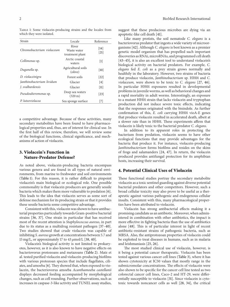
As a bisindole, violacein (Figure 1) is produced by diverse gen-era of bacterial strains, including Collimonas [1], Duganella[2], Janthinobacterium [3–5],Microbulbifer sp. [6], and Pseu-doalteromonas [7–9]. These violacein producers are variedphylogenetically and so are the locales from which theyhave been isolated. As shown in Table 1, these include quitea selection of environs as these bacteria have been foundassociated with the surfaces of sea sponges [7] and therhizosphere of olive groves [10] and even within glaciers[4, 11, 12]. Perhaps the best known genus, however, is Chro-mobacterium [13, 14], which includes the strain C. violaceum[15].
2. Violacein as an Indicator ofQuorum Sensing
In most of violacein-producing bacterial strains isolatedfrom nature, this bisindole is a secondary metabolite thatis associated with biofilm production [5]. Moreover, itsproduction withinC. violaceum and other strains is regulatedby quorum sensing mechanisms [16]. Because it is easy tovisualize, violacein production by C. violaceum has becomea useful indicator of quorum sensing molecules and theirinhibitors [17–20].
Secondary metabolites often serve functions other thanthe bacteria’s immediate needs in growth and propagation.Many of these molecules are biologically active, and somehave toxic properties to competing species giving the bacteria
Hindawi Publishing CorporationBioMed Research InternationalVolume 2015, Article ID 465056, 8 pageshttp://dx.doi.org/10.1155/2015/465056
2 BioMed Research International
Table 1: Some violacein-producing strains and the locales fromwhich they were isolated.
Strain Locale Reference
Chromobacterium violaceumRiver
Waste watertreatment plant
[14][21]
Collimonas sp. Arctic coastalwaters [1]
Duganella sp. Agricultural soils(olive) [10]
D. violaceinigra Forest soils [22]Janthinobacterium lividum Glacier [4]J. svalbardensis Glacier [11]
Pseudoalteromonas sp. Deep sea waters(320m) [23]
P. luteoviolacea Sea sponge surface [7]
a competitive advantage. Because of these activities, manysecondary metabolites have been found to have pharmaco-logical properties and, thus, are of interest for clinical use. Inthe first half of this review, therefore, we will review someof the biological functions, clinical significance, and mech-anisms of action of violacein.
3. Violacein’s Function inNature-Predator Defense?
As noted above, violacein-producing bacteria encompassvarious genera and are found in all types of natural envi-ronments, from marine to freshwater and soil environments(Table 1). For this reason, it is rather difficult to pinpointviolacein’s main biological or ecological role. One possiblecommonality is that violacein producers are generally sessilebacteria whichmakes themmore vulnerable to predation [6].This leads to the idea that violacein serves as some sort ofdefensemechanism for its producing strain or that it providesthese sessile bacteria some competitive advantage.
Consistentwith this, violaceinwas shown to have antibac-terial properties particularly towards Gram-positive bacterialstrains [36, 37]. One strain in particular that has receivedmost of the recent attention is Staphylococcus aureus, in partdue to its status as a multidrug resistant pathogen [37–40].Two studies showed that crude violacein was capable ofinhibiting S. aureus growth at concentrations between 5.7 and15mg/L, or approximately 17 to 43 𝜇mol/L [38, 40].
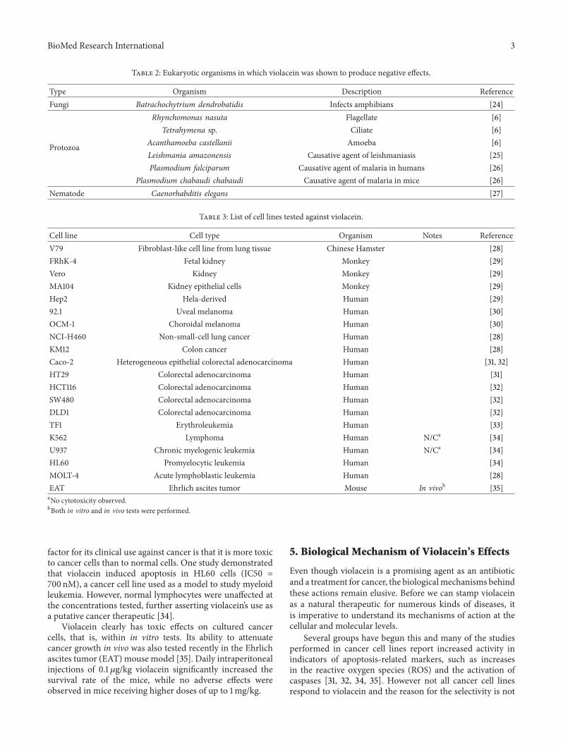
Violacein’s biological activity is not limited to prokary-otes, however, as it is also known to have negative effects onbacteriovorus protozoans and metazoans (Table 2). Matz etal. tested purified violacein and violacein-producing biofilmswith various protozoan species that include flagellates, cili-ates, and amoeba [6].They showed that, when exposed to vio-lacein, the bacteriovorus amoeba Acanthamoeba castellaniidisplays decreased feeding accompanied by morphologicalchanges, such as cell rounding. Further observations, such asincreases in caspase-3-like activity and TUNEL assay studies,
suggest that these predacious microbes are dying via anapoptotic-like cell death [41].
Like many protists, the soil nematode C. elegans is abacteriovorus predator that ingests a wide variety of microor-ganisms [42]. Although C. elegans is best known as a premiergenetic model organism that has propelled such importantdiscoveries as RNAi,microRNAs, and programmed cell death[43–45], it is also an excellent tool to understand violacein’sbiological activity on bacterial predators. For example, C.elegans fed E. coli as a prey strain grows normally andhealthily in the laboratory. However, two strains of bacteriathat produce violacein, Janthinobacterium sp. HH01 and C.violaceum, were shown to be toxic to C. elegans [27, 46].In particular HH01 exposures resulted in developmentalproblems in juvenileworms, aswell as behavioral changes anda rapid mortality in adult worms. Interestingly, an exposureto a mutant HH01 strain that lacks violacein and tryptophanproduction did not induce severe toxic effects, indicatingthat the responses originated with the bisindole. As furtherconfirmation of this, E. coli carrying HH01 vioA-E genesthat produce violacein resulted in accelerated death, albeit ata slower rate than in HH01. These experiments affirm thatviolacein is likely toxic to the bacterial predator C. elegans.
In addition to its apparent roles in protecting thebacterium from predation, violacein seems to have otherecological functions that may provide advantages for thebacteria that produce it. For instance, violacein-producingJanthinobacterium forms biofilms and resides on the skinsof frogs and salamanders [24, 47]. In return, the violaceinproduced provides antifungal protection for its amphibianhosts, increasing their survival.
4. Potential Clinical Uses of Violacein
These functional studies portray the secondary metaboliteviolacein as a toxic sentinel guarding against diverse potentialbacterial predators and other competitors. However, such abroad cellular toxicity may also prove to be useful as a ther-apeutic against various pathogenic and endogenous cellularinsults. Consistent with this, many pharmacological proper-ties have been attributed to violacein.
Violacein has strong antibacterial effects making it apromising candidate as an antibiotic.Moreover, when admin-istered in combination with other antibiotics, the impact ismore effective in fighting bacteria than the use of antibioticsalone [48]. This is of particular interest in light of recentantibiotic-resistant strains of pathogenic bacteria, such asMRSA. Also, the antiprotozoan properties of violacein couldbe exploited to treat diseases in humans, such as in malariaand leishmaniasis [25, 26].
The most studied clinical use of violacein, however, isit being a potential cancer therapeutic. Violacein has beentested against various cancer cell lines (Table 3), where it hasshown cytotoxicity at IC50 values that mostly range in thesubmicromolar concentrations. The effects of violacein werealso shown to be specific for the cancer cell line tested as twocolorectal cancer cell lines, Caco-2 and HT-29, were differ-entially susceptible to violacein [31]. Since violacein is cyto-toxic towards noncancer cells as well [28, 34], the critical
BioMed Research International 3
Table 2: Eukaryotic organisms in which violacein was shown to produce negative effects.
Type Organism Description ReferenceFungi Batrachochytrium dendrobatidis Infects amphibians [24]
Protozoa
Rhynchomonas nasuta Flagellate [6]Tetrahymena sp. Ciliate [6]
Acanthamoeba castellanii Amoeba [6]Leishmania amazonensis Causative agent of leishmaniasis [25]Plasmodium falciparum Causative agent of malaria in humans [26]
Plasmodium chabaudi chabaudi Causative agent of malaria in mice [26]Nematode Caenorhabditis elegans [27]
Table 3: List of cell lines tested against violacein.
Cell line Cell type Organism Notes ReferenceV79 Fibroblast-like cell line from lung tissue Chinese Hamster [28]FRhK-4 Fetal kidney Monkey [29]Vero Kidney Monkey [29]MA104 Kidney epithelial cells Monkey [29]Hep2 Hela-derived Human [29]92.1 Uveal melanoma Human [30]OCM-1 Choroidal melanoma Human [30]NCI-H460 Non-small-cell lung cancer Human [28]KM12 Colon cancer Human [28]Caco-2 Heterogeneous epithelial colorectal adenocarcinoma Human [31, 32]HT29 Colorectal adenocarcinoma Human [31]HCT116 Colorectal adenocarcinoma Human [32]SW480 Colorectal adenocarcinoma Human [32]DLD1 Colorectal adenocarcinoma Human [32]TF1 Erythroleukemia Human [33]K562 Lymphoma Human N/Ca [34]U937 Chronic myelogenic leukemia Human N/Ca [34]HL60 Promyelocytic leukemia Human [34]MOLT-4 Acute lymphoblastic leukemia Human [28]EAT Ehrlich ascites tumor Mouse In vivob [35]aNo cytotoxicity observed.bBoth in vitro and in vivo tests were performed.
factor for its clinical use against cancer is that it is more toxicto cancer cells than to normal cells. One study demonstratedthat violacein induced apoptosis in HL60 cells (IC50 =700 nM), a cancer cell line used as a model to study myeloidleukemia. However, normal lymphocytes were unaffected atthe concentrations tested, further asserting violacein’s use asa putative cancer therapeutic [34].
Violacein clearly has toxic effects on cultured cancercells, that is, within in vitro tests. Its ability to attenuatecancer growth in vivo was also tested recently in the Ehrlichascites tumor (EAT) mouse model [35]. Daily intraperitonealinjections of 0.1 𝜇g/kg violacein significantly increased thesurvival rate of the mice, while no adverse effects wereobserved in mice receiving higher doses of up to 1mg/kg.
5. Biological Mechanism of Violacein’s Effects
Even though violacein is a promising agent as an antibioticand a treatment for cancer, the biologicalmechanisms behindthese actions remain elusive. Before we can stamp violaceinas a natural therapeutic for numerous kinds of diseases, itis imperative to understand its mechanisms of action at thecellular and molecular levels.
Several groups have begun this and many of the studiesperformed in cancer cell lines report increased activity inindicators of apoptosis-related markers, such as increasesin the reactive oxygen species (ROS) and the activation ofcaspases [31, 32, 34, 35]. However not all cancer cell linesrespond to violacein and the reason for the selectivity is not
4 BioMed Research International
well understood. Moreover, among the leukemia cell linestested in the literature, violacein showed selective cytotoxicityagainst HL60 and TF1 (Table 3), but the pathways that lead tocell death were very different in the two cells. In HL60 cells,an exposure to violacein led to phosphorylation of p38 MAPkinase, upregulation of the NF𝜅B pathway, and activation ofcaspases [34]. It was also found that TGF𝛼 receptor activationwas required for these downstream effects. TF1 cells, on theother hand, did not seem to follow the canonical apoptoticpathway as treatmentwith inhibitors of proapoptotic caspasesdid not prevent cell death [33].
Conclusions on violacein’s mechanism of action areclearly scant at this point. However, the fact that violaceinhas cytotoxic effects on such a wide variety of organisms andcells hints at a common target or pathway. Studying the effectof these bisindoles at the genetic level on model eukaryoticorganisms, such as C. elegans, will help in elucidating itsmechanism of action and enrich our knowledge of violaceinas a clinical therapeutic. Owing to its versatile activity againstmany human ailments and infectious agents, however, it isnot surprising that this bisindole has garneredmore attentionrecently from the scientific community. One factor that maycontribute to reducing violacein’s application, though, is itsrelatively low level production by natural strains. Conse-quently, the latter half of this review will be primarily givento the discussion of current research into the production andpurification of violacein and its related derivatives.
6. Production of Violacein byNatural Host Strains
Since violacein is produced naturally by various bacterialspecies, the use of these strains for its production seemed likea clear choice. However,many factors were found to influencethe yields, including the agitation and aeration [7], the inocu-lum size [2, 10], and the nutrients available [2, 10, 49, 50]. Itshould be noted that the violacein concentrations reportedwithin many of the articles are based upon the extinctioncoefficient as determined by the authors using spectropho-tometric analyses, with extinction coefficient values rangingbetween 10.955 and 74.3 L/(g-cm) in the literature [2, 49,51, 52]. A recent article by Rodrigues et al. highlighted thediscrepancy caused by spectrophotometric-based determi-nations of violacein and deoxyviolacein concentrations andstated that this could result in violacein concentrations thatare inflated by as much as 680% [51]. To address this in theirstudy, therefore, Rettori and Duran (1998) relied on HPLCmeasurements, a technique which was proposed in an earlierstudy to be used in parallel with NMR, UV-Vis spectroscopy,and mass spectroscopy when characterizing violacein and itsproduction by bacterial strains [52]. Consequently, to avoidany potential confusion to the readers, this report will providethe violacein and deoxyviolacein concentrations reportedand state whether they were determined via HPLC or withan extinction coefficient.
One group applied response surface methodologies(RSM) to identify the best conditions to produce violaceinwith C. violaceum [49]. They initially analyzed 16 variablesbut eventually limited them to three—glucose, tryptone, and
yeast extract. While the latter two improved both the cellmass and violacein yields as they were increased, glucose wasfound to be negatively correlated with the violacein yields,and limiting its addition to the culture was advantageous.Using this technique, they were able to increase the dry cellweight (DCW) from7.5 to 21 g/L and the violacein yields from170 to 430mg/L with an extinction coefficient of 56.01 L/(g-cm). Although there was a significant improvement in thevolumetric productivity of violacein, defined as the mg ofproduct per liter of culture, it should be noted that this doesnot represent a greater specific productivity of this bisindole,that is,mg product per gramof cells, as the cellmass increasedby 2.8-fold while the violacein concentration increased byonly 2.5-fold. Yet, similar protocols could potentially beemployed with other strains to increase the cell density and,thus, raise the volumetric productivity.
A more recent study using RSM with another violacein-producing strain, Pseudoduganella sp. B2, previouslyDuganella sp. B2, found similar results as the concentrationof beef extract usedwas amajor impetus for violacein produc-tion [2]. They also identified the culture pH and the concen-trations of tryptophan and potassium nitrate as major play-ers influencing the final violacein yields. The importanceof tryptophan is not surprising as this is the precursor forviolacein,while the impact of nitratewas thought to be relatedto nitrogen source availability for the growing bacterialcultures. Under the optimal conditions found, the authorsclaimed Duganella sp. B2 was capable of producing 1.6 g/Lcrude violacein, typically referring to the naturally producedmixture of violacein and deoxyviolacein. This value is underscrutiny, however, as the extinction coefficient used (10.955 L/(g-cm)) is the lowest reported in the literature [2, 49, 51, 52].
7. Production of Violacein within E. coli andOther Heterologous Hosts
Since the genes required for the production of violacein areknown to exist within a single operon, that is, vioABCDE[53], many groups have sought to clone and express thesewithin other bacterial hosts, including E. coli [8, 51, 53].The Pemberton group has focused on plasmid stability, anissue when trying to generate bacterial products in long termand in nonnatural hosts. They found, for instance, whenthe violacein gene cluster was cloned into pHC79, a cosmidvector, that it was unstable and was lost in as much as 60%of the bacterial population when grown for 15 generations inthe absence of antibiotic pressure [54]. They claimed that thesame was true when they expressed the vioABCDE operon ina pUC18 vector but were able to generate a stable constructusing a broad host range IncP plasmid [54, 55].This plasmid,pPSX-Vio+, was stable without antibiotics for more than 100generations [54], making it a potentially useful tool for theproduction of violacein.
In a subsequent study, they found that the amount ofviolacein produced by E. coli was dependent upon the host,with E. coli strain JM109 producing 3.9-fold more violaceinthan E. coli strain DH5𝛼 when harboring the same plasmid[55]. The production of the alpha amylase protein, AmyA,from Streptomyces lividans was likewise found to be better in
BioMed Research International 5
NH
Violacein Deoxyviolacein Oxyviolacein
O
O
OHOH
HO
HN
NH
NHNH
OO
OO
HNHN
NH N
H
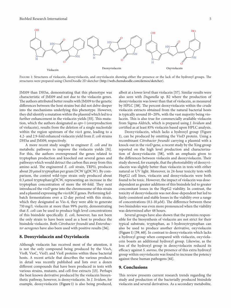
Figure 1: Structures of violacein, deoxyviolacein, and oxyviolacein showing either the presence or the lack of the hydroxyl groups. Thestructures were prepared using ChemDoodle 2D sketcher (http://web.chemdoodle.com/demos/sketcher).
JM109 than DH5𝛼, demonstrating that this phenotype wascharacteristic of JM109 and not due to the violacein genes.The authors attributed better results with JM109 to the geneticdifferences between the host strains but did not delve deeperinto the mechanisms underlying this phenotype. However,they did identify amutationwithin the plasmidwhich led to afurther enhancement in the violacein yields [55]. This muta-tion, which the authors designated as opv-1 (overproductionof violacein), results from the deletion of a single nucleotidewithin the region upstream of the vioA gene, leading to a4.2- and 2.9-fold enhanced violacein yield from E. coli strainsDH5𝛼 and JM109, respectively.
A more recent study sought to engineer E. coli and itsmetabolic pathways to improve the violacein yields [51].For this, the authors overexpressed the genes related totryptophan production and knocked out several genes andpathways which would detract the carbon flux away from thisamino acid. The engineered E. coli strain, TRP11, producesabout 20𝜇mol tryptophan per gramDCW(gDCW). By com-parison, the control wild-type strain only produced about0.3 𝜇mol tryptophan/gDCW, representing an increase in thetryptophan concentration of more the 60-fold. They nextintroduced the vioD gene into the chromosome of this strainand a plasmid expressing the vioABCE genes. Performing fed-batch fermentations over a 12-day period with this strain,which they designated as Vio-4, they were able to generate710mg/L violacein at more than 99% purity, demonstratingthat E. coli can be used to produce high level concentrationsof this bisindole specifically. E. coli, however, has not beenthe only strain to have been used as a host to produce thebisindole violacein. Both Citrobacter freundii and Enterobac-ter aerogenes have also been used with positive results [56].
8. Deoxyviolacein and Oxyviolacein
Although violacein has received most of the attention, itis not the only compound being produced by the VioA,VioB, VioC, VioD, and VioE proteins within these bacterialhosts. A recent article that describes the various productsin detail was recently published and lists over a dozendifferent compounds that have been produced in tests withvarious strains, mutants, and cell-free extracts [13]. Perhapsthe best known derivative produced by the violacein biosyn-thetic pathway, however, is deoxyviolacein. In J. lividum, forexample, deoxyviolacein (Figure 1) is also being produced,
albeit at a lower level than violacein [57]. Similar results werealso seen with Duganella sp. B2 where the production ofdeoxyviolacein was lower than that of violacein, as measuredby HPLC [58]. The percent deoxyviolacein within the crudeviolacein extracts obtained from the natural bacterial hostsis typically around 10∼20%, with the vast majority being vio-lacein. This is also true for commercially available violaceinfrom Sigma-Aldrich, which is prepared using J. lividum andcertified as at least 85% violacein based upon HPLC analysis.
Deoxyviolacein, which lacks a hydroxyl group (Figure1), can be produced by omitting the VioD protein. Using arecombinant Citrobacter freundii carrying a plasmid with aknock-out in the vioD gene, a recent study by the Xing groupreported on the high level production and characteriza-tion of deoxyviolacein [58], with an emphasis given tothe differences between violacein and deoxyviolacein. Theirstudy showed, for example, that the photostability of deoxyvi-olacein was slightly better than violacein in tests with eithernatural or UV light. Moreover, in 24-hour toxicity tests withHepG2 cell lines, violacein and deoxyviolacein were bothfound to be toxic. However, the impact of violacein was dose-dependent as greater additions of this bisindole led to greaterconcomitant losses in the HepG2 viability. In contrast, thetoxicity of deoxyviolacein was not dose-dependent but led tofairly consistent and stable losses in the viability over a rangeof concentrations (0.1–10𝜇M). The difference between thesetwo bisindoles was even more pronounced when the viabilitywas determined after 48 hours.
Several groups have also shown that the proteins respon-sible for the biosynthesis of violacein are not strict for theirtypical substrate, tryptophan, as 5-hydroxytryptophan canalso be used to produce another derivative, oxyviolacein(Figure 1) [59, 60]. In contrast to deoxyviolacein which lacksa hydroxyl group when compared with violacein, oxyviola-cein boasts an additional hydroxyl group. Likewise, as theloss of the hydroxyl group in deoxyviolacein reduced itsefficacy against S. aureus, the presence of this extra hydroxylgroup within oxyviolacein was found to increase the potencyagainst these human pathogens [61].
9. Conclusions
This review presents current research trends regarding thestudy and production of the bacterially produced bisindoleviolacein and several derivatives. As a secondary metabolite,
6 BioMed Research International
violacein has been found to possess a wide variety of bio-logical activities, including anticancerous properties. Thesecharacteristics have led to renewed interest in this compoundand its production by both wild-type and recombinantbacterial strains. As presented in this report, the productionand characterization of violacein are not without their ownobstacles and struggles, andmuchwork still needs to be done.This is particularly true regarding the mode of activity ofviolacein which needs to be studied more in depth. Currenttrends in molecular genetics are aiding in this as research-ers are now capable of engineering bacterial host that canoverproduce this bisindole within fermentations. As workwith this compound and its derivatives progresses, it is anti-cipated that violacein will become more readily available forthe scientific community and clinical studies.
Conflict of Interests
The authors declare that there is no conflict of interestsregarding the publication of this paper.
Authors’ Contribution
Seong Yeol Choi and Kyoung-hye Yoon contributed equallyto this paper. All authors participated in preparation of thispaper.
Acknowledgments
SeongYeol Choi andRobert J.Mitchell received support fromthe Korea Institute for Advancement of Technology underthe EUREKA Programme (Grant no. N019800009) andthe Korea Health Industry Development Institute (KHIDI)(Grant no. HI13C13550000). Jin Il Lee and Kyoung-hye Yoonare supported by a New Investigator Grant from the NationalResearch Foundation of Korea (Grant no. 201405553). Theauthors appreciate the support.
References
[1] S. Hakvag, E. Fjærvik, G. Klinkenberg et al., “Violacein-pro-ducing Collimonas sp. from the sea surface microlayer of costalwaters in Trøndelag, Norway,” Marine Drugs, vol. 7, no. 4, pp.576–588, 2009.
[2] H. S.Wang, P. X. Jiang, Y. Lu et al., “Optimization of culture con-ditions for violacein production by a new strain ofDuganella sp.B2,” Biochemical Engineering Journal, vol. 44, no. 2-3, pp. 119–124, 2009.
[3] B. A. Jude, J. Tanner, T. Koko, and E. C. McLaughlin, “Analysis,characterization, and synthesis of violacein from Janthinobac-terium isolate extracts,” Abstracts of Papers of the AmericanChemical Society, vol. 244, 2012.
[4] Y. Lu, L. Wang, Y. Xue et al., “Production of violet pigment bya newly isolated psychrotrophic bacterium from a glacier inXinjiang, China,” Biochemical Engineering Journal, vol. 43, no.2, pp. 135–141, 2009.
[5] F. Pantanella, F. Berlutti, C. Passariello, S. Sarli, C. Morea, andS. Schippa, “Violacein and biofilm production in Janthinobac-terium lividum,” Journal of Applied Microbiology, vol. 102, no. 4,pp. 992–999, 2007.
[6] C. Matz, J. S. Webb, P. J. Schupp et al., “Marine biofilm bacteriaevade eukaryotic predation by targeted chemical defense,” PLoSONE, vol. 3, no. 7, Article ID e2744, 2008.
[7] L. H. Yang, H. Xiong, O. O. Lee, S.-H. Qi, and P.-Y. Qian, “Effectof agitation on violacein production in Pseudoalteromonasluteoviolacea isolated from a marine sponge,” Letters in AppliedMicrobiology, vol. 44, no. 6, pp. 625–630, 2007.
[8] X. Zhang and K. Enomoto, “Characterization of a gene clusterand its putative promoter region for violacein biosynthesis inPseudoalteromonas sp. 520P1,” Applied Microbiology and Bio-technology, vol. 90, no. 6, pp. 1963–1971, 2011.
[9] S. A. Mccarthy, R. M. Johnson, D. kakimoto, and T. sakata,“Effects of various agents on the pigment (violacein) and anti-biotic production of Alteromonas luteoviolacea,” Bulletin of theJapanese Society of Scientific Fisheries, vol. 51, no. 7, pp. 1115–1121,1985.
[10] S. Aranda, M. Montes-Borrego, and B. B. Landa, “Purple-pigmented violacein-producing Duganella spp. inhabit therhizosphere of wild and cultivated olives in Southern Spain,”Microbial Ecology, vol. 62, no. 2, pp. 446–459, 2011.
[11] J. A. Avgustin, D. Z. Bertok, R. Kostanjsek, and G. Avgustin,“Isolation and characterization of a novel violacein-like pig-ment producing psychrotrophic bacterial species Janthinobac-terium svalbardensis sp. nov,” Antonie van Leeuwenhoek, Inter-national Journal of General andMolecularMicrobiology, vol. 103,no. 4, pp. 763–769, 2013.
[12] S. J. Kim, S. C. Shin, S. G. Hong et al., “Genome sequence ofJanthinobacterium sp. strain PAMC 25724, isolated from alpineglacier cryoconite,” Journal of Bacteriology, vol. 194, no. 8, pp.2096–2096, 2012.
[13] T. Hoshino, “Violacein and related tryptophanmetabolites pro-duced by Chromobacterium violaceum: biosynthetic mecha-nism and pathway for construction of violacein core,” AppliedMicrobiology and Biotechnology, vol. 91, no. 6, pp. 1463–1475,2011.
[14] M. O. Moss, C. Ryall, and N. A. Logan, “The classification andcharacterization of chromobacteria from a lowland river,” Jour-nal of General Microbiology, vol. 105, no. 1, pp. 11–21, 1978.
[15] N. D. Duran andC. F.M.Menck, “Chromobacterium violaceum:a review of pharmacological and industiral perspectives,” Criti-cal Reviews in Microbiology, vol. 27, no. 3, pp. 201–222, 2001.
[16] K. H. McClean, M. K. Winson, L. Fish et al., “Quorum sensingand Chromobacterium violaceum: exploitation of violacein pro-duction and inhibition for the detection of N-acylhomoserinelactones,”Microbiology, vol. 143, no. 12, pp. 3703–3711, 1997.
[17] S. A. Burt, V. T. A. Ojo-Fakunle, J. Woertman, and E. J. A.Veldhuizen, “The natural antimicrobial carvacrol inhibits quo-rum sensing inChromobacterium violaceum and reduces bacte-rial biofilm formation at sub-lethal concentrations,” PLoS ONE,vol. 9, no. 4, Article ID e93414, 2014.
[18] P. S. Rajesh and V. Ravishankar Rai, “Quorum quenchingactivity in cell-free lysate of endophytic bacteria isolated fromPterocarpus santalinus Linn., and its effect on quorum sensingregulated biofilm in Pseudomonas aeruginosa PAO1,”Microbio-logical Research, vol. 169, no. 7-8, pp. 561–569, 2014.
[19] J. C. Taganna, J. P. Quanico, R. M. G. Perono, E. C. Amor,and W. L. Rivera, “Tannin-rich fraction from Terminalia cat-appa inhibits quorum sensing (QS) in Chromobacterium vio-laceum and the QS-controlled biofilm maturation and LasAstaphylolytic activity in Pseudomonas aeruginosa,” Journal ofEthnopharmacology, vol. 134, no. 3, pp. 865–871, 2011.

BioMed Research International 7
[20] M. Duran, A. Faljoni-Alario, and N. Duran, “Chromobacteriumviolaceum and its important metabolites—review,” Folia Micro-biologica, vol. 55, no. 6, pp. 535–547, 2010.
[21] W. A. Ahmad, N. Z. Yusof, N. Nordin, Z. A. Zakaria, and M. F.Rezali, “Production and characterization of violacein by locallyisolated Chromobacterium violaceum grown in agriculturalwastes,” Applied Biochemistry and Biotechnology, vol. 167, no. 5,pp. 1220–1234, 2012.
[22] W. J. Li, Y. Q. Zhang, D. J. Park et al., “Duganella violaceinigrasp. nov., a novel mesophilic bacterium isolated from forest soil,”International Journal of Systematic and Evolutionary Microbiol-ogy, vol. 54, part 5, pp. 1811–1814, 2004.
[23] S. Yada, Y. Wang, Y. Zou et al., “Isolation and characterizationof two groups of novel marine bacteria producing violacein,”Marine Biotechnology, vol. 10, no. 2, pp. 128–132, 2008.
[24] R. M. Brucker, R. N. Harris, C. R. Schwantes et al., “Amphibianchemical defense: antifungal metabolites of the microsym-biont Janthinobacterium lividum on the salamander Plethodoncinereus,” Journal of Chemical Ecology, vol. 34, no. 11, pp. 1422–1429, 2008.
[25] L. L. Leon, C. C. Miranda, A. O. de Souza, and N. Duran,“Antileishmanial activity of the violacein extracted from Chro-mobacterium violaceum,” Journal of Antimicrobial Chemother-apy, vol. 48, no. 3, pp. 449–450, 2001.
[26] S. C. P. Lopes, Y. C. Blanco,G. Z. Justo et al., “Violacein extractedfrom Chromobacterium violaceum inhibits Plasmodium growthin vitro and in vivo,” Antimicrobial Agents and Chemotherapy,vol. 53, no. 5, pp. 2149–2152, 2009.
[27] C. Hornung, A. Poehlein, F. S. Haack et al., “The Janthinobac-terium sp. HH01 Genome Encodes a Homologue of the V.cholerae CqsA and L. pneumophila LqsA Autoinducer Syn-thases,” PLoS ONE, vol. 8, no. 2, Article ID e55045, 2013.
[28] P. da Silva Melo, S. S. Maria, B. de Campos Vidal, M. Haun, andN. Duran, “Violacein cytotoxicity and induction of apoptosis inV79 cells,” In Vitro Cellular & Developmental Biology—Animal,vol. 36, no. 8, pp. 539–543, 2000.
[29] C. R. Andrighetti-Frohner, R. V. Antonio, T. B. Creczynski-Pasa, C. R. M. Barardi, and C. M. O. Simoes, “Cytotoxicity andpotential antiviral evaluation of violacein produced byChromo-bacterium violaceum,”Memorias do Instituto Oswaldo Cruz, vol.98, no. 6, pp. 843–848, 2003.
[30] V. S. Saraiva, J.-C. Marshall, J. Cools-Lartigue, and M. N.Burnier Jr., “Cytotoxic effects of violacein in human uveal mela-noma cell lines,”Melanoma Research, vol. 14, no. 5, pp. 421–424,2004.
[31] D. D. de Carvalho, F. T. M. Costa, N. Duran, and M. Haun,“Cytotoxic activity of violacein in human colon cancer cells,”Toxicology in Vitro, vol. 20, no. 8, pp. 1514–1521, 2006.
[32] L. L. Kodach, C. L. Bos, N. Duran, M. P. Peppelenbosch, C.V. Ferreira, and J. C. H. Hardwick, “Violacein synergisticallyincreases 5-fluorouracil cytotoxicity, induces apoptosis andinhibits Akt-mediated signal transduction in human colorectalcancer cells,” Carcinogenesis, vol. 27, no. 3, pp. 508–516, 2006.
[33] K. C. S. Queiroz, R. Milani, R. R. Ruela-de-Sousa et al., “Vio-lacein induces death of resistant leukaemia cells via kinomereprogramming, endoplasmic reticulum stress and Golgi appa-ratus collapse,” PLoS ONE, vol. 7, no. 10, Article ID e45362, 2012.
[34] C. V. Ferreira, C. L. Bos, H. H. Versteeg, G. Z. Justo, N. Duran,and M. P. Peppelenbosch, “Molecular mechanism of violacein-mediated human leukemia cell death,” Blood, vol. 104, no. 5, pp.1459–1464, 2004.
[35] N. Bromberg, J. L. Dreyfuss, C. V. Regatieri et al., “Growth inhi-bition and pro-apoptotic activity of violacein in Ehrlich ascitestumor,” Chemico-Biological Interactions, vol. 186, no. 1, pp. 43–52, 2010.
[36] H. C. Lichstein and V. F. van de Sand, “Violacein, an antibioticpigment produced by Chromobacterium violaceum,” Journal ofInfectious Diseases, vol. 76, no. 1, pp. 47–51, 1945.
[37] Y. Nakamura, T. Sawada, Y. Morita, and E. Tamiya, “Isolation ofa psychrotrophic bacterium from the organic residue of a watertank keeping rainbow trout and antibacterial effect of violetpigment produced from the strain,” Biochemical EngineeringJournal, vol. 12, no. 1, pp. 79–86, 2002.
[38] Y. Nakamura, C. Asada, and T. Sawada, “Production of antibac-terial violet pigment by psychrotropic bacterium RT102 strain,”Biotechnology and Bioprocess Engineering, vol. 8, no. 1, pp. 37–40, 2003.
[39] N. G. Vynne, M. Mansson, and L. Gram, “Gene sequence basedclustering assists in dereplication of Pseudoalteromonas luteovi-olacea strains with identical inhibitory activity and antibioticproduction,”Marine Drugs, vol. 10, no. 8, pp. 1729–1740, 2012.
[40] S. Subramaniam, V. Ravi, and A. Sivasubramanian, “Synergisticantimicrobial profiling of violacein with commercial antibioticsagainst pathogenic micro-organisms,” Pharmaceutical Biology,vol. 52, no. 1, pp. 86–90, 2014.
[41] P. D. Melo, S. S. Maria, B. D. Vidal, M. Haun, and N. Duran,“Violacein cytotoxicity and induction of apoptosis in V79 cells,”In Vitro Cellular & Developmental Biology—Animal, vol. 36, no.8, pp. 539–543, 2000.
[42] S. Brenner, “The genetics of Caenorhabditis elegans,” Genetics,vol. 77, no. 1, pp. 71–94, 1974.
[43] A. Fire, S. Xu, M. K. Montgomery, S. A. Kostas, S. E. Driver, andC. C.Mello, “Potent and specific genetic interference by double-stranded RNA in caenorhabditis elegans,” Nature, vol. 391, no.6669, pp. 806–811, 1998.
[44] R. C. Lee, R. L. Feinbaum, andV. Ambros, “TheC. elegans heter-ochronic gene lin-4 encodes small RNAs with antisense com-plementarity to lin-14,” Cell, vol. 75, no. 5, pp. 843–854, 1993.
[45] M. O. Hengartner and H. R. Horvitz, “Programmed cell deathin Caenorhabditis elegans,” Current Opinion in Genetics andDevelopment, vol. 4, no. 4, pp. 581–586, 1994.
[46] L. R. Swem, D. L. Swem, C. T. O’Loughlin et al., “A quorum-sensing antagonist targets bothmembrane-bound and cytoplas-mic receptors and controls bacterial pathogenicity,” MolecularCell, vol. 35, no. 2, pp. 143–153, 2009.
[47] R. N. Harris, R. M. Brucker, J. B.Walke et al., “Skin microbes onfrogs prevent morbidity and mortality caused by a lethal skinfungus,”The ISME Journal, vol. 3, no. 7, pp. 818–824, 2009.
[48] S. Subramaniam, V. Ravi, and A. Sivasubramanian, “Synergisticantimicrobial profiling of violacein with commercial antibioticsagainst pathogenic micro-organisms,” Pharmaceutical Biology,vol. 52, no. 1, pp. 86–90, 2014.
[49] A. S. Mendes, J. E. de Carvalho, M. C. T. Duarte, N. Duran, andR. E. Bruns, “Factorial design and response surface optimiza-tion of crude violacein forChromobacterium violaceum produc-tion,” Biotechnology Letters, vol. 23, no. 23, pp. 1963–1969, 2001.
[50] R. Riveros, M. Haun, and N. Duran, “Effect of growthconditions on production of violacein by Chromobacteriumviolaceum (BB-78 strain),” Brazilian Journal of Medical andBiological Research, vol. 22, no. 5, pp. 569–577, 1989.
[51] A. L. Rodrigues, N. Trachtmann, J. Becker et al., “Systemsmeta-bolic engineering of Escherichia coli for production of the
8 BioMed Research International
antitumor drugs violacein and deoxyviolacein,”Metabolic Engi-neering, vol. 20, pp. 29–41, 2013.
[52] D. Rettori and N. Duran, “Production, extraction and purifica-tion of violacein: an antibiotic pigment produced by Chromo-bacterium violaceum,” World Journal of Microbiology and Bio-technology, vol. 14, no. 5, pp. 685–688, 1998.
[53] P. R. August, T.H.Grossman, C.Minor et al., “Sequence analysisand functional characterization of the violacein biosyntheticpathway from Chromobacterium violaceum,” Journal of Molec-ular Microbiology and Biotechnology, vol. 2, no. 4, pp. 513–519,2000.
[54] D. S. Sarovich and J. M. Pemberton, “pPSX: a novel vector forthe cloning and heterologous expression of antitumor antibioticgene clusters,” Plasmid, vol. 57, no. 3, pp. 306–313, 2007.
[55] A. Ahmetagic and J. M. Pemberton, “Stable high level expres-sion of the violacein indolocarbazole anti-tumour gene clusterand the Streptomyces lividans amyA gene inE. coliK12,”Plasmid,vol. 63, no. 2, pp. 79–85, 2010.
[56] P.-X. Jiang, H.-S. Wang, C. Zhang, K. Lou, and X.-H. Xing,“Reconstruction of the violacein biosynthetic pathway fromDuganella sp. B2 in different heterologous hosts,”AppliedMicro-biology and Biotechnology, vol. 86, no. 4, pp. 1077–1088, 2010.
[57] A. L. Rodrigues, Y. Gocke, C. Bolten, N. L. Brock, J. S.Dickschat, and C. Wittmann, “Microbial production of thedrugs violacein and deoxyviolacein: analytical developmentand strain comparison,” Biotechnology Letters, vol. 34, no. 4, pp.717–720, 2012.
[58] P.-X. Jiang, H.-S. Wang, S. Xiao et al., “Pathway redesign fordeoxyviolacein biosynthesis inCitrobacter freundii and charact-erization of this pigment,”AppliedMicrobiology and Biotechnol-ogy, vol. 94, no. 6, pp. 1521–1532, 2012.
[59] C. Sanchez, A. F. Brana, C. Mendez, and J. A. Salas, “Reevalua-tion of the violacein biosynthetic pathway and its relationship toindolocarbazole biosynthesis,” ChemBioChem, vol. 7, no. 8, pp.1231–1240, 2006.
[60] T. Hoshino and N. Ogasawara, “Biosynthesis of violacein:evidence for the intermediacy of 5-hydroxy-L-tryptophan andthe structure of a new pigment, oxyviolacein, produced by themetabolism of 5-hydroxytryptophan,” Agricultural and Biologi-cal Chemistry, vol. 54, no. 9, pp. 2339–2346, 1990.
[61] H. S. Wang, F. Z. Wang, X. F. Zhu et al., “Biosynthesis and cha-racterization of violacein, deoxyviolacein and oxyviolacein inheterologous host, and their antimicrobial activities,” Biochem-ical Engineering Journal, vol. 67, pp. 148–155, 2012.

Submit your manuscripts athttp://www.hindawi.com
Hindawi Publishing Corporationhttp://www.hindawi.com Volume 2014
Anatomy Research International
PeptidesInternational Journal of
Hindawi Publishing Corporationhttp://www.hindawi.com Volume 2014
Hindawi Publishing Corporation http://www.hindawi.com
International Journal of
Volume 2014
Zoology
Hindawi Publishing Corporationhttp://www.hindawi.com Volume 2014
Molecular Biology International
GenomicsInternational Journal of
Hindawi Publishing Corporationhttp://www.hindawi.com Volume 2014
The Scientific World JournalHindawi Publishing Corporation http://www.hindawi.com Volume 2014
Hindawi Publishing Corporationhttp://www.hindawi.com Volume 2014
BioinformaticsAdvances in
Marine BiologyJournal of
Hindawi Publishing Corporationhttp://www.hindawi.com Volume 2014
Hindawi Publishing Corporationhttp://www.hindawi.com Volume 2014
Signal TransductionJournal of
Hindawi Publishing Corporationhttp://www.hindawi.com Volume 2014
BioMed Research International
Evolutionary BiologyInternational Journal of
Hindawi Publishing Corporationhttp://www.hindawi.com Volume 2014
Hindawi Publishing Corporationhttp://www.hindawi.com Volume 2014
Biochemistry Research International
ArchaeaHindawi Publishing Corporationhttp://www.hindawi.com Volume 2014
Hindawi Publishing Corporationhttp://www.hindawi.com Volume 2014
Genetics Research International
Hindawi Publishing Corporationhttp://www.hindawi.com Volume 2014
Advances in
Virolog y
Hindawi Publishing Corporationhttp://www.hindawi.com
Nucleic AcidsJournal of
Volume 2014
Stem CellsInternational
Hindawi Publishing Corporationhttp://www.hindawi.com Volume 2014
Hindawi Publishing Corporationhttp://www.hindawi.com Volume 2014
Enzyme Research
Hindawi Publishing Corporationhttp://www.hindawi.com Volume 2014
International Journal of
Microbiology