Embed Size (px)
Citation preview

Confocal scanning laser micrograph of the inner retina showing astrcytes (green) and capillaries (red). Alan Stitt, Queens University, Belfast.
Website: http://www.pharmweb.net/ukicrs.html
new
slet
terUKI
Controlled Release Society
Editors - Neena Washington
and Lance Shaw
VOLUMESeptember 2003
Membership offi cer and treasurer elect - Dr Susie Berrill,
Senior Scientifi c Offi cer, Ventura Ltd.
Website Administrator - Dr Tom Li, Principal Scientist, Drug
Delivery R&D, Renovo Ltd. [email protected]
Committee member - Dr Karl Malcolm, Lecturer in
Pharmaceutics, Queen’s University of Belfast.
UKICRS 2003 Conference Co-organiser - Dr Catriona
O’Driscoll, Professor of Pharmaceuticals, Department of
Pharmaceutics, Trinity College, Dublin.
UKICRS Newsletter Co-editor - Dr Neena Washington,
Scientifi c Advisor for Imaging, AstraZeneca.
The last 12 months have again seen the UKICRS involved in a series of very exciting activities,
particularly in terms of reassessing our organisational strategy and in contributing to a range of
meetings. Since the last newsletter we have carried on our very successful collaboration with the
Academy of Pharmaceutical Sciences in running a UKICRS session at the British Pharmaceutical
Conference in Manchester, September 2002. While we are grateful to all our speakers we were
particularly delighted to welcome Professor Alan Hoffman and Professor Kinam Park, both of whom
travelled from the US specifi cally to contribute to the conference. The session was on the topic
of “Polymers in Drug Delivery – Recent Advances and New Horizons” and you can read about
this standing-room only success story later in the newsletter. We also held a fascinating one-day
meeting in Belfast on drug delivery to the brain; again, you can fi nd out more details regarding the
speakers and attendees later in the newsletter. Finally, we having been gearing ourselves up to the
CRS meeting in July, offering Clive Wilson whatever support he feels we can give him in making
this event a huge success. We are delighted by the astonishing number of abstracts that have
been submitted to the meeting and are also very pleased to be involved with the fi rst ever ‘Pearls
of Wisdom’ session. The meeting promises to be lively and scientifi cally fascinating, buoyed on by
Clive’s indefatigable and infectious enthusiasm.
In terms of our own organisation we have introduced new membership arrangements that have
meant that our lists are now up to date and composed of active and participating individuals rather
than being an accumulation of names stretching back several years; we feel that this is much better
for the long term future of the organisation. In addition we are looking at our website and logo and
will hopefully have something spectacular to show our members over the coming months.
Finally, I can announce that the current executive of chair, secretary and treasurer will be standing
down from our current posts at the end of the current year to allow a new trio to run the organisation.
I am delighted to announce that Dr David Brayden will take over as chair, Dr Yvonne Perrie as
Secretary and Dr Susie Berrill as Treasurer. Perhaps on behalf of the rest of the committee and
the membership I can wish these three every success and also thank the present secretary Rupi
Pannu and treasurer Ali Siahboomi for their outstanding work and dedication. All three of us have
greatly enjoyed serving the membership and know that the organisation is in very safe hands and will
continue to thrive in the future.
Duncan Craig
Chairman’saddress
Dear Colleagues
Chair - Professor Duncan Craig, Chair in Biophysical
Pharmacy, Queen’s University of Belfast.
Chair elect - Dr David Brayden, Lecturer in Pharmacology,
Veterinary School, University College, Dublin.
Secretary elect, external liaisons and membership
offi cer - Dr Yvonne Perrie, Lecturer, Aston Pharmacy
School, University of aston in Birmingham.
UKICRS Committee Members 2003/2004
to the latest issue of the UKICRS newsletter. As always we aim to provide
an informative and entertaining read for you. We have our work cut-out
a bit more this year though, having slim-lined our membership to active
members who are all hopefully avid readers! The lifeblood of our organisation
and discipline is the sharing of ideas and discussion through the forum
of scientifi c meetings. In the last year the society has organised two such
events, a session at the British Pharmaceutical Conference and our own
one-day meeting. We have reports from Manchester and Belfast. Next we
have an article giving us an insight into the EPSRC’s Life Sciences Interface
Programme. We continue our series of interviews with eminent scientists,
this year we have the thoughts of Professor Geoff Tucker. Articles on three
diverse analytical techniques follow, giving an informative introduction to the
techniques discussed. We then continue our interview series exploring the
views of our members. This year we talk to fi ve scientists who have changed
career direction, going from Academia to Industry or vice versa. We round off
with an update from Clive Wilson on the preparations for the CRS meeting in
Glasgow later this year.
We hope you fi nd the Newsletter stimulating and we are interested in any
feedback or suggestions for articles. Please e-mail these to the editors, at the
addresses detailed under our profi les.
Finally, we encourage new members, please pass on the membership form
to any interested colleagues.
Lance Shaw Neena Washington
Welcome
Editor’sreview
Lance Neena
Co-opt Committee Member - Professor Martyn Davies,
Head of the School of Pharmaceutical Sciences, University of
Nottingham. [email protected]
Co-opt Committee Member - Dr Karen Lewis,
Pharmaceutical Development, GlaxoSmithKline Verona.
Secretary - Dr Rupi Pannu, Product Development,
AstraZeneca R&D.
Treasurer - Dr Ali Rajabi-Siahboomi, Global Technical
Director, Modifi ed Release (Europe, Middle East and Africa),
Colorcon Ltd.
CRS 2003 Conference Liaisons Offi cer - Professor Clive
Wilson, JP Todd Professor of Pharmaceutics, Institute for
Biomedical Sciences, University of Strathclyde.
UKICRS Newsletter Co-editor - Dr Lance Shaw, Senior
Process Engineer, GlaxoSmithKline Consumer Healthcare.
UKICRS Newsletter Co-editor - Professor Ijeoma Uchegbu,
Professor of Drug Delivery, Department of Pharmaceutical
Sciences, University of Strathclyde.
Deputy Treasurer/ External Liaisons Offi cer and UKICRS
2004 Conference Co-organiser - Dr Jayne Lawrence, Reader
in Drug Delivery, Department of Pharmacy, King’s College London
BPC 2002
Yvonne Perrie Duncan Craig
The ever-popular UKICRS organised
science symposium, held at the British
Pharmaceutical Conference, Manchester
International Conference Centre on
Tuesday 24th September 2002, drew a
full house with the topic, Polymers in
Drug Delivery. The UKICRS committee,
led by Dr Tom Li, Dr Karl Malcolm and Dr
Susie Berrill invited a series of excellent
speakers from the UK and US, who all
provided excellent presentations, which
synergistically built to an extremely
interesting and stimulating days session.
Opening the morning session, chaired by
Prof Duncan Craig (Queen’s University,
Belfast) and Dr Tommy Dolan (Pfi zer),
Prof Kinam Park (University of Purdue)
presented a fascinating account on
the progress of novel hydrogels in drug
delivery. In particular, issues such as
imprintable polymers were discussed,
outlining the ways in which advances
in polymer technology are feeding in
to the pharmaceutical sciences. The
advantages of polymer-based drug
delivery were then expanded by
the second presentation of the day,
“Optimising the intracellular traffi cking of
polymer therapeutics designed for drug,
protein or gene therapy” by Prof Ruth
Duncan (University of Cardiff). Within this
presentation, Prof Duncan provided clear
and comprehensible evidence of the
ability of polymer systems to transport
their cargo through the cytoplasm and
delivery macromolecules, such as DNA,
to the nucleus – a component vital in
gene and antisense therapy. Closing the
morning session Prof Ijeoma Uchegbu
(University of Strathclyde) illustrated the
covalent conversion of soluble polymers,
such as glycol chitosan, into amphiphilic
molecules. These polymers were
shown to self-assemble into micelles,
vesicles and nanoparticles depending
on the surfactants hydrophobic nature.
Discussing the application of these
systems, Prof Uchegbu, demonstrated
how her group exploited these
novel polymers to produce effi cient,
biocompatible drug and gene delivery
systems. The session then broke for a
freshly prepared, (packed) lunch which
provided an excellent opportunity for
networking and, depending on your taste,
optimising the contents of your lunch
goody bag – trading of fruit for chocolate
appeared to top the choice.
With trading suspended, the afternoon
session commenced. Chaired by Dr
Karl Malcolm (Queen’s University,
Belfast), and Professor Clive Wilson
(Strathclyde University, Glasgow), this
began with an exploration in to recent
drug delivery applications of pH-
sensitive polymers and gels presented
by Professor Allan Hoffman, (University
of Washington). Re-iterating the major
obstacles in macromolecular drug
delivery, Prof Hoffman highlighted to
the audience the signifi cant challenge
of intracellular traffi cking – avoiding
lysosomal degradation. Noting that
both passive and receptor-mediated
endocytosis, unless circumvented,
results in drug capture within endosomal
compartments and destruction of the
drug in most cases, Prof Hoffman
described how, using novel pH-responsive
polymeric carriers, can circumvent this.
Oligonucleotides, that were internalised
through the asialoglycoprotein receptor
of hepatocytes, were shown to bypass
lysosomal degradation when delivered
using poly(alkylacrylic acid) based
polymers. Using this delivery strategy
cell tranfection in vivo was improved. In
the second of the afternoon’s session,
Dr Tony D’Emanuele discussed the
use of dendritic systems in drug
delivery with their potential to deliver a
range of macromolecular drugs being
demonstrated. The many potential
applications, including gene delivery,
clearly indicate that the full versatility of
dendrimers is only beginning to be realised
within the pharmaceutical sciences. Last
but not least, Professor Leigh Canham
presented an elegant talk on “Porous
silicon: a biodegradable semiconductor for
controlled release”.
Following the session, many of the
attendees seized the opportunity to
continue their discussions by retiring to
the poster and wine session where the
theme of drug delivery was continued.
CONFERENCE
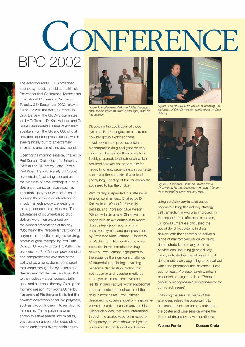
Figure 1: Prof Kinam Park, Prof Allan Hoffman and Dr Karl Malcolm (from left to right) discuss the session.
Figure 2: Dr Antony D’Emanuele describing the attributes of Dendrimers for applications in drug delivery.
Figure 3: Prof Allan Hoffman, involved in a dynamic audience discussion on drug delivery via pH-sensitive polymers and gels.
Professor Ivan Lieberburg (Elan
Corporation) illustrated the importance of
understanding the biology of the target
site in his presentation about amyloid
protein as a drug discovery target in the
treatment of Alzheimer’s disease. Amyloid
precursor protein is broken down into
beta amyloid, thought to be the main
cause of Alzheimer’s disease, by the
action of beta and gamma secretases.
Elan now have oligopeptide inhibitors of
beta secretase with nanomolar specifi city
in development, with clinical studies
predicted to take place in 2004. Gamma
secretase has been more complicated as
a target site and clinical studies are still
some way off. Another approach to the
treatment of is that of immunotherapy.
Active immunotherapy using beta amyloid
worked well in pre-clinical studies, but
the Phase I human studies were stopped
as some patients showed meningo-
encephalitis. This was thought to be
due to activation of T cells, with the
activity directed against the C terminus
of the beta amyloid. Clinical studies are
currently on hold, but in the future, smaller
fragments of beta amyloid may be used to
reduce or eliminate this issue.
Dr John Kirk (Queen’s University Belfast)
described the role of the blood brain
barrier in health and disease using multiple
sclerosis (MS) as an example. In MS
nerve sheaths are demyelinated and
the integrity of the blood brain barrier is
affected, resulting in a loss of function
and progressive disability of the patient.
Dr Kirk described studies using confocal
microscopy and immunofl uorescence
on autopsy samples of MS sufferers to
elucidate the molecular basis of the blood
brain barrier’s dysfunction. Tight junction
abnormality was found in 42% of active
lesions, 23% of inactive lesions and
13% in normal-appearing white matter,
suggesting that there may be a pre-clinical phase of the disease before the patient
realises that something is wrong. He also discussed current therapy for MS, outlining
how intravenous steroids may reduce infl ammation during acute episodes and how
immuno-modulatory drugs, such as beta interferon, may slow progression of the
disease, but concluded that there were no therapies currently available which prevent
progression of the disease or restore lost function, highlighting the necessity for future
research in this area.
Professor Lisbeth Illum (IDentity) discussed the biological aspects of brain delivery
via the intra-nasal route. Following nasal application, the drug is likely to be quickly
absorbed across the extracellular route into the cerebro-spinal fl uid or the olfactory
lobes of the brain, resulting in speedy onset of action, although some slower
intracellular absorption routes may also be important. Typical brain bioavailabilities,
however, are very low and are typically less than 1%. Differences in rat and human
physiology were described, such as nasal cavity size and orientation of the head,
which affects uptake of nasally-administered drugs to the cerebro-spinal fl uid. In
general, the uptake of these drugs into the rat brain is greater than into the human
brain and this must be borne in mind when analysing pre-clinical studies. Professor
Illum reviewed some of the formulation approaches used in attempting to improve
drug delivery to the brain via the nose, such as the use of bioadhesive chitosan
formulations, and concluded that there are still many formulation challenges and
opportunities for this route of drug administration.
Dr David Begley (King’s College London) gave an informative review about the location
of and role of effl ux transporters in the blood-brain barrier and how their action
can affect the delivery of drugs to the brain. Several effl ux transporters have been
identifi ed as being active in the blood-brain barrier, for example, P-glycoprotein (PGP),
multi-drug resistance protein (MDR) and breast cancer resistance protein (BCRP),
which, interestingly, appears to be one half of PGP, raising the question of whether
it needs to be dimerised for biological activity. PGP itself was described as possibly
acting via recognition of a disturbance in the membrane, caused by the presence
of a foreign molecule such as a drug, and acting to fi x it, rather than by recognising
specifi c molecules, which may help in explaining the wide range of substrates of
PGP. Importantly, Dr Begley described how the expression of PGP may change in
disease states such as cancer or AIDS, indicating the need for appropriate models
to be developed when designing formulations and therapeutic regimens for these
conditions.
Dr Wolfgang Staddler (University of Graz) discussed stereometabolism in an in vitro
model of the blood brain barrier. In particular he described the roles of ABC A1 and
SR-B1 during oxy-sterol transport through the capillary endothelial cells of the blood
brain barrier. ABC A1 mediates lipid traffi cking via exocytosis on the surface on cells
reviewsUKICRS 9th Annual Symposium
Access of therapeutics to the brainThe 9th Annual Symposium of the UKICRS took place in
January 2003, with the theme “Access of therapeutics to the brain”. The symposium covered biological aspects of brain delivery as well as formulation challenges and opportunities.
BELFAST 2003
and also retroendocytosis during intracellular processing. SR B1 is primarily expressed
in the liver, but also in the brain, and has been reported to be co-localised with caveolae
in porcine brain, with caveolae also being involved in signalling. Dr Staddler used as
an example the metabolism of cholesterol. In the brain cholesterol is metabolised to
24-hydroxycholesterol, which can then cross the blood-brain barrier and circulate to
the liver and bile. It in unclear whether cholesterol itself can cross from the brain into
the blood. This type of specifi c cellular traffi cking may have implications for the fate of
therapeutic molecules in the brain, if they are substrates for such systems.
Professor David Rees (Synt:em) described his company’s exciting new formulation
strategy to transport drug across biological membranes, including the blood-brain
barrier. The “Pep:trans” system is derived from protegrin, a mammalian peptide. This
“Pep:trans” system can interact with mammalian cell membranes, creating pores,
thus allowing the inward diffusion of the drug molecule. The two advantages of this
system are that it may interact with the cells without causing lysis, ie barrier integrity is
maintained, and also that it bypasses MDR/ PGP, thus maximising the effective dose
of the drug. Several examples of the use of this system with various drug molecules
were described. An opioid drug was shown to have improved pharmacological activity
including fast onset of pain relief; an increased cytotoxic T-cell response was observed
after vaccination with protein antigens; the effi cacy of paclitaxel in treating brain cancer
was increased with no peripheral side effects being observed.
Professor Jörg Kreuter (J.W. Goethe-Universität) described the use of nanoparticles
for the transport of drugs to the brain following intravenous injection. In particular, his
group have studied the use of drug-loaded cyanoacrylate nanoparticles coated with
polysorbate 80 (Tween 80), a widely-used surfactant. A range of drug molecules have
shown increased effectiveness when administered in this way, such as the hexapeptide
dalargin, the dipeptide kytorphin and the large molecule tubocurarine. Comparative
studies with the drug administered either alone, in uncoated nanoparticles or in solution
with Tween 80 showed no effect, indicating that for optimal effect, all three components
were required, ie the drug-loaded nanoparticles needed to be coated with the Tween
80. The mode of action of the Tween 80 in increased the brain absorption is not fully
elucidated yet, but is thought to be related to the coating of the iv-injected nanoparticles
with apoprotein E or B, followed by interaction with the low density lipoprotein receptor,
leading to endocytic uptake.
Dr Sylvia Wissing (Freie Universität, Berlin) also discussed the use of nanoparticulates
in targetting the brain. She reviewed the use of drug nanocrystals (essentially very fi ne
drug particles stabilised with a surfactant), solid lipid nanoparticles (SLN), nano-lipid
crystals (NLC) and lipid-drug conjugate nanoparticles (LDC). Nanocrystals tend to be
captured by macrophages after administration, which may not be desirable depending
on the intended target site. SLN and NLC are both produced using an oil in water
emulsion process, utilising solid lipids and a mixture of solid and liquid lipids respectively.
The solid system resulted in a good quality “wall” but lower drug loading, whereas
inclusion of a liquid phase resulted in a less structured “wall”, those allowing greater drug
inclusion. NLC is a newer formulation which has been reported to have a drug carrying
capacity of up to 50% of a lipid-soluble drug. All these systems can be prepared using
conventional processing techniques in large quantities, obviously an advantage to the
pharmaceutical industry.
Taken together, it was
a very informative day
with an appropriate
balance between the
biological aspects of
brain delivery and the
formulations challenges
posed thereby. The
conference was well-
attended, even on a cold
wet day in Belfast, and
the delegates expressed
uniform appreciation
of the programme.
Along with many of my
colleagues, I am looking
forward to the next
UKICRS meeting.
The LSI team Paul Tomsen, Andy Rendell, John Hand, Anne Farrow
and myself working with colleagues in other research councils have now
completed 42 university visits. At each of these meetings we have run cross
departmental workshops where academics from the physical sciences and
engineering disciplines have had the opportunity to discuss research ideas with life
science colleagues and then to identify what the barriers to working at this interface
were. Importantly, groups at these meetings were also able to identify solutions to
some of the barriers. Many of these solutions have been taken up by individuals, peer
reviewers, host universities and the EPSRC. It was from these meetings that the seeds
were sown for:-
■ Post Doctoral Mobility ■ Discipline Hoppers
■ The Complexity Sandpit ■ LSI Doctoral Training Centres
We have also run a number of meetings for young physical scientists looking at career
options in life sciences and in partnership with learned societies and other research
funders we have held a number of subject focussed workshops. Each of these events
has repeatedly shown how much interest and excitement is generated when researchers
have the opportunity to discuss research at this interface.
The Life Sciences interface Programme was established in April 1999 with a budget of £5m and 4 objectives:
■ Funding high quality research at the boundary between engineering and physical
sciences and the life sciences
■ Ensuring EPSRC is providing basic EPS needed to underpin the life sciences
■ Maximising the opportunities for advances in EPS from discoveries in life sciences
■ Providing suitable training at the interface
Over the last 4 years much has been achieved by the research community although
there remain large challenges. This article provides a timely opportunity to review what
has been achieved to date.
The programme’s focus since its formation has been on people, contact time and
research quality.
EPSRC’SLIFE SCIENCES
interface programme
PEOPLE
CONTACTtime
The research community working at the interface have made very good
use of the fellowship opportunities funded by the EPSRC. Professor
McLeish Leeds, and Prof Tuberfi eld Oxford, both have Senior fellowships
supported by the programme, with more applications currently under consideration. The
Programme in partnership with the MRC and more recently BBSRC has supported 55
discipline hopping awards and recently in an extension to this opportunity funded the
fi rst tranche of Institutional discipline bridging awards to 6 universities in the UK. These
awards will give a named facilitator at each of the successful institutions £250k to invest
in developing stronger collaborations at this interface within their host institution.
Last year the Programme announced the establishment of 2 doctoral training centres
at Oxford and Edinburgh both of which have secured funding for 5 annual intakes of
10 PhD students a year each studying for a physical sciences PhD while developing
knowledge of life sciences. It is hoped that a further 5 LSI doctoral training centres will
be announced shortly.
The Programme has made a virtue of fl exible funding; there is no norm for research
grant support in the Programme. Awards range from £2k to £10m.
Researchers have been encouraged to think fi rst about the research RESEARCHQuality
THEfuture
question they wish to address and then to cost this appropriately.
Similarly we have encouraged early discussion of outline proposals
as the programme supports grants announced through EPSRC,
BBSRC and MRC. The Programme now supports 6 research centres in
■ Medical imaging and signal processing to clinical science-Oxford led
■ Nanotechnology-Cambridge
■ Bio nanotechnology -Oxford
■ Bio manufacturing- UCL
■ Mathematics and Biology- Warwick
■ Tissue Engineering- Manchester/ Liverpool
EPSRC Council has confi rmed it’s strong ongoing
support for LSI and agreed a commitment budget
of £33M for the coming year. This includes EPSRC’s
investment in the joint council programmes on
Proteomics, Stem Cells and Brain Science announced
as part of SR2002. A challenge for the future is to demonstrate to EPSRC Council and
others that the programme as making a difference in the UK. Any help and/or examples
on this are always welcome. We will be exploring this with the community through a
number of regional meetings in May.
Finally the LSI team is here to work with the research and user community, should you wish to discuss any issues
with regard to the Programme or funding opportunities you are strongly encouraged to contact us.
Lesley Thompson
Programme Manager Life Sciences Interface
EPSRC
Polaris House
North Star Avenue
Swindon SN2 1ET
e-mail: [email protected]
Tel: 01793 444317
Fax: 01793 444456
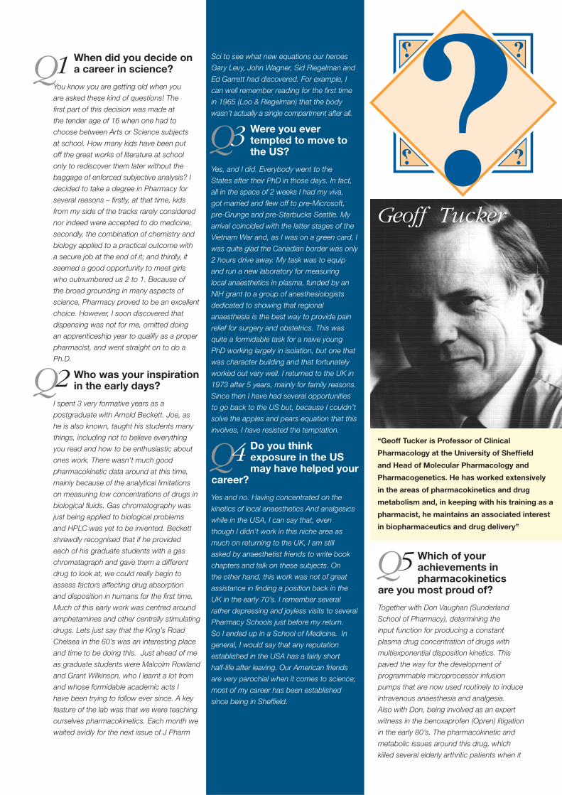
When did you decide on a career in science?
You know you are getting old when you
are asked these kind of questions! The
fi rst part of this decision was made at
the tender age of 16 when one had to
choose between Arts or Science subjects
at school. How many kids have been put
off the great works of literature at school
only to rediscover them later without the
baggage of enforced subjective analysis? I
decided to take a degree in Pharmacy for
several reasons – fi rstly, at that time, kids
from my side of the tracks rarely considered
nor indeed were accepted to do medicine;
secondly, the combination of chemistry and
biology applied to a practical outcome with
a secure job at the end of it; and thirdly, it
seemed a good opportunity to meet girls
who outnumbered us 2 to 1. Because of
the broad grounding in many aspects of
science, Pharmacy proved to be an excellent
choice. However, I soon discovered that
dispensing was not for me, omitted doing
an apprenticeship year to qualify as a proper
pharmacist, and went straight on to do a
Ph.D.
Who was your inspiration in the early days?
I spent 3 very formative years as a
postgraduate with Arnold Beckett. Joe, as
he is also known, taught his students many
things, including not to believe everything
you read and how to be enthusiastic about
ones work. There wasn’t much good
pharmacokinetic data around at this time,
mainly because of the analytical limitations
on measuring low concentrations of drugs in
biological fl uids. Gas chromatography was
just being applied to biological problems
and HPLC was yet to be invented. Beckett
shrewdly recognised that if he provided
each of his graduate students with a gas
chromatagraph and gave them a different
drug to look at, we could really begin to
assess factors affecting drug absorption
and disposition in humans for the fi rst time.
Much of this early work was centred around
amphetamines and other centrally stimulating
drugs. Lets just say that the King’s Road
Chelsea in the 60’s was an interesting place
and time to be doing this. Just ahead of me
as graduate students were Malcolm Rowland
and Grant Wilkinson, who I learnt a lot from
and whose formidable academic acts I
have been trying to follow ever since. A key
feature of the lab was that we were teaching
ourselves pharmacokinetics. Each month we
waited avidly for the next issue of J Pharm
Sci to see what new equations our heroes
Gary Levy, John Wagner, Sid Riegelman and
Ed Garrett had discovered. For example, I
can well remember reading for the fi rst time
in 1965 (Loo & Riegelman) that the body
wasn’t actually a single compartment after all.
Were you ever tempted to move to the US?
Yes, and I did. Everybody went to the
States after their PhD in those days. In fact,
all in the space of 2 weeks I had my viva,
got married and fl ew off to pre-Microsoft,
pre-Grunge and pre-Starbucks Seattle. My
arrival coincided with the latter stages of the
Vietnam War and, as I was on a green card, I
was quite glad the Canadian border was only
2 hours drive away. My task was to equip
and run a new laboratory for measuring
local anaesthetics in plasma, funded by an
NIH grant to a group of anesthesiologists
dedicated to showing that regional
anaesthesia is the best way to provide pain
relief for surgery and obstetrics. This was
quite a formidable task for a naive young
PhD working largely in isolation, but one that
was character building and that fortunately
worked out very well. I returned to the UK in
1973 after 5 years, mainly for family reasons.
Since then I have had several opportunities
to go back to the US but, because I couldn’t
solve the apples and pears equation that this
involves, I have resisted the temptation.
Do you think exposure in the US may have helped your
career?
Yes and no. Having concentrated on the
kinetics of local anaesthetics And analgesics
while in the USA, I can say that, even
though I didn’t work in this niche area as
much on returning to the UK, I am still
asked by anaesthetist friends to write book
chapters and talk on these subjects. On
the other hand, this work was not of great
assistance in fi nding a position back in the
UK in the early 70’s. I remember several
rather depressing and joyless visits to several
Pharmacy Schools just before my return.
So I ended up in a School of Medicine. In
general, I would say that any reputation
established in the USA has a fairly short
half-life after leaving. Our American friends
are very parochial when it comes to science;
most of my career has been established
since being in Sheffi eld.
Q1
Geoff Tucker
Which of your achievements in pharmacokinetics
are you most proud of?
Together with Don Vaughan (Sunderland
School of Pharmacy), determining the
input function for producing a constant
plasma drug concentration of drugs with
multiexponential disposition kinetics. This
paved the way for the development of
programmable microprocessor infusion
pumps that are now used routinely to induce
intravenous anaesthesia and analgesia.
Also with Don, being involved as an expert
witness in the benoxaprofen (Opren) litigation
in the early 80’s. The pharmacokinetic and
metabolic issues around this drug, which
killed several elderly arthritic patients when it
Q2
Q3
Q4
Q5
“Geoff Tucker is Professor of Clinical
Pharmacology at the University of Sheffi eld
and Head of Molecular Pharmacology and
Pharmacogenetics. He has worked extensively
in the areas of pharmacokinetics and drug
metabolism and, in keeping with his training as a
pharmacist, he maintains an associated interest
in biopharmaceutics and drug delivery”
was introduced, fi nally woke drug regulatory
authorities up to the need for more PK and
metabolic information on new drugs. More
recently, together with my colleague Amin
Rostami, I think we have had some infl uence
on the FDA with respect to bioequivalence
assessment – convincing them that systemic
exposure rather than actual drug release
from a product is the key regulatory issue.
We were also involved at a very early stage
in arguing for the rational selection of drug
interaction studies based on advances in
the knowledge of cytochrome P450 enzyme
selectivity. I can also lay claim to have
narrowly ‘missed’ being the fi rst to publish
on alpha1-acid glycoprotein as the major
plasma binding protein for basic drugs and
the CYP2D6 genetic polymorphism – but
those are long stories.
In your opinion have UK based scientists lost or
gained prominence relative to scientists in the rest of the world in the last 30 years?
When I go to international conferences on
subjects around my areas of interest it is
fairly clear that the academic UK presence
is less than it used to be. This is certainly
true for clinical pharmacology, and there is at
least cause for concern in drug metabolism
and PK. The decline of UK clinical
pharmacology has not been helped by some
of its erstwhile clinical leaders, who have
gone on to ‘higher’ administrative roles and
failed to foster a younger generation. Also,
the dominance of the ‘gene jockeys’ in UK
Medical Schools has wiped out a generation
of clinical investigators skilled in the
assessment of the functional aspects of drug
response. The older guys in drug metabolism
and PK still carry the fl ag. But where is the
next generation of academics going to come
from when a junior lecturer with a PhD earns
less than a fresh undergraduate joining the
pharmaceutical industry? On the other hand,
DMPK departments in the UK and global
pharmaceutical industry have fl ourished in
the last 5 – 10 years. This is because the
penny has fi nally dropped with at least some
research directors that ADME properties
have to be optimised along with receptor
binding, and because of academic effort
towards understanding the fundamental
structure and function of relevant human
enzymes and transporters.
How would you go about encouraging your younger
colleagues to reach for the high standards that you have set?
I don’t know about that – I have been
fortunate and privileged to work with some
superb colleagues and graduate students.
It helps, I guess. To have an ‘editor’s eye’
when it comes to detail, to be totally honest
about the limitations of a study and not to be
sloppy and selective in acknowledging the
prior body of knowledge.
Do you fi nd it relatively easy to fund your work?
No. The obsession with molecular biology
over the past 10 – 15 years has been a
problem for anyone primarily interested in
‘functional studies’. The MRC still invests
most of its money in large groups of gene
‘stamp collectors’, and it is only relatively
recently that the reality-checks on the
hype around the genome and much of
the rubbish talked about the imminence
of ‘personalised medicine’ has begun to
kick in. The RAE is not a four-letter word,
but it should be. At least in my neck of the
woods, the establishment is still motivated
by grant income and journal impact factors.
I am more interested in ‘bang for buck’, i.e.
outcome rather than just income measures,
and journal impact factors do not relate to
the impact of individual papers (Rostami
& Tucker – ‘Journal impact factors: A
bioequivalence issue?’ Brit J Clin Pharmacol
51: 111-118, 2001).
Do you think the pharmaceutical industry does
enough to encourage high quality research in the UK?
As someone who has benefi ted from
signifi cant industrial funding over the years,
through CASE awards etc, I think the UK
industry does this reasonably well. They can
sometimes be somewhat divisive by setting
academic groups off against each other,
but then this is not helped by an academic
climate (the RAE and the funding structure
that goes along with this) that does not
encourage effective collaboration between
Universities for the optimum benefi t of UK
plc.
What is your opinion of the proliferation of university spin-out
companys?
Having just set up my own University-based
company (plug for Simcyp.com) I am clearly
happy with the principle. However, this has
not been an experience I would like to repeat
in a hurry. Despite what they say about
encouraging entrepreneurship, many (but not
all) universities may make diffi culties for their
associated companies by taking too much
out of them (e.g. by charging excessive
overheads), and have not thought through
the mechanics of the process suffi ciently nor
indeed some of the potential ethical issues
relating to the academia-business interface.
Ideally, the research of both university
department and the company should benefi t
from the symbiosis. In practice, because
income generated by the company does
not contribute directly to the departmental
income score in the RAE, this can create
some ambiguities and tension.
How do you view the frontier of controlled-release and drug
delivery technology? Can we afford the sophisticated tailoring that modern technology could ultimately provide?
The number of products that have reached
the patient seems far fewer than many have
expected from the enormous research effort
that has gone into sophisticated delivery
systems and drug targeting. Certainly gene
therapy has been a spectacular failure so far,
and drug-antibody systems are still wrestling
with the problems of tissue specifi city and
bunker-busting to penetrate poorly-perfused
solid tumours. Cost-benefi t has to be
assessed on an individual product basis.
Together with colleagues I recently patented
a system requiring chrono-release of an
orphan drug with a well-defi ned clinical need.
On approaching two companys to provide
the pharmaceutical technology to develop
the product, one eventually declined for
pharmacoeconomic reasons but the second
is taking it forward.
If you had the chance to do it all again, would you?
Most certainly, if the clock was turned back.
Not so sure I would choose the same route if
I were starting over now. Academic freedom
is not what it was. In many UK institutions
young academics are increasingly expected
to make their individual research mark within
large frameworks of often diverse disciplines
held together in contrived ‘clusters’ and
constrained by proscribed ‘research themes’
defi ned by control freaks. Any interest in
good teaching has largely to be its own
reward, since it doesn’t factor greatly in the
promotion game.
Q6
Q7
Q8
Q9
Q11
Q12
Q10
MicrodissectionLASER CAPTURE
The recovery of purifi ed populations
of cells from tissue allows analysis of
gene or protein expression in discrete
cell populations. Previous technologies
to achieve this included protease
or enzymatic digestions that were
cumbersome, yielded uncertain cell
purities, and carried the risk of activating
or degrading the target cells. The advent
of laser capture microdissection (LCM)
technologies provides more elegant
methods for isolating specifi c cells types
from cell mixes in tissue sections.
The fi rst LCM system was developed
in the mid 1990s at the NIH and was
subsequently advanced commercially in
partnership with Arcturus Engineering.
This technology involves placing a
transparent thermoplastic fi lm over a
tissue section and visualising the cells
microscopically. A low-power infrared
laser melts the fi lm over the cells of
interest, and the selected cells remain
attached and are captured when the
fi lm is removed. This is the LCM system
most commonly seen in the current
literature. Multiple other LCM systems
are now on the market, however. Not
all of them require cell contact with a
laser-activated membrane. Indeed, many
employ objective- or prism-focused laser
beams to dissect cells of interest within
a tissue section. Cell retrieval can then
occur passively if the system utilises an
upright microscope allowing the laser to
cut from above and the cells to fall into
collecting caps underneath the specimen.
Alternatively LCM systems using inverted
microscopes can employ “laser pressure
catapulting (LPC)” to retrieve cells. LPC
involves launching cells into a receiving
cap above the tissue sample using a
“photonic cloud” generated by a laser
beam focused just beneath the tissue
plane.
LCM systems bundle multiple
components including laser source,
microscope, video camera, and computer,
and they typically cost in the range of
$100,000. Fluorescent microscope
attachments are available for many
models and increase the cost, as do
vibration resistant tables. A motorised
stage in often included to allow the tissue
section on the stage to move around a
fi xed laser. Cells captured by LCM can be
analysed for gene expression or protein
expression, and the power of LCM has
increased even further as successful
methods for genome wide profi ling and
proteomic analysis in tiny samples have
evolved.
Some problems with LCM technology
include relatively poor image quality.
Several elements of the technology
contribute to this including the use of
inverted microscopes in many systems
and the requirement for laser cutting on
completely dry sections without cover
slips or liquid. Also, tissue staining has to
be done with care and sparingly, because
the stains can degrade RNA quality. All
of these issues can impair the quality of
the image and make it diffi cult to dissect
the area of interest. LCM systems,
which do not use inverted microscopes
provide improved image quality. Other
problems with LCM include non-specifi c
retrieval of non dissected cells which
can occur with both contact and non
contact systems of LCM. This can be
guarded against by careful inspection of
the captured cells - an essential quality
assurance step in all LCM protocols (see
fi gure). Overall, though, current systems
of LCM work well are relatively easy to
use and provide powerful systems for
analysis of specialised or cancerous cells
retrieved from their native or pathologic
environment.
A more detailed but easy to read review of LCM has been published in The Scientist last year (The Scientist16[10]:42, May. 13, 2002 (http://www.the scientist.com/yr2002/may/profi le_020513.html).
John V. Fahy, M.D.
Prescott G. Woodruff, M.D.
Division of Pulmonary and Critical care Medicine
Department of Medicine and the Cardiovascular Research Institute
University of California San Francisco.
A
B
C
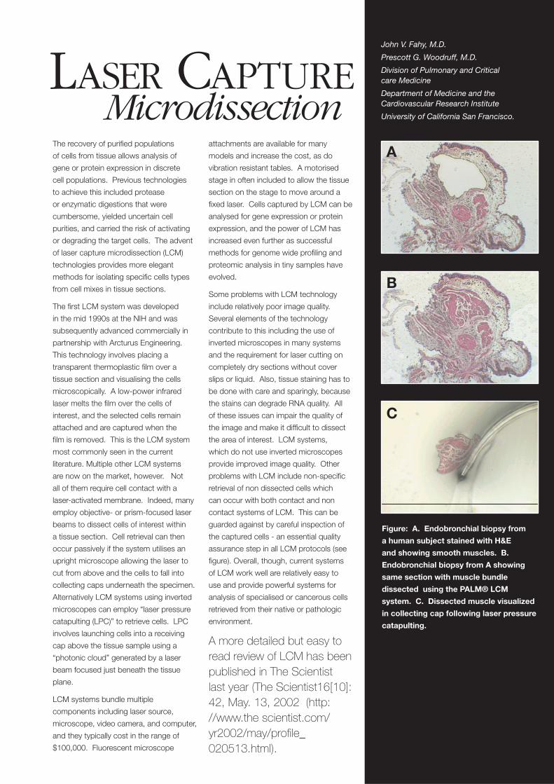
Figure: A. Endobronchial biopsy from
a human subject stained with H&E
and showing smooth muscles. B.
Endobronchial biopsy from A showing
same section with muscle bundle
dissected using the PALM® LCM
system. C. Dissected muscle visualized
in collecting cap following laser pressure
catapulting.
IntroductionIn recent years the confocal microscope has become a standard
tool in many biomedical laboratories. The confocal microscope,
originally conceived in 1955 (1), was introduced more broadly as
a specialist piece of research equipment in the early 1990’s but
has now become a biomedical research tool used routinely by
non-specialists. At the same time the scientifi c and technological
development of the technique continues at an amazing pace,
constantly re-defi ning the boundary between cutting-edge
research and routine application. The rate of technological
development also means that the manufacturers have to release
the next generation of system every few years in order to be able
to integrate the latest technological developments.
The equipment, i.e. the confocal microscope (CM) itself, is only
part of a larger array of interdependent technologies that include
e.g. fl uorescent probes, sample preparation techniques, digital
signal processing electronics, optical elements, fi lters, lasers,
detector technology and software development.
For the “standard user” the use of the CM has reached a point
where it is often treated as a black box. The superior image
quality and relative ease of use have meant that CM has started
to replace conventional epi-fl uorescence microscopy as a
standard microscopy technique.
For these users the advanced features of the latest generation
of CM systems are much less important than reliability and
ease of use for the detection of a few standard probes. For a
mid tier of users additional features such as fl exibility in terms
of (simultaneous) detection of a wider range of fl uorescent
probes, microscopy stages which allow live cell imaging,
ease of use of acquisition and image processing software are
increasingly important. Finally, the specialist user will want to
be able to customize microscopes to accommodate e.g. multi-
/two photon lasers, fl uorescence lifetime imaging, or spectral
analysis; acquired images will often be processed and analysed
with dedicated software packages to extract information from
multidimensional stacks of images that carry spectral, spatial,
and time resolved information.
This brief guide only provides an ‘executive’
overview of confocal microscopy and related
techniques and technology. High quality
detailed information on confocal microscopy
and related topics is available freely on the
internet. We hope this article
and the information in the
resources/reference
section will serve
as starting point for
those who seek more
in depth information about confocal
microscopy and related technologies.
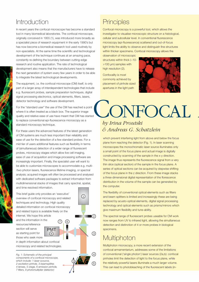
Fig. 1: Schematic of the principal components of a confocal microscope, which include: 1-laser scource, 2-excitation pinhole, 3-beamsplitter, 4-lenses, 5-stage, 6-emission pinhole, 7 fi lters, 9 photomulitplier (detector)
PrinciplesConfocal microscopy is a powerful tool, which allows the
investigator to visualise microscopic structure on a histological,
cellular and subcellular level. In conventional fl uorescence
microscopy (epi-fl uorescence) scattered and out-of-focus
light limits the ability to observe and distinguish fi ne structures
within thicker specimens. Confocal microscopy allows the
observation of microscopic
structures within thick (~10-
>100 µm) samples with
high resolution (2).
Confocality is most
commonly achieved by
placement of pinhole sized
apertures in the light path
1
2
3
4
5
67
8PMT
Z
which prevent interfering light from above and below the focus
plane from reaching the detector (Fig. 1). In laser scanning
microscopes the monochromatic laser source illuminates only
a small point of the focus plane and actual image is digitally
constructed by scanning of the sample in the x-y direction.
The image thus represents the fl uorescence signal from a very
thin slice (optical section) of the sample in the focus plane. A
series of optical sections can be acquired by stepwise shifting
of the focus plane in the z direction. From these image stacks
a three-dimensional digital representation of the fl orescence
distribution in the volume of the sample can be generated by
the computer.
The fl exibility of conventional optical elements such as fi lters
and beam splitters is limited and increasingly these are being
replaced by acusto-optical elements, digital signal processing
technology and optical elements such as prisms/mirrors which
give maximum fl exibility and tune-ability.
The spectral range of fl uorescent probes useable for CM work
now ranges from UV to infrared light, allowing the simultaneous
detection and distinction of 4 or more probes in biological
specimens.
MultiphotonMultiphoton microscopy, a more recent extension of the
confocal armamentarium, addresses some of the limitations
of conventional (‘single photon’) laser sources (2a,b): confocal
pinholes limit the detection of light to the focus plane, while
the relatively powerful lasers illuminate a much larger volume.
This can lead to photobleaching of the fl uorescent labels (in-
CONFOCAL Mby Irina Proutski & Andreas G. Schatzlein
Principles & ApplicationsMICROSCOPY
and out-of-focus) and the creation of phototoxic products (free-
radicals). Furthermore, the scattering/absorption of the excitation
laser light is higher for light of shorter wavelengths, which in turn
means that red and infrared dyes are more suited for the study of
thicker specimens.
In multiphoton microscopy infrared light is used to excite probes
usually exited by UV or visible light. As the energy of a photon
is inversely proportional to its frequency (E = h.v) this is only
possible because a fl uorescent molecule absorbs two or more
photons in a narrow time window to reach the same excitation
state. Multiphoton laser sources create femtosecond and
picosecond light pulses with very high photon densities that
allow multiple photon absorption events only in a very narrow
focal spot. Consequently, the spatial resolution is improved and
photobleaching and toxicity from out-of-focus fl uorophores
minimized, making the technique particularly suited for live cell
work, high resolution applications, and ‘deep’ samples.
Applications
The confocal microscope provides a tool to extract
multidimensional (spatiotemporal and spectral) information
about the distribution of fl uorescent probes in a specifi c
specimen. The technology does not exist in a vacuum but, only
in conjunction with suitable upstream (e.g. sample preparation
techniques, probe selection) and downstream technologies
(image processing, 3D reconstruction), does it become part of an
integrated application (7a.b).
In particular the rapid development of new fl uorescent probes
has helped the success of confocal microscopy. In addition
to hundreds of small molecule probes (4a) biological
macromolecule such as fl uorescent proteins have become
increasingly important (4b,c,d) . In the last ten years the
numbers of scientifi c papers referencing fl uorescent proteins
based on the jellyfi sh Aequorea victoria green fl uorescent
protein (GFP) has exploded from below 100 to several
thousand.
GFP and related probes have been cloned and can be
expressed in virtually all organisms from bacteria to whole
animals. They have evolved into an important biological tool
that impinges on almost every area of biological research.
Important applications involve the tagging of cellular proteins
to study their distribution in cells or transgenic animals,
measurement of gene expression and regulation, and studying
of tumour growth in living animals.
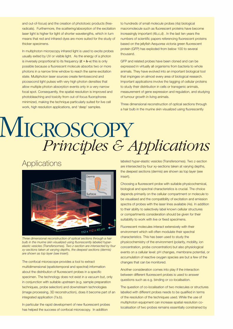
Three dimensional reconstruction of optical sections through
a hair bulb in the murine skin visualized using fl uorescently
labeled hyper-elastic vesicles (Transfersomes). Two z-section
are intersected by four xy-sections taken at varying depths,
the deepest sections (dermis) are shown as top layer (see
insert).
Choosing a fl uorescent probe with suitable physicochemical,
biological and spectral characteristics is crucial. The choice
depends primarily on the cellular compartment or molecule to
be visualised and the compatibility of excitation and emission
spectra of probes with the laser lines available (4e). In addition
to their ability to selectively label known cellular structures
or compartments consideration should be given for their
suitability to work with live or fi xed specimens.
Fluorescent molecules interact extensively with their
environment which will often modulate their spectral
characteristics. This has been used to study the
physicochemistry of the environment (polarity, mobility, ion
concentration, probe concentration) but also physiological
events on a cellular level: pH changes, membrane potential, or
accumulation of reactive oxygen species are but a few of the
changes that can be monitored.
Another consideration comes into play if the interaction
between different fl uorescent probes is used to answer
questions such as e.g. binding or co-localisation.
The question of co-localisation of two molecules or structures
labelled with different probes needs to be qualifi ed in terms
of the resolution of the techniques used. While the use of
multiphoton equipment can increase spatial resolution co-
localisation of two probes remains essentially constrained by
Three dimensional reconstruction of optical sections through a hair bulb in the murine skin visualized using fl uorescently labeled hyper-elastic vesicles (Transfersomes). Two z-section are intersected by four xy-sections taken at varying depths, the deepest sections (dermis) are shown as top layer (see insert).
the laws of optics, i.e. usually well above the wavelength of laser
source used.
Fluorescence resonance energy transfer (FRET) is a technique
which brings a leap in resolution, i.e. into the range of
nanometres (6a).
FRET describes the direct (without photon emission) transfer
of emission energy between a fl uorescent donor and a suitable
acceptor molecule. The transfer of energy can only occur if both
molecules are in close proximity, defi ned by the Förster radius
(10-100 Angstrom). Co-localisation would thus be determined
by observation of fl uorescence emission with the spectral
characteristics of the acceptor probe on excitation of the donor
probe.
Another characteristic of fl uorescent molecules which can yield
information about the probe’s environment is the lifetime of the
fl uorescence signal (6b). Any interaction with other molecules
will modulate fl uorescence lifetime because of the energy
transfer from the exited fl uorophore. While fl uorescence intensity
and spectrum are frequently strongly dependent on probe
concentration (e.g. eximer formation, quenching) fl uorescence
lifetime imaging (FLIM) directly observes effects that involve
energy transfer. Applications of FLIM include mapping of cell
physiology, but the lack of concentration dependence also
simplify FRET measurements. In combination with conventional
‘intensity’ imaging FLIM helps to distinguish between multiple
fl uorescence probes which may be diffi cult to distinguish based
on their spectral characteristics alone.
In addition, most sources of autofl uorescence have a relatively
short fl uorescence lifetime (few nanoseconds) whereas
fl uorescent probes often have long lifetimes on the order of
hundreds of nanoseconds. Measurement of the fl uorescence
signal after the decay of the fl uorescence signal can thus increase
signal-to-noise ratio dramatically.
LimitationsModern confocal microscopes hide their technological
sophistication behind a simple interface; even inexperienced
users can usually get impressive images in a short space of time.
Nevertheless caution and an understanding of the limitations
of the technique are required in interpreting results and when
planning more complex experiments. The most problematic area
is probably associated with the quantitative interpretation of the
confocal images, i.e. the correlation of fl uorescence intensity with
actual analyte concentration. Because of the complexity of the
technique it is extremely diffi cult to control all factors that can
potentially infl uence the process (10).
In conclusion it is clear that confocal microscopy is a powerful
tool which has changed the way we conduct biomedical
research. It has opened a window through which we visualize
life on a cellular and even molecular level in a way that has
not previously been possible and which is at the same time
aesthetically pleasing and scientifi cally exiting.
Reference & ResourcesThe web links/URLs provided in this resources section will provide a
starting point for the further exploration of confocal microscopy and
its application. A “Google” search for “confocal microscopy” comes
up with over 40,000 hits covering virtually every aspect of confocal
microscopy. We have selected a few (free access) URLs that may be
interesting and useful for those who want to explore the technique in
more detail.
1. Minsky, M., Memoir on Inventing the Confocal Scanning Microscopehttp://www.ai.mit.edu/people/minsky/papers/confocal.microscope.txt
2. A collection of PowerPoint slides by Dr. J. Paul Robinson (Purdue University Cytometry Laboratories) from a graduate class “Introduction to Confocal Microscopy and Image Analysis” covering most aspects of confocal microscopy can be found atUrl: http://www.cyto.purdue.edu/fl owcyt/educate/pptslide.htm
3. Multiphotona. Steve Potter’s reference list:
Url: http://www.neuro.gatech.edu/potter/mplsmAbs.htmlb. Good illustrations and brief explanation of multiphoton
http://www-celanphy.sci.kun.nl/Bruce%20web/scanning%20microscopy.htm
4. Fluorescent probes
General fl uorescent probesa. Molecular probes encyclopedic product catalogue is an invaluable
resource for fl uorescent dyes and their use in different applicationshttp://www.probes.com
Fluorescent proteinsb. GFP applications, books, and link page
Url: http://pantheon.cis.yale.edu/~wfm5/gfp_gateway.html
c. Zhang et al. Nature Reviews Molecular Cell Biology 3, 906-918 (2002)http://www.nature.com/cgi-taf/DynaPage.taf?fi le=/nrm/journal/v3/n12/full/nrm976_fs.html
d. Whole body imaging of tumour developmenthttp://www.anticancer.com/metamouse.html
Fluorescence spectra plottinge. Biorad database ”…has been designed to allow the user to superimpose
graphical fl uorochrome data from various fl uorochromes onto a normalised axis.” http://fl uorescence.nexus-solutions.net/
5. Stains FileA more general resource for histological dyes and staining techniques.http://stainsfi le.info
6. Fluorescence Resonance Energy Transfer (FRET) & Fluorescence Lifetime Imaging (FLIM) Microscopy
a. FRET: Selvin (2000) Review Nature structural biologyhttp://www.physics.uiuc.edu/People/Faculty/Selvin/articles/Nature2000.pdf
b. FLIM: Lifetime Imaging Techniques for Optical Microscopyhttp://www.becker-hickl.de/pdf/tcvgbh1.pdf
7. Sites with links covering including Optical Microscopy, Digital Imaging, and Photomicrography, Manufacturers etc.
a. Molecular Expressions’ web sitehttp://micro.magnet.fsu.edu/primer/resources/confocal.html
b. Confocal microscopy on the webhttp://swehsc.pharmacy.arizona.edu/exppath/micro/confocal.html
8. E-mail list servers & archives provide an excellent resource for in-depth advice from experienced users on all things microscopy/confocal. You can also subscribe to the e-mail list to keep up to date.
a. Confocal e-mail list archive (University at Buffalo) http://listserv.acsu.buffalo.edu/cgi-bin/wa?S1=confocal
b. Microscopy e-mail list archive http://www.msa.microscopy.com/MicroscopyListserver/SearchMLArchive.html
c. Multiphoton e-mail listhttp://groups.yahoo.com/group/mplsm-users/
9. Confocal related bookshttp://www.vaytek.com/books.html
10. J. Pawley’s “Critical Aspects of Fluorescence Confocal Microscopy” gives insight into some of the limitations of confocal microscopy.http://www.microscopyu.com/articles/confocal/pawley39steps.html
The development of increasingly complex drug delivery systems, combined with the enhanced understanding of disease processes at the cellular and molecular level, has led to a considerable need for the development of novel analytical approaches whereby increasingly small quantities of sample may be characterised. The challenges
associated with this are compounded by the need to study
systems in a state that is as near to the clinical or practical
situation as possible, hence such analysis may be required when
the material of interest is embedded within a complex structure
such as a dosage form, an isolated tissue or indeed a patient.
A Basic Guide toRAMAN MICROSCOPY
- Biomedical applicationsTo this effect there is strong evidence that Raman microscopy
represents an extremely powerful addition to the current range
of available approaches1,2. In brief, this method involves the
combination of Raman spectroscopy with optical microscopy
such that one may obtain spectra on highly localised regions
(scale of scrutiny ca. 1 µm) in complex multicomponent samples,
thereby allowing both identifi cation and characterisation of
individual components.
Raman spectroscopy is now a well-established analytical
technique whereby inelastic scattering from a molecule
irradiated with monochromatic light is measured as a function
of displacement of the scattered radiation, measured as a shift
in vibrational frequency from the incident excitation wavelength.
This technique has been widely used in the biomedical fi eld
due to the ability to both identify (via fi ngerprinting) and to
characterise species of interest by examination of the frequency
and intensity of the scattering signal which may be related
to the vibrational frequencies of specifi c bonds within the
sample. By coupling the spectrometer to an optical microscope
the technique effectively becomes a microprobe with spatial
resolution of ca. 1 µm, thereby enabling the operator to obtain
spectra for highly specifi c regions of a sample, to spatially
resolve the distribution of species with a characteristic vibrational
frequency or to map a sample whereby full spectra are obtained
throughout a selected area, hence providing a very powerful
means of species identifi cation and characterisation as a function
of location. In addition, Raman microscopy may also be used
in confocal mode to allow three-dimensional imaging (subject to
corrections for refractive index effects through the sample). It
should be noted that FT-IR microscopy is also available but lacks
the spatial resolution of the Raman technique and is also highly
sensitive to the presence of water and glass which may dominate
large regions of the spectral response, hence this method is of
less immediate interest within the context of the present account.
Raman microprobes have been in use since the 1970s but
the use within the biomedical fi eld has been limited due to the
weak signals obtained, the possibility of sample damage via
the laser and the strong fl uorescence background that is often
observed for biological samples. However, these diffi culties
have now been largely overcome. Sample damage can be
minimised by the judicious choice of experimental parameters
for example, by the use of line focussing to limit the excitation
laser power density at the sample and thus reducing thermal
damage. The problem of fl uorescence can sometimes be
by Duncan Craigand John J. McGarvey
effectively eliminated by the methods used for signal acquisition
and processing1 or by judicious choice of excitation wavelength.
For the latter, the current trend is to use longer wavelength lasers
(in the NIR wavelength region), away from electronic transitions
of the scattering molecule or of impurities therein, thus avoiding
fl uorescence entirely in many cases. . The problem of signal
strength has been addressed by improving the instrumentation
used. Specifi cally the development of both multi-channel
detector techniques and the use of notch fi lters with high
excitation light rejection capability has signifi cantly broadened the
applicability of modern Raman techniques. Other approaches for
improving sensitivity include resonance Raman scattering, used
particularly for nucleic acids and proteins, although this carries
a danger of sample degradation by the UV radiation required for
resonance enhancement, or by using surface-enhanced Raman
scattering whereby the sample is adjacent tonanoscale metal
surfaces, resulting in orders of magnitude increases in scattering
intensities.
There have been several studies using Raman microscopy for
the study of cells and tissues, all of which set a very encouraging
precedent, although it should be emphasised that the number of
studies in relation to the breadth of the biomedical fi eld is actually
quite limited. Indeed, to date the vast majority of studies on
biomedical topics use the conventional (macroscopic) technique,
sometimes with the excitation beam focussed to a similar spatial
resolution as that used for the microscopy approach (although
we do include examples of such studies below as they do still
illustrate the potential of the technique).
Previous workers have shown that the technique may be used
to characterise single cells. For example, Puppels et al.3-5
were able to obtain spectra for the nucleus and cytoplasm
of an eosinophilic granulocyte and a spectrum for a single
chromosome, while more recently real-time intracellular water
movement in single living cells have been visualised using a
derivative of the Raman technique (nonlinear coherent anti-
Stokes Raman scattering microscopy)6, while Schuster et al.7
have used the method to study population distributions in
bacterial cultures by characterising the chemical composition of
individual cells. Interestingly, ocular tissue has been particularly
well studied, using both conventional Raman spectroscopy
and Raman microscopy. Studies include those of animal and
human lenses, including the study of ageing on lens structure,
the characterisation of cataracts and the distribution of water
and proteins within lens the lens capsule8,9. The retina has also
been extensively studied from the perspective of accumulation
of age-related pigments10 and excitation state of cone visual
pigments11,12 .
The approach has also been used to examine crystalline
materials formed as a result of disease state. For example, the
spatial composition of kidney stones13, gallstones containing
cholesterol and bilirubin14 and atherosclerotic plaques containing
apatites15. The gallstone study is of particular interest as the
authors were able to show in layered gallstones that the outer
layer was composed of cholesterol, a centre containing bilirubin
(both acid and salt forms) and an intermediate layer containing
calcium phosphate, cholesterol and bilirubin. The authors also
demonstrated that small regions composed of calcium salts of
fatty acids were interspersed within the various layers. While
such an analysis is to some extent possible using alternative
techniques, it is diffi cult to suggest a method that can both
identify and spatially map these differing components within a
single set of experiments.
A further relevant area is the study of the presence of “foreign”
materials within a biological sample. Examples of such studies
include the identifi cation of materials remaining from surgical
operations, including fragments of PTFE following heart graft
surgery16 and traces of PGA in bone biopsy tissue following
reconstructive surgery2. Similarly, trace pieces of polymethyl
methacrylate (PMMA) and polyethylene have been identifi ed in
lung lavage material taken from a patient working in a particle-
laden environment17. The presence of drugs in biological material
may also be included in this category. For example, Nabiev et
al.18 studied the uptake of the cytotoxic drug doxorubicin. In
particular, these authors were able to differentiate between the
presence and conformation of the drug in the cytoplasm and the
nucleus, demonstrating that the drug was in a different binding
state within the nucleus and cytoplasm, this being ascribed to
DNA binding of the drug within the former. Interestingly, the
vibrational spectrum of the doxorubicin within the cytoplasm
was different to that in the free state, implying that the drug
does have a target within the cytoplasm as well as the nucleus.
Similarly, Morjani et al.19 showed that the topoisomerase inhibitor
intoplicine binds to DNA in the cell nucleus but remains in the free
state within the cytoplasm. More recent studies20 have included
the use of confocal Raman studies to examine the conformation
of poly(dG-dC).poly(dG-dC), a model for DNA, in two cationic
liposome complexes, thereby enabling the authors to model
the structure of the lipoplex and conformation of the nucleic
acid. The same authors21 were also able to locate DNA within
lipocomplexes, noting that the DNA concentration is higher near
the core of the vesicle. These highly interesting studies may
have profound implications for understanding the structure of
gene delivery systems, thereby enhancing the rational design of
these dosage forms. A limited number of studies have examined
drug diffusion through biological materials, including a study by
Suci et al.22 whereby the movement of chlorhexidine digluconate
through a Candida albicans biofi lm was monitored. Furthermore,
Dong et al.23 studied the interaction between micro-crystals of
human urokinase, a trypsin-like protease inhibitor that has been
strongly associated with tumour cells, and a range of inhibitors,
allowing the authors to defi ne the binding groups of the inhibitors
and to defi ne the rank order of inhibitors in terms of their binding
constants.
Our own interest has been developed as a result of a
collaboration between the authors, along with pharmaceutical
scientists Susan Barker and Vicky Kett, and colleagues in the
faculty of medicine here at Queen’s University Belfast. We are
in the process of installing our instrument and look forward to
performing some fascinating studies in the months to come.
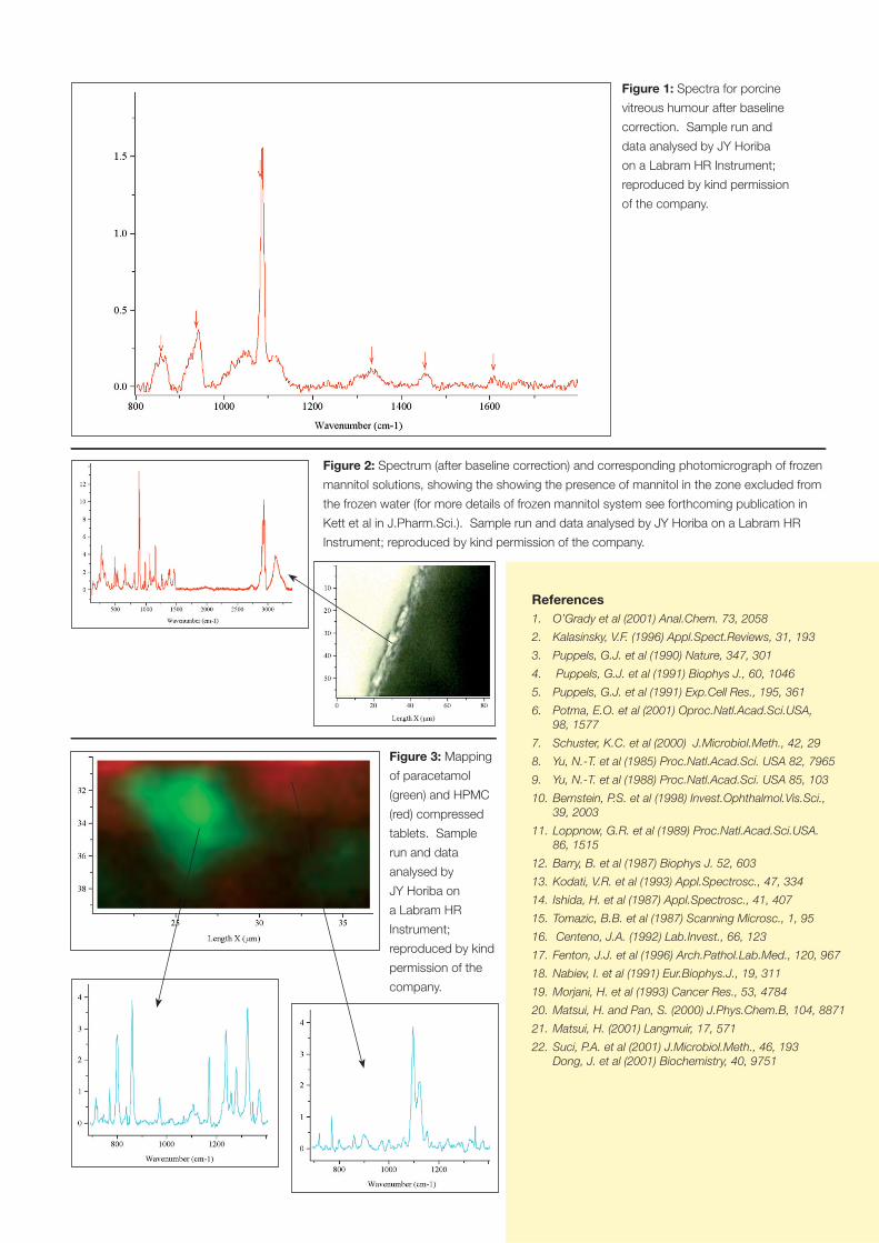
In particular, we intend to examine drug delivery systems,
ophthalmic fl uids and tissues and lung tissue and include images
from some of our (very preliminary) studies here (Figures 1-3).
References 1. O’Grady et al (2001) Anal.Chem. 73, 2058
2. Kalasinsky, V.F. (1996) Appl.Spect.Reviews, 31, 193
3. Puppels, G.J. et al (1990) Nature, 347, 301
4. Puppels, G.J. et al (1991) Biophys J., 60, 1046
5. Puppels, G.J. et al (1991) Exp.Cell Res., 195, 361
6. Potma, E.O. et al (2001) Oproc.Natl.Acad.Sci.USA, 98, 1577
7. Schuster, K.C. et al (2000) J.Microbiol.Meth., 42, 29
8. Yu, N.-T. et al (1985) Proc.Natl.Acad.Sci. USA 82, 7965
9. Yu, N.-T. et al (1988) Proc.Natl.Acad.Sci. USA 85, 103
10. Bernstein, P.S. et al (1998) Invest.Ophthalmol.Vis.Sci., 39, 2003
11. Loppnow, G.R. et al (1989) Proc.Natl.Acad.Sci.USA. 86, 1515
12. Barry, B. et al (1987) Biophys J. 52, 603
13. Kodati, V.R. et al (1993) Appl.Spectrosc., 47, 334
14. Ishida, H. et al (1987) Appl.Spectrosc., 41, 407
15. Tomazic, B.B. et al (1987) Scanning Microsc., 1, 95
16. Centeno, J.A. (1992) Lab.Invest., 66, 123
17. Fenton, J.J. et al (1996) Arch.Pathol.Lab.Med., 120, 967
18. Nabiev, I. et al (1991) Eur.Biophys.J., 19, 311
19. Morjani, H. et al (1993) Cancer Res., 53, 4784
20. Matsui, H. and Pan, S. (2000) J.Phys.Chem.B, 104, 8871
21. Matsui, H. (2001) Langmuir, 17, 571
22. Suci, P.A. et al (2001) J.Microbiol.Meth., 46, 193 Dong, J. et al (2001) Biochemistry, 40, 9751
Figure 1: Spectra for porcine
vitreous humour after baseline
correction. Sample run and
data analysed by JY Horiba
on a Labram HR Instrument;
reproduced by kind permission
of the company.
Figure 2: Spectrum (after baseline correction) and corresponding photomicrograph of frozen
mannitol solutions, showing the showing the presence of mannitol in the zone excluded from
the frozen water (for more details of frozen mannitol system see forthcoming publication in
Kett et al in J.Pharm.Sci.). Sample run and data analysed by JY Horiba on a Labram HR
Instrument; reproduced by kind permission of the company.
Figure 3: Mapping
of paracetamol
(green) and HPMC
(red) compressed
tablets. Sample
run and data
analysed by
JY Horiba on
a Labram HR
Instrument;
reproduced by kind
permission of the
company.
If you believe the plethora of literature out there on “change” it seems that the human disposition is to avoid this situation if at all possible. However, in this article we explore the motivations of fi ve
scientists that opted for a career change, going from Academia to Industry or vice versa.
Prof
iles
PEO
PLES
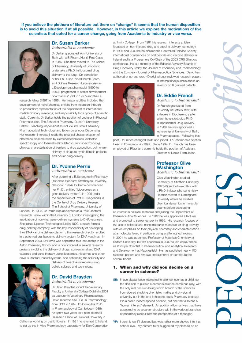
Dr. Susan BarkerIndustrialist to Academic:
Dr Barker graduated from University of Bath with a B.Pharm.(Hons) First Class in 1986. She then moved to The School of Pharmacy, University of London to undertake a Ph.D. in liposomal drug delivery to the lung. On completion of her Ph.D. she joined Merck Sharp and Dohme Research Laboratories as a Development pharmacist (1990 to 1993), progressed to senior development pharmacist (1993 to 1997) and then a
research fellow (1997 to 1999). Her responsibilities included the development of novel chemical entities from inception through to production; representation of the department at international multidisciplinary meetings; and responsibility for a group of scientifi c staff. Currently, Dr Barker holds the position of Lecturer in Physical Pharmaceutics, The School of Pharmacy, Queen’s University Belfast. Teaching responsibilities include Industrial Pharmacy; Pharmaceutical Technology and Extemporaneous Dispensing. Her research interests include the physical characterisation of pharmaceutical materials by electrical techniques (dielectric spectroscopy and thermally-stimulated current spectroscopy), physical characterisation of barriers to drug absorption, pulmonary
delivery of drugs to cystic fi brosis patients and ocular drug delivery.
Dr. Yvonne PerrieIndustrialist to Academic:
After obtaining a B.Sc degree in Pharmacy (1st class Honours; Strathclyde University, Glasgow; 1994), Dr Perrie commenced her Ph.D., entitled “Liposomes as a gene delivery system”, in 1995 under the supervision of Prof G. Gregoriadis in the Centre of Drug Delivery Research, The School of Pharmacy, University of
London. In 1998, Dr Perrie was appointed as a Post-Doctoral Research Fellow within the University of London investigating the application of non-viral gene-delivery systems to DNA vaccines. She joined Lipoxen Technologies Ltd in 1999, a newly formed drug-delivery company, with the key responsibility of developing their DNA vaccine delivery platform; this research directly resulted in a patented oral liposome delivery system for DNA vaccines. In September 2000, Dr Perrie was appointed to a lectureship in the Aston Pharmacy School and is now involved in several research projects involving the delivery of drugs, conventional and DNA vaccines and gene therapy using liposomes, niosomes and other novel surfactant-based systems, and enhancing the solubility and
delivery of bioactive molecules using colloid science and technology.
Dr. David BraydenIndustrialist to Academic:
Dr David Brayden joined the Veterinary Faculty at University College Dublin in 2001 as Lecturer in Veterinary Pharmacology. David received his B.Sc. in Pharmacology from UCD in 1984. Following his Ph.D. in Pharmacology at Cambridge (1989), he spent two years as a post-doctoral Research Fellow at Stanford University in
California working on cystic fi brosis. In 1991 he returned to Ireland to set up the In Vitro Pharmacology Laboratory for Elan Corporation
at Trinity College. From 1991 his research interests at Elan focussed on non-injected drug and vaccine delivery technology. In 1995 and 2000 he co-chaired the Controlled Release Society international conferences on oral peptide and vaccine delivery in Ireland and is a Programme Co-Chair of the 2003 CRS Glasgow conference. He is a member of the Editorial Advisory Boards of Drug Discovery Today, the Journal of Pharmacy and Pharmacology and the European Journal of Pharmaceutical Sciences. David has authored or co-authored 40 original peer-reviewed research papers
in international journals and is an inventor on 6 granted patents.
Dr. Eddie FrenchAcademic to Industrialist:
Dr French graduated from University of Bath in 1986 with a degree in Biochemistry after which he undertook a Ph.D. in Transdermal Drug Delivery. In 1989, he was appointed a lectureship at University of Bath, in Pharmaceutics. Following this
post, Dr French changed fi elds and joined RP Scherer as a Section Head in Formulation in 1992. Since 1994, Dr. French has been employed at Pfi zer and currently holds the position of Assistant
Director of Liquid Formulation.
Professor Clive WashingtonAcademic to Industrialist:
Clive Washington studied Chemistry at Sheffi eld University (1975-8) and followed this with a Ph.D. in laser photochemistry. He then moved to Nottingham University where he studied chemical dynamics in molecular beams before developing
an interest in colloidal materials and joining the Department of Pharmaceutical Sciences. In 1987 he was appointed a lecturer and promoted to senior lecturer in 1996. His research focuses on the use of colloidal and nanostructured materials for drug delivery, with an emphasis on their physical chemistry and characterisation at a molecular level, in particular using scattering techniques. In 2001 he was appointed Professor of Materials Chemistry at Salford University, but left academia in 2002 to join AstraZeneca as Principal Scientist in Pharmaceutical and Analytical Research and Development at Macclesfi eld. He has published nearly 100 research papers and reviews and authored or contributed to several books.
1. When and why did you decide on a career in science?
SB: I have always been interested in science, even as a child, so the decision to pursue a career in science came naturally, with the only real decision being which branch of the sciences. I considered studying chemistry, maths and physics at university but in the end I chose to study Pharmacy because it is a broad-based applied science, but one that also has a “human interest” element. An additional bonus was that there appeared to be a career structure within the various branches of pharmacy (useful from the perspective of a teenager).
YP: I don’t know if I decided but was rather more shoved into it at school level. My careers tutor suggested my plans to be an
Pof
iles
PEO
PLES
astronaut and/or a pop idol was apparently unrealistic. Where was Pete Waterman then?!?
DB: At 2nd level, I was intrigued by Biology, although my better subjects were languages. At College, during the B.Sc. course one of my electives in 2nd Year was pharmacology and I got very excited about linking drug action to physiology – and especially enjoyed sloshing around in practicals.
EF: I decided on a career in science whilst doing my O’Levels. I had a lot of interest, a very inquisitive mind and little ability in any other fi eld. I would have never have made it as a stand up comic or professional sportsman which I would have chosen but for lack of talent.
CW: For me, there was never really any question – I got my fi rst chemistry set when I was six and after that I never looked back – particularly after the incident with the fl aming methylated spirit on the dining table……
2. Why did you decide on a career change from industry to academia?
SB: After nine years working in industry, I felt that I needed a change. As with most things, familiarity breeds contempt and I was beginning to feel less challenged with the somewhat repetitive nature of drug development. Additionally, the opportunities for performing fundamental research, rather than development-associated projects, were limited. Academia offers more freedom in the research area than industry, so I thought I’d try academia.
YP: My original plan was to enter academia however during my time as a Post-doc I was lucky enough to be offered an Industrial position in Lipoxen Technologies Ltd a new start-up co-founded by Prof Gregory Gregoriadis. I really enjoyed my time with Lipoxen and because the company was, at the time, newly established I had the exciting chance of watching it grow. Also since I was involved in the research from very early stages I was very much involved in all aspects of the project I was developing. This provided a challenging and exciting environment. So why did I leave? Well it was not an easy decision but I had developed a taste for the educational side of science (madness I know!) so with this in mind I decided to spread my wings and head north!
DB: During my industrial research career, I was creating academic collaborations for the company and gradually I started getting more fun out of supervising postgraduates and also from presenting my group’s data in written and oral format. Gradually, I suppose that my bias moved back
towards the philosophy of research, i.e., adding to knowledge. It therefore became more likely that I would move to academia.
EF: Two major reasons. One was money, second was a need for a change to something more structured.
CW: To a large extent I was pushed by the problems within academia as much as I was attracted by the potential of industry. I had made the decision to pursue an academic career when I was doing my fi rst degree at Sheffi eld; I followed this with a Ph.D., postdoctoral work in chemistry, lectureship at Nottingham, the usual path. However this was a time of enormous change in the University environment, with factors such as the Research Assessment Exercise and the move towards bigger grants in focused centres. The academia that I found myself in around 2001 was a very different place to that which I had originally perceived. Of course it was a gradual change, but eventually I realised that I simply wasn’t having fun any more – for me the main reason for doing the job. Then, of course, there were the obvious benefi ts of working in the private sector. Finally, I had been exposed to industry rather more than most academics – I had worked with AstraZeneca on collaborative projects for many years; I knew and respected the people and the environment. This made it much less like jumping into a black hole.
3. What were the main factors that contributed to your decision to change careers?
SB: As I said before, it was mainly an issue of wanting to feel challenged and motivated. Obviously, there were considerations of fi nance and geography, but luckily those factors didn’t turn out to be insuperable.
YP: Mainly just timing really (also the pop idol thing was going nowhere!)
DB: A lectureship was advertised at the University in 2001 and I was on the look-out for a suitable move. Previously, the university had invited me to contribute lectures to courses and to co-supervise postgrads and I happened to have a collaborating professor on site there, who was ready to start new projects with me. However, I never considered a Veterinary Faculty until I realised that I would have the same pharmacology research opportunities as being in Medicine or Science Faculties.
EF: I had done my Degree, Ph.D. and was a lecturer, all at Bath. I needed a change. It was probably a mistake to stay in the same place for so long. I am naturally a fairly unstructured sort of person and was young and impressionable at the time. I needed more guidance than I was getting at University. I was not sure what area
of research I wanted to go into and in academia you need focussed drive. Also my salary was basically not enough to survive on in Bath.
4. What prompted you to make this career change at the time that you did?
SB: Nine years is a long term to spend in one environment, so I was ready for a change. However, I had been thinking about making a career change for several months before I did so. Pharmacy is a relatively small world, so the opportunity to move also had to be present.
YP: I only decided to apply for an academic position in response to as series of adverts for Pharmaceutics Lecturers in the Pharmaceutical Journal.
DB: In my industrial career I had become a project manager and had moved away from the science. Although I learned a lot on how to lead and run projects – especially the joint ventures with US biotech start-ups, I missed the actual design of experiments and being close to the research and the researchers. At the same time, the company started moving away from niche areas of oral peptide and vaccine delivery and these were the areas I wanted to continue in.
EF: Timing was down to three main things,
a) I had just completed my three year probation so was a ‘qualifi ed’ lecturer.
b) My wife had just had our fi rst child and we needed somewhere bigger to live. My salary at the time was not high enough to get a mortgage for a one bedroom fl at in Bath. Scherer offered to double my salary.
c) The Conservatives were re-elected and I did not see the way forward for universities.
CW: I had the offer of a chair of chemistry at Salford University. This came at an opportune time because my wife originally worked at AZ Charnwood but her job function was becoming more focused at Alderley Park. She was commuting several days a week, and a permanent change was likely. Consequently the chair came up at an opportune time and we moved to Cheshire. The Salford job looked promising initially as there was signifi cant investment in the Department and several other new members of staff had been recruited at the same time. However, after about six months the university management decided to suspend the chemistry courses due to declining student numbers in chemistry nationally, and many staff members took redundancy packages or early retirement. This really prompted us to take a hard look at the future, and examine all the options. Moving to industry had been at
the back of my mind for a few years, and this brought it to the forefront. I suppose you could say that I felt I’d done my ‘National Service’ and it was time to move to something with more potential.
5. How would you compare the working cultures of these two different areas?
SB: Industry has a much more controlled environment, i.e. everything has to be approved by several layers of management before anything can be done, whereas in academia, individual researchers do have more freedom to follow their own interests. Academia tends to have a “freer” environment, such that individuals can manage their own time and resources most appropriately. However, industry does have a focus that may not be apparent in academia. In the pharmaceutical industry, the company’s goals are set and all departments work to the same end, whereas in academia, everyone’s research is different, so a common focus is not really possible.
YP: In some respects in Academia you are left more to your own devises in terms of the research paths you take, and meetings always seem to take twice as long!
DB: In academia, you are wearing three hats of trying to be good at research, teaching and administration. I can work on what I want to – assuming I can get funding. In a way, you are working for yourself in a one person company so personal motivation might be higher. I enjoy working with academic staff spread over a wider age bracket as this helps put perspective on things. My group has research targets, but they are realistic and are set by me in accordance with what is required for the funding agencies and for postgraduate higher degrees. A university is primarily a learning environment and one is continually teaching, mentoring and generating enthusiasm in undergraduate and postgraduate students – and I really work at this part as it’s new to me.
EF: I feel to be successful at university requires that you are highly self motivated and know where you are going and can provide your own structure to move forward. It is somewhat a self-centred approach as you focus mainly on what you are doing. In industry you also have to be self-motivated but in general a team helps defi ne some of the structure for you especially when you are more junior. Industry is a much more collaborative team environment as in general the end goal is much bigger than in university.
CW: When I fi rst joined AstraZeneca everyone asked me if I had experienced a culture shock. I responded that I hadn’t; after all I’d known and worked with people here for years, so I knew what to expect. However, now I’ve been here about six
months, I can see that there are a lot of cultural differences – a ‘creeping culture shock’, perhaps. People here are more supportive; there is a feeling of community, which arises from working on common projects. To me this is very evident since it wasn’t a notable feature of academia. People are equally committed both here and in academia; it’s just that, in academia, competition for resource between research groups is so severe that it dominates the landscape; you spend more time competing than researching. Here we all have a common game plan, although, of course, there can never be enough resource to do everything that everyone would like. But running an academic research group is very much like being self-employed; if you fail to secure grants or compete for space, your personal career development can be severely curtailed. That, I suppose, is another reason I left; suddenly I realised that I was trusting my career to an intrinsically high-risk environment.
6. What do you consider to be the up-sides and the down-sides of working in each area?
SB: The most signifi cant benefi t to working in industry is the money - both personal and also the money available to buy high quality equipment and resources. On the other hand, the most signifi cant drawbacks are the lack of personal freedom and the corporate culture.
From the academic perspective, money is a big issue. I have found that most academics spend a large portion of their time writing grant applications to obtain money to fund their research, most of which are not then funded due to the limited funds available. In my opinion, the current funding system needs some degree of overhaul to make the whole process easier and more transparent, particularly in the case of younger staff at the beginning of their academic career. A defi nite up-side of academic life is the chance to meet and work with scientists in other disciplines that may not be possible in industry. For example, I am currently collaborating with scientists from Mechanical Engineering, Chemical Engineering, Ophthalmology and Respiratory Medicine.
YP: The down-side of academia is the students (only joking!). More seriously, academia is messier and I often feel less focused. I tend to jump from one thing to another then to the next too often - but on the fl ip side this helps keep things fresh!
DB: Academic up-sides include fl exible working conditions, freedom to work on what you want, the buzz out of teaching well, travel, and having a permanent job! Academic down-sides include grant writing for a lot of each year, a rather arbitrary promotion
system. Industry up-sides include having the money to do the science, expertise available in the company to help, seeing key projects through to the next phase and potentially rapid rises through the ranks. Down-sides include less job security, having a different boss every year and the likelihood of your pet project being canned just before it is about to deliver!
EF: Industry: Up-sides - Working conditions, nice kit, salary, team focus, clear goals, structured hours. Down sides - deadlines that sometime prevent exploratory research, drive to focussed fi elds in which you may not want to be, sometimes over pressured.
Academia: Up-sides - students (keep you young), freedom to choose subjects to focus on, fl exibility of hours. Down-sides - salary, pressure to succeed, lack of funding, effort to get funding. Limited opportunities for growth, almost entirely self-driven and a lack of support.
CW: In theory academia gives you a lot of freedom to explore ideas and be completely self-determined, although if your ideas go nowhere, so do you! In reality your freedom is actually determined by your money-earning capacity. I think the biggest drawback of academia is that you’re actually doing four separate jobs – fundraising, research, teaching, and administration, so it’s hard to put a useful effort into any one. The people who succeed in academia often end up doing sixty hours a week or more just to fi t it all in, whereas at AstraZeneca we have a stronger emphasis on work-life balance and family-friendly working. In industry you are much more focused on specifi c problems, and if everyone agrees that something is really worth doing, then the time and resource are generally made available to do it. Of course the compensation package is much better as well; the academic package is offensive by comparison. The only downside to working in industry is that some of the broadest elements of intellectual freedom are removed, due to the need to support the business. I can’t just decide one day that I want to work on superconducting ceramics or quantum optics. I have to focus on the company’s objectives, but so far that hasn’t led to any lack of stimulating problems.
7. What do you miss from your previous fi eld and what do you enjoy most in your current area?
SB: I miss the apparent speed of work - because time is money to the industry, development goals are set with very tight timelines, often to the day several years in the future, so there is always a sense of urgency. There is not usually the same sense of urgency in academia (except perhaps at exam time, when marking
dominates everyone’s life for a few weeks!).
I most enjoy being responsible for myself, my own time management and work planning.
YP: At present I don’t have much time for my personal research time and I miss doing my own lab work, but I plan to improve this as most my teaching is now in place. However what I do enjoy is the fact I supervise a few research projects so there is always new and interesting data coming in from the students.
DB: I miss a lot of interaction with biotech companies. Although I carry out some consultancy, unless you are directly part of the industrial scene, it’s harder to keep up with current industrial developments from the outside. I also had lots of research contacts in other companies that I got to know well as scientists and friends. In the university I enjoy most those rare occasions when you know you have managed to convey some pharmacological principle in a creative way to students in a lecture and that they really understand the concept.
EF: Miss students and fl exibility. Enjoy team environment.
CW: I used to quite enjoy teaching – the contact with a continual stream of new people was very stimulating. It was teaching that made me realise that you don’t know if you actually understand something until you try to explain it to someone else. I think the most enjoyable part of my present job is the opportunity to work on projects that may become household names and improve peoples’ quality of life. Also it’s very gratifying to work in an environment where there is a substantial amount of support and development. I’ve been on more training courses in the last six months than all my years in academia together!
8. Of the skills gained from your previous experience in industry / academia, which have been the most benefi cial to you in your current fi eld?
SB: I think it is the transferable skills that have been the most useful, e.g. organisational skills and communication skills. An in-depth knowledge of the drug development process has helped in terms of lecture materials and also in some of the collaborations I have with other scientists, being able to discuss the industrial perspective has been invaluable. In a more general sense, it is always benefi cial to departments to have people with different ideas and experiences, so hopefully I make a contribution in academia to that effect.
YP: Working at the Academic/Industrial interface I learned quickly what each side expects.
DB: Industry gives great training in time management, project management, report writing, running meetings, budgeting, communication skills and an innate ability to focus on the essential deliverables of projects. These are real assets in academia. When we write collaborative grants, a lot of the time I will end up coordinating the proposal as the requirements are more instinctive to me.
EF: Method of thinking. I was taught to challenge everything, assume nothing, and work out how to prove things with data.
CW: My current position is that of Principal Scientist and my main responsibility is to provide leading-edge science to underpin the business. I’ve been very lucky in getting to this point because it means that my job is mainly about doing science and solving problems, so naturally the knowledge base I accumulated has been invaluable. But it’s no use being able to do all this if you can’t explain it to anyone, so the communication and teaching skills I developed as a lecturer have been equally important. Also I had a lot of exposure to intellectual property while I was at Nottingham, and that has been very useful.
9. What is your opinion on the proliferation of University Spin-out and Biotechnology Start-up companies?
SB: A lot of these companies don’t seem to perform very well, or indeed do close after the initial support money has run out. It would seem more appropriate for fewer companies to be set up, but with a more structured business plan and also with much more scientifi c backing - a good idea is not suffi cient.
YP: I learned a lot from my time at Lipoxen and I think start-ups are a useful “grey area” between Academia and Industry. They are ideally suited for those of us who enjoy taking basic research developed in Academia and seeing it put to good use.
DB: There is a lot of emphasis on academics becoming entrepreneurs and I support this. As long as academics keep the company business separate from their public sector-funded research, I don’t see a problem. The university doesn’t want to see the academics being spun-out however, so the current positive trend is for licensing deals and handing over the technology to a start-up company, which is then run by industrial people. In the end, I see the main role for academic research is science for public good and to increase knowledge. This can still be compatible with start-ups as long as the academic knows where to draw the
line between academic and business aspects. In all this, we mustn’t forget that academics have a key educational role in preparing our new young scientists for industry, for other careers, and to instil the genuinely liberal values that emerge from third level education - these key functions must not be squeezed out in favour of the sole pursuit of university money-making ventures. Some university administrators tend to forget this in their excitement over making lucrative royalty deals on an academic’s research!
EF: Mixed. Up-side is benefi t for academics and institutions giving a more tangible out put. Down-side is I think it stunts freedom and is very bad for the students.
CW: There’s little point in doing scientifi c or medical research if it’s not ultimately going to be exploited for the benefi t of society. Even areas that most people consider to be of no immediate benefi t, such as cosmology or particle physics, may ultimately be built into a useful technology if we look suffi ciently far into the future. For many new ideas, spin-out from the university is the only way of exploiting them, as they go through a precompetitive or venture phase. Even when they are earning money they may not fi t the profi le of preexisting companies. The trouble is that a lot of tensions arise as the company culture tries to grow within the university environment. I think it’s essential that the company becomes independent and free-standing as soon as possible, as universities are not good places to run businesses from, especially if you’re still trying to do research and teaching. Contributing to the exploitation of your work is an important part of being an academic, but unfortunately it can lead to the researcher being sidetracked into running a full-time business, and his efforts are then lost from the research community. However I have a cynical eye to many spin-out activities. In some departments every member of staff seems to feel the need to have a company, and one suspects that they have little of value to sell, and had pumped it up – they probably realised that tapping into venture capital was the only way they’d ever be able to drive a Ferrari. I think it’s a good thing when a new technology or product is developed by spin-off, but I’m much less enthusiastic about companies who offer, for example, specifi c service functions that could equally well be done in the context of sponsored research within the university research group itself.
10. Do you think that the drive for academics to form Spin-out companies from their research is a benefi t to research in general? Why?
SB: I think that dissemination of research
fi ndings and commercialisation of inventions are very important and historically academics have been very bad at doing this. However, I do feel that the pendulum has swung too much in the opposite direction with universities actively encouraging (almost forcing) academics to set up private companies (albeit for a hefty share in any profi ts). It does seem to me that some universities are in danger of forgetting their core business, which is education and research.
YP: Yes. It helps academics nurture and advance their projects out into the commercial domain.
DB: I do, and the main reason is that new research ideas can be translated into a practical outcome of benefi t to society. It is also more likely that the technology being developed will have a sound mechanistic basis if it has been thought of by an academic scientist who has being working in the area for some years. I have some sympathy with the view, however, that we are neglecting a lot of great creative scientists in universities who spend their lives fi nding out how things work, but cannot get funding because society does not see a useful short-term profi table outcome. These individuals appear to be getting lost within the current government priority of linking research funding directly to industrial application and employment.
EF: Not really - Focuses researchers too quickly. Takes away the blue sky/risk edge and makes the aims too close to that of industry. Need to fi nd a way to reward the inventors but allow them to continue to focus effort on 5 - 20 years in the future.
CW: Actually I tend to think the opposite. It helps in the development of the specifi c technology but much of the profi t is simply not fed back into the universities. This is partly because profi t has to be fed back to shareholders, and partly because, in the early days at least, universities were very naïve about the value of their intellectual property. That has changed now, of course, and most are much sharper in this respect. Unfortunately, even when a university has a major share in a valuable spin-off, the income from it need not be fed back to the original research area. Also an academic supporting a spin-off will have less time to devote to fundamental work – I’ve seen several examples where commercial success led to the academic concerned either leaving the University sector or moving to a part-time contract. However, I suppose you could call this fi ne detail – the opposite view is that, if academia has commercial success, it will invigorate the sector overall. Personally I’m not convinced about this argument. If you look at the world-leading quality of British university research, then why is the academic sector always in such a
poor fi nancial position with contracting and closing departments, grey-listed Universities, and lagging salaries?
11. Would you recommend scientists to experience both Academia and Industry within their career? Why?
SB: I think the most important thing is to have an intellectually challenging occupation, whether that is in academia or industry. I would certainly encourage scientists to consider both academia and industry before making career choices, but also to remember that it’s not a life-sentence - you can move between branches and back again.
YP: Yes defi nitely, you don’t know what you like until you try it!
DB: Yes, indeed I would go so far as to recommend sabbatical teaching and research stints in each type of environment. This might help academic and industrial researchers to respect the differences a bit more and to appreciate the qualities required to survive in both types of system. For example, some academics are quite sceptical about the quality of research being carried out in industry – yet they appear not have noticed that a very high proportion of papers in the British Journal of Pharmacology are from industrial laboratories requiring the same level of peer review as those papers from academic colleagues. Some industrial researchers still believe erroneously that academics cannot work to funding deadlines and that they are unreliable collaborators who can never deliver!
EF: There is defi nitely cross fertilisation of working practices/styles that would benefi t someone from exposure to both. It also helps with understanding where folk are coming from.
CW: I would always recommend a broad experience base, but I think it’s risky to try to plan a career, which has a late change to industry. In my experience ‘mixed’ careers are usually unplanned, rather than planned, which implies that they were a response to an unexpected problem or opportunity. It’s true that I’ve benefi ted a great deal from my academic past, although I feel in retrospect that perhaps I should have made the change sooner. Personally I think that many aspects of trying to climb the academic ladder are too risky, particularly the postdoctoral system, which is a national disgrace. We all know horror stories of postdocs who are at the top of their pay scale and simply allowed their career to run on too long on short contracts, hoping that a permanent academic job would come along eventually. Despite recent changes in employment law, universities
are still not required to provide security to fi xed-contract postdoctoral staff. My advice is that, unless you have a head of department grooming you for an upcoming post, leave as soon as your thesis has been written up, or perhaps do your BTA (Been To America) for a year. There are very few companies that are as open-minded as AstraZeneca about employing senior academics, and it can be very hard to convince an employer that an academic has the qualities a specifi c industrial job needs.
12. If you had the chance to do it all again would you? What would you do differently?
SB: I think I probably would have gone into industry when I did (after my Ph.D.), but made the switch to academia earlier. Also, I would probably have gone abroad (to Europe) either to study or to work.
YP: Perhaps! What would I do differently? Take singing lessons.
DB: I would do it again and in the same order - even though I never planned it that way. Somebody deciding which career path to go for should ask which environment is best suited to their instincts and personality. Personally, I think the transition back to academia from industry in my own case has been liberating and I have had the advantage of bringing some good working habits from a goal-driven environment
EF: Either I would move about for my Ph.D. or postdocs or I would do a degree (poss Ph.D.) then some industry then go back to academia. I am now far better mentally equipped to be an academic as I have learnt some discipline.
CW: I think I might make two types of change. The fi rst is to realise the power of working at the best centres with the top people on projects that are recognised as world-class. I chose my postgraduate and postdoctoral projects because they interested me; I really wasn’t clued up about the politics of science at that time. I think a spell at somewhere like Oxford or Imperial would have been a good idea. Secondly I would move around more. There are very few employers, academic or industrial, in which you can make rapid upward progress. Most people advance by moving on rather than hanging around. It’s obvious really; why restrict yourself to the opportunities in a single place if you can take advantage of the opportunities in the whole country?
This, of course, assumes that I’d decide to follow the same sort of career path, but just try to do it more effectively. If I could do everything again then I’d probably choose a completely different path. No one wants to have the same experiences twice. But it would be so different that, I suspect, your readers probably wouldn’t be interested in it.
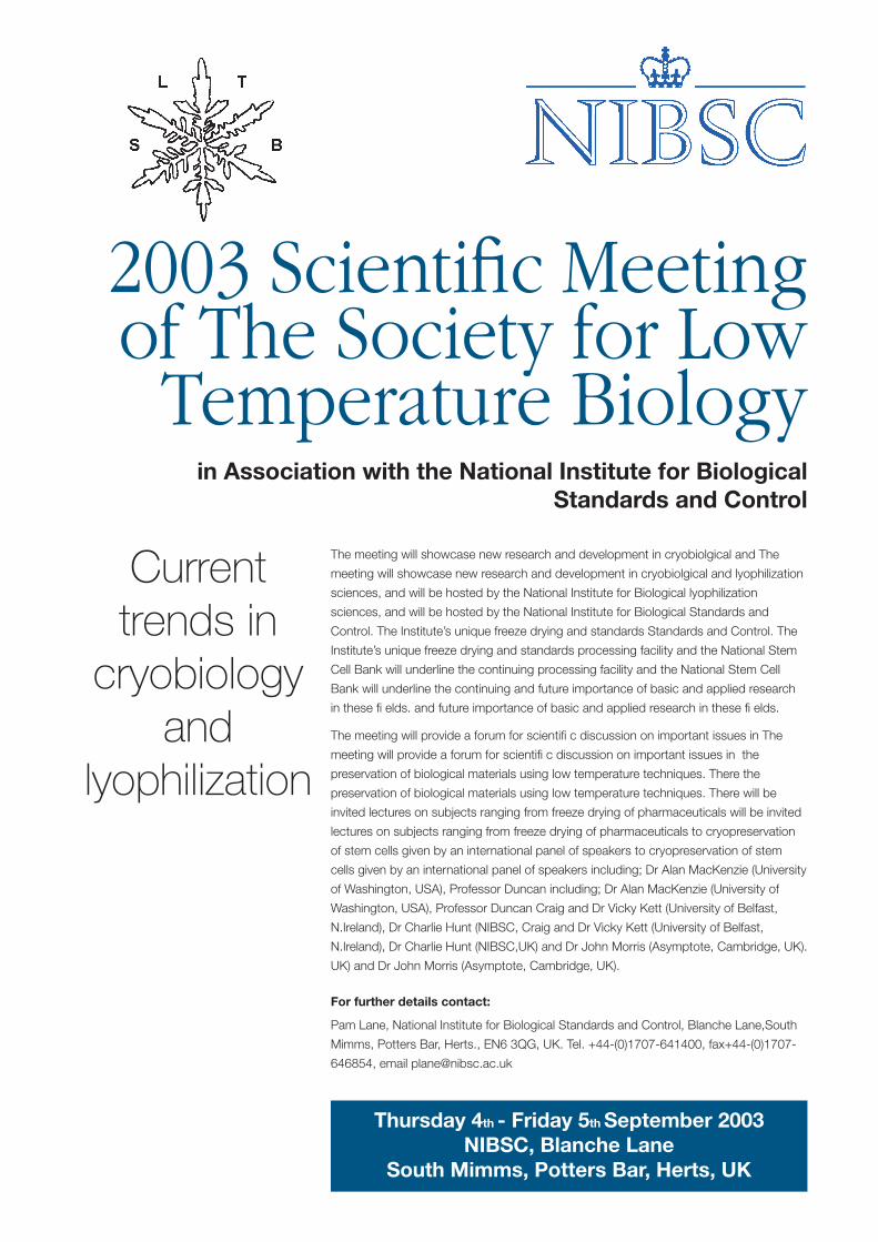
The meeting will showcase new research and development in cryobiolgical and The
meeting will showcase new research and development in cryobiolgical and lyophilization
sciences, and will be hosted by the National Institute for Biological lyophilization
sciences, and will be hosted by the National Institute for Biological Standards and
Control. The Institute’s unique freeze drying and standards Standards and Control. The
Institute’s unique freeze drying and standards processing facility and the National Stem
Cell Bank will underline the continuing processing facility and the National Stem Cell
Bank will underline the continuing and future importance of basic and applied research
in these fi elds. and future importance of basic and applied research in these fi elds.
The meeting will provide a forum for scientifi c discussion on important issues in The
meeting will provide a forum for scientifi c discussion on important issues in the
preservation of biological materials using low temperature techniques. There the
preservation of biological materials using low temperature techniques. There will be
invited lectures on subjects ranging from freeze drying of pharmaceuticals will be invited
lectures on subjects ranging from freeze drying of pharmaceuticals to cryopreservation
of stem cells given by an international panel of speakers to cryopreservation of stem
cells given by an international panel of speakers including; Dr Alan MacKenzie (University
of Washington, USA), Professor Duncan including; Dr Alan MacKenzie (University of
Washington, USA), Professor Duncan Craig and Dr Vicky Kett (University of Belfast,
N.Ireland), Dr Charlie Hunt (NIBSC, Craig and Dr Vicky Kett (University of Belfast,
N.Ireland), Dr Charlie Hunt (NIBSC,UK) and Dr John Morris (Asymptote, Cambridge, UK).
UK) and Dr John Morris (Asymptote, Cambridge, UK).
For further details contact:
Pam Lane, National Institute for Biological Standards and Control, Blanche Lane,South
Mimms, Potters Bar, Herts., EN6 3QG, UK. Tel. +44-(0)1707-641400, fax+44-(0)1707-
646854, email [email protected]
Thursday 4th - Friday 5th September 2003NIBSC, Blanche Lane
South Mimms, Potters Bar, Herts, UK
Current trends in
cryobiologyand
lyophilization
2003 Scientifi c Meeting of The Society for Low
Temperature Biologyin Association with the National Institute for Biological
Standards and Control
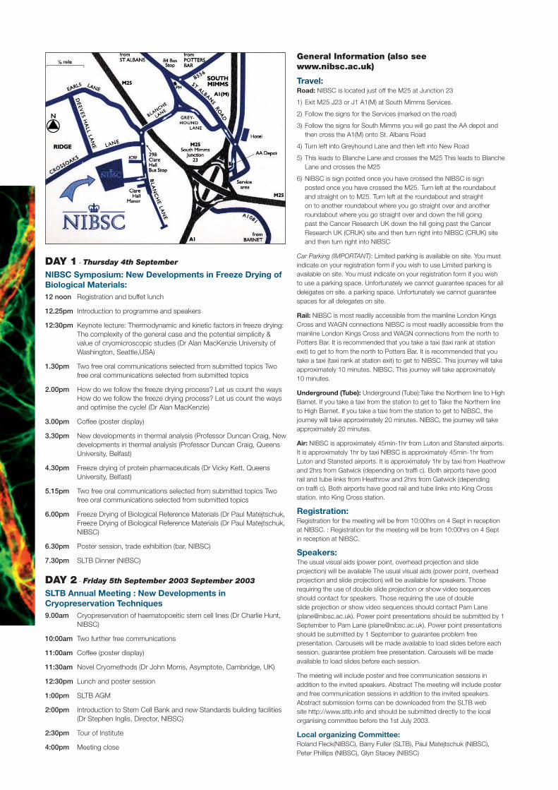
General Information (also see www.nibsc.ac.uk)
Travel:Road: NIBSC is located just off the M25 at Junction 23
1) Exit M25 J23 or J1 A1(M) at South Mimms Services.
2) Follow the signs for the Services (marked on the road)
3) Follow the signs for South Mimms you will go past the AA depot and then cross the A1(M) onto St. Albans Road
4) Turn left into Greyhound Lane and then left into New Road
5) This leads to Blanche Lane and crosses the M25 This leads to Blanche Lane and crosses the M25
6) NIBSC is sign posted once you have crossed the NIBSC is sign posted once you have crossed the M25. Turn left at the roundabout and straight on to M25. Turn left at the roundabout and straight on to another roundabout where you go straight over and another roundabout where you go straight over and down the hill going past the Cancer Research UK down the hill going past the Cancer Research UK (CRUK) site and then turn right into NIBSC (CRUK) site and then turn right into NIBSC
Car Parking (IMPORTANT): Limited parking is available on site. You must indicate on your registration form if you wish to use Limited parking is available on site. You must indicate on your registration form if you wish to use a parking space. Unfortunately we cannot guarantee spaces for all delegates on site. a parking space. Unfortunately we cannot guarantee spaces for all delegates on site.
Rail: NIBSC is most readily accessible from the mainline London Kings Cross and WAGN connections NIBSC is most readily accessible from the mainline London Kings Cross and WAGN connections from the north to Potters Bar. It is recommended that you take a taxi (taxi rank at station exit) to get to from the north to Potters Bar. It is recommended that you take a taxi (taxi rank at station exit) to get to NIBSC. This journey will take approximately 10 minutes. NIBSC. This journey will take approximately 10 minutes.
Underground (Tube): Underground (Tube):Take the Northern line to High Barnet. If you take a taxi from the station to get to Take the Northern line to High Barnet. If you take a taxi from the station to get to NIBSC, the journey will take approximately 20 minutes. NIBSC, the journey will take approximately 20 minutes.
Air: NIBSC is approximately 45min-1hr from Luton and Stansted airports. It is approximately 1hr by taxi NIBSC is approximately 45min-1hr from Luton and Stansted airports. It is approximately 1hr by taxi from Heathrow and 2hrs from Gatwick (depending on traffi c). Both airports have good rail and tube links from Heathrow and 2hrs from Gatwick (depending on traffi c). Both airports have good rail and tube links into King Cross station. into King Cross station.
Registration:Registration for the meeting will be from 10:00hrs on 4 Sept in reception at NIBSC. : Registration for the meeting will be from 10:00hrs on 4 Sept in reception at NIBSC.
Speakers:The usual visual aids (power point, overhead projection and slide projection) will be available The usual visual aids (power point, overhead projection and slide projection) will be available for speakers. Those requiring the use of double slide projection or show video sequences should contact for speakers. Those requiring the use of double slide projection or show video sequences should contact Pam Lane ([email protected]). Power point presentations should be submitted by 1 September to Pam Lane ([email protected]). Power point presentations should be submitted by 1 September to guarantee problem free presentation. Carousels will be made available to load slides before each session. guarantee problem free presentation. Carousels will be made available to load slides before each session.
The meeting will include poster and free communication sessions in addition to the invited speakers. Abstract The meeting will include poster and free communication sessions in addition to the invited speakers. Abstract submission forms can be downloaded from the SLTB web site http://www.sltb.info and should be submitted directly to the local organising committee before the 1st July 2003.
Local organizing Committee:Roland Fleck(NIBSC), Barry Fuller (SLTB), Paul Matejtschuk (NIBSC), Peter Phillips (NIBSC), Glyn Stacey (NIBSC)
DAY 1 - Thursday 4th September
NIBSC Symposium: New Developments in Freeze Drying of Biological Materials:12 noon Registration and buffet lunch
12.25pm Introduction to programme and speakers
12:30pm Keynote lecture: Thermodynamic and kinetic factors in freeze drying: The complexity of the general case and the potential simplicity & value of cryomicroscopic studies (Dr Alan MacKenzie University of Washington, Seattle,USA)
1.30pm Two free oral communications selected from submitted topics Two free oral communications selected from submitted topics
2.00pm How do we follow the freeze drying process? Let us count the ways How do we follow the freeze drying process? Let us count the ways and optimise the cycle! (Dr Alan MacKenzie)
3.00pm Coffee (poster display)
3.30pm New developments in thermal analysis (Professor Duncan Craig, New developments in thermal analysis (Professor Duncan Craig, Queens University, Belfast)
4.30pm Freeze drying of protein pharmaceuticals (Dr Vicky Kett, Queens University, Belfast)
5.15pm Two free oral communications selected from submitted topics Two free oral communications selected from submitted topics
6.00pm Freeze Drying of Biological Reference Materials (Dr Paul Matejtschuk, Freeze Drying of Biological Reference Materials (Dr Paul Matejtschuk, NIBSC)
6.30pm Poster session, trade exhibition (bar, NIBSC)
7.30pm SLTB Dinner (NIBSC)
DAY 2 - Friday 5th September 2003 September 2003
SLTB Annual Meeting : New Developments in Cryopreservation Techniques9.00am Cryopreservation of haematopoeitic stem cell lines (Dr Charlie Hunt,
NIBSC)
10:00am Two further free communications
11:00am Coffee (poster display)
11:30am Novel Cryomethods (Dr John Morris, Asymptote, Cambridge, UK)
12:30pm Lunch and poster session
1:00pm SLTB AGM
2:00pm Introduction to Stem Cell Bank and new Standards building facilities (Dr Stephen Inglis, Director, NIBSC)
2:30pm Tour of Institute
4:00pm Meeting close

Registration and Booking Form
The cost of registration is £50 (SLTB members) £75 (non-members)
I am/am not a member of the SLTB (please delete as appropriate).
Accommodation
30 Rooms have been block booked for the 3 and 4 September at the Holiday Inn, 30 Rooms have been block booked for the 3 and 4 September at the Holiday Inn, Swanland Road, South Mimms, Potters Bar, at a cost of £85 per night with breakfast Swanland Road, South Mimms, Potters Bar, at a cost of £85 per night with breakfast included. A form for this is attached. included. A form for this is attached.
Enclosed is a list of other hotels for you to contact directly for bookings.
Parking at the Conference Site
I wish to request a parking space at NIBSC (please tick box) I wish to request a parking space at NIBSC (please tick box)
Conference Dinner
The dinner will be held at NIBSC and starts at 19.30hrs. The dinner will be held at NIBSC and starts at 19.30hrs.
If you would be interested in attending (please tick box) If you would be interested in attending (please tick box)The cost of this is yet to be fi nalized but we will inform you The cost of this is yet to be fi nalized but we will inform you as soon as possible. as soon as possible.
I enclose a cheque for the amount of £ for the registration.
Signed Date
NB: Non-UK participants may pay at registration.
Name:
Contact Address:
Tel.No:
Fax No:
e-maill:
Please forward this form to:Pam Lane, NIBSC, South Mimms, Herts, EN6 3QG, UK
tel 44 (0)1707 641400 • fax 44 (0)1707 646854 • e-mail [email protected].
Please note the seating numbers are strictly limited and bookings can only be confi rmed on receipt of payment before 31 July 2003
please cut out or photocopy
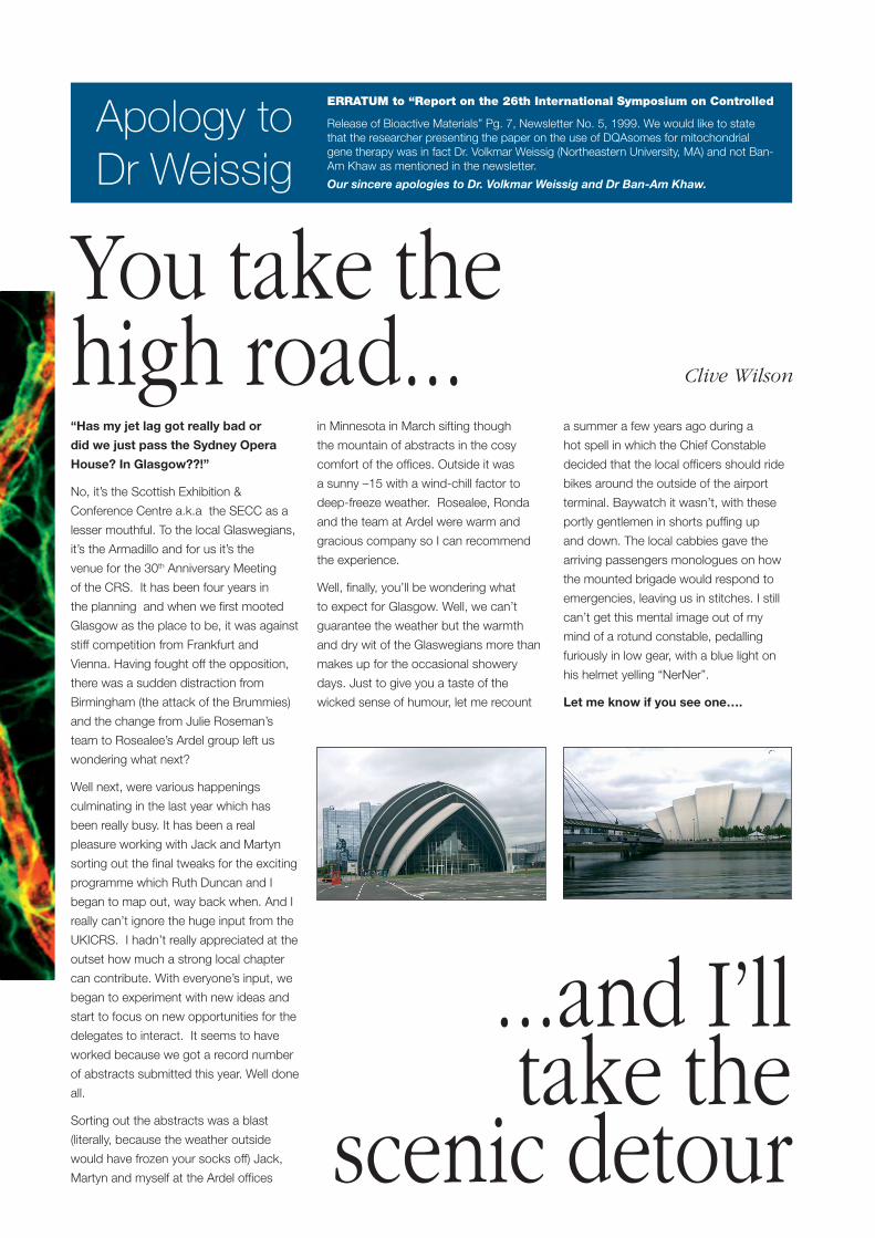
“Has my jet lag got really bad or
did we just pass the Sydney Opera
House? In Glasgow??!”
No, it’s the Scottish Exhibition &
Conference Centre a.k.a the SECC as a
lesser mouthful. To the local Glaswegians,
it’s the Armadillo and for us it’s the
venue for the 30th Anniversary Meeting
of the CRS. It has been four years in
the planning and when we fi rst mooted
Glasgow as the place to be, it was against
stiff competition from Frankfurt and
Vienna. Having fought off the opposition,
there was a sudden distraction from
Birmingham (the attack of the Brummies)
and the change from Julie Roseman’s
team to Rosealee’s Ardel group left us
wondering what next?
Well next, were various happenings
culminating in the last year which has
been really busy. It has been a real
pleasure working with Jack and Martyn
sorting out the fi nal tweaks for the exciting
programme which Ruth Duncan and I
began to map out, way back when. And I
really can’t ignore the huge input from the
UKICRS. I hadn’t really appreciated at the
outset how much a strong local chapter
can contribute. With everyone’s input, we
began to experiment with new ideas and
start to focus on new opportunities for the
delegates to interact. It seems to have
worked because we got a record number
of abstracts submitted this year. Well done
all.
Sorting out the abstracts was a blast
(literally, because the weather outside
would have frozen your socks off) Jack,
Martyn and myself at the Ardel offi ces
in Minnesota in March sifting though
the mountain of abstracts in the cosy
comfort of the offi ces. Outside it was
a sunny –15 with a wind-chill factor to
deep-freeze weather. Rosealee, Ronda
and the team at Ardel were warm and
gracious company so I can recommend
the experience.
Well, fi nally, you’ll be wondering what
to expect for Glasgow. Well, we can’t
guarantee the weather but the warmth
and dry wit of the Glaswegians more than
makes up for the occasional showery
days. Just to give you a taste of the
wicked sense of humour, let me recount
a summer a few years ago during a
hot spell in which the Chief Constable
decided that the local offi cers should ride
bikes around the outside of the airport
terminal. Baywatch it wasn’t, with these
portly gentlemen in shorts puffi ng up
and down. The local cabbies gave the
arriving passengers monologues on how
the mounted brigade would respond to
emergencies, leaving us in stitches. I still
can’t get this mental image out of my
mind of a rotund constable, pedalling
furiously in low gear, with a blue light on
his helmet yelling “NerNer”.
Let me know if you see one….
ERRATUM to “Report on the 26th International Symposium on Controlled
Release of Bioactive Materials” Pg. 7, Newsletter No. 5, 1999. We would like to state that the researcher presenting the paper on the use of DQAsomes for mitochondrial gene therapy was in fact Dr. Volkmar Weissig (Northeastern University, MA) and not Ban-Am Khaw as mentioned in the newsletter.
Our sincere apologies to Dr. Volkmar Weissig and Dr Ban-Am Khaw.
Apology to Dr Weissig
You take the high road...
...and I’ll take the
scenic detour
Clive Wilson
Please note the following: Membership is currently free and it is our intention for this
to remain the case for the foreseeable future. Should
the UKICRS committee decide to introduce a fee then
the members will be given due notice and advised that
their existing membership will continue until the end of
the existing membership period without charge, after
which they will have the option of rejoining under the new
arrangements.
UKICRS membership does not imply membership of the
parent Controlled Release Society, for which there is a
separate procedure.
Membership is open to any individual, irrespective of
experience, who has an interest in the controlled release of
drugs and bioactive materials and the development of novel
delivery strategies.
Membership information will be treated in confi dence and
will not be disclosed to a third party with the exception of
the parent organisation (the Controlled Release Society)
for purposes of forwarding membership benefi ts and
information regarding CRS activities.
Membership is for a period lasting until the end of the
calendar year following of the date of registration, i.e. an
individual joining in 2002 will have a membership period up
until 31st December 2003. Members can discontinue their
membership at any point of their choosing by informing the
UKICRS to that effect.
Participation in the January Annual UKICRS Meeting
carries automatic membership until the end of the following
calendar year.
The benefi ts of membership may change on an annual basis
but at present we are able to offer the following:
■ Early alerts to forthcoming meetings
■ Hard copy of the UKICRS newsletter
■ Free CD of the main CRS meeting proceedings*
■ Reduced registration fee for the BPC** These items are currently available but, as they involve the participation of other organisations, we can only guarantee their continued availability on a year-by-year basis
In addition to the above by joining the organisation (or
renewing your membership) you will be offering your support
and encouragement to the UKICRS. As this organisation is
run by volunteers on a non-profi t basis, the endorsement of
members of the pharmaceutical and related communities is
greatly appreciated.
The current membership list ceased to be in use from
1st January 2003, although the UKICRS may retain the list
for mailing purposes only.
See back cover for application form
As part of our ongoing development as an organisation, the UKICRS is formalising the
defi nition of membership so as to concentrate our efforts and resources on individuals
who are actively interested in the groups’ activities. To this effect we are inviting
existing members and those new to the group to simply register their interest on the
enclosed form. In this way we believe that we can deliver a more effective programme
of activities to our members and avoid unnecessary contact with those who no longer
have an interest in the area.
fi nd out more about us at:http://www.pharmweb.net/ukicrs.html
Application forUKICRS MEMBERSHIP

Title
First name
Surname
Contact address
Email address
Tel Fax
Place of emplyment/study
Job title
Your research interests
Are you a member of the CRS, our parent organisation?
If yes, what is your membership number?
Date
continued from inside back cover
Please return completed form to: Dr Susie Berrill, Vectura Ltd, 1 Prospect West, Chippenham, Wiltshire SN14 6FH Fax: +44 (0)1249 667701
Application forUKICRS MEMBERSHIP
Please cut out or photocopy