Embed Size (px)
Citation preview
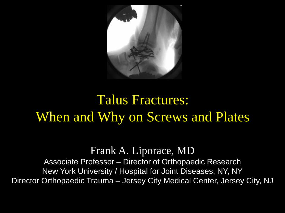
Talus Fractures:
When and Why on Screws and Plates
Frank A. Liporace, MD Associate Professor – Director of Orthopaedic Research
New York University / Hospital for Joint Diseases, NY, NY
Director Orthopaedic Trauma – Jersey City Medical Center, Jersey City, NJ
Disclosures
• Please refer to program
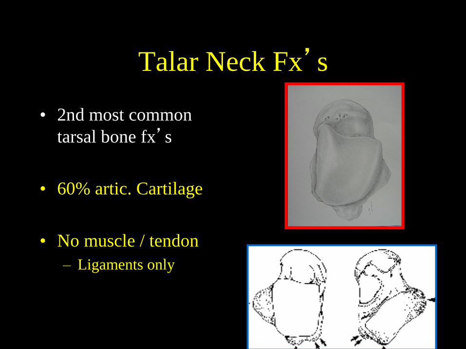
Talar Neck Fx’s
• 2nd most common
tarsal bone fx’s
• 60% artic. Cartilage
• No muscle / tendon
– Ligaments only
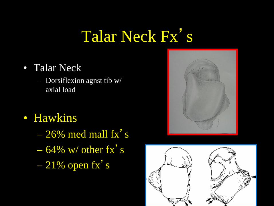
Talar Neck Fx’s
• Talar Neck – Dorsiflexion agnst tib w/
axial load
• Hawkins
– 26% med mall fx’s
– 64% w/ other fx’s
– 21% open fx’s
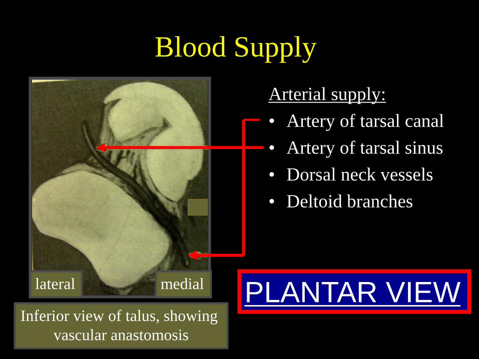
Blood Supply
Arterial supply:
• Artery of tarsal canal
• Artery of tarsal sinus
• Dorsal neck vessels
• Deltoid branches
medial lateral
Inferior view of talus, showing
vascular anastomosis
PLANTAR VIEW
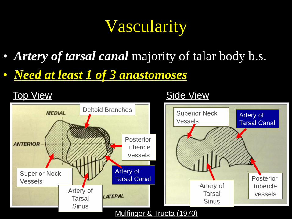
Vascularity
• Artery of tarsal canal majority of talar body b.s.
• Need at least 1 of 3 anastomoses
Side View Top View
Deltoid Branches
Posterior
tubercle
vessels
Artery of
Tarsal
Sinus
Artery of
Tarsal Canal
Superior Neck
Vessels
Superior Neck
Vessels
Artery of
Tarsal
Sinus
Artery of
Tarsal Canal
Posterior
tubercle
vessels
Mulfinger & Trueta (1970)
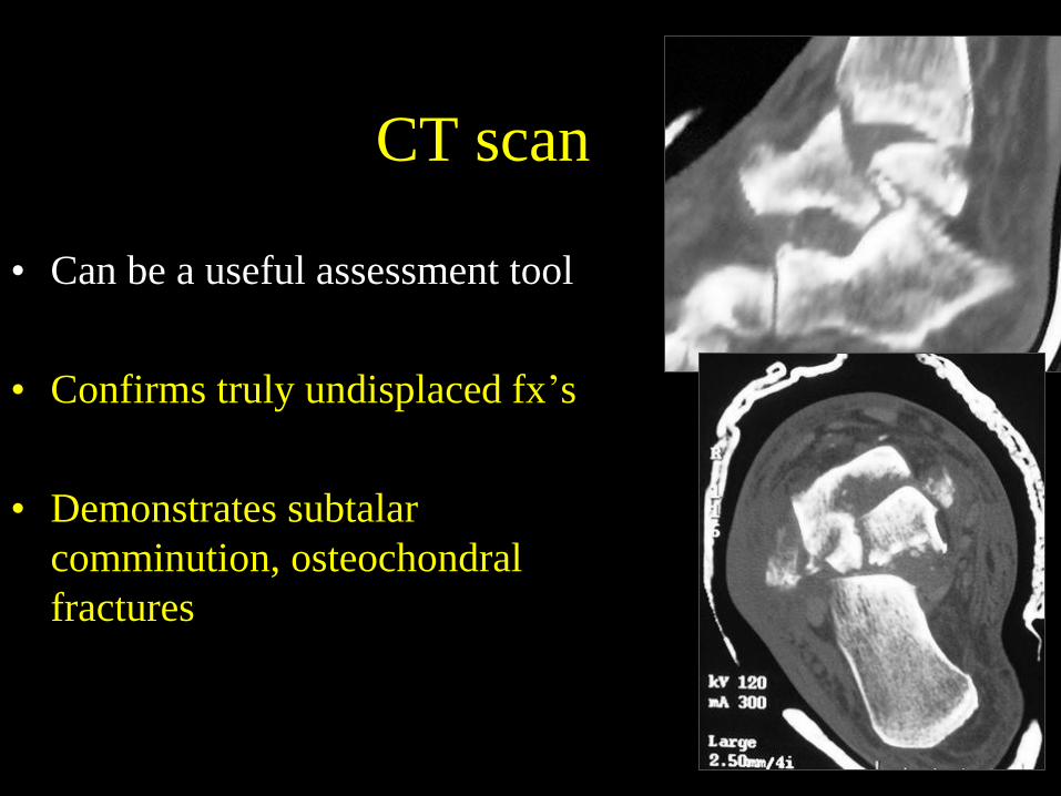
CT scan
• Can be a useful assessment tool
• Confirms truly undisplaced fx’s
• Demonstrates subtalar
comminution, osteochondral
fractures
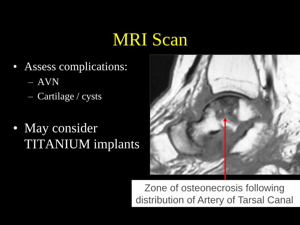
MRI Scan
• Assess complications:
– AVN
– Cartilage / cysts
• May consider
TITANIUM implants
Zone of osteonecrosis following
distribution of Artery of Tarsal Canal
Maybe NOT an Emergency?
• Lindvall, Haidukewych, Di Pasquale, Herscovici, Sanders: JBJS - A 2004
– DELAY IN REDUCTION & FIXATION DOES NOT AFFECT:
• UNION
• ON
• OUTCOME
• Vallier, Nork, Barei, et al: JBJS - A 2004
– NO CORRELATION WITH TIMING OF FIXATION & ON!!!
Open vs Closed
Front or Back
Approach for Reduction
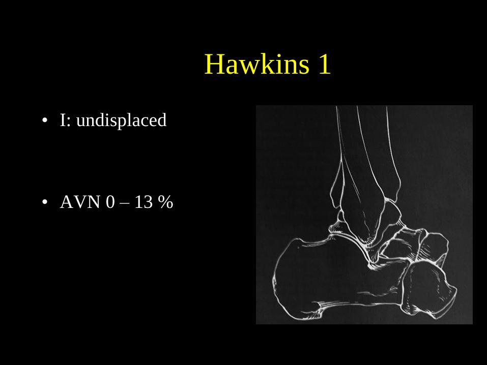
Hawkins 1
• I: undisplaced
• AVN 0 – 13 %
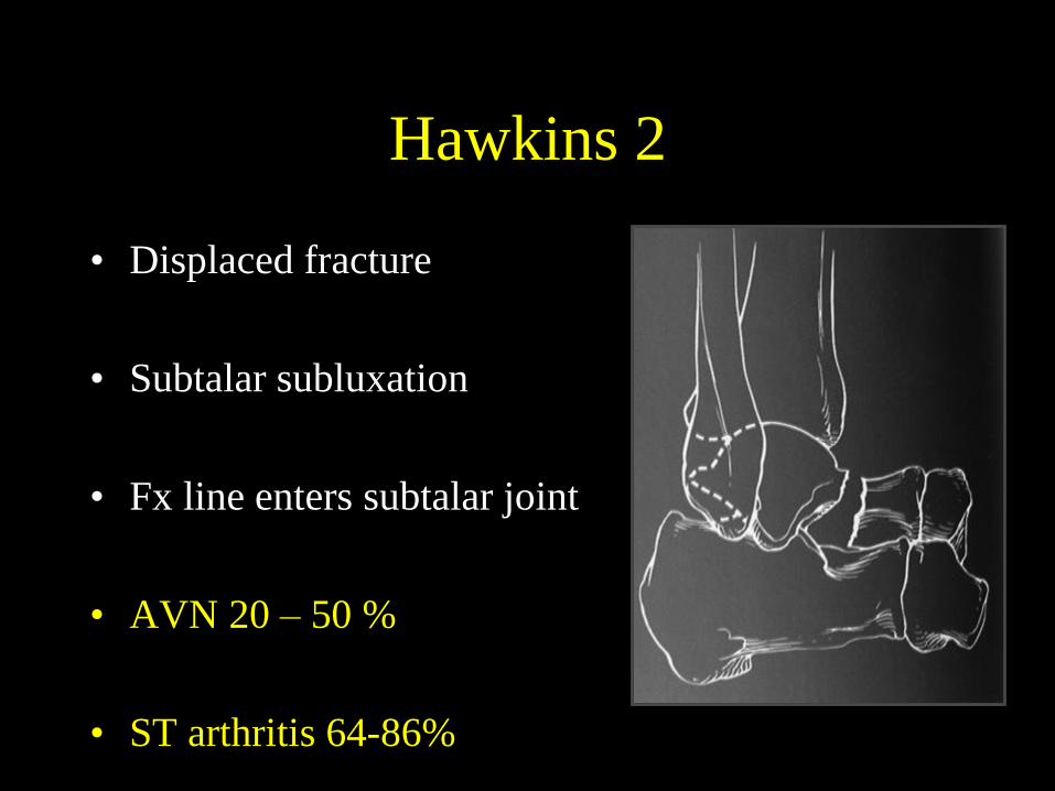
Hawkins 2
• Displaced fracture
• Subtalar subluxation
• Fx line enters subtalar joint
• AVN 20 – 50 %
• ST arthritis 64-86%
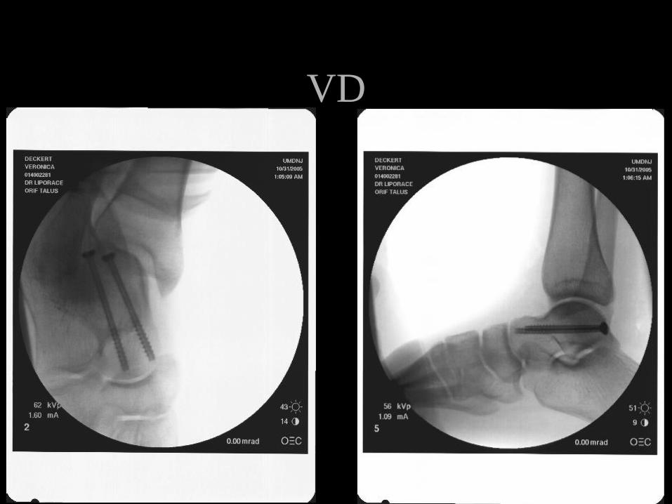
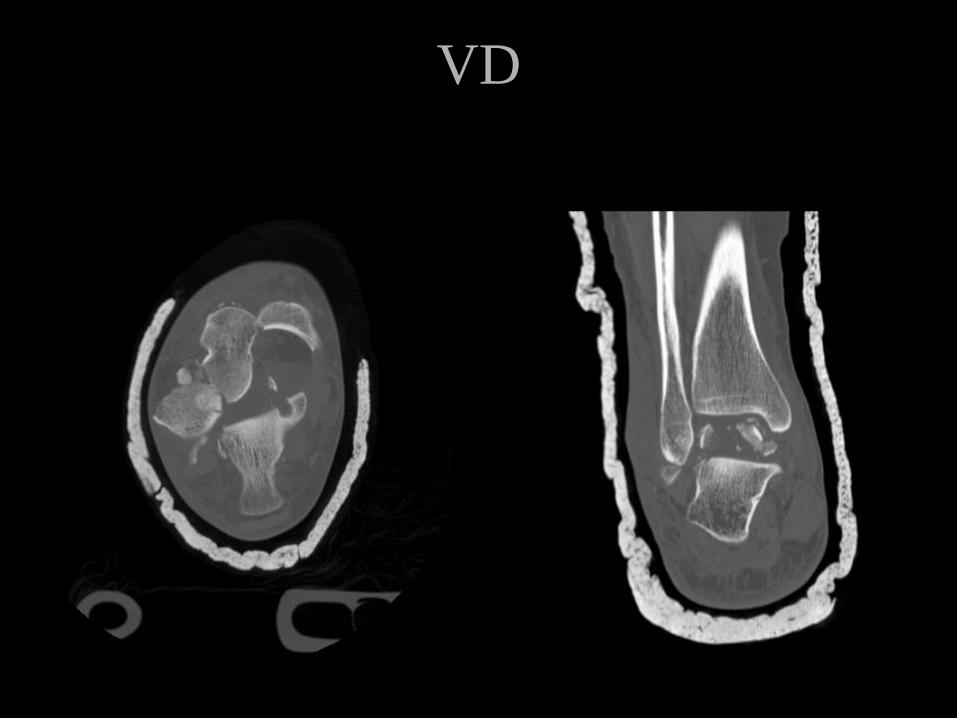
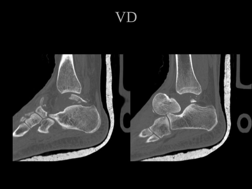
VD
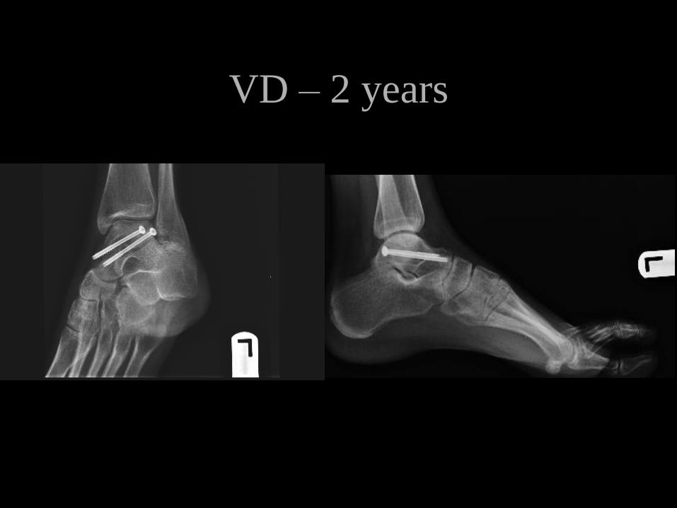
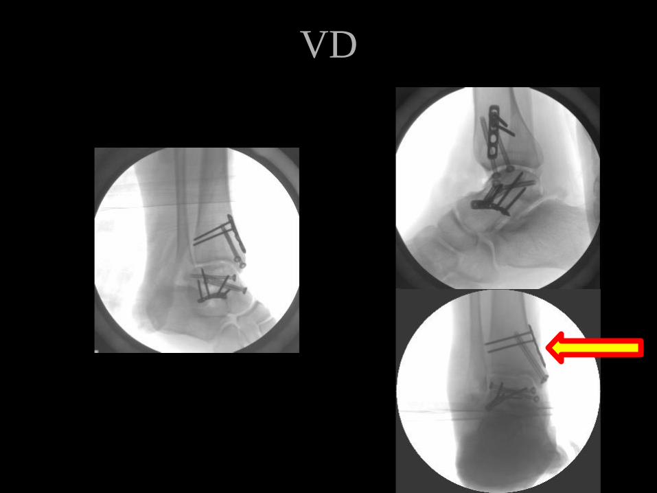
VD – 2 years
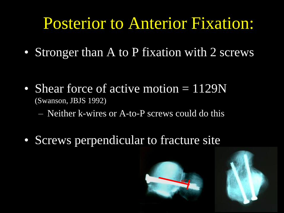
Posterior to Anterior Fixation:
• Stronger than A to P fixation with 2 screws
• Shear force of active motion = 1129N (Swanson, JBJS 1992)
– Neither k-wires or A-to-P screws could do this
• Screws perpendicular to fracture site
90°
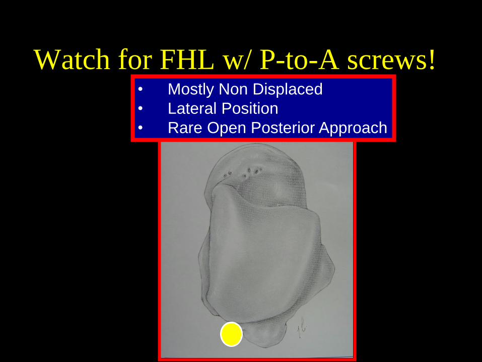
Watch for FHL w/ P-to-A screws! • Mostly Non Displaced
• Lateral Position
• Rare Open Posterior Approach
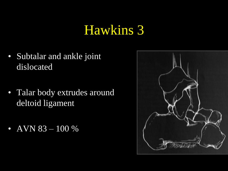
Hawkins 3
• Subtalar and ankle joint
dislocated
• Talar body extrudes around
deltoid ligament
• AVN 83 – 100 %
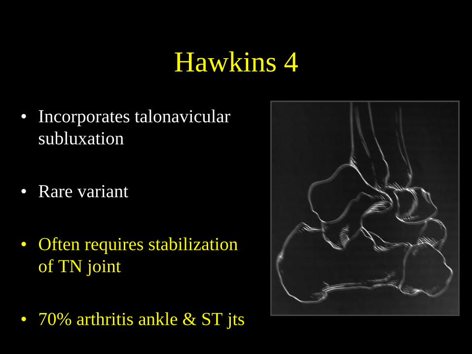
Hawkins 4
• Incorporates talonavicular
subluxation
• Rare variant
• Often requires stabilization
of TN joint
• 70% arthritis ankle & ST jts
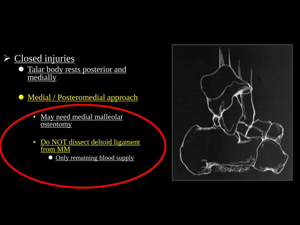
Closed injuries Talar body rests posterior and
medially
Medial / Posteromedial approach
• May need medial malleolar
osteotomy
• Do NOT dissect deltoid ligament from MM Only remaining blood supply
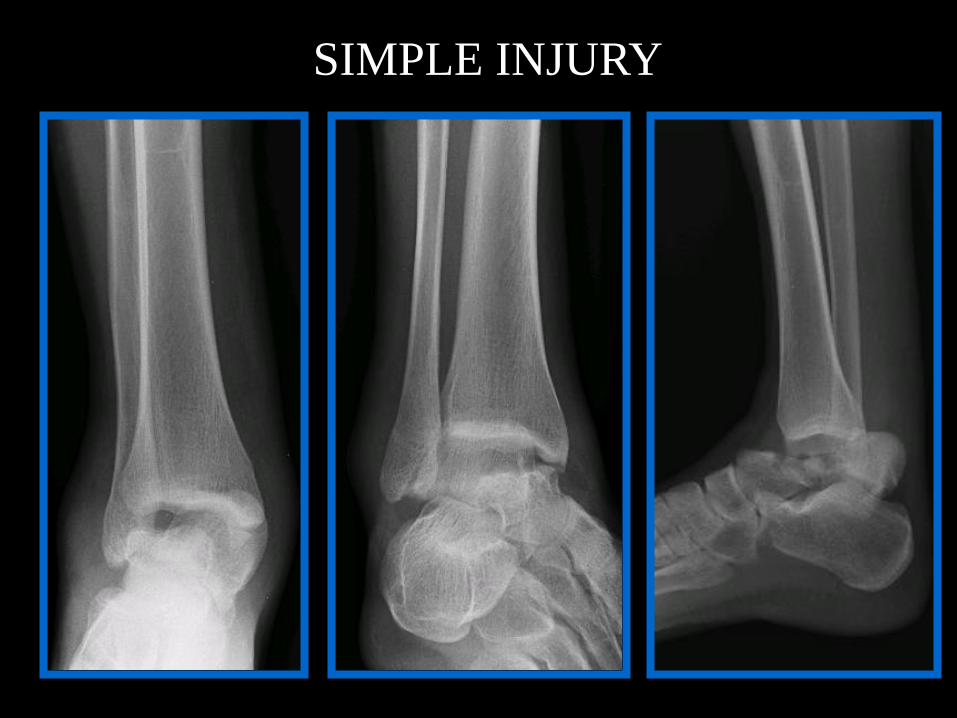
SIMPLE INJURY
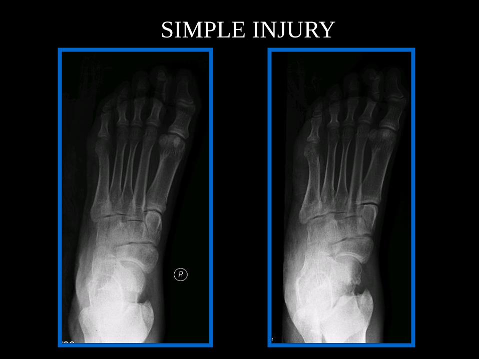
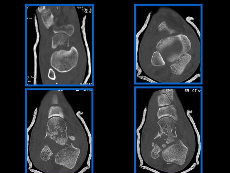
SIMPLE INJURY
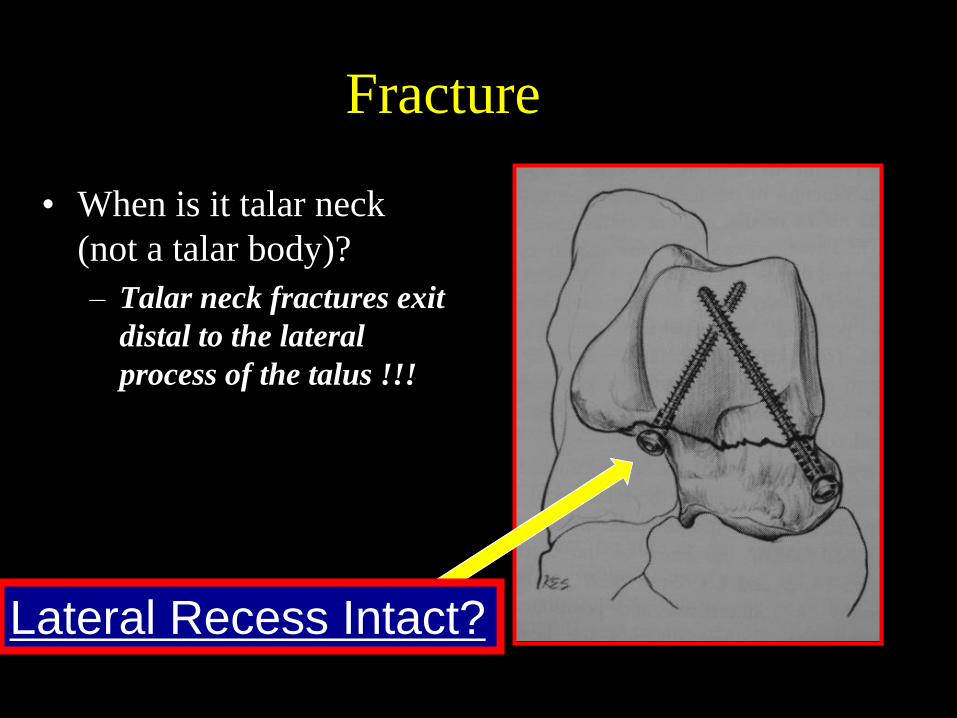
Fracture
• When is it talar neck
(not a talar body)?
– Talar neck fractures exit
distal to the lateral
process of the talus !!!
Lateral Recess Intact?
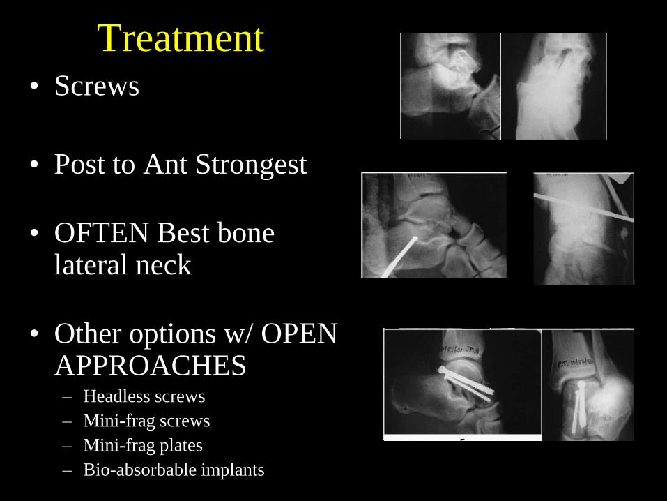
Treatment • Screws
• Post to Ant Strongest
• OFTEN Best bone lateral neck
• Other options w/ OPEN APPROACHES – Headless screws
– Mini-frag screws
– Mini-frag plates
– Bio-absorbable implants
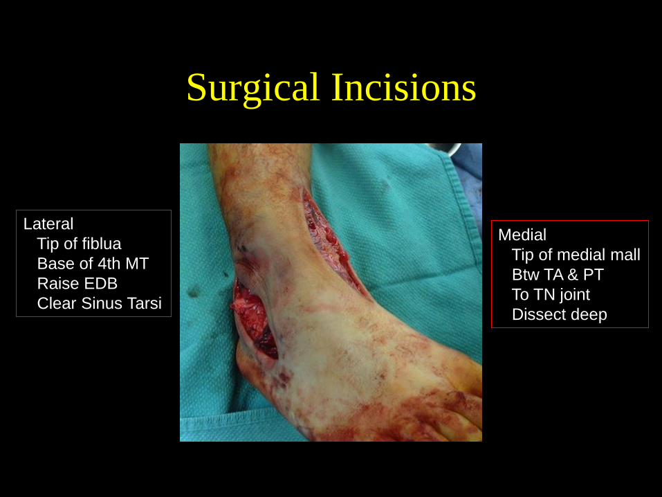
Surgical Incisions
Lateral
Tip of fiblua
Base of 4th MT
Raise EDB
Clear Sinus Tarsi
Medial
Tip of medial mall
Btw TA & PT
To TN joint
Dissect deep
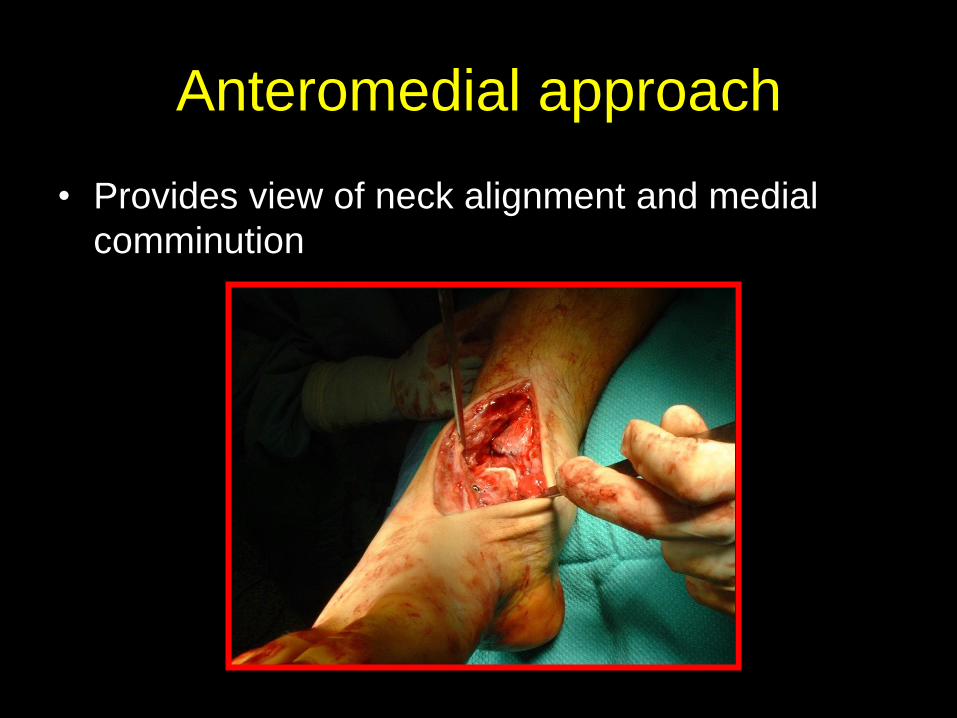
Anteromedial approach
• Provides view of neck alignment and medial
comminution
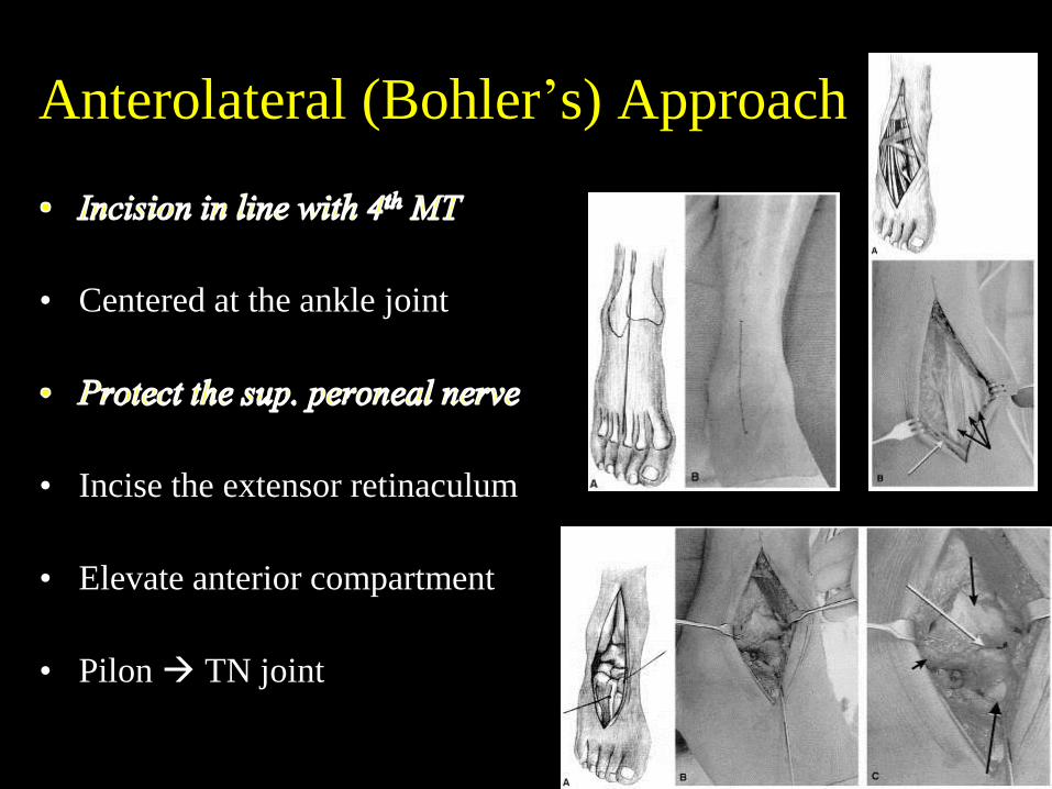
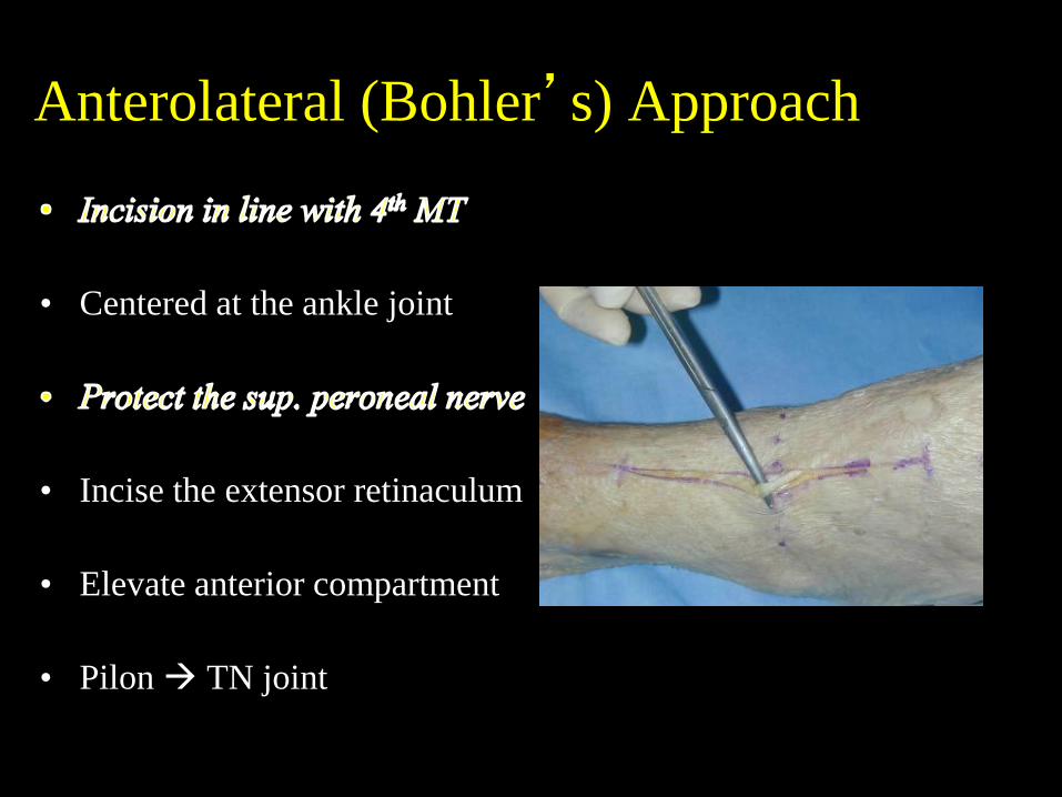
Anterolateral (Bohler’s) Approach
• Centered at the ankle joint
• Incise the extensor retinaculum
• Elevate anterior compartment
• Pilon TN joint
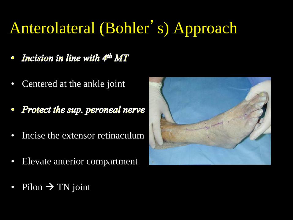
Anterolateral (Bohler’s) Approach
• Centered at the ankle joint
• Incise the extensor retinaculum
• Elevate anterior compartment
• Pilon TN joint
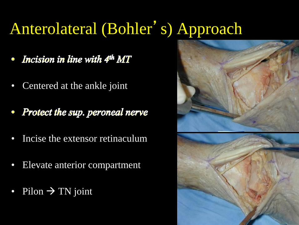
Anterolateral (Bohler’s) Approach
• Centered at the ankle joint
• Incise the extensor retinaculum
• Elevate anterior compartment
• Pilon TN joint
Anterolateral (Bohler’s) Approach
• Centered at the ankle joint
• Incise the extensor retinaculum
• Elevate anterior compartment
• Pilon TN joint
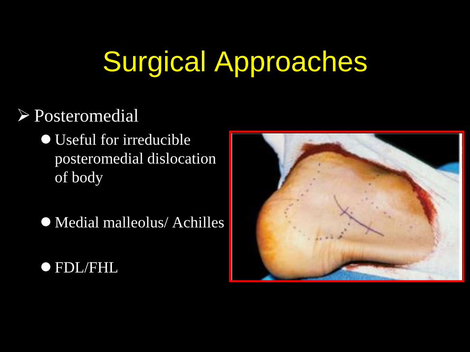
Surgical Approaches
Posteromedial
Useful for irreducible
posteromedial dislocation
of body
Medial malleolus/ Achilles
FDL/FHL
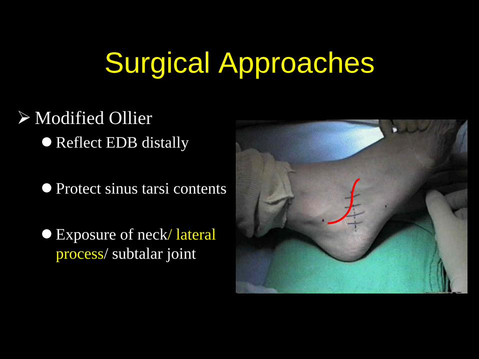
Surgical Approaches
Modified Ollier
Reflect EDB distally
Protect sinus tarsi contents
Exposure of neck/ lateral
process/ subtalar joint
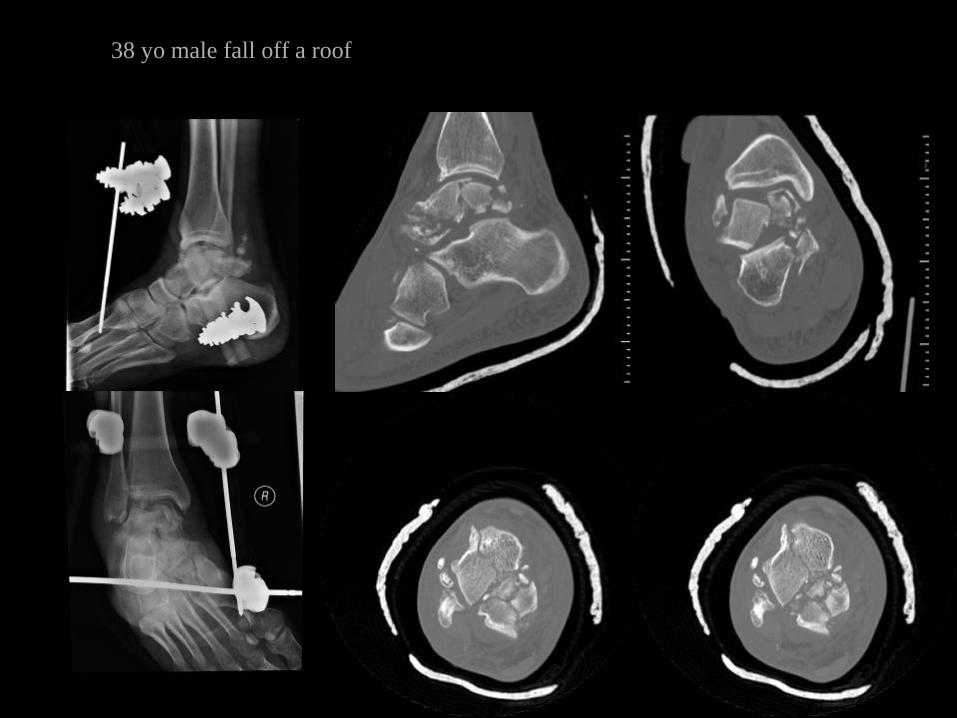
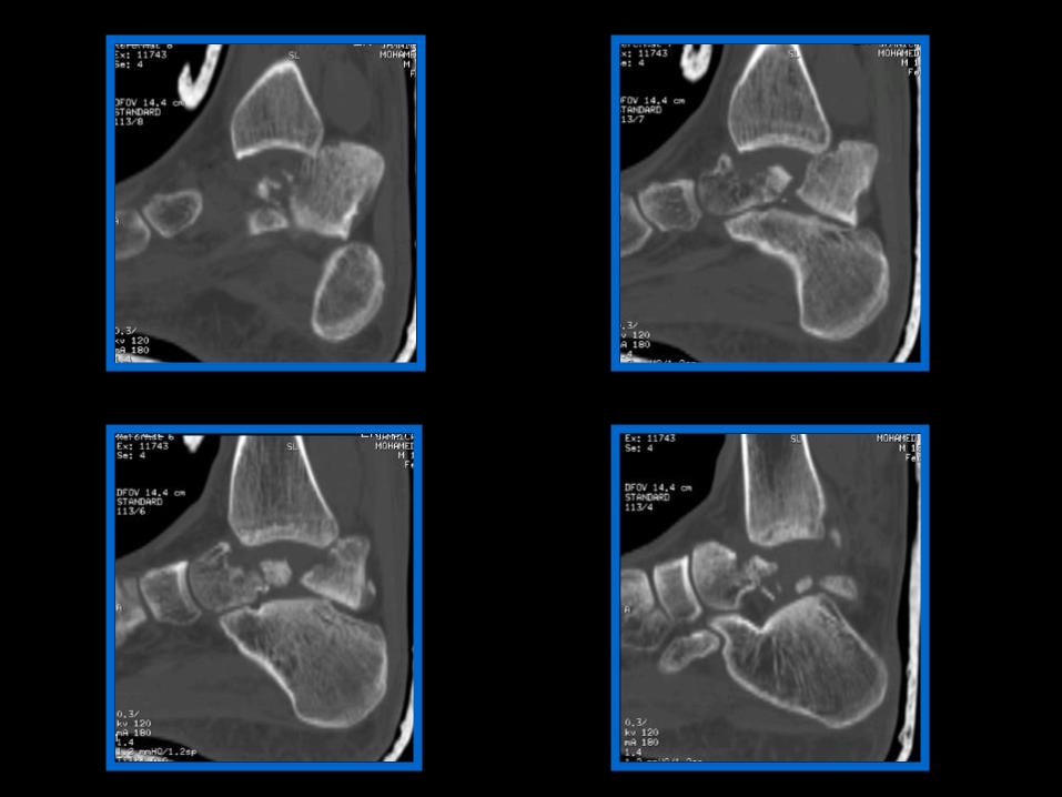
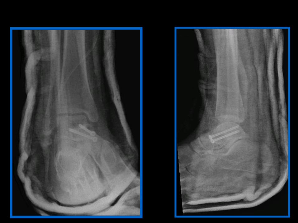
38 yo male fall off a roof
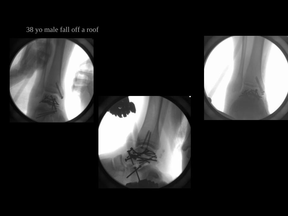
38 yo male fall off a roof
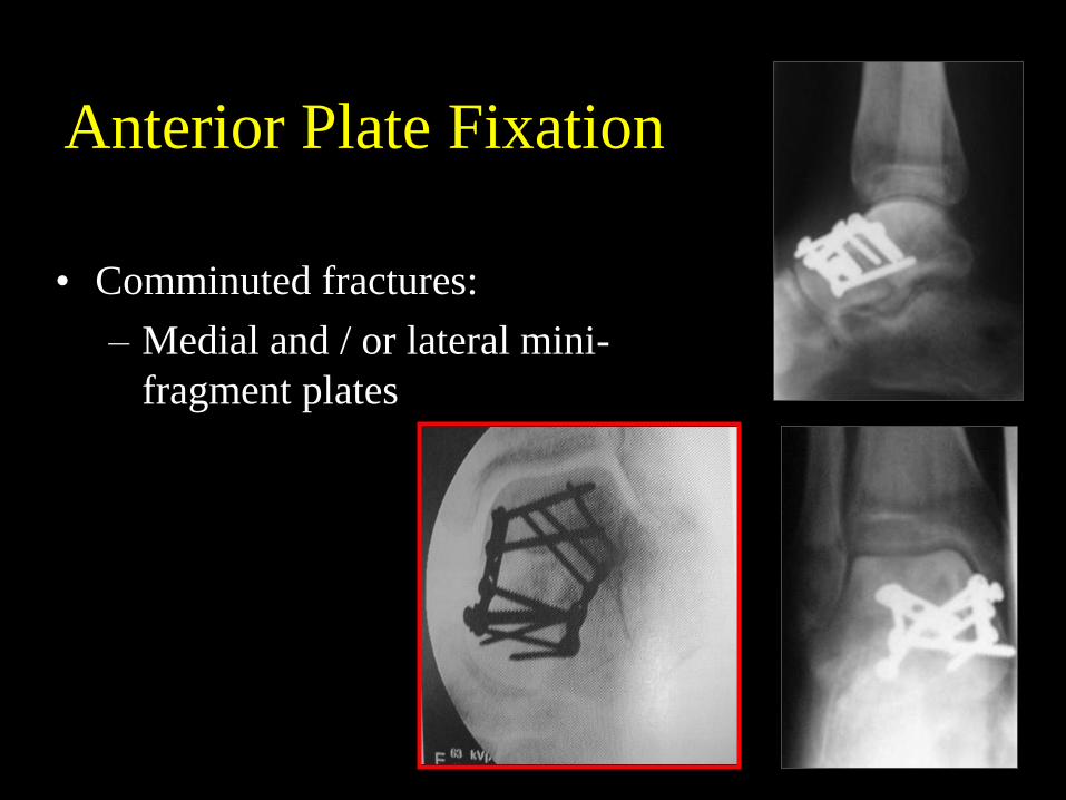
Anterior Plate Fixation
• Comminuted fractures:
– Medial and / or lateral mini-
fragment plates
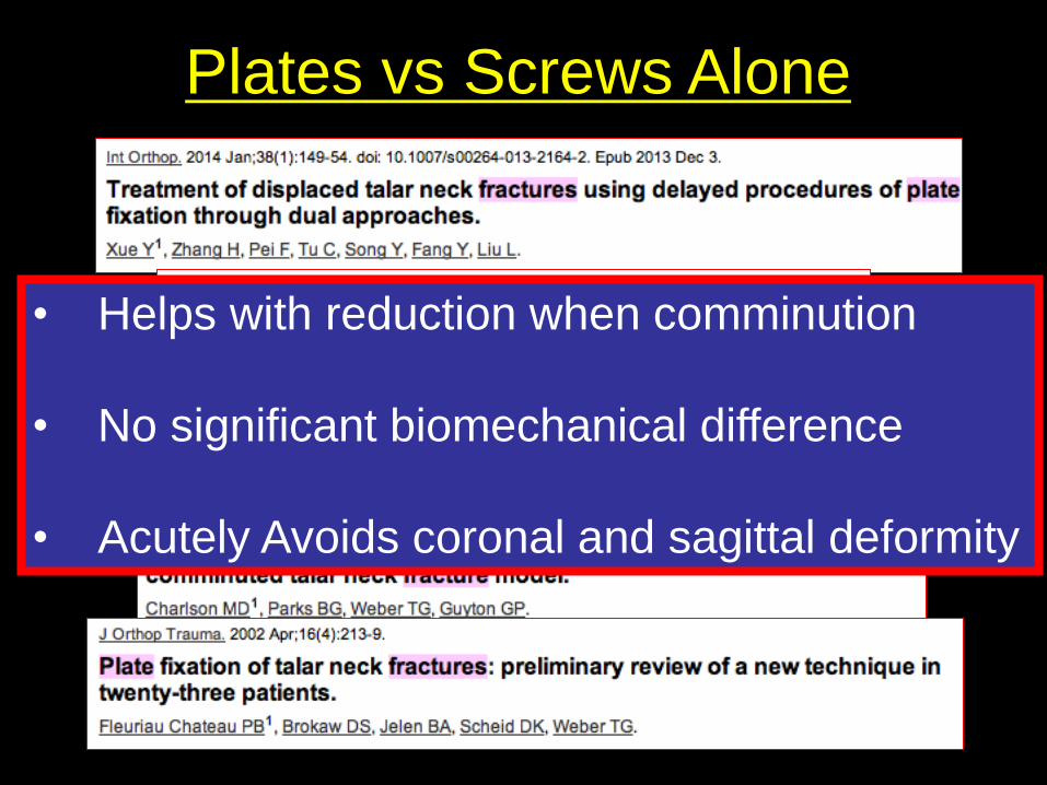
• Helps with reduction when comminution
• No significant biomechanical difference
• Acutely Avoids coronal and sagittal deformity
Plates vs Screws Alone
VD
VD
VD
VD
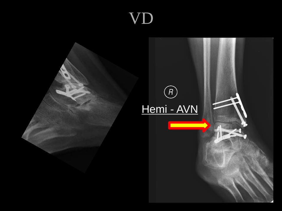
Hemi - AVN
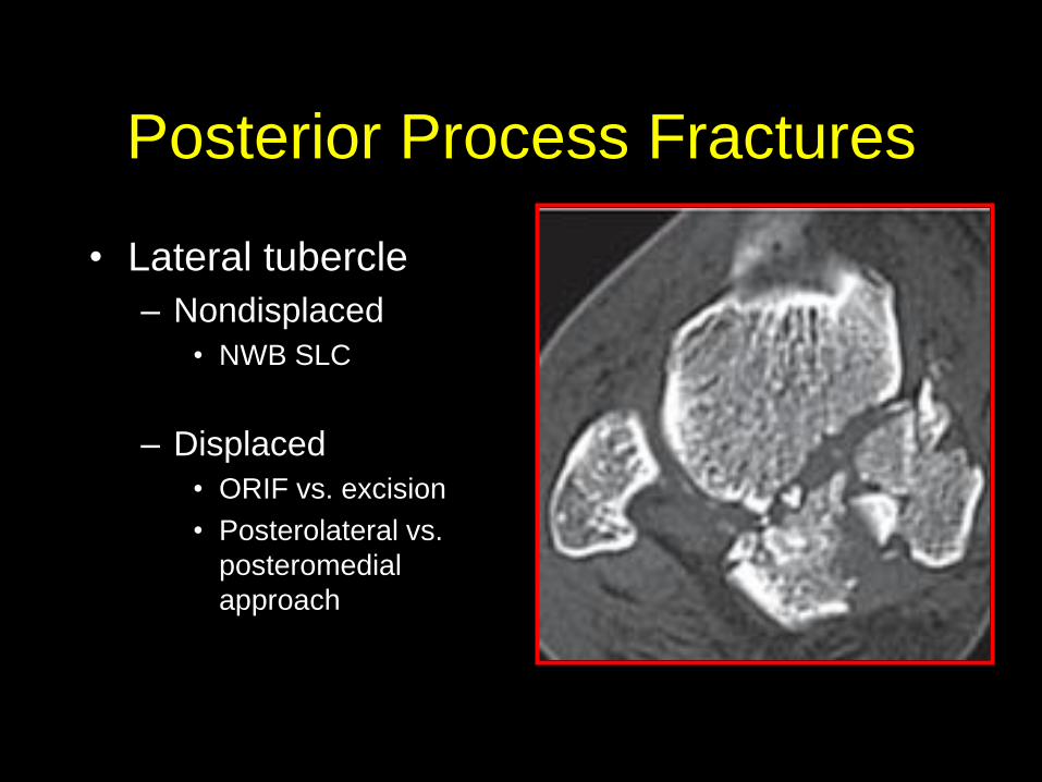
Posterior Process Fractures
• Lateral tubercle
– Nondisplaced
• NWB SLC
– Displaced
• ORIF vs. excision
• Posterolateral vs.
posteromedial
approach
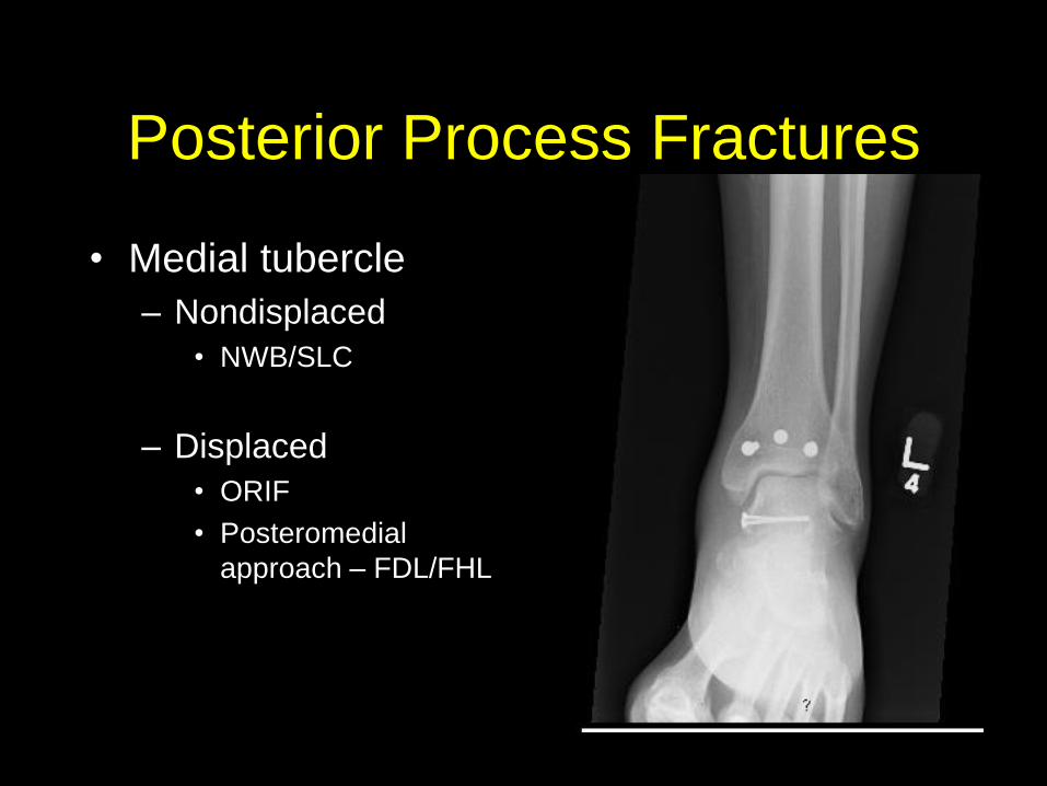
Posterior Process Fractures
• Medial tubercle
– Nondisplaced
• NWB/SLC
– Displaced
• ORIF
• Posteromedial
approach – FDL/FHL
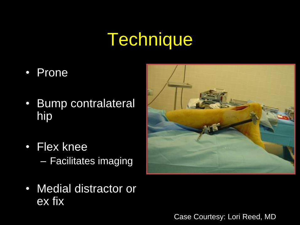
Technique
• Prone
• Bump contralateral hip
• Flex knee – Facilitates imaging
• Medial distractor or ex fix
Case Courtesy: Lori Reed, MD
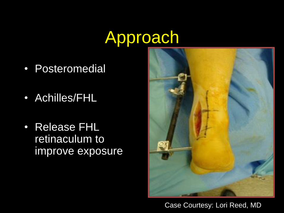
Approach
• Posteromedial
• Achilles/FHL
• Release FHL retinaculum to improve exposure
Case Courtesy: Lori Reed, MD
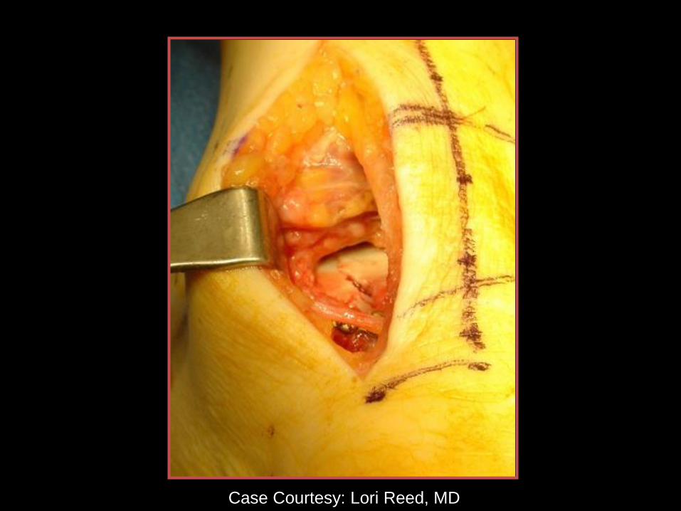
Case Courtesy: Lori Reed, MD
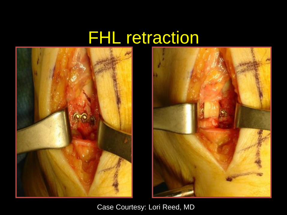
FHL retraction
Case Courtesy: Lori Reed, MD
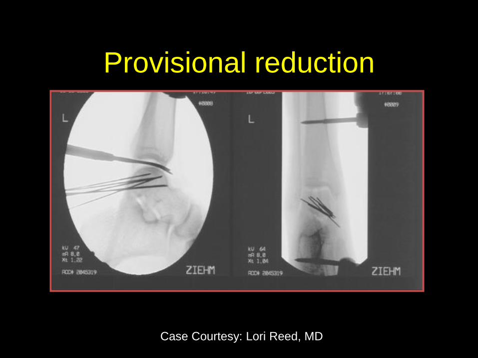
Provisional reduction
Case Courtesy: Lori Reed, MD
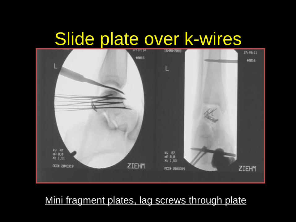
Slide plate over k-wires
Mini fragment plates, lag screws through plate
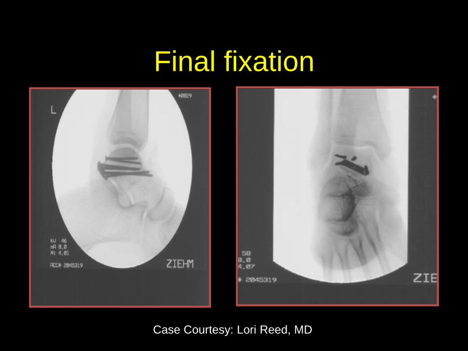
Final fixation
Case Courtesy: Lori Reed, MD
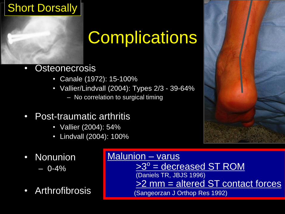
Complications
• Osteonecrosis • Canale (1972): 15-100%
• Vallier/Lindvall (2004): Types 2/3 - 39-64%
– No correlation to surgical timing
• Post-traumatic arthritis • Vallier (2004): 54%
• Lindvall (2004): 100%
• Nonunion – 0-4%
• Arthrofibrosis
Malunion – varus >3o = decreased ST ROM (Daniels TR, JBJS 1996)
>2 mm = altered ST contact forces (Sangeorzan J Orthop Res 1992)
Short Dorsally
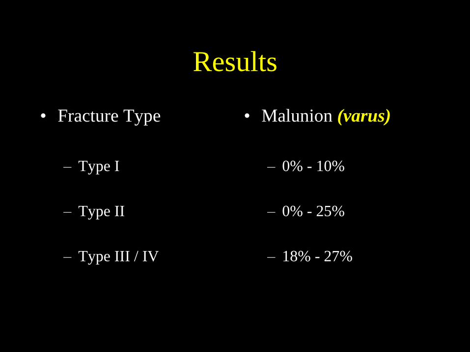
Results
• Fracture Type
– Type I
– Type II
– Type III / IV
• Malunion (varus)
– 0% - 10%
– 0% - 25%
– 18% - 27%
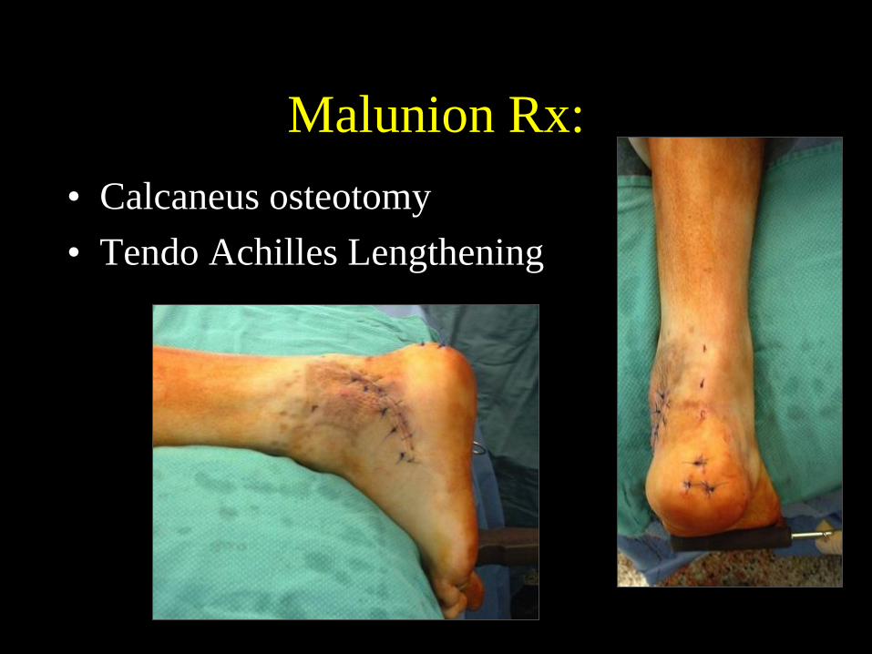
Malunion Rx:
• Calcaneus osteotomy
• Tendo Achilles Lengthening
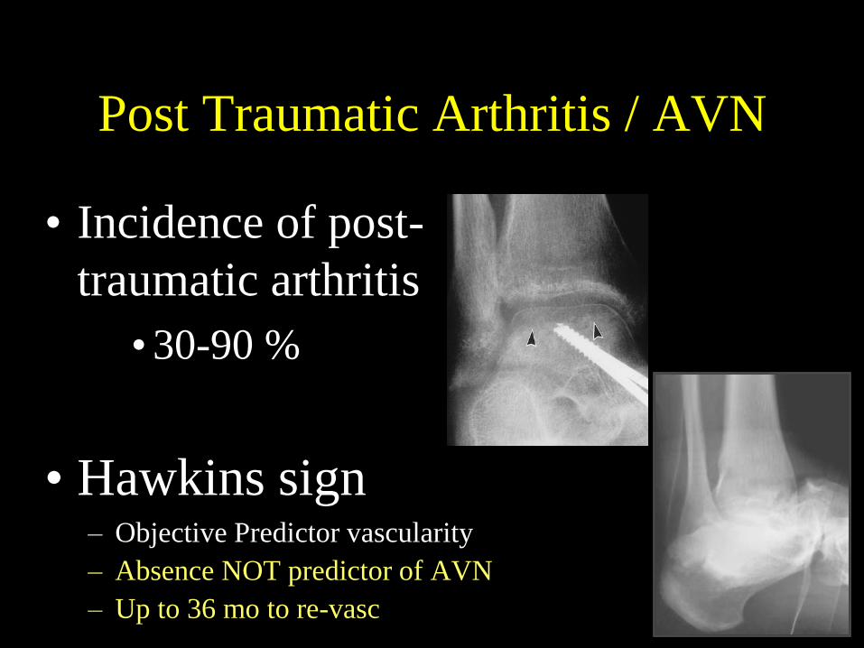
Post Traumatic Arthritis / AVN
• Incidence of post-
traumatic arthritis
•30-90 %
• Hawkins sign – Objective Predictor vascularity
– Absence NOT predictor of AVN
– Up to 36 mo to re-vasc
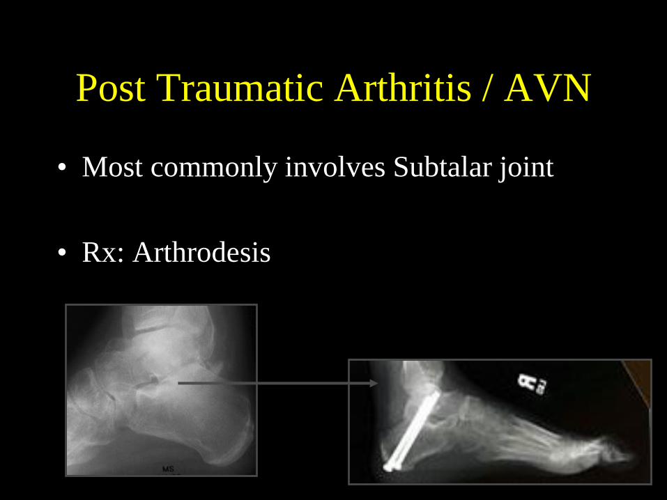
Post Traumatic Arthritis / AVN
• Most commonly involves Subtalar joint
• Rx: Arthrodesis
Take home
• Talus fractures with variable results
• More dislocations w/ neck fx Higher AVN
• Body Fx’s may be poor prognosis
• Stable fixation required
– Consider plates with COMMINUTION
• Lateral side of neck more often key
– Medial side comminuted and may promote varus
• SIMILAR stability plates and screws
• NOT AN EMERGENCY
– Unless skin compromise / irreducible dislocation
THANK YOU