Embed Size (px)
Citation preview
Journal of Healthcare Engineering · Vol. 3 · No. 2 · 2012 Page 243–260 243
Bioresorbable Plates and Screws for ClinicalApplications: A Review
Sandra Pina* and José M.F. FerreiraUniversity of Aveiro, Deptartment of Ceramics and Glass Engineering, Centro deInvestigação de Materiais Cerâmicos e Compósitos, 3810-193 Aveiro, Portugal
Submitted January 2011. Accepted for publication November 2011.
ABSTRACTBioresorbable implants are being widely used for fracture fixation in orthopaedic surgery and themarket is expanding rapidly worldwide. Bioresorbable materials slowly dissolve in the humanbody, such that a second operation to remove the synthetic material is not needed. Bioresorbableimplants have expanded the armamentarium of the surgeon, especially in the field of sportsmedicine. Interference screws, plates, pins, suture anchors, meniscal repair implants, and simplefracture fixation implants are the most commonly used resorbable implants for anterior cruciateligament reconstruction, shoulder surgery, meniscal repair, and fracture care. However, manyclinicians continue to rely on metal fixation, mainly due to the high mechanical strength and tothe complications reported with some of the available resorbable implant materials. The goal ofthe present paper is to present an overview on the available resorbable materials and theirapplications with a particular focus on new developments and trends in the field.
Keywords: bioresorbable, internal fixation, plates and screws, calcium phosphates, polymers
1. INTRODUCTIONA key goal of orthopaedic medicine is to restore the structure of damaged or diseasedbone tissue to a natural state. Internal fixation implants that are stronger, moreacceptable to the body, cheaper and durable have been developed to improve bonefracture osteosynthesis, to attach soft tissues or tissue grafts to bone for more than twodecades. Such implants comprise screws, plates, pins, staples and suture anchors whichare commonly fabricated of metals such as stainless steel and titanium and its alloys.However, there are intrinsic problems with the use of these metallic implants, such asstress-shielding phenomenon, pain, and local irritation [1–3].
*Corresponding author: Sandra Pina, University of Aveiro, Dept. of Ceramics and Glass Engineering, CICECO, 3810-193 Aveiro, Portugal. Phone: (+351) 234 370 200. Fax: (+351) 234 370 985. E-mail: [email protected]. Other author: [email protected].
Retained metallic implants are always at the risk of endogenous infection [4]. Inaddition, metal plate-screws might lead to destruction and osteoporosis in thesurrounding bone tissue [5]. For these reasons, there is need for a second surgery toremove the metallic fixation after the bone has healed [2].
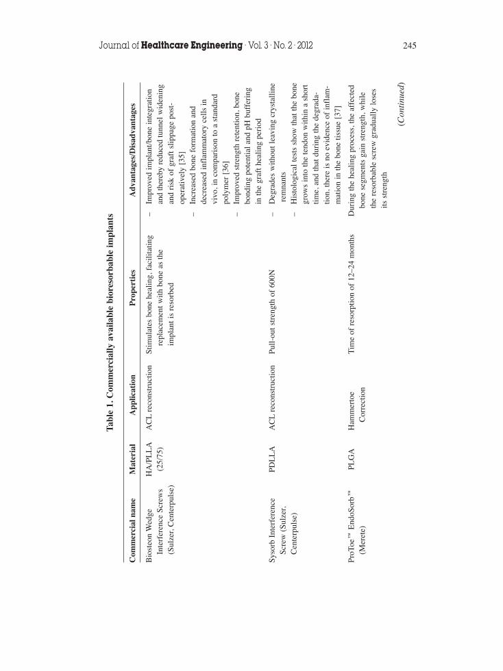
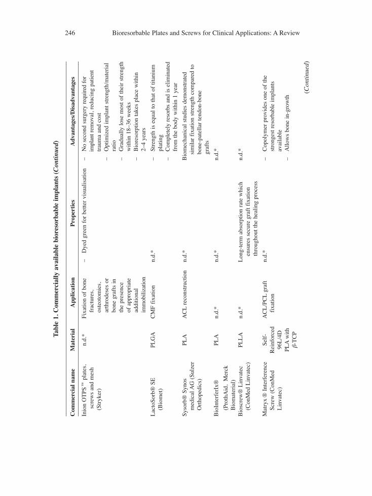
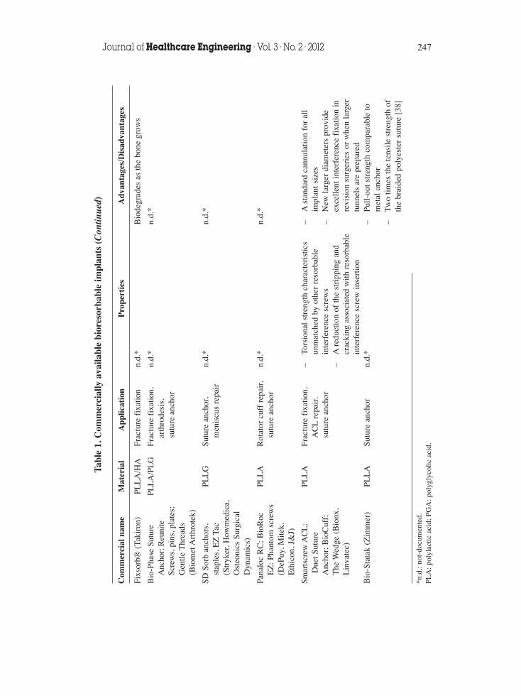
Bioresorbable and biodegradable fracture fixation implants have been considered asan effective fixation system with several advantages over metallic fixation, including noneed to remove the implants after osseous healing, radiolucency, no corrosion, noaccumulation of metal in tissues, less pain and reduced stress-shielding since theimplants bear less load initially and gradually transfer the load as they degrade [6–12].These devices are most often manufactured from polylactides (polylactic acid, PLA),polyglycolides (polyglycolic acid, PGA) and their co-polymer compositions as they arehighly resorbable [13–14]. Commercially available resorbable implants aresummarized in Table 1.
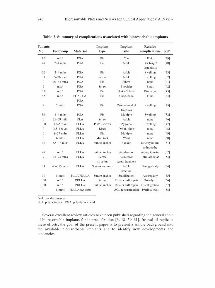
Bioresorbable materials allow a newly formed tissue to grow into any surfaceirregularities [15–17]. Thus, a resorbable implant is free of toxic and mutagenic effects.Nonetheless, there are some problems related to the use of these implants, such as aninflammatory response, rapid loss of initial implant strength, higher refracture rates,inadequate stiffness of the implants, and weakness in comparison to metallic implants[1, 18]. Table 2 summarizes some of the problems observed with the implementationsof common bioresorbable materials.
Biodegradable implants are characterized by materials that show disintegration afterimplantation but with no proof of its elimination from the body [19]. Thebiodegradation process depends on contact with body fluids, temperature, motion,molecular weight, crystal form and geometry of material, and the tissue that isimplanted [20–21]. The ideal biodegradable material provides appropriate strengthwhilst degrading in a predictable fashion throughout the healing process withoutcausing adverse reactions [22].
The first study on the use of biodegradable implants was published in 1966 byKulkarni et al. [23], who studied the biocompatibility of poly-L-lactic (PLLA) inanimals. The material proved to be non-toxic and gradually degraded. The use of PLLAplates and screws to fix mandibular fractures in dogs was studied by Kulkarni et al.[24]. Another study presented the results of PLLA sutures in mandibular fractures withno serious tissue reactions [25].
A common use of biodegradable interference screws is for the anterior cruciateligament reconstruction. A recent study showed the potential of these screws as analternative to titanium screws for the fixation of autologous bone grafts in dentalimplants [26].
The limitations of biodegradable implants are mainly in their mechanical propertieswhich are lower than those of conventional metal implants, leading to low confidencelevels regarding the stability of reduced fractures. Also, the construction of screws andpins for the necessary compression between the implant and the bone is somehowdifficult [27–28]. The biocompatibility is another limitation of these materials sincethey sometimes provoke an adverse tissue response that has the characteristic of aninflammatory, bacterial foreign-body reaction [29–34].
244 Bioresorbable Plates and Screws for Clinical Applications: A Review
Journal of Healthcare Engineering · Vol. 3 · No. 2 · 2012 245
Tabl
e 1.
Com
mer
cial
ly a
vaila
ble
bior
esor
babl
e im
plan
ts
Com
mer
cial
nam
eM
ater
ial
App
licat
ion
Pro
pert
ies
Adv
anta
ges/
Dis
adva
ntag
es
Bio
steo
n W
edge
H
A/P
LL
AA
CL
reco
nstr
uctio
nSt
imul
ates
bon
e he
alin
g, f
acili
tatin
g–
Impr
oved
impl
ant/b
one
inte
grat
ion
Inte
rfer
ence
Scr
ews
(25/
75)
repl
acem
ent w
ith b
one
as th
e an
d th
ereb
y re
duce
d tu
nnel
wid
enin
g(S
ulze
r, C
ente
rpul
se)
impl
ant i
s re
sorb
edan
d ri
sk o
f gr
aft s
lippa
ge p
ost-
oper
ativ
ely
[35]
–In
crea
sed
bone
for
mat
ion
and
decr
ease
d in
flam
mat
ory
cells
invi
vo, i
n co
mpa
riso
n to
a s
tand
ard
poly
mer
[36
]–
Impr
oved
str
engt
h re
tent
ion,
bon
ebo
ndin
g po
tent
ial a
nd p
H b
uffe
ring
in th
e gr
aft h
ealin
g pe
riod
Syso
rb I
nter
fere
nce
PDL
LA
AC
Lre
cons
truc
tion
Pull-
out s
tren
gth
of 6
00N
–D
egra
des
with
out l
eavi
ng c
ryst
allin
eSc
rew
(Su
lzer
,re
mna
nts
Cen
terp
ulse
)–
His
tolo
gica
l tes
ts s
how
that
the
bone
grow
s in
to th
e te
ndon
with
in a
sho
rttim
e, a
nd th
at d
urin
g th
e de
grad
a-tio
n, th
ere
is n
o ev
iden
ce o
f in
flam
-m
atio
n in
the
bone
tiss
ue [
37]
ProT
oe
End
oSor
bPL
GA
Ham
mer
toe
Tim
e of
res
orpt
ion
of 1
2–24
mon
ths
Dur
ing
the
heal
ing
proc
ess,
the
affe
cted
(Mer
ete)
Cor
rect
ion
bone
seg
men
ts g
ain
stre
ngth
, whi
leth
e re
sorb
able
scr
ew g
radu
ally
lose
sits
str
engt
h
(Con
tinu
ed)
246 Bioresorbable Plates and Screws for Clinical Applications: A Review
Tabl
e 1.
Com
mer
cial
ly a
vaila
ble
bior
esor
babl
e im
plan
ts (
Con
tinue
d)
Com
mer
cial
nam
eM
ater
ial
App
licat
ion
Pro
pert
ies
Adv
anta
ges/
Dis
adva
ntag
es
Inio
n O
TPS
pl
ates
,n.
d.*
Fixa
tion
of b
one
–D
yed
gree
n fo
r be
tter
visu
alis
atio
n–
No
seco
nd s
urge
ry r
equi
red
for
scre
ws
and
mes
hfr
actu
res,
impl
ant r
emov
al, r
educ
ing
patie
nt(S
tryk
er)
oste
otom
ies,
trau
ma
and
cost
ar
thro
dese
s or
–O
ptim
ized
impl
ant s
tren
gth/
mat
eria
lbo
ne g
raft
s in
ra
tioth
e pr
esen
ce
–G
radu
ally
lose
mos
t of
thei
r st
reng
thof
app
ropr
iate
w
ithin
18–
36 w
eeks
addi
tiona
l–
Bio
reso
rptio
n ta
kes
plac
e w
ithin
im
mob
iliza
tion
2–4
year
s
Lac
toSo
rb®
SEPL
GA
CM
F fi
xatio
nn.
d.*
–St
reng
th is
equ
al to
that
of
titan
ium
(Bio
met
)pl
atin
g–
Com
plet
ely
reso
rbs
and
is e
limin
ated
from
the
body
with
in 1
yea
r
Syso
rb®
Syno
s
PLA
AC
Lre
cons
truc
tion
n.d.
*B
iom
echa
nica
l stu
dies
dem
onst
rate
dm
edic
al A
G (
Sulz
ersi
mila
r fi
xatio
n st
reng
th c
ompa
red
toO
rtho
pedi
cs)
bone
-pat
ella
r te
ndon
-bon
e gr
afts
Bio
Inte
rfer
Ix®
PLA
n.d.
*n.
d.*
n.d.
*
(Pro
thA
id,
Mer
ckB
iom
ater
ial)
Bio
scre
w®
Lin
vate
cPL
LA
n.d.
*L
ong-
term
abs
orpt
ion
rate
whi
ch
n.d.
*(C
onM
ed L
inva
tec)
ensu
res
secu
re g
raft
fix
atio
nth
roug
hout
the
heal
ing
proc
ess
Mat
ryx
®In
terf
eren
ceSe
lf-
AC
L/P
CL
graf
t n.
d.*
–C
opol
ymer
pro
vide
s on
e of
the
Scre
w (
Con
Med
Rei
nfor
ced
fixa
tion
stro
nges
t res
orba
ble
impl
ants
Lin
vate
c)96
L/4
D
avai
labl
ePL
Aw
ith
–A
llow
s bo
ne in
-gro
wth
β-T
CP
(Con
tinu
ed)
Journal of Healthcare Engineering · Vol. 3 · No. 2 · 2012 247
Tabl
e 1.
Com
mer
cial
ly a
vaila
ble
bior
esor
babl
e im
plan
ts (
Con
tinue
d)
Com
mer
cial
nam
eM
ater
ial
App
licat
ion
Pro
pert
ies
Adv
anta
ges/
Dis
adva
ntag
es
Fixs
orb®
(Tak
iron
)PL
LA
/HA
Frac
ture
fix
atio
nn.
d.*
Bio
degr
ades
as
the
bone
gro
ws
Bio
-Pha
se S
utur
ePL
LA
/PL
GFr
actu
re f
ixat
ion,
n.d.
*n.
d.*
Anc
hor;
Reu
nite
arth
rode
sis,
Scre
ws,
pin
s, p
late
s;su
ture
anc
hor
Gen
tle T
hrea
ds(B
iom
et A
rthr
otek
)
SD S
orb
anch
ors,
PL
LG
Sutu
re a
ncho
r,n.
d.*
n.d.
*st
aple
s, E
Z T
acm
enis
cus
repa
ir(S
tryk
er, H
owm
edic
a,O
steo
nics
Sur
gica
lD
ynam
ics)
Pana
loc
RC
; Bio
Roc
PLL
AR
otat
or c
uff
repa
ir,
n.d.
*n.
d.*
EZ
; Pha
ntom
scr
ews
sutu
re a
ncho
r(D
ePuy
, Mite
k,E
thic
on, J
&J)
Smar
tscr
ew A
CL
;PL
LA
Frac
ture
fix
atio
n,–
Tors
iona
l str
engt
h ch
arac
teri
stic
s–
Ast
anda
rd c
annu
latio
n fo
r al
lD
uet S
utur
e A
CL
repa
ir,
unm
atch
ed b
y ot
her
reso
rbab
leim
plan
t siz
es
Anc
hor;
Bio
Cuf
f;su
ture
anc
hor
inte
rfer
ence
scr
ews
–N
ew la
rger
dia
met
ers
prov
ide
The
Wed
ge (
Bio
nx,
–A
redu
ctio
n of
the
stri
ppin
g an
d ex
celle
nt in
terf
eren
ce f
ixat
ion
inL
inva
tec)
crac
king
ass
ocia
ted
with
res
orba
ble
revi
sion
sur
geri
es o
r w
hen
larg
erin
terf
eren
ce s
crew
inse
rtio
ntu
nnel
s ar
e pr
epar
edB
io-S
tata
k (Z
imm
er)
PLL
ASu
ture
anc
hor
n.d.
*–
Pull-
out s
tren
gth
com
para
ble
tom
etal
anc
hor
–Tw
o tim
es th
e te
nsile
str
engt
h of
th
e br
aide
d po
lyes
ter
sutu
re [
38]
*n.d
.: no
t-do
cum
ente
d.PL
A: p
olyl
actic
aci
d; P
GA
: pol
ygly
colic
aci
d.
Several excellent review articles have been published regarding the general topicof bioresorbable implants for internal fixation [6, 18, 59–61]. Instead of replicatethese efforts, the goal of the present paper is to present a simple background into the available bioresorbable implants and to identify new developments andtendencies.
248 Bioresorbable Plates and Screws for Clinical Applications: A Review
Table 2. Summary of complications associated with bioresorbable implants
Patients Implant Implant Results/(%) Follow-up Material type site complications Ref.
1.3 n.d.* PGA Pin Toe Fluid [39]
49 2–4 mths PGA Pin Ankle Discharge/ [40]
Osteolysis
6.3 2–4 mths PGA Pin Ankle Swelling [33]
11 5–26 wks PGA Screw Ankle Swelling [32]
0 10–44 mths PGA Pin Elbow none [41]
5 n.d.* PGA Screw Shoulder Sinus [42]
6.8 n.d.* PGA Pin Ankle/Elbow Discharge [43]
6.5 n.d.* PGA/PLA, Pin Canc. bone Fluid [44]
PGA
4 2 mths PGA Pin Osteo-chondral Swelling [45]
fractures
7.9 2–4 mths PGA Pin Multiple Swelling [32]
0 21–59 mths PLA Screw Ankle none [46]
100 3.3–5.7 yrs PLLA Plates/screws Zygoma Swelling [47]
0 3.5–6.6 yrs PLLA Discs Orbital floor none [48]
0 8–37 mths PLLA Pin Multiple none [49]
0 6 mths PLLA Mini tack Wrist none [50]
16 3.5–18 mths PLLA Suture anchor Bankart Osteolysis and [51]
arthropathy
47 n.d.* PLLA Suture anchor Stabilization Asymptomatic [52]
2 15–23 mths PLLA Screw ACL recon Intra-articular [53]
-struction screw fragment
31 40–115 mths PLLA Screws and rods Ankle Foreign-body [54]
reaction
19 8 mths PLLA/PDLLA Suture anchor Stabilization Arthropathy [55]
100 n.d.* PDLLA Screw Rotator cuff repair Osteolysis [56]
100 n.d.* PDLLA Suture anchor Rotator cuff repair Disintegration [57]
4 8 mths PDLLA (Sysorb) – ACL reconstruction Pretibial cyst [58]
*n.d.: not documented.PLA: polylactic acid; PGA: polyglycolic acid.
2. VARIETY OF BIORESORBABLE MATERIALS2.1. Ceramics: Calcium PhosphatesCalcium phosphates (CaPs) are found widely in the earth crust and are characterized aswhite solids unless doped or containing elements that pass in the lattice structure of therespective compound. CaPs are chemical compounds similar to the inorganic part ofmajor normal (bones, teeth and antlers) and pathological calcified tissues of mammals[62–64]. CaPs can be categorized into bioactive and bioresorbable materials [65–66]. Abioactive biomaterial enables establishing direct chemical bonds with bone andsurrounding tissues, and could provide good stabilization for materials that are subjectto mechanical loading. β-tricalcium phosphate (β-TCP) and hydroxyapatite (HA) arethe most commonly used CaPs as ceramics. β-TCP is biodegradable and able topromote osteogenesis and new bone formation. HA is highly crystalline and is the moststable and least soluble CaP in an aqueous solution down to a pH of 4.2 [65]. Theresorption of a ceramic HA is believed to be slow (1 to 2% per year), and onceimplanted into the body, HA may remain integrated into the regenerated bone tissue,while β-TCP is completely reabsorbed [67–68].
Unfortunately, CaPs have poor mechanical properties that do not allow load-bearingapplications. However, advantages are achieved by combining these materials withpolymers that are generally bioinert to provide the composite bioactivity, in order toform a composite with optimized properties. The composite implants are able to form achemical bond with the host tissue, and the fixation of implants is accelerated [69–71].For example, CaP/poly-DL-lactide-co-glycolide composite biomaterials exhibit goodadhesion onto human cells, indicating a high level of biocompatibility [70–71].Additionally, previous studies with composite materials consisting of PLLA/TCP orPLLA/HA showed a rapid resorption and replacement by newly formed bone tissue[72–74].
The elastic modulus of the composite can also be adjusted to approach that of thehuman bone by altering the content of ceramic. It is known that the match of elasticmodulus between implants and the human bone favours the evasion of stress-shieldingand the sequent bone absorption, which is often caused by implants with high elasticmodulus [75].
2.2. PolymersBioresorbable fixation materials commonly used in orthopaedic applications are PGA,PLA, poly lactide-co-glycolide (PLGA) co-polymers in various ratios, polydioxanone(PDS), propylene (PP), polysulphone (PS), and polycarbonate (PC). Among them,PGA, PLA and their co-polymers have received the most attention, in part because theycan be self-reinforced to achieve better strength properties [76]. The mechanicalproperties of these materials changes over time in a physiologic environment asdetermined by the molecular weight and degree of crystallinity. Hence, the molecularweight and crystallinity can be altered to optimize mechanical strength of an implant.For example, polymers with a higher degree of crystallinity are stronger and degradeslower than amorphous polymers with the same chemical composition [77].
Journal of Healthcare Engineering · Vol. 3 · No. 2 · 2012 249
2.2.1. Polyglycolide - PGAPGA was the first bioresorbable polymer for reinforcing pins, screws and plates forbone surgery suggested by Schmitt and Polistina in 1969 [78]. PGA is a hard andcrystalline polymer with an average molecular weight of 20000 to 145000, a meltingpoint of 224–230°C, and a glass transition temperature of 36°C [79]. It is degraded inhydrolysis, and is broken down by nonspecific esterases and carboxy peptidases. Itsmechanical strength is lost in 6 weeks, and it is totally resorbed in a few monthsdepending on the molecular weight, purity, and crystallinity in addition to the size andshape of the implant [80–81]. However, adverse tissue responses to fixation implantsmade of PGA have been reported [82–85], with the incidence rate varying from 2.0 to46.7% [60]. The highest incidence has been observed in fractures of the distal radiusand the scaphoid bone [86–88]. Another work reported adverse tissue reaction in 5.3%(107 reactions) of operations using self-reinforced-PGA implants [89]. Nevertheless,the frequency of foreign-body reactions significantly decreased when the dye wasomitted from the PGA implant material [89–90]. The risk of adverse tissue reactions hasdeterred the use of PGA implants in favour of PLA, for example, which have lower rateof degradation.
PGA has been used mostly in sutures, rods and screws in fracture fixation ofcancellous bone due to the rapid loss of mechanical strength of the implants [32, 34, 91].
2.2.2. Polylactide - PLAPLA is a semicrystalline polymer with molecular weights of 180000 to 530000, amelting point of about 174°C, and glass transition temperature of 57°C [92].Depending on the L and D configuration, it can exist in several distinct forms, such asPLLA and poly-D-lactide (PDLA) [93], and it is also degraded via hydrolysis.P(L/D)LA: PLLA is hydrophobic and crystallic and thus resistant to hydrolysis anddegradation. By adding D-isomers into an L-isomer based polymerization system,polymer chains widen and cannot be packed as tightly as PLLA polymer chains. Thisresults in a less crystallic and more rapidly degraded material [94].
PLLA interference screws and plates have been used successfully to fixate and healtissue and bone, for injuries such as ligament damage and skeletal fractures. In the areaof high-strength fracture fixation, PLLA is favoured by product specialists because ofits slow rate of complete resorption into the body, although it does not have sufficientlyhigh strength characteristics for use in the fixation of larger fractures such as those inthe humerus and femur. Much of the referenced PLLA research has focused onveterinary applications using rabbits [95–96]. PDLLA (poly-DL-lactide) also showscharacteristics that could be employed in high-strength situations, but PLLA is thepreferred material for use in fracture fixation implants due to its higher strengthcompared with PDLLA [97].
In experimental studies, the biocompatibility of PLA has been well tolerated by thehost tissue [98–101]. PDLLA and PLLA were well-tolerated and the tissue responseinside muscle was similar to that of stainless steel [102]. A good biocompatibility hasalso been observed with PDLLA implants in craniomaxillofacial surgery [103]. PDLLA
250 Bioresorbable Plates and Screws for Clinical Applications: A Review
pins were compared with PDLLA (70:30) with β-TCP (10%) and no different reactionin synovial membrane, lymph nodes, or bone formation was observed with eitherpolymer [104]. Complete degradation of both materials occurred within 36 months. Theimplant channel was filled with cancellous bone or scar tissue.
However, some problems related to foreign-body reactions were reported althoughthey should not be generalized to all PLLA materials. Eitenmüller et al. [105] usedPLLA plates for fixation of ankle fractures, and observed that 52% of the patientsdemonstrated an aseptic soft tissue problem caused by delayed clearance of thedegrading PLA particles. In a second protocol, smaller plates and screws did notcause any soft tissue reactions. Bergsma et al. [47] reported a late tissue response toPLLA bone plates and screws used in the fixation of ten zygomatic fractures inhumans. Intraosseally implanted self-reinforced-PLLA screws and pins have beenshown to cause similar, mild foreign-body reactions as corresponding metallicdevices, without signs of inflammatory reactions during follow-up of 48 weeks[98, 106].
The total resorption time of PLA is considerably longer than PGA [102]. PGAscrews have been shown to completely disappear within 6 months while PLLA has avery long degradation time and has been shown to persist in tissues for as long as fiveyears post implantation [60]. Therefore, many resorbable orthopaedic implants arecurrently manufactured from PLLA. Toxicity is then minimized and biocompatibility isexceptional [13, 107]. The incidence of adverse tissue reaction with PLA-basedimplants is lower from 0 to 1% [89].
PGA also differs from PLA in that PGA is a stronger acid and behaves morehydrophilically than PLA which is more hydrophobic because of its methyl groups. Thedecrease of pH values in the tissues adjacent to degrading biodegradable polymers maycontribute to adverse effects, an issue that could be addressed by the incorporation ofbasic salts within the polymer [108].
2.2.3. Co-polymersPGA and PLA can be combined to form a full range of PLGA polymers. Both L- andDL-lactides have been used for co-polymerization. Properties can be controlled byvarying the ratio of glycolide to lactide for different compositions [109]. The rates ofhydration and hydrolysis can be increased when the crystalline PGA is co-polymerizedwith PLA.
The degradation time of the co-polymer depends on the ratio of monomers used insynthesis. In general, the higher content of glycolide, the faster is the rate ofdegradation. For example, the degradation time is 5 months for a 85:15 PDLA:PGAco-polymer [110]. However, an exception to this rule is the 50:50 ratio of PGA:PLA,which exhibits the fastest degradation [111].
There are some concerns about the potential aseptic inflammatory wear debrisgenerated during implant resorption. Caminear et al. [112] used 82:18 PLLA:PGAcopolymer implants to fix distal chevron osteotomies in 15 patients and only one patientdeveloped postoperatively a giant cell granuloma needing debridement. Andrews and
Journal of Healthcare Engineering · Vol. 3 · No. 2 · 2012 251
Veznedaroglu evaluated the incidences of infection in a group of 296 patients inwhich 146 received craniotomy fixation with titanium implants and 150 receivedcraniotomy fixation with a PDLLA co/polymer [113]. 43 patients in the titanium groupand 37 patients in the polymer group also received postoperative irradiation. Theincidence rate of infection was 4.6% for the titanium group and 4.0% for the resorbablepolymer group.
Some resorbable membranes made of PLA:PGA have been used for guided boneregeneration (GBR) procedure [114–115]. These membranes generally start to resorbafter 4 to 6 weeks. However, their stiffness and duration are questionable. Sandberg et al.[116] noted that some resorbable membranes used in their study demonstrated a lack ofstiffness, resulting in collapse of the membrane into the defect area, causing the newlyformed bone to take on an hourglass shape.
3. CLINICAL EMPLOYMENTS OF BIORESORBABLE IMPLANTSThe main applications of bioresorbable implants are to stabilize fractures, osteotomies,bone grafts and fusions mostly in trabecular bones, as well as to reattach ligaments,tendons, meniscal tears and other soft tissue structures [117–118]. The midfacialskeleton would seem to be an acceptable location for the use of bioresorbable implants,given the relatively easy access to fractures of this region and the low biomechanicalstresses to which they would be exposed to. Andrews and Veznedaroglu studied theincidence of infection in patients after receiving craniotomy fixation with titanium andresorbable PDLA implants [113].
Bioresorbable fixation implants have also been used for the fixation of facial bonesin orthognathic surgery, offering clinical advantages over titanium plates by eliminatingthe possible need for a second operation for their removal [119]. Fedorowicz et al. [119]evaluated the effectiveness of bioresorbable implants used in orthognathic surgery.Adverse effects were observed in two plate exposures between the third and ninthmonths, and occurred mainly in the posterior maxillary region. Known causes ofinfection were associated with loosened screws and wound dehiscence [119].
Bioresorbable implants have been also employed for management of foot and anklefractures [7, 30–31, 34, 105, 120]. Eitenmuller et al. [105] investigated the suitabilityof PLLA screws and plates for the treatment of ankle fractures. Fractures healed within6 weeks, but 52% of the patients experienced an aseptic soft tissue problem caused bydelayed clearance of the degrading PLA particles. Prompted by these problems, theauthors treated 7 patients with volume reduced plates and screws with flat heads, andnone of the patients experienced any soft tissue reactions. The authors concluded thatthe use of PLLA screws and plates is acceptable for the fixation of ankle fractures, andsoft tissue inflammatory reactions can be avoided by using implants with reducedvolume of biodegradable material.
Bioresorbable implants have also been used in treating knee [121], wrist [122] andhand [1] injuries. Soft tissue reconstruction in complex knee injuries were performedby using meniscal tacks and biodegradable suture anchors [121]. Bioresorbableimplants were successfully applied in the repair and reconstruction of many intra-
252 Bioresorbable Plates and Screws for Clinical Applications: A Review
articular and extra-articular abnormalities in the shoulder, such as shoulder instability,rotator cuff tears, and biceps lesions or biceps tendon tenodesis [123].
Applications of bioresorbable implants in spinal reconstructive surgery have beenreported [59, 124–127]. For example, PLA screws were used for anterior cervicaldecompression and fusion procedures [125, 128]. Deguchi et al. [126] evaluated thebiomechanical stability of PLLA pins in the posterior lumbar spine in comparison withother spinal implants, and showed that the PLLA pin construct provided improvedstability to the spine, although it was not as stiff as the screw construct due to the slidingmotion of the pins during testing.
Bioresorbable materials are also used in paediatric orthopaedics [45, 129–133].Svensson et al. [45] reported the use of biodegradable osteosynthetic materials in 50 children with transphyseal or osteochondral fractures. Two patients had non-unionof articular radial head fractures, possibly related to a foreign-body reaction. Illi et al.[132] evaluated the efficiency of PLLA implants in 32 children, aged 11 months to17 years, with 15 cases of craniofacial malformations, 16 cases of neurotraumatologicallesions and 1 case of refixation of an osteochondral flake of the patella. The follow-uptime ranged from 3 months to 5.6 years, with an average of 3 years. The stabilityachieved was comparable to that of metal implants. No foreign-body reaction or localinfections were observed, and it was not necessary to remove any of the resorbableimplants. Furthermore, there was no interference with skull growth. In a study byEppley et al. [133], where resorbable PLLA-PGA (LactoSorb) plate and screw fixationfor craniofacial surgery was applied in 1883 infants and young children, it was observedthat device-related complications requiring reoperation occurred in less than 0.5% ofthe implanted patients, which is less frequent than that reported for metallic bonefixation. Significant infectious complications occurred in 0.2%, device instabilityprimarily resulting from postoperative trauma occurred in 0.3%, and self-limiting localforeign-body reactions occurred in 0.7% of the treated patients. The overall reoperationrate attributable to identifiable device-related problems was 0.3%.
Bioresorbable membranes are used in several oral surgical procedures, such assinus lifts [134–135] and GBR for the treatment of periodontal intraosseous defects[136–137].
4. CONCLUSIONSBioresorbable fracture implants are effective fixation devices offering significantadvantages over the traditional metal implants. They retain their strength long enoughto support healing of bone, and then gradually and harmlessly disintegrate in thepatient’s body. These implants can also be engineered to alter their material propertiesand degradation characteristics. Future developments of these materials as orthopaedicimplants should be focused on the reduction of the foreign-body reaction andenhancement of the mechanical strength.
CONFLICT OF INTERESTThe authors indicated no potential conflicts of interest.
Journal of Healthcare Engineering · Vol. 3 · No. 2 · 2012 253
ACKNOWLEDGEMENTSThanks are due to Centro de Investigação de Materiais Cerâmicos e Compósitos for thesupport and to the Portuguese Foundation for Science and Technology for thefellowship grant of Sandra Pina (SFRH/BPD/64119/2009).
REFERENCES[1] Hughes TB. Bioabsorbable implants in the treatment of hand fractures: an update. Clinical
Orthopedics. 2006, 445:169–174.
[2] Waris E, Konttinen YT, Ashammakhi N. Bioabsorbable fixation devices in trauma and bone surgery:current clinical standing. Expert Rev Med Devices. 2004, 1:229–240.
[3] Viljanen J, Kinnunen J, Bondestam S, Majola A, Rokkanen P, Tormala P. Bone changes afterexperimental osteotomies fixed with absorbable self-reinforced poly-L-lactide screws or metallicscrews studied by plain radiographs, quantitative computed tomography and magnetic resonanceimaging. Biomaterials. 1995, 16:1353–1358.
[4] Gristina AG. Biomaterial centered infection: microbial adhesion vs tissue integration. Science.1987,237:1588–1595.
[5] Litsky AS. Clinical reviews: bioabsorbable implants for orthopaedic fracture fixation. Journal ofApplied Biomaterials. 1993, 4:109–111.
[6] Pietrzak WS. Principles of development and use of absorbable internal fixation. Tissue Engineering.2000, 6:425–433.
[7] Kukk A, Nurmi JT. A retrospective follow-up of ankle fracture patients treated with a biodegradableplate and screws. Foot and Ankle Surgery. 2009, 15:192–197.
[8] Stockmann P, Bohm H, Driemel O, Muhling J, Pistner H. Resorbable versus titanium osteosynthesisdevices in bilateral sagittal split ramus osteotomy of the mandible - the results of a two centrerandomised clinical study with an eight-year follow-up. Journal of Cranio-Maxillo-Facial Surgery.2010, doi: 10.1016/j-jcms.2010.01.002.
[9] Mittal R, Morley J, Dinopoulos H, Drakoulakis EG, Vermani E, Giannoudis PV. Use of bio-resorbableimplants for stabilisation of distal radius fractures: the United Kingdom patients’ perspective. Injury-International Journal of the Care of the Injured. 2005, 36(2):333–338.
[10] Agins HJ, Alcock NW, Bansal M, Salvati EA, Wilson PD, Pellicci PM. Metallic wear in failed titaniumalloy total hip replacements: A histological and quantative analysis. Journal of Bone and Joint SurgeryAmerican. 1988, 70:347-56.
[11] Kim YK, Yeo HH, Lim SC. Tissue response to titanium plates: A transmitted electron microscopicstudy. Journal of Oral Maxillofacial Surgery. 1997, 55:322-6.
[12] Alpert B, Seligson D. Removal of asymptomatic bone plates used for orthognathic surgery and facialfractures. Journal of Oral Maxillofacial Surgery. 1996, 54:618–21.
[13] Pistner H, Bend DR, Mühling J, Reuther J. Poly (l-lactide): a long-term degradation study in vivo: PartIII. Analytical characterization Biomaterials. 1993, 14:291–298
[14] De Jong WH, Bersgma JE, Robinson JE, Bos RR. Tissue response to partially in vitro predegradedpoly(L-lactide) implants. Biomaterials. 2005, 26:1781–1791.
[15] Gross KA, Berndt CC. Phosphates: geochemical, geobiological and materials importance. In:Mineralogical Society of America, Washington DC, 2002, 631–672.
[16] Cao W, Hench LL. Bioactive materials. Ceramics International. 1996, 22:493–507.
[17] Neo M, Kotani S, Fujita Y, et al. Differences in ceramic-bone interface between surface-activeceramics and resorbable ceramics: A study by scanning and transmission electron microscope. Journalof Biomedical Materials Research. 1992, 26:255–267.
[18] Dhillon MS, Lokesh AV. Bioabsorbable implants in orthopaedics. Indian Journal Orthopaedics. 2009,40:205–209.
254 Bioresorbable Plates and Screws for Clinical Applications: A Review
[19] Vert M, Li MS, Spenlehauer G, Guerin P. Bioresorbability and biocompatibility of aliphatic polyesters.Journal of Materials Science. 1992, 3:432–446.
[20] Blasier RD, Bucholz R, Cole WG, Wohnson L, Makela E. Bioresorbable implants: applications inorthopaedic surgery. Instr Course Lec AAOS1997, 46:531–546.
[21] Yetkin H, Senkoylu A, Cila E, Özturk A, Simsek A. Biodegradable Implants in Orthopaedics andTraumatology. Turk Journal of Medical Science. 2000, 30:297–301.
[22] Peltoniemi H. Biocompatibility and fixation properties of absorbable miniplates and screws ingrowing calvarium: An experimental study in sheep. Helsinki, University Central Hospital, 2000.
[23] Kulkarni RK, Pani KC, Neuman C, Leonard F. Polyactic acid for surgical implants. Arch Surgery.1966, 93:839–843.
[24] Kulkarni RK, Moore EG, Hegyeli AF, Leonard F. Biodegradable poly(lactic acid) polymers. Journalof Biomedical Materials Research. 1971, 5:169–181.
[25] Cutright DE, Hunsuck EE, Beasley JD. Fracture reduction using a biodegradable material, polylacticacid. Journal of Oral Surgery. 1971, 29:393–397.
[26] Raghoebar GM, Liem R, Bos R, Van Der Wal J, Vissink A. Resorbable screws for fixation ofautologous bone grafts. Clinical Oral Impl Research. 2006, 17:288–293.
[27] Claes L. Requirements on resorbable implant materials. In: Clinical implant materials. Elsevier,Amsterdam, 1989.
[28] Claes L. Mechanical characterization of biodegradable implants. In: Biodegradable implants inorthopaedic surgery. Technik Kommunikation, Berlin, 1990, 83–93.
[29] Böstman S, Vainionpää E, Hirvensalo A. Biodegradable internal fixation for malleolar fractures. Aprospective randomised trial. Journal of Bone Joint Surgery. 1987, 69:615–619.
[30] Bostman, Hirvensalo E, Vainionpaa S. Ankle fractures treated using biodegradable internal fixation.Clinical Orthopaedics. 1989, 238:195–203.
[31] Hirvensalo E. Fracture fixation with biodegradable rods: fourty-one cases of severe ankle fractures.Acta Orthopaedica Scandinav. 1989, 60:601–606.
[32] Böstman O, Hirvensalo E, Mäkinen J, Rokkanen P. Foreign body reactions to fracture fixationimplants of biodegradable synthetic polymers. Journal of Bone Joint Surgery. , 1990, 72B:592–596.
[33] Böstman O. Intense granulomatous inflammatory lesions associated with absorbable internal fixationdevices made of polyglycolide in ankle fractures. Clinical Orthopaedics. 1992, 278:193–198.
[34] Rokkanen P, Böstman O, Vainionpää S, Vihtonen K, Törmälä P, Laiho J. Biodegradable implants infracture fixation: early results of treatment of fractures of the ankle. Lancet. 1985, 1:1422–1424.
[35] Lind M. Tibial bone tunnel widening is reduced by polylactate/hydroxyapatite interference screwscompared to metal screws after ACL reconstruction with hamstring grafts. Knee, 2009,doi:10.1016/j.knee.2009.04.003.
[36] Hunt J, Callaghan J. Polymer-hydroxyapatite composite versus polymer interference screws in anteriorcruciate ligament reconstruction in a large animal model. Knee Surgery and Sports TraumatologyArthroscospy. 2008, 16:655–660.
[37] SulzerMedica, 1999, http://www.staehelin.ch/SMJ/full-text-1.html
[38] Barber F, Herbert M, Click J. The ultimate strength of suture anchors. Arthroscopy. 1995, 11.
[39] Andersen S, Gehrchen PM, Brems E. Chevron osteotomy with biodegradable fixation of halluxvalgus. Acta Orthopaedica Scandinav.1990, 61: 45.
[40] Bostman. Osteolytic changes accompanying degradation of absorbable fracture fixation implants.Journal of Bone Joint Surgery.1991, 73:679–682.
[41] Pelto K, Hirvensalo A, Bostman O. Treatment of radial head fractures with absorbable polyglycolidepins: a study on the security of the fixation in 38 cases. Journal Orthopaedics Trauma. 1994, 8:94–98.
[42] Partio EK, Bostman O, Hirvensalo A. The indication for the fixation of fractures with totallyabsorbable SR-PGA screws. Acta Orthopaedica Scandinav. 1990, 61:43.
Journal of Healthcare Engineering · Vol. 3 · No. 2 · 2012 255
[43] Hirvensalo A, Bostman O, Patio E, Tormala P, Rokkanen P. Self-reinforced polyglycolide rods in 768fractures and osteotomies. Acta Orthopaedics Scandinav. 1990, 61:43.
[44] Rokkanen PU. Absorbable materials in orthopaedic surgery. Annuals Medica. 1991, 23:109–115.
[45] Svensson P, Janarv P, Hirsch G. Internal fixation with biodegradable rods in pediatric fractures: oneyear follow-up of 50 patients. Journal of Pediatrics Orthopaedics. 1994, 14:220–224.
[46] Bucholtz R, Henry S, Henley M. Fixation with bioabsorbable screws for the treatment of fractures ofthe ankle. Journal of Bone Joint Surgery. 1994, 76:319–324.
[47] Bergsma JE, de Bruijn WC, Rozema FR, Bos RR, Boering G. Late degradation tissue response topoly(L-lactide) bone plates and screws. Biomaterials. 1995, 16:25–31.
[48] Cordewener F, Bos RR, Rozema FR, Houtman W. Poly (l-lactide) implants for repair of human orbitalfloor defects. Journal Oral Maxillofacial Surgery. 1996, 54:9–13.
[49] Pihlajamaki H, Bostman O, Hirvensalo E, Tormala P, Rokkanen PU. Absorbable pins of self-reinforced poly-l-lactic acid for fixation of fractures and osteotomies. Journal of Bone Joint Surgery.1992, 74:853–857.
[50] Vihtonen K, Juutilainen T, Patiala H, Rockkanen P, Pellinen M, Tormala P. Fixation of ruptured ulnarcollateral ligament of the first metacarpophalangeal joint with totally absorbable mini tack. 4th WorldBiomater Congress. Berlin, Germany, 1992.
[51] Athwal GS, Shridharani SM, O’Driscoll SW. Osteolysis and arthropathy of the shoulder after use ofbioabsorbable knotless suture anchors: a report of four cases. Journal of Bone Joint Surgery American.2006, 88:1840–1845.
[52] Muller M, Kaab MJ, Villiger C, Holzach P. Osteolysis after open shoulder stabilization using a new bio-resorbable bone anchor: a prospective, non-randomized clinical trial. Injury. 2002,33B:30–36.
[53] Shafer BL, Simonian PT. Broken poly-L-lactic acid interference screw after ligament reconstruction.Arthroscopy. 2002, 18:35.
[54] Voutilainen NH, Hess M, Toivonen TS, Krogerus LA, Partio EK, Patiala HV. A long-term clinicalstudy on dislocated ankle fractures fixed with self-reinforced polylevolactide implants. Journal LongTerm Eff Med Implants. 2002, 12:35–52.
[55] Freehill M, Harms D, Huber S, Atlihan D, Buss D. Poly-l-lactic acid tack synovitis after arthroscopicstabilization of the shoulder. American Journal Sports Medicine. 2003, 31:643–647.
[56] Gaenslen E, Satterlee C, Hinson G. Magnetic resonance imaging for evaluation of failed repairs of therotator cuff. Journal of Bone Joint Surgery. 1996, 78:1391–1396.
[57] Kelly J. Disintegration of an absorbable rotator cuff anchor six weeks after implantation. Arthroscopy.2005, 21:495–497.
[58] Martinek V, Friederich NF. Tibial and pretibial cyst formation after anterior cruciate ligamentreconstruction with bioabsorbable interference screw fixation. Arthroscopy. 1999, 15:317–320.
[59] Vaccaro AR, Singh K, Haid R. The use of bioabsorbable implants in the spine. Spine Journal. 2003,3:227–237.
[60] Bostman O, Pihlajamaki H. Clinical biocompatibility of biodegradable orthopaedic implants forinternal fixation: a review. Biomaterials. 2000, 21:2615–2621.
[61] Pietrzak WS, Sarver D, Verstynen M. Bioresorbable implants - practical considerations. Bone. 1996,19:109S–119S.
[62] Dorozhkin SV. Calcium orthophosphates. Journal of Materials Science. 2007, 42(4):1061–1095.
[63] Addadi L, Weiner S. Control and design principles in biological mineralization. Angew Chemistry.1992, 104:159–176.
[64] Bauerlein E. Biomineralization: Wiley-VHC, Weinheim, 2000.
[65] Bohner M. Calcium orthophosphates in medicine: from ceramics to calcium phosphate cements.Injury-International Journal of the Care of the Injured. 2000, 31:37–47.
256 Bioresorbable Plates and Screws for Clinical Applications: A Review
[66] Bohner M. Resorbable biomaterials as bone graft substitutes. Materials Today. 2010, 13:24–30.
[67] Ginebra MP, Traykova T, Planell JA. Calcium phosphate cements as bone drug delivery systems: Areview. Journal of Controlled Release. 2006, 113(2):102–110.
[68] Takahashi Y, Yamamoto M, Tabata Y. Osteogenic differentiation of mesenchymal stem cells inbiodegradable sponges composed of gelatin and beta-tricalcium phosphate. Biomaterials. 2005,26:35–87.
[69] Elgendy H, Norman M, Keaton A, Laurencin C. Osteoblast-like cell (MC3T3-E1) proliferation onbioerodible polymers: an approach towards the development of a bone-bioerodible polymer compositebiomaterials. Biomaterials. 1993, 14:263–269.
[70] Ignjatovic N, Ninkov P, Kojic V. Cytotoxicity and fibroblast properties during in vitro test of biphasiccalcium phosphate/poly-dl-lactide-co-glycolide biocomposites and different phosphate materials.Microscopy Research Technology. 2006, 69:976–982.
[71] Durucan C, Brown P. Calcium-deficient hydroxyapatite-PLGA composites: mechanical andmicrostructural investigation. Journal of Biomedical Materials Research. 2000, 51:726–734.
[72] Higashi S, Yamamuro T, Nakamura T, Ikada Y, Hyon S, Jamshidi K. Polymer-hydroxyapatitecomposites for biodegradable bone fillers. Biomaterials. 1986, 7:183–187.
[73] Fischer J, Ruffieux K, Jeschkeit S, Heidemann W, Gerlach K, Wintermantel E. In vivo versus in vitroevaluation of poly(D, L)lactide rods including calcium phosphate particles. In: InternationalSymposium on Biodegradable Materials, Hamburg, 1996.
[74] Prokop A, Helling H, Fischbach R. Neue biodegradierbare Tricalciumphosphat- Polylactidstifte zurRefixation osteochondraler Fragmente. Erste radiologische Ergebnisse einer tierexperimentellen. 3rdEuropean Trauma Congress, Amsterdam, 1998.
[75] Zhang G, Latour RAJ, Kennedy JM, Del Schutte HJ, Friedman RJ. Long-term compressive propertydurability of carbon fibre-reinforced polyetheretherketone composite in physiological saline.Biomaterials. 1996, 17:781–789.
[76] Gunja NJ, Athanasiou KA. Biodegradable materials in arthroscopy. Sports Medicine Arthroscopy.2006, 14:112–119.
[77] Siparski PN, Gramen P, Gall K, Dambrosia R. Bioabsorbable polymers used in knee arthroscopy, part1: basic science and application. Tech Knee Surgery. 2006, 5:193–198.
[78] Schmitt E, Polistina R. Polyglycolic acid prosthetic devices. 1969, US Patent 3 463 158.
[79] Frazza EJ, Schmitt E. A new absorbable suture. Journal of Biomedical Materials ResearchSymposium. 1971, 1:43–58.
[80] Vainionpää S, Rokkanen P, Törmälä P. Surgical applications of biodegradable polymers in humantissues. Prog Polym Science. 1989, 14:679–716.
[81] Gerlach KL, Eitenmüller J. In vivo evaluation of 8 different polymers for use as osteosynthesismaterial in maxillo-facial surgery: Biomaterials and Clinical Applications. Amsterdam: ElsevierScience Publisher B.V., 1987.
[82] Bostman O, Pihlajamaki HK. Adverse tissue reactions to bioabsorble fixation devices. ClinicalOrthopaedics. 2000, 371:216–227.
[83] Hovis WD, Bucholz RW. Polyglycolide bioabsorbable screws in the treatment of ankle fractures. FootAnkle International. 1997, 18:128–131.
[84] Frokjaer J, Nue Moller B. Biodegradable fixation of ankle fractures. Complications in a prospectivestudy of 25 cases. Acta Orthopaedica Scandinav. 1992, 63:434–436.
[85] Bostman O, Makela EA, Sodergard J, Hirvensalo E, Tormala P, Rokkanen P. Absorbablepolyglycolide pins in internal fixation of fractures in children. Journal of Pediatric Orthopaedics.1993, 13:242–245.
[86] Casteleyn PP, Handelberg F, Haentjens P. Biodegradable rods versus Kirschner wire fixation of wristfractures. A randomised trial. Journal of Bone and Joint Surgery. 1992, 74:858–861.
Journal of Healthcare Engineering · Vol. 3 · No. 2 · 2012 257
[87] Hoffmann R, Krettek C, Hetkamper A, Haas N, Tscherne H. Osteosynthesis of distal radius fractureswith biodegradable fracture rods. Results of two years follow-up. Der Unfallchirurg. 1992,95:99–105.
[88] Pelto-Vasenius K, Hirvensalo E, Böstman OM, Rokkanen PU. Fixation of scaphoid delayed union andnon-union with absorbable polyglycolide pin or Herbert screw. Consolidation and functional results.Archives of Orthopaedic and Trauma Surgery. 1995, 114:347–351.
[89] Böstman OM, Pihlajamäki HK. Adverse tissue reactions to bioabsorbable fixation devices. Clinicalorthopaedics and Related Research. 2000, 371:216–227.
[90] Partio EK. Absorbable screws in the fixation of cancellous bone fractures and arthrodeses. HelsinkiUniversity Central Hospital, 1992.
[91] Partio EK, Böstman O, Hirvensalo E, Vainionpää S, Vihtonen K, Pätiälä H. Self-reinforced absorbablescrews in fixation of displaced ankle fractures: A prospective clinical study of 152 patients. Journal ofOrthopaedics Trauma. 1992, 6:209–215.
[92] Vert M, Christel P, Chabot F, Leray J. Macromolecular Biomaterials: Bioresorbable plastic materialsfor bone surgery. Boca Raton, FL, CRC Press, 1984.
[93] Holten C. Lactic acid. Verlag Chemie, Weinheim, 1971.
[94] Ashammakhi N, Peltoniemi H, Waris E. Developments in craniomaxillofacial surgery: Use of self-reinforced bioabsorbable osteofixation devices. Plast Reconstr Surgery. 2001, 108:167–180.
[95] Shikinami Y, Matsusueb Y, Nakamura T. The complete process of bioresorption and bone replacementusing devices made of forged composites of raw hydroxyapatite particles/poly L-lactide (F-u-HA/PLLA). Biomaterials. 2005, 26:5542–5551.
[96] Hochuli-Vieira E, Cabrini GM, Pereira-Filho VA, Gabrielli MF, Padilha JG. Rigid Internal Fixationwith Titanium versus Bioresorbable Miniplates in the Repair of Mandibular Fractures in Rabbits.International Journal Oral Maxillofacial Surgery. 2005, 34:167–173.
[97] Farrar D. Business Briefing: Medical Device Manufacturing and Technology, 2005.
[98] Majola A, Vainionpää S, Vihtonen K. Absorption, biocompatibility, and fixation properties ofpolylactic acid in bone tissue: an experimental study in rats. Clinical Orthopaedics and RelatedResearch. 1991, 260–9.
[99] Cutright DE, Hunsuck EE. The repair of fractures of the orbital floor using biodegradable polylacticacid. Oral Surgery and Oral Medicine. 1972, 33:28–34.
[100] Matsusue Y, Hanafusa S, Yamamuro T, Shikinami Y, Ikada Y. Tissue reaction of bioabsorbable ultrahigh strength poly (L-lactide) rod. A long-term study in rabbits. Clinical Orthopaedcis and RelatedResearch. 1995, 246–53.
[101] Nordström P, Pihlajamäki H, Toivonen T, Törmälä P, Rokkanen P. Tissue response to polyglycolideand polylactide pins in cancellous bone. Arch Orthopaedic Trauma Surgery. 1998, 117: 197–204.
[102] Kulkarni RK, Moore EG, Hegyeli AF, Leonard F. Biodegradable poly(lactic acid) polymers. Journalof Biomedical Materials Research. 1971, 5B:169–181.
[103] Ashammakhi N, Peltoniemi H, Waris E. Developments in craniomaxillofacial surgery: use of self-reinforced bioabsorbable osteofixation devices. Plastic Reconstruction Surgery. 2001,108:167–80.
[104] Prokop A, Jubel A, Helling HJ. Soft tissue reactions of different biodegradable polylactide implants.Biomaterials. 2004, 5:259–267.
[105] Eitenmuller J, David A, Pommer A, Muhr G. Surgical treatment of ankle joint fractures withbiodegradable screws and plates of poly-L-lactide. Chirurgery.1996, 67:413–418.
[106] Viljanen J, Pihlajamaki H, Tormala P, Rokkanen P. Comparison of the tissue response to absorbableself-reinforced polylactide screws and metallic screws in the fixation of cancellous bone osteotomies:an experimental study on the rabbit distal femur. Journal of Orthopaedics Research. 1997,15:398–407.
[107] Cutright DE, Hunsuck EE. Tissue reaction to the biodegradable PLA suture. Oral Surgery. 1971,31:134.
258 Bioresorbable Plates and Screws for Clinical Applications: A Review
[108] Agrawal CM, Athanasiou KA. Technique to control pH in vicinity of biodegrading PLA-PGAimplants. Journal of Biomedical Materials Research. 1997, 38:105–114.
[109] Cohn D, Younes H, Marom G. Amorphous and crystalline morphologies in glycolic acid and lacticacid polymers. Polymer. 1987, 28:2018.
[110] Lewis DH. Controlled release of bioactive agents from lactide/glycolide polymers. In: R CML (ed).Biodegradable Polymers as Drug Delivery Systems, Marcel Dekker, New York, 1990, 1–43.
[111] Park TG, Lu W, Crotts G. Importance of in vitro experimental conditions on protein release kinetics,stability and polymer degradation in protein encapsulated poly (D, L-lactic acid-co-glycolic acid)microspheres. Journal Control Release. 1995, 33:211–222.
[112] Caminear DS, Pavlovich R, Jr, Pietrzak WS. Fixation of the chevron osteotomy with an absorbablecopolymer pin for treatment of hallux valgus deformity. Journal of Foot Ankle Surgery. 2005,44:203–210.
[113] Andrews DW, Veznedaroglu E. Neurosurgical applications of resorbable fixation. In: Symposium oncraniofacial distraction with biodegradable devices, Columbia University College of Physicians &Surgeons, New York, 2000.
[114] Simion M, Scarano A, Gionso L, Picttelli A. Guided bone regeneration using resorbable andnonresorbable membranes: A comparative histological study in humans. International Journal OralMaxillofacial Implant. 1996, 11:735-42.
[115] Rosen P, Reynolds M. Guided bone regeneration for dehiscence and fenestration defects on implantsusing an absorbable polymer barrier. Journal Periodontology. 2001, 72:250-6.
[116] Sandberg E, Dahlin C, Linde A. Bone regeneration by the osteopromotion technique usingbioabsorbable membranes: An experimental study in rats. Journal Oral Maxillofacial Surgery. 1993,51:1106-14.
[117] Bostman O. Current concepts review: absorbable implants for the fixation of fractures. Journal ofBone Joint Surgery American. 1991, 73:148–153.
[118] Tunc DC, Rohovsky MW, Zadwadsky JP, Spieker JE, Strauss ED. Evaluation of body absorbablescrew in avulsion type fractures. Annual Meeting of the Society for Biomaterials, 1986.
[119] Fedorowicz Z, Nasser M, Newton T, Oliver R. Resorbable versus titanium plates for orthognathicsurgery. Cochrane Database of Systematic Reviews 2007 (2). Art. No.: CD006204. DOI:10.1002/14651858.CD006204.pub2
[120] Frokjaer J, Nue Moller B. Biodegradable fixation of displaced ankle fractures. Acta OrthopaedicaScandinav. 1989, 231:12.
[121] Burkhart SS. The evolution of clinical applications of biodegradable implants in arthroscopic surgery.Biomaterials. 2000, 21:2631–2634.
[122] van Manen CJ, Dekker ML, van Eerten PV, Rhemrev SJ, van Olden GD, van der Elst M. Bio-resorbable versus metal implants in wrist fractures: a randomised trial. Arch Orthop Trauma Surgery.2008, 128:1413–1417.
[123] McFarland EG, Park HB, Keyurapan E, Gill HS, Selhi HS. Suture anchors and tacks for shouldersurgery. Part 1: biology and biomechanics. American Journal Sports Medicin., 2005, 33: 1918–1923.
[124] Coe JD, Vaccaro AR. Instrumented transforaminal lumbar interbody fusion with bioresorbablepolymer implants and iliac crest autograft. Spine. 2005, 1: 76–83.
[125] Brunon J, Duthel R, Fotso MJ, Tudor C. Anterior osteosynthesis of the cervical spine by phusilinebioresorbable screws and plates. Neuro-Chirurgie. 1994, 40:196–202.
[126] Deguchi M, Cheng B, Sato K, Matsuyama Y. Biomechanical evaluation of translaminar facet jointfixation. Spine. 1998, 23:1307–18.
[127] Johnsson R, Axelsson P, Stromqvist B. Posterolateral lumbar fusion using facet joint fixation withbiodegradable rods: a pilot study. Spine Journal. 1997, 6:144–8.
[128] Subach BR, Haid RW, Rodts GE, Branch CE, Alexander JT. Posterior lumbar interbody fusion (PLIF)using an impacted, bioabsorbable device. American Association of Neurological Surgeons, Congressof Neurological Surgeons. Orlando, FL, 1999.
Journal of Healthcare Engineering · Vol. 3 · No. 2 · 2012 259
260 Bioresorbable Plates and Screws for Clinical Applications: A Review
[129] Bostman, Makela EA, Tormala P, Rokkanen P. Transphyseal fracture fixation using biodegradablepins. Journal Bone Joint Surgery. 1989, 71: 701–707.
[130] Hope PG, Williamson DM, Coates CJ, Cole WG. Biodegradable pin fixation of elbow fractures inchildren: a randomized trial. Journal Bone Joint Surgery. 1991, 73:965–968.
[131] Partio ES, Merikanto J, Heikkila JT. Totally absorbable screws in fixation of subtalar extraarticulararthrodesis in children with spastic neuromuscular disease: preliminary report of a randomizedprospective study of 14 arthrodeses fixed with absorbable metallic screws. Journal PaediatricOrthopaedics. 1992, 12:646–650.
[132] Illi OE, Gitzelmann C, Gasser B, Misteli F, Ruedi M. Five years of experience with biodegradableimplants in paediatric surgery. Journal of Materials Science: Materials in Medicine. 1994, 5:417–423.
[133] Eppley BL, Morales L, Wood R. Resorbable PLLA-PGA Plate and Screw Fixation in PediatricCraniofacial Surgery: Clinical Experience in 1883 Patients. Plastic Reconst Surgery. 2004,114:850–856.
[134] Pjetursson BE, Tan WC, Zwahlen M, Lang NP. A systematic review of the success of sinus floorelevation and survival of implants inserted in combination with sinus floor elevation. Part I - Lateraltechnique. Journal Clinical Periodontology. 2008, 35:216–240
[135] Listl S, Faggion CMJ. An economic evaluation of different sinus lift techniques. Journal ClinicalPeriodontology. 2010, 37:777–787.
[136] Zybutz M, Laurell L, Rapoport D, Persson G. Treatment of intrabony defects with resorbablematerials, non-resorbable materials and flap debridement. Journal Clinical Periodontology. 2000,27:169–178.
[137] Listl S, Tu Y-K, Faggion CJ. A cost-effectiveness evaluation of enamel matrix derivatives alone or inconjunction with regenerative devices in the treatment of periodontal intra-osseous defects. JournalClinical Periodontology. 2010, 37:920–927.

International Journal of
AerospaceEngineeringHindawi Publishing Corporationhttp://www.hindawi.com Volume 2014
RoboticsJournal of
Hindawi Publishing Corporationhttp://www.hindawi.com Volume 2014
Hindawi Publishing Corporationhttp://www.hindawi.com Volume 2014
Active and Passive Electronic Components
Control Scienceand Engineering
Journal of
Hindawi Publishing Corporationhttp://www.hindawi.com Volume 2014
International Journal of
RotatingMachinery
Hindawi Publishing Corporationhttp://www.hindawi.com Volume 2014
Hindawi Publishing Corporation http://www.hindawi.com
Journal ofEngineeringVolume 2014
Submit your manuscripts athttp://www.hindawi.com
VLSI Design
Hindawi Publishing Corporationhttp://www.hindawi.com Volume 2014
Hindawi Publishing Corporationhttp://www.hindawi.com Volume 2014
Shock and Vibration
Hindawi Publishing Corporationhttp://www.hindawi.com Volume 2014
Civil EngineeringAdvances in
Acoustics and VibrationAdvances in
Hindawi Publishing Corporationhttp://www.hindawi.com Volume 2014
Hindawi Publishing Corporationhttp://www.hindawi.com Volume 2014
Electrical and Computer Engineering
Journal of
Advances inOptoElectronics
Hindawi Publishing Corporation http://www.hindawi.com
Volume 2014
The Scientific World JournalHindawi Publishing Corporation http://www.hindawi.com Volume 2014
SensorsJournal of
Hindawi Publishing Corporationhttp://www.hindawi.com Volume 2014
Modelling & Simulation in EngineeringHindawi Publishing Corporation http://www.hindawi.com Volume 2014
Hindawi Publishing Corporationhttp://www.hindawi.com Volume 2014
Chemical EngineeringInternational Journal of Antennas and
Propagation
International Journal of
Hindawi Publishing Corporationhttp://www.hindawi.com Volume 2014
Hindawi Publishing Corporationhttp://www.hindawi.com Volume 2014
Navigation and Observation
International Journal of
Hindawi Publishing Corporationhttp://www.hindawi.com Volume 2014
DistributedSensor Networks
International Journal of