Embed Size (px)
Citation preview

SKZ
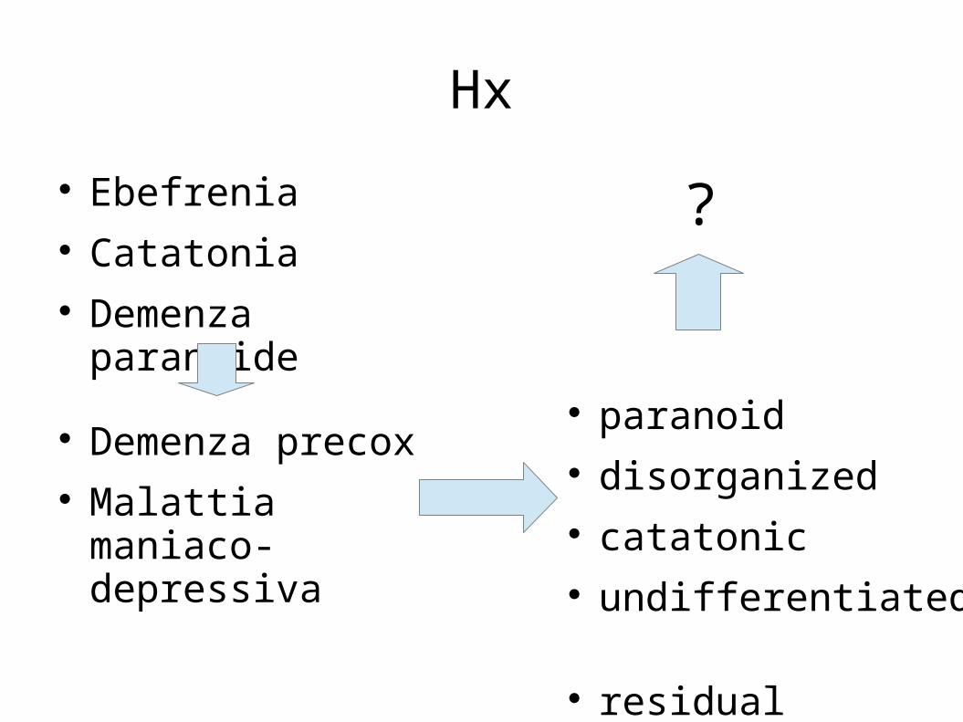
Hx
Ebefrenia Catatonia Demenza paranoide
Demenza precox Malattia maniaco-
depressiva
paranoid disorganized catatonic undifferentiated residual
?
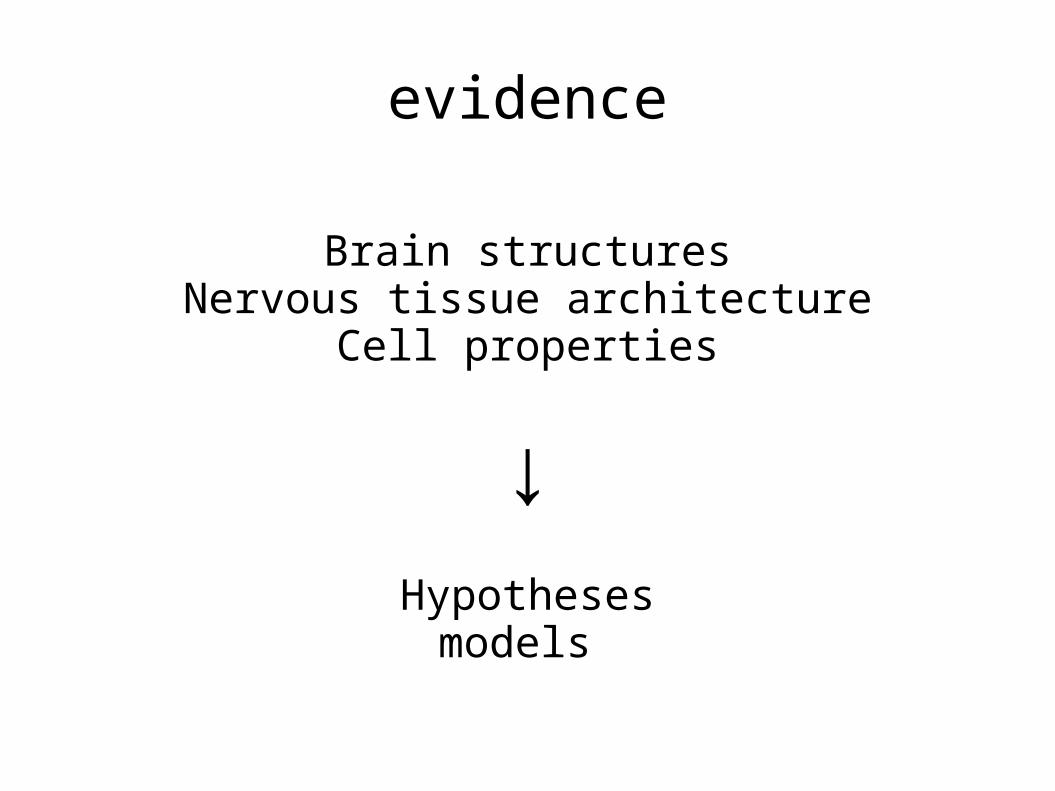
evidence
Brain structuresNervous tissue architecture
Cell properties
↓Hypotheses
models
Background
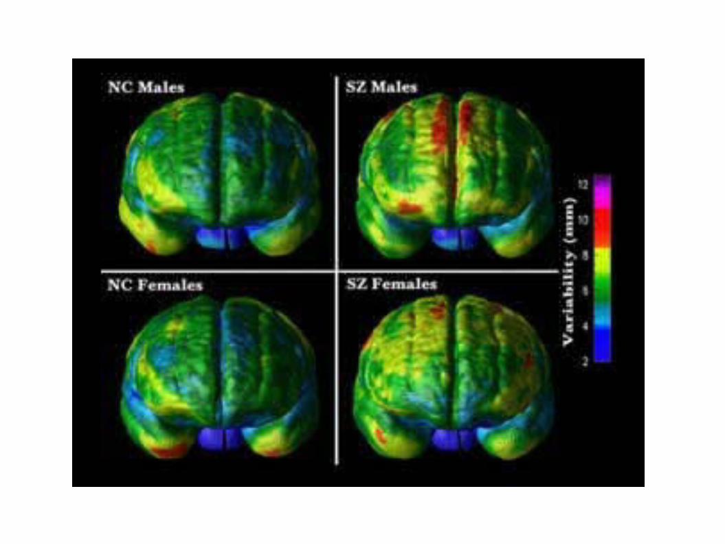
http://www.schizophrenia.com/research/schiz.brain.htmhttp://www.loni.ucla.edu/
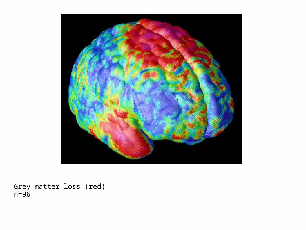
Grey matter loss (red)n=96
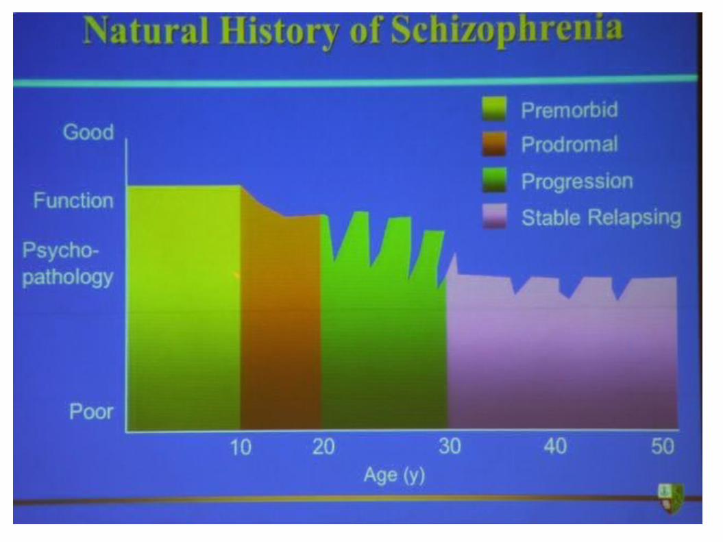
SKZ
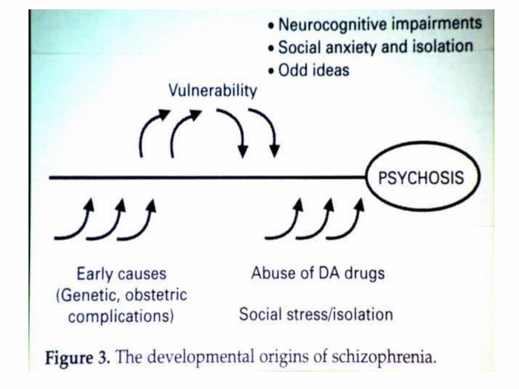
Interplay between biological and psychosocial factors
New conception of schizophrenia as a brain disorder (last 20 years)
Not to dismiss environmental stressors, but rather to put these in the perspective of a brain disorder in evolution
SKZ
Behavioral aberrations → diffuse abnormality in several brain systems
Frontal and temporal lobes
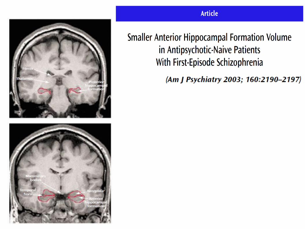
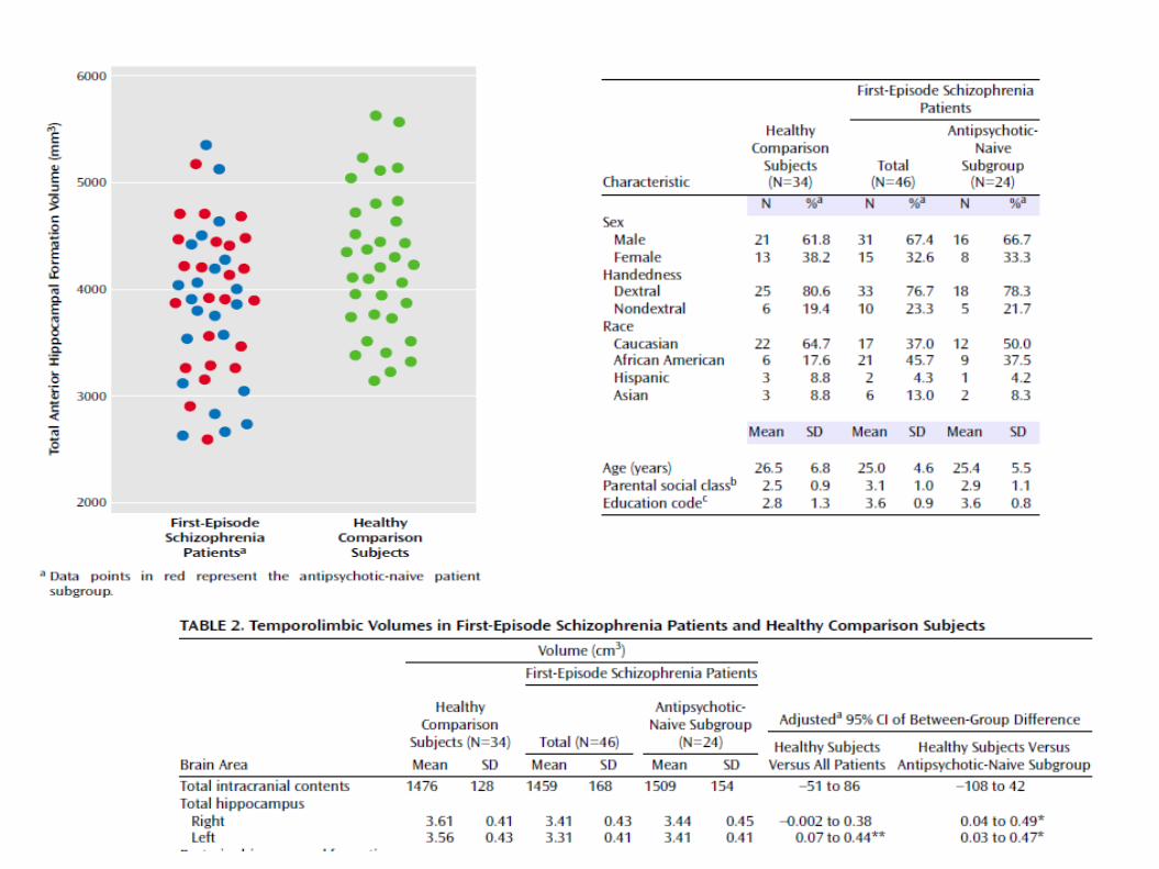
Reduced volume was reported in multiple regions including the superior temporal gyrus, hippocampus, and thalamus.
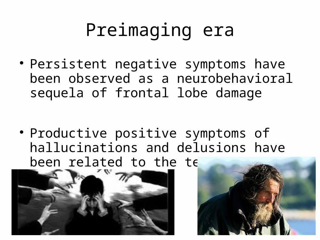
Preimaging era
Persistent negative symptoms have been observed as a neurobehavioral sequela of frontal lobe damage
Productive positive symptoms of hallucinations and delusions have been related to the temporolimbic system
SKZ
The exception is basal ganglia regions ↑ volume
→ related to the effects of dopamine receptor antagonists ?

Blood flow
Patients did not show the normal pattern of more anterior than posterior flow.
Decreased frontal metabolic activity has been associated with duration of illness and negative symptoms
Improvement in clinical status correlated with a shift toward lower left hemispheric metabolism relative to that in the right hemisphere
Metabolism and blood flow
PET described abnormal cerebral blood flow in the parahippocampal gyrus, associated with positive symptoms.
SPECT blood flow changes in the hippocampus, parahippocampus, and amygdala → Hallucinations
Symmetrical temporal lobe perfusion (lower in the left than the right) in patients with auditory hallucinations (inconsistencies).
Activation studies
Healthy controls showed the expected greater left hemispheric increase for the verbal task and greater right hemispheric increase for the spatial task.
Patients with schizophrenia had a bilaterally
symmetrical activation for the verbal task and greater left hemispheric activation for the spatial task.
Conclusion 1
SKZ is a multifactorial brain disease Neurodevelopment is crucial to the disease and
sensitive to external stimula Front lobes and the limbic system are central to
SKZ but results from nimg did not yeald comprehensive results
Thus → go into into further details, propose and check models
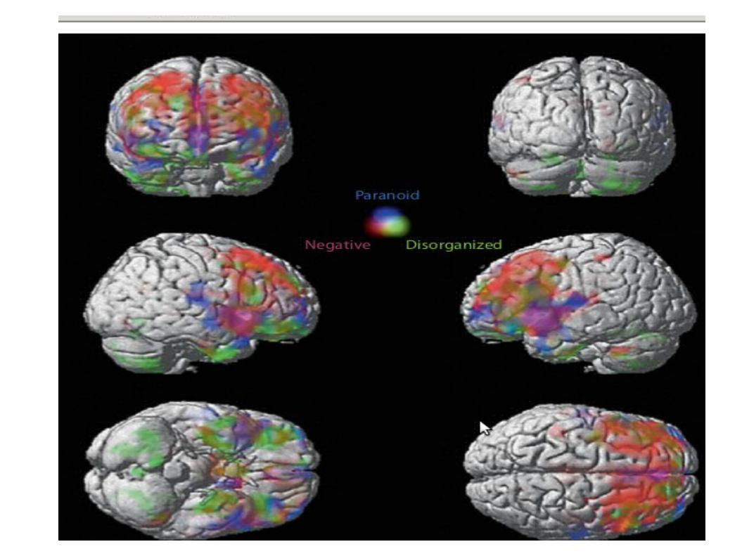
Specificity of symptoms ?
Role of interneurons
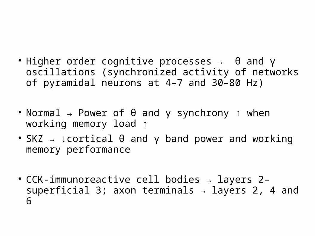
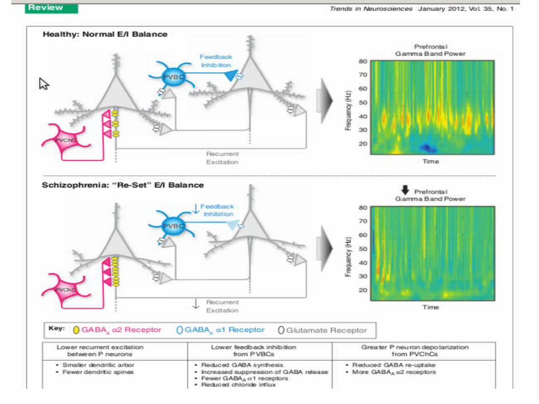
Higher order cognitive processes → θ and γ oscillations (synchronized activity of networks of pyramidal neurons at 4–7 and 30–80 Hz)
Normal → Power of θ and γ synchrony ↑ when working memory load ↑
SKZ → ↓cortical θ and γ band power and working memory performance
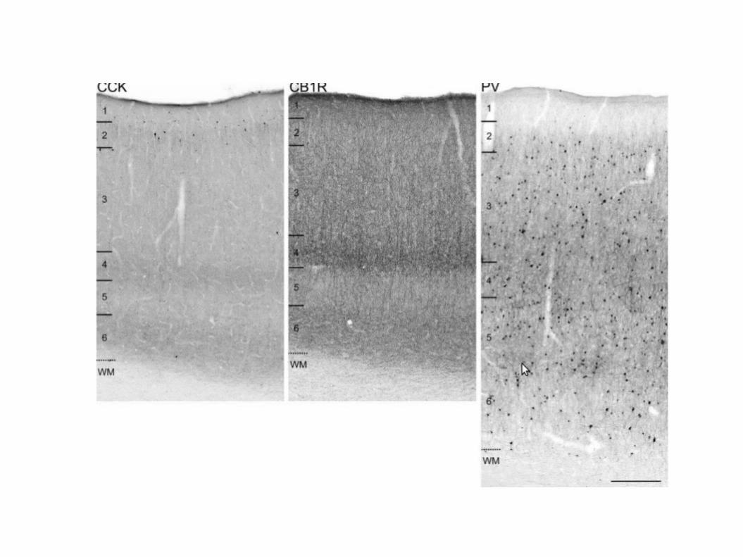
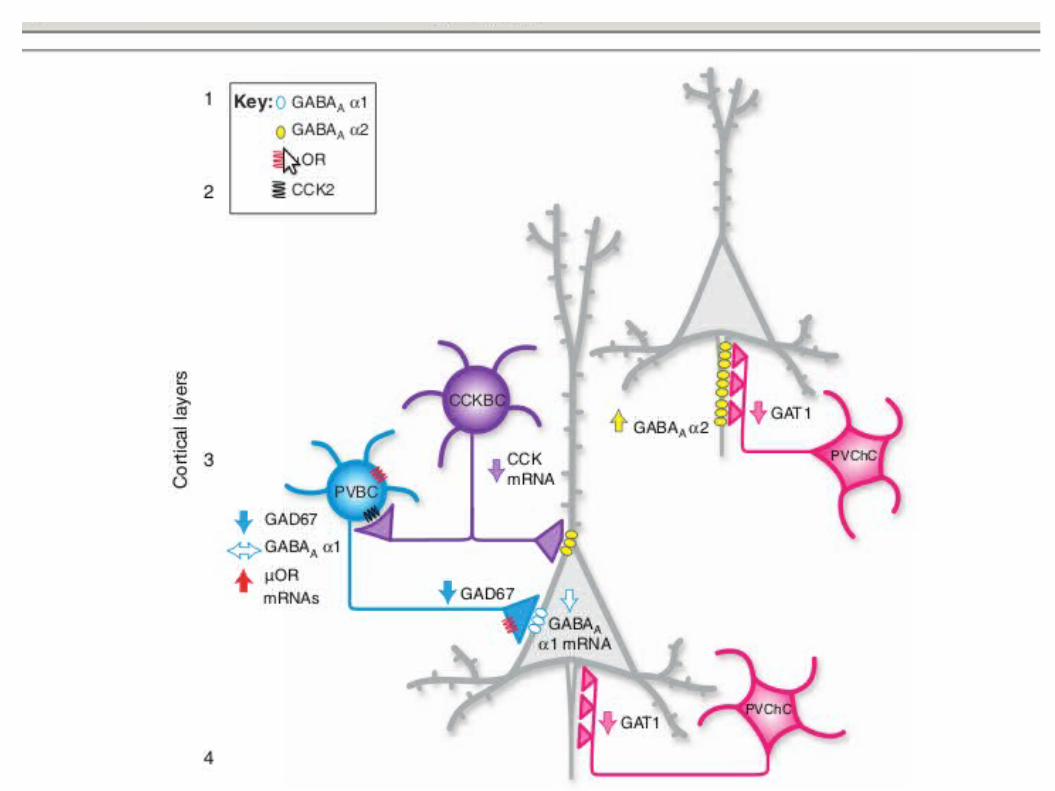
CCK-immunoreactive cell bodies → layers 2–superficial 3; axon terminals → layers 2, 4 and 6
Cortical θ and γ oscillations require strong inhibition provided by GABAergic inputs to the perisomatic region of pyramidal cells
Perisomatic inhibitory inputs to pyramidal neurons are furnished primarily by GABAergic basket cells that contain calcium-binding protein PV (PVb cells) or neuropeptide cholecystokinin (CCKb cells)
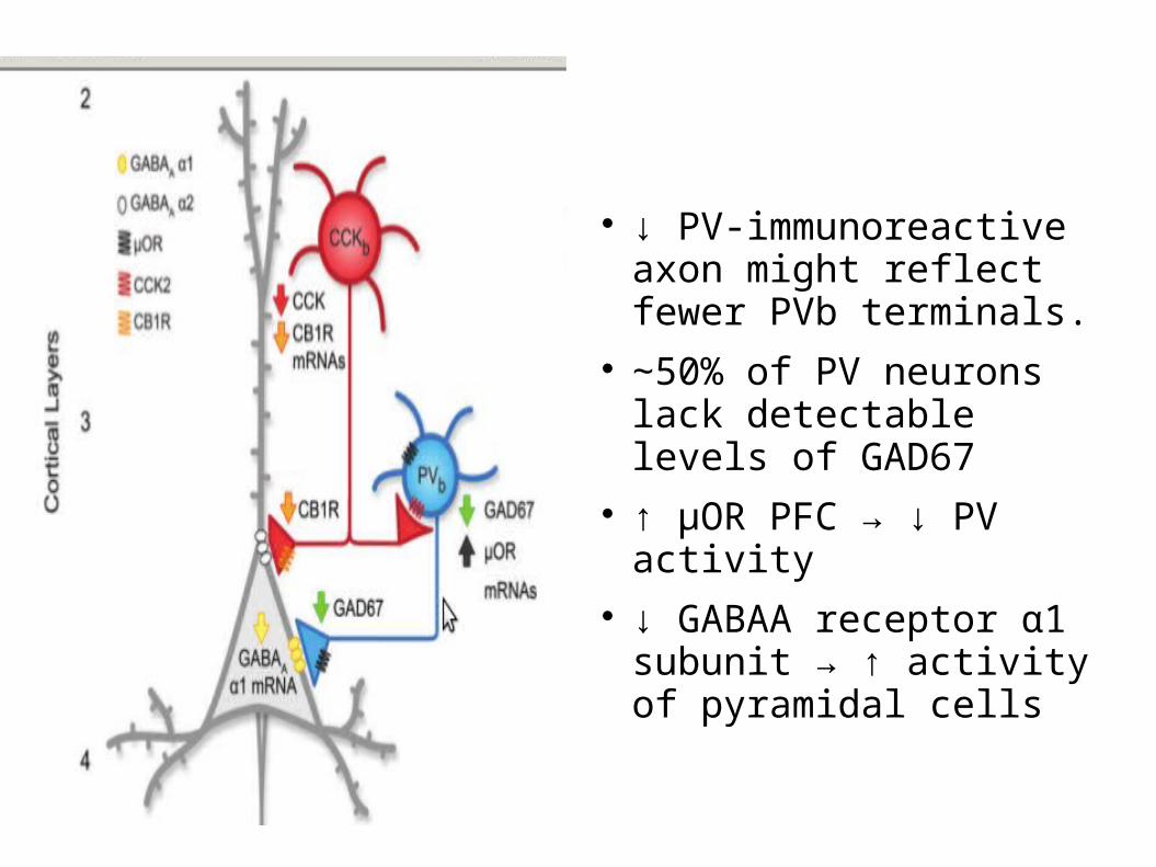
↓ PV-immunoreactive axon might reflect fewer PVb terminals.
∼50% of PV neurons lack detectable levels of GAD67
↑ μOR PFC → ↓ PV activity
↓ GABAA receptor α1 subunit → ↑ activity of pyramidal cells

CB1R ↓ PFC statistically significant in layers deep 3–4 and 6
That results in ↓ GABAergic transmission
Is GABA that central ?
Cognitive deficits are present and progressive years before the onset of psychosis
the degree of cognitive impairment is the best predictor of long-term functional outcome
gamma frequency (30–80 Hz) oscillations in DLPFC neural networks are thought to be a key neural substrate for cognition
↓ GABA in DLPFC → ∆ gamma frequency
GAD65 and GAD67 drive the synthesis of GABA in the brain
GAD67 mRNA and protein have been found consistently to be lower in the DLPFC of subjects with schizophrenia
frontal lobe GABA levels tended to be correlated with working memory performance in subjects with early-stage schizophrenia
Conclusion 2
GABAergic transmission in the mPFC could be a main biological target for explaining the cognitive deficits in SKZ
▲ in the GABAergic transmission would result in ↓ efficacy of the mPFC functioning
Thus → check in other regions, find common pathways
SKZ KEY MAP POINTS
PFC NACC HIPPOCAMPUS/AMYGDALA
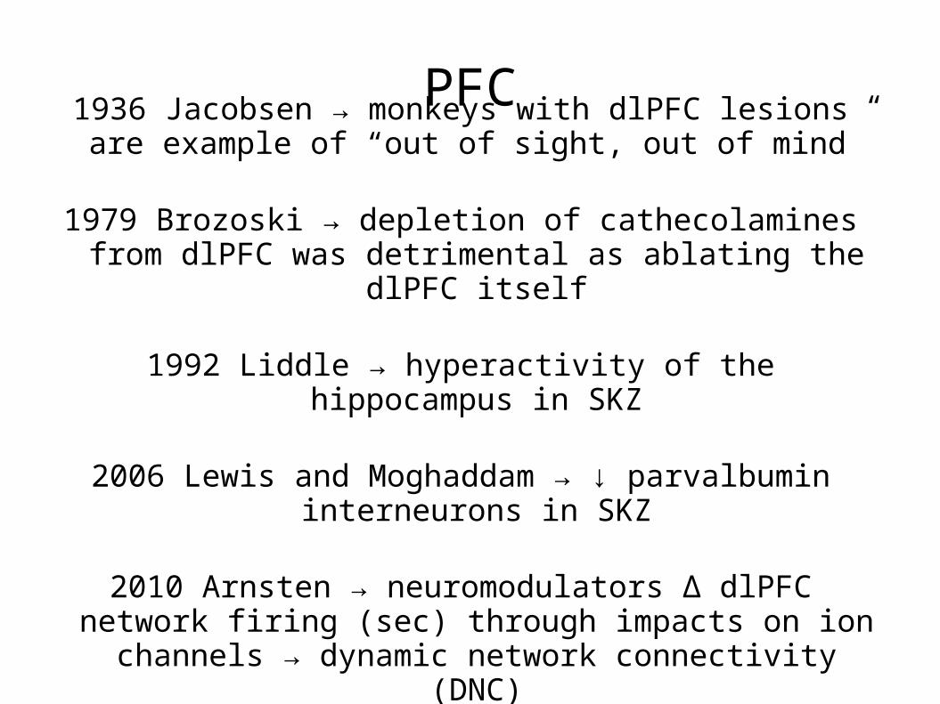
PFC1936 Jacobsen → monkeys with dlPFC lesions are example
of “out of sight, out of mind”
1979 Brozoski → depletion of cathecolamines from dlPFC was detrimental as ablating the dlPFC itself
1992 Liddle → hyperactivity of the hippocampus in SKZ
2006 Lewis and Moghaddam → ↓ parvalbumin interneurons in SKZ
2010 Arnsten → neuromodulators ∆ dlPFC network firing (sec) through impacts on ion channels → dynamic
network connectivity (DNC)
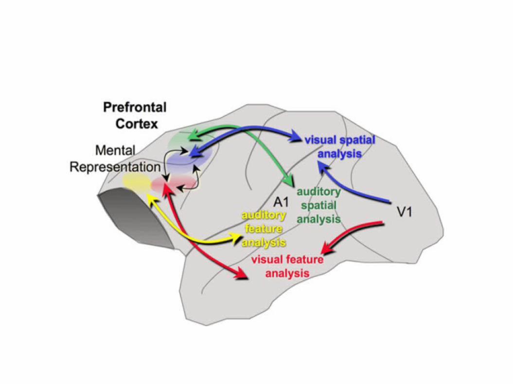
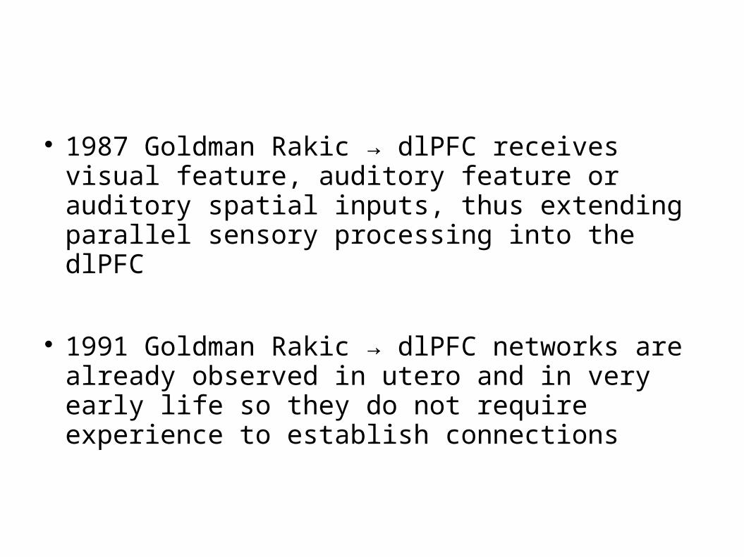
1987 Goldman Rakic → dlPFC receives visual feature, auditory feature or auditory spatial inputs, thus extending parallel sensory processing into the dlPFC
1991 Goldman Rakic → dlPFC networks are already observed in utero and in very early life so they do not require experience to establish connections
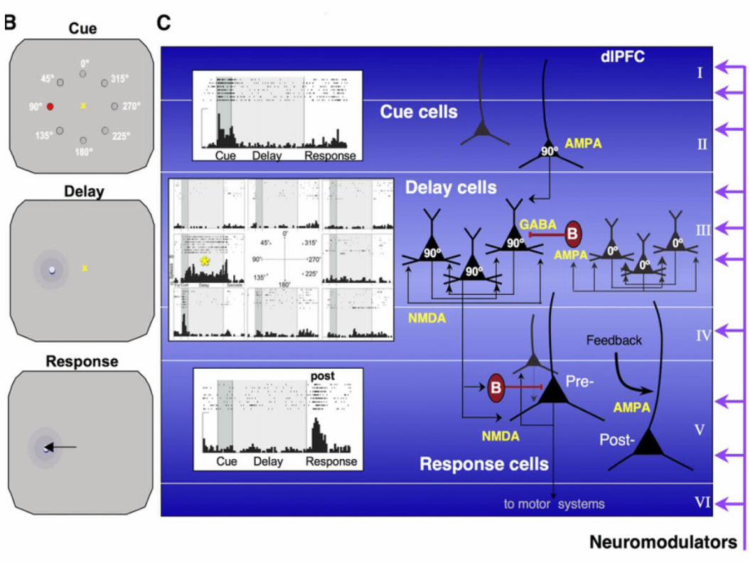
Maier 2008 → A large portion of neurons in the principal sulcal dlPFC show spatially tuned, persistent firing across the delay period
Thus, neurons in dlPFC can represent visual space in the absence of sensory stimulation
Goldman Rakic, 1996 → behavioural inhibition and cognitive control depend on these mechanisms
Seo and lee, 2009 → emotions reward, changing rules and goals require the activation of the PFC
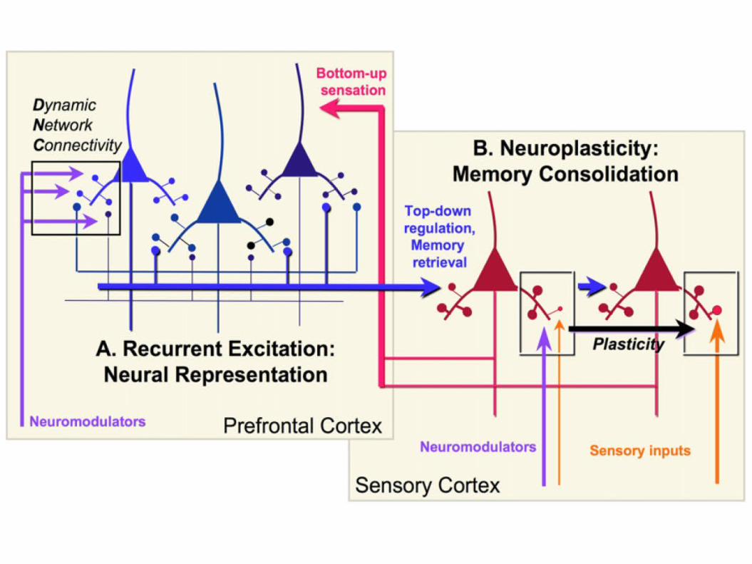
Nearby neurons with similar spatial tuning excite each other via connections on spines to maintain firing without the need for bottom-up regulatory stimulation
This persistent firing is highly dependent on NMDA receptors, including those with NR2B subunits (slow kinetics)
AMPA receptors are involved in the activity of GABA interneurons
Schizophrenia → ↓ GABAergic action, with loss of spines and neuropil in layer III
There is evidence of ↓ parvalbumin-containing neurons in schizophrenic patients
→ profound working memory impairment and thought disorder
Layer III pyramidal cells are also an early target of neurofibrillary tangles and neurodegeneration in Alzheimer's disease

Layer V neurons also exhibit alterations in schizophrenia
They project to the striatum and engage in cortico-cortical connections
Lidow, 1998 → Some delay cells and most response cells reside in layer V, they are selectively influenced by D2 receptors and D2 receptor mRNA is enriched in layer V
Ford 2002 → alterations in discharge feedback from PFC may contribute to hallucinations
Corlett, 2007 → errors in feedback may also play a role in delusions
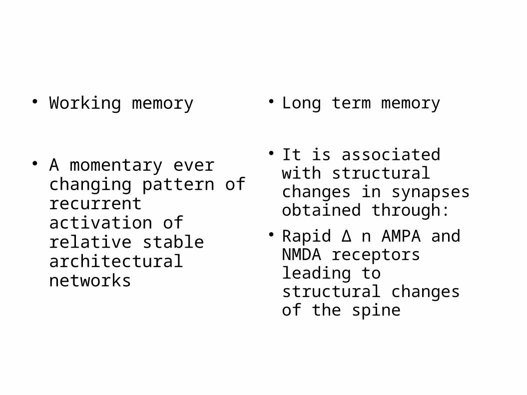
Working memory
A momentary ever changing pattern of recurrent activation of relative stable architectural networks
Long term memory
It is associated with structural changes in synapses obtained through:
Rapid ∆ n AMPA and NMDA receptors leading to structural changes of the spine
Long term memory
Requires Ca++ and cAMP which are facilitated by neuromodulators and lead to trascriptional events in the nucleus (Barco, 2003)
In this way the cortex is thought to accumulate a lifetime of experience in remote memory storage
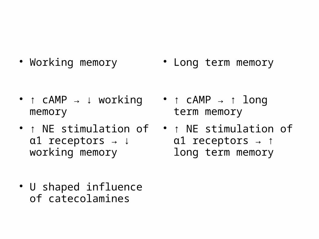
Working memory
↑ cAMP → ↓ working memory
↑ NE stimulation of α1 receptors → ↓ working memory
U shaped influence of catecolamines
Long term memory
↑ cAMP → ↑ long term memory
↑ NE stimulation of α1 receptors → ↑ long term memory

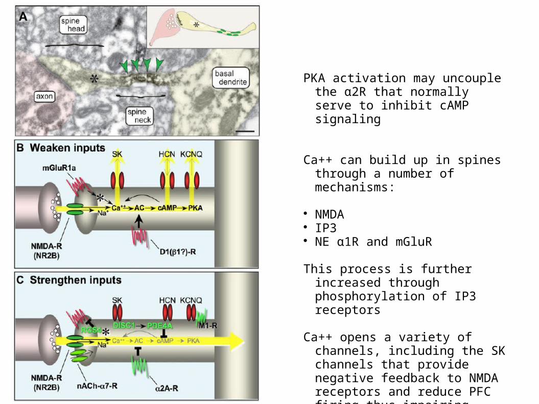
PKA activation may uncouple the α2R that normally serve to inhibit cAMP signaling
Ca++ can build up in spines through a number of mechanisms:
NMDA IP3 NE α1R and mGluR
This process is further increased through phosphorylation of IP3 receptors
Ca++ opens a variety of channels, including the SK channels that provide negative feedback to NMDA receptors and reduce PFC firing thus impairing working memory
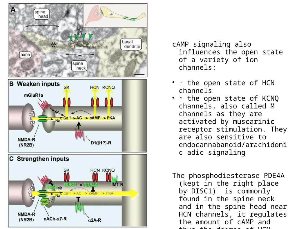
cAMP signaling also influences the open state of a variety of ion channels:
↑ the open state of HCN channels ↑ the open state of KCNQ
channels, also called M channels as they are activated by muscarinic receptor stimulation. They are also sensitive to endocannabanoid/arachidonic adic signaling
The phosphodiesterase PDE4A (kept in the right place by DISC1) is commonly found in the spine neck and in the spine head near HCN channels, it regulates the amount of cAMP and thus the degree of HCN channel opening.
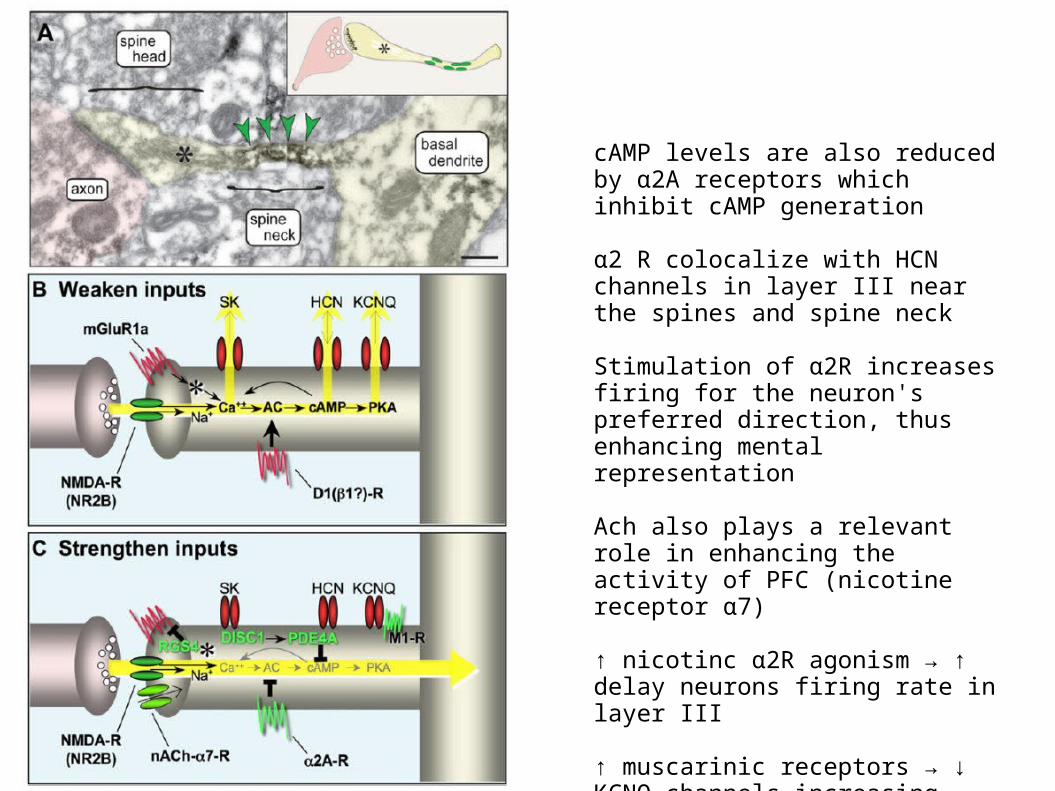
cAMP levels are also reduced by α2A receptors which inhibit cAMP generation
α2 R colocalize with HCN channels in layer III near the spines and spine neck
Stimulation of α2R increases firing for the neuron's preferred direction, thus enhancing mental representation
Ach also plays a relevant role in enhancing the activity of PFC (nicotine receptor α7)
↑ nicotinc α2R agonism → ↑ delay neurons firing rate in layer III
↑ muscarinic receptors → ↓ KCNQ channels increasing neuron excitability
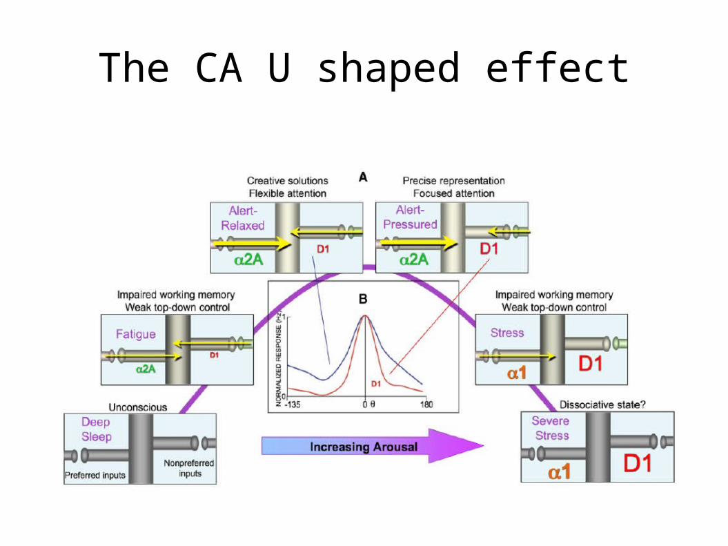
The CA U shaped effect
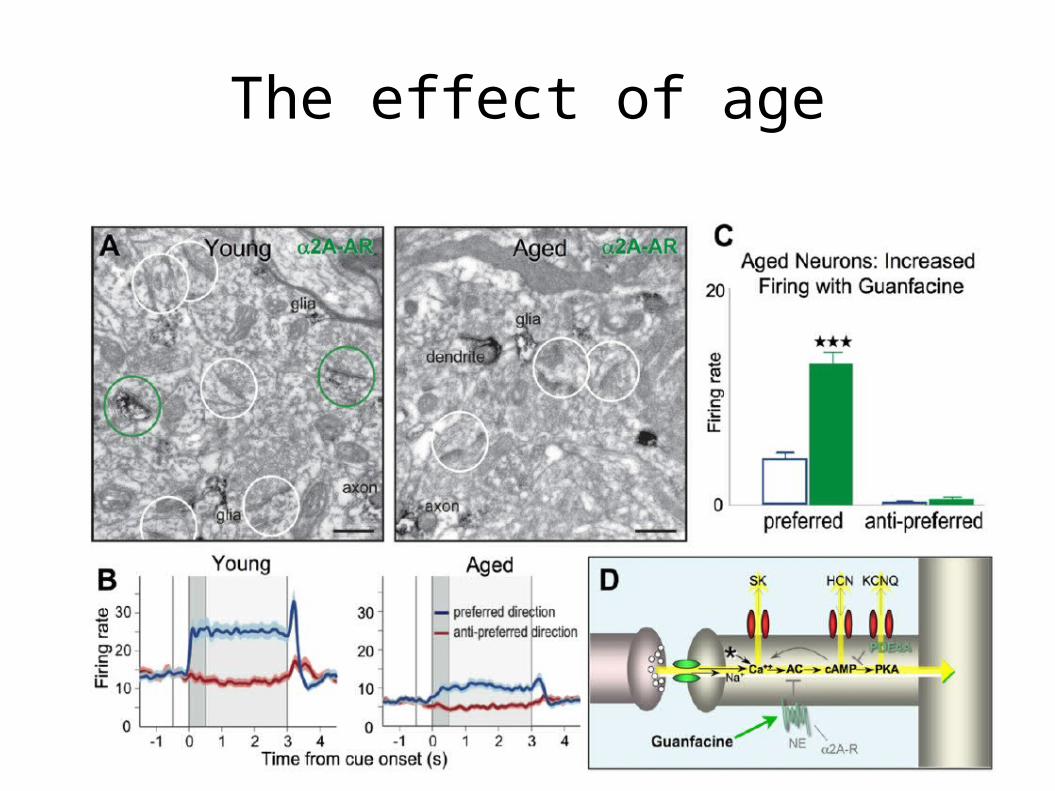
The effect of age
Skz
Atrophy in layer III of dlPFC Loss of neuropil, dendritic spines Weakened GABAergic lateral inhibition Atrophy in layer V dlPFC
↓cortical DA and ↑ subcortical DA COMT genotype CA++ signaling
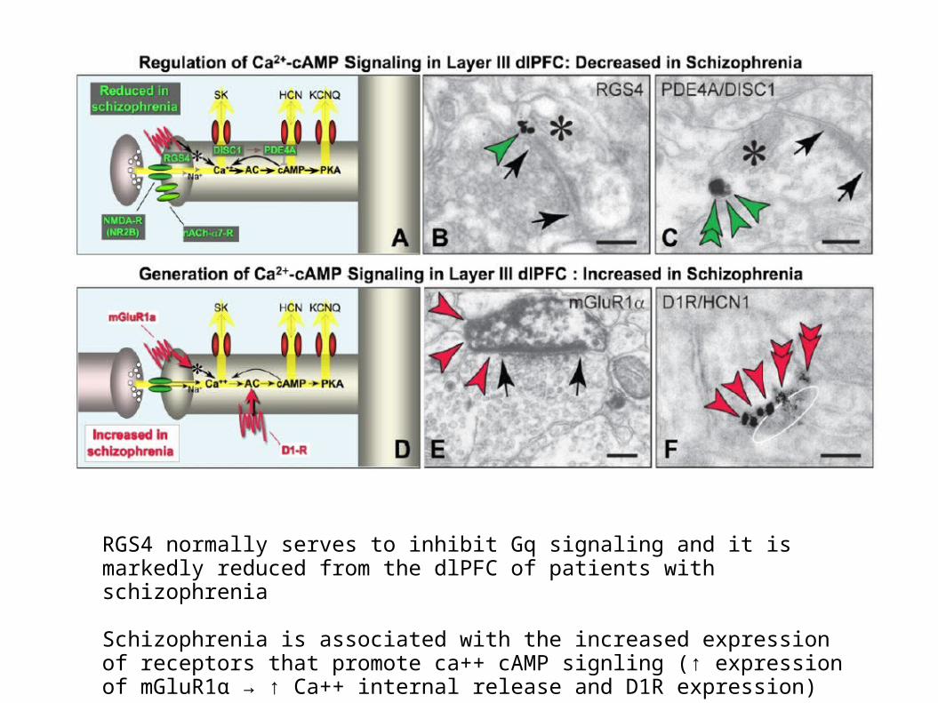
RGS4 normally serves to inhibit Gq signaling and it is markedly reduced from the dlPFC of patients with schizophrenia
Schizophrenia is associated with the increased expression of receptors that promote ca++ cAMP signling (↑ expression of mGluR1α → ↑ Ca++ internal release and D1R expression)
Conclusion 3Dendrite activity in the mPFC is central to representation of stimuli before reaction and after their termination
These events may be the basis for delusional thoughts and hallucinations
The Ca++ homeostasis and PKA activation in dendrites of layer III in mPFC is central to the correct firing rate during delay period
Thus → check more in delay period and representation, check other parts of the brain
NAc
The central structure for integration of contextual stimuli processed in the hippocampus and executive functions from the PFC
This system is modulated by the DA in the ventral tegmental area to control goal directed behavior
→ Latent inhibition
NAc
Latent inhibition → proactive interference of non-reinforced stimulus pre-exposure on the subsequent performance of a learning task involving this stimulus (Lubow 1973)
↓ the attention for that stimulus but does not affect the association strength (Wagner and Rescorla 1972)
NAc
Latent inhbition → stimulus that are not associated with any particular event are actively ignored in their affective counterparts
→ SKZ ?
NAc
1919 Kraepelin → disorder of attention” is “conspicuously developed” in patients with dementia praecox
1911 Bleuler → schizophrenia as the loss of “selectivity [...] among the sensory impressions”
NAc
Normal rat + low doses of amphetamine → ↓ LI, reversed by AP
Normal rat + high doses of amphetamine → normal LI
Normal rat + haloperidol → ↑ LI
The effect of drugs is only visible when the LI is to be recalled, not when it is formed
NAc
LI … conditioning … ↓ LI → affective association with the stimulus
{LI + (hal | cloz)} … conditioning … ↓ LI → affective association with the stimulus
LI … {conditioning + (hal | cloz)} … ≈ LI → no affective association with the stimulus (Peters and Joseph 1993)
→ the DA system is not involved in the acquisition of LI but in it recovery when the same stimulus is given
NAc
The target of LI disruption by amphetamines is the NAc (Solomon and Staton,1982)
↑ DA in NAc → ↓ LI → switch to the actual association
↓ DA in NAc → ↑ LI →| switch to the actual association
NAc
Not involved in the LI formation but activated in the conditioning stage, when the previously non-reinforced stimulus is followed by reinforcement
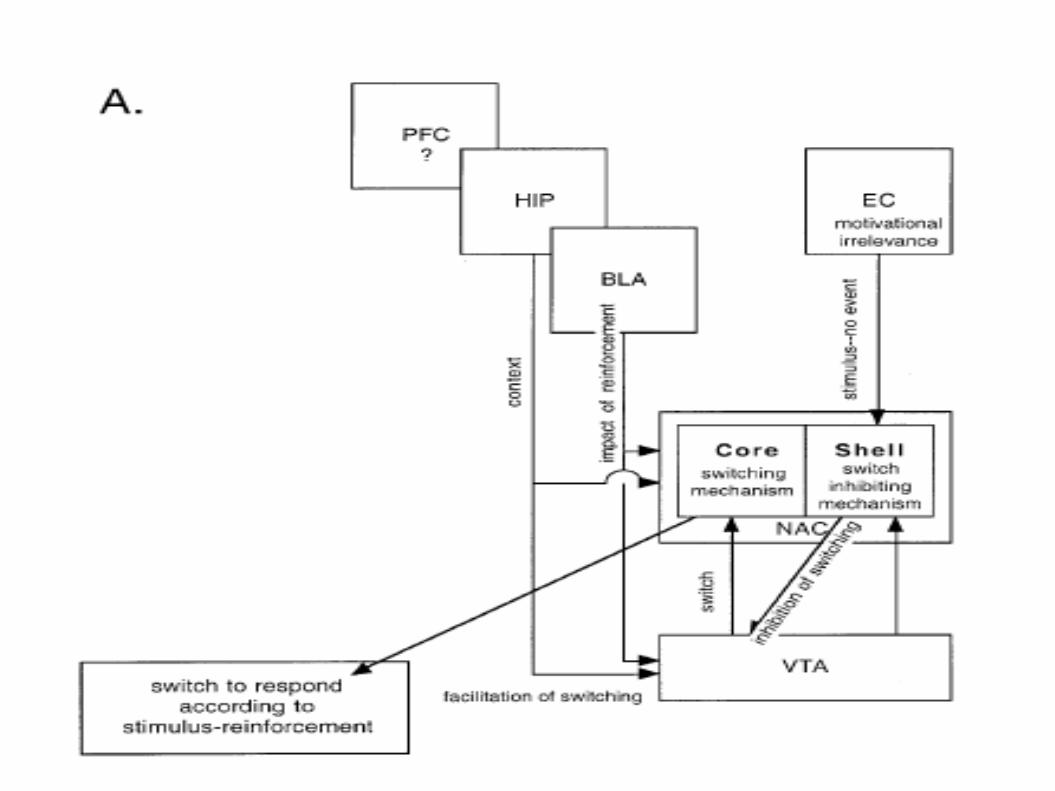
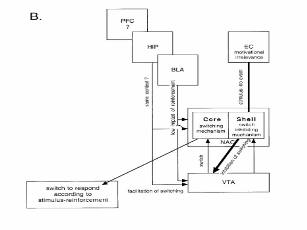
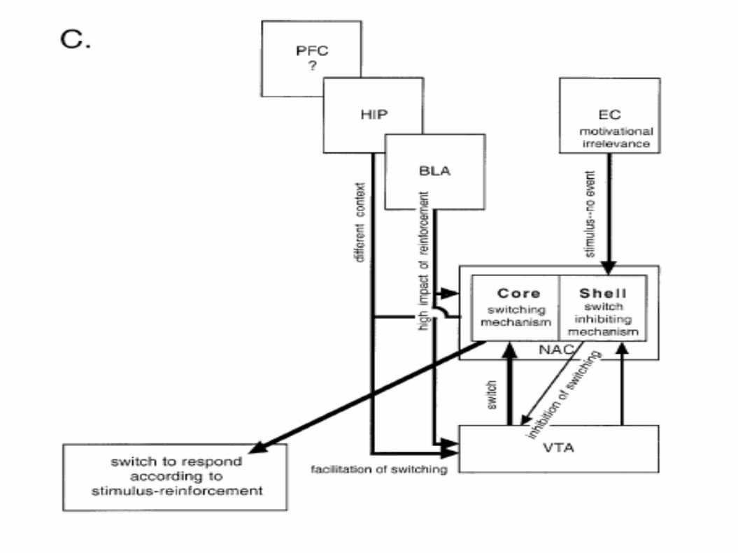
Shell → promotes LI ↓ Core → promotes LI ↑
→ a shell lesion will destroy LI, a large NAc lesion will promote perseveration
Hippocampus
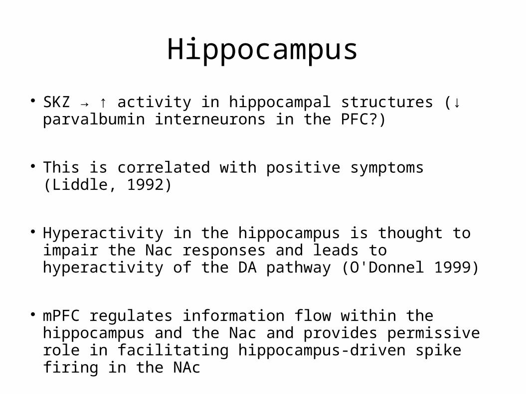
SKZ → ↑ activity in hippocampal structures (↓ parvalbumin interneurons in the PFC?)
This is correlated with positive symptoms (Liddle, 1992)
Hyperactivity in the hippocampus is thought to impair the Nac responses and leads to hyperactivity of the DA pathway (O'Donnel 1999)
mPFC regulates information flow within the hippocampus and the Nac and provides permissive role in facilitating hippocampus-driven spike firing in the NAc
Hippocampus
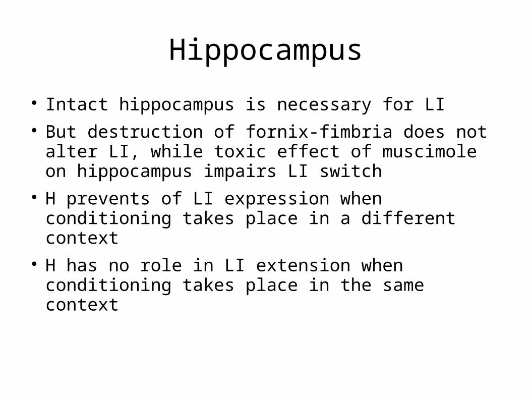
Intact hippocampus is necessary for LI But destruction of fornix-fimbria does not alter
LI, while toxic effect of muscimole on hippocampus impairs LI switch
H prevents of LI expression when conditioning takes place in a different context
H has no role in LI extension when conditioning takes place in the same context
Other relevant regions
Disruption of the ehtorhinal cortex → ↓ LI
This event is reverted by hal → disruption of LI in the NAc after lesion to the cortex is mediated by an ↑ DA in the Nac
Lesions of the PFC or BLA do not affect LI