Embed Size (px)
Citation preview
6/5/2010
1
Sex Cord Stromal Tumors of the TestisCharles Zaloudek, M.D.
Professor, Department of PathologyUniversity of California, San Francisco
505 Parnassus Ave., M563San Francisco, CA 94143
Sex Cord Stromal Tumors of the Testis
• Uncommon testicular neoplasms• Account for about 5% of testicular tumors• Wide age range, children to elderly• Usually present because of a testicular
mass• Some are functional, but most are not• Classification similar to that for analogous
ovarian tumors
Leydig cell tumor
Malignant Leydig cell tumor
Sertoli cell tumorLipid rich Sertoli cell tumorSclerosing Sertoli cell tumorLarge cell calcifying Sertoli cell tumor
Malignant Sertoli cell tumor
Granulosa cell tumorJuvenile granulosa cell tumorAdult type granulosa cell tumor
Tumors of fibroma/thecoma group
Unclassified sex cord-gonadal stromal tumor
Classification of Testicular Sex Cord-Stromal Tumor s
Leydig Cell Tumors
• Most common testicular sex cord-stromal tumor
• Bimodal age distribution• Adults over a wide age range, 20-60 years• Children• Reported in patients with germline
fumarate hydratase mutations (hereditary leiomyomatosis and renal cell carcinoma syndrome)
6/5/2010
2
Leydig Cell Tumors in Adults
• Mean age in large series mid 30’s• Usual presentation is with a testicular
mass• About 30% have gynecomastia, which may
bring the patient to attention• Other presentations include testicular pain
and infertility• About 1/3 have increased androgens and
1/3, mainly those with gynecomastia, have increased estrogens
Leydig Cell Tumors in Children
• 4% of testicular tumors in boys < 12• 3/40 in Kim, Young and Scully series of
Leydig cell tumors• Small tumors• Most present with precocious
pseudopuberty due to androgen secretion• About 10% have gynecomastia, and
elevated estrogens• Leydig cell tumors in children are benign
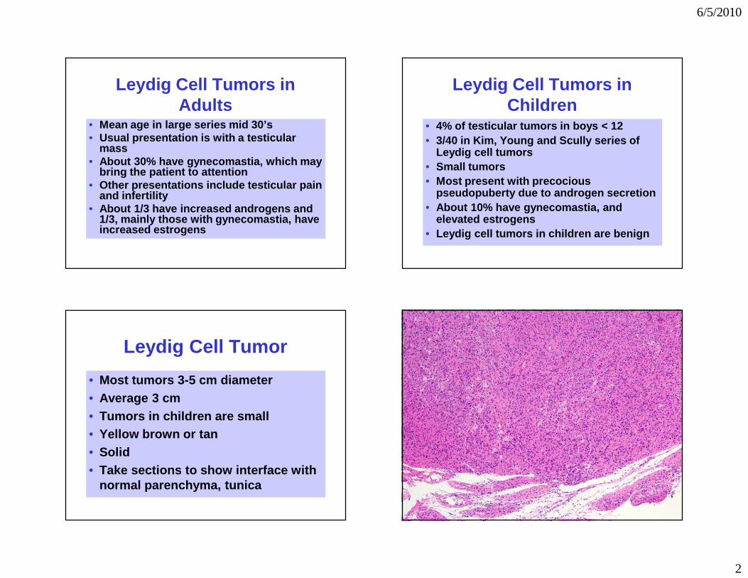
Leydig Cell Tumor
• Most tumors 3-5 cm diameter• Average 3 cm• Tumors in children are small• Yellow brown or tan• Solid• Take sections to show interface with
normal parenchyma, tunica
6/5/2010
3
6/5/2010
4
2
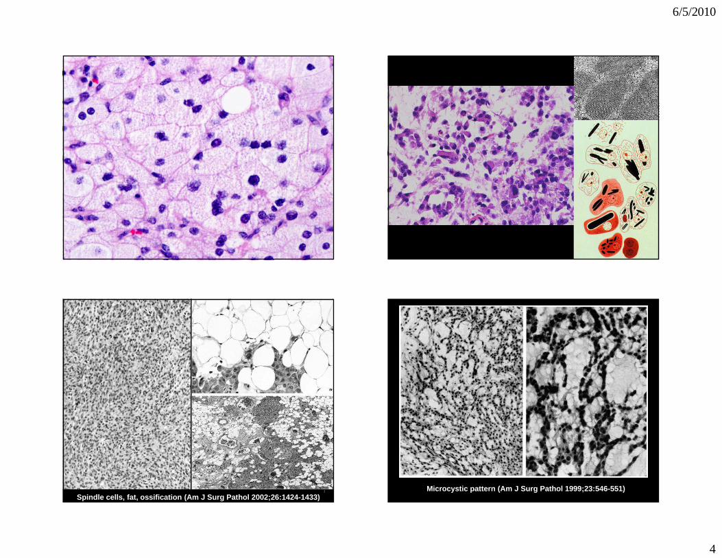
Spindle cells, fat, ossification (Am J Surg Pathol 2002;26:1424-1433)Microcystic pattern (Am J Surg Pathol 1999;23:546-5 51)
6/5/2010
5
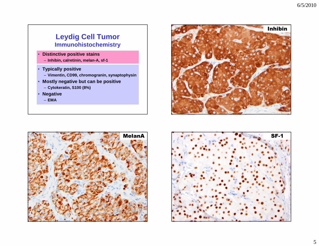
Leydig Cell TumorImmunohistochemistry
• Distinctive positive stains– Inhibin, calretinin, melan-A, sf-1
• Typically positive– Vimentin, CD99, chromogranin, synaptophysin
• Mostly negative but can be positive– Cytokeratin, S100 (8%)
• Negative– EMA
Inhibin
MelanA SF-1
6/5/2010
6
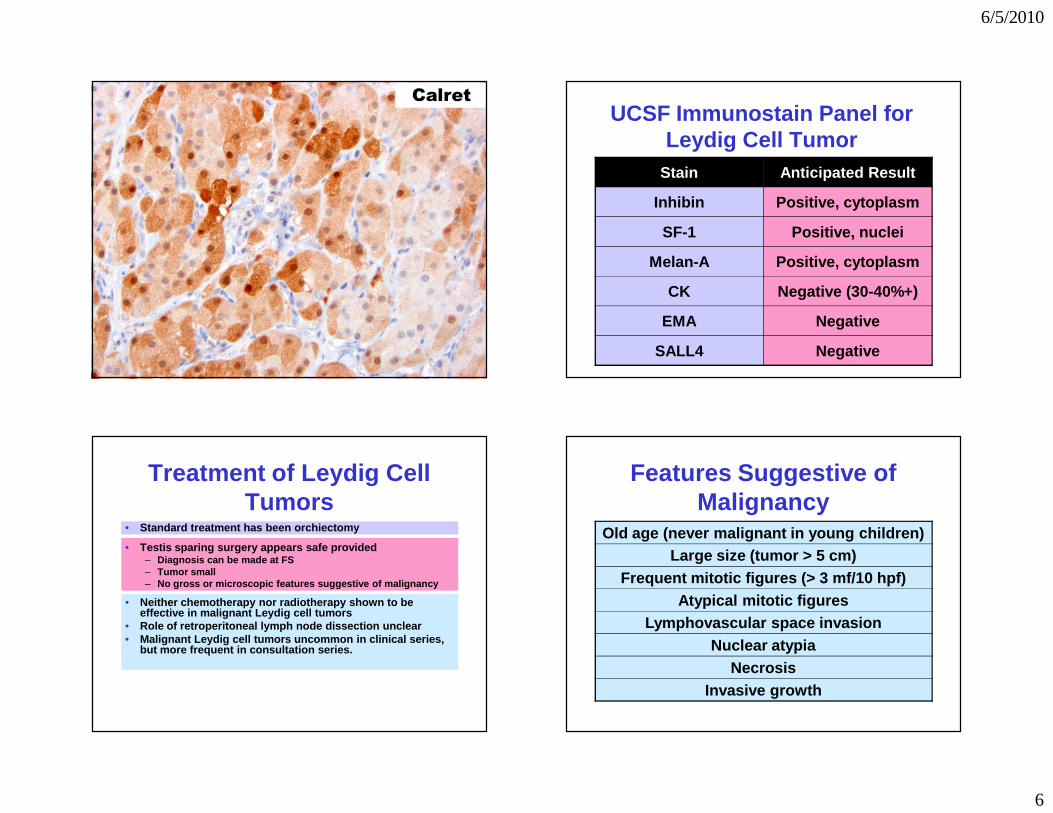
CalretUCSF Immunostain Panel for
Leydig Cell TumorStain Anticipated Result
Inhibin Positive, cytoplasm
SF-1 Positive, nuclei
Melan-A Positive, cytoplasm
CK Negative (30-40%+)
EMA Negative
SALL4 Negative
Treatment of Leydig Cell Tumors
• Standard treatment has been orchiectomy
• Testis sparing surgery appears safe provided– Diagnosis can be made at FS– Tumor small– No gross or microscopic features suggestive of mali gnancy
• Neither chemotherapy nor radiotherapy shown to be effective in malignant Leydig cell tumors
• Role of retroperitoneal lymph node dissection uncle ar• Malignant Leydig cell tumors uncommon in clinical s eries,
but more frequent in consultation series.
Features Suggestive of Malignancy
Old age (never malignant in young children)Large size (tumor > 5 cm)
Frequent mitotic figures (> 3 mf/10 hpf)Atypical mitotic figures
Lymphovascular space invasionNuclear atypia
NecrosisInvasive growth
6/5/2010
7
6/5/2010
8
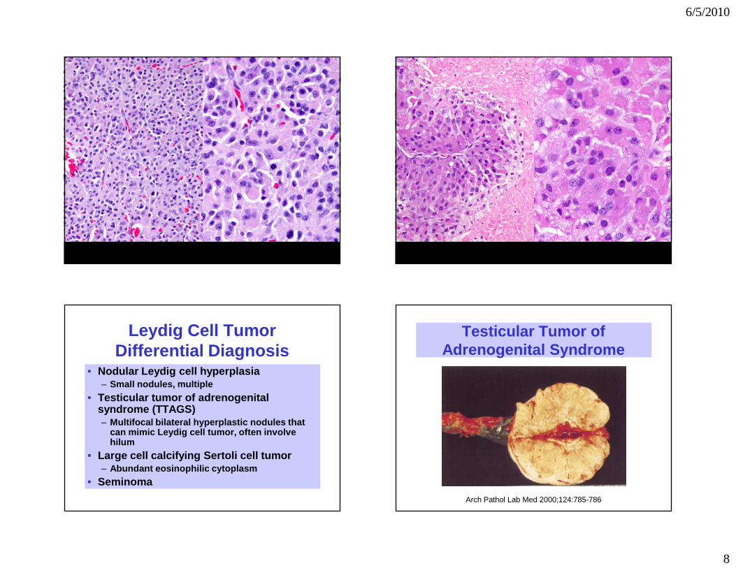
Leydig Cell TumorDifferential Diagnosis
• Nodular Leydig cell hyperplasia– Small nodules, multiple
• Testicular tumor of adrenogenital syndrome (TTAGS)– Multifocal bilateral hyperplastic nodules that
can mimic Leydig cell tumor, often involve hilum
• Large cell calcifying Sertoli cell tumor– Abundant eosinophilic cytoplasm
• Seminoma
Testicular Tumor of Adrenogenital Syndrome
Arch Pathol Lab Med 2000;124:785-786
6/5/2010
9
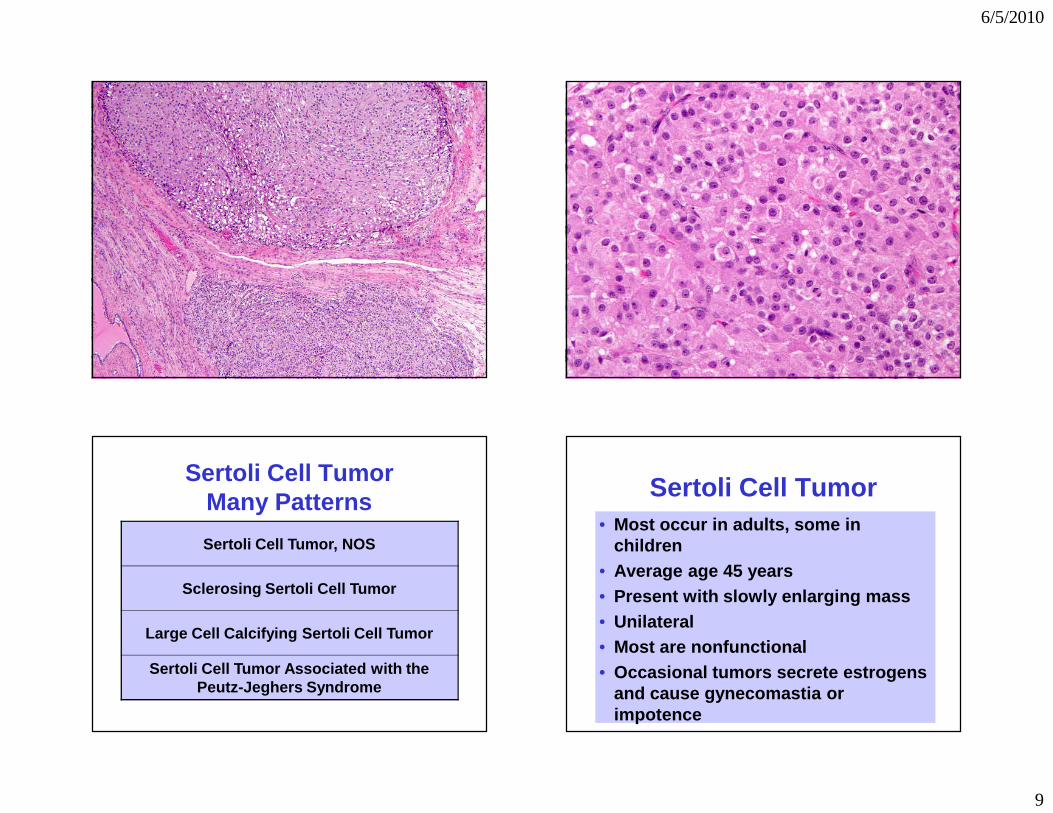
Sertoli Cell TumorMany Patterns
Sertoli Cell Tumor, NOS
Sclerosing Sertoli Cell Tumor
Large Cell Calcifying Sertoli Cell Tumor
Sertoli Cell Tumor Associated with the Peutz-Jeghers Syndrome
Sertoli Cell Tumor• Most occur in adults, some in
children• Average age 45 years• Present with slowly enlarging mass• Unilateral• Most are nonfunctional• Occasional tumors secrete estrogens
and cause gynecomastia or impotence
6/5/2010
10
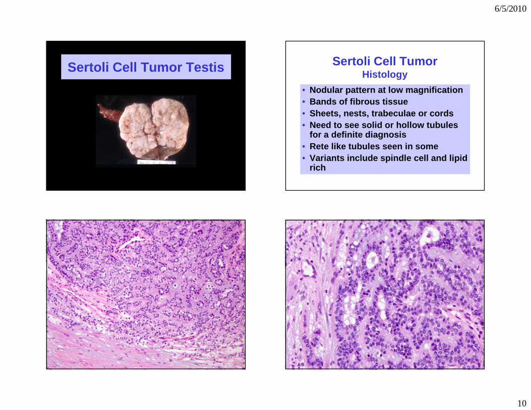
Sertoli Cell Tumor Testis Sertoli Cell TumorHistology
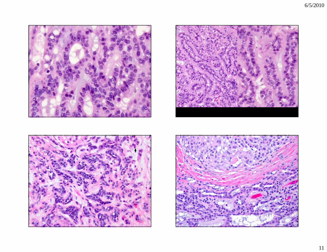
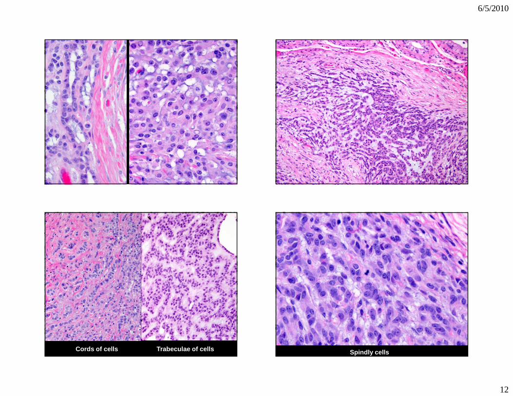
• Nodular pattern at low magnification• Bands of fibrous tissue• Sheets, nests, trabeculae or cords• Need to see solid or hollow tubules
for a definite diagnosis• Rete like tubules seen in some• Variants include spindle cell and lipid
rich
6/5/2010
11
6/5/2010
12
Cords of cells Trabeculae of cells Spindly cells
6/5/2010
13
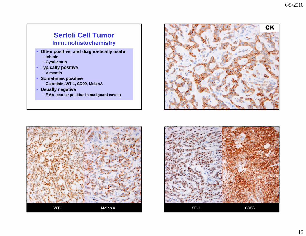
Sertoli Cell TumorImmunohistochemistry
• Often positive, and diagnostically useful– Inhibin– Cytokeratin
• Typically positive– Vimentin
• Sometimes positive– Calretinin, WT-1, CD99, MelanA
• Usually negative– EMA (can be positive in malignant cases)
CK
WT-1 Melan A SF-1 CD56
6/5/2010
14
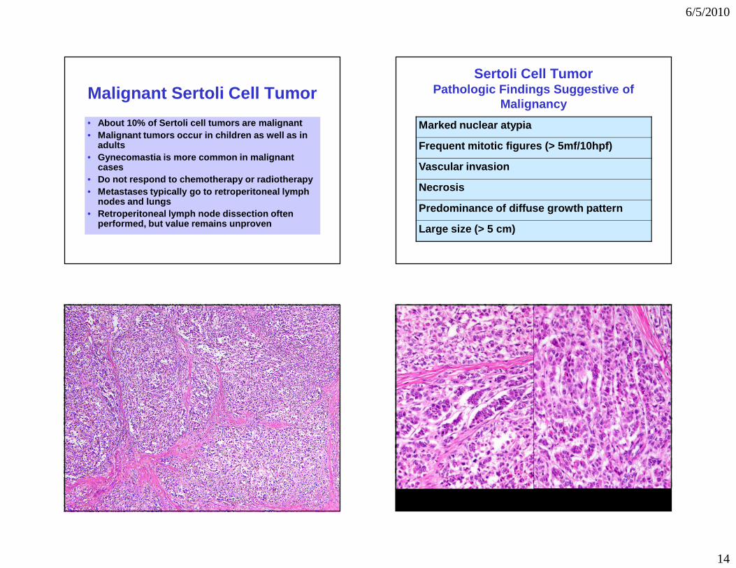
Malignant Sertoli Cell Tumor
• About 10% of Sertoli cell tumors are malignant• Malignant tumors occur in children as well as in
adults• Gynecomastia is more common in malignant
cases• Do not respond to chemotherapy or radiotherapy• Metastases typically go to retroperitoneal lymph
nodes and lungs• Retroperitoneal lymph node dissection often
performed, but value remains unproven
Sertoli Cell TumorPathologic Findings Suggestive of
Malignancy
Marked nuclear atypia
Frequent mitotic figures (> 5mf/10hpf)
Vascular invasion
Necrosis
Predominance of diffuse growth pattern
Large size (> 5 cm)
6/5/2010
15
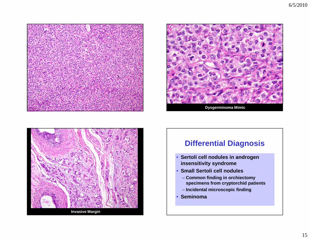
Dysgerminoma Mimic
Invasive Margin
Differential Diagnosis
• Sertoli cell nodules in androgen insensitivity syndrome
• Small Sertoli cell nodules– Common finding in orchiectomy
specimens from cryptorchid patients– Incidental microscopic finding
• Seminoma
6/5/2010
16
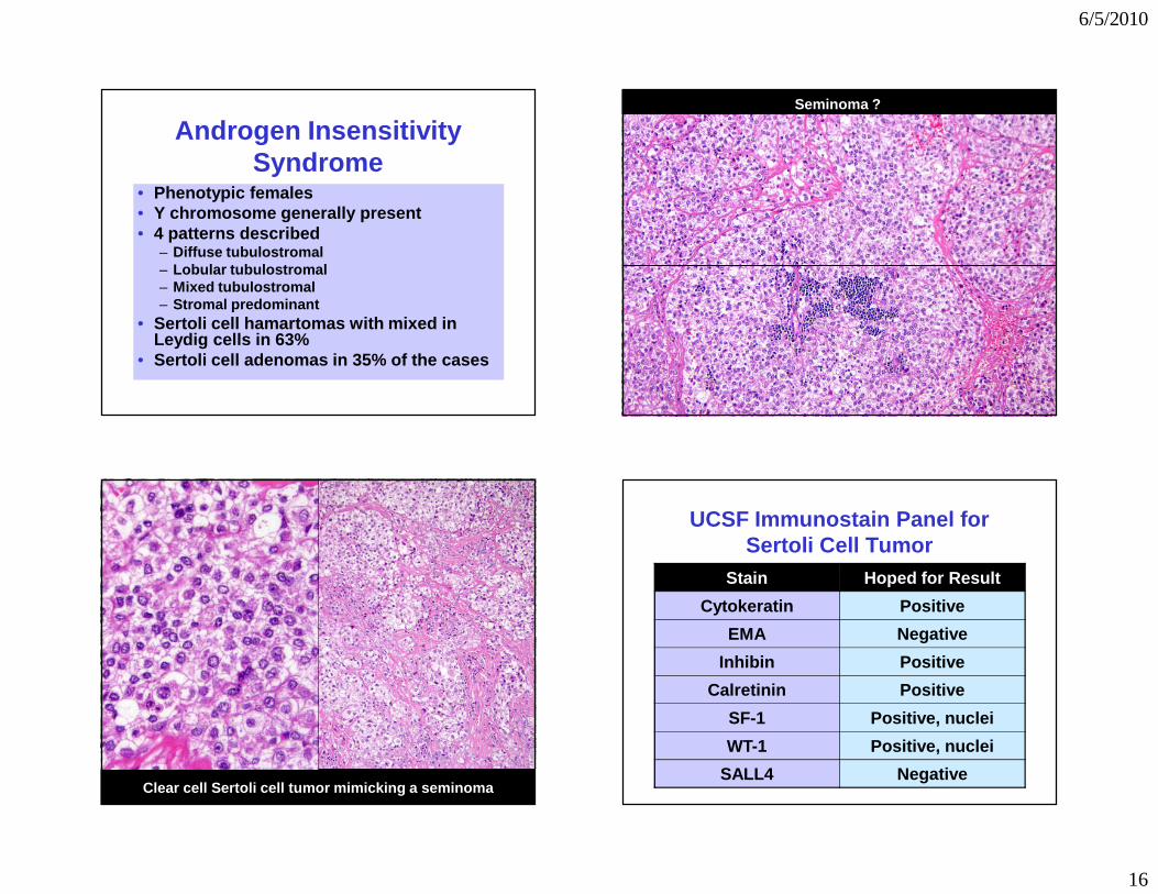
Androgen Insensitivity Syndrome
• Phenotypic females• Y chromosome generally present• 4 patterns described
– Diffuse tubulostromal– Lobular tubulostromal– Mixed tubulostromal– Stromal predominant
• Sertoli cell hamartomas with mixed in Leydig cells in 63%
• Sertoli cell adenomas in 35% of the cases
Seminoma ?
Clear cell Sertoli cell tumor mimicking a seminoma
UCSF Immunostain Panel for Sertoli Cell Tumor
Stain Hoped for Result
Cytokeratin Positive
EMA Negative
Inhibin Positive
Calretinin Positive
SF-1 Positive, nuclei
WT-1 Positive, nuclei
SALL4 Negative
6/5/2010
17
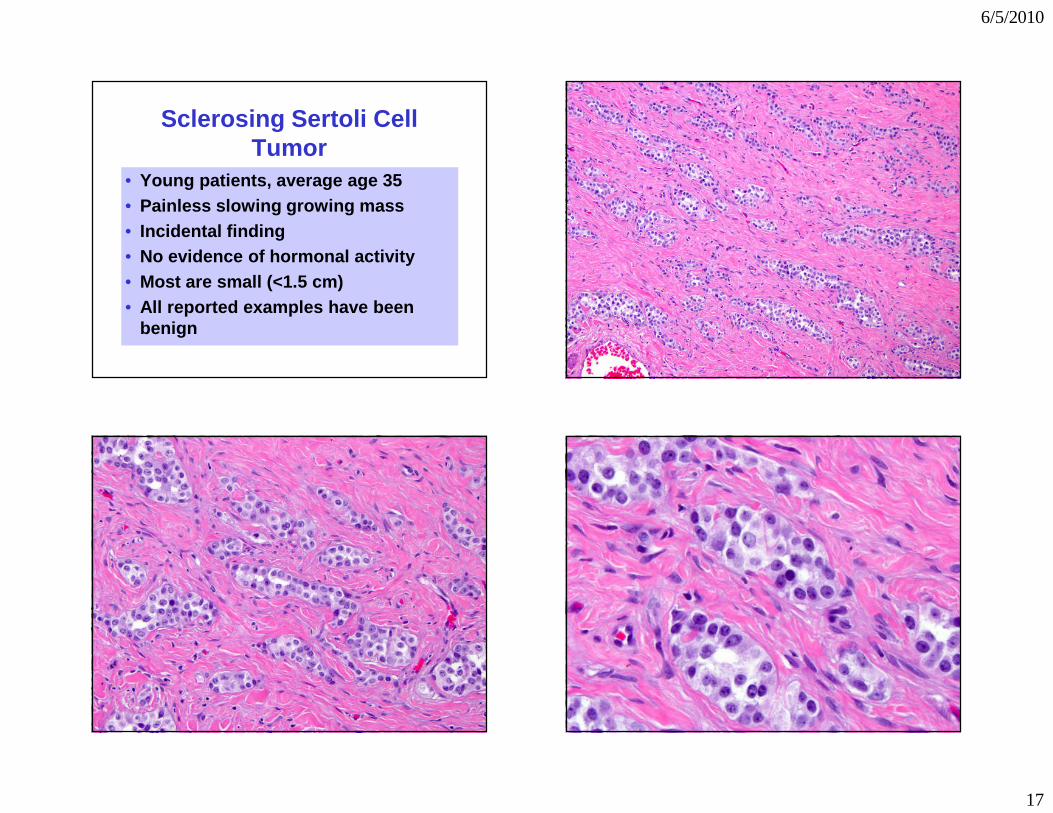
Sclerosing Sertoli Cell Tumor
• Young patients, average age 35• Painless slowing growing mass• Incidental finding• No evidence of hormonal activity• Most are small (<1.5 cm)• All reported examples have been
benign
6/5/2010
18
Large Cell Calcifying Sertoli Cell Tumor
• Patients are young, with an average age of 16 years
• Can occur over a wide range, from children to older adults
• Usual presentation is with a painless slowly enlarging testicular mass
• Hormones secreted by some tumors, or the surrounding Leydig cells, can cause symptoms such as gynecomastia or precocious pseudopuberty
• About 40% of patients have the Carney syndrome or Peutz-Jeghers syndrome
Carney Complex
• Many possible findings, listed in handout
• Complex has a genetic basis• LCCSCT in Carney patients occur at
a young age• LCCSCT are small and tend to be
bilateral and multifocal
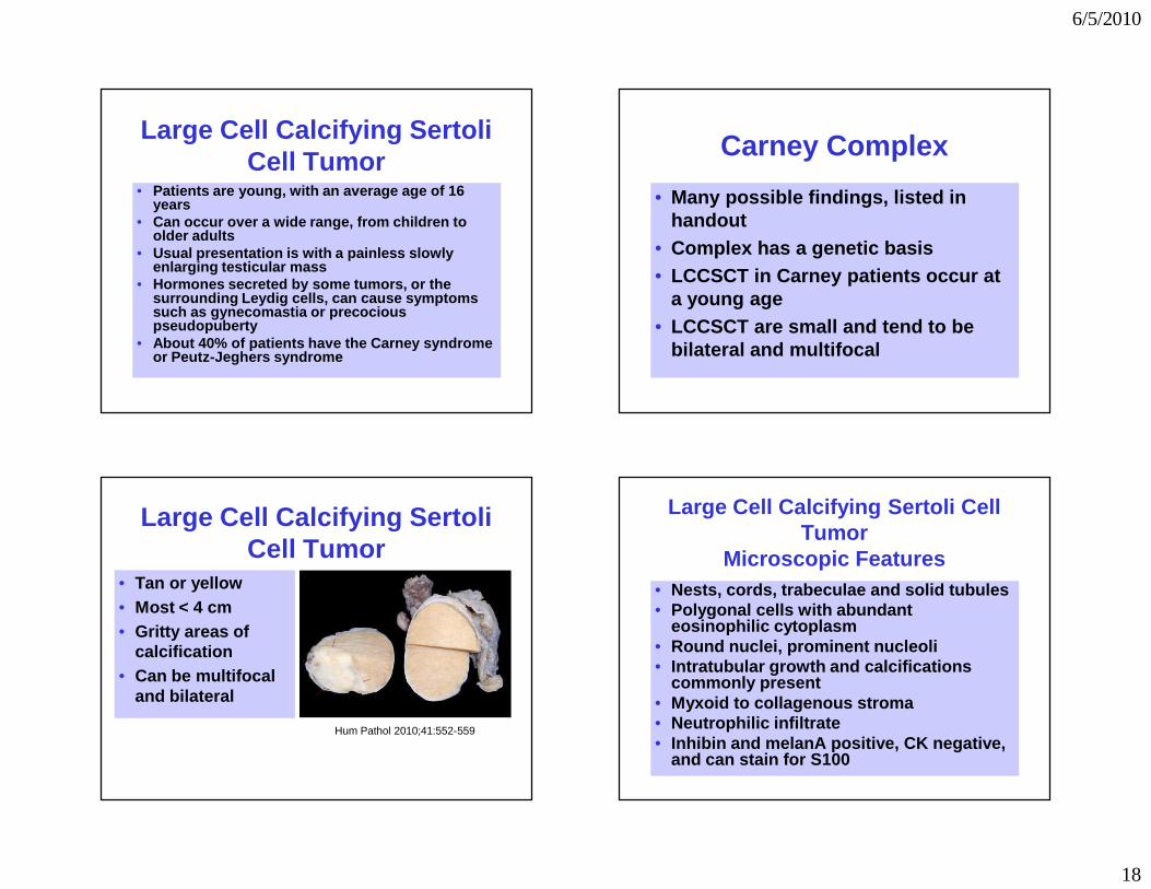
Large Cell Calcifying Sertoli Cell Tumor
• Tan or yellow• Most < 4 cm• Gritty areas of
calcification• Can be multifocal
and bilateral
Hum Pathol 2010;41:552-559
Large Cell Calcifying Sertoli Cell Tumor
Microscopic Features• Nests, cords, trabeculae and solid tubules• Polygonal cells with abundant
eosinophilic cytoplasm• Round nuclei, prominent nucleoli• Intratubular growth and calcifications
commonly present• Myxoid to collagenous stroma• Neutrophilic infiltrate• Inhibin and melanA positive, CK negative,
and can stain for S100
6/5/2010
19
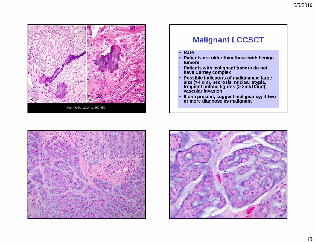
Hum Pathol 2010;41:552-559
Malignant LCCSCT• Rare• Patients are older than those with benign
tumors• Patients with malignant tumors do not
have Carney complex• Possible indicators of malignancy: large
size (>4 cm), necrosis, nuclear atypia, frequent mitotic figures (> 3mf/10hpf), vascular invasion
• If one present, suggest malignancy; if two or more diagnose as malignant
6/5/2010
20
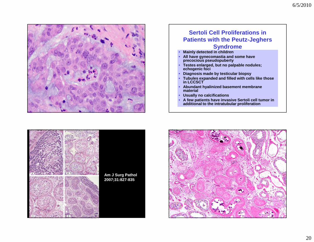
Sertoli Cell Proliferations in Patients with the Peutz-Jeghers
Syndrome• Mainly detected in children• All have gynecomastia and some have
precocious pseudopuberty• Testes enlarged, but no palpable nodules;
echogenic foci• Diagnosis made by testicular biopsy• Tubules expanded and filled with cells like those
in LCCSCT• Abundant hyalinized basement membrane
material• Usually no calcifications• A few patients have invasive Sertoli cell tumor in
additional to the intratubular proliferation
Am J Surg Pathol 2007;31:827-835
6/5/2010
21
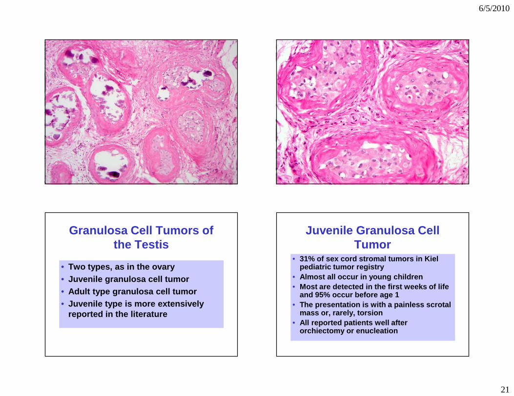
Granulosa Cell Tumors of the Testis
• Two types, as in the ovary• Juvenile granulosa cell tumor• Adult type granulosa cell tumor• Juvenile type is more extensively
reported in the literature
Juvenile Granulosa Cell Tumor
• 31% of sex cord stromal tumors in Kiel pediatric tumor registry
• Almost all occur in young children• Most are detected in the first weeks of life
and 95% occur before age 1• The presentation is with a painless scrotal
mass or, rarely, torsion• All reported patients well after
orchiectomy or enucleation
6/5/2010
22
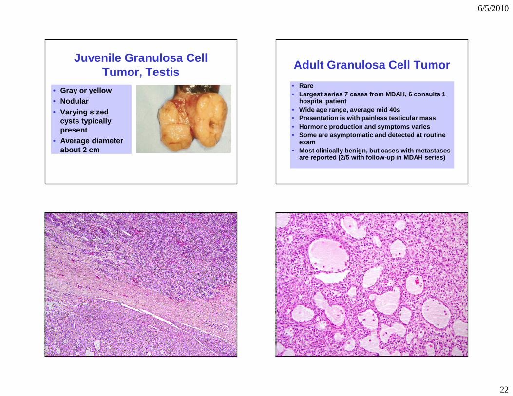
Juvenile Granulosa Cell Tumor, Testis
• Gray or yellow• Nodular• Varying sized
cysts typically present
• Average diameter about 2 cm
Adult Granulosa Cell Tumor
• Rare• Largest series 7 cases from MDAH, 6 consults 1
hospital patient• Wide age range, average mid 40s• Presentation is with painless testicular mass• Hormone production and symptoms varies• Some are asymptomatic and detected at routine
exam• Most clinically benign, but cases with metastases
are reported (2/5 with follow-up in MDAH series)
6/5/2010
23
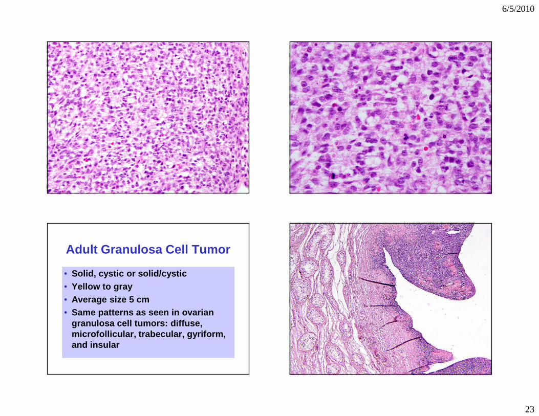
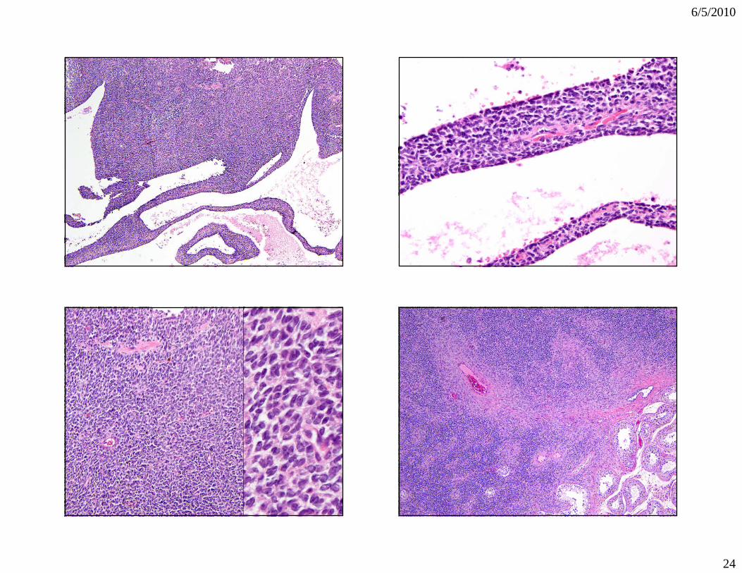
Adult Granulosa Cell Tumor
• Solid, cystic or solid/cystic• Yellow to gray• Average size 5 cm• Same patterns as seen in ovarian
granulosa cell tumors: diffuse, microfollicular, trabecular, gyriform, and insular
6/5/2010
24
6/5/2010
25
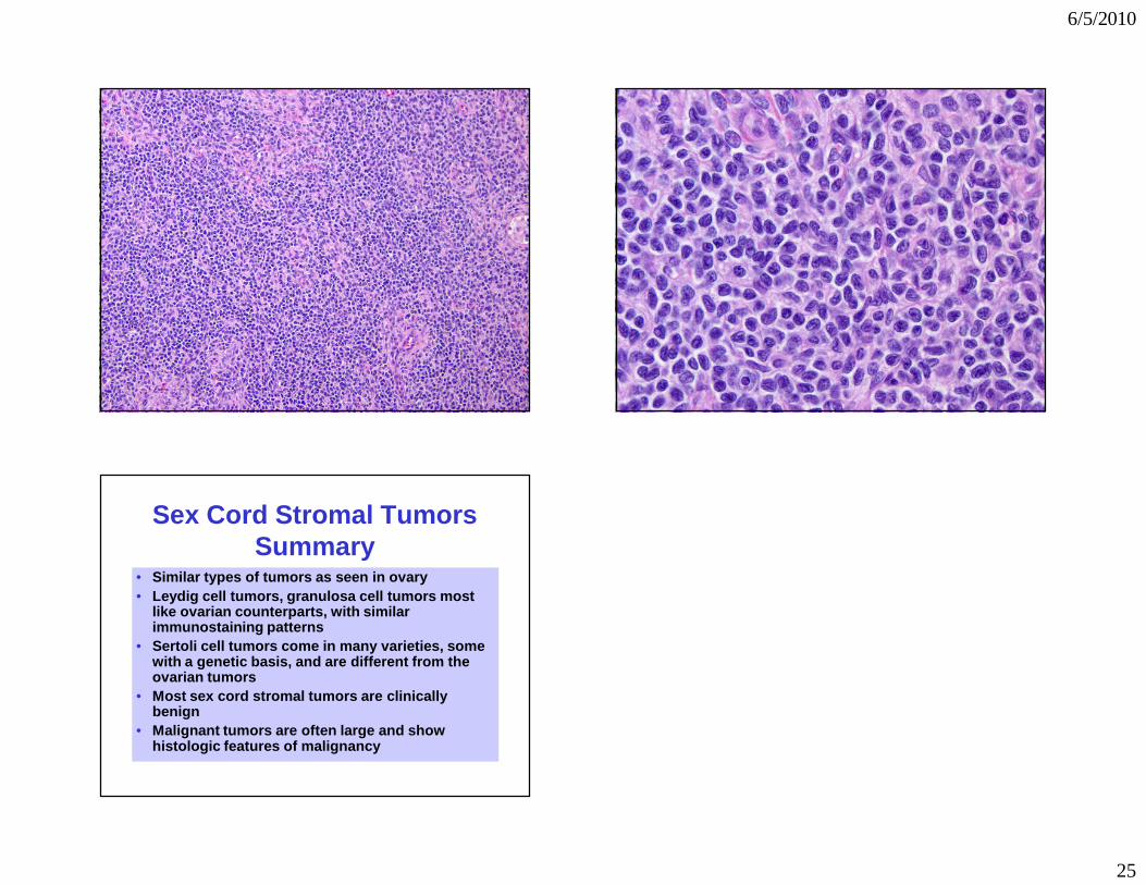
Sex Cord Stromal TumorsSummary
• Similar types of tumors as seen in ovary• Leydig cell tumors, granulosa cell tumors most
like ovarian counterparts, with similar immunostaining patterns
• Sertoli cell tumors come in many varieties, some with a genetic basis, and are different from the ovarian tumors
• Most sex cord stromal tumors are clinically benign
• Malignant tumors are often large and show histologic features of malignancy