Embed Size (px)
Citation preview
CUAJ • April 2010 • Volume 4, Issue 2© 2010 Canadian Urological Association
E19
Canadian consensus guidelines for the management of testiculargerm cell cancer
Can Urol Assoc J 2010;4(2):E19-E38
Testicular tumours are uncommon but constitute animportant group of malignancies in young men.Worldwide, it is estimated that there were more than
48 500 new cases and 8900 deaths from the disease in2002.1 The vast majority are primary germ cell tumours(GCTs) and the incidence has doubled in the past 30 years(with most of the increase in seminomas).2 While mostpatients present with early-stage and highly curable dis-ease, the continued rise in the incidence of these tumourspresents a major challenge.
Germ cell testicular tumours are the most common solidmalignancies in males between the ages of 20 and 35; it isestimated that in 2008 there will be 900 new cases and 30deaths from testicular cancer in Canada.3
Germ cell cancer is a rare disease that requires experttreatment. Clear evidence has emerged that patients withgerm cell cancer benefit from treatment in centres withspecial experience in the field.4 However, it is also of con-siderable importance that clear, comprehensive and up-to-date consensus guidelines are available which representthe current “state of the art” in diagnosis and managementof germ cell cancer. The European Germ Cell CancerConsensus Group published guidelines in 2004 (updatedin 2008) and these reflect the “European” approach to man-agement of patients with GCTs.5-7 In October 2007, the 1stCanadian Germ Cell Cancer Consensus Conference washeld in Toronto with support from the Canadian Partnershipagainst Cancer (CPAC), the Canadian Institute of HealthResearch, multiple provincial cancer agencies, the Dell’ElceTesticular Cancer Research Fund from the Princess MargaretFoundation and industry sponsors. The initiative wasendorsed by the Canadian Urological Association, theCanadian Association of Medical Oncologists and theCanadian Association of Radiation Oncologists. There were
a total of 46 attendees from across Canada and interna-tional invitees (Dr. Peter Albers, Dr. Robert Huddart andDr. Craig Nichols). The group reviewed and discussed thecurrent literature and the Canadian experience with germcell cancer. The group developed this Canadian ConsensusGuideline to cover many aspects of the diagnosis and man-agement of germ cell cancer.
1. Diagnosis and staging
Clinical presentation of germ cell tumour
Most patients present with a primary tumour in the testis.Delay in diagnosing germ cell cancer, which has been shownto affect outcome, may be caused either by patients whoignore symptoms for too long or by physicians who fail tomake the correct diagnosis.8 In a minority of patients, theprimary tumour manifestation is located extragonadally (i.e.,in the retroperitoneum or in the mediastinum).5
Consensus recommendations
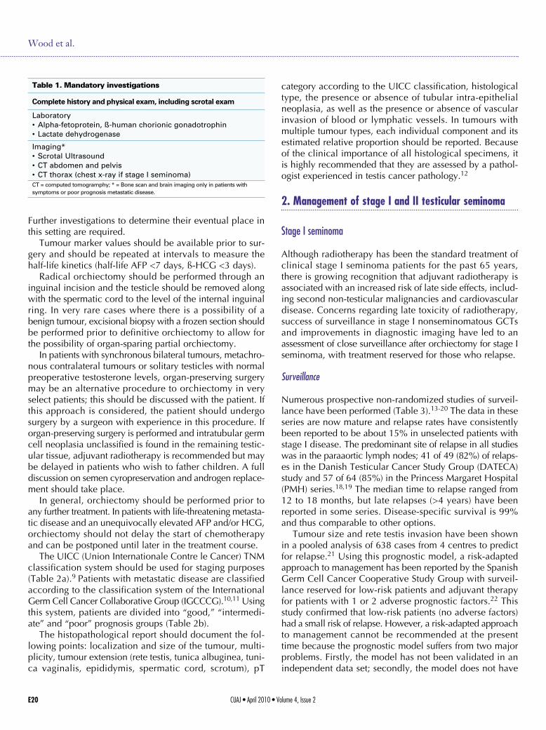
There are mandatory diagnostic and staging examinations(Table 1). These include scrotal examination, determina-tion of the serum tumour markers alpha-fetoprotein (AFP),ß-human chorionic gonadotrophin (HCG) and lactate dehy-drogenase (LDH), scrotal ultrasound to image the testis,computed tomography (CT) scan of the thorax, abdomenand pelvis (chest x-ray should be used instead of CT tho-rax in stage I seminoma). Bone scan and CT scan of thebrain are indicated in patients with symptoms suggestiveof central nervous system or bone involvement and in patientswith poor prognosis disease. Other imaging procedures,such as magnetic resonance imaging (MRI) and positronemission tomography (PET), should not be routinely used.
Lori Wood, MD;* Christian Kollmannsberger, MD, FRCSC;† Michael Jewett, MD, FRCSC;‡ Peter Chung, MD;§Sebastian Hotte, MD, FRCSC;± Martin O’Malley, MD;¥ Joan Sweet, MD;# Lynn Anson-Cartwright, CCRA;§ EricWinquist, MD, FRCSC;β Scott North, MD, FRCSC;α Scott Tyldesley, MD;Δ Jeremy Sturgeon, MD, FRCSC;µ MaryGospodarowicz, MD, FRCSC;§ Roanne Segal, MD;** Tina Cheng, MD;† Peter Venner, MD, FRCSC;α MalcolmMoore, MD, FRCSC;†† Peter Albers, MD;‡‡ Robert Huddart, MD;§§ Craig Nichols, MD;±± Padraig Warde, MB§
CONSENSUS GUIDELINE
CUAJ • April 2010 • Volume 4, Issue 2E20
Wood et al.
Further investigations to determine their eventual place inthis setting are required.
Tumour marker values should be available prior to sur-gery and should be repeated at intervals to measure thehalf-life kinetics (half-life AFP <7 days, ß-HCG <3 days).
Radical orchiectomy should be performed through aninguinal incision and the testicle should be removed alongwith the spermatic cord to the level of the internal inguinalring. In very rare cases where there is a possibility of abenign tumour, excisional biopsy with a frozen section shouldbe performed prior to definitive orchiectomy to allow forthe possibility of organ-sparing partial orchiectomy.
In patients with synchronous bilateral tumours, metachro-nous contralateral tumours or solitary testicles with normalpreoperative testosterone levels, organ-preserving surgerymay be an alternative procedure to orchiectomy in veryselect patients; this should be discussed with the patient. Ifthis approach is considered, the patient should undergosurgery by a surgeon with experience in this procedure. Iforgan-preserving surgery is performed and intratubular germcell neoplasia unclassified is found in the remaining testic-ular tissue, adjuvant radiotherapy is recommended but maybe delayed in patients who wish to father children. A fulldiscussion on semen cyropreservation and androgen replace-ment should take place.
In general, orchiectomy should be performed prior toany further treatment. In patients with life-threatening metasta-tic disease and an unequivocally elevated AFP and/or HCG,orchiectomy should not delay the start of chemotherapyand can be postponed until later in the treatment course.
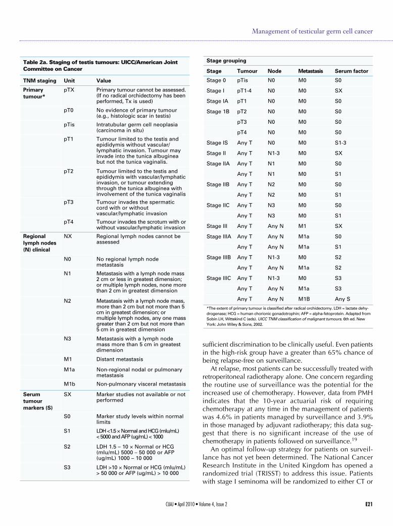
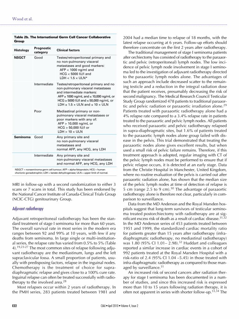
The UICC (Union Internationale Contre le Cancer) TNMclassification system should be used for staging purposes(Table 2a).9 Patients with metastatic disease are classifiedaccording to the classification system of the InternationalGerm Cell Cancer Collaborative Group (IGCCCG).10,11 Usingthis system, patients are divided into “good,” “intermedi-ate” and “poor” prognosis groups (Table 2b).
The histopathological report should document the fol-lowing points: localization and size of the tumour, multi-plicity, tumour extension (rete testis, tunica albuginea, tuni-ca vaginalis, epididymis, spermatic cord, scrotum), pT
category according to the UICC classification, histologicaltype, the presence or absence of tubular intra-epithelialneoplasia, as well as the presence or absence of vascularinvasion of blood or lymphatic vessels. In tumours withmultiple tumour types, each individual component and itsestimated relative proportion should be reported. Becauseof the clinical importance of all histological specimens, itis highly recommended that they are assessed by a pathol-ogist experienced in testis cancer pathology.12
2. Management of stage I and II testicular seminoma
Stage I seminoma
Although radiotherapy has been the standard treatment ofclinical stage I seminoma patients for the past 65 years,there is growing recognition that adjuvant radiotherapy isassociated with an increased risk of late side effects, includ-ing second non-testicular malignancies and cardiovasculardisease. Concerns regarding late toxicity of radiotherapy,success of surveillance in stage I nonseminomatous GCTsand improvements in diagnostic imaging have led to anassessment of close surveillance after orchiectomy for stage Iseminoma, with treatment reserved for those who relapse.
Surveillance
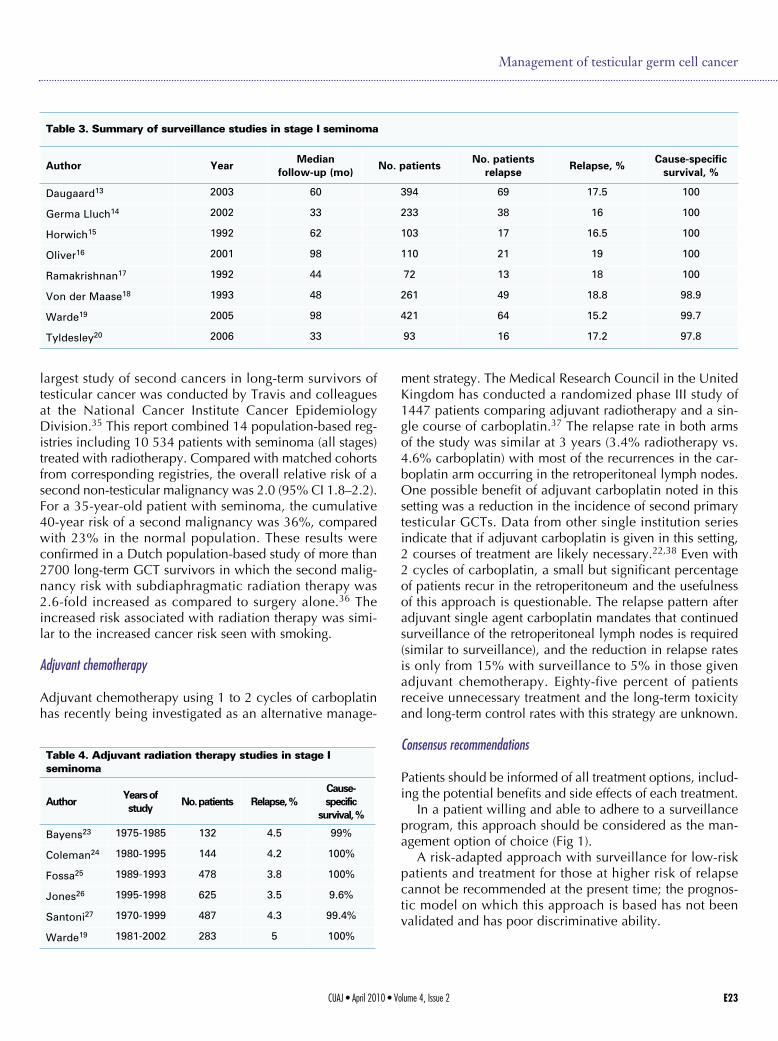
Numerous prospective non-randomized studies of surveil-lance have been performed (Table 3).13-20 The data in theseseries are now mature and relapse rates have consistentlybeen reported to be about 15% in unselected patients withstage I disease. The predominant site of relapse in all studieswas in the paraaortic lymph nodes; 41 of 49 (82%) of relaps-es in the Danish Testicular Cancer Study Group (DATECA)study and 57 of 64 (85%) in the Princess Margaret Hospital(PMH) series.18,19 The median time to relapse ranged from12 to 18 months, but late relapses (>4 years) have beenreported in some series. Disease-specific survival is 99%and thus comparable to other options.
Tumour size and rete testis invasion have been shownin a pooled analysis of 638 cases from 4 centres to predictfor relapse.21 Using this prognostic model, a risk-adaptedapproach to management has been reported by the SpanishGerm Cell Cancer Cooperative Study Group with surveil-lance reserved for low-risk patients and adjuvant therapyfor patients with 1 or 2 adverse prognostic factors.22 Thisstudy confirmed that low-risk patients (no adverse factors)had a small risk of relapse. However, a risk-adapted approachto management cannot be recommended at the presenttime because the prognostic model suffers from two majorproblems. Firstly, the model has not been validated in anindependent data set; secondly, the model does not have
Table 1. Mandatory investigations
Complete history and physical exam, including scrotal exam
Laboratory• Alpha-fetoprotein, ß-human chorionic gonadotrophin• Lactate dehydrogenase
Imaging*• Scrotal Ultrasound• CT abdomen and pelvis• CT thorax (chest x-ray if stage I seminoma)CT = computed tomogramphy; * = Bone scan and brain imaging only in patients withsymptoms or poor prognosis metastatic disease.
CUAJ • April 2010 • Volume 4, Issue 2 E21
sufficient discrimination to be clinically useful. Even patientsin the high-risk group have a greater than 65% chance ofbeing relapse-free on surveillance.
At relapse, most patients can be successfully treated withretroperitoneal radiotherapy alone. One concern regardingthe routine use of surveillance was the potential for theincreased use of chemotherapy. However, data from PMHindicates that the 10-year actuarial risk of requiringchemotherapy at any time in the management of patientswas 4.6% in patients managed by surveillance and 3.9%in those managed by adjuvant radiotherapy; this data sug-gest that there is no significant increase of the use ofchemotherapy in patients followed on surveillance.19
An optimal follow-up strategy for patients on surveil-lance has not yet been determined. The National CancerResearch Institute in the United Kingdom has opened arandomized trial (TRISST) to address this issue. Patientswith stage I seminoma will be randomized to either CT or
Management of testicular germ cell cancer
Table 2a. Staging of testis tumours: UICC/American JointCommittee on Cancer
TNM staging Unit Value
Primarytumour*
pTX Primary tumour cannot be assessed.(If no radical orchidectomy has beenperformed, Tx is used)
pT0 No evidence of primary tumour(e.g., histologic scar in testis)
pTis Intratubular germ cell neoplasia(carcinoma in situ)
pT1 Tumour limited to the testis andepididymis without vascular/lymphatic invasion. Tumour mayinvade into the tunica albugineabut not the tunica vaginalis.
pT2 Tumour limited to the testis andepididymis with vascular/lymphaticinvasion, or tumour extendingthrough the tunica albuginea withinvolvement of the tunica vaginalis
pT3 Tumour invades the spermaticcord with or withoutvascular/lymphatic invasion
pT4 Tumour invades the scrotum with orwithout vascular/lymphatic invasion
Regionallymph nodes(N) clinical
NX Regional lymph nodes cannot beassessed
N0 No regional lymph nodemetastasis
N1 Metastasis with a lymph node mass2 cm or less in greatest dimension;or multiple lymph nodes, none morethan 2 cm in greatest dimension
N2 Metastasis with a lymph node mass,more than 2 cm but not more than 5cm in greatest dimension; ormultiple lymph nodes, any one massgreater than 2 cm but not more than5 cm in greatest dimension
N3 Metastasis with a lymph nodemass more than 5 cm in greatestdimension
M1 Distant metastasis
M1a Non-regional nodal or pulmonarymetastasis
M1b Non-pulmonary visceral metastasis
Serumtumourmarkers (S)
SX Marker studies not available or notperformed
S0 Marker study levels within normallimits
S1 LDH <1.5 × Normal and HCG (mIu/mL) < 5000 and AFP (ug/mL) < 1000
S2 LDH 1.5 – 10 × Normal or HCG(mIu/mL) 5000 – 50 000 or AFP(ug/mL) 1000 – 10 000
S3 LDH >10 × Normal or HCG (mIu/mL)> 50 000 or AFP (ug/mL) > 10 000
Stage grouping
Stage Tumour Node Metastasis Serum factor
Stage 0 pTis N0 M0 S0
Stage I pT1-4 N0 M0 SX
Stage IA pT1 N0 M0 S0
Stage 1B pT2 N0 M0 S0
pT3 N0 M0 S0
pT4 N0 M0 S0
Stage IS Any T N0 M0 S1-3
Stage II Any T N1-3 M0 SX
Stage IIA Any T N1 M0 S0
Any T N1 M0 S1
Stage IIB Any T N2 M0 S0
Any T N2 M0 S1
Stage IIC Any T N3 M0 S0
Any T N3 M0 S1
Stage III Any T Any N M1 SX
Stage IIIA Any T Any N M1a S0
Any T Any N M1a S1
Stage IIIB Any T N1-3 M0 S2
Any T Any N M1a S2
Stage IIIC Any T N1-3 M0 S3
Any T Any N M1a S3
Any T Any N M1B Any S
*The extent of primary tumour is classified after radical orchidectomy. LDH = lactate dehy-drogenase; HCG = human chorionic gonadotrophin; AFP = alpha-fetoprotein. Adapted fromSobin LH, Wittekind C (eds). UICC TNM classification of malignant tumours. 6th ed. NewYork: John Wiley & Sons, 2002.
MRI in follow-up with a second randomization to either 3scans or 7 scans in total. This study has been endorsed bythe National Cancer Institute of Canada-Clinical Trials Group(NCIC-CTG) genitourinary Group.
Adjuvant radiotherapy
Adjuvant retroperitoneal radiotherapy has been the stan-dard treatment of stage I seminoma for more than 60 years.The overall survival rate in most series in the modern eraranges between 92 and 99% at 10 years, with few if anydeaths from seminoma. In large single or multi-institution-al series, the relapse rate has varied from 0.5% to 5% (Table4).19,23-27 The most common sites of relapse following adju-vant radiotherapy are the mediastinum, lungs and the leftsupraclavicular fossa. A small proportion of patients, usu-ally with predisposing factors, relapse in the inguinal nodes.Chemotherapy is the treatment of choice for supra-diaphragmatic relapse and gives close to a 100% cure rate.Inguinal relapse can often be treated successfully with radio-therapy to the involved area.28
Most relapses occur within 2 years of radiotherapy. Inthe PMH series, 283 patients treated between 1981 and
2004 had a median time to relapse of 18 months, with thelatest relapse occurring at 6 years. Follow-up efforts shouldtherefore concentrate on the first 2 years after radiotherapy.
The traditional management of stage I seminoma patientsafter orchiectomy has consisted of radiotherapy to the paraaor-tic and pelvic (retroperitoneal) lymph nodes. The low inci-dence of pelvic lymph node involvement in stage I semino-ma led to the investigation of adjuvant radiotherapy directedto the paraaortic lymph nodes alone. The advantages ofsuch an approach include decreased scatter to the remain-ing testicle and a reduction in the integral radiation dosethat the patient receives, presumably decreasing the risk ofsecond malignancy. The Medical Research Council TesticularStudy Group randomized 478 patients to traditional paraaor-tic and pelvic radiation or paraaortic irradiation alone.29
Patients treated with paraaortic radiotherapy alone had a4% relapse rate compared to a 3.4% relapse rate in patientstreated to the paraaortic and pelvic lymph nodes. All patientswho received paraaortic and pelvic radiotherapy relapsedin supra-diaphragmatic sites, but 1.6% of patients treatedto the paraaortic lymph nodes alone group failed with dis-ease in the pelvis. This trial demonstrated that treating theparaaortic nodes alone gives excellent results, but whenused a small risk of pelvic failure remains. Therefore, if thistreatment approach is adopted, regular imaging with CT ofthe pelvic lymph nodes must be performed to ensure that ifpelvic relapse occurs, it is detected at an early stage. Datafrom the Christie Hospital in Manchester, United Kingdom,where no routine evaluation of the pelvis is carried out afterparaaortic radiation alone, has shown that the median sizeof the pelvic lymph nodes at time of detection of relapse is5 cm (range 2.5 to 9 cm).30 The advantage of paraaorticradiotherapy alone is therefore not clear, particularly in com-parison to surveillance.
Data from the MD Anderson and the Royal Marsden hos-pitals suggest that long-term survivors of testicular semino-ma treated postorchiectomy with radiotherapy are at sig-nificant excess risk of death as a result of cardiac disease.31,32
In the MD Anderson series of 453 patients treated between1951 and 1999, the standardized cardiac mortality ratiofor patients greater than 15 years after radiotherapy (infra-diaphragmatic radiotherapy, no mediastinal radiotherapy)was 1.80 (95% CI 1.01– 2.98).32 Huddart and colleaguesreported a similar increase in cardiac events in a cohort of992 patients treated at the Royal Marsden Hospital with arisk-ratio of 2.4 (95% CI 1.04 –5.45) in those treated withinfra-diaphragmatic radiotherapy as compared to those man-aged by surveillance.31
An increased risk of second cancers after radiation ther-apy for stage I seminoma has been documented in a num-ber of studies, and since this increased risk is expressedmore than 10 to 15 years following radiation therapy, it isoften not apparent in series with shorter follow-up.33,34 The
CUAJ • April 2010 • Volume 4, Issue 2E22
Wood et al.
Table 2b. The International Germ Cell Cancer CollaborativeGroup
HistologyPrognosticcategory
Clinical factors
NSGCT Good Testes/retroperitoneal primary andno non-pulmonary visceralmetastases and good markers:AFP < 1000 ng/ml andHCG < 5000 IU/l andLDH < 1.5 × ULN*
Intermediate Testes/retroperitoneal primary and nonon-pulmonary visceral metastasesand intermediate markers:AFP ≥ 1000 ng/mL and ≤ 10,000 ng/mL orHCG ≥ 5000 IU/l and ≤ 50,000 ng/mL orLDH ≥ 1.5 × ULN and ≤ 10 × ULN
Poor Mediastinal primary or non-pulmonary visceral metastases orpoor markers with any of:AFP > 10,000 ng/mL orHCG > 50,000 IU/l orLDH > 10 × ULN
Seminoma Good Any primary site andno non-pulmonary visceralmetastases andnormal AFP, any HCG, any LDH
Intermediate Any primary site andnon-pulmonary visceral metastasesand normal AFP, any HCG, any LDH
NSGCT = nonseminoma germ cell tumour; AFP = alpha-fetoprotein; HCG = human chorionic gonadotrophin; LDH = lactate dehydrogenase; ULN = upper limit of normal.
CUAJ • April 2010 • Volume 4, Issue 2 E23
largest study of second cancers in long-term survivors oftesticular cancer was conducted by Travis and colleaguesat the National Cancer Institute Cancer EpidemiologyDivision.35 This report combined 14 population-based reg-istries including 10 534 patients with seminoma (all stages)treated with radiotherapy. Compared with matched cohortsfrom corresponding registries, the overall relative risk of asecond non-testicular malignancy was 2.0 (95% CI 1.8–2.2).For a 35-year-old patient with seminoma, the cumulative40-year risk of a second malignancy was 36%, comparedwith 23% in the normal population. These results wereconfirmed in a Dutch population-based study of more than2700 long-term GCT survivors in which the second malig-nancy risk with subdiaphragmatic radiation therapy was2.6-fold increased as compared to surgery alone.36 Theincreased risk associated with radiation therapy was simi-lar to the increased cancer risk seen with smoking.
Adjuvant chemotherapy
Adjuvant chemotherapy using 1 to 2 cycles of carboplatinhas recently being investigated as an alternative manage-
ment strategy. The Medical Research Council in the UnitedKingdom has conducted a randomized phase III study of1447 patients comparing adjuvant radiotherapy and a sin-gle course of carboplatin.37 The relapse rate in both armsof the study was similar at 3 years (3.4% radiotherapy vs.4.6% carboplatin) with most of the recurrences in the car-boplatin arm occurring in the retroperitoneal lymph nodes.One possible benefit of adjuvant carboplatin noted in thissetting was a reduction in the incidence of second primarytesticular GCTs. Data from other single institution seriesindicate that if adjuvant carboplatin is given in this setting,2 courses of treatment are likely necessary.22,38 Even with2 cycles of carboplatin, a small but significant percentageof patients recur in the retroperitoneum and the usefulnessof this approach is questionable. The relapse pattern afteradjuvant single agent carboplatin mandates that continuedsurveillance of the retroperitoneal lymph nodes is required(similar to surveillance), and the reduction in relapse ratesis only from 15% with surveillance to 5% in those givenadjuvant chemotherapy. Eighty-five percent of patientsreceive unnecessary treatment and the long-term toxicityand long-term control rates with this strategy are unknown.
Consensus recommendations
Patients should be informed of all treatment options, includ-ing the potential benefits and side effects of each treatment.
In a patient willing and able to adhere to a surveillanceprogram, this approach should be considered as the man-agement option of choice (Fig 1).
A risk-adapted approach with surveillance for low-riskpatients and treatment for those at higher risk of relapsecannot be recommended at the present time; the prognos-tic model on which this approach is based has not beenvalidated and has poor discriminative ability.
Management of testicular germ cell cancer
Table 3. Summary of surveillance studies in stage I seminoma
Author YearMedian
follow-up (mo)No. patients
No. patientsrelapse
Relapse, %Cause-specific
survival, %
Daugaard13 2003 60 394 69 17.5 100
Germa Lluch14 2002 33 233 38 16 100
Horwich15 1992 62 103 17 16.5 100
Oliver16 2001 98 110 21 19 100
Ramakrishnan17 1992 44 72 13 18 100
Von der Maase18 1993 48 261 49 18.8 98.9
Warde19 2005 98 421 64 15.2 99.7
Tyldesley20 2006 33 93 16 17.2 97.8
Table 4. Adjuvant radiation therapy studies in stage Iseminoma
AuthorYears ofstudy
No. patients Relapse, %Cause-specific
survival, %
Bayens23 1975-1985 132 4.5 99%
Coleman24 1980-1995 144 4.2 100%
Fossa25 1989-1993 478 3.8 100%
Jones26 1995-1998 625 3.5 9.6%
Santoni27 1970-1999 487 4.3 99.4%
Warde19 1981-2002 283 5 100%
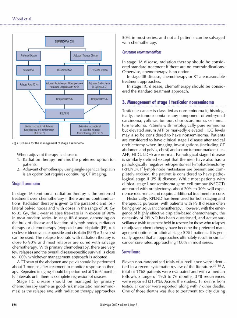
When adjuvant therapy is chosen: 1. Radiation therapy remains the preferred option for
patients. 2. Adjuvant chemotherapy using single-agent carboplatin
is an option but requires continuing CT imaging.
Stage II seminoma
In stage IIA seminoma, radiation therapy is the preferredtreatment over chemotherapy if there are no contraindica-tions. Radiation therapy is given to the paraaortic and ipsi-lateral pelvic nodes and with doses in the range of 30 Gyto 35 Gy, the 5-year relapse free-rate is in excess of 90%in most modern series. In stage IIB disease, depending onthe bulk of disease and location of lymph nodes, radiationtherapy or chemotherapy (etoposide and cisplatin [EP] × 4cycles or bleomycin, etoposide and cisplatin [BEP] × 3 cycles)can be used. The relapse-free rate with radiation therapy isclose to 90% and most relapses are cured with salvagechemotherapy. With primary chemotherapy, there are veryfew relapses and the overall disease-specific survival is closeto 100% whichever management approach is adopted.
A CT scan of the abdomen and pelvis should be performedabout 3 months after treatment to monitor response to ther-apy. Repeated imaging should be performed at 3 to 6 month-ly intervals until there is complete regression of disease.
Stage IIC disease should be managed by primarychemotherapy (same as good-risk metastatic nonsemino-mas) as the relapse rate with radiation therapy approaches
50% in most series, and not all patients can be salvagedwith chemotherapy.
Consensus recommendations
In stage IIA disease, radiation therapy should be consid-ered standard treatment if there are no contraindications.Otherwise, chemotherapy is an option.
In stage IIB disease, chemotherapy or RT are reasonabletreatment approaches.
In stage IIC disease, chemotherapy should be consid-ered the standard treatment approach.
3. Management of stage I testicular nonseminoma
Testicular cancer is classified as nonseminoma if, histolog-ically, the tumour contains any component of embryonalcarcinoma, yolk sac tumour, choriocarcinoma, or imma-ture teratoma. Patients with histologically pure seminomabut elevated serum AFP or markedly elevated HCG levelsmay also be considered to have nonseminoma. Patientsare considered to have clinical stage I disease after radicalorchiectomy when imaging investigations (including CTabdomen and pelvis, chest) and serum tumour markers (i.e.,AFP, HCG, LDH) are normal. Pathological stage I diseaseis similarly defined except that the men have also had apathologically negative retroperitoneal lymphadenectomy(RPLND). If lymph node metastases are present and com-pletely excised, the patient is considered to have patho-logical stage II (PS II) disease. While most patients withclinical stage I nonseminoma germ cell tumour (NSGCT)are cured with orchiectomy, about 20% to 30% will expe-rience recurrence and require additional treatment for cure.
Historically, RPLND has been used for both staging andtherapeutic purposes, with patients with PS II disease oftenbeing given adjuvant chemotherapy. However, with the emer-gence of highly effective cisplatin-based chemotherapy, thenecessity of RPLND has been questioned, and active sur-veillance (with treatment held in reserve for those who relapse)or adjuvant chemotherapy have become the preferred man-agement options for clinical stage (CS) I patients. It is gen-erally agreed that all approaches ultimately result in similarcancer cure rates, approaching 100% in most series.
Surveillance
Eleven non-randomized trials of surveillance were identi-fied in a recent systematic review of the literature.39-48 Atotal of 1768 patients were evaluated and with a medianfollow-up range of 19.5 to 76 months, 378 recurrenceswere reported (21.4%). Across the studies, 13 deaths fromtesticular cancer were reported, along with 7 other deaths.One of those deaths was due to treatment toxicity during
CUAJ • April 2010 • Volume 4, Issue 2E24
Wood et al.
SEMINOMA CS I
Preferred Option
Surveillance
Relapse Rate 15%
Possible Option
Adjuvant Radiotherapy of RetroperitonealPara-aortic Lympatics with 20 GY
Relapse Rate 5%
RELAPSE
Limited Locoregional Relapse:Radiotherapy or Chemotherapy
(BEP or EP)
Extensive Locoregionalor Systemic Relapse:
Chemotherapy (BEP or EP)
Relapse Rate 5%
Adjuvant Carboplantin(1 Cylce AUC 7)
Preferred Option
Adjuvant Therapy Chosen
Fig 1. Schema for the management of stage I seminoma.
CUAJ • April 2010 • Volume 4, Issue 2 E25
Management of testicular germ cell cancer
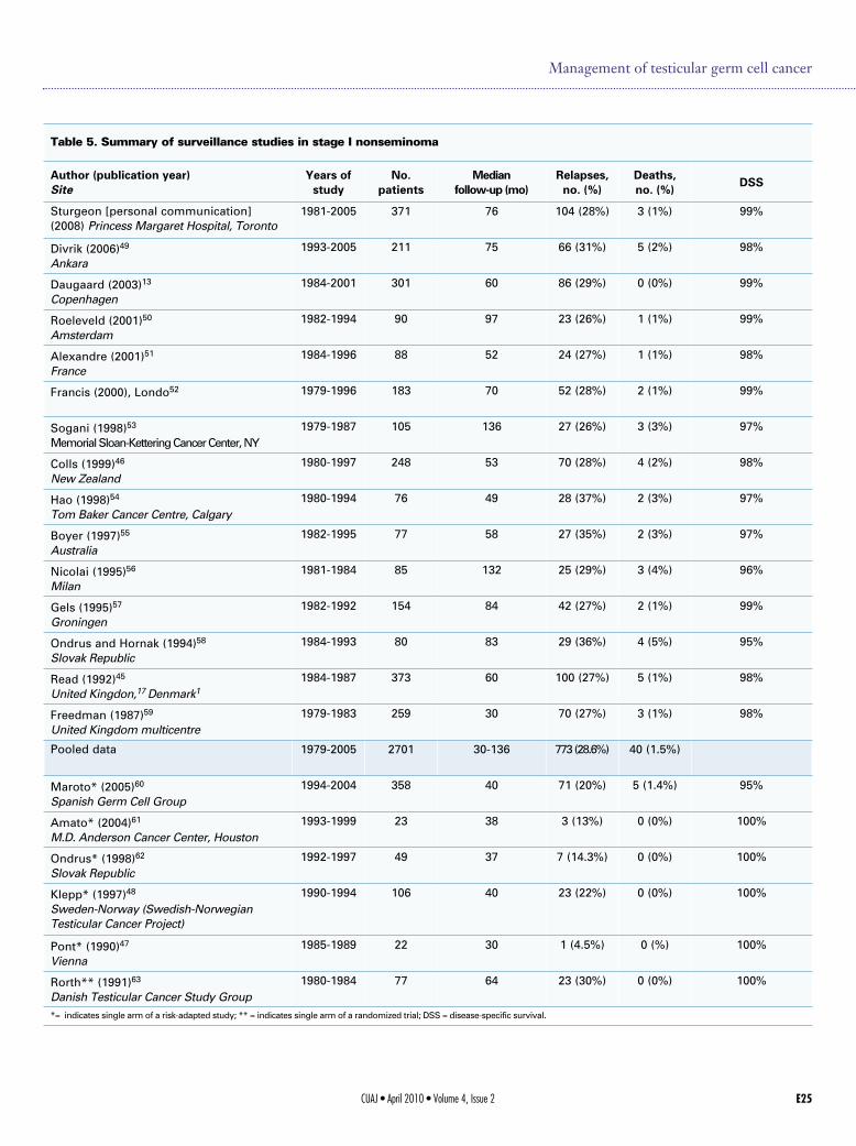
Table 5. Summary of surveillance studies in stage I nonseminoma
Author (publication year)Site
Years ofstudy
No.patients
Medianfollow-up (mo)
Relapses,no. (%)
Deaths,no. (%)
DSS
Sturgeon [personal communication](2008) Princess Margaret Hospital, Toronto
1981-2005 371 76 104 (28%) 3 (1%) 99%
Divrik (2006)49
Ankara1993-2005 211 75 66 (31%) 5 (2%) 98%
Daugaard (2003)13
Copenhagen1984-2001 301 60 86 (29%) 0 (0%) 99%
Roeleveld (2001)50
Amsterdam1982-1994 90 97 23 (26%) 1 (1%) 99%
Alexandre (2001)51
France1984-1996 88 52 24 (27%) 1 (1%) 98%
Francis (2000), Londo52 1979-1996 183 70 52 (28%) 2 (1%) 99%
Sogani (1998)53
Memorial Sloan-Kettering Cancer Center, NY1979-1987 105 136 27 (26%) 3 (3%) 97%
Colls (1999)46
New Zealand1980-1997 248 53 70 (28%) 4 (2%) 98%
Hao (1998)54
Tom Baker Cancer Centre, Calgary1980-1994 76 49 28 (37%) 2 (3%) 97%
Boyer (1997)55
Australia1982-1995 77 58 27 (35%) 2 (3%) 97%
Nicolai (1995)56
Milan1981-1984 85 132 25 (29%) 3 (4%) 96%
Gels (1995)57
Groningen1982-1992 154 84 42 (27%) 2 (1%) 99%
Ondrus and Hornak (1994)58
Slovak Republic1984-1993 80 83 29 (36%) 4 (5%) 95%
Read (1992)45
United Kingdon,17 Denmark11984-1987 373 60 100 (27%) 5 (1%) 98%
Freedman (1987)59
United Kingdom multicentre1979-1983 259 30 70 (27%) 3 (1%) 98%
Pooled data 1979-2005 2701 30-136 773 (28.6%) 40 (1.5%)
Maroto* (2005)60
Spanish Germ Cell Group1994-2004 358 40 71 (20%) 5 (1.4%) 95%
Amato* (2004)61
M.D. Anderson Cancer Center, Houston1993-1999 23 38 3 (13%) 0 (0%) 100%
Ondrus* (1998)62
Slovak Republic1992-1997 49 37 7 (14.3%) 0 (0%) 100%
Klepp* (1997)48
Sweden-Norway (Swedish-NorwegianTesticular Cancer Project)
1990-1994 106 40 23 (22%) 0 (0%) 100%
Pont* (1990)47
Vienna1985-1989 22 30 1 (4.5%) 0 (%) 100%
Rorth** (1991)63
Danish Testicular Cancer Study Group1980-1984 77 64 23 (30%) 0 (0%) 100%
*= indicates single arm of a risk-adapted study; ** = indicates single arm of a randomized trial; DSS = disease-specific survival.
salvage treatment. The survival outcomes are summarizedin Table 5.13,45-63 The presence of microscopic vascular orlymphatic invasion in the primary tumour is the most impor-tant factor predicting relapse and the presence or absenceof this factor has been used to divide patients: those withhigh-risk disease (a third of the cases) who have about a50% risk of relapse, and those with low-risk disease whohave about a 15% to 20% risk of relapse.45
Retroperiteonal lymph node dissection
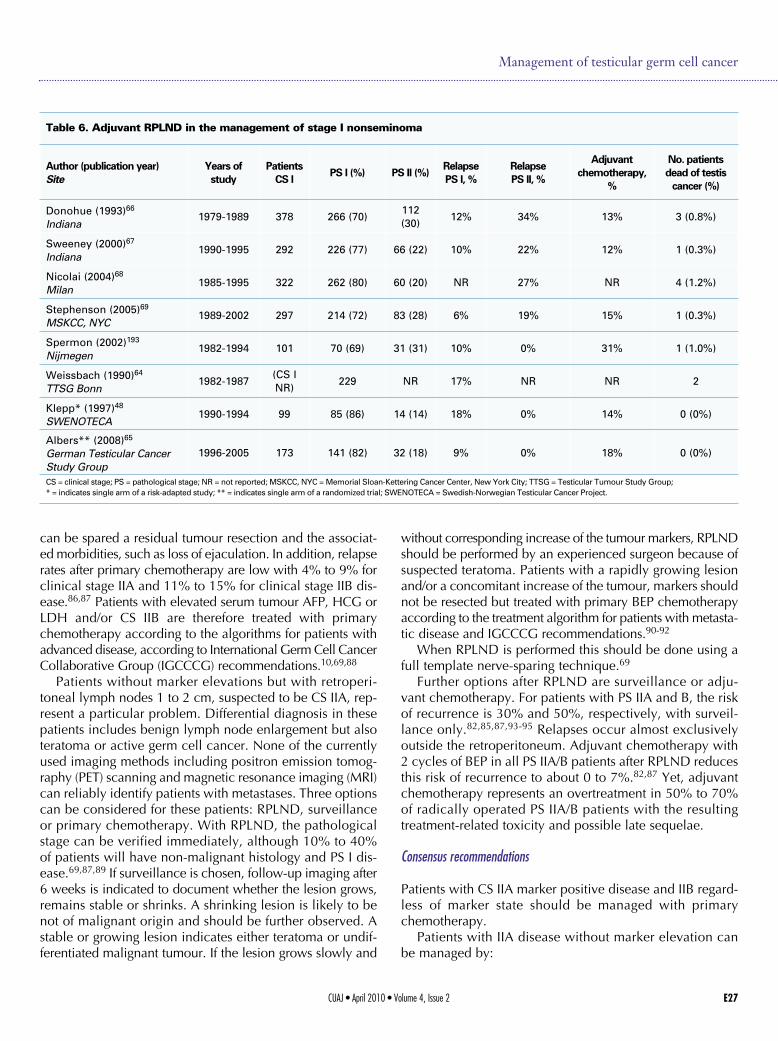
Two non-randomized studies of adjuvant RPLND in themanagement of stage I NSGCT were identified in a recentsystematic review of the literature.39,48,64 Across these stud-ies, 344 patients were followed for a median time rangingfrom 21 to 40 months, and a total of 41 recurrences werefound. There was 1 death from testicular cancer and 1 otherdeath from unrelated causes. In addition, Albers and col-leagues recently reported the results of a randomized clin-ical trial (RCT) comparing 1 course of BEP versus RPLNDin 382 patients with CS I RPLND (all risk groups).65 In the173 patients who received RPLND, 18.5% had stage II dis-ease at surgery and these patients were given 2 courses ofBEP. In those patients treated with adjuvant chemothera-py, no relapses were observed. In patients managed withRPLND alone, 13 recurrences were observed, 7 of whichoccurred in the retroperitoneum. Outcomes from publishedstudies are shown in Table 6.48,64-70
Adjuvant chemotherapy
One RCT and 7 non-randomized studies with 10 treatmentarms (total 873 evaluable patients) were identified in a recentmeta-analysis.39,48,65,71-75 Because the RCT compared adju-vant chemotherapy to RPLND, only the chemotherapy armwas included in the meta-analysis. Although the follow-up
times of the included studies varied, all had sufficient fol-low-up that almost all recurrences that would occur amongthese patients were included. Across the 8 studies, 23 recur-rences were reported, corresponding to an overall estimat-ed recurrence rate of 3.8% (95% CI, 2.6% to 5.5%; p =0.42; I2=2.6%). For patients treated with BEP or cisplatin,vinblastine, and bleomycin (PVB), the estimated recurrencerates were 3.9% (95% CI, 1.6% to 9%), 3.9% (95% CI,2.1% to 7%), and 7.2% (95% CI, 2.1% to 22.1%) for 1, 2,and 3 cycles of adjuvant chemotherapy, respectively. Tworecurrences with 2 cycles of BEP or PVB and 1 with 3 cyclesof BEP were pure mature teratoma. In the randomized trialby Albers and colleagues, one course BEP versus RPLND,the two-year recurrence-free survival was 99.46%.65 Theresults of the studies are summarized in Table 7.65,71,74-81
Consensus recommendations
Patients should be informed of all treatment options, includ-ing the potential benefits and side effects of each treatment.
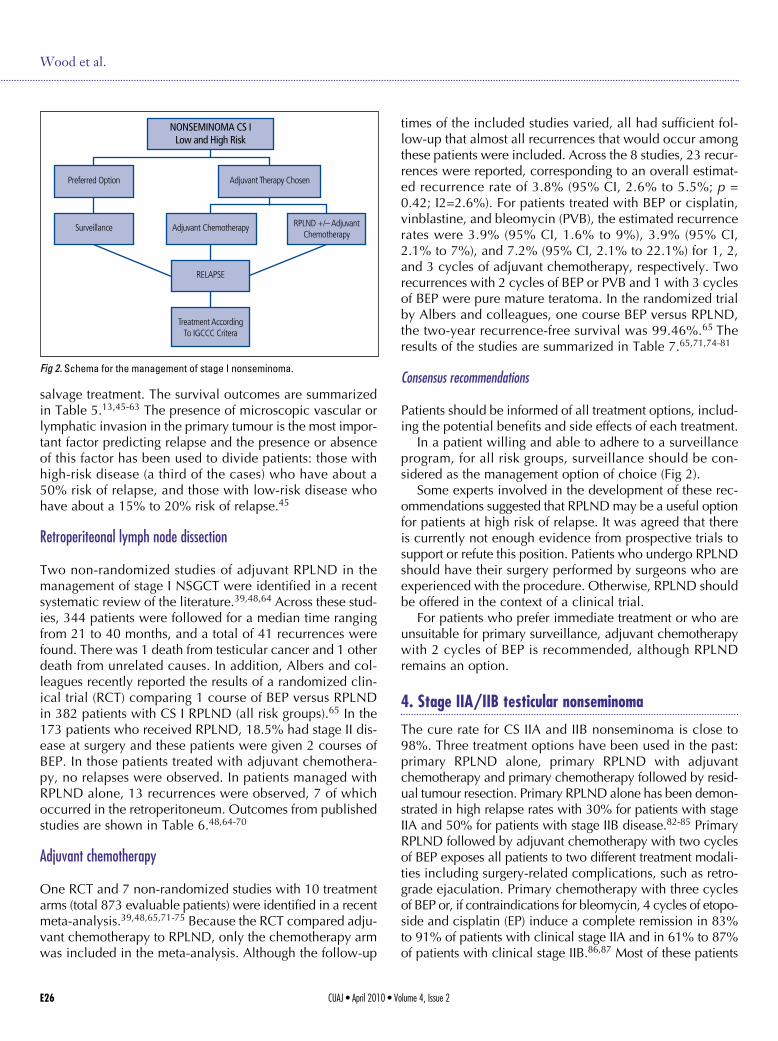
In a patient willing and able to adhere to a surveillanceprogram, for all risk groups, surveillance should be con-sidered as the management option of choice (Fig 2).
Some experts involved in the development of these rec-ommendations suggested that RPLND may be a useful optionfor patients at high risk of relapse. It was agreed that thereis currently not enough evidence from prospective trials tosupport or refute this position. Patients who undergo RPLNDshould have their surgery performed by surgeons who areexperienced with the procedure. Otherwise, RPLND shouldbe offered in the context of a clinical trial.
For patients who prefer immediate treatment or who areunsuitable for primary surveillance, adjuvant chemotherapywith 2 cycles of BEP is recommended, although RPLNDremains an option.
4. Stage IIA/IIB testicular nonseminoma
The cure rate for CS IIA and IIB nonseminoma is close to98%. Three treatment options have been used in the past:primary RPLND alone, primary RPLND with adjuvantchemotherapy and primary chemotherapy followed by resid-ual tumour resection. Primary RPLND alone has been demon-strated in high relapse rates with 30% for patients with stageIIA and 50% for patients with stage IIB disease.82-85 PrimaryRPLND followed by adjuvant chemotherapy with two cyclesof BEP exposes all patients to two different treatment modali-ties including surgery-related complications, such as retro-grade ejaculation. Primary chemotherapy with three cyclesof BEP or, if contraindications for bleomycin, 4 cycles of etopo-side and cisplatin (EP) induce a complete remission in 83%to 91% of patients with clinical stage IIA and in 61% to 87%of patients with clinical stage IIB.86,87 Most of these patients
CUAJ • April 2010 • Volume 4, Issue 2E26
Wood et al.
Fig 2. Schema for the management of stage I nonseminoma.
Figure 2: Schema for Management of Stage I Non-Seminoma
NONSEMINOMA CS ILow and High Risk
Preferred Option
Surveillance Adjuvant Chemotherapy
RELAPSE
Treatment AccordingTo IGCCC Critera
RPLND +/– AdjuvantChemotherapy
Adjuvant Therapy Chosen
CUAJ • April 2010 • Volume 4, Issue 2 E27
can be spared a residual tumour resection and the associat-ed morbidities, such as loss of ejaculation. In addition, relapserates after primary chemotherapy are low with 4% to 9% forclinical stage IIA and 11% to 15% for clinical stage IIB dis-ease.86,87 Patients with elevated serum tumour AFP, HCG orLDH and/or CS IIB are therefore treated with primarychemotherapy according to the algorithms for patients withadvanced disease, according to International Germ Cell CancerCollaborative Group (IGCCCG) recommendations.10,69,88
Patients without marker elevations but with retroperi-toneal lymph nodes 1 to 2 cm, suspected to be CS IIA, rep-resent a particular problem. Differential diagnosis in thesepatients includes benign lymph node enlargement but alsoteratoma or active germ cell cancer. None of the currentlyused imaging methods including positron emission tomog-raphy (PET) scanning and magnetic resonance imaging (MRI)can reliably identify patients with metastases. Three optionscan be considered for these patients: RPLND, surveillanceor primary chemotherapy. With RPLND, the pathologicalstage can be verified immediately, although 10% to 40%of patients will have non-malignant histology and PS I dis-ease.69,87,89 If surveillance is chosen, follow-up imaging after6 weeks is indicated to document whether the lesion grows,remains stable or shrinks. A shrinking lesion is likely to benot of malignant origin and should be further observed. Astable or growing lesion indicates either teratoma or undif-ferentiated malignant tumour. If the lesion grows slowly and
without corresponding increase of the tumour markers, RPLNDshould be performed by an experienced surgeon because ofsuspected teratoma. Patients with a rapidly growing lesionand/or a concomitant increase of the tumour, markers shouldnot be resected but treated with primary BEP chemotherapyaccording to the treatment algorithm for patients with metasta-tic disease and IGCCCG recommendations.90-92
When RPLND is performed this should be done using afull template nerve-sparing technique.69
Further options after RPLND are surveillance or adju-vant chemotherapy. For patients with PS IIA and B, the riskof recurrence is 30% and 50%, respectively, with surveil-lance only.82,85,87,93-95 Relapses occur almost exclusivelyoutside the retroperitoneum. Adjuvant chemotherapy with2 cycles of BEP in all PS IIA/B patients after RPLND reducesthis risk of recurrence to about 0 to 7%.82,87 Yet, adjuvantchemotherapy represents an overtreatment in 50% to 70%of radically operated PS IIA/B patients with the resultingtreatment-related toxicity and possible late sequelae.
Consensus recommendations
Patients with CS IIA marker positive disease and IIB regard-less of marker state should be managed with primarychemotherapy.
Patients with IIA disease without marker elevation canbe managed by:
Management of testicular germ cell cancer
Table 6. Adjuvant RPLND in the management of stage I nonseminoma
Author (publication year)Site
Years ofstudy
PatientsCS I
PS I (%) PS II (%)RelapsePS I, %
RelapsePS II, %
Adjuvantchemotherapy,
%
No. patientsdead of testiscancer (%)
Donohue (1993)66
Indiana1979-1989 378 266 (70)
112(30)
12% 34% 13% 3 (0.8%)
Sweeney (2000)67
Indiana1990-1995 292 226 (77) 66 (22) 10% 22% 12% 1 (0.3%)
Nicolai (2004)68
Milan1985-1995 322 262 (80) 60 (20) NR 27% NR 4 (1.2%)
Stephenson (2005)69
MSKCC, NYC1989-2002 297 214 (72) 83 (28) 6% 19% 15% 1 (0.3%)
Spermon (2002)193
Nijmegen1982-1994 101 70 (69) 31 (31) 10% 0% 31% 1 (1.0%)
Weissbach (1990)64
TTSG Bonn1982-1987
(CS INR)
229 NR 17% NR NR 2
Klepp* (1997)48
SWENOTECA1990-1994 99 85 (86) 14 (14) 18% 0% 14% 0 (0%)
Albers** (2008)65
German Testicular CancerStudy Group
1996-2005 173 141 (82) 32 (18) 9% 0% 18% 0 (0%)
CS = clinical stage; PS = pathological stage; NR = not reported; MSKCC, NYC = Memorial Sloan-Kettering Cancer Center, New York City; TTSG = Testicular Tumour Study Group; * = indicates single arm of a risk-adapted study; ** = indicates single arm of a randomized trial; SWENOTECA = Swedish-Norwegian Testicular Cancer Project.

1. RPLND with consideration of adjuvant chemothera-py if node positive.
2. Surveillance with surgery for stable or growing lesions(if becomes marker positive use primary chemother-apy approach.
5. Treatment of advanced or metastatic disease
Patients with advanced or metastatic GCTs should alwaysbe considered potentially curable. Survival outcomes appearto be better in specialized centres and this may be relatedto experience, case selection, volume, and/or the organi-zation of multidisciplinary care.4,11,96 Referral of all patientswith advanced GCTs for consultation to a specialized cen-tre is strongly recommended. Patients with advanced dis-ease can be stratified into three prognostic groups usingthe IGCCC criteria (Table 2b).10 Prognostic variables includehistology (nonseminoma vs. seminoma), site of the primarytesticular (retroperitoneal and other), presence or absenceof non-pulmonary visceral metastases (brain, bone or liver)and degree of marker elevation (AFP, β-HCG and LDH).
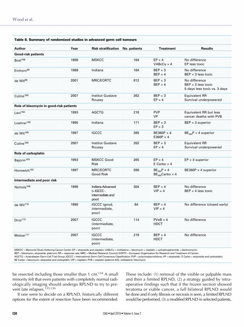
Standard chemotherapy for all patients is BEP chemother-apy.97-100 The efficacy of the 5-day schedule of BEP withetoposide 100 mg/m2/day and cisplatin 20 mg/m2/day for5 days and bleomycin 30 IU weekly is of equivalent effi-cacy to the same drugs given on a 3-day schedule (etopo-side 165 mg/m2/day given for 3 days, cisplatin 50 mg/m2/daygiven for 2 days, and bleomycin 30 IU weekly.99 BEP givenover 3 days, however, has increased short-term gastroin-testinal toxicity and long-term ototoxicity.99,101 Carboplatinshould not be substituted for cisplatin due to inferior out-comes.102,103 Thus, the original 5-day BEP regimen, there-fore, is the preferred option for the management of advancedGCTs. Modifications in BEP to reduce toxicity or improveconvenience should be avoided as they may also reduceefficacy. A summary of the randomized trials in advanceddisease is shown in Table 8.
In patients with IGCCC “good” prognosis disease, 3 cyclesof BEP should be given.97,98,104,105 If there is a contraindi-cation to bleomycin, 4 cycles of EP can be given, but hasbeen associated with a nonstatistically significant but high-er death rate in one RCT.100,106,107
CUAJ • April 2010 • Volume 4, Issue 2E28
Wood et al.
Table 7. Adjuvant chemotherapy for stage I nonseminoma germ cell tumour (selected studies)
Author(publication year)
No.patients
Riskfactors Regimen
Medianfollow-up (mo) Relapses
Adjuvant chemotherapy with 2 cycles of cisplatin-based combination chemotherapy
Oliver et al (1992)76 22 EC, VI, no yolk sactumour
EBC 3i × 2 43 1 (5%)
Madej et al. (1991)77 30 VI, LI, RT invasion,involvement ofepidydimis
PVB × 3 NR 0
Pont et al. (1996)75 29 VI BEP × 2 79 2 (6.9%)
Cullen et al. (1996)74 114 EC, VI, no yolk sactumour
BEP × 2 48 2 (1.7%)
Studer et al. (2000)78 59 EC, VI, capsulepenetration
PVB or BEP × 2 93 2 (3%)
Chevreau et al.(2004)79
40 VI, EC BEP × 2 113 0
Dearnaley et al.(2005)71
115 EC, VI, no yolk sactumour
BOP × 2 70 2 (1.7%)
Adjuvant chemotherapy with 1 cycle of BEP (experimental)
Gilbert et al. (2006)80 22 VI, EC, LI, no Yolksac tumour
BEP × 1CEB × 1
122 0
Albers et al. (2008)65 191 VI BEP × 1 56 2 (1%)
Westermann et al.(2008)81
40 VI, LI, EC BEP × 1 99 1 (2.5%)
EC = embryonal component; VI = vascular invasion; LI = lymphatic invasion; RT = rete testes; NR = not reported; EBC = etoposide, bleomycin, and carboplatin; PVB = cisplatin [Platinol-AQ], vinblastine, bleomycin; BEP = bleomycin, etoposide, platinum; BOP = bleomycin, vincristine, cisplatin; CEB = carboplatin, etoposide and bleomycin.
CUAJ • April 2010 • Volume 4, Issue 2 E29
In patients with IGCCC “intermediate” or “poor” prog-nosis disease, 4 cycles of the BEP are considered the stan-dard therapy.108,109 etoposide, ifosfamide and cisplatin (VIP)has been compared to BEP in this patient population andshows similar cancer outcomes but more genitourinary tox-icity and myelosuppression and, thus, represents an alter-native to BEP for patients with a contraindication tobleomycin or who develop pulmonary compromise whilereceiving BEP.110 For “intermediate” or “poor” prognosispatients, there is no evidence to date that the use of high-dose chemotherapy with autologous stem cell transplant issuperior to standard BEP for 4 cycles.111,112 When chemother-apy is given, it should be given without dose reductions at21-day intervals. Only in exceptional circumstances shouldthe chemotherapy be delayed or dose-reduced. Primaryprophylaxis for complications of neutropenia with granu-locyte-colony stimulating factor is generally not recom-mended as per the American Society of Clinical Oncology2006 Evidence-Based Clinical Practice Guidelines.113 Inthis guideline, primary prophylaxis is not recommended ifthe risk of febrile neutropenia is less than 20%; however,it can be considered in patients that are at high risk basedon age, co-morbidities, disease characteristics and myelo-toxicity of the regimen (i.e., ifosfamide-based chemothera-py). Secondary prophylaxis can be considered if there wereinfectious complications in the prior cycle and to maintaindense intensity.114 Prophylactic antibiotic treatment hasbeen shown to reduce febrile neutropenia during chemother-apy with no change in mortality and may be considered insome patients.115,116
In patients with life-threatening “poor” prognosis disease,orchiectomy should not delay the initiation of curative ther-apy and can be performed at the end of therapy.117-120 It isrecommended that these patients be referred to special-ized centres for optimal multidisciplinary management andsupportive care.
Monitoring during chemotherapy
During chemotherapy, monitoring tumour markers just priorto each treatment cycle is mandatory. If there is no tumourmarker elevation prior to chemotherapy, radiological imag-ing should be performed after 2 cycles.
As long as tumour markers are declining, a full course ofchemotherapy should be completed. If there is a slow tumourmarker decline or stable tumour markers, earlier radiologi-cal restaging can be considered. If there is unequivocal tumourmarker rise, even in the presence of radiological regression,a switch to salvage chemotherapy may be necessary. Patientsin this setting, who have evidence of progressive diseasewith first-line chemotherapy, have a worse prognosis.121
If there is an expected tumour marker decline but themetastases are growing radiologically, growing teratoma
syndrome should be considered.92,122 In most cases, thefull course of chemotherapy should be completed and resec-tion of the growing and residual masses should be donepost-chemotherapy. Very rarely, rapid radiological progres-sion in the setting of decreasing tumour marker decline isseen which would necessitate surgical resection prior tothe completion of chemotherapy.
Post-chemotherapy, radiological restaging should be per-formed in all patients. If the expected tumour marker declineis seen, all residual masses should be treated appropriate-ly. If the tumour markers plateau and are at a low level,they should be followed closely. If there is a persistent plateauor tumour marker decline, residual masses should be treat-ed appropriately.
It is not uncommon for patients with a markedly elevat-ed HCG prior to treatment to take longer for the HCG tonormalize or plateau at the end of chemotherapy.123
Post-chemotherapy residual masses: nonseminomatous germ celltumours
In many patients who have completed chemotherapy andhave normalized their tumour markers, residual massesare seen on repeat radiological imaging. Histology of resid-ual masses after first-line chemotherapy will be necrosis in40% to 50%, mature teratoma in 35% to 40% and viablecancer in 10% to 15%.124,125 The incidence of viable can-cer is likely even higher after salvage chemotherapy.124,126
Some of the factors that have been found to predict for noviable tumour in the residual mass include: no teratomain the primary tumour, pre-chemotherapy normal tumourmarkers, a small pre-chemotherapy mass, a large shrink-age of the mass with chemotherapy, and size of residualmass ≤ 10 mm.125,127,128 To date, however, no imaging pro-cedures, including PET scan, nor any one predictive factoror predictive model exists to reliably predict the histologyof residual masses.125,127-133
In patients with normal tumour markers and residualmasses, the residual masses should be resected. Two areasof controversy exist, however, where the literature doesnot give clear answers: (1) what constitutes a “residual mass”and (2) the extent of surgical resection. No consensus couldbe reached on the first controversy. All Canadian genitouri-nary oncology specialists felt that a full discussion regard-ing the risks and benefits of surgery post-chemotherapymust be undertaken with all patients and individual fac-tors, such as the risk of relapse, ability for follow-up, qual-ity of life and patient preferences, must be taken in to account.All felt the radiological imaging had to be reviewed by expe-rienced and knowledgeable radiologists, uro-oncologistsand/or medical oncologists. All Canadian genitourinaryoncology specialists thought any residual mass ≥1 cm shouldbe resected.126 Some thought that any residual mass should
Management of testicular germ cell cancer
be resected including those smaller than 1 cm.134 A smallminority felt that even patients with completely normal radi-ologically imaging should undergo RPLND to try to pre-vent late relapses.135,136
If one were to decide on a RPLND, historically differentoptions for the extent of resection have been recommended.
These include: (1) removal of the visible or palpable massand then a limited RPLND, (2) a strategy guided by intra-operative findings such that if the frozen section showedteratoma or viable cancer, a full bilateral RPLND wouldbe done and if only fibrosis or necrosis is seen, a limited RPLNDwould be performed, (3) a modified RPLND in selected patients,
CUAJ • April 2010 • Volume 4, Issue 2E30
Wood et al.
Table 8. Summary of randomized studies in advanced germ cell tumours
Author Year Risk stratification No. patients Treatment Results
Good-risk patients
Bosl106 1988 MSKCC 164 EP × 4VABcCy × 4
No differenceEP less toxic
Einhorn98 1989 Indiana 184 BEP × 3BEP × 4
No differenceBEP × 3 less toxic
de Wit99 2001 MRC/EORTC 812 BEP × 3BEP × 4
No differenceBEP × 3 less toxic5 days less toxic vs. 3 days
Culine100 2007 Institut GustaveRoussy
262 BEP × 3EP × 4
Equivalent RRSurvival underpowered
Role of bleomycin in good-risk patients
Levi194 1993 AGCTG 218 PVPVP
Equivalent RR but lesscancer deaths with PVB
Loehrer105 1995 Indiana 171 BEP × 3EP × 3
BEP × 3 superior
de Wit195 1997 IGCCC 395 BE360P × 4E360P × 4
BE360P × 4 superior
Culine100 2007 Institut GustaveRoussy
262 BEP × 3EP × 4
Equivalent RRSurvival underpowered
Role of carboplatin
Bajorin103 1993 MSKCC GoodRisk
265 EP × 4E Carbo × 4
EP × 4 superior
Horowich102 1997 MRC/EORTCGood Risk
598 BE360P × 4BE360Carbo × 4
BE360P × 4 superior
Intermediate and poor risk
Nichols108 1998 Indiana Advanced(= IGCCC -intermediate andpoor)
304 BEP × 4VIP × 4
No differenceBEP × 4 less toxic
de Wit110 1998 IGCCC (good,intermediate,poor)
84 BEP × 4VIP × 4
No difference (closed early)
Droz112 2007 IGCCC(intermediate,poor)
114 PVeB × 4HDCT
No difference
Motzer111 2007 IGCCC(intermediate,poor)
219 BEP × 4HDCT
No difference
MSKCC = Memorial Sloan-Kettering Cancer Center; EP = etoposide and cisplatin; VABcCy = vinblastine + bleomycin + cisplatin + cyclophosphamide + dactinomycin; BEP = bleomycin, etoposide, platinum; RR = response rate; MRC = Mediacl Research Council; EORTC = European Organization for Research and Treatment of Cancer; AGCTG = Australasian Germ Cell Trial Group; IGCCC = International Germ Cell Consensus Classification; PVP = polyvinylpyrrolidone; VP = etoposide ; E Carbo = etoposide and carboplatin; BE Carbo = bleomycin, etoposide and carboplatin; VIP = cisplatin; PVB = cisplatin [platinol-AQ], vinblastine, bleomycin.
CUAJ • April 2010 • Volume 4, Issue 2 E31
(4) or a full bilateral RPLND in all patients.137-142 Most Canadiangenitourinary oncology specialists felt that in the setting ofpost-chemotherapy residual masses, a full bilateral RPLNDshould be done in most cases; however, this decision mustbe reviewed carefully in each individual patient.
If persistent retroperitoneal disease is present and the deci-sion to perform a RPLND is made, surgery should be done4 to 8 weeks after completing chemotherapy. Whatever theextent of surgery, complete resection of the residual massesimpacts prognosis and every attempt at complete surgicalresection must be made.126,132,143 If technically possible, anerve-sparing procedure should be done. Surgery should beperformed by trained, experienced, expert uro-oncologistswhich may require referral to specialist centre. Perioperativeand postoperative complications must also be minimized,especially pulmonary toxicity in patients who have receivedbleomycin.144 Laparoscopic RPLND should not be consid-ered a standard of care at the present time.145-147
Resection of residual tumour outside the retroperitoneumshould be considered on an individual basis. In most cases,the retroperitoneum should be operated on first. Concordancein the pathology between the retroperitoneum and othermetastatic sites ranges from 50% to 89%.148-152 Thus, if thehistology in the resected residual retroperitoneal massesshows complete necrosis, both surveillance and resectionof the remaining non-retroperitoneal residual lesions areacceptable options. If the histology in the resected residualretroperitoneal masses shows mature teratoma, resectionof the remaining non-retroperitoneal masses should be done.If the histology in the resected residual retroperitoneal mass-es shows viable malignancy, salvage chemotherapy and/orfurther surgery may be options depending on the individ-ual clinical scenario.
Post-chemotherapy residual masses: seminoma
Post-chemotherapy residual masses in advanced seminomaare not uncommon and most do not have to be treated. Inpatients with residual masses ≥3 cm, an FDG-PET scan shouldbe obtained to gain further information regarding the via-bility of the residual mass.153,154 In patients with residualmasses less than 3 cm, the likelihood of viable malignancyis low and thus surveillance is reasonable.155-157 In patientswith residual masses less than <3 cm, the use of FDG-PETscanning is optional. The PET scan should be done 4 to 12weeks after day 21 of the last chemotherapy cycle.
If the PET scan is negative, no other active treatment isrequired and the patient can be surveyed. If the PET scanis positive, however, one must consider the possibility ofviable disease. The Canadian consensus was that surgicalresection is the management of choice.158,159 Radiationtherapy may be given in some cases although the overallbenefit of radiotherapy may be minimal.160 The advantages
of surgery include the ability to assess the response tochemotherapy, stage accurately and potentially providecure. The disadvantage of surgery is the high frequency ofdesmoplastic reaction associated with seminoma whichmay make surgery more technically difficult and increasecomplication rates.161 The extent of surgical resection inseminoma is usually a resection of the residual mass ormultiple biopsies and does not usually include a full ormodified RPLND.
Consolidation chemotherapy after secondary surgery
If the pathology from completely resected residual massesshows necrosis or mature teratoma, no further treatment isrequired. When viable cancer is found, the role of furtherchemotherapy is not clear. One retrospective analysis showedan improvement in progression-free survival with adjuvantchemotherapy however no improvement in overall sur-vival.162 A second retrospective analysis also showed alonger disease-free interval in those patients who had viablecancer in their post-chemotherapy RPLND who receivedchemotherapy compared to those who did not.124 Thereare no direct prospective data to help guide the decision.
Brain metastases
About 2% to 3% of patients with germ cell malignancy willpresent with brain metastases and up to 10% to 15% maydevelop it during the course of their illness. There are threepatterns of presentations: (1) at the time of initial diagnosisin the presence of other systemic disease, (2) at relapse inthe brain only, (3) at relapse including the brain and othersites of systemic disease.163,164 Patients who present withbrain metastases at their initial diagnosis have a survivalprobability in the range of 30%, whereas patients who devel-op brain metastases as one of multiple sites of relapse havea shorter survival.164 The patients that appear to have thebest outcome are those with isolated brain metastases atthe time of recurrence; although this is a rare group.25,165,166
The optimal sequence of chemotherapy, radiotherapy andsurgery is not known and management should be performedon an individual basis. In a multivariate analysis, radiother-apy together with chemotherapy improved the overall prog-nosis of patients who present with brain metastases versuseither treatment alone, which is different than another reportshowing no benefit from the addition of radiotherapy.25,166
If the goal is cure, systemic chemotherapy should be usedin all patients. It is not clear whether high-dose chemother-apy is of greater benefit to these patients.167 The roles ofsecondary resection of a solitary residual mass in the brainand the use of brain radiotherapy are also unclear.
Management of testicular germ cell cancer
Consensus recommendations
All patients with advanced GCT should be treated for cureand referral to specialized centre should be strongly con-sidered.
Patients with IGCCC good-risk disease should receive 3cycles of BEP.
Patients with IGCCC intermediate and poor-risk diseaseshould receive 4 cycles of BEP.
During chemotherapy, patients need to be monitoredon a regular basis with serial tumour marker estimation.Post-chemotherapy, all patients should have radiologicalrestaging to determine if there are residual masses.
In NSGCT cases, post-chemotherapy residual massesgreater than 1 cm should be resected.
No consensus could be reached on role of surgical resec-tion of masses <1 cm or where complete response isachieved.
If surgery performed for residual disease a full bilateralRPLND should be performed
Residual mass post-treatment seminoma: 1. Greater than 3 cm with PET scan positive: surgical
resection.2. <3 cm or greater than 3 cm with PET scan negative:
observe.Patients with brain metastases should be managed in a
specialized centre and may require multimodality treat-ment including surgical resection.
6. Treatment of relapsed and refractory disease
Patients with GCTs who relapse represent a heterogeneousgroup of patients. Depending on the histology and initialpresenting stage, patients may either relapse while on sur-veillance, post-radiation therapy, post-RPLND or post-chemotherapy.
Chemotherapy naïve
Nonseminoma patients may relapse while on surveillance orpost primary RPLND for clinical stage I disease. Seminomapatients may relapse after adjuvant carboplatin chemothera-py, adjuvant radiation therapy or while on surveillance.Seminoma patients who relapse while on surveillance or afteradjuvant carboplatin may be candidates for radiation therapyif the relapse is localized to the RPLN and if they meet thecriteria for radiation therapy as per stage II seminoma.
All other patients should be risk stratified into by theIGCCCG criteria and treated accordingly with standard doseBEP. Seminoma patients who have previously received onlyradiation therapy have an excellent chance of cure withstandard dosed cisplatin-based chemotherapy.168
Relapse post-cisplatin-based chemotherapy
Patients who relapse after cisplatin-based chemotherapycan be treated with either further standard dose chemother-apy or consideration of high-dose chemotherapy (HDCT)and autologous stem cell transplantation (ASCT). In thispatient population, prognostic factors have been identifiedthat affect outcome. These are similar, but not identical to,those factors used to risk-stratify chemotherapy-naïve patients.These factors include: site of the primary cancer (gonadalbetter than non-gonadal); histology of the primary tumour(seminoma better than nonseminoma); response to first-line cisplatin-based chemotherapy (complete response orpartial response marker negative better than partial responsemarker positive or progressive disease); progression-freeinterval after first-line chemotherapy (greater than 6 monthsbetter than less than 6 months); sites of metastatic diseaseprior to salvage treatment (lung or nodal better than othervisceral sites of disease); and level of tumour markers atrelapse (AFP <100/ng/mL, HCG <1000 u/L as opposed tohigher than this).169-174
No data exits as to whether or not or how one couldtailor the approach to treating relapsed patients based onthese prognostic factors. However, consensus participantsnoted that many of the phase II trials of standard dosechemotherapy have largely been conducted in good-riskpatients, making it difficult to extrapolate results to the poorrisk population.
Standard dose chemotherapy
Regimens of choice for standard dose salvage treatmentinclude 4 cycles of either VIP, Vinblastine, ifosfamide andcisplatin (VeIP) or Paclitaxel, ifosfamide and cisplatin(TIP).170,171,173,175-179 With these regimens, long-term dis-ease control can be achieved in between 15% to 60% ofpatients. Given that no randomized trials exist comparingthese chemotherapy combinations, no recommendationcan be made as to which regimen is superior or should beused as the standard management. Most of the older datawith VIP and VeIP is in all-risk patients and most of thenewer data with TIP is in good-risk patients at relapse. Thus,it is less certain if the data in good-risk relapsed patientswith TIP would be as useful in determining a regimen inthe poorer risk population; however, it is not an unreason-able choice.
There has been one randomized trial of HDCT versusstandard dose salvage chemotherapy in good-risk relapsedpatients. This trial compared 4 cycles of cisplatin, ifosfamideand etoposide (or vinblastine) (PEI) compared to 3 cyclesof PEI followed by high dose carboplatin, etoposide andcyclophosphamide and ASCT. This trial showed no advan-tage to HDCT in this group.174
CUAJ • April 2010 • Volume 4, Issue 2E32
Wood et al.
CUAJ • April 2010 • Volume 4, Issue 2 E33
High-dose chemotherapy and autologous transplantation
High-dose chemotherapy has been shown in phase II trialsto be an effective salvage strategy in poor-risk patients witha suggestion of an improvement in survival as comparedto standard dose salvage chemotherapy, albeit throughmatched-pair analysis rather than randomizedtrials.180,181High-dose chemotherapy has also been shownto be a potentially curative option for patients with secondor subsequent relapses.181-183
Patients who failed to be cured with a standard doseoption and are well enough to tolerate it, HDCT with trans-plantation should be offered before declaring the relapseddisease incurable. For patients undergoing HDCT, high dosecarboplatin and etoposide regimen is the conditioning reg-imen of choice.184 While there have been no randomizedstudies that have determined the optimal conditioning reg-imen, there have been reports of excess toxicity if an oxaza-phosphorine (e.g., cyclophosphamide) is included in theregimen.183,184 Also, not including a third drug in the regi-men allows higher doses of the two most active agents (car-boplatin and etoposide) to be administered. Patients whoare proceeding to transplant should be offered standarddose chemotherapy before the transplant to determine ifthey have chemotherapy sensitive disease, to debulk thedisease before the transplant, and to keep the disease undercontrol while logistical arrangements for the transplant canbe put into place. It appears the best results for HDCT havebeen obtained if a tandem transplant has been performedand thus, patients should have enough stem cells collectedfor a planned tandem procedure.184 High-dose chemother-apy and transplantation should be performed in special-ized centres where there is adequate volume and expertiseto be able to offer the best supportive and pre/post trans-plant care.
Multiply refractory patients
Patients who relapse after standard dose and high dosechemotherapy may still respond to chemotherapy agents,usually with a view to palliating the disease rather thancuring it. Drugs having efficacy include oral etoposide,paclitaxel, gemcitabine, oxaliplatin or combinations of thesedrugs.185 While infrequent, some patients being treated inthe third-line setting may have long-term disease controlor cure in particular if the tumour lesions can be com-pletely resected.184
Salvage surgery
In patients who normalize their markers but have residualdisease radiographically, all residual masses should be con-sidered for salvage surgery. Patients who still harbor viable
disease in their post-chemotherapy masses have a worseprognosis but the administration of adjuvant chemotherapydoes not appear to improve outcomes in this setting.143,186
Patients who fail to normalize their markers or have pro-gressive disease post-salvage systemic treatment may becandidates for salvage or “desperation” surgery if it is feltthat all the disease can be resected. This will often requirea multidisciplinary surgical team and should be performedby surgeons skilled in these operations working in special-ized centres.
Later relapses
Late relapse is defined as disease recurrence more than 2years after completion of first-line therapy. The risk of laterelapse in all comers is approximately 1.5% for seminomaand 3% for nonseminoma patients.187 These patients havedisease that is more chemotherapy resistant and immedi-ate surgical resection of recurrent disease should be under-taken if feasible, irrespective of the level of tumour mark-ers.188-192 If surgical resection is not feasible, biopsy of thelesions should be undertaken to determine the histologyand chemotherapy should be administered directed to thehistology that is found on biopsy. If viable GCT is found,TIP has shown activity in late relapsers who were not sur-gical candidates.173
Relapsing patients with mediastinal primary
Patients with mediastinal primary GCT who relapse have apoor prognosis. In transplant series, very few if any of thesepatients have successfully been salvaged with a transplant,yet the toxicities of the treatment are still present.
Consensus recommendations
Seminoma patients who relapse while on surveillance orafter adjuvant carboplatin may be candidates for radiationtherapy if the relapse is localized to the RPLN and meetsthe criteria for radiation therapy as per stage II seminomarecommendations.
All other chemotherapy-naïve relapse patients shouldbe stratified by the IGCCCG criteria and treated accord-ingly to the recommendations for first-line management ofadvanced GCT.
Patients who relapse after cisplatin-based chemotherapycan be treated with either further standard dose chemother-apy or consideration of high-dose chemotherapy and autol-ogous stem cell transplantation (HDCT with ASCT).
No evidence that either approach is better.Many in the group felt that it would be reasonable to
offer standard dose chemotherapy to good-risk patients asmany will be cured, especially based on the more recent
Management of testicular germ cell cancer
data using TIP, and reserve the transplant for third line.For patients undergoing HDCT, high dose carboplatin
and etoposide regimen is the conditioning regimen of choice.High-dose chemotherapy and transplantation could be
offered as third-line therapy.Patients who relapse after standard dose and high-dose
chemotherapy should be offered third-line treatment as long-term disease control may still be achieved.
Salvage surgery should always be considered in patientswith residual resectable disease.
Patients with late relapse (nonseminoma) should havesurgical resection of disease if possible.
Patients with mediastinal primary GCT who relapse shouldrarely if ever be offered transplantation.
*Division of Medical Oncology, Queen Elizabeth II Health Sciences Centre, Halifax, NS; †Departmentof Medical Oncology, British Columbia Cancer Agency, Vancouver, BC; ‡Division of Urology, UniversityHealth Network, University of Toronto, Toronto, ON; §Department of Radiation Oncology, RadiationMedicine Program, Princess Margaret Hospital, University of Toronto, Toronto, ON; ±Division ofMedical Oncology, Juravinski Cancer Centre, Hamilton, ON; ¥Department of Medical Imaging,University Health Network, University of Toronto, Toronto, ON; #Department of Pathology, UniversityHealth Network, University of Toronto, Toronto, ON; βDepartment of Medicine, London HealthSciences Centre, London, ON; αDepartment of Medical Oncology, Cross Cancer Institute, Edmonton,AB; ΔDepartment of Radiation Oncology, Vancouver Cancer Clinic, Vancouver, BC; µDivision ofMedical Oncology, McGill University Health Centre, Montréal, QC; **Department of Medicine,Division of Medical Oncology, The Ottawa Hospital Regional Cancer Centre, Ottawa, ON; ††Departmentof Medicine, University of Toronto, Princess Margaret Hospital, Toronto, ON; ‡‡Department ofUrology, Charité – Universitätsmedizin, Freie Universität Berlin, Berlin; §§Academic Radiotherapy,Institute of Cancer Research and Royal Marsden Hospital, Sutton, Surrey; ±±Oregon Health &Science University Cancer Institute, Portland, OR
Competing interests: None declared.
This paper has been peer-reviewed.
References
1. International Agency for Reseach on Cancer. World Health Organization. Available at: http://www-dep.iarc.fr/. Accessed March 9, 2010.
2. McGlynn KA, Devesa SS, Sigurdson AJ, et al. Trends in the incidence of testicular germ cell tumour s inthe United States. Cancer 2003;97:63-70.
3. Canadian Cancer Society. Available at: www.cancer.ca. Accessed March 17, 2010. 4. Collette L, Sylvester RJ, Stenning SP, et al. Impact of the treating institution on survival of patients with
“poor-prognosis” metastatic nonseminoma. European Organization for Research and Treatment ofCancer Genito-Urinary Tract Cancer Collaborative Group and the Medical Research Council TesticularCancer Working Party. J Natl Cancer Inst 1999;91:839-46.
5. Schmoll HJ, Souchon R, Krege S, et al. European consensus on diagnosis and treatment of germ cell cancer:a report of the European Germ Cell Cancer Consensus Group (EGCCCG). Ann Oncol 2004;15:1377-99.
6. Krege S, Beyer J, Souchon R, et al. European consensus conference on diagnosis and treatment ofgerm cell cancer: a report of the second meeting of the European Germ Cell Cancer Consensus group(EGCCCG): part I. Eur Urol 2008;53:478-96.
7. Krege S, Beyer J, Souchon R, et al. European consensus conference on diagnosis and treatment ofgerm cell cancer: a report of the second meeting of the European Germ Cell Cancer Consensus Group(EGCCCG): part II. Eur Urol 2008;53:497-513.
8. Richie JP. Impact of diagnostic delay in testis cancer: Results of a large population-based study: HuygheE, Muller A, Mieusset R, Bujan L, Bachaud JM, Chevreau C, Plante P, Thonneau P, Human FertilityResearch Group, Paule de Viguier Hospital, Toulouse University III, France; Urology and AndrologyDepartment, Paule de Viguier Hospital, Toulouse, France. Urol Oncol 2008;26:220-1.
9. Sobin LH, Wittekind CH, eds. TNM classification of malignant tumours (UICC). 6th ed. Hoboken, NJ:Wiley, John & Sons; 2002.
10. International Germ Cell Consensus Classification: a prognostic factor-based staging system for metastaticgerm cell cancers. International Germ Cell Cancer Collaborative Group. J Clin Oncol 1997;15:594-603.
11. Aass N, Klepp O, Cavallin-Stahl E, et al. Prognostic factors in unselected patients with nonseminomatousmetastatic testicular cancer: a multicenter experience. J Clin Oncol 1991;9:818-26.
12. Winstanley AM, Mikuz G, Debruyne F, et al. Handling and reporting of biopsy and surgical specimens oftesticular cancer. Eur Urol 2004;45:564-73.
13. Daugaard G, Petersen PM, Rorth M. Surveillance in stage I testicular cancer. Apmis 2003;111:76-83;discussion 83-75.
14. Germa-Lluch JR, Garcia del Muro X, Maroto P, et al. Clinical pattern and therapeutic results achieved in1490 patients with germ-cell tumours of the testis: the experience of the Spanish Germ-Cell CancerGroup (GG). Eur Urol 2002;42:553-62; discussion 562-3.
15. Horwich A, Alsanjari N, A’Hern R, et al. Surveillance following orchidectomy for stage I testicular semi-noma. Br J Cancer 1992;65:775-8.
16. Oliver R, Boubilkova L, Ong J. Fifteen-year follow-up of the Anglian Germ Cell Cancer group adjuvantstudies of carboplatin as an alternative to radiation or surveillance for stage 1 seminoma. Proc ASCO2001;19Abstract 780.
17. Ramakrishnan S, Champion AE, Dorreen MS, et al. Stage I seminoma of the testis: is post-orchidectomy sur-veillance a safe alternative to routine postoperative radiotherapy? Clin Oncol (R Coll Radiol) 1992;4:284-6.
18. von der Maase H, Specht L, Jacobsen GK, et al. Surveillance following orchidectomy for stage I semino-ma of the testis. Eur J Cancer 1993;29A:1931-4.
19. Warde PR, Chung P, Sturgeon J, et al. Should surveillance be considered the standard of care in stage Iseminoma? J Clin Oncol 2005;23(16suppl):4520.
20. Tyldesley S, Voduc D, McKenzie M, et al. Surveillance of stage I testicular seminoma: British ColumbiaCancer Agency Experience 1992 to 2002. Urology 2006;67:594-8.
21. Warde P, Specht L, Horwich A, et al. Prognostic factors for relapse in stage I seminoma managed bysurveillance: a pooled analysis. J Clin Oncol 2002;20:4448-52.
22. Aparicio J, Germa JR, Garcia del Muro X, et al. Risk-adapted management for patients with clinicalstage I seminoma: the Second Spanish Germ Cell Cancer Cooperative Group study. J Clin Oncol2005;23:8717-23.
23. Bayens YC, Helle PA, Van Putten WL, et al. Orchidectomy followed by radiotherapy in 176 stage I and IItesticular seminoma patients: benefits of a 10-year follow-up study. Radiother Oncol 1992;25:97-102.
24. Coleman JM, Coleman RE, Turner AR, et al. The management and clinical course of testicular semino-ma: 15 years’ experience at a single institution. Clin Oncol (R Coll Radiol) 1998;10:237-41.
25. Fossa SD, Bokemeyer C, Gerl A, et al. Treatment outcome of patients with brain metastases from malig-nant germ cell tumour s. Cancer 1999;85:988-97.
26. Jones WG, Fossa SD, Mead GM, et al. Randomized trial of 30 versus 20 Gy in the adjuvant treatment ofstage I Testicular Seminoma: a report on Medical Research Council Trial TE18, European Organisation forthe Research and Treatment of Cancer Trial 30942 (ISRCTN18525328). J Clin Oncol 2005;23:1200-8.
27. Santoni R, Barbera F, Bertoni F, et al. Stage I seminoma of the testis: a bi-institutional retrospectiveanalysis of patients treated with radiation therapy only. BJU Int 2003;92:47-52; discussion 52.
28. Warde P, Gospodarowicz MK, Panzarella T, et al. Stage I testicular seminoma: results of adjuvant irradi-ation and surveillance. J Clin Oncol 1995;13:2255-62.
29. Fossa SD, Horwich A, Russell JM, et al. Optimal planning target volume for stage I testicular seminoma:A Medical Research Council randomized trial. Medical Research Council Testicular Tumour WorkingGroup. J Clin Oncol 1999;17:1146.
30. Logue JP, Harris MA, Livsey JE, et al. Short course paraaortic radiation for stage I seminoma of thetestis. Int J Radiat Oncol Biol Phys 2003;57:1304-9.
31. Huddart RA, Norman A, Shahidi M, et al. Cardiovascular disease as a long-term complication of treat-ment for testicular cancer. J Clin Oncol 2003;21:1513-23.
32. Zagars GK, Ballo MT, Lee AK, et al. Mortality after cure of testicular seminoma. J Clin Oncol2004;22:640-7.
33. van Leeuwen FE, Stiggelbout AM, van den Belt-Dusebout AW, et al. Second cancer risk following testic-ular cancer: a follow-up study of 1,909 patients. J Clin Oncol 1993;11:415-24.
34. Moller H, Friis S, Kjaer SK. Survival of Danish cancer patients 1943-1987. Male genital organs. APMISSuppl 1993;33:122-36.
35. Travis LB, Fossa SD, Schonfeld SJ, et al. Second cancers among 40,576 testicular cancer patients:focus on long-term survivors. J Natl Cancer Inst 2005;97:1354-65.
CUAJ • April 2010 • Volume 4, Issue 2E34
Wood et al.
CUAJ • April 2010 • Volume 4, Issue 2 E35
36. van den Belt-Dusebout AW, de Wit R, Gietema JA, et al. Treatment-specific risks of second malignanciesand cardiovascular disease in 5-year survivors of testicular cancer. J Clin Oncol 2007;25:4370-8.
37. Oliver RT, Ong J, Shamash J, et al. Long-term follow-up of Anglian Germ Cell Cancer Group surveillance ver-sus patients with Stage 1 nonseminoma treated with adjuvant chemotherapy. Urology 2004;63:556-61.
38. Steiner H, Muller T, Gozzi C, et al. Two cycles of cisplatin-based chemotherapy for low-volume retroperi-toneal stage II nonseminomatous germ cell tumours. BJU Int 2006;98:349-52.
39. Hotte S, Mayhew LA, Jewett M, et al. Cancer Care Ontario. Management of Stage I NonseminomatousTesticular Cancer: Guideline Recommendations. Available at www.cancercare.on.ca/pdf/pebc3-19s.pdf.Accessed March 9, 2010.
40. Read G, Johnson RJ, Wilkinson PM, et al. Prospective study of follow up alone in stage I teratoma ofthe testis. Br Med J (Clin Res Ed) 1983;287:1503-5.
41. Sogani PC, Whitmore WF Jr, Herr HW, et al. Orchiectomy alone in the treatment of clinical stage I non-seminomatous germ cell tumour of the testis. J Clin Oncol 1984;2:267-70.
42. Pizzocaro G, Zanoni F, Milani A, et al. Orchiectomy alone in clinical stage I nonseminomatous testiscancer: a critical appraisal. J Clin Oncol 1986;4:35-40.
43. Peckham MJ, Brada M. Surveillance following orchidectomy for stage I testicular cancer. Int J Androl1987;10:247-54.
44. Sturgeon JF, Jewett MA, Alison RE, et al. Surveillance after orchidectomy for patients with clinical stageI nonseminomatous testis tumour s. J Clin Oncol 1992;10:564-8.
45. Read G, Stenning SP, Cullen MH, et al. Medical Research Council prospective study of surveillance forstage I testicular teratoma. Medical Research Council Testicular Tumour s Working Party. J Clin Oncol1992;10:1762-8.
46. Colls BM, Harvey VJ, Skelton L, et al. Late results of surveillance of clinical stage I nonseminoma germ celltesticular tumours: 17 years’ experience in a national study in New Zealand. BJU Int 1999;83:76-82.
47. Pont J, Holtl W, Kosak D, et al. Risk-adapted treatment choice in stage I nonseminomatous testiculargerm cell cancer by regarding vascular invasion in the primary tumour : a prospective trial. J Clin Oncol1990;8:16-20.
48. Klepp O, Dahl O, Flodgren P, et al. Risk-adapted treatment of clinical stage 1 non-seminoma testis can-cer. Eur J Cancer 1997;33:1038-44.
49. Divrik RT, Akdogan B, Ozen H, et al. Outcomes of surveillance protocol of clinical stage I nonseminoma-tous germ cell tumour s-is shift to risk adapted policy justified? J Urol 2006;176(4 Pt 1):1424-9; dis-cussion 1429-30.
50. Roeleveld TA, Horenblas S, Meinhardt W, et al. Surveillance can be the standard of care for stage I non-seminomatous testicular tumour s and even high risk patients. J Urol 2001;166:2166-70.
51. Alexandre J, Fizazi K, Mahe C, et al. Stage I non-seminomatous germ-cell tumours of the testis: identifi-cation of a subgroup of patients with a very low risk of relapse. Eur J Cancer 2001;37:576-82.
52. Francis R, Bower M, Brunstrom G, et al. Surveillance for stage I testicular germ cell tumours: resultsand cost benefit analysis of management options. Eur J Cancer 2000;36:1925-32.
53. Sogani PC, Perrotti M, Herr HW, et al. Clinical stage I testis cancer: long-term outcome of patients onsurveillance. J Urol 1998;159:855-8.
54. Hao D, Seidel J, Brant R, et al. Compliance of clinical stage I nonseminomatous germ cell tumourpatients with surveillance. J Urol 1998;160(3 Pt 1):768-71.
55. Boyer MJ, Cox K, Tattersall MH, et al. Active surveillance after orchiectomy for nonseminomatous testic-ular germ cell tumour s: late relapse may occur. Urology 1997;50:588-92.
56. Nicolai N, Pizzocaro G. A surveillance study of clinical stage I nonseminomatous germ cell tumour s ofthe testis: 10-year followup. J Urol 1995;154:1045-9.
57. Gels ME, Hoekstra HJ, Sleijfer DT, et al. Detection of recurrence in patients with clinical stage I non-seminomatous testicular germ cell tumour s and consequences for further follow-up: a single-center 10-year experience. J Clin Oncol 1995;13:1188-94.
58. Ondrus D, Hornak M. Orchiectomy alone for clinical stage I nonseminomatous germ cell tumour s of thetestis (NSGCTT): a minimum follow-up period of 5 years. Tumour i 1994;80:362-4.
59. Freedman LS, Parkinson MC, Jones WG, et al. Histopathology in the prediction of relapse of patientswith stage I testicular teratoma treated by orchidectomy alone. Lancet 1987;2:294-8.
60. Maroto P, Garcia del Muro X, Aparicio J, et al. Multicentre risk-adapted management for stage I non-seminomatous germ cell tumours. Ann Oncol 2005;16:1915-20.
61. Amato RJ, Ro JY, Ayala AG, et al. Risk-adapted treatment for patients with clinical stage I nonsemino-matous germ cell tumour of the testis. Urology 2004;63:144-8; discussion 148-9.
62. Ondrus D, Matoska J, Belan V, et al. Prognostic factors in clinical stage I nonseminomatous germ celltesticular tumour s: rationale for different risk-adapted treatment. Eur Urol 1998;33:562-6.
63. Rorth M, Jacobsen GK, von der Maase H, et al. Surveillance alone versus radiotherapy after orchiecto-my for clinical stage I nonseminomatous testicular cancer. Danish Testicular Cancer Study Group. J ClinOncol 1991;9:1543-8.
64. Weissbach L, Boedefeld EA, Horstmann-Dubral B. Surgical treatment of stage-I non-seminomatous germcell testis tumour . Final results of a prospective multicenter trial 1982-1987. Testicular Tumour StudyGroup. Eur Urol 1990;17:97-106.
65. Albers P, Siener R, Krege S, et al. Randomized Phase III Trial Comparing Retroperitoneal Lymph NodeDissection With One Course of Bleomycin and Etoposide Plus Cisplatin Chemotherapy in the AdjuvantTreatment of Clinical Stage I Nonseminomatous Testicular Germ Cell Tumour s: AUO Trial AH 01/94 bythe German Testicular Cancer Study Group. J Clin Oncol 2008;26:2966-72.
66. Donohue JP, Thornhill JA, Foster RS, et al. Retroperitoneal lymphadenectomy for clinical stage A testis can-cer (1965 to 1989): modifications of technique and impact on ejaculation. J Urol 1993;149:237-43.
67. Sweeney CJ, Hermans BP, Heilman DK, et al. Results and outcome of retroperitoneal lymph node dissec-tion for clinical stage I embryonal carcinoma—predominant testis cancer. J Clin Oncol 2000;18:358-62.
68. Nicolai N, Miceli R, Artusi R, et al. A simple model for predicting nodal metastasis in patients with clini-cal stage I nonseminomatous germ cell testicular tumour s undergoing retroperitoneal lymph node dis-section only. J Urol 2004;171:172-6.
69. Stephenson AJ, Bosl GJ, Bajorin DF, et al. Retroperitoneal lymph node dissection in patients with lowstage testicular cancer with embryonal carcinoma predominance and/or lymphovascular invasion. J Urol2005;174:557-60; discussion 560.
70. Spermon JR, Roeleveld TA, van der Poel HG, et al. Comparison of surveillance and retroperitoneal lymphnode dissection in Stage I nonseminomatous germ cell tumour s. Urology 2002;59:923-9.
71. Dearnaley DP, Fossa SD, Kaye SB, et al. Adjuvant bleomycin, vincristine and cisplatin (BOP) for high-risk stage I non-seminomatous germ cell tumours: a prospective trial (MRC TE17). Br J Cancer2005;92:2107-13.
72. Pectasides D, Skarlos D, Dimopoulos AM, et al. Two cycles of carboplatin-based adjuvant chemotherapyfor high-risk clinical stage I and stage IM non-seminomatous germ cell tumours of the testis: a HECOGtrial. Anticancer Res 2003;23:4239-44.
73. Bohlen D, Borner M, Sonntag RW, et al. Long-term results following adjuvant chemotherapy in patientswith clinical stage I testicular nonseminomatous malignant germ cell tumour s with high risk factors. JUrol 1999;161:1148-52.
74. Cullen MH, Stenning SP, Parkinson MC, et al. Short-course adjuvant chemotherapy in high-risk stage Inonseminomatous germ cell tumour s of the testis: a Medical Research Council report. J Clin Oncol1996;14:1106-13.
75. Pont J, Albrecht W, Postner G, et al. Adjuvant chemotherapy for high-risk clinical stage I nonseminoma-tous testicular germ cell cancer: long-term results of a prospective trial. J Clin Oncol 1996;14:441-8.
76. Oliver RT, Raja MA, Ong J, et al. Pilot study to evaluate impact of a policy of adjuvant chemotherapyfor high risk stage 1 malignant teratoma on overall relapse rate of stage 1 cancer patients. J Urol1992;148:1453-5; discussion 1455-6.
77. Madej G, Pawinski A. Risk-related adjuvant chemotherapy for stage I non-seminoma of the testis. ClinOncol (R Coll Radiol) 1991;3:270-2.
78. Studer UE, Burkhard FC, Sonntag RW. Risk adapted management with adjuvant chemotherapy inpatients with high risk clinical stage i nonseminomatous germ cell tumour . J Urol 2000;163:1785-7.
79. Chevreau C, Mazerolles C, Soulie M, et al. Long-term efficacy of two cycles of BEP regimen in high-riskstage I nonseminomatous testicular germ cell tumour s with embryonal carcinoma and/or vascular inva-sion. Eur Urol 2004;46:209-14; discussion 214-5.
80. Gilbert DC, Norman AR, Nicholl J, et al. Treating stage I nonseminomatous germ cell tumours with a sin-gle cycle of chemotherapy. BJU Int 2006;98:67-9.
81. Westermann DH, Schefer H, Thalmann GN, et al. Long-term followup results of 1 cycle of adjuvantbleomycin, etoposide and cisplatin chemotherapy for high risk clinical stage I nonseminomatous germcell tumour s of the testis. J Urol 2008;179:163-6.
82. Williams SD, Stablein DM, Einhorn LH, et al. Immediate adjuvant chemotherapy versus observationwith treatment at relapse in pathological stage II testicular cancer. N Engl J Med 1987;317:1433-8.
83. Weissbach L, Hartlapp JH. Adjuvant chemotherapy of metastatic stage II nonseminomatous testis tumour.J Urol 1991;146:1295-8.
84. Donohue JP, Thornhill JA, Foster RS, et al. The role of retroperitoneal lymphadenectomy in clinical stageB testis cancer: the Indiana University experience (1965 to 1989). J Urol 1995;153:85-9.
85. Kennedy BJ, Torkelson JL, Fraley EE. Adjuvant chemotherapy for stage II nonseminomatous germ cellcancer of the testis. Cancer 1994;73:1485-9.
86. Kuczyk M, Machtens S, Stief C, et al. Management of the post-chemotherapy residual mass in patientswith advanced stage non-seminomatous germ cell tumour s (NSGCT). Int J Cancer 1999;83:852-5.
87. Weissbach L, Bussar-Maatz R, Flechtner H, et al. RPLND or primary chemotherapy in clinical stage IIA/Bnonseminomatous germ cell tumour s? Results of a prospective multicenter trial including quality of lifeassessment. Eur Urol 2000;37:582-94.
88. Stephenson AJ, Bosl GJ, Motzer RJ, et al. Nonrandomized comparison of primary chemotherapy andretroperitoneal lymph node dissection for clinical stage IIA and IIB nonseminomatous germ cell testicularcancer. J Clin Oncol 2007;25:5597-602.
Management of testicular germ cell cancer
89. Sheinfeld J, Motzer RJ, Rabbani F, et al. Incidence and clinical outcome of patients with teratoma inthe retroperitoneum following primary retroperitoneal lymph node dissection for clinical stages I and IIAnonseminomatous germ cell tumour s. J Urol 2003;170(4 Pt 1):1159-62.
90. Horwich A, Stenning S. Initial chemotherapy for stage II testicular non-seminoma. World J Urol1994;12:148-50.
91. Peckham MJ, Hendry WF. Clinical stage II non-seminomatous germ cell testicular tumours. Results ofmanagement by primary chemotherapy. Br J Urol 1985;57:763-8.
92. Logothetis CJ, Samuels ML, Selig DE, et al. Primary chemotherapy followed by a selective retroperi-toneal lymphadenectomy in the management of clinical stage II testicular carcinoma: a preliminaryreport. J Urol 1985;134:1127-30.
93. Pizzocaro G, Monfardini S. No adjuvant chemotherapy in selected patients with pathologic stage II non-seminomatous germ cell tumour s of the testis. J Urol 1984;131:677-80.
94. Pizzocaro G, Piva L, Salvioni R, et al. Adjuvant chemotherapy in resected stage-II nonseminomatousgerm cell tumour s of testis. In which cases is it necessary? Eur Urol 1984;10:151-8.
95. Kondagunta GV, Sheinfeld J, Mazumdar M, et al. Relapse-free and overall survival in patients withpathologic stage II nonseminomatous germ cell cancer treated with etoposide and cisplatin adjuvantchemotherapy. J Clin Oncol 2004;22:464-7.
96. Feuer EJ, Frey CM, Brawley OW, et al. After a treatment breakthrough: a comparison of trial and popu-lation-based data for advanced testicular cancer. J Clin Oncol 1994;12:368-77.
97. Saxman SB, Finch D, Gonin R, et al. Long-term follow-up of a phase III study of three versus four cyclesof bleomycin, etoposide, and cisplatin in favorable-prognosis germ-cell tumour s: the Indian Universityexperience. J Clin Oncol 1998;16:702-6.
98. Einhorn LH, Williams SD, Loehrer PJ, et al. Evaluation of optimal duration of chemotherapy in favorable-prognosis disseminated germ cell tumour s: a Southeastern Cancer Study Group protocol. J Clin Oncol1989;7:387-91.
99. de Wit R, Roberts JT, Wilkinson PM, et al. Equivalence of three or four cycles of bleomycin, etoposide,and cisplatin chemotherapy and of a 3- or 5-day schedule in good-prognosis germ cell cancer: a ran-domized study of the European Organization for Research and Treatment of Cancer Genitourinary TractCancer Cooperative Group and the Medical Research Council. J Clin Oncol 2001;19:1629-40.
100. Culine S, Kerbrat P, Kramar A, et al. Refining the optimal chemotherapy regimen for good-risk metastat-ic nonseminomatous germ-cell tumour s: a randomized trial of the Genito-Urinary Group of the FrenchFederation of Cancer Centers (GETUG T93BP). Ann Oncol 2007;18:917-24.
101. Fossa SD, de Wit R, Roberts JT, et al. Quality of life in good prognosis patients with metastatic germcell cancer: a prospective study of the European Organization for Research and Treatment of CancerGenitourinary Group/Medical Research Council Testicular Cancer Study Group (30941/TE20). J ClinOncol 2003;21:1107-18.
102. Horwich A, Sleijfer DT, Fossa SD, et al. Randomized trial of bleomycin, etoposide, and cisplatin com-pared with bleomycin, etoposide, and carboplatin in good-prognosis metastatic nonseminomatous germcell cancer: a Multiinstitutional Medical Research Council/European Organization for Research andTreatment of Cancer Trial. J Clin Oncol 1997;15:1844-52.
103. Bajorin DF, Sarosdy MF, Pfister DG, et al. Randomized trial of etoposide and cisplatin versus etoposideand carboplatin in patients with good-risk germ cell tumour s: a multiinstitutional study. J Clin Oncol1993;11:598-606.
104. Toner GC, Stockler MR, Boyer MJ, et al. Comparison of two standard chemotherapy regimens for good-prognosis germ-cell tumours: a randomised trial. Australian and New Zealand Germ Cell Trial Group.Lancet 2001;357:739-45.
105. Loehrer PJ Sr, Johnson D, Elson P, et al. Importance of bleomycin in favorable-prognosis disseminatedgerm cell tumour s: an Eastern Cooperative Oncology Group trial. J Clin Oncol 1995;13:470-6.
106. Bosl GJ, Geller NL, Bajorin D, et al. A randomized trial of etoposide + cisplatin versus vinblastine +bleomycin + cisplatin + cyclophosphamide + dactinomycin in patients with good-prognosis germ celltumours. J Clin Oncol 1988;6:1231-8.
107. Xiao H, Mazumdar M, Bajorin DF, et al. Long-term follow-up of patients with good-risk germ cell tumours treated with etoposide and cisplatin. J Clin Oncol 1997;15:2553-8.
108. Nichols CR, Catalano PJ, Crawford ED, et al. Randomized comparison of cisplatin and etoposide andeither bleomycin or ifosfamide in treatment of advanced disseminated germ cell tumour s: an EasternCooperative Oncology Group, Southwest Oncology Group, and Cancer and Leukemia Group B Study. JClin Oncol 1998;16:1287-93.
109. de Wit R, Collette L, Sylvester R, et al. Serum alpha-fetoprotein surge after the initiation of chemotherapy fornon-seminomatous testicular cancer has an adverse prognostic significance. Br J Cancer 1998;78:1350-5.
110. de Wit R, Stoter G, Sleijfer DT, et al. Four cycles of BEP vs four cycles of VIP in patients with intermedi-ate-prognosis metastatic testicular non-seminoma: a randomized study of the EORTC Genitourinary TractCancer Cooperative Group. European Organization for Research and Treatment of Cancer. Br J Cancer1998;78:828-32.
111. Motzer RJ, Nichols CJ, Margolin KA, et al. Phase III randomized trial of conventional-dose chemotherapywith or without high-dose chemotherapy and autologous hematopoietic stem-cell rescue as first-line treat-ment for patients with poor-prognosis metastatic germ cell tumour s. J Clin Oncol 2007;25:247-56.
112. Droz JP, Kramar A, Biron P, et al. Failure of high-dose cyclophosphamide and etoposide combined withdouble-dose cisplatin and bone marrow support in patients with high-volume metastatic nonseminomatousgerm-cell tumours: mature results of a randomised trial. Eur Urol 2007;51:739-46; discussion 747-8.
113. Smith TJ, Khatcheressian J, Lyman GH, et al. 2006 update of recommendations for the use of whiteblood cell growth factors: an evidence-based clinical practice guideline. J Clin Oncol 2006;24:3187-205.
114. Fossa SD, Kaye SB, Mead GM, et al. Filgrastim during combination chemotherapy of patients with poor-prognosis metastatic germ cell malignancy. European Organization for Research and Treatment ofCancer, Genito-Urinary Group, and the Medical Research Council Testicular Cancer Working Party, Cambridge,United Kingdom. J Clin Oncol 1998;16:716-24.
115. Cullen M, Steven N, Billingham L, et al. Antibacterial prophylaxis after chemotherapy for solid tumour sand lymphomas. N Engl J Med 2005;353:988-98.
116. Cullen MH, Billingham LJ, Gaunt CH, et al. Rational selection of patients for antibacterial prophylaxisafter chemotherapy. J Clin Oncol 2007;25:4821-8.
117. Chong C, Logothetis CJ, von Eschenbach A, et al. Orchiectomy in advanced germ cell cancer followingintensive chemotherapy: a comparison of systemic to testicular response. J Urol 1986;136:1221-3.
118. Leibovitch I, Baniel J, Rowland RG, et al. Malignant testicular neoplasms in immunosuppressed patients.J Urol 1996;155:1938-42.
119. Ondrus D, Hornak M, Breza J, et al. Delayed orchiectomy after chemotherapy in patients with advancedtesticular cancer. Int Urol Nephrol 2001;32:665-7.
120. Simmonds PD, Mead GM, Lee AH, et al. Orchiectomy after chemotherapy in patients with metastatictesticular cancer. Is it indicated? Cancer 1995;75:1018-24.
121. Fossa SD, Stenning SP, Gerl A, et al. Prognostic factors in patients progressing after cisplatin-basedchemotherapy for malignant non-seminomatous germ cell tumours. Br J Cancer 1999;80:1392-9.
122. Andre F, Fizazi K, Culine S, et al. The growing teratoma syndrome: results of therapy and long-term fol-low-up of 33 patients. Eur J Cancer 2000;36:1389-94.
123. Zon RT, Nichols C, Einhorn LH. Management strategies and outcomes of germ cell tumour patients withvery high human chorionic gonadotropin levels. J Clin Oncol 1998;16:1294-7.
124. Fox EP, Weathers TD, Williams SD, et al. Outcome analysis for patients with persistent nonteratoma-tous germ cell tumour in postchemotherapy retroperitoneal lymph node dissections. J Clin Oncol1993;11:1294-9.
125. Toner GC, Panicek DM, Heelan RT, et al. Adjunctive surgery after chemotherapy for nonseminomatousgerm cell tumour s: recommendations for patient selection. J Clin Oncol 1990;8:1683-94.
126. Hendry WF, Norman AR, Dearnaley DP, et al. Metastatic nonseminomatous germ cell tumour s of thetestis: results of elective and salvage surgery for patients with residual retroperitoneal masses. Cancer2002;94:1668-76.
127. Carver BS, Bianco FJ Jr, Shayegan B, et al. Predicting teratoma in the retroperitoneum in men undergoingpost-chemotherapy retroperitoneal lymph node dissection. J Urol 2006;176:100-3; discussion 103-4.
128. Donohue JP, Rowland RG, Kopecky K, et al. Correlation of computerized tomographic changes and his-tological findings in 80 patients having radical retroperitoneal lymph node dissection after chemothera-py for testis cancer. J Urol 1987;137:1176-9.
129. Debono DJ, Heilman DK, Einhorn LH, et al. Decision analysis for avoiding postchemotherapy surgery inpatients with disseminated nonseminomatous germ cell tumour s. J Clin Oncol 1997;15:1455-64.
130. Vergouwe Y, Steyerberg EW, de Wit R, et al. External validity of a prediction rule for residual mass his-tology in testicular cancer: an evaluation for good prognosis patients. Br J Cancer 2003;88:843-7.
131. Steyerberg EW, Keizer HJ, Habbema JD. Prediction models for the histology of residual masses afterchemotherapy for metastatic testicular cancer. ReHiT Study Group. Int J Cancer 1999;83:856-9.
132. Steyerberg EW, Keizer HJ, Fossa SD, et al. Prediction of residual retroperitoneal mass histology afterchemotherapy for metastatic nonseminomatous germ cell tumour : multivariate analysis of individualpatient data from six study groups. J Clin Oncol 1995;13:1177-87.
133. Vergouwe Y, Steyerberg EW, Foster RS, et al. Predicting retroperitoneal histology in postchemotherapytesticular germ cell cancer: a model update and multicentre validation with more than 1000 patients.Eur Urol 2007;51:424-32.
134. Oldenburg J, Alfsen GC, Lien HH, et al. Postchemotherapy retroperitoneal surgery remains necessary inpatients with nonseminomatous testicular cancer and minimal residual tumour masses. J Clin Oncol2003;21:3310-7.
135. Fossa SD, Ous S, Lien HH, et al. Post-chemotherapy lymph node histology in radiologically normalpatients with metastatic nonseminomatous testicular cancer. J Urol 1989;141:557-9.
136. Carver BS, Shayegan B, Serio A, et al. Long-term clinical outcome after postchemotherapy retroperi-toneal lymph node dissection in men with residual teratoma. J Clin Oncol 2007;25:1033-7.
CUAJ • April 2010 • Volume 4, Issue 2E36
Wood et al.
CUAJ • April 2010 • Volume 4, Issue 2 E37
137. Hendry WF, A’Hern RP, Hetherington JW, et al. Paraaortic lymphadenectomy after chemotherapy formetastatic non-seminomatous germ cell tumours: prognostic value and therapeutic benefit. Br J Urol1993;71:208-13.
138. Aprikian AG, Herr HW, Bajorin DF, et al. Resection of postchemotherapy residual masses and limitedretroperitoneal lymphadenectomy in patients with metastatic testicular nonseminomatous germ celltumour s. Cancer 1994;74:1329-34.
139. Wood DP Jr, Herr HW, Heller G, et al. Distribution of retroperitoneal metastases after chemotherapy inpatients with nonseminomatous germ cell tumour s. J Urol 1992;148:1812-5; discussion 1815-6.
140. Rabbani F, Goldenberg SL, Gleave ME, et al. Retroperitoneal lymphadenectomy for post-chemothera-py residual masses: is a modified dissection and resection of residual masses sufficient? Br J Urol1998;81:295-300.
141. Beck SD, Foster RS, Bihrle R, et al. Is full bilateral retroperitoneal lymph node dissection always neces-sary for postchemotherapy residual tumour ? Cancer 2007;110:1235-40.
142. Carver BS, Shayegan B, Eggener S, et al. Incidence of metastatic nonseminomatous germ cell tumouroutside the boundaries of a modified postchemotherapy retroperitoneal lymph node dissection. J ClinOncol 2007;25:4365-9.
143. Stenning SP, Parkinson MC, Fisher C, et al. Postchemotherapy residual masses in germ cell tumourpatients: content, clinical features, and prognosis. Medical Research Council Testicular Tumour WorkingParty. Cancer 1998;83:1409-19.
144. Baniel J, Foster RS, Rowland RG, et al. Complications of post-chemotherapy retroperitoneal lymph nodedissection. J Urol 1995;153(3 Pt 2):976-80.
145. Rassweiler JJ, Frede T, Lenz E, et al. Long-term experience with laparoscopic retroperitoneal lymphnode dissection in the management of low-stage testis cancer. Eur Urol 2000;37:251-60.
146. Janetschek G, Peschel R, Hobisch A, et al. Laparoscopic retroperitoneal lymph node dissection. J Endourol2001;15:449-53; discussion 453-5.
147. Steiner H, Peschel R, Janetschek G, et al. Long-term results of laparoscopic retroperitoneal lymph nodedissection: a single-center 10-year experience. Urology 2004;63:550-5.
148. Hartmann JT, Candelaria M, Kuczyk MA, et al. Comparison of histological results from the resection ofresidual masses at different sites after chemotherapy for metastatic non-seminomatous germ cell tumours.Eur J Cancer 1997;33:843-7.
149. Steyerberg EW, Donohue JP, Gerl A, et al. Residual masses after chemotherapy for metastatic testicularcancer: the clinical implications of the association between retroperitoneal and pulmonary histology. Re-analysis of Histology in Testicular Cancer (ReHiT) Study Group. J Urol 1997;158:474-8.
150. Tognoni PG, Foster RS, McGraw P, et al. Combined post-chemotherapy retroperitoneal lymph node dis-section and resection of chest tumour under the same anesthetic is appropriate based on morbidity andtumour pathology. J Urol 1998;159:1833-5.
151. Gels ME, Hoekstra HJ, Sleijfer DT, et al. Thoracotomy for postchemotherapy resection of pulmonaryresidual tumour mass in patients with nonseminomatous testicular germ cell tumour s: aggressive sur-gical resection is justified. Chest 1997;112:967-73.
152. Brenner PC, Herr HW, Morse MJ, et al. Simultaneous retroperitoneal, thoracic, and cervical resection ofpostchemotherapy residual masses in patients with metastatic nonseminomatous germ cell tumour s ofthe testis. J Clin Oncol 1996;14:1765-9.
153. De Santis M, Bokemeyer C, Becherer A, et al. Predictive impact of 2-18fluoro-2-deoxy-D-glucose positronemission tomography for residual postchemotherapy masses in patients with bulky seminoma. J ClinOncol 2001;19:3740-4.
154. De Santis M, Becherer A, Bokemeyer C, et al. 2-18fluoro-deoxy-D-glucose positron emission tomogra-phy is a reliable predictor for viable tumour in postchemotherapy seminoma: an update of the prospec-tive multicentric SEMPET trial. J Clin Oncol 2004;22:1034-9.
155. Albers P, Weissbach L, Krege S, et al. Prediction of necrosis after chemotherapy of advanced germ celltumour s: results of a prospective multicenter trial of the German Testicular Cancer Study Group. J Urol2004;171:1835-8.
156. Herr HW, Sheinfeld J, Puc HS, et al. Surgery for a post-chemotherapy residual mass in seminoma. JUrol 1997;157:860-2.
157. Ravi R, Ong J, Oliver RT, et al. The management of residual masses after chemotherapy in metastaticseminoma. BJU Int 1999;83:649-53.
158. Puc HS, Heelan R, Mazumdar M, et al. Management of residual mass in advanced seminoma: resultsand recommendations from the Memorial Sloan-Kettering Cancer Center. J Clin Oncol 1996;14:454-60.
159. Hofmockel G, Gruss A, Theiss M. Chemotherapy in advanced seminoma and the role of postcytostaticretroperitoneal lymph node dissection. Urol Int 1996;57:38-42.
160. Duchesne GM, Stenning SP, Aass N, et al. Radiotherapy after chemotherapy for metastatic semino-ma—a diminishing role. MRC Testicular Tumour Working Party. Eur J Cancer 1997;33:829-35.
161. Mosharafa AA, Foster RS, Leibovitch BC. Is the postchemotherapy resection of seminomatous elementsassociated with higher acute morbidity? J Urol 2002;167:172.
162. Fizazi K, Tjulandin S, Salvioni R, et al. Viable malignant cells after primary chemotherapy for dissemi-nated nonseminomatous germ cell tumour s: prognostic factors and role of postsurgery chemothera-py—results from an international study group. J Clin Oncol 2001;19:2647-57.
163. Azar JM, Schneider BP, Einhorn LH. Is the blood-brain barrier relevant in metastatic germ cell tumour ?Int J Radiat Oncol Biol Phys 2007;69:163-6.
164. Spears WT, Morphis JG 2nd, Lester SG, et al. Brain metastases and testicular tumour s: long-term sur-vival. Int J Radiat Oncol Biol Phys 1992;22:17-22.
165. Bokemeyer C, Nowak P, Haupt A, et al. Treatment of brain metastases in patients with testicular can-cer. J Clin Oncol 1997;15:1449-54.
166. Hartmann JT, Bamberg M, Albers P. Multidisciplinary treatement and prognosis of patients with centralnervous metastases (CNS) from testicular germ cell tumour (GCT) origin. Proc Am Soc Clin Oncol2003;22:400.
167. Kollmannsberger C, Nichols C, Bamberg M, et al. First-line high-dose chemotherapy +/- radiation thera-py in patients with metastatic germ-cell cancer and brain metastases. Ann Oncol 2000;11:553-9.
168. Miller KD, Loehrer PJ, Gonin R, et al. Salvage chemotherapy with vinblastine, ifosfamide, and cisplatinin recurrent seminoma. J Clin Oncol 1997;15:1427-31.
169. Schmoll HJ, Beyer J. Prognostic factors in metastatic germ cell tumour s. Semin Oncol 1998;25:174-85.170. Motzer RJ, Sheinfeld J, Mazumdar M, et al. Paclitaxel, ifosfamide, and cisplatin second-line therapy for
patients with relapsed testicular germ cell cancer. J Clin Oncol 2000;18:2413-8.171. Loehrer PJ Sr, Gonin R, Nichols CR, et al. Vinblastine plus ifosfamide plus cisplatin as initial salvage
therapy in recurrent germ cell tumour . J Clin Oncol 1998;16:2500-4.172. Kaye SB, Mead GM, Fossa S, et al. Intensive induction-sequential chemotherapy with BOP/VIP-B com-
pared with treatment with BEP/EP for poor-prognosis metastatic nonseminomatous germ cell tumour :a Randomized Medical Research Council/European Organization for Research and Treatment of Cancerstudy. J Clin Oncol 1998;16:692-701.
173. Kondagunta GV, Bacik J, Donadio A, et al. Combination of paclitaxel, ifosfamide, and cisplatin is aneffective second-line therapy for patients with relapsed testicular germ cell tumour s. J Clin Oncol2005;23:6549-55.
174. Pico JL, Rosti G, Kramar A, et al. A randomised trial of high-dose chemotherapy in the salvage treat-ment of patients failing first-line platinum chemotherapy for advanced germ cell tumours. Ann Oncol2005;16:1152-9.
175. Motzer RJ, Cooper K, Geller NL, et al. The role of ifosfamide plus cisplatin-based chemotherapy as sal-vage therapy for patients with refractory germ cell tumour s. Cancer 1990;66:2476-81.
176. Loehrer PJ Sr, Lauer R, Roth BJ, et al. Salvage therapy in recurrent germ cell cancer: ifosfamide and cis-platin plus either vinblastine or etoposide. Ann Intern Med 1988;109:540-6.
177. Droz JP, Ribrag V, Ghosn M, et al. Phase II trial with etoposide (VP16) plus ifosfamide plus high-dosecisplatin (VIhP regimen) in refractory germ cell tumour s. Prog Clin Biol Res 1989;303:739-47.
178. Mardiak J, Salek T, Sycova-Mila Z, et al. Paclitaxel plus ifosfamide and cisplatin in second-line treat-ment of germ cell tumour s: a phase II study. Neoplasma 2005;52:497-501.
179. Mead GM, Cullen MH, Huddart R, et al. A phase II trial of TIP (paclitaxel, ifosfamide and cisplatin)given as second-line (post-BEP) salvage chemotherapy for patients with metastatic germ cell cancer: amedical research council trial. Br J Cancer 2005;93:178-84.
180. Beyer J, Stenning S, Gerl A, et al. High-dose versus conventional-dose chemotherapy as first-salvagetreatment in patients with non-seminomatous germ-cell tumour s: a matched-pair analysis. Ann Oncol2002;13:599-605.
181. Motzer RJ, Mazumdar M, Sheinfeld J, et al. Sequential dose-intensive paclitaxel, ifosfamide, carbo-platin, and etoposide salvage therapy for germ cell tumour patients. J Clin Oncol 2000;18:1173-80.
182. Vaena DA, Abonour R, Einhorn LH. Long-term survival after high-dose salvage chemotherapy for germcell malignancies with adverse prognostic variables. J Clin Oncol 2003;21:4100-4.
183. Lorch A, Kollmannsberger C, Hartmann JT, et al. Single versus sequential high-dose chemotherapy inpatients with relapsed or refractory germ cell tumour s: a prospective randomized multicenter trial ofthe German Testicular Cancer Study Group. J Clin Oncol 2007;25:2778-84.
184. Einhorn LH, Williams SD, Chamness A, et al. High-dose chemotherapy and stem-cell rescue for metasta-tic germ-cell tumour s. N Engl J Med 2007;357:340-8.
185. Kollmannsberger C, Beyer J, Liersch R, et al. Combination chemotherapy with gemcitabine plus oxali-platin in patients with intensively pretreated or refractory germ cell cancer: a study of the GermanTesticular Cancer Study Group. J Clin Oncol 2004;22:108-14.
186. Hartmann JT, Schmoll HJ, Kuczyk MA, et al. Postchemotherapy resections of residual masses frommetastatic non-seminomatous testicular germ cell tumour s. Ann Oncol 1997;8:531-8.
187. Oldenburg J, Martin JM, Fossa SD. Late relapses of germ cell malignancies: incidence, management,and prognosis. J Clin Oncol 2006;24:5503-11.
188. Baniel J, Foster RS, Gonin R, et al. Late relapse of testicular cancer. J Clin Oncol 1995;13:1170-6.189. Gerl A, Clemm C, Schmeller N, et al. Late relapse of germ cell tumour s after cisplatin-based chemother-
apy. Ann Oncol 1997;8:41-7.
Management of testicular germ cell cancer
190. Shahidi M, Norman AR, Dearnaley DP, et al. Late recurrence in 1263 men with testicular germ cell tumours. Multivariate analysis of risk factors and implications for management. Cancer 2002;95:520-30.
191. Oldenburg J, Alfsen GC, Waehre H, et al. Late recurrences of germ cell malignancies: a population-based experience over three decades. Br J Cancer 2006;94:820-7.
192. Rutherford EE, Ferguson JL, Geldart TR, et al. Late relapse of metastatic non-seminomatous testiculargerm cell tumours. Clin Radiol 2006;61:907-15.
193. Spermon JR, De Geus-Oei LF, Kiemeney LA, et al. The role of (18)fluoro-2-deoxyglucose positron emis-sion tomography in initial staging and re-staging after chemotherapy for testicular germ cell tumours.BJU Int 2002;89:549-56.
194. Levi JA, Raghavan D, Harvey V, et al. The importance of bleomycin in combination chemotherapy forgood-prognosis germ cell carcinoma. Australasian Germ Cell Trial Group. J Clin Oncol 1993;11:1300-5.
195. de Wit R, Sylvester R, Tsitsa C, et al. Tumour marker concentration at the start of chemotherapy is astronger predictor of treatment failure than marker half-life: a study in patients with disseminated non-seminomatous testicular cancer. Br J Cancer 1997;75:432-5.
Correspondence: Dr. Padraig Warde, Department of Radiation Oncology, 610 University Ave.,Toronto, ON M5G 2M9; fax: 416-946-4568; [email protected]
CUAJ • April 2010 • Volume 4, Issue 2E38
Wood et al.

![Porcine vesical acellular matrix graft of tunica albuginea for penile … · 2016-08-26 · sue [2, 3]. The acellular matrix, using urinary tract tis-sue or small intestinal submucosa](https://img.pdfslide.us/doc/110x75/5f9142224c3f14202461bc23/porcine-vesical-acellular-matrix-graft-of-tunica-albuginea-for-penile-2016-08-26.jpg)
![SUBMISSION TO IEEE TRANSACTIONS ON CLOUD COMPUTING 1 ... · MapReduce [5], characterized by its remarkable sim-plicity, fault tolerance, and scalability, is becoming a popular programming](https://img.pdfslide.us/doc/110x75/5f5278cc5e19d6683730e93f/submission-to-ieee-transactions-on-cloud-computing-1-mapreduce-5-characterized.jpg)
![Scaling of critical velocity for bubble raft fracture ...dennin/preprints/KD11... · (1999)]. One of the main reasons is the relative sim plicity of the microscale physics. Foams](https://img.pdfslide.us/doc/110x75/5ec5286423e6f87db35de0e5/scaling-of-critical-velocity-for-bubble-raft-fracture-denninpreprintskd11.jpg)


![UG440: Wireless Xpress BGX220P SLEXP8031A Kit User's Guide...By using the [Energy Profiler ] in Sim-plicity Studio, current consumption and voltage can be measured in real time. More](https://img.pdfslide.us/doc/110x75/6112f22521953676a0264462/ug440-wireless-xpress-bgx220p-slexp8031a-kit-users-guide-by-using-the-energy.jpg)