Embed Size (px)
Citation preview
Chapter 22Neurological Complications of Chemotherapyin Lymphoma and Leukemia Patients
Eudocia C. Quant, David C. Fisher, and Patrick Y. Wen
Introduction
In recent decades, important progress in the treatment of leukemia and lymphomahas resulted in patients surviving longer, thus increasing the likelihood that theywill develop long-term neurologic complications from their therapy. These neuro-logic sequelae significantly affect the quality of life of patients with hematologicmalignancies. Many of these complications were previously attributable to cranialirradiation. However, this modality is increasingly being replaced by intensive sys-temic and intrathecal chemotherapy regimens. The balance between efficacy andtoxicity is a major focus of recent clinical trials. In patients with primary central ner-vous system lymphoma (PCNSL), neurologic complications also play a major rolein limiting potential therapies. This chapter will review the most common neurologiccomplications of chemotherapy in leukemia and lymphoma.
Treatment for Leukemia and Lymphoma
Detailed discussion of the treatment regimens used in hematologic malignancies isbeyond the scope of this chapter. These therapies are reviewed in Chapters 1 and 2,as well as in a number of other sources [1–7].
Leukemia
Patients with acute lymphocytic leukemia (ALL) or acute myelogenous leukemia(AML) may be treated with chemotherapy alone or with high dose chemotherapy
P.Y. Wen (B)Division of Neuro-Oncology, Department of Neurology, Dana Farber Cancer Institute, Center forNeuro-Oncology, Brigham and Women’s Hospital, Boston, MA 02115, USAe-mail: [email protected]
357T. Batchelor, L.M. DeAngelis (eds.), Lymphoma and Leukemiaof the Nervous System, DOI 10.1007/978-1-4419-7668-0_22,C© Springer Science+Business Media, LLC 2012
358 E.C. Quant et al.
followed by hematopoietic stem cell transplantation, depending on age and risk fac-tors. Chemotherapy regimens for ALL include high-dose methotrexate, cytarabine,cyclophosphamide, steroids, vincristine, L-asparaginase and/or an anthracycline,such as daunorubicin [1]. Patients with Philadelphia-chromosome (bcr-abl) posi-tive ALL are also treated with imatinib, nilotinib, or dasatinib. The leptomeningesare a common site of extramedullary involvement in ALL, and therefore treatmentsare often administered to prevent or eradicate CNS leukemia [8]. CNS involve-ment is identified at the time of diagnosis in less than 10% of adults with ALL[8], and in these patients, CNS toxicity from aggressive systemic and CNS-directedregimens is often the dose limiting side effect of treatment. In patients withoutCNS involvement, CNS prophylaxis may include potentially neurotoxic treatments,such as intrathecal chemotherapy, high-dose systemic chemotherapy and/or cranialirradiation.
In adult AML, standard induction regimens include cytarabine and an anthracy-cline [2]. Postremission therapy may include more chemotherapy, such as high-dosecytarabine (HiDAC), autologous or allogeneic stem cell transplantation. Since CNSinvolvement is less common in AML, prophylactic therapy is usually not indicated.However, examination of the cerebrospinal fluid should be considered in subtypeswith an increased risk of leptomeningeal leukemia or brain myeloid sarcoma, suchas monocytic subtypes, patients with extramedullary disease, inversion of chromo-some 16 and t(8:21) genotypes, CD7- and CD56-positive immunophenotypes, andpatients with very high blast counts [9]. Therapies for patients with CNS involve-ment include high-dose cytarabine or methotrexate, intrathecal chemotherapy and/orcranial irradiation.
Chronic myeloid leukemia (CML) is characterized by a reciprocal translocationof chromosomes 9q and 22q, leading to the formation of the fusion oncogene bcr-abl [5]. Imatinib, a small molecule inhibitor of bcr-abl, is first-line therapy for newlydiagnosed CML. Despite high rates of complete cytogenetic response on imatinib,the only proven curative treatment is an allogeneic hematopoietic stem cell trans-plant. CNS involvement is rare in CML, but may occur during blast crisis [10, 11].
Chronic lymphocytic leukemia (CLL) follows a more indolent course. Treatmentmay be withheld until a patient becomes symptomatic, although early treatment isjustified in patients with poor prognosis [4, 12]. First line therapies for CLL oftencontain a nuceloside analog, such as fludarabine in combination with rituximab.CLL rarely involves the nervous system [13].
Primary CNS Lymphoma
As discussed Chapters 7, 8, and 9, the treatment of primary CNS lymphoma(PCNSL) generally involves chemotherapy, with or without whole-brain radiationtherapy. The precise chemotherapeutic agents vary, but most regimens contain high-dose methotrexate [3, 14]. This may be given alone [15] or in combination withchemotherapeutic agents, such as procarbazine, cytosine arabinoside and vincristine[3]. Other regimens include high-dose chemotherapy with autologous stem cellsupport [16], blood-brain barrier disruption [17], or anti-CD20 immunotherapies,
22 Neurological Complications of Chemotherapy 359
such as rituximab [18]. Intrathecal chemotherapy was previously standard treatmentfor patients with proven leptomeningeal disease. However, more recent regimenshave generally excluded intrathecal chemotherapy since high-dose methotrexatepenetrates the blood-brain-barrier sufficiently to achieve therapeutic levels in theCSF [19–21].
Almost all the drugs used to treat PCNSL and whole brain radiation therapy mayresult in neurotoxicity. In a retrospective review of 194 patients with PCNSL, the 5-year cumulative incidence of neurotoxicity was 24% [22]. Of particular importanceis the leukoencephalopathy that occurs with high-dose methotrexate and radiationtherapy. Since this complication tends to be much more severe when methotrexateis administered during or after radiation therapy, most regimens attempt to reducethis complication by administering the chemotherapy before radiation therapy [3,14]. However, patients may develop leukoencephalopathy, even when they receivemethotrexate before radiation therapy [14]. In one of the largest series, 32% ofpatients receiving combined modality treatment developed delayed neurotoxicity[23]. This complication was especially common in patients over the age of 60 [24].To avoid this complication, there has been a trend towards treating patients, espe-cially the elderly, with chemotherapy alone, and deferring radiation therapy untilthe time of relapse. Retrospective analysis suggests that this approach may decreasethe incidence of neurotoxicity, but at the cost of an increase rate of relapse [25].The challenge for future treatments for PCNSL is to optimize tumor control, whilelimiting the incidence of delayed neurotoxicity.
Systemic Lymphoma
Hodgkin disease (HD) rarely involves the central nervous system; so CNS-specifictherapy is not required. Therefore, treatment-induced CNS toxicity, as seen inpatients with PCNSL, is unusual. Most of the neurologic complications in HDpatients result from indirect involvement of the nervous system included in a radi-ation field or as a result of the neurotoxic effects of chemotherapeutic agents.Patients with Stage I and II HD are generally treated with ABVD chemother-apy (doxorubicin, bleomycin, vinblastine and dacarbazine) alone or combined withradiation therapy, while Stage III and IV patients generally receive chemotherapyalone [usually ABVD, which has largely supplanted the more toxic MOPP regimen(mechlorethamine, vincristine, procarbazine and prednisone)] [6, 26]. Other com-monly used regimens for advanced stage patients include Stanford V (doxorubicin,vinblastine, mechlorethamine, vincristine, bleomycin, etoposide, and prednisone)and BEACOPP (bleomycin, etoposide, doxorubicin, cyclophosphamide, vincristine,procarbazine and prednisone). For refractory and relapsed HD, most patients receivehigh-dose therapy using regimens, such as CBV (Cytoxan, BCNU and etoposide)or BEAM (BCNU, etoposide, cytosine arabinoside and melphalan), and autologoushematopoietic stem cell transplantation [27, 28].
Non-Hodgkin lymphomas (NHL) have a greater propensity to involve the CNSthan HD. The treatment regimens for PCNSL are often used for NHL involving the
360 E.C. Quant et al.
CNS, and as a result, the neurologic complications are also similar to those observedin patients with PCNSL.
Patients with indolent lymphomas generally have a good prognosis and may beobserved for a period of time before therapy is instituted [29, 30]. Patients with lim-ited stage disease are generally treated with radiation, while advanced stage patientsreceive single-agent chemotherapy such as chlorambucil, bendamustine or fludara-bine, or combination chemotherapies, such as CVP (cyclophosphamide, vincristine,prednisone) and CHOP (cyclophosphamide, doxorubicin, vincristine and pred-nisone). These chemotherapeutic regimens may be combined with the monoclonalanti-CD20 antibody rituximab. Other treatment options with activity in indolentNHL include alpha-interferon, single-agent rituximab, and radioimmunotherapywith 131I tositumomab or 90Y ibritumomab.
Therapy for aggressive NHL is complex. In general, localized and advancedstage aggressive NHL (e.g., diffuse large B cell lymphoma) is treated with ritux-imab added to CHOP chemotherapy (R-CHOP) [31]. There is interest in autologousstem cell transplantation as consolidation after CHOP-based therapy for patientswith high-risk disease, but studies on this treatment approach are ongoing [32, 33].Therapy for highly aggressive NHL, such as Burkitt’s lymphoma and lymphoblas-tic lymphoma, is complicated by their tendency to involve the CNS. The risk ofCNS involvement in these highly aggressive forms of NHL is 30–50% [34]. As aresult, most regimens include some form of CNS prophylaxis. Current treatmentregimens are comprised of multiple, dose-intensive agents and incorporate high-dose systemic methotrexate or cytosine arabinoside for CNS prophylaxis or therapy[7]. These regimens are often supplemented with intrathecal chemotherapy, but theuse of cranial irradiation has been largely abandoned to reduce the risk of neuro-toxicity. Nonetheless, chemotherapy-induced neurotoxicity still remains a concern.An example of a regimen commonly used in Burkitt’s lymphoma is CODOX-M(cyclophosphamide, vincristine, doxorubicin and high-dose methotrexate), alter-nating with IVAC (ifosfamide, etoposide and high-dose cytarabine). Patients onCODOX-M/IVAC also receive intrathecal cytarabine and methotrexate. The risk ofsevere neurologic toxicity from clinical trials of CODOX-M/IVAC ranged from 3 to27% in patients with high-risk disease, with sensory, motor, cortical, and cerebellartoxicities all reported [35, 36].
Neurologic Complications of Chemotherapy
Chemotherapeutic Agents that Commonly Cause NeurologicComplications
Methotrexate
Methotrexate is a dihydrofolate reductase inhibitor, preventing the conversion offolic acid to tetrahydrofolate, which is required for purine and thymidine synthesis(Table 22.1). It inhibits DNA synthesis in the S-phase of the cell cycle and is used in
22 Neurological Complications of Chemotherapy 361
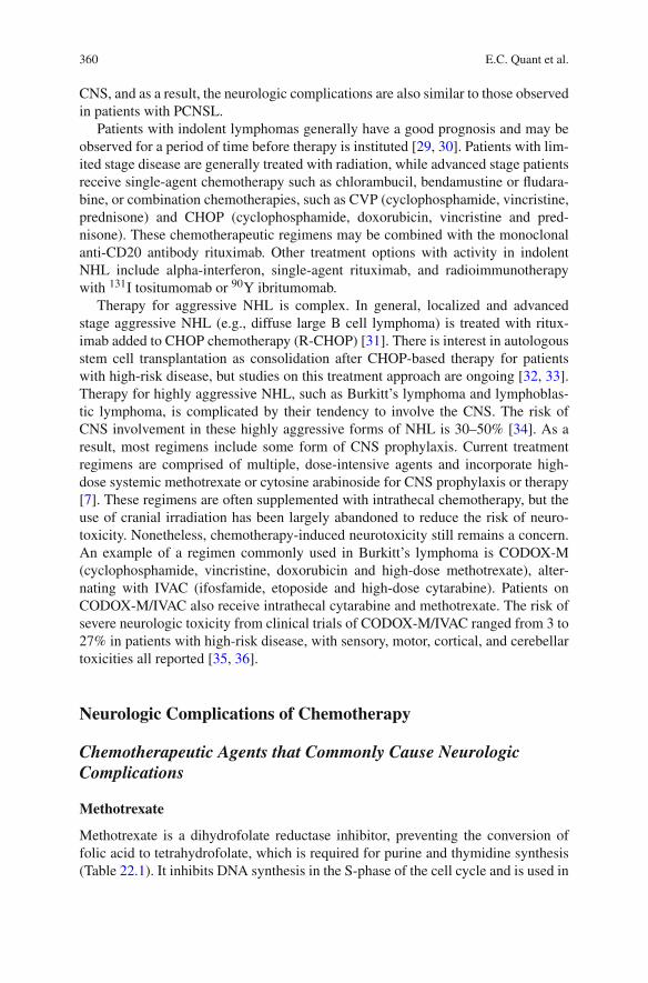
Table 22.1 Neurologic complications of chemotherapy for patients with leukemia or lymphoma
Acute encephalopathyCisplatinCyclophosphamideCytosine arabinoside (HD)DenileukinDoxorubicin (IT)Etoposide (HD)
FludarabineGM-CSFIfosfamideInterferonsMechloramineMethotrexate
Procarbazine (HD)ThalidomideThiotepa (HD)Vinca alkaloids
Mood/Personality ChangeCladribineCorticosteroids
InterferonsMechlorethamine
ThalidomideProcarbazine
HeadacheATRACisplatinCladribineCorticosteroidsCytosine arabinosideDenileukin
IbritumomabInterferonsMechlorethamineMethotrexate (IT)Oprelvekin (Neumega)
FludarabineRituximabThalidomideTositumomab
SeizuresBusulphan (HD)BCNUChlorambucil (HD)Cytosine arabinoside (HD or IT)
ErythropoietinFludarabineGM-CSFIfosfamide
MechloramineMethotrexateThalidomideVinca alkaloids
DementiaCytosine arabinoside Fludarabine
Interferon-alphaMethotrexateProcarbazine
Peripheral neuropathyCisplatinCladribineEtoposide
OprelvekinProcarbazine
ThalidomideVinca alkaloids
Cranial neuropathyCisplatin Methotrexate Vincristine
Hearing lossCisplatin Mechlorethamine
Visual disturbanceChlorambucilCorticosteroidsCyclophosphamide
Cytosine arabinosideEtoposideFludarabine
Interleukin-2Vincristine
Cortical blindnessCisplatinErythropoietin
FludarabineGM-CSF
Methotrexate (HD)Vinca alkaloids
MyelopathyBCNU (HD)CisplatinCorticosteroids
Cytosine arabinoside (IT)Interferon-alphaMethotrexate (IT)
Mitoxantrone (IT)Thiotepa (IT)Vincristine (IT)
362 E.C. Quant et al.
Table 22.1 (continued)
Extrapyramidal syndromesIfosfamide Interferon-alpha Vincristine
Cerebellar syndromeCytosine arabinoside (HD)Ifosfamide
Procarbazine ThalidomideVinca alkaloids
DizzinessCladribineCyclophosphamide
Rituximab Tositumomab
Vasculopathy and strokeBleomycinCisplatin
DoxorubicinErythropoietin
Methotrexate
Aseptic meningitisCytosine arabinoside (IT) Etoposide (IT)
Methotrexate (IT)Thiotepa (IT)
HD, high-dose; IT, intrathecal.
the treatment of leukemias, lymphomas, and leptomeningeal disease. Methotrexatecrosses the blood-brain barrier relatively poorly, but significant CNS concentra-tions can be achieved when the drug is administered intrathecally, or when highintravenous doses are used [19, 37]. The clinical expression of its neurotoxicity isdetermined by the dosage, route of administration, and the use of other therapeuticmodalities with overlapping neurotoxicities, such as irradiation.
Intrathecal methotrexate toxicity: Aseptic meningitis is the most common neu-rotoxicity associated with intrathecal methotrexate [38–40]. This occurs in approx-imately 10% of patients, although some series have reported incidences as high as50% [39, 41]. Symptoms usually begin 2–4 h after the drug is injected and maylast for 12–72 h. It is characterized by headache, nuchal rigidity, back pain, nausea,vomiting, fever, and lethargy, and is indistinguishable from other types of chemicalmeningitis. Cerebrospinal fluid (CSF) analysis shows a lymphocytic pleocytosis andan elevated protein, but cultures are negative. The symptoms are usually self-limitedand require no specific treatment. Most patients have no sequelae, although rarely itmay lead to delayed leukoencephalopathy [42]. Aseptic meningitis can be preventedto some extent by injecting methotrexate with hydrocortisone or using oral corticos-teroids. Some patients who developed aseptic meningitis have been retreated withmethotrexate without problems.
Transverse myelopathy, a much less common complication of intrathecalmethotrexate, is characterized by back or leg pain followed by paraplegia, sensoryloss, and sphincter dysfunction [41, 43, 44]. The symptoms usually occur between30 min and 48 h after treatment, but may occur up to 2 weeks later [41, 44]. Themajority of cases show clinical improvement, but the extent of recovery is variable[45]. The pathogenesis is unknown. Pathologically, there is vacuolar demyelina-tion and necrosis in the spinal cord, without inflammatory or vascular changes [46].
22 Neurological Complications of Chemotherapy 363
If present, abnormalities on magnetic imaging resonance (MRI) tend to be non-specific with spinal cord swelling, hyperintensity on T2-weighted sequences andcontrast enhancement [47]. Initial imaging is often normal. This complication ismore common in patients receiving concurrent radiotherapy or frequent treatmentsof intrathecal methotrexate.
Rarely, intrathecal methotrexate produces an acute encephalopathy [42], espe-cially if CSF outflow is obstructed, or if the methotrexate is injected directly intocerebral white matter as a result of a misplaced ventricular catheter [48]. Intrathecalmethotrexate can also cause delayed leukoencephalopathy [49, 50], posteriorreversible encephalopathy syndrome [51], seizures [52], subacute focal neuro-logic deficits [53], cranial nerve palsies, lumbosacral polyradiculopathy [54–56],noncardiogenic pulmonary edema [57], and sudden death [41, 58–60].
Accidental overdosage of intrathecal methotrexate (particularly doses > 500 mg)may result in myelopathy, encephalopathy, seizures, and death [61]. Prompt recog-nition and early treatment are essential to improve outcomes [62]. The use of rapidCSF drainage by lumbar puncture, CSF exchange, ventriculolumbar perfusion [63],carboxypeptidase G2 [64], high-dose intravenous leucovorin, and alkaline diuresishave allowed occasional patients to survive [59, 60].
Weekly low-dose methotrexate neurotoxicity: Up to 25% of patients receivingweekly, low dose methotrexate may experience headaches, dizziness, and sub-tle cognitive impairment [65]. These symptoms resolve when the methotrexate isdiscontinued.
High-dose methotrexate neurotoxicity: High-dose methotrexate may cause acute,subacute or chronic neurotoxicity. Renal function, hydration and alkalization, use ofleucovorin rescue, coadminstration of other antineoplastic agents and pharmacoge-nomics may significantly influence the risk of toxicity from high-dose methotrexate[66].
Acute high-dose methotrexate neurotoxicity is characterized by somnolence,confusion, and seizures within 24 h of treatment. Symptoms usually resolve spon-taneously without sequelae, and patients can often continue to receive this drug[41, 60, 67]. Although more commonly described with intrathecal methotrex-ate, an acute syndrome with symptoms and imaging findings similar to posteriorreversible encephalopathy syndrome has also been described in patients receiv-ing high-dose methotrexate [68, 69]. In one series, however, the patients also hadhypomagnesemia, raising the possibility that the electrolyte abnormality may havebeen a contributing factor.
Weekly or biweekly treatments with moderate to high-dose methotrexate mayproduce a subacute stroke-like syndrome characterized by transient focal neuro-logic deficits, confusion, and occasionally seizures [70, 71]. Typically, the disorderdevelops 6 days after high-dose methotrexate, lasts 15 min to 72 h, and resolvesspontaneously without sequelae. Neuroimaging studies are usually normal, althoughareas of restricted diffusion [72] and non-enhancing hyperintense T2 lesions in thewhite mater have been observed on MRI [73]. CSF is normal, but the EEG demon-strates diffuse slowing. Methotrexate may be administered subsequently withoutthe encephalopathy recurring. The pathogenesis of this syndrome is unknown, but
364 E.C. Quant et al.
may be related to reduced cerebral glucose metabolism [74], reduced biogenicamine synthesis [75], excess adenosine release through inhibition of AICAR-formyltransferase by methotrexate [76], or increased levels of homocysteine [66].
Chronic leukoencephalopathy has been reported in a number of patients follow-ing high-dose methotrexate and is discussed further below.
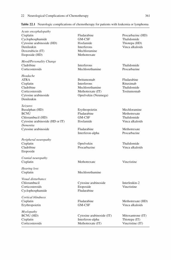
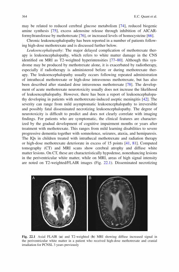
Leukoencephalopathy: The major delayed complication of methotrexate ther-apy is leukoencephalopathy, which refers to white matter damage in the CNSidentified on MRI as T2-weighted hyperintensities [77–80]. Although this syn-drome may be produced by methotrexate alone, it is exacerbated by radiotherapy,especially if radiotherapy is administered before or during methotrexate ther-apy. The leukoencephalopathy usually occurs following repeated administrationof intrathecal methotrexate or high-dose intravenous methotrexate, but has alsobeen described after standard dose intravenous methotrexate [78]. The develop-ment of acute methotrexate neurotoxicity usually does not increase the likelihoodof leukoencephalopathy. However, there has been a report of leukoencephalopa-thy developing in patients with methotrexate-induced aseptic meningitis [42]. Theseverity can range from mild asymptomatic leukoencephalopathy to irreversibleand possibly fatal disseminated necrotizing leukoencephalopathy. The degree ofneurotoxicity is difficult to predict and does not clearly correlate with imagingfindings. For patients who are symptomatic, the clinical features are character-ized by the gradual development of cognitive impairment months or years aftertreatment with methotrexate. This ranges from mild learning disabilities to severeprogressive dementia together with somnolence, seizures, ataxia, and hemiparesis.The IQs in children treated with intrathecal methotrexate and radiation therapyor high-dose methotrexate deteriorate in excess of 15 points [41, 81]. Computedtomography (CT) and MRI scans show cerebral atrophy and diffuse whitematter lesions. On CT, these are characteristically hypodense, nonenhancing lesionsin the periventricular white matter, while on MRI, areas of high signal intensityare noted on T2-weighted/FLAIR images (Fig. 22.1). Disseminated necrotizing
A B
Fig. 22.1 Axial FLAIR (a) and T2-weighted (b) MRI showing diffuse increased signal inthe periventricular white matter in a patient who received high-dose methotrexate and cranialirradiation for PCNSL 3 years previously
22 Neurological Complications of Chemotherapy 365
leukoencephalopathy produces a more rapid neurologic decline, and the MRI showsmore extensive white matter damage [82]. Pathologic lesions range from loss ofoligodendrocytes and gliosis to a necrotizing leukoencephalopathy [78]. There isdemyelination, axonal swelling, dystrophic mineralization of axonal debris, andfibrinoid necrosis of small blood vessels [41]. Occasionally, children may havea mineralizing microangiopathy, characterized by calcification of capillaries andvenules, especially in the basal ganglia [83]. The clinical course is variable. Manypatients stabilize, but the course is progressive in some patients and may lead todeath, particularly in disseminated necrotizing leukoencephalopathy. No effectivetreatment is available.
The cause of the leukoencephalopathy is unknown. Possibilities include injury tocerebral vascular endothelium, increasing blood-brain barrier permeability, deple-tion of reduced folates in the brain, inhibition of cerebral glucose or proteinmetabolism, inhibition of catecholamine synthesis [41, 74], or disturbance ofmyelin metabolism [84]. In addition, cranial irradiation may either potentiate thetoxic effects of methotrexate or disrupt the blood-brain barrier, allowing higherconcentrations of methotrexate to reach the brain.
Vinca Alkaloids: Vincristine (Oncovin), Vinblastine (Velban), Vindesine,and Vinorelbine (Navelbine)
Vincristine is a vinca alkaloid derived from the periwinkle plant, which is frequentlyused to treat leukemia, HD, and NHL. It binds to tubulin and prevents micro-tubule formation, thereby arresting cells in metaphase. Its main toxicity is an axonalneuropathy, resulting from disruption of the microtubules within axons and interfer-ence with axonal transport. The neuropathy involves both sensory and motor fibers,although small sensory fibers are especially affected [85, 86].
Virtually all patients receiving vincristine develop some degree of neuropathy.The clinical features resemble those of other axonal neuropathies, such as diabeticneuropathy. The earliest symptoms are usually paresthesias in the fingertips andfeet, with or without muscle cramps. These symptoms often develop after severalweeks of treatment, but they may occur after the first dose. Furthermore, symptomsmay appear even after the drug has been discontinued, and progress for severalmonths before improving. Initially, objective sensory findings tend to be relativelyminor compared to the subjective complaints, but loss of ankle jerks is common.Occasionally, there may be profound weakness, with bilateral foot drop, wrist drop,and loss of all sensory modalities. Neurophysiologic studies are compatible witha primarily axonal neuropathy [87]. Severe neuropathies are particularly likely todevelop in older or cachectic patients, those who have received prior irradiation tothe peripheral nerves or concomitant hematopoietic colony-stimulating factors [88],patients with hepatic insufficiency [89], and those who have pre-existing neurologicconditions, such as Charcot-Marie-Tooth [90, 91]. There is generally no effectivetreatment. Patients with mild neuropathy can usually continue to receive full dosesof vincristine, but when symptoms increase in severity and interfere with neurologic
366 E.C. Quant et al.
function, dose reduction or discontinuation of the drug may be necessary. The nat-ural history following discontinuation of treatment is gradual improvement, whichmay take up to several months [86]. Children tend to recover more quickly thanadults.
Autonomic neuropathies can also develop in patients receiving vincristine.Colicky abdominal pain and constipation occur in almost 50% of patients, and,rarely, a paralytic ileus may result [86]. Consequently, patients receiving vincristineshould take prophylactic stool softeners or laxatives. Less commonly, patients maydevelop impotence, postural hypotension, or an atonic bladder.
Cranial neuropathies may occasionally be caused by vincristine [60]. The mostcommon nerve to be involved is the oculomotor nerve, resulting in ptosis and oph-thalmoplegia. Other nerves that may be involved include the optic [92], recurrentlaryngeal, facial, and auditory. Vincristine may also cause retinal damage and nightblindness. Some patients may experience jaw and parotid pain.
CNS complications are rare as vincristine poorly penetrates the blood-brainbarrier. Accidental administration of vincristine into the CSF produces a rapidlyascending myelopathy, coma and usually, death [93]. Rarely, vincristine may causeSIADH, resulting in hyponatremia, confusion, and seizures [94]. CNS complica-tions unrelated to SIADH may also occur, including seizures [95], encephalopathy,transient cortical blindness [96], ataxia, athetosis, tremor and parkinsonism [41,60, 97].
The related vinca alkaloids vindesine, vinblastine, and vinorelbine tend to haveless neurotoxicity. This may be related to differences in lipid solubility, plasmaclearance, terminal half-life, and sensitivities of axoplasmic transport [41, 60]. Likevincristine, both vinblastine and vinorelbine inhibit microtubule assembly, but theyhave less affinity for neural tissue and are less neurotoxic. In fact, vinorelbine isassociated with mild paresthesias in only about 20% of patients [59], and severe neu-ropathy is rare, occurring most often in patients treated previously with paclitaxel[98].
Chemotherapeutic Agents that Occasionally Cause NeurologicComplications
Cytosine Arabinoside (Cytarabine, Ara-C)
Cytosine arabinoside is a pyrimidine analogue converted by deoxycytidine kinaseto its active metabolite Ara-CTP that inhibits DNA polymerase and incorporatesitself into the DNA molecule resulting in premature chain termination. This agenthas little neurotoxicity when used at conventional doses. High doses (3 g/m2 every12 h) cause an acute cerebellar syndrome in 10–25% of patients [99–101]. Patientsabove the age of 50 with abnormal liver or renal function, underlying neuro-logic dysfunction, or receiving more than 30 g of the drug are especially likelyto develop cerebellar involvement [59, 60, 102]. Typically, the patients developsomnolence and occasionally encephalopathy 2–5 days after completing treatment.
22 Neurological Complications of Chemotherapy 367
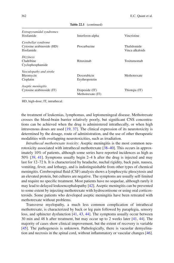
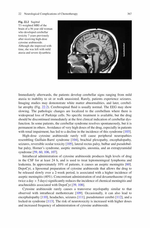
Fig. 22.2 SagittalT1-weighted MRI of thebrain of a 56 year old womanwho developed cerebellartoxicity 7 years previouslyafter receiving high-dosecytosine arabinoside.Although she improved withtime, she was left with mildataxia and severe dysarthria
Immediately afterwards, the patients develop cerebellar signs ranging from mildataxia to inability to sit or walk unassisted. Rarely, patients experience seizures.Imaging studies may demonstrate white matter abnormalities, and later, cerebel-lar atrophy (Fig. 22.2). Cerebrospinal fluid is usually normal. The EEG may showslowing. The pathologic changes are localized to the cerebellum where there iswidespread loss of Purkinje cells. No specific treatment is available, but the drugshould be discontinued immediately at the first clinical indication of cerebellar dys-function. In some patients, the cerebellar syndrome resolves spontaneously, but it ispermanent in others. Avoidance of very high doses of the drug, especially in patientswith renal impairment, has led to a decline in the incidence of this syndrome [103].
High-dose cytosine arabinoside rarely will cause peripheral neuropathiesresembling Guillain-Barré syndrome [104], brachial plexopathy, encephalopathy,seizures, reversible ocular toxicity [105], lateral rectus palsy, bulbar and pseudobul-bar palsy, Horner’s syndrome, aseptic meningitis, anosmia, and an extrapyramidalsyndrome [59, 60, 106, 107].
Intrathecal administration of cytosine arabinoside produces high levels of drugin the CSF for at least 24 h, and is used to treat leptomeningeal lymphoma andleukemia. In approximately 10% of patients, it causes an aseptic meningitis [60].DepoCyt, a liposomal preparation of cytosine arabinoside that allows the drug tobe released slowly over a 2-week period, is associated with a higher incidence ofaseptic meningitis (40%). Concomitant administration of oral dexamethasone (4 mgtwice a day × 5 days) significantly reduces the incidence of chemical meningitis andarachnoiditis associated with DepoCyt [39, 108].
Cytosine arabinoside rarely causes a transverse myelopathy similar to thatobserved with intrathecal methotrexate [109]. Occasionally, it can also lead toencephalopathy [110], headaches, seizures [111], pseudotumor cerebri [112], and alocked-in syndrome [113]. The risk of neurotoxicity is increased with higher dosesand increased frequency of administration of cytosine arabinoside.
368 E.C. Quant et al.
Ifosfamide (Ifex)
Ifosfamide is an analogue of cyclophosphamide occasionally used to treat lym-phoma. The most common neurotoxicity associated with ifosfamide is encephalopa-thy [114, 115]. Decreased attention, sometimes with agitation, may develop withinhours of administration and typically lasts 1–4 days. Estimations of incidence gen-erally range from 10 to 25% without evidence of a dose-response curve. Thepathophysiology of ifosfamide encephalopathy is unknown, but intoxication withchloracetaldehyde, a metabolic product of ifosfamide, is likely the critical factor.Small, non-randomized trials suggest that thiamine [116] or methylene blue [117]may prevent or treat the encephalopathy. Rarely, encephalopathy can progress tocoma or death [118, 119]. Rarely, ifosfamide causes extrapyramidal signs, cerebellarsigns, weakness, incontinence, or seizures [115, 119].
Chemotherapeutic Agents that Rarely Cause NeurologicComplications
Anthracycline Antibiotics (Doxorubicin [Adriamycin], Daunorubicin,Epirubicin, Idarubicin, Mitoxantrone)
Doxorubicin is an anthracycline antibiotic, which is frequently used to treat NHLas part of the CHOP regimen. It can cause arrhythmias and cardiomyopathies,which, in turn, can result in cerebrovascular complications [120]. Doxorubicin incombination with cyclosporine can lead to coma and death [60]. Accidental intrathe-cal injection can cause a myelopathy and encephalopathy [41, 121]. Idarubicin,epirubicin, and daunorubicin do not appear to be neurotoxic.
Bleomycin
Bleomycin is a mixture of polypeptide antibiotics that cuts DNA strands. Rarely,cardiovascular and cerebrovascular ischemia has been associated with multi-drugregimens including bleomycin. A causal link between bleomycin and stroke remainsspeculative.
Chlorambucil
This is an alkylating agent used for the treatment of HD and NHL. It usually has littleneurotoxicity, but can cause encephalopathy, myoclonus [122, 123], and seizureswhen taken in very high doses [124]. Ocular toxicity, including keratitis, retinaledema, and hemorrhages have also been described following oral administration ofchlorambucil [125].
Cladribine (2-Chlordeoxyadenosine)
This drug inhibits DNA polymerase and ligase and ribonucleotide reductase, result-ing in DNA strand breakage. It is used for low-grade NHL. It has little neurotoxicity
22 Neurological Complications of Chemotherapy 369
at conventional doses, but can produce a paraparesis [126], reversible blurred vision[127], or confusion [128] at high doses.
Cyclophosphamide (Cytoxan)
This is an alkylating agent used in the treatment of NHL and refractory leukemia.Standard dose cyclophosphamide has little neurotoxicity. High-dose cyclophos-phamide can cause reversible visual blurring, dizziness, and confusion [41].
Etoposide (VP-16)
This is a topoisomerase II inhibitor used in the treatment of refractory lymphoma.It does not readily penetrate the blood-brain barrier and generally has little neu-rotoxicity, even in high doses. Rarely, it can cause a peripheral neuropathy, milddisorientation, seizures, transient cortical blindness, or optic neuritis [41, 129].Intrathecal etoposide is associated with a mild transient arachnoiditis appearingwithin 1–5 days of treatment [130].
Fludarabine (Fludara)
Fludarabine, an inhibitor of DNA polymerase and ribonucleotide reductase, is usedto treat indolent lymphomas. Neurotoxicity is uncommon, and appears to be dose-related. Over one-third of patients receiving more than 96 mg/m2/day of intravenousfludarabine develop severe neurotoxicity, while less than 0.5% of patients receivingstandard doses of fludarabine (< 40 mg/m2/day) develop neurologic complications[131]. At low doses, fludarabine can cause headaches, somnolence, confusion, andparesthesias [41, 131–133]. Patients with mild neurologic complications usuallyimprove when the drug is discontinued, but some patients have permanent deficits[134]. At high doses, fludarabine can cause a delayed progressive encephalopathywith visual loss, tremor, ataxia, seizures, paralysis, and coma [131, 135, 136]. Someof these patients progress to a persistent vegetative state and occasionally, death.A few cases have been attributed to progressive multifocal leukoencephalopathydue to JC virus [137–140]. Patients may also develop a severe myelopathy, lead-ing to quadriparesis. MRI may show diffuse or multifocal areas of non-enhancing,increased T2 signal in the white matter and brainstem [131, 132]. Pathologically,there is multifocal demyelination and necrosis [133].
Mechlorethamine (Nitrogen Mustard)
This is an alkylating agent used to treat HD. Rarely, it causes sleepiness, headaches,weakness, hearing loss, and encephalopathy [41]. At high doses used for hematopoi-etic stem cell transplantation, it has been reported to cause confusion and seizures[141]. Intra-carotid administration produces a uveitis and cerebral necrosis [41,134]. Older age and concomitant use of cyclophosphamide or procarbazine areassociated with an increased risk of neurotoxicity.
370 E.C. Quant et al.
Pentostatin
This is an inhibitor of adenosine deaminase and is used for the treatment of cuta-neous T-cell lymphomas. At low doses, lethargy and fatigue are common. Higherdoses can cause a severe encephalopathy, seizures, and coma [59, 131].
Nitrosoureas (Carmustine, Lomustine)
Carmustine is the prototype for this group of drugs and is used in the treatmentof lymphomas. At conventional doses, carmustine does not cause neurotoxicity.However, patients who receive high dose intravenous carmustine can develop a pro-found encephalopathy, with or without a myelopathy that may progresses to comaand death [142].
Procarbazine (Matulane)
This is a weak monoamine oxidase inhibitor that probably acts as an alky-lating agent. It is used to treat both HD and NHL. At standard oral doses,it can cause a mild reversible encephalopathy and neuropathy, and rarely psy-chosis and stupor [41, 143]. The incidence of encephalopathy may be increasedin patients receiving “high dose” procarbazine [144]. Seventeen percent ofpatients develop a peripheral neuropathy with paresthesias and loss of deep ten-don reflexes, which is usually reversible [145]. Other rare side effects includeataxia, orthostatic hypotension, and weakness of the intrinsic hand muscles.Procarbazine also potentiates the sedative effects of narcotics, phenothiazines,and barbiturates. Intravenous and intracarotid procarbazine produces a severeencephalopathy.
Thiotepa (Thioplex)
This is an alkylating agent occasionally used to treat leptomeningeal lymphoma.Intrathecal thiotepa can cause aseptic meningitis, and very rarely, a myelopa-thy [146]. Both thiotepa and its metabolite, TEPA, are lipid soluble and readilycross the blood-brain barrier. High intravenous doses of thiotepa can produce anencephalopathy that can be fatal [59].
Neurologic Complications of Corticosteroids
Corticosteroids are used frequently in lymphoma patients. Corticosteroids have adirect cytolytic effect against neoplastic lymphocytes and are part of many treat-ment regimens. In addition, corticosteroids reduce peritumoral edema in patientswith CNS lymphoma and spinal cord edema in patients with epidural spinal cordcompression.
The side effects of prolonged steroid therapy are well known [147, 148]. Theincidence of complications increases with higher doses and prolonged therapy, but
22 Neurological Complications of Chemotherapy 371
individual susceptibility varies significantly. One of the most common complica-tions of corticosteroids is a steroid myopathy [149, 150]. Proximal muscle weaknessis initially apparent in the hip girdle, and patients may complain of difficulty gettingup from a chair or climbing stairs. In severe cases, the pectoral girdle and neck mus-cles may also be involved. Electromyography (EMG) is usually normal and serumcreatine kinase levels are typically not elevated.
CNS complications also occur frequently. Corticosteroids often produce alter-ations in mood [151]. An improved sense of well-being, anxiety, irritability,insomnia, difficulty concentrating, and depression are all relatively common.Occasionally, patients may develop steroid psychosis [152]. This usually takes theform of acute delirium, but the psychosis may resemble mania, depression, orschizophrenia.
Other common neurologic complications of corticosteroids include tremors,visual blurring, reduced sense of taste and smell, and cerebral atrophy onneuroimaging studies. Rare complications include hiccups [153], dementia,seizures, and cord compression as a result of epidural lipomatosis [154].
Steroid withdrawal can also produce a variety of symptoms, which can be quitedisabling. These include headaches, lethargy, nausea, vomiting, anorexia, myalgia,arthralgia, and postural hypotension. Rarely pseudotumor may occur.
Neurologic Complications of Biologic Agents
Alpha Interferon
This is a glycoprotein cytokine with antiviral, cytotoxic, and immunomodulatoryactivities. It is used therapeutically in patients with leukemia. Systemic toxicitiesinclude flu-like symptoms and myelosuppression. The flu-like symptoms, whichinclude lethargy and headaches, tend to be worse at the onset of therapy, andusually improve with time. Neurotoxicity tends to be dose-related. It is generallymild when low doses of alpha-interferon are used [155]. At higher doses, alpha-interferon can cause headaches, dizziness, confusion, lethargy, hallucinations, andseizures [156–158]. These effects are more common in older patients [159] and inpatients with prior psychiatric histories [160]. Neuroimaging studies are usually nor-mal. EEG may show diffuse slowing [161] and rarely, epileptiform activity [162].These neurotoxicities are usually reversible, but occasionally a permanent demen-tia or a persistent vegetative state may result [156, 157]. Rarely, alpha interferonhas been associated with oculomotor palsy, visual hallucinations, retinopathy [163],parkinsonism [134] and spastic diplegia [164].
A high incidence of neuropsychiatric toxicity has been noted in patientstreated with recombinant interferon alpha-2b (rIFNα2b). In one study of CMLpatients receiving rIFNα2b and low dose cytarabine, 24% of patients expe-rienced grade 3 or 4 neuropsychiatric toxicity that affected daily functioning[160]. All patients recovered upon withdrawal of interferon alpha-2b. Patients
372 E.C. Quant et al.
with a psychiatric history were more likely to develop severe neuropsychi-atric toxicity than patients without a psychiatric history. The mechanism ofinterferon neurotoxicity is unknown, but may include induction of proinflamma-tory cytokines, hyperactivity of corticotropin-releasing hormone-mediated stresspathway, and competition with naturally occurring neurotrophic hormones andopioids [134, 165].
Intrathecal administration of alpha interferon produces an acute reaction withinhours of the first injection, consisting of headache, nausea, vomiting, fever,and dizziness. The symptoms usually resolve over the next 12–24 h. A severeencephalopathy develops in a significant number of patients within several daysof the onset of treatment. This is dose dependent and tends to be worse in patientswho have received cranial irradiation [156].
Pegylated rIFN-α2b (PEG Intron) has similar toxicities as rIFN-α2b [166].
All-Trans Retinoic Acid (ATRA, Vesanoid)
This is a biologic agent used to treat acute promyelocytic leukemia. ATRA dif-ferentiates promyelocytes into mature cells. Pseudotumor cerebri is an uncommoncomplication, but headache is seen frequently [167, 168].
Monoclonal Antibodies
Rituximab (Rituxan)
This is a genetically engineered chimeric murine/human monoclonal antibodydirected against the CD20 antigen found on the surface of normal and malignantB lymphocytes. It is used for the treatment of NHL. Neurologic complications areuncommon, but some patients complain of headaches, myalgia, dizziness [169], orparesthesias [170].
Iodine-131 Tositumomab (Bexxar)
This is a radiolabeled immunoglobulin G-2a murine monoclonal antibody directedagainst the CD20 antigen. In addition to the cytotoxic effects induced by the anti-body, the presence of iodine-131 results in focused targeting of beta radiation to thetumor and surrounding tissue. Iodine-131 tositumomab is used to treat NHL and iswell tolerated. A minority of patients experience headache or myalgia and a fewdevelop hypothyroidism [171, 172].
Yttrium-90 Ibritumomab Tiuxetan (Zevalin)
This is also a radiolabeled murine monoclonal antibody directed against CD20 usedto treat NHL. It is well tolerated, but a few patients may complain of headaches ordizziness [173, 174].
22 Neurological Complications of Chemotherapy 373
Denileukin difitox (Ontak)
This is a fusion toxin used to treat cutaneous T-cell lymphoma, expressing theCD25 component for the IL-2 receptor. The most common complication is a vascu-lar leak syndrome, but some patients experience myalgias, dizziness, paresthesias,nervousness, confusion, and insomnia [175, 176].
Targeted Agents
Imatinib (Gleevec)
This is an oral tyrosine kinase inhibitor of the fusion protein bcr-abl found inPhiladelphia chromosome-positive leukemias. The most common neurotoxitiesare muscle cramping and myalgias, although symptoms are typically mild andrespond to treatment with calcium, magnesium, or quinine [177, 178]. Other rarecomplications include muscle edema, rhabdomyolysis with myoglobinuria [179],and subdural hematomas [180]. Related tyrosine kinase inihibitors, dasatinib andnilotinib, do not seem to cause neurotoxicity as frequently as imatinib [181].
References
1. Onciu M (2009) Acute lymphoblastic leukemia. Hematol Oncol Clin North Am 23:655–674
2. Shipley JL, Butera JN (2009) Acute myelogenous leukemia. Exp Hematol 37:649–658
3. Morris PG, Abrey LE (2009) Therapeutic challenges in primary CNS lymphoma. LancetNeurol 8:581–592
4. Hsi ED (2009) The leukemias of mature lymphocytes. Hematol Oncol Clin North Am23:843–871
5. Milojkovic D, Apperley J (2008) State-of-the-art in the treatment of chronic myeloidleukaemia. Curr Opin Oncol 20:112–121
6. Evens AM, Hutchings M, Diehl V (2008) Treatment of Hodgkin lymphoma: the past,present, and future. Nat Clin Pract Oncol 5:543–556
7. Sweetenham JW (2008) Highly aggressive lymphomas in adults. Hematol Oncol Clin NorthAm 22:965–978, ix
8. Thomas X, Le QH (2008) Central nervous system involvement in adult acute lymphoblasticleukemia. Hematology 13:293–302
9. Liesveld JL, Lichtman MA (2006) Acute myelogenous leukemia. In: Lichtman MA, BeutlerE, Kipps TJ, Seligsohn U, Kaushansky K, Prchal JT (eds) Williams hematology, 7th edn.McGraw-Hill Medical, New York, NY, pp 1183–1236
10. Pfeifer H, Wassmann BJ, Hofmann WK (2003) Risk and prognosis of central nervous systemleukemia in patients with Philadelphia chromosome-positive acute leukemias treated withimatinib mesylate. Clin Cancer Res 9:4674–4681
11. Rytting ME, Wierda WG (2004) Central nervous system relapse in two patients withchronic myelogenous leukemia in myeloid blastic phase on imatinib mesylate therapy. LeukLymphoma 45:1623–1626
12. Chiorazzi N, Rai KR, Ferrarini M (2005) Chronic lymphocytic leukemia. N Engl J Med352:804–815
374 E.C. Quant et al.
13. Denier C, Tertian G, Ribrag V et al (2009) Multifocal deficits due to leukemic meningoradi-culitis in chronic lymphocytic leukemia. J Neurol Sci 277:130–132
14. Batchelor T, Loeffler JS (2006) Primary CNS lymphoma. J Clin Oncol 24:1281–128815. Batchelor T, Carson K, O’Neil A et al (2003) Treatment of primary CNS lymphoma with
methotrexate and deferred radiotherapy: a report of NABTT 96-07. J Clin Oncol 21(6):1044–1049
16. Ferreri AJ, Crochhiolo R, Assanelli A et al (2008) High-dose chemotherapy supportedby autologous stem cell transplantation in patients with primary central nervous systemlymphoma: facts and opinions. Leuk Lymphoma 49:2042–2047
17. McAllister LD, Doolittle ND, Guastadisegni PE et al (2000) Cognitive outcomes and long-term follow-up results after enhanced chemotherapy delivery for primary central nervoussystem lymphoma. Neurosurgery 46:51–60; discussion 60–1
18. Shah GD, Yahalom J, Correa DD et al (2007) Combined immunochemotherapy with reducedwhole-brain radiotherapy for newly diagnosed primary CNS lymphoma. J Clin Oncol25:4730–4735
19. Glantz MJ, Cole BF, Recht L et al (1998) High-dose intravenous methotrexate for patientswith nonleukemic leptomeningeal cancer: is intrathecal chemotherapy necessary? J ClinOncol 16:1561–1567
20. Tetef ML, Margolin KA, Doroshow JH et al (2000) Pharmacokinetics and toxicity of high-dose intravenous methotrexate in the treatment of leptomeningeal carcinomatosis. CancerChemother Pharmacol 46:19–26
21. Vassal G, Valteau D, Bonnay M et al (1990) Cerebrospinal fluid and plasma methotrexatelevels following high-dose regimen given as a 3-hour intravenous infusion in children withnon-Hodgkin’s lymphoma. Pediatr Hematol Oncol 7:71–77
22. Omuro AM, Ben-Porat LS, Panageas KS et al (2005) Delayed neurotoxicity in primarycentral nervous system lymphoma. Arch Neurol 62:1595–1600
23. Abrey LE, DeAngelis LM, Yahalom J (1998) Long-term survival in primary CNS lym-phoma. J Clin Oncol 16(3):859–863
24. Abrey LE, Yahalom J, DeAngelis LM et al (2000) Treatment for primary CNS lymphoma:the next step. J Clin Oncol 18(17):3144–3150
25. Gavrilovic IT, Hormigo A, Yahalom J et al (2006) Long-term follow-up of high-dosemethotrexate-based therapy with and without whole brain irradiation for newly diagnosedprimary CNS lymphoma. J Clin Oncol 24:4570–4574
26. Seam P, Janik JE, DeAngelis LM et al (2009) Role of chemotherapy in Hodgkin’slymphoma. Cancer J 15:150–154
27. Brice P (2008) Managing relapsed and refractory Hodgkin lymphoma. Br J Haematol 141:3–13
28. Quddus F, Armitage JO (2009) Salvage therapy for Hodgkin’s lymphoma. Cancer J 15:161–163
29. Gribben JG (2007) How I treat indolent lymphoma. Blood 109:4617–462630. Feuerlein K, Zucca E, Ghielmini M et al (2009) First-line treatment of follicular lymphoma:
a patient-oriented algorithm. Leuk Lymphoma 50:325–33431. Michallet AS, Coiffier B (2009) Recent developments in the treatment of aggressive non-
Hodgkin lymphoma. Blood Rev 23:11–2332. Friedberg JW, Fisher RI (2008) Diffuse large B-cell lymphoma. Hematol Oncol Clin North
Am 22:941–952, ix33. Wrench D, Gribben JG (2008) Stem cell transplantation for non-Hodgkin’s lymphoma.
Hematol Oncol Clin North Am 22:1051–1079, xi34. Lim HY, Thiel E, Glantz MJ (2008) To protect and defend: central nervous system
prophylaxis in patients with non-Hodgkin’s lymphoma. Curr Opin Oncol 20:495–50135. Mead GM, Sydes MR, Walewski J et al (2002) An international evaluation of CODOX-
M and CODOX-M alternating with IVAC in adult Burkitt’s lymphoma: results of UnitedKingdom lymphoma group LY06 study. Ann Oncol 13:1264–1274
22 Neurological Complications of Chemotherapy 375
36. Magrath I, Adde M, Shad A et al (1996) Adults and children with small non-cleaved-celllymphoma have a similar excellent outcome when treated with the same chemotherapyregimen. J Clin Oncol 14:925–934
37. Lippens RJ, Winograd B (1988) Methotrexate concentration levels in the cerebrospinal fluidduring high-dose methotrexate infusions: an unreliable prediction. Pediatr Hematol Oncol5:115–124
38. Geiser CF, Bishop Y, Jaffe N et al (1975) Adverse effects of intrathecal methotrexate inchildren with acute leukemia in remission. Blood 45:189–195
39. Glantz MJ, Jaeckle KA, Chamberlain MC et al (1999) A randomized controlled trial compar-ing intrathecal sustained-release cytarabine (DepoCyt) to intrathecal methotrexate in patientswith neoplastic meningitis from solid tumors. Clin Cancer Res 5:3394–3402
40. Phillips PC (1991) Methotrexate toxicity. In: Rottenberg DA (ed) Neurological complica-tions of cancer treatment. Butterworth-heinemann, Boston, MA, pp 115–134
41. DeAngelis LM, Posner JB (2009) Side effects of chemotherapy: Neurologic complicationsof cancer. Oxford University Press, New York, NY, pp 447–510
42. Boogerd W, vd Sande JJ, Moffie D et al (1988) Acute fever and delayed leukoencephalopa-thy following low dose intraventricular methotrexate. J Neurol Neurosurg Psychiatry51(10):1277–1283
43. Bates S, McKeever P, Masur H et al (1985) Myelopathy following intrathecal chemotherapyin a patient with extensive Burkitt’s lymphoma and altered immune status. Am J Med 78:697–702
44. McLean DR, Clink HM, Ernst P et al (1994) Myelopathy after intrathecal chemotherapy. Acase report with unique magnetic resonance imaging changes. Cancer 73:3037–3040
45. Gagliano RG, Costanzi JJ (1976) Paraplegia following intrathecal methotrexate: report of acase and review of the literature. Cancer 37:1663–1668
46. Clark AW, Cohen SR, Nissenblatt MJ et al (1982) Paraplegia following intrathecalchemotherapy: neuropathologic findings and elevation of myelin basic protein. Cancer50:42–47
47. Counsel P, Khangure M (2007) Myelopathy due to intrathecal chemotherapy: magneticresonance imaging findings. Clin Radiol 62:172–176
48. Lemann W, Wiley RG, Posner JB (1988) Leukoencephalopathy complicating intraventric-ular catheters: clinical, radiographic and pathologic study of 10 cases. J Neurooncol 6:67–74
49. Baehring JM, Fulbright RK (2008) Delayed leukoencephalopathy with stroke-like presenta-tion in chemotherapy recipients. J Neurol Neurosurg Psychiatry 79:535–539
50. Riva D, Giorgi C, Nichellli F et al (2002) Intrathecal methotrexate affects cognitive functionin children with medulloblastoma. Neurology 59:48–53
51. Dicuonzo F, Salvati A, Palma M et al (2009) Posterior reversible encephalopathy syndromeassociated with methotrexate neurotoxicity: conventional magnetic resonance and diffusion-weighted imaging findings. J Child Neurol 24:1013–1018
52. Winick NJ, Bowman WP, Kamen BA et al (1992) Unexpected acute neurologic toxicity inthe treatment of children with acute lymphoblastic leukemia. J Natl Cancer Inst 84:252–256
53. Yim YS, Mahoney DH Jr, Oshman DG (1991) Hemiparesis and ischemic changes of thewhite matter after intrathecal therapy for children with acute lymphocytic leukemia. Cancer67:2058–2061
54. Anderson SC, Baquies GD, Jackson A et al (2002) Ventral polyradiculopathy with pediatricacute lymphocytic leukemia. Muscle Nerve 25(1):106–110
55. Koh S, Nelson MD Jr, Kovanlikaya A et al (1999) Anterior lumbosacral radiculopathy afterintrathecal methotrexate treatment. Pediatr Neurol 21:576–578
56. Pascual AM, Coret F, Casanova B et al (2008) Anterior lumbosacral polyradiculopathy afterintrathecal administration of methotrexate. J Neurol Sci 267(1-2):158–161
57. Bernstein ML, Sobel DB, Wimmer RS (1982) Noncardiogenic pulmonary edema followinginjection of methotrexate into the cerebrospinal fluid. Cancer 50:866–868
376 E.C. Quant et al.
58. Shore T, Barnett MJ, Phillips GL (1990) Sudden neurologic death after intrathecalmethotrexate. Med Pediatr Oncol 18:159–161
59. Paleologos N (1998) Complications of chemotherapy. In: Biller J (ed) Iatrogenic neurology.Butterworth-heinemann, Boston, MA, pp 439–460
60. Dietrich J, Wen PY (2008) Neurologic complications of chemotherapy. In: Schiff D, KesariS, Wen PY (eds) Cancer neurology in clinical practice, Humana Press, Totowa, NJ, pp 287–326
61. Ettinger LJ (1982) Pharmacokinetics and biochemical effects of a fatal intrathecalmethotrexate overdose. Cancer 50:444–450
62. Finkelstein Y, Zevin S, Heyol J et al (2004) Emergency treatment of life-threateningintrathecal methotrexate overdose. Neurotoxicology 25:407–410
63. Spiegel RJ, Cooper PR, Blun RH et al (1984) Treatment of massive intrathecal methotrexateoverdose by ventriculolumbar perfusion. N Engl J Med 311:386–388
64. Widemann BC, Balis FM, Shalabi A et al (2004) Treatment of accidental intrathe-cal methotrexate overdose with intrathecal carboxypeptidase G2. J Natl Cancer Inst96(20):1557–1559
65. Wernick R, Smith DL (1989) Central nervous system toxicity associated with weekly low-dose methotrexate treatment. Arthritis Rheum 32:770–775
66. Schmiegelow K (2009) Advances in individual prediction of methotrexate toxicity: a review.Br J Haematol 146:489–503
67. Rubnitz JE, Relling MV, Harrison PL et al (1998) Transient encephalopathy followinghigh-dose methotrexate treatment in childhood acute lymphoblastic leukemia. Leukemia12:1176–1181
68. Sanchez-Carpintero R, Narbona J, Lopez de Mesa R et al (2001) Transient posteriorencephalopathy induced by chemotherapy in children. Pediatr Neurol 24:145–148
69. Erbetta A, Salmaggi A, Sghirlanzoni A et al (2008) Clinical and radiological features ofbrain neurotoxicity caused by antitumor and immunosuppressant treatments. Neurol Sci29:131–137
70. Martino RL, Benson AB, 3rd, Merritt JA et al (1984) Transient neurologic dysfunction fol-lowing moderate-dose methotrexate for undifferentiated lymphoma. Cancer 54:2003–2005
71. Walker RW, Allen JC, Rosen G et al (1986) Transient cerebral dysfunction secondary tohigh-dose methotrexate. J Clin Oncol 4:1845–1850
72. Inaba H, Khan RB, Laninghan FH et al (2008) Clinical and radiological characteristics ofmethotrexate-induced acute encephalopathy in pediatric patients with cancer. Ann Oncol19:178–184
73. Borgna-Pignatti C, Battisti L, Marradi P et al (1992) Transient neurologic disturbances in achild treated with moderate-dose methotrexate. Br J Haematol 81:448
74. Phillips PC, Dhawan Y, Strother SC et al (1987) Reduced cerebral glucose metabolism andincreased brain capillary permeability following high-dose methotrexate chemotherapy: apositron emission tomographic study. Ann Neurol 21:59–63
75. Millot F, Dhondt JL, Hayte JM et al (1992) Impairment of cerebral biogenic amine synthesisin a patient receiving high-dose methotrexate. Am J Pediatr Hematol Oncol 14:276–278
76. Chabner BA, Allegra CJ, Curt GA et al (1985) Polyglutamation of methotrexate. Ismethotrexate a prodrug? J Clin Invest 76:907–912
77. Pizzo PA, Poplack DG, Blyer WA et al (1979) Neurotoxicities of current leukemia therapy.Am J Pediatr Hematol Oncol 1:127–140
78. Rubinstein LJ, Herman MM, Long TF et al (1975) Disseminated necrotizing leukoen-cephalopathy: a complication of treated central nervous system leukemia and lymphoma.Cancer 35:291–305
79. Shuper A, Stark B, Kornreich L et al (2000) Methotrexate treatment protocols and the centralnervous system: significant cure with significant neurotoxicity. J Child Neurol 15:573–580
80. Lai R, Abrey LE, Rosenblum MK et al (2004) Treatment-induced leukoencephalopathy inprimary CNS lymphoma: a clinical and autopsy study. Neurology 62:451–456
22 Neurological Complications of Chemotherapy 377
81. Ochs J, Mulhern R, Fairclough D et al (1991) Comparison of neuropsychologic function-ing and clinical indicators of neurotoxicity in long-term survivors of childhood leukemiagiven cranial radiation or parenteral methotrexate: a prospective study. J Clin Oncol 9:145–151
82. Oka M, Terae S, Kobayashi R et al (2003) MRI in methotrexate-related leukoencephalopa-thy: disseminated necrotising leukoencephalopathy in comparison with mild leukoen-cephalopathy. Neuroradiology 45:493–497
83. Price RA, Birdwell DA (1978) The central nervous system in childhood leukemia. III.Mineralizing microangiopathy and dystrophic calcification. Cancer 42:717–728
84. Davidson A, Payne G, Leach MO et al (2000) Proton magnetic resonance spectroscopy((1)H-MRS) of the brain following high-dose methotrexate treatment for childhood cancer.Med Pediatr Oncol 35:28–34
85. Casey EB, Jellife AM, Le Quesne PM et al (1973) Vincristine neuropathy. Clinical andelectrophysiological observations. Brain 96:69–86
86. Legha SS (1986) Vincristine neurotoxicity. Pathophysiology and management. Med Toxicol1:421–427
87. McLeod JG, Penny R (1969) Vincristine neuropathy: an electrophysiological and histologi-cal study. J Neurol Neurosurg Psychiatry 32:297–304
88. Weintraub M, Adde MA, Venzon DJ et al (1996) Severe atypical neuropathy associated withadministration of hematopoietic colony-stimulating factors and vincristine. J Clin Oncol14:935–940
89. Sandler SG, Tobin W, Henderson ES et al (1969) Vincristine-induced neuropathy. A clinicalstudy of fifty leukemic patients. Neurology 19:367–374
90. Nishikawa T, Kawakami K, Kumamoto T et al (2008) Severe neurotoxicities in a case ofCharcot-Marie-Tooth disease type 2 caused by vincristine for acute lymphoblastic leukemia.J Pediatr Hematol Oncol 30:519–521
91. Naumann R, Moham J, Reuner U et al (2001) Early recognition of hereditary motor andsensory neuropathy type 1 can avoid life-threatening vincristine neurotoxicity. Br J Haematol115:323–325
92. Munier F, Perentes E, Herbort CP et al (1992) Selective loss of optic nerve beta-tubulin invincristine-induced blindness. Am J Med 93:232–234
93. Gaidys WG, Dickerman JD, Walters CL et al (1983) Intrathecal vincristine. Report of a fatalcase despite CNS washout. Cancer 52:799–801
94. Robertson GL, Bhoopalam N, Zelkowitz LJ (1973) Vincristine neurotoxicity and abnormalsecretion of antidiuretic hormone. Arch Intern Med 132:717–720
95. Hurwitz RL, Mahoney DH Jr, Armstrong DL et al (1988) Reversible encephalopathy andseizures as a result of conventional vincristine administration. Med Pediatr Oncol 16:216–219
96. Byrd RL, Rohrbaugh TM, Raney RB Jr et al (1981) Transient cortical blindness secondaryto vincristine therapy in childhood malignancies. Cancer 47:37–40
97. Boranic M, Raci F (1979) A Parkinson-like syndrome as side effect of chemotherapy withvincristine and adriamycin in a child with acute leukaemia. Biomedicine 31:124–125
98. Fazeny B, Zifko U, Meryn S et al (1996) Vinorelbine-induced neurotoxicity in patients withadvanced breast cancer pretreated with paclitaxel – a phase II study. Cancer ChemotherPharmacol 39:150–156
99. Herzig RH, Hines JD, Herzig GP et al (1987) Cerebellar toxicity with high-dose cytosinearabinoside. J Clin Oncol 5(6):927–932
100. Hwang TL, Yung WK, Estey EH et al (1985) Central nervous system toxicity with high-doseAra-C. Neurology 35:1475–1479
101. Winkelman MD, Hines JD (1983) Cerebellar degeneration caused by high-dose cytosinearabinoside: a clinicopathological study. Ann Neurol 14:520–527
102. Gottlieb D, Bradstock K, Koutts J et al (1987) The neurotoxicity of high-dose cytosinearabinoside is age-related. Cancer 60:1439–1441
378 E.C. Quant et al.
103. Smith GA, Damon LE, Rugo HS et al (1997) High-dose cytarabine dose modificationreduces the incidence of neurotoxicity in patients with renal insufficiency. J Clin Oncol15:833–839
104. Johnson NT, Crawford SW, Sargur M (1987) Acute acquired demyelinating polyneuropa-thy with respiratory failure following high-dose systemic cytosine arabinoside and marrowtransplantation. Bone Marrow Transplant 2:203–207
105. Ritch PS, Hansen RM, Heuer DK (1983) Ocular toxicity from high-dose cytosine arabi-noside. Cancer 51:430–432
106. Baker WJ, Royer GL Jr, Weirs RB (1991) Cytarabine and neurologic toxicity. J Clin Oncol9:679–693
107. Luque FA, Selhorst JB, Petruska P (1987) Parkinsonism induced by high-dose cytosinearabinoside. Mov Disord 2:219–222
108. Glantz MJ, LaFollette S, Jaeckle KA et al (1999) Randomized trial of a slow-release versus astandard formulation of cytarabine for the intrathecal treatment of lymphomatous meningitis.J Clin Oncol 17:3110–3116
109. Dunton SF, Nitschke R, Spruce WE et al (1986) Progressive ascending paralysis follow-ing administration of intrathecal and intravenous cytosine arabinoside. A pediatric oncologygroup study. Cancer 57:1083–1088
110. Resar LM, Phillips PC, Kastan MB et al (1993) Acute neurotoxicity after intrathecal cytosinearabinoside in two adolescents with acute lymphoblastic leukemia of B-cell type. Cancer71:117–123
111. Eden OB, Goldie W, Wood T et al (1978) Seizures following intrathecal cytosine arabinosidein young children with acute lymphoblastic leukemia. Cancer 42:53–58
112. Fort JA, Smith LD (1999) Pseudotumor cerebri secondary to intermediate-dose cytarabineHCl. Ann Pharmacother 33:576–578
113. Kleinschmidt-DeMasters BK, Yeh M (1992) “Locked-in syndrome” after intrathe-cal cytosine arabinoside therapy for malignant immunoblastic lymphoma. Cancer 70:2504–2507
114. Watkin SW, Husband DJ, Green JA et al (1989) Ifosfamide encephalopathy: a reappraisal.Eur J Cancer Clin Oncol 25:1303–1310
115. Zalupski M, Baker LH (1988) Ifosfamide. J Natl Cancer Inst 80:556–566116. Buesa JM, Garcia-Teijido P, Losa R et al (2003) Treatment of ifosfamide encephalopathy
with intravenous thiamin. Clin Cancer Res 9:4636–4637117. Pelgrims J, De Vos F, Van den Brande J et al (2000) Methylene blue in the treatment and
prevention of ifosfamide-induced encephalopathy: report of 12 cases and a review of theliterature. Br J Cancer 82:291–294
118. Gieron MA et al (1988) Severe encephalopathy associated with ifosfamide administration intwo children with metastatic tumors. J Neurooncol 6(1):29–30
119. Shuper A, Stein J, Goshen J et al (2000) Subacute central nervous system degeneration in achild: an unusual manifestation of ifosfamide intoxication. J Child Neurol 15:481–483
120. Schachter S, Freeman R (1982) Transient ischemic attack and adriamycin cardiomyopathy.Neurology 32:1380–1381
121. Arico M, Nespoli L, Porta F et al (1990) Severe acute encephalopathy following inadvertentintrathecal doxorubicin administration. Med Pediatr Oncol 18:261–263
122. Vandenberg SA, Kulig K, Spoerke DG et al (1988) Chlorambucil overdose: accidentalingestion of an antineoplastic drug. J Emerg Med 6:495–498
123. Wyllie AR, Bayliff CD, Kovacs MJ (1997) Myoclonus due to chlorambucil in two adultswith lymphoma. Ann Pharmacother 31:171–174
124. Salloum E, Khan KK, Cooper DL (1997) Chlorambucil-induced seizures. Cancer 79:1009–1013
125. Burns LJ (1992) Ocular toxicities of chemotherapy. Semin Oncol 19:492–500126. Chabner BA, Longo DL (1996) Cancer chemotherapy and biotherapy: principles and
practice, Lippincott-Raven, Philadelphia, PA
22 Neurological Complications of Chemotherapy 379
127. Kende G, Sirkin SR, Thomas PR et al (1979) Blurring of vision: a previously undescribedcomplication of cyclophosphamide therapy. Cancer 44:69–71
128. Tashima CK (1975) Immediate cerebral symptoms during rapid intravenous administrationof cyclophosphamide (NSC-26271). Cancer Chemother Rep 59:441–442
129. Leff RS, Thompson JM, Daly MB et al (1988) Acute neurologic dysfunction after high-doseetoposide therapy for malignant glioma. Cancer 62:32–35
130. Chamberlain MC, Tsao-wei DD, Groshen S (2006) Phase II trial of intracerebrospinal fluidetoposide in the treatment of neoplastic meningitis. Cancer 106:2021–2027
131. Cheson BD, Vena DA, Foss FM et al (1994) Neurotoxicity of purine analogs: a review.J Clin Oncol 12:2216–2228
132. Cohen RB, Abdsallah JM, Gray JR et al (1993) Reversible neurologic toxicity in patientstreated with standard-dose fludarabine phosphate for mycosis fungoides and chronic lym-phocytic leukemia. Ann Intern Med 118:114–116
133. Spriggs DR, Stopa E, Mayer RJ et al (1986) Fludarabine phosphate (NSC 312878) infu-sions for the treatment of acute leukemia: phase I and neuropathological study. Cancer Res46:5953–5958
134. Hammack JE, Cascino TL (1998) Chemotherapy and other common drug-induced toxicitiesof the central nervous system in patients with cancer. In: Vecht CJ (ed) Handbook of clinicalneurology. Elsevier Science, Amsterdam, pp 481–514
135. Chun HG, Leyland-Jones BR, Caryk SM et al (1986) Central nervous system toxicity offludarabine phosphate. Cancer Treat Rep 70:1225–1228
136. Warrell RP Jr, Berman E (1986) Phase I and II study of fludarabine phosphate in leukemia:therapeutic efficacy with delayed central nervous system toxicity. J Clin Oncol 4:74–79
137. Cid J, Revilla M, Cervera A et al (2000) Progressive multifocal leukoencephalopathyfollowing oral fludarabine treatment of chronic lymphocytic leukemia. Ann Hematol79:392–395
138. Kiewe P, Seyfert S, Korper S et al (2003) Progressive multifocal leukoencephalopathy withdetection of JC virus in a patient with chronic lymphocytic leukemia parallel to onset offludarabine therapy. Leuk Lymphoma 44:1815–1818
139. Rodriguez L, Ribera JM, Batlle M et al (2002) Progressive multifocal leukoencephalopa-thy shortly after the diagnosis of follicular lymphoma in a patient treated with fludarabine.Haematologica 87:ECR26
140. Saumoy M, Castells G, Escoda L et al (2002) Progressive multifocal leukoencephalopa-thy in chronic lymphocytic leukemia after treatment with fludarabine. Leuk Lymphoma 43:433–436
141. Sullivan KM, Storb R, Shulman HM et al (1982) Immediate and delayed neurotoxicity aftermechlorethamine preparation for bone marrow transplantation. Ann Intern Med 97:182–189
142. Burger PC, Kamenar E, Schold SC et al (1981) Encephalomyelopathy following high-doseBCNU therapy. Cancer 48:1318–1327
143. Weiss HD, Walker MD, Wiernik PH (1974) Neurotoxicity of commonly used antineoplasticagents (second of two parts). N Engl J Med 291:127–133
144. Postma TJ, van Groenigen CJ, Witjes RJ et al (1998) Neurotoxicity of combinationchemotherapy with procarbazine, CCNU and vincristine (PCV) for recurrent glioma.J Neurooncol 38(1):69–75
145. Spivack SD (1974) Drugs 5 years later: procarbazine. Ann Intern Med 81:795–800146. Gutin PH, Levi JA, Wiernik PH et al (1977) Treatment of malignant meningeal disease with
intrathecal thioTEPA: a phase II study. Cancer Treat Rep 61:885–887147. Drappatz J, Schiff D, Kesaru S et al (2007) Medical management of brain tumor patients.
Neurol Clin 25:1035–1071148. DeAngelis LM, Posner JB (2009b) Supportive care and its complications: Neurologic
complications of cancer. Oxford University Press, New York, NY, pp 95–140149. Batchelor TT, Taylor LP, Thaler HT et al (1997) Steroid myopathy in cancer patients.
Neurology 48:1234–1238
380 E.C. Quant et al.
150. Dropcho EJ, Soong SJ (1991) Steroid-induced weakness in patients with primary braintumors. Neurology 41:1235–1239
151. Stiefel FC, Breitbart WS, Holland JC (1989) Corticosteroids in cancer: neuropsychiatriccomplications. Cancer Invest 7:479–491
152. Lewis DA, Smith RE (1983) Steroid-induced psychiatric syndromes. A report of 14 casesand a review of the literature. J Affect Disord 5:319–332
153. Baethge BA, Lidsky MD (1986) Intractable hiccups associated with high-dose intravenousmethylprednisolone therapy. Ann Intern Med 104:58–59
154. Haddad SF, Hitchon PW, Godersky JC (1991) Idiopathic and glucocorticoid-induced spinalepidural lipomatosis. J Neurosurg 74:38–42
155. Caraceni A, Gangeri L, Martini C et al (1998) Neurotoxicity of interferon-alpha in melanomatherapy: results from a randomized controlled trial. Cancer 83:482–489
156. Meyers CA, Obbens EA, Scheibel RS et al (1991) Neurotoxicity of intraventricularlyadministered alpha-interferon for leptomeningeal disease. Cancer 68:88–92
157. Meyers CA, Scheibel RS, Forman AD (1991) Persistent neurotoxicity of systemicallyadministered interferon-alpha. Neurology 41:672–676
158. Rohatiner AZ, Prior PF, Burton AC et al (1983) Central nervous system toxicity ofinterferon. Br J Cancer 47:419–422
159. Spiegel RJ (1987) The alpha interferons: clinical overview. Semin Oncol 14:1–12160. Hensley ML, Peterson B, Silver RT et al (2000) Risk factors for severe neuropsychi-
atric toxicity in patients receiving interferon alfa-2b and low-dose cytarabine for chronicmyelogenous leukemia: analysis of Cancer and Leukemia Group B 9013. J Clin Oncol18:1301–1308
161. Smedley H, Katrak M, Sikora K et al (1983) Neurological effects of recombinant humaninterferon. Br Med J (Clin Res Ed) 286:262–264
162. Suter CC, Westmoreland BF, Shabrough FW et al (1984) Electroencephalographic abnor-malities in interferon encephalopathy: a preliminary report. Mayo Clin Proc 59:847–850
163. Hejny C, Sternberg P, Lawson DH et al (2001) Retinopathy associated with high-doseinterferon alfa-2b therapy. Am J Ophthalmol 131:782–787
164. Vesikari T, Nuutila A, Cantell K (1988) Neurologic sequelae following interferon therapy ofjuvenile laryngeal papilloma. Acta Paediatr Scand 77:619–622
165. Raison CL, Demetrashvili M, Capuron L et al (2005) Neuropsychiatric adverse effects ofinterferon-alpha: recognition and management. CNS Drugs 19:105–123
166. Michallet M, Maloisel F, Delain M et al (2004) Pegylated recombinant interferon alpha-2b vs recombinant interferon alpha-2b for the initial treatment of chronic-phase chronicmyelogenous leukemia: a phase III study. Leukemia 18:309–315
167. Adamson PC, Reaman G, Finklestein JZ et al (1997) Phase I trial and pharmacokineticstudy of all-trans-retinoic acid administered on an intermittent schedule in combination withinterferon-alpha2a in pediatric patients with refractory cancer. J Clin Oncol 15:3330–3337
168. Tallman MS, Andersen JW, Schiffer CA et al (1997) All-trans-retinoic acid in acutepromyelocytic leukemia. N Engl J Med 337:1021–1028
169. Maloney DG, Press OW (1998) Newer treatments for non-Hodgkin’s lymphoma: mono-clonal antibodies. Oncology (Williston Park) 12:63–76
170. Foran JM, Rohatiner AZ, Cunninghan D et al (2000) European phase II study of rituximab(chimeric anti-CD20 monoclonal antibody) for patients with newly diagnosed mantle-celllymphoma and previously treated mantle-cell lymphoma, immunocytoma, and small B-celllymphocytic lymphoma. J Clin Oncol 18:317–324
171. Kaminski MS, Estes J, Zasadny KR et al (2000) Radioimmunotherapy with iodine (131)Itositumomab for relapsed or refractory B-cell non-Hodgkin lymphoma: updated results andlong-term follow-up of the University of Michigan experience. Blood 96:1259–1266
172. Vose JM, Wahl RL, Saleh M et al (2000) Multicenter phase II study of iodine-131 tositu-momab for chemotherapy-relapsed/refractory low-grade and transformed low-grade B-cellnon-Hodgkin’s lymphomas. J Clin Oncol 18:1316–1323
22 Neurological Complications of Chemotherapy 381
173. Dillman RO, Hendrix CS (2003) Unique aspects of supportive care using monoclonalantibodies in cancer treatment. Support Cancer Ther 1:38–48
174. Morschhauser F, Radford J, Van Hoof A et al (2008) Phase III trial of consolidation ther-apy with yttrium-90-ibritumomab tiuxetan compared with no additional therapy after firstremission in advanced follicular lymphoma. J Clin Oncol 26:5156–5164
175. Apisarnthanarax N (2002) Treatment of cutaneous T cell lymphoma: current status andfuture directions. Am J Clin Dermatol 3:193–215
176. Olsen E, Duvic M, Frankal A et al (2001) Pivotal phase III trial of two dose levels ofdenileukin diftitox for the treatment of cutaneous T-cell lymphoma. J Clin Oncol 19:376–388
177. Deininger MW, O’Brien SG, Ford JM et al (2003) Practical management of patients withchronic myeloid leukemia receiving imatinib. J Clin Oncol 21:1637–1647
178. Kantarjian H, Sawyers C, Hochhaus A et al (2002) Hematologic and cytogenetic responsesto imatinib mesylate in chronic myelogenous leukemia. N Engl J Med 346:645–652
179. Penel N, Blay JY, Adenis A et al (2008) Imatinib as a possible cause of severe rhabdomyol-ysis. N Engl J Med 358:2746–2747
180. Song KW, Rifkind J, Al-Beirouti B et al (2004) Subdural hematomas during CML therapywith imatinib mesylate. Leuk Lymphoma 45:1633–1636
181. Schiff D, Wen PY, van den Bent MJ et al (2009) Neurological adverse effects caused bycytotoxic and targeted therapies. Nat Rev Clin Oncol 6:596–603