Embed Size (px)
Citation preview

This is an Open Access document downloaded from ORCA, Cardiff University's institutional
repository: http://orca.cf.ac.uk/110332/
This is the author’s version of a work that was submitted to / accepted for publication.
Citation for final published version:
Menzel, Claudia, Holzeisen, Thomas, Laffleur, Flavia, Zaichik, Sergey, Abdulkarim, Muthanna,
Gumbleton, Mark and Bernkop-Schnürch, Andreas 2018. In vivo evaluation of an oral self-
emulsifying drug delivery system (SEDDS) for exenatide. Journal of Controlled Release 277 , pp.
165-172. 10.1016/j.jconrel.2018.03.018 file
Publishers page: http://dx.doi.org/10.1016/j.jconrel.2018.03.018
<http://dx.doi.org/10.1016/j.jconrel.2018.03.018>
Please note:
Changes made as a result of publishing processes such as copy-editing, formatting and page
numbers may not be reflected in this version. For the definitive version of this publication, please
refer to the published source. You are advised to consult the publisher’s version if you wish to cite
this paper.
This version is being made available in accordance with publisher policies. See
http://orca.cf.ac.uk/policies.html for usage policies. Copyright and moral rights for publications
made available in ORCA are retained by the copyright holders.

In vivo evaluation of an oral self-emulsifying drug delivery system (SEDDS) for exenatide
Claudia Menzel1, Thomas Holzeisenl, Flavia Laffleur1, Sergey Zaichik1, Muthanna Abdulkarim2, Mark
Gumbleton2, and Andreas Bernkop-Schnürch1*
1 Center for Chemistry and Biomedicine,
Department of Pharmaceutical Technology,
Institute of Pharmacy, University of Innsbruck,
Innrain 80/82, 6020 Innsbruck, Austria
2 School of Pharmacy and Pharmaceutical Sciences
Cardiff University
Cardiff, CF 10 3 NB
United Kingdom
*Corresponding Author:
Center for Chemistry and Biomedicine,
Department of Pharmaceutical Technology,
Institute of Pharmacy, University of Innsbruck,
Innrain 80/82, 6020 Innsbruck, Austria
Tel.: +43-512-507 58601
Fax: +43-512-507 58699
e-mail: [email protected]
2
Graphical Abstract

3
Abstract
Background: The aim of the study was to develop an oral self-emulsifying drug delivery system
(SEDDS) for exenatide and to evaluate its in vivo efficacy.
Methods: Exenatide was lipidised via hydrophobic ion pairing with sodium docusate (DOC) and
incorporated in SEDDS consisting of 35 % Cremophor EL, 25 % Labrafil 1944, 30 % Capmul-
PG 8 and 10 % propylene glycol. Exenatide/DOC was characterized in terms of lipophilicity
evaluating the octanol/water phase distribution (logP). Exenatide/DOC SEDDS were
characterized via droplet size analysis, drug release characteristics (log DSEDDS/release medium
determination) and mucus permeation studies. Furthermore, the impact of orally administered
exenatide/DOC SEDDS on blood glucose level was investigated in vivo on healthy male
Sprague-Dawley rats.
Results: Hydrophobic ion pairing in a molar ratio of 1:4 (exenatide:DOC) increased the effective
logP of exenatide from -1.1 to 2.1. SEDDS with a payload of 1% exenatide/DOC had a mean
droplet size of 45.87 ± 2.9 nm and a Log D SEDDS/release medium of 1.9 ±0.05. Permeation
experiments revealed 2.7-fold improved mucus diffusion for exenatide/DOC SEDDS compared
to exenatide in solution. Orally administered exenatide/DOC SEDDS showed a relative
bioavailability (versus s.c.) of 14.62 % ± 3.07 % and caused a significant (p < 0.05) 20.6 %
decrease in AUC values of blood glucose levels.
Conclusion: According to these results, hydrophobic ion pairing in combination with SEDDS
represents a promising tool for oral peptide delivery.

4
Introduction
During the last decades, rates of diabetes mellitus type 2 (T2D) have increased tremendously
making it one of the most common causes of death in the world, especially in countries with a
high percentage of obesity [1] [2]. Many approaches have been applied in the treatment of this
metabolic disorder that is characterized by elevated blood sugar levels, insufficient insulin
secretion and/or insulin resistance. One of the more recent developments was the launch of
exenatide (marketed as Byetta, Bydureon) in 2005 by Eli Lilly and Company. This 39-amino
acid, glucogon-like peptide-1 glucagon-like peptide-1 agonist increases insulin secretion,
suppresses pancreatic glucagon release and slows down gastric emptying [3]. As a peptide drug it
was developed to be administered by subcutaneous (s.c.) injection. However, adherence to
medication is critical for T2D patients, and injectable options are recognised to deter patient
compliance [4]. The long-acting injectables also fail to fully recapitulate the temporal nature of
the endogenous hormones released from intestinal L-cells during feeding and also the localized
first-pass hepatic actions. Injectables have a greater risk of hypo-glycaemia and side-effects such
as nausea. Consequently, there is a huge demand for oral delivery systems.
One promising approach to improve oral bioavailability of such peptide drugs is the development
of self-emulsifying drug delivery systems (SEDDS) [5]. Belonging to the group of lipid-based
formulations, SEDDS can help overcome the main reasons for low oral absorption of peptide
drugs, namely enzymatic degradation by intestinal proteases, poor mucus permeating properties
and low cellular uptake. For example, it has been already proven, that hydrolyzing enzymes such
as pepsin, trypsin, chymotrypsin and elastase are not soluble in the oily SEDDS droplets and that
therapeutic peptides incorporated in SEDDS are therefore protected against enzymatic
degradation [6]. Further,, SEDDS bear a slippery surface facilitating mucus permeation even for

5
large molecules [7]. Moreover, the absorption membrane permeating properties of SEDDS have
been shown in various studies [8].
So far, however, this promising technology has not been utilized for oral delivery of exenatide. In
former studies, oral carrier systems for exenatide included nanoparticles, micelles and
microspheres showing promising results and indicating that the use of SEDDS could
provide a valuable contribution to the topic [9-12]. It was therefore the aim of this study to
increase oral bioavailability of exenatide by incorporating the drug in a SEDDS formulation. A
sufficient degree of lipophilicity of a peptide drug is crucial for successful incorporation into the
oily droplet core, as such the strategy of hydrophobic ion pairing based on ionic interactions
between the peptide and oppositely charged surfactants was utilized [13]. Subsequently, the most
promising ion pair was incorporated in a SEDDS formulation that had already shown promise to
enhance permeation and provide stability against hydrolysis by lipase [14]. The resulting drug
delivery system was investigated, inter alia, in terms of drug release, mucus permeating
properties and safety. Furthermore, the efficacy of this novel delivery system was evaluated in
rats in vivo.
Materials and Methods
Materials
Exenatide was purchased from Carbosynth Limited (United Kingdom). Capmul PG-8 EP/NF
(propylene glycol monocaprylate, HLB = 6.7) was a gift from Abitec (USA) and Labrafil M 1944
CS was donated by Gattefosse (France). Cremophor EL (polyethoxylated-35 castor oil, HLB =
13), propylene glycol, sodium docusate, sodium dodecylsulfate, sodium oleate, sodium

6
taurocholate and all other chemicals, reagents and solvents were purchased from Sigma Aldrich
(Germany).
Quantification of exenatide
The quantification of exenatide was performed via HPLC. In brief, samples containing exenatide
were analyzed on a Nucleosil 100-5 C18 column (250 × 4 mm) at 40 °C with gradient elution
(0.5 mL/min): 0–20 min; linear gradient; from 58% A/42% B to 26% A/74% B (eluent A: 0.1 %
trifluoracetic aicid in water; eluent B: 0.1% trifluoroacetic acidin 80% acetonitrile) with signal
dtermination by UV/VIS at 278 nm. Retention time was 8.7 min. The calibration curve was
established with exenatide in a range from 7.8 to 1000 μg/mL. The detection and quantification
limit was investigated in a validation study and was specified with 2 μg/mL and 3.9 μg/mL.
Hydrophobic ion pairing of exenatide
The strategy of hydrophobic ion pairing was applied to increase lipophilicity of exenatide and
facilitate incorporation into SEDDS. In order to identify the most promising candidate, different
surfactants were tested. Exenatide acetate was dissolved in 0.555 mM acetic acid (pH 4) to a
final concentration of 2 mg/mL. Sodium dodecylsulfate, sodium deoxycholate, sodium docusate,
sodium oleate and sodium taurocholate were dissolved in 0.555 mM acetic acid (pH 4) and added
dropwise to the exenatide solution under light shaking in a ratio of 1:1 (v/v). Surfactant solutions
were prepared in different concentrations in order to obtain molar ratios of 1:1, 1:2, 1:4 and 1:6
(exenatide:surfactant). The precipitated ion-pair was separated by centrifugation at 13000 rpm
(MiniSpin®, Eppendorf Austria GmbH). The supernatant was analyzed for remaining un-
precipitated exenatide via HLPC as described above. The ion pair was washed with water,
lyophilized at −30 °C and 0.01 mbar (Christ Gamma 1-16 LSC Freeze dryer) and stored at
−24 °C.

7
Determination of LogP (octanol/water) of exenatide/DOC and exenatide/SDS ion pairs
Ion pairs of exenatide with SDS and DOC in molar ratios of 1:1, 1:4 and 1:6 were prepared as
described above and the pH adjusted to 6.8 with NaOH. Afterwards, octanol was added to the
mixture in a volume ratio of 1:1 (octanol:aqueous phase). The mixtures were placed on a
thermomixer (900 rpm, 25 °C) for 30 min followed by phase separation via centrifugation (5 min,
13000 rpm, MiniSpin®, Eppendorf Austria GmbH)). The amount of exenatide in octanol and
water phase was determined via HPLC as described above. Octanol samples (10 μL) were diluted
prior to injection in HPLC with isopropanol (80 μL) and water (110 μL) to yield a final volume
of 200 μL. The same experiment was performed with aqueous exenatide solution that was
adjusted to pH 6.8 with NaOH. Furthermore, 100 % values were generated in order to exclude the
presence of exenatide in the interphase.
Preparation and characterization of SEDDS
For SEDDS development, the ion-pair exenatide/DOC in a ratio of 1:4 has been chosen. A
previously developed SEDDS formulation showing protection against enzymatic degradation was
employed. The proportions of the individual components were slightly adapted in order to
identify the most suitable composition for the incorporation of the ion pair. Therefore, different
ratios of Cremophor EL, Labrafil 1944, Capmul PG-8 and propylene glycol were homogenized
by vigorous stirring. The lyophilized exenatide/DOC was dissolved in a concentration of 1 % in
the pre-concentrate for in vitro studies. This concentration was found to be the maximum soluble
payload in the SEDDS formulation referring to an exenatide payload of 0.71 %. For in vivo
studies the ion-pair was dissolved in a concentration of 0.0842 % in order to reach an applicable
volume of 250 µL. For in vitro studies SEDDS were emulsified in 50 mM phosphate buffer pH
6.8 in a concentration of 20 % (m/v) under gentle shaking. The time of emulsification was

8
evaluated visually. The mean droplet size and the zeta potential were measured by dynamic light
scattering using a PSS NICOMP TM 380 DLS (Santa Barbara, CA, USA). SEDDS without
incorporated exenatide served as control.
Determination of log D SEDDS/release medium of exenatide/DOC
Log D SEDDS/release medium was calculated as a measure of the distribution of exenatide/DOC
between the oily droplet phase of SEDDS and the release medium. Therefore, lyophilized
exenatide/DOC (molar ratio 1:4) was dispersed in water, 50 mM phosphate buffer pH 6.8 or 100
mM HCl pH 1 in a concentration of 1 mg/mL. The suspensions were stirred for 6 hours and
centrifuged (13000 rpm, MiniSpin®, Eppendorf Austria GmbH). The supernatant was analyzed
by HPLC with respect to solubility of the ion pair in the respective release medium (CRM). The
solubility of the ion pair in the SEDDS concentrate corresponds to the maximum payload of 1 %
(CSEDDS). D values were calculated according to equation 1. The drug concentration in SEDDS
depending on the ratio between SEDDS (VSEDDS) and aqueous release medium (VRM) was
calculated according to equation 2.
log = log �
� � % = % + � �� ∗
Release studies
Release studies were performed via dialysis membrane method. Exenatide/DOC containing
SEDDS pre-concentrate was emulsified in 50 mM phosphate buffer pH 6.8 (20 %, m/v), and 1.75
mL of the resulting emulsion was further diluted with phosphate buffer to a total volume of 2.5
mL in order to approach sink conditions and to minimize the membrane effect leading to a
(equation 1)
(equation 2)

9
final exenatide concentration of 1 mg/mL. The same volume and concentration of exenatide
acetate in 50 mM phosphate buffer pH 6.8 served as control. Test solutions were filled into
dialysis tubes and placed in beakers with 10 mL 50 mM phosphate buffer pH 6.8. The release of
exenatide to the outer phase beyond the confines of the dialysis tube was analyzed via HPLC as
described above over 6 h.
Mucus diffusion studies
Investigation of mucus permeating properties of exenatide/DOC SEDDS were performed with
transwell® with ThinCerts (surface 33.6 mm2, pore size 0.4 μm, pore density (Greiner-BioOne,
Austria)) in 24-well plates and an amount of 60 mg of mucus [15]. Briefly, 250 µL of
exenatide/DOC SEDDS (20 % in 50 mM phosphate buffer pH 6.8) and the corresponding amount
of exenatide in 50 mM phosphate buffer pH 6.8 (1.426 mg/mL) were layered carefully over the
mucus in the donor compartment. The acceptor chamber was filled with 500 µL 50 mM
phosphate buffer pH 6.8. The plate was incubated at 37 °C under continuous shaking at 20 rpm
on an orbital shaker. At predetermined time points 100 μL aliquotes were withdrawn from the
acceptor compartment and replaced with the same volume of fresh buffer. The experiment was
performed over a time interval of 4 hours corresponding to the time of a complete mucus
turnover in the small intestine [16, 17]. The amount of permeated exenatide was calculated via
HPLC as described above with reference to a 100% control that was obtained by the same
procedure but without mucus in the donor chamber.
Resazurin assay
In order to examine cytotoxic potential of exenatide/DOC SEDDS a resazurin assay on Caco-2
cells was performed [18]. Briefly, Caco-2 cells were seeded on a 24 well plate (d = 1 × 10⁵
cells/well; 500 μL per well) in minimum essential medium (MEM) supplemented with 10% (v/v)

10
fetal calf serum (FCS) and penicillin/streptomycin solution (100 units/0.1 mg/L) and cultured for
2 weeks at 37 °C in an atmosphere of 5% CO2 and 95% relative humidity. During this period the
medium was changed every other day. Subsequently, cells were washed with phosphate buffered
saline (PBS) with a temperature of 37°C and treated with 500 µL of test solutions of different
concentrations from 2.5 mg/mL to 7 mg/mL. Pure MEM served as negative control and 1%
(w/v) Triton X 100 as positive control. After incubation time of 12 h cells were washed with PBS
and treated with a 5% (m/v) resazurin solution. Cells were incubated for additional 2 h and
fluorescence of the supernatant was measured at 540 nm excitation wavelength and 590 nm
emission wavelength (TECAN Infinite M200, Austria GmbH).
In vivo studies
In vivo studies were approved by the Ethical Committee of Austria and performed according to
the Principles of Laboratory Animal Care. Male Sprague-Dawley rats with a mean body weight
of 250–300 g were obtained from Janvier Labs (Saint Berthevin, France). For pharmacokinetic
studies, rats were randomly devided into 2 groups (n=3). The first group served as positive
control and received 60 µL of s.c. exenatide injection (0.333 mg/mL in PBS). The second group
received 150 µg of exenatide via oral gavage in form of 250 µL SEDDS pre-concentrate with
exenatide/DOC (0.0842 %, m/m). Blood samples were collected at time intervals of 0, 0.5, 1, 1.5,
2, 3.5, 6 and 10 hours following exenatide administration. Exenatide was extracted from plasma
according to a protocol from Phoenix Pharmaceuticals Inc.. In brief, samples were acidified by
equal volume of acidic buffer and centrifuged for 20 minutes at 17,000 x g (4°C). The collected
supernatant was loaded into a C18 column that was pretreated with the same buffer. Exenatide
was eluted from the column utilizing a second buffer supplied by Phoenix Pharmaceuticals Inc..
Then, exenatide buffer solution was freeze dried and exenatide concentration was measured by
Chemiluminescent Elisa technique (Phoenix Pharmaceuticals Inc). The concentration-time curve

11
was plotted and AUCs were measured by trapezoidal rule for all exenatide systems. Relative
bioavailability was evaluated according to equation 3.
��� ���� �� � �� ����� [%] = [�� ]� ��∗[ � �]�.[�� ]�. ∗[ � �]� �� ∗ (equation 3)
For pharmacodynamics studies, rats were randomly divided into 4 groups (n = 3). The first group
served as positive control and received 60 µL of s.c. exenatide injection (0.333 mg/mL in PBS)
and 10 min later 2 g/kg glucose by i.p. route (50% dextrose solution in sterile water). The second
and third group received 150 µg of exenatide via oral gavage followed by 2 g/kg glucose i.p. 3
hours later. The second group received the drug in form of 250 µL exenatide solution (0.6 mg/mL
in PBS) and the third group in form of 250 µL SEDDS pre-concentrate with exenatide/DOC
(0.0842 %, m/m). The fourth group served as negative control and received only 2 g/kg glucose
via i.p. route. The animals were fasted 12 h prior to oral administration and had free access to
water during the experiment. At predetermined time points blood samples were taken from the
tail vein and blood glucose was measured immediately by Glucometer AccuCheck ® Active (F.
Hoffmann-La Roche AG).
Statistical data analysis
The software Sigma Plot version 12.3 was used for the statistical data analysis. One way
ANOVA and Bonferroni t-test were performed with p<0.05 as the minimal level of significance.

12
Results and discussion
Preparation and characterization of ion-pairs
The strategy of hydrophobic ion-pairing was used in the present study to increase lipophilicity
and hence solubility of exenatide in the SEDDS pre-concentrate. Since complex formation via
ion pairing is based on ionic interactions without any chemical modification the peptide
functionality is maintained. In this study different surfactants in different ratios were tested. Oleic
acid, sodium deoxycholate and sodium taurocholate did not lead to any ion pair formation
indicating that obviously only surfactants with a permanent and pH-independent negative charge
precipitate with exenatide in acid environment. Therefore, sodium dodecyl sulfate and sodium
docusate were added in different ratios to exenatide solutions in 0.555 mM acetic acid pH 4.
Upon addition of surfactant to the peptide solution an immediate precipitation of ion pair was
observed. The extent of ion pair formation was evaluated by quantifying un-precipitated peptide
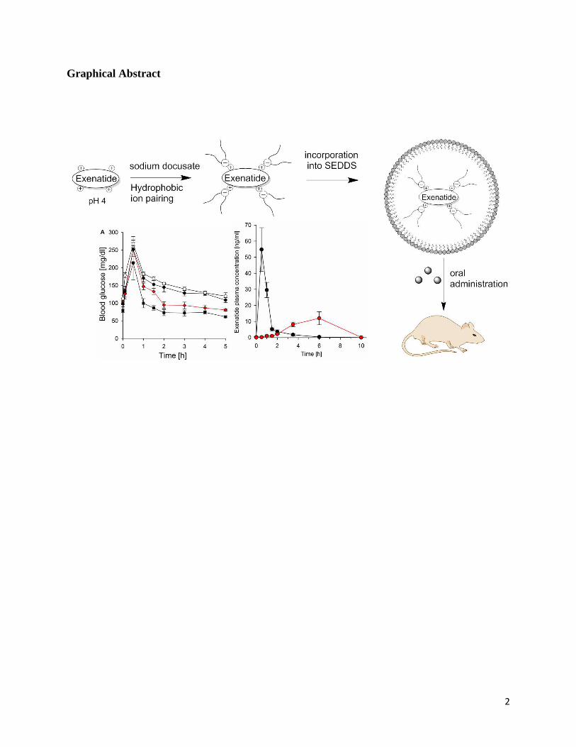
in the supernatant via HPLC. Figure 1 shows the percentage of precipitated exenatide depending
on the amount of added surfactant.
13
Figure 1: Precipitation of exenatide from 0.555 mM acetic acid (2 mg/mL) with DOC (●) and SDS (○). Precipitation of exenatide from 0.555 mM acetic acid (2 mg/mL) with DOC (○) and SDS (●). The precipitated exenatide-surfactant ion pair was isolated by centrifugation and the remaining exenatide analysed by HPLC. Data are shown as mean ± SD (n = 3).
In a molar ratio of 1:4 (surfactant:exenatide) both surfactants reached the maximum of
precipitated ion pair. This result is in accordance with the structure of exenatide: At pH 4, four
positive net charges can be assumed at position 1, 12, 20 and 27 in the amino acid sequence that
can form ionic complexes with four molecules of negatively charged surfactant. For each tested
ratio, DOC showed a higher rate of ion pair formation indicating superiority over SDS as
surfactant. This was also confirmed by the partitioning coefficient (logP) values of
exenatide/SDS and exenatide/DOC in different ratios (Figure 2). Determining the octanol/ water
partitioning coefficient of ion pairs provides information about their lipophilicity that is crucial
for efficient incorporation in SEDDS. The control experiment with exenatide alone proved the
hydrophilic nature of the peptide with a log P value of -1.14. With increasing amounts of
surfactant, log P values rapidly increases to 1.6 (SDS) or 2.1 (DOC). Both surfactants increased

14
lipophilicity most at a ratio of 1:4. At a ratio of 1:6 (surfactant:exenatide) log p values slightly
decreased. This might be due to formation of micellar complexes diffusing into the water phase.
According to these results, docusate in a molar ratio of 1:4 (exenatide:DOC) was chosen for
further experiments.
Figure 2: Log P (octanol/water) of aqueous exenatide solution pH 6.8 (black bar) and of exenatide/DOC (white bars) and exenatide/SDS (grey bars) at pH 6.8 depending on ratios between peptide and surfactant. Values are means of at least three experiments ± SD.
15
SEDDS development and characterization
Most of orally administered SEDDS formulations are prone to hydrolysis by pancreatic lipase, an
enzyme naturally occurring in duodenum, cleaving triglycerides in positions 1 and 3, resulting in
two free fatty acids and one 2- monoacylglyceride [19] [14]. The resulting deconstruction of the
emulsion can lead to enzymatic degradation of the ion pair under physiological conditions and an
unsatisfying in vitro/ in vivo correlation. In order to improve the in vivo situation, only lipolysis
stable components have been chosen in this study. The main oily components of the final SEDDS
concentrate are Labrafil 1944 and Capmul PG-8, both lacking classical triglyceride structure
being therefore stable against digestion. Furthermore, all components proved to be stable against
hydrolysis by lipase as confirmed in a former study [14]. The formation of stable SEDDS in the
gastro-intestinal tract needs a surfactant concentration ranging from 30-60 % (m/m). Cremophor-
EL was chosen because of its high HLB value ensuring high self-emulsifying properties and
small droplet sizes [20] and being less toxic compared to other ionic surfactants [21]. For
developing the SEDDS pre-concentrate, different ratios of surfactant and oily components were
tested. Each formulation contained propylene glycol as a co-solvent in a concentration of 10%.
According to a former study, the amount of gastrointestinal fluid in fasted rats is 3.2 ± 1.8 mL
[22]. In this study the amount of applied SEDDS formulation to the animals was 250 µL
corresponding to a SEDDS concentration of 7.8 % (m/v). However, as the SEDDS formulation is
not emulsified in the whole gastrointestinal fluid, a concentration of 20 % (m/v) in aqueous phase
was applied to test in vitro the emulsifying properties. Finding the most suitable composition for
the pre-concentrate, the focus was set on fast emulsification and a small droplet size with a low
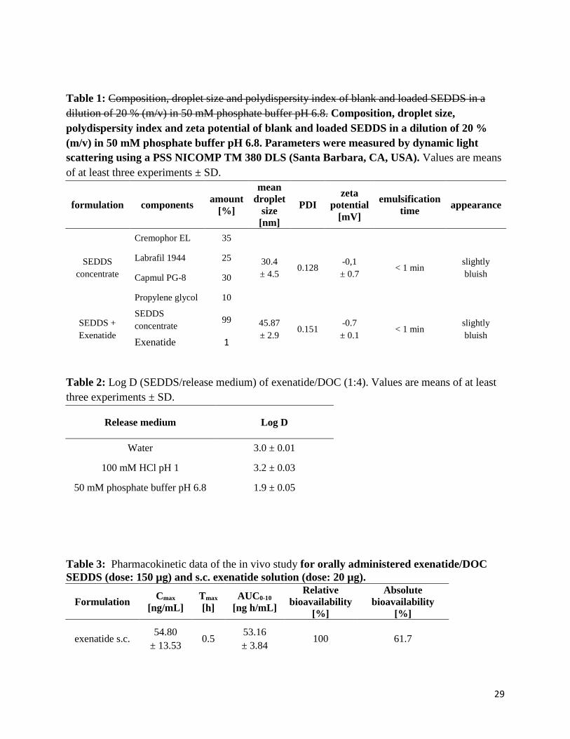
PDI value. Table 1 shows the pre-concentrate with the most favorable self-emulsifying properties
with and without incorporated ion pair. This composition was used for any further experiments.
The increase in mean droplet size due to addition of the ion pair previous to emulsification

16
indicates successful incorporation into the SEDDS droplets. Due to incorporation of the ion pair
into SEDDS, no precipitation was observed. Zeta potential measurements revealed an almost
uncharged surface for blank SEDDS and exenatide/DOC SEDDS.
Table 1: Composition, droplet size and polydispersity index of blank and loaded SEDDS in a dilution of 20 % (m/v) in 50 mM phosphate buffer pH 6.8. Composition, droplet size, polydispersity index and zeta potential of blank and loaded SEDDS in a dilution of 20 % (m/v) in 50 mM phosphate buffer pH 6.8. Parameters were measured by dynamic light scattering using a PSS NICOMP TM 380 DLS (Santa Barbara, CA, USA). Values are means of at least three experiments ± SD.
formulation components amount [%]
mean droplet
size [nm]
PDI zeta
potential [mV]
emulsification time appearance
SEDDS concentrate
Cremophor EL 35
30.4 ± 4.5
0.128 -0,1 ± 0.7
< 1 min slightly bluish
Labrafil 1944 25
Capmul PG-8 30
Propylene glycol 10
SEDDS + Exenatide
SEDDS concentrate
99 45.87 ± 2.9
0.151 -0.7 ± 0.1
< 1 min slightly bluish
Exenatide 1
Release studies
Release studies show the time dependent release of the drug from the oily SEDDS droplets into
the release medium. For quantification of the drug released from SEDDS the oily droplets need to
be separated. This was achieved using a separating dialysis membrane. The release profile of
exenatide/DOC SEDDS is displayed in Figure 3. As can be seen in the graph, this method has a
tremendous impact on the resulting release profile. Currently there is no valid method to
circumvent this problem determining the drug release from SEDDS in vitro to a satisfying extent.
In order to study this membrane effect and its influence on the release profile, the increase in

17
drug concentration in the acceptor medium was quantified with exenatide in the donor
compartment without any formulation. The time dependent increase in quantified drug from the
acceptor medium shows clearly the influence of the membrane. Taking this effect into account,
the release profile from the SEDDS formulation does not show any sustained release and is
simply based on the effect of the distribution of the ion pair between the oily phase and the
aqueous phase resulting in a lower drug concentration in the aqueous donor medium and
consequently in a flatter concentration gradient. As no sustained release could be observed, the
presence of fluid-crystalline partial structures in the SEDDS droplets is very unlikely, which was
confirmed by polarized microscopic analysis.
Figure 3: In vitro release of exenatide from solution (50 mM phosphate buffer pH 6.8) [●] and from exenatide/DOC SEDDS [○] at 37 °C over 6 h. Values are means of at least three experiments ± SD.

18
This data interpretation is confirmed by latest observations in this field [23]. The authors
postulate in this article that drug release from SEDDS is based on a simple diffusion process from
the oily droplet phase into an aqueous phase taking place within seconds. The drug release is
thereby just controlled by the partition coefficient (log D) of the ion pair between the lipophilic
SEDDS phase and the release medium. In the release study this phase distribution is set up
immediately after each sampling. Table 2 shows the log D SEDDS/release medium values dependent on
different release media. In the experimental setup, phosphate buffer was used as release medium
and the ratio between SEDDS and release medium was 1:35. The phase distribution between
SEDDS and buffer should be 70 % vs 30 %. Taken the membrane effect into account, the
immediate release of 20 % exenatide after 30 min is confirming this assumption.
Table 2: Log D (SEDDS/release medium) of exenatide/DOC (1:4). Values are means of at least three experiments ± SD.
Release medium Log D
Water 3.0 ± 0.01
100 mM HCl pH 1 3.2 ± 0.03
50 mM phosphate buffer pH 6.8 1.9 ± 0.05
Predicting the in vivo situation, a total volume of gastrointestinal fluid in rats of 3.2 mL (plus
0.25 mL water given by oral gavage to wash down SEDDS formulation) was assumed, meaning
that 85.2 % of the ion pair remain in the SEDDS droplets and only 14.8 % are released
immediately probably not reaching the epithelium.
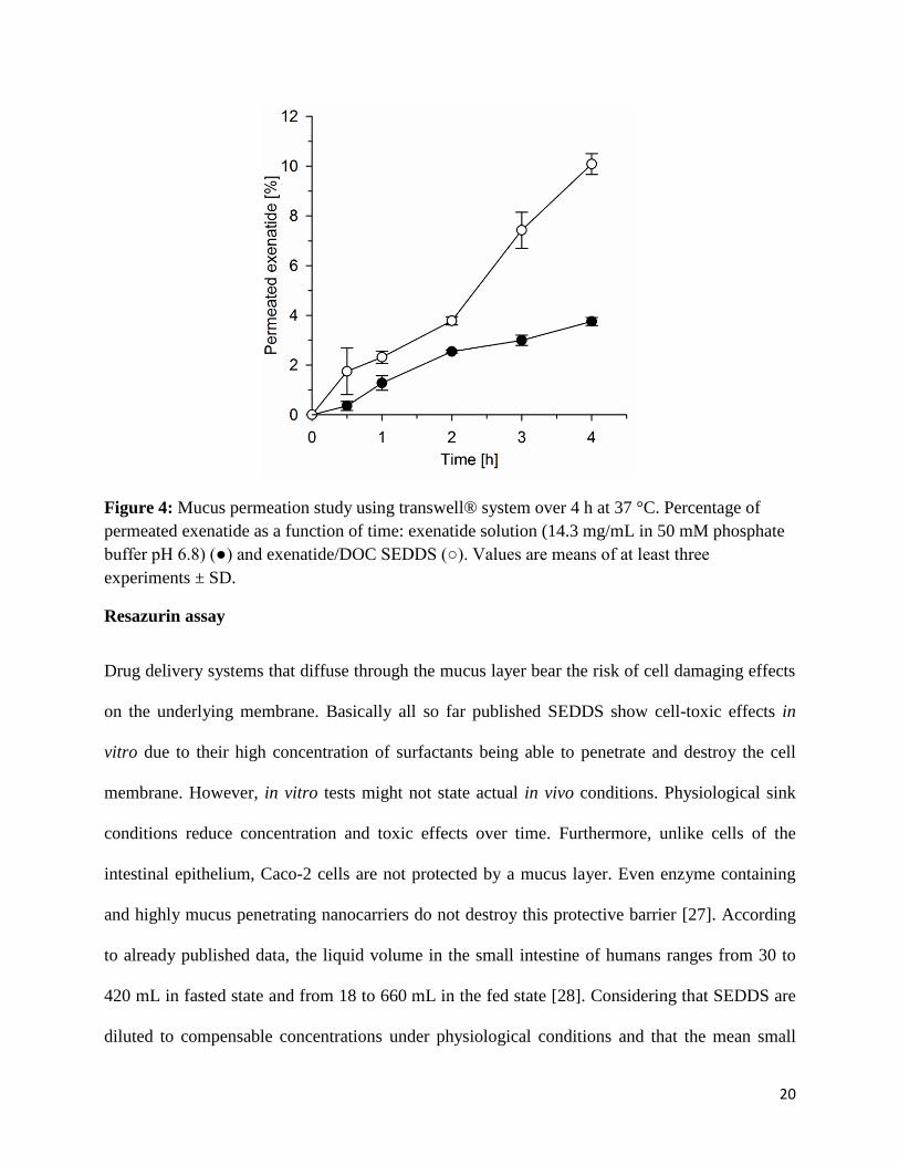
Mucus diffusion studies
One of the main reasons for poor drug absorption of peptide drugs is the mucus gel layer that is
composed of negatively charged glycoproteins covering the gastro intestinal tract. In order to

19
reach the epithelium drugs must overcome this natural barrier. Many substances and especially
peptide drugs get entrapped in this layer by ionic interactions with the glycoproteins. SEDDS
showed in various studies the potential to overcome this barrier due to their lipophilic nature, no
or negative surface charge and small droplet size [7, 24, 25]. Results from permeation studies are
provided in Figure 4 and show 2.7-fold improved mucus permeation for exenatide containing
SEDDS compared to the drug without formulation after four hours. The permeation enhancing
properties of Cremophor-EL itself might also contribute to this result. This surfactant showed
also the ability to open tight junctions of cells improving drug uptake [26]. However, the exact
mechanism of drug uptake from and/or of SEDDS has so far not been thoroughly understood.
Possible mechanisms are drug release outside the cell followed by uptake of the drug alone,
fusion of droplets with the bilayer of the cell membrane, passage of SEDDS through the
membrane by opening of tight junctions or the uptake of the entire SEDDS droplet via
endocytosis or even transcytosis.
20
Figure 4: Mucus permeation study using transwell® system over 4 h at 37 °C. Percentage of permeated exenatide as a function of time: exenatide solution (14.3 mg/mL in 50 mM phosphate buffer pH 6.8) (●) and exenatide/DOC SEDDS (○). Values are means of at least three experiments ± SD.
Resazurin assay
Drug delivery systems that diffuse through the mucus layer bear the risk of cell damaging effects
on the underlying membrane. Basically all so far published SEDDS show cell-toxic effects in
vitro due to their high concentration of surfactants being able to penetrate and destroy the cell
membrane. However, in vitro tests might not state actual in vivo conditions. Physiological sink
conditions reduce concentration and toxic effects over time. Furthermore, unlike cells of the
intestinal epithelium, Caco-2 cells are not protected by a mucus layer. Even enzyme containing
and highly mucus penetrating nanocarriers do not destroy this protective barrier [27]. According
to already published data, the liquid volume in the small intestine of humans ranges from 30 to
420 mL in fasted state and from 18 to 660 mL in the fed state [28]. Considering that SEDDS are
diluted to compensable concentrations under physiological conditions and that the mean small

21
intestinal passage time is 4 hours, a maximum concentration of 5 mg/mL (m/v) and an incubation
time of 4 hours was chosen in former studies [14]. In the present study, parameters were adjusted
to a maximum concentration of 7 mg/mL and an incubation time of 12 hours in order to cover
covering a larger range of concentration and dwell time in the body in order to study the full
cytotoxic potential over a longer time period in case of accumulation in the body. Figure 5
shows the influence of exenatide/DOC SEDDS on cell viability depending on SEDDS
concentration. A positive correlation between SEDDS concentration and cell toxicity could be
observed. At a concentration of 5 mg/mL cell viability was still at 70 %. This result is in good
agreement with former published results [14]. In the mentioned study, a blank SEDDS
formulation containing the same compounds as exenatide/DOC SEDDS showed a cell viability of
94.2 % after 4 hours. Considering the 3-fold longer incubation time in the present study, the 20 %
lower survival rate is well explainable. Results obtained from the cell disrupting substance
Triton x proved the reliability of the chosen method as measured fluorescence and hence
cell viability was remarkably decreased compared to the 100 % value of MEM treated cells

22
Figure 5: Cytotoxicity of exenatide/DOC SEDDS after 12 h incubation in concentrations of 2.5 mg/mL, 3 mg/mL, 5 mg/mL and 7 mg/mL10 mg/mL. Values are means of at least three experiments ± SD.
In vivo study
Results from pharmacokinetic studies are provided in Table 3 revealing a relative bioavailability
of exenatide/DOC SEDDS vs s.c. exenatide solution of 14.62 ± 3.07 %. As illustrated in Figure
6, plasma concentrations of exenatide s.c. reached their maximum after 0.5 h, whereas the plasma
concentrations of exenatide/DOC SEDDS were at their highest after 6 h due to a sustained
absorption from gastrointestinal tract. According to literature, orally applied exenatide solution
has a very low bioavailability with no detectable exenatide in plasma [29] [30]. Due to
incorporation into SEDDS a relative bioavailability of almost 15 % could be achieved being in
the same range [30] or even exceeding [29] results from former studies that used particulate
carrier systems for oral exenatide application. According to data from literature, absolute
bioavailability of exenatide s.c. compared to i.v. is 61.7 %, hence absolute bioavailability of
exenatide/DOC SEDDS vs i.v. application is likely 9.0 %. Accordingly, exenatide/DOC SEDDS

23
showed a strikingly improved absolute bioavailability compared to already published peptide
containing SEDDS showing absolute bioavailability values below 2.5 % (oral vs i.v.) [14] [26].
Table 3: Pharmacokinetic data of the in vivo study for orally administered exenatide/DOC SEDDS (dose: 150 µg) and s.c. exenatide solution (dose: 20 µg).
Formulation Cmax [ng/mL]
Tmax [h]
AUC0-10
[ng h/mL]
Relative bioavailability
[%]
Absolute bioavailability
[%]
exenatide s.c. 54.80
± 13.53 0.5
53.16 ± 3.84
100 61.7
exenatide/DOC SEDDS
8.12 ± 1.48 11.99 ±
4.10
6 58.79
± 15.83 14.62 ± 3.07
9 ± 1.89
Figure 6: Exenatide plasma concentration-time curve for orally administered exenatide/DOC SEDDS (dose: 150 µg) (○) and s.c. exenatide solution (dose: 20 µg) (●). Illustrated values are the means of at least three experiments ± standard deviation

24
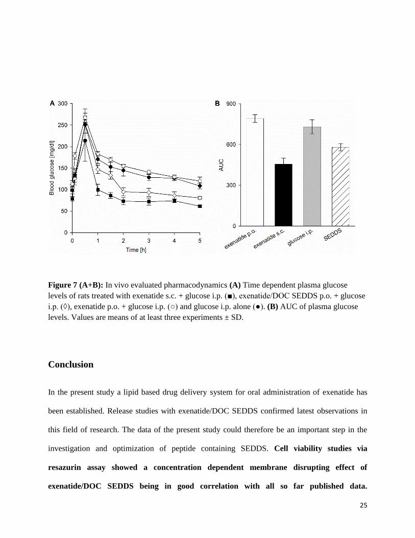
Since exenatide shows high activity, pharmacodynamic effects can be measured even at low
plasma concentrations. Exenatide binds to the GLP-1 receptor causing a glucose-dependent
insulin secretion followed by enhanced insulin response [31]. Therefore, time dependent blood
glucose levels were measured after glucose challenge. According to the manufacturer, exenatide
should be administered prior to meal times [31]. For s.c injection an interval of 40 min between
administration and glucose challenge was chosen and 3 hours for oral administered formulations.
The measured blood glucose levels over time are shown in Figure 7 (A). As the experiment was
performed with healthy rats, each group showed a fast decrease in blood glucose levels after the
initial glucose peak due to sufficient insulin response. However, this insulin response was
significantly increased in animals treated by s.c injection and orally administered SEDDS
formulation (p < 0.05). Thereby, the SEDDS caused a 20 % lower blood glucose level one hour
after glucose challenge compared to the control only treated with exenatide solution. Af ter two
hours, this effect was most pronounced with 38.9 % lower blood glucose levels in the SEDDS
group compared to the control group. Even after 5 hours blood glucose levels in the SEDDS
group were lower compared to the control. This might be due to the comparably long half-life of
the drug. Exenatide levels can be measured for approximately 10 hours bearing the risk of
hypoglycemia [32]. Figure 7 (B) shows the calculated AUC values of plasma glucose levels. In
the SEDDS group the AUC was 20.6 % smaller than in the control group. Furthermore, no
significant difference between the AUC of orally applied exenatide solution compared to the
control was observed (p < 0.05). According to these results there is strong evidence that
incorporation in SEDDS could enhance oral bioavailability of exenatide. Due to the high potency
of this drug, the usual problems of SEDDS formulations, e.g. low payload and low oral drug
absorption compared to s.c application, do not matter that much.
25
Figure 7 (A+B): In vivo evaluated pharmacodynamics (A) Time dependent plasma glucose levels of rats treated with exenatide s.c. + glucose i.p. (■), exenatide/DOC SEDDS p.o. + glucose i.p. (◊), exenatide p.o. + glucose i.p. (○) and glucose i.p. alone (●). (B) AUC of plasma glucose levels. Values are means of at least three experiments ± SD.
Conclusion
In the present study a lipid based drug delivery system for oral administration of exenatide has
been established. Release studies with exenatide/DOC SEDDS confirmed latest observations in
this field of research. The data of the present study could therefore be an important step in the
investigation and optimization of peptide containing SEDDS. Cell viability studies via
resazurin assay showed a concentration dependent membrane disrupting effect of
exenatide/DOC SEDDS being in good correlation with all so far published data.

26
Considering in vivo situation in the human body, however, SEDDS are way more diluted
and cells protected by a mucus barrier providing likely sufficient safety. Mucus diffusion
experiments as well as Pharmacokinetic pharmacokinetic and pharmacodynamic studies
revealed promising results indicating high in vitro / in vivo correlation: In vitro permeation
studies revealed 2.7-fold improved mucus permeation for exenatide containing SEDDS
compared to the drug without the formulation. In vivo evaluation showed a 20.6 %
decreased AUC of plasma glucose in the SEDDS group vs. the control group. This might be
on the one hand due to the high lipophilicity of the created ion pair being incorporated into
SEDDS and on the other hand due to the high potency of the peptide, enhancing insulin response
even at low plasma concentrations. Considering that only healthy animals were tested in this
study, effects on plasma glucose levels might be even more pronounced in type-2 diabetics.
References
[1] A. Axmon, G. Ahlström, P. Höglund, Prevalence and treatment of diabetes mellitus and hypertension
among older adults with intellectual disability in comparison with the general population, BMC
Geriatrics, 17 (2017) 272.
[2] S. Smyth, A. Heron, Diabetes and obesity: the twin epidemics, Nature Medicine, 12 (2006) 75.
[3] M.C. Bunck, M. Diamant, A. Cornér, B. Eliasson, J.L. Malloy, R.M. Shaginian, W. Deng, D.M. Kendall,
M.-R. Taskinen, U. Smith, H. Yki-Järvinen, R.J. Heine, One-Year Treat e t With Exe atide I proves β-Cell
Function, Compared With Insulin Glargine, in Metformin-Treated Type 2 Diabetic Patients: A
randomized, controlled trial, Diabetes Care, 32 (2009) 762-768.
[4] M. Brod, M. Rousculp, A. Cameron, Understanding compliance issues for daily self-injectable
treatment in ambulatory care settings, Patient preference and adherence, 2 (2008) 129-136.
[ ] O. Zupa čič, A. Bernkop-Schnürch, Lipophilic peptide character – What oral barriers fear the most,
Journal of Controlled Release, 255 (2017) 242-257.
[6] G. Hetényi, J. Griesser, M. Moser, F. Demarne, V. Jannin, A. Bernkop-Schnürch, Comparison of the
protective effect of self-emulsifying peptide drug delivery systems towards intestinal proteases and
glutathione, Int J Pharm, 523 (2017) 357-365.
[7] J. Griesser, G. Hetényi, H. Kadas, V. Jannin, F. Demarne, A. Bernkop-Schnüch, Self-emulsifying peptide
drug delivery systems: How to make them highly mucus permeating, Int J Pharm, 538 (2018) 159-166.
[8] G. Leonaviciute, A. Bernkop-Schnurch, Self-emulsifying drug delivery systems in oral (poly)peptide
drug delivery, Expert Opin Drug Deliv, 12 (2015) 1703-1716.

27
[9] Y. Shi, X. Sun, L. Zhang, K. Sun, K. Li, Y. Li, Q. Zhang, Fc-modified exenatide-loaded nanoparticles for
oral delivery to improve hypoglycemic effects in mice, Scientific reports, 8 (2018) 726.
[10] P. Wang, X. Zhuo, W. Chu, X. Tang, Exenatide-loaded microsphere/thermosensitive hydrogel long-
acting delivery system with high drug bioactivity, International journal of pharmaceutics, 528 (2017) 62-
75.
[11] L. Soudry-Kochavi, N. Naraykin, T. Nassar, S. Benita, Improved oral absorption of exenatide using an
original nanoencapsulation and microencapsulation approach, Journal of controlled release : official
journal of the Controlled Release Society, 217 (2015) 202-210.
[12] C. Chen, X. Zhu, Y. Dou, J. Xu, J. Zhang, T. Fan, J. Du, K. Liu, Y. Deng, L. Zhao, Y. Huang, Exendin-4
Loaded Nanoparticles with a Lipid Shell and Aqueous Core Containing Micelles for Enhanced Intestinal
Absorption, Journal of biomedical nanotechnology, 11 (2015) 865-876.
[13] J. Griesser, G. Hetényi, M. Moser, F. Demarne, V. Jannin, A. Bernkop-Schnürch, Hydrophobic ion
pairing: Key to highly payloaded self-emulsifying peptide drug delivery systems, Int J Pharm, 520 (2017)
267-274.
[ ] O. Zupa čič, J.A. Grieβi ger, J. Rohrer, I. Pereira de Sousa, L. Da i ger, A. Parte hauser, N.E. Sündermann, F. Laffleur, A. Bernkop-Schnürch, Development, in vitro and in vivo evaluation of a self-
emulsifying drug delivery system (SEDDS) for oral enoxaparin administration, European Journal of
Pharmaceutics and Biopharmaceutics, 109 (2016) 113-121.
[15] H. Friedl, S. Dünnhaupt, F. Hintzen, C. Waldner, S. Parikh, J.P. Pearson, M.D. Wilcox, A. Bernkop-
Schnürch, Development and evaluation of a novel mucus diffusion test system approved by self-
nanoemulsifying drug delivery systems, J Pharm Sci, 102 (2013) 4406-4413.
[16] S.K. Lai, Y.-Y. Wang, J. Hanes, Mucus-penetrating nanoparticles for drug and gene delivery to
mucosal tissues, Advanced drug delivery reviews, 61 (2009) 158-171.
[17] C.-M. Lehr, F.G.J. Poelma, H.E. Junginger, J.J. Tukker, An estimate of turnover time of intestinal
mucus gel layer in the rat in situ loop, International journal of pharmaceutics, 70 (1991) 235-240.
[18] P. Jennings, C. Koppelstaetter, S. Aydin, T. Abberger, A.M. Wolf, G. Mayer, W. Pfaller, Cyclosporine A
induces senescence in renal tubular epithelial cells, American journal of physiology. Renal physiology,
293 (2007) F831-838.
[19] A. Müllertz, A. Ogbonna, S. Ren, T. Rades, New perspectives on lipid and surfactant based drug
delivery systems for oral delivery of poorly soluble drugs, J Pharm Pharmacol, 62 (2010) 1622-1636.
[20] M.A. Rahman, A. Hussain, M.S. Hussain, M.A. Mirza, Z. Iqbal, Role of excipients in successful
development of self-emulsifying/microemulsifying drug delivery system (SEDDS/SMEDDS), Drug Dev Ind
Pharm, 39 (2013) 1-19.
[21] C. Leichner, C. Menzel, F. Laffleur, A. Bernkop-Schnürch, Development and in vitro characterization
of a papain loaded mucolytic self-emulsifying drug delivery system (SEDDS), Int J Pharm, 530 (2017) 346-
353.
[22] E.L. McConnell, A.W. Basit, S. Murdan, Measurements of rat and mouse gastrointestinal pH, fluid
and lymphoid tissue, and implications for in-vivo experiments, Journal of Pharmacy and Pharmacology,
60 (2008) 63-70.
[23] A. Bernkop-Schnürch, A. Jalil, Do drug release studies from SEDDS make any sense?, Journal of
Controlled Release, 271 (2018) 55-59.
[24] J. Rohrer, A. Partenhauser, S. Hauptstein, C.M. Gallati, B. Matuszczak, M. Abdulkarim, M.
Gumbleton, A. Bernkop-Schnürch, Mucus permeating thiolated self-emulsifying drug delivery systems,
European Journal of Pharmaceutics and Biopharmaceutics, 98 (2016) 90-97.
[ ] O. Zupa čič, J. Rohrer, H. Tha h La , J.A. Grießi ger, A. Ber kop-Schnürch, Development and in
vitro characterization of self-emulsifying drug delivery system (SEDDS) for oral opioid peptide delivery,
Drug Dev Ind Pharm, 43 (2017) 1694-1702.

28
[26] F. Hintzen, G. Perera, S. Hauptstein, C. Müller, F. Laffleur, A. Bernkop-Schnürch, In vivo evaluation of
an oral self-microemulsifying drug delivery system (SMEDDS) for leuprorelin, Int J Pharm, 472 (2014) 20-
26.
[27] C. Menzel, A. Bernkop-Schnürch, Enzyme decorated drug carriers: Targeted swords to cleave and
overcome the mucus barrier, Adv Drug Deliv Rev, 124 (2018) 164-174.
[28] D.M. Mudie, G.L. Amidon, G.E. Amidon, Physiological parameters for oral delivery and in vitro
testing, Molecular pharmaceutics, 7 (2010) 1388-1405.
[29] B. Zhang, D. He, Y. Fan, N. Liu, Y. Chen, Oral Delivery of Exenatide via Microspheres Prepared by
Cross-Linking of Alginate and Hyaluronate, PloS one, 9 (2014) e86064.
[30] H.-N. Nguyen, S.-P. Wey, J.-H. Juang, K. Sonaje, Y.-C. Ho, E.-Y. Chuang, C.-W. Hsu, T.-C. Yen, K.-J. Lin,
H.-W. Sung, The glucose-lowering potential of exendin-4 orally delivered via a pH-sensitive nanoparticle
vehicle and effects on subsequent insulin secretion in vivo, Biomaterials, 32 (2011) 2673-2682.
[31] Byetta package insert. Available at http://www.azpicentral.com/byetta/pi_byetta.pdf#page=1. [Last
Accessed: May 2nd.
[32] L. Prasad-Reddy, D. Isaacs, A clinical review of GLP-1 receptor agonists: efficacy and safety in
diabetes and beyond, Drugs in Context, 4 (2015) 212283.
29
Table 1: Composition, droplet size and polydispersity index of blank and loaded SEDDS in a dilution of 20 % (m/v) in 50 mM phosphate buffer pH 6.8. Composition, droplet size, polydispersity index and zeta potential of blank and loaded SEDDS in a dilution of 20 % (m/v) in 50 mM phosphate buffer pH 6.8. Parameters were measured by dynamic light scattering using a PSS NICOMP TM 380 DLS (Santa Barbara, CA, USA). Values are means of at least three experiments ± SD.
formulation components amount [%]
mean droplet
size [nm]
PDI zeta
potential [mV]
emulsification time appearance
SEDDS concentrate
Cremophor EL 35
30.4 ± 4.5
0.128 -0,1 ± 0.7
< 1 min slightly bluish
Labrafil 1944 25
Capmul PG-8 30
Propylene glycol 10
SEDDS + Exenatide
SEDDS concentrate
99 45.87 ± 2.9
0.151 -0.7 ± 0.1
< 1 min slightly bluish
Exenatide 1
Table 2: Log D (SEDDS/release medium) of exenatide/DOC (1:4). Values are means of at least three experiments ± SD.
Release medium Log D
Water 3.0 ± 0.01
100 mM HCl pH 1 3.2 ± 0.03
50 mM phosphate buffer pH 6.8 1.9 ± 0.05
Table 3: Pharmacokinetic data of the in vivo study for orally administered exenatide/DOC SEDDS (dose: 150 µg) and s.c. exenatide solution (dose: 20 µg).
Formulation Cmax [ng/mL]
Tmax [h]
AUC0-10
[ng h/mL]
Relative bioavailability
[%]
Absolute bioavailability
[%]
exenatide s.c. 54.80
± 13.53 0.5
53.16 ± 3.84
100 61.7

30
exenatide/DOC SEDDS
8.12 ± 1.48 11.99 ±
4.10
6 58.79
± 15.83 14.62 ± 3.07
9 ± 1.89
Figure 1: Precipitation of exenatide from 0.555 mM acetic acid (2 mg/mL) with DOC (●) and SDS (○). Precipitation of exenatide from 0.555 mM acetic acid (2 mg/mL) with DOC (○) and SDS (●). The precipitated exenatide-surfactant ion pair was isolated by centrifugation and the remaining exenatide analysed by HPLC. Data are shown as mean ± SD (n = 3).

31
Figure 2: Log P (octanol/water) of aqueous exenatide solution pH 6.8 (black bar) and of exenatide/DOC (white bars) and exenatide/SDS (grey bars) at pH 6.8 depending on ratios between peptide and surfactant. Values are means of at least three experiments ± SD.

32
Figure 3: In vitro release of exenatide from solution (50 mM phosphate buffer pH 6.8) [●] and from exenatide/DOC SEDDS [○] at 37 °C over 6 h. Values are means of at least three experiments ± SD.

33
Figure 4: Mucus permeation study using transwell® system over 4 h at 37 °C. Percentage of permeated exenatide as a function of time: exenatide solution (14.3 mg/mL in 50 mM phosphate buffer pH 6.8) (●) and exenatide/DOC SEDDS (○). Values are means of at least three experiments ± SD.

34
Figure 5: Cytotoxicity of exenatide/DOC SEDDS after 12 h incubation in concentrations of 2.5 mg/mL, 3 mg/mL, 5 mg/mL and 7 mg/mL10 mg/mL. Values are means of at least three experiments ± SD.
Figure 6: Exenatide plasma concentration-time curve for orally administered exenatide/DOC SEDDS (dose: 150 µg) (○) and s.c. exenatide solution (dose: 20 µg) (●). Illustrated values are the means of at least three experiments ± standard deviation

35
Figure 7 (A+B): In vivo evaluated pharmacodynamics (A) Time dependent plasma glucose levels of rats treated with exenatide s.c. + glucose i.p. (■), exenatide/DOC SEDDS p.o. + glucose i.p. (◊), exenatide p.o. + glucose i.p. (○) and glucose i.p. alone (●). (B) AUC of plasma glucose levels. Values are means of at least three experiments ± SD.