Embed Size (px)
Citation preview
Ck positive cells in draining pulmonaryveins of patients with non-small cell
lung cancer: the impact of intraoperative surgical manipolationon the detection of tumour cells and its relation to CXCR4 expression and
Microvascular Density
G.Liguori,G.Pirozzi^, I. Forte, G.Botti, R.Franco, A.La Rocca*, G.Rocco*
Istituto Nazionale Tumori Napoli
Dip.Anatomia Patologica; Dip.Onc.Sperimentale^; Dip.Toraco-Polmonare*
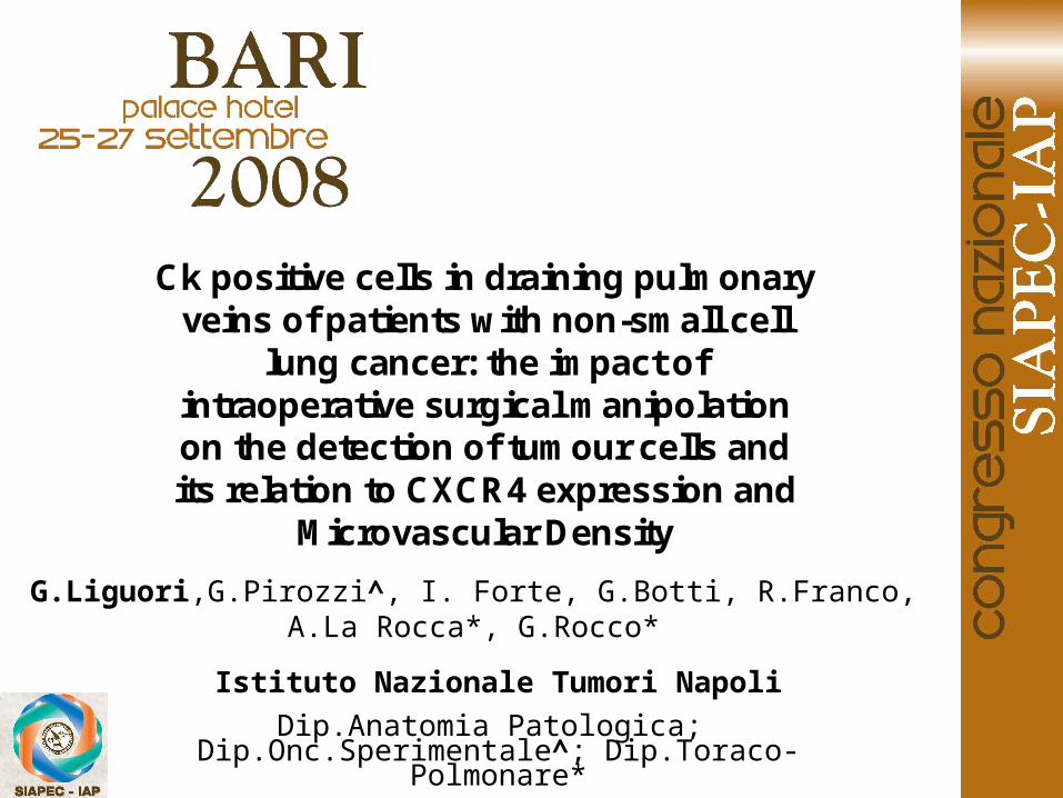
• Cancer cell dissemination in cancer is an essential event for the hematogenous metastasis of solid tumors.
• High frequency of cancer cells could be detected in the bloodstream during surgery.
• Tumor cell migration could be critically regulated by chemokines and their receptors, mainly CXCR4.
• Micro Vasculature Density (MVD), represent a measuring factor of metastatic potential.
INTRODUCTION
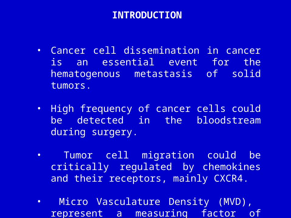
PURPOSE
-Evaluation of CXCR4 and CD31 expression in 39 patients with non-small cell lung cancer
-Relation between CXCR4 and CD31 expression ( Microvascular Density)
- To confirm CD31 and CXCR4 expression as a potential high risk for metastasis and poor prognosis.
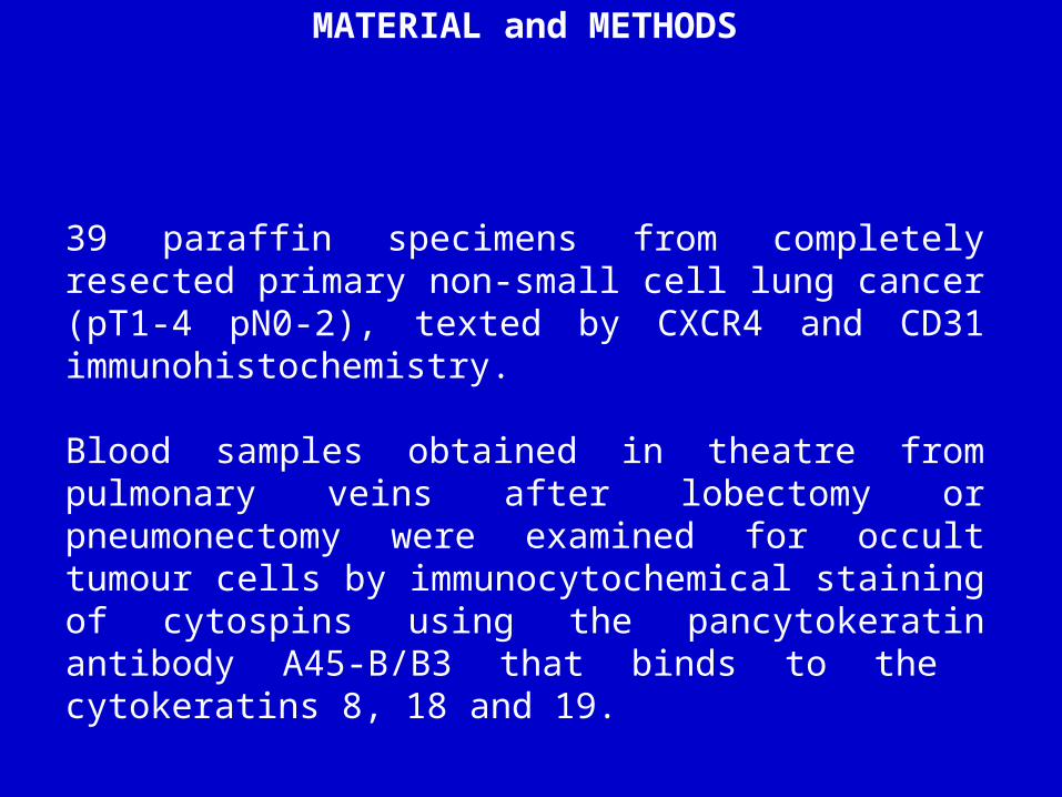
MATERIAL and METHODS
39 paraffin specimens from completely resected primary non-small cell lung cancer (pT1-4 pN0-2), texted by CXCR4 and CD31 immunohistochemistry.
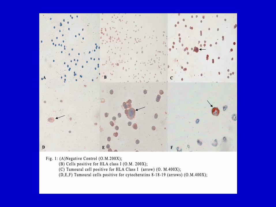
Blood samples obtained in theatre from pulmonary veins after lobectomy or pneumonectomy were examined for occult tumour cells by immunocytochemical staining of cytospins using the pancytokeratin antibody A45-B/B3 that binds to the cytokeratins 8, 18 and 19.
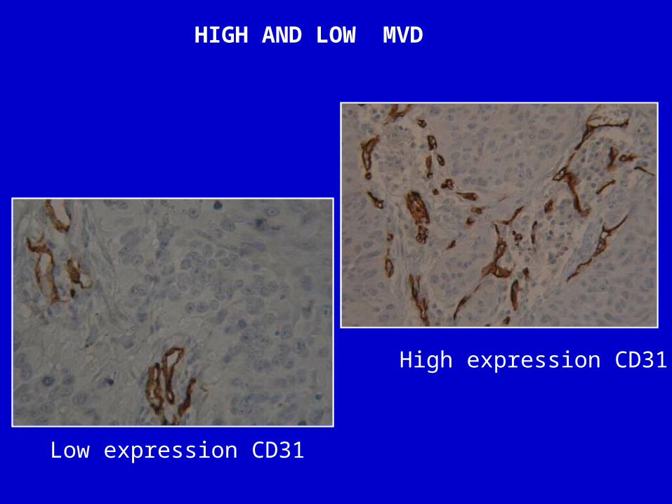
HIGH AND LOW MVD
Low expression CD31
High expression CD31
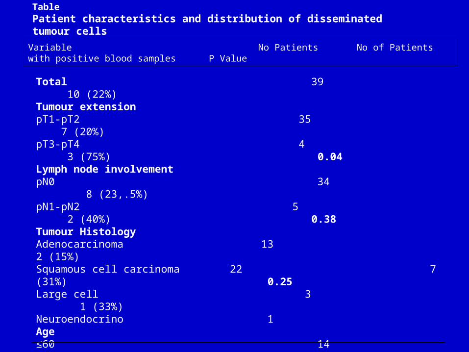
TablePatient characteristics and distribution of disseminated tumour cells
Total 39 10 (22%)Tumour extension pT1-pT2 35 7 (20%)pT3-pT4 4 3 (75%) 0.04Lymph node involvementpN0 34 8 (23,.5%)pN1-pN2 5 2 (40%) 0.38 Tumour Histology Adenocarcinoma 13 2 (15%) Squamous cell carcinoma 22 7 (31%) 0.25Large cell 3 1 (33%)Neuroendocrino 1 Age≤60 14 4 (28%) >60 25 6 (24%) 0.51 SexFemale 12 3 (25)Male 27 7 (33%) 0.63Expression of CD31Low 26 1 (3%)High 13 9 (69%) 2.9x10⁻⁵
Variable No Patients No of Patients with positive blood samples P Value
RESULTS
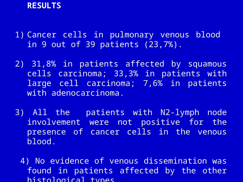
1) Cancer cells in pulmonary venous blood in 9 out of 39 patients (23,7%).
2) 31,8% in patients affected by squamous cells carcinoma; 33,3% in patients with large cell carcinoma; 7,6% in patients with adenocarcinoma.
3) All the patients with N2-lymph node involvement were not positive for the presence of cancer cells in the venous blood.
4) No evidence of venous dissemination was found in patients affected by the other histological types.
CONCLUSION
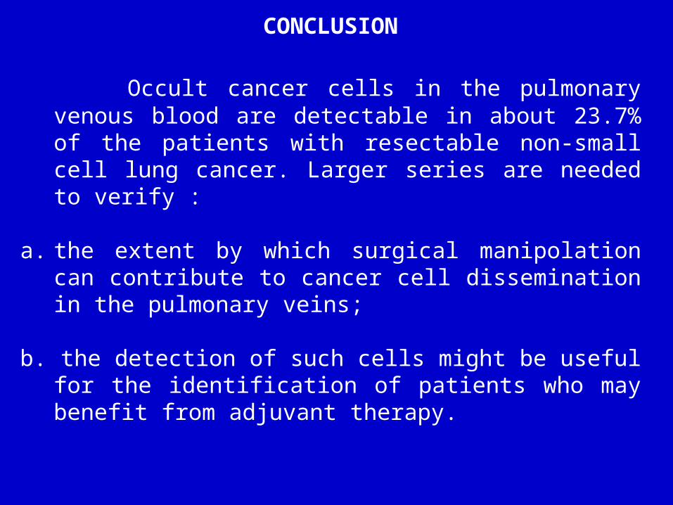
Occult cancer cells in the pulmonary venous blood are detectable in about 23.7% of the patients with resectable non-small cell lung cancer. Larger series are needed to verify :
a. the extent by which surgical manipolation can contribute to cancer cell dissemination in the pulmonary veins;
b. the detection of such cells might be useful for the identification of patients who may benefit from adjuvant therapy.
References•Sienel W., Seen-Hibler R.et al. Tumour cells in the tumour draining vein of patients with non-small cell lung cancer: detection rate and clinical significance. European Journal of Cardio-Thoracic Surgery 23 (2003) 451-456•Kubuschok B., Passlick B. et al.Disseminating Tumor Cells in Lymph Nodes as a Determinant for Survival in Surgically Resected Non-Small Cell Lung Cancer. Journal of Clinical Oncology, Vol 17, N°1 ,1999: pp 19-24•Naruke T., Goya T. et al. Prognosis and survival in resected lung cancer based on the new international staging system. J. Thoracic Cardiovascular Surgery 96:440-447,1988•Gould V., Warren W. et al. Malignant cells of epithelial phenotype limited thoracic lymph nodes. Eur J Cabcer 26:1121-1126,1990•Kasper M., Stosiek P., et al. Histological evaluation of three new monoclonal anti-cytocheratin antibodies. Eur J Cancer Clin Oncol 1987;23 (2):137-47•Pantel K., Schlimok G et al. Methodoligal analysis of immunocytochemical screening for disseminated epithelial tumor cells in bone marrow. J Hematother 1994;3(3):165-73


![Case Report - Hindawi Publishing Corporationdownloads.hindawi.com/journals/crinm/2012/616813.pdf4 Case Reports in Neurological Medicine [2] R.Franco, A.Fernandez-Vazquez, J.L.Rodriguez-Peralto](https://img.pdfslide.us/doc/110x75/5f3c0895979e6b6da30e12b7/case-report-hindawi-publishing-4-case-reports-in-neurological-medicine-2-rfranco.jpg)