Embed Size (px)
Citation preview
THE UNIVERSITY OF NORTH
CAROLINA at CHAPEL HILL
SEPTEMBER 2013
Claudia da Costa Leite, MD, PhD Thomas Bouldin, MD
NEURORADIOLOGY-NEUROPATHOLOGY CONFERENCE
CASE 1
6 y-o female with headaches and vomiting for 2 months.
Headaches are worse in the morning.
Spinal MRI: no evidence of enhancement.
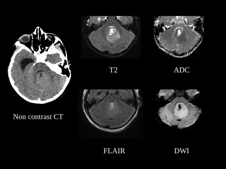
Non contrast CT
T2 ADC
FLAIR DWI
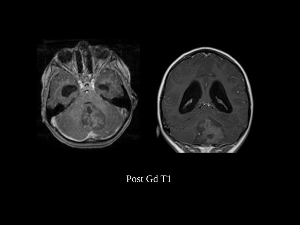
Post Gd T1
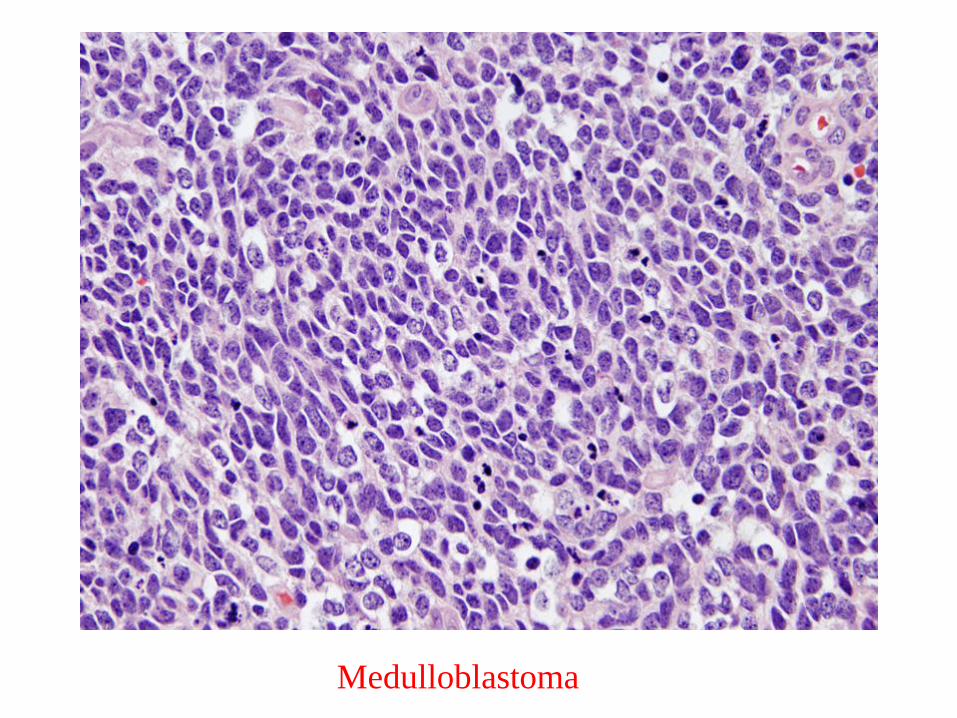
Medulloblastoma
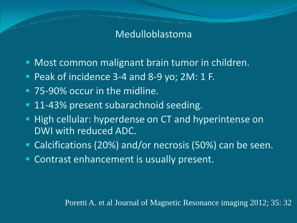
Medulloblastoma
Most common malignant brain tumor in children.
Peak of incidence 3-4 and 8-9 yo; 2M: 1 F.
75-90% occur in the midline.
11-43% present subarachnoid seeding.
High cellular: hyperdense on CT and hyperintense on DWI with reduced ADC.
Calcifications (20%) and/or necrosis (50%) can be seen.
Contrast enhancement is usually present.
Poretti A. et al Journal of Magnetic Resonance imaging 2012; 35: 32
CASE 2
75 y-o male with septic shock and delirium,
Previous hystory of SCC in the left external year 11 years ago that reccur; patient received radiation treatment. SCC in the right ear 4 years ago, SCC in a vocal cord followed by a SCC in the left lateral tongue 3 years ago and SCC in the left post-auricular region 1 year ago.
Chest x-ray Diffuse infiltrate –diagnosed as parainfluenza 3 pneumonia.
Case 2-Continuation
Clinical condition worsened: persistent delirium, respiratory and renal insufficiency, anasarca and the patient had a cardiopulmonary arrest.
Autopsy has been request.
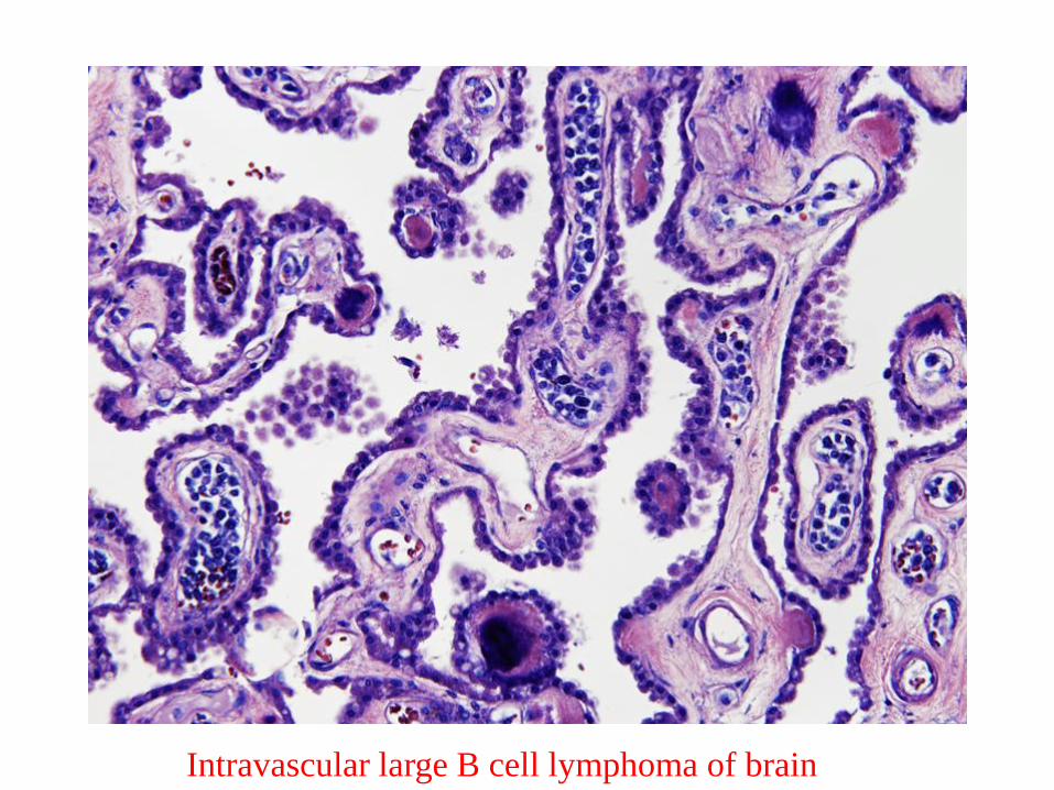
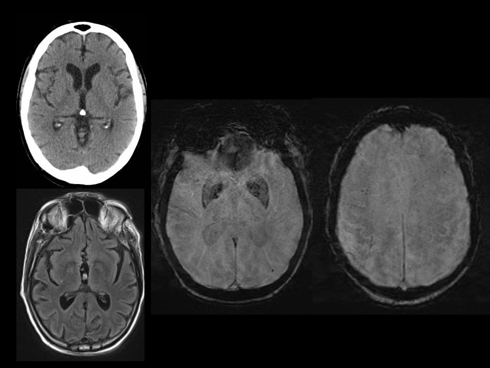
Intravascular large B cell lymphoma of brain
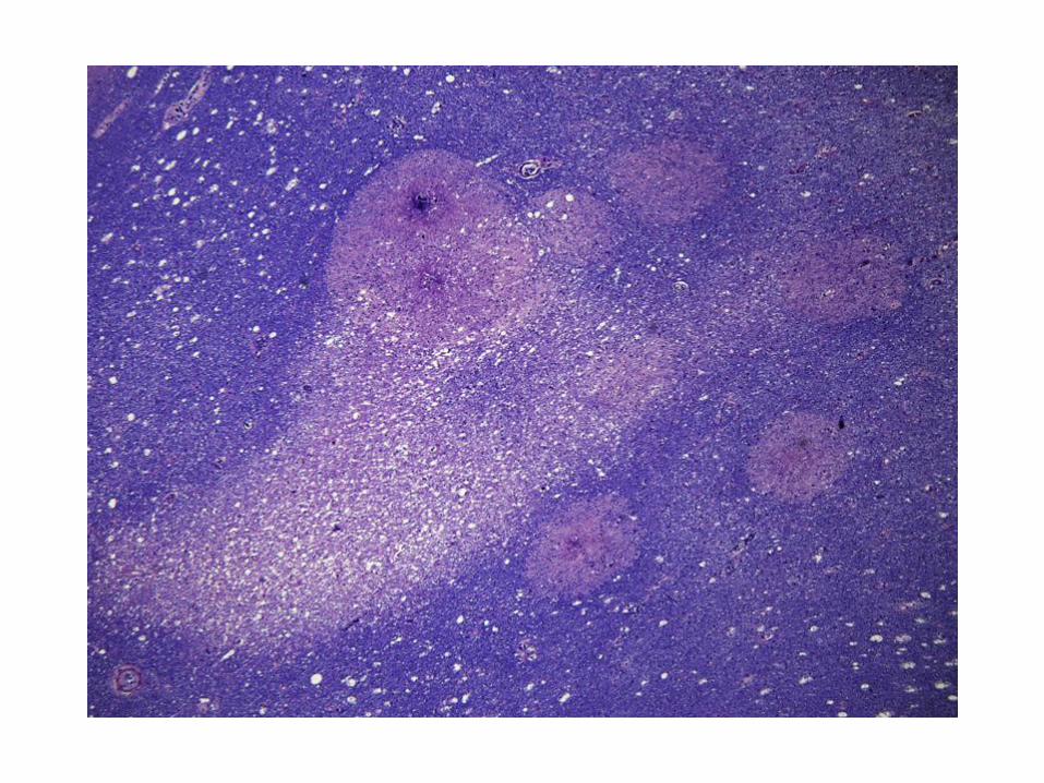
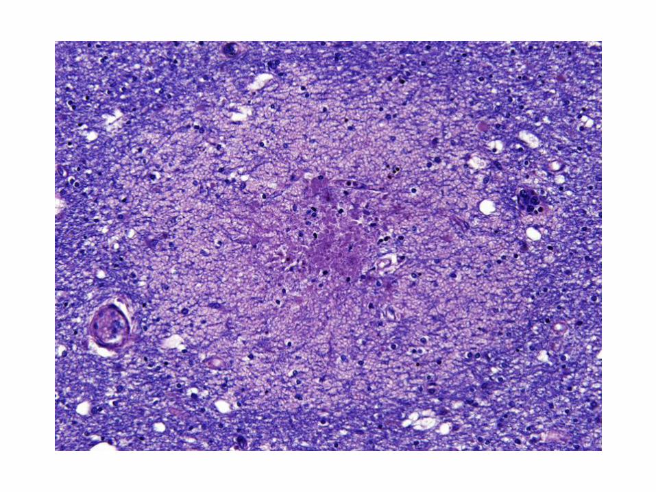
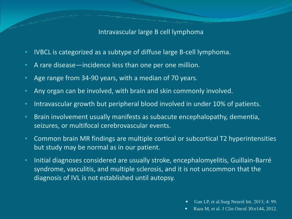
Intravascular large B cell lymphoma
• IVBCL is categorized as a subtype of diffuse large B-cell lymphoma.
• A rare disease—incidence less than one per one million.
• Age range from 34-90 years, with a median of 70 years.
• Any organ can be involved, with brain and skin commonly involved.
• Intravascular growth but peripheral blood involved in under 10% of patients.
• Brain involvement usually manifests as subacute encephalopathy, dementia, seizures, or multifocal cerebrovascular events.
• Common brain MR findings are multiple cortical or subcortical T2 hyperintensities but study may be normal as in our patient.
• Initial diagnoses considered are usually stroke, encephalomyelitis, Guillain-Barré syndrome, vasculitis, and multiple sclerosis, and it is not uncommon that the diagnosis of IVL is not established until autopsy.
Gan LP, et al.Surg Neurol Int. 2013; 4: 99.
Raza M, et al. J Clin Oncol 30:e144, 2012.
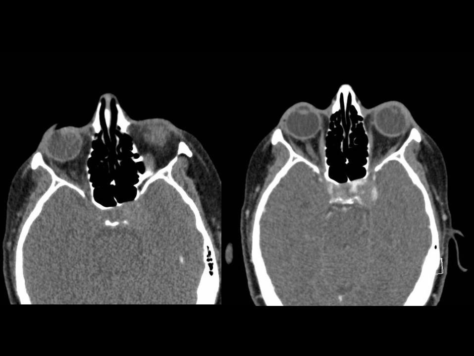
CASE 3
50 y-o female with left cranial nerve III palsy.
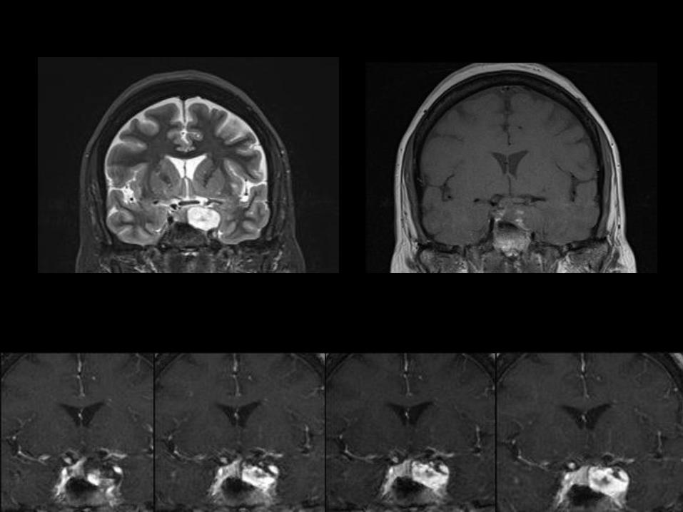
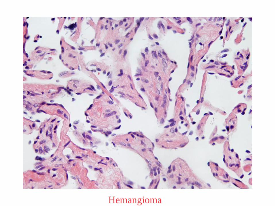
Hemangioma
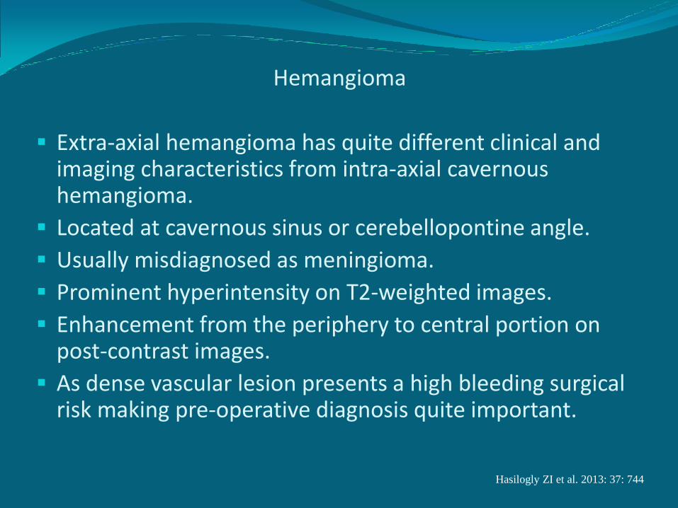
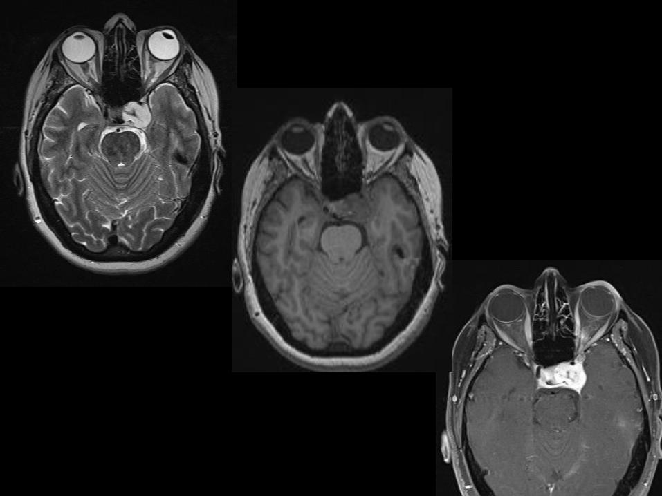
Hemangioma
Extra-axial hemangioma has quite different clinical and imaging characteristics from intra-axial cavernous hemangioma.
Located at cavernous sinus or cerebellopontine angle.
Usually misdiagnosed as meningioma.
Prominent hyperintensity on T2-weighted images.
Enhancement from the periphery to central portion on post-contrast images.
As dense vascular lesion presents a high bleeding surgical risk making pre-operative diagnosis quite important.
Hasilogly ZI et al. 2013: 37: 744
CASE 4
28 y-o male with back strain who developed radicular pain radiating to his left leg with a L3-L4 dermatomal distribution.
Only non contrast (outside) MRI study is available.
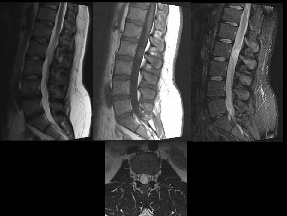
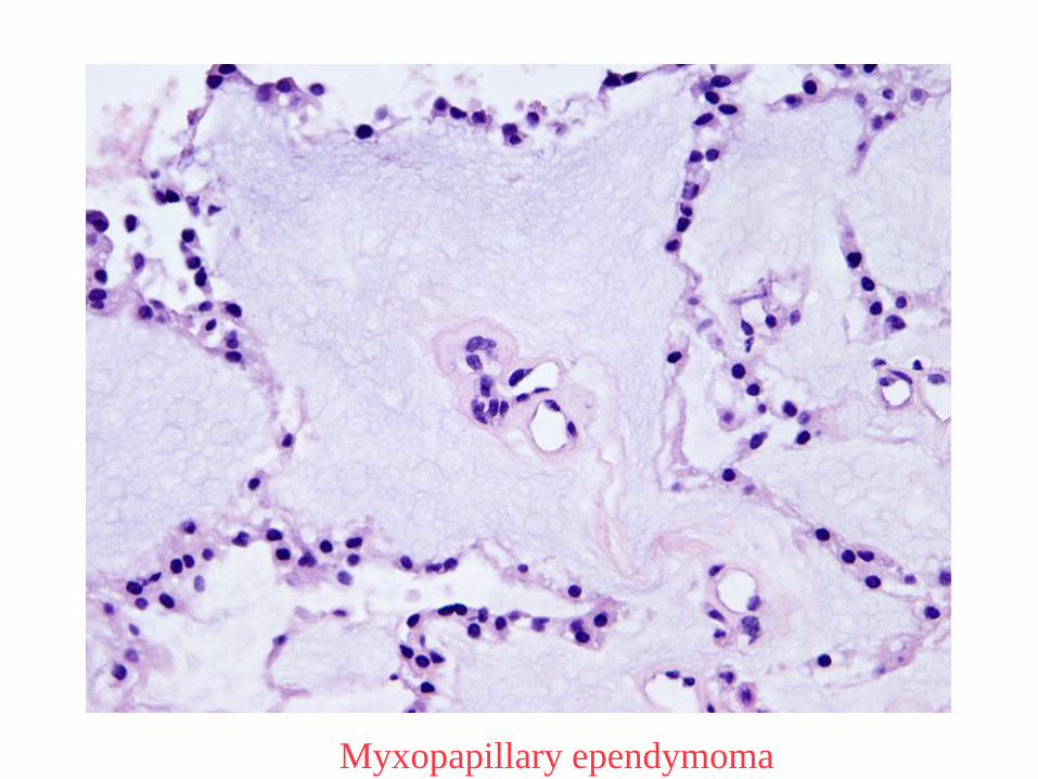
Myxopapillary ependymoma
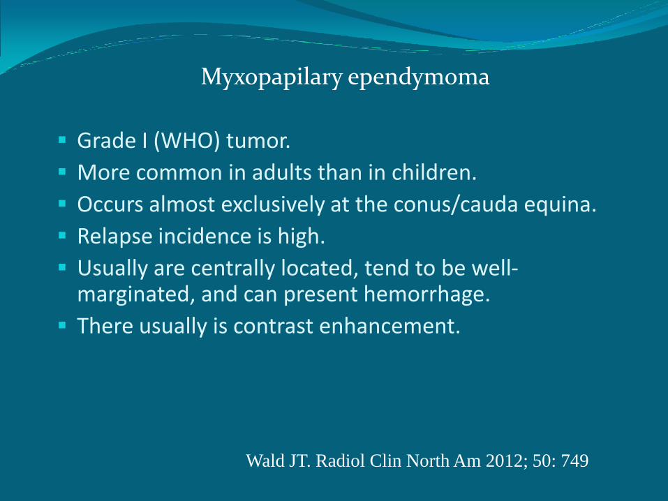
Myxopapilary ependymoma
Grade I (WHO) tumor.
More common in adults than in children.
Occurs almost exclusively at the conus/cauda equina.
Relapse incidence is high.
Usually are centrally located, tend to be well- marginated, and can present hemorrhage.
There usually is contrast enhancement.
Wald JT. Radiol Clin North Am 2012; 50: 749
Case 5
22 y-o female, headaches 2 months ago, followed by nausea, and vomiting. One month ago suffered loss of vision in both eyes without light perception.
Case 5-Continuation
Chest and abdomen CT are normal.
Patient had a brain craniotomy.
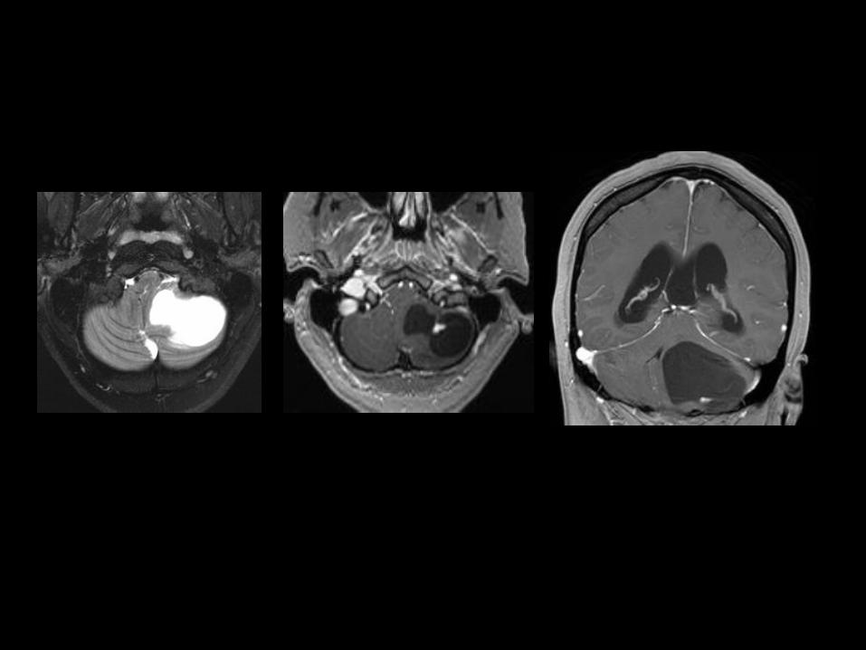
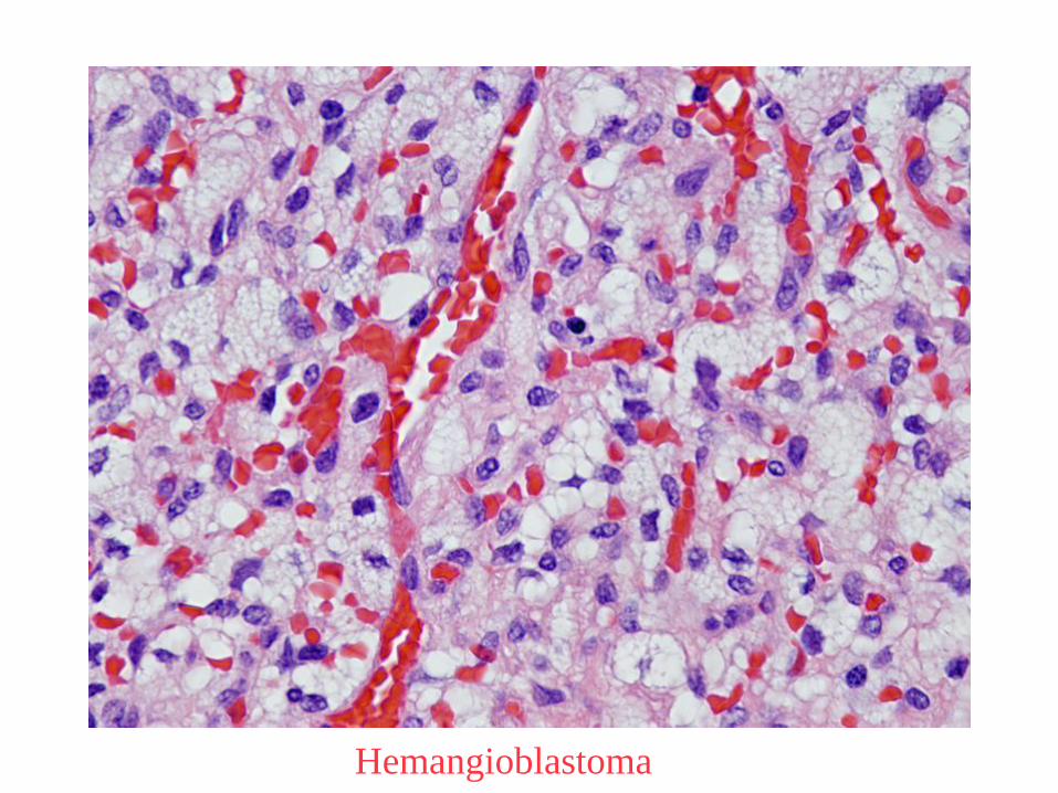
Hemangioblastoma
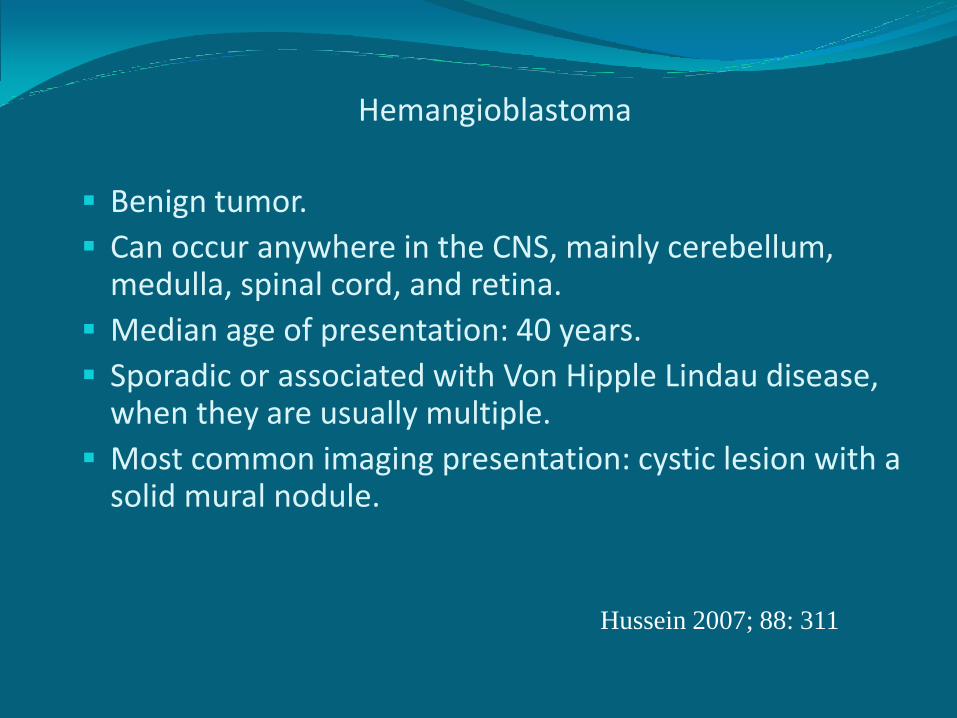
Hemangioblastoma
Benign tumor.
Can occur anywhere in the CNS, mainly cerebellum, medulla, spinal cord, and retina.
Median age of presentation: 40 years.
Sporadic or associated with Von Hipple Lindau disease, when they are usually multiple.
Most common imaging presentation: cystic lesion with a solid mural nodule.
Hussein 2007; 88: 311