Embed Size (px)
Citation preview
Department of Obstetrics and Gynecology
Helsinki University Central Hospital
University of Helsinki, Finland
DIAGNOSIS AND MANAGEMENT OF PATIENTS WITH CLINICALLY
SUSPECTED ACUTE PELVIC INFLAMMATORY DISEASE
Pontus Molander
ACADEMIC DISSERTATION
To be presented by permission of the Medical Faculty of the University of Helsinki for public
discussion in the Auditorium of the Department of Obstetrics and Gynecology, Helsinki University
Central Hospital, Haartmaninkatu 2, Helsinki, on June 6th, 2003, at 12 noon.
Supervised by
Professor Jorma Paavonen
Department of Obstetrics and Gynecology
University of Helsinki, Finland
Docent Bruno Cacciatore
Department of Obstetrics and Gynecology
University of Helsinki, Finland
Reviewed by
Professor Pentti K. Heinonen
Department of Obstetrics and Gynecology
University of Tampere, Finland
Docent Aydin Tekay
Department of Obstetrics and Gynecology
University of Oulu, Finland
Official opponent
Docent Jorma Penttinen
Department of Obstetrics and Gynecology
University of Kuopio, Finland
ISBN 952-91-5887-4 (paperback)
ISBN 952-10-1183-1 (PDF)
Yliopistopaino 2003

3
To Eva, Jan, and Jessica
4
CONTENTS
List of original publications 7
Abbreviations 8
1. Introduction 10
2. Review of the literature 12
2.1. Definition and history of acute PID 12
2.2. Microbiology 14
2.3. Pathogenesis 17
2.4. Epidemiology 20
2.5. Clinical manifestations 27
2.5.1. Subclinical disease 27
2.5.2. Endometritis 28
2.5.3. Mild and moderate PID 30
2.5.4. Severe PID 30
2.5.5. Perihepatitis 32
2.5.6. Periappendicitis 33
2.6. Diagnosis 33
2.6.1. Clinical diagnosis 34
2.6.2. Endometrial biopsy 37
2.6.3. Laboratory diagnosis 38
2.6.4. Ultrasonographic diagnosis 40
2.6.5. Other imaging modalities 42
2.6.6. Laparoscopic diagnosis 43
5
2.6.7. Differential diagnosis 47
2.7. Treatment 50
2.7.1. Conservative 50
2.7.2. Surgical 53
2.8. Prevention 55
2.9. Long-term sequelae of PID 57
3. Aims of the study 59
4. Material and methods 60
4.1. Subjects 60
4.2. Methods 61
4.2.1. Clinical diagnosis of acute PID (Studies I-IV) 61
4.2.2. Ultrasonography (Studies I-IV) 62
4.2.3. Magnetic resonance imaging (Study I) 63
4.2.4. Laparoscopy (Studies I-IV) 64
4.2.5. Observer reproducibility (Study V) 65
4.3. Biostatistical analyses 66
5. Results 68
5.1. Diagnosis of pelvic inflammatory disease and acute appendicitis 68
5.1.1. MRI in diagnosis of acute PID (Study I) 68
5.1.2. Power Doppler TVS in diagnosis of acute PID (Study III) 70
5.1.3. TVS in diagnosis of acute appendicitis (Study IV) 72
5.2. Laparoscopic management of pelvic inflammatory disease (Study II) 74
5.3. Accuracy of laparoscopic findings in pelvic inflammatory disease (Study V) 77
6. Discussion 78
6.1. Diagnosis of pelvic inflammatory disease and acute appendicitis 78
6
6.1.1. MRI in diagnosis of acute PID 78
6.1.2. Transvaginal sonography in diagnosis of acute PID 79
6.1.3. Transvagnial and transabdominal sonography in diagnosis of acute appendicitis 82
6.2. Laparoscopic management of pelvic inflammatory disease 84
6.3. Accuracy of laparoscopic findings in pelvic inflammatory disease 86 7. Conclusion and future prospects 88 8. Summary 91
Acknowledgements 93
References 96
7
List of original publications
This thesis is based on the following original publications:
I Tukeva T, Aronen HJ, Karjalainen PT, Molander P, Paavonen T, Paavonen J. MR imaging in
pelvic inflammatory disease: comparison with laparoscopy and US. Radiology 1999;210:209-
216.
II Molander P, Cacciatore B, Sjöberg J, Paavonen J. Laparoscopic management of suspected acute
pelvic inflammatory disease. J Am Assoc Gyn Lap 2000;7:107-110.
III Molander P, Sjöberg J, Paavonen J, Cacciatore B. Transvaginal power Doppler findings in
laparoscopically proven acute pelvic inflammatory disease. Ultrasound Obstet Gynecol
2001;17:233-238.
IV Molander P, Paavonen J, Savelli L, Sjöberg J, Cacciatore B. Transvaginal sonography in the
diagnosis of acute appendicitis. Ultrasound Obstet Gynecol 2002;20:496-501.
V Molander P, Finne P, Sjöberg J, Sellors J, Paavonen J. Observer agreement study of laparoscopic
diagnosis of pelvic inflammatory disease using photographs. Obstet Gynecol (in press).
8
Abbreviations
AFS American Fertility Society
ASS acute salpingitis score
AV aerobic vaginitis
BV bacterial vaginosis
CDC Centers for Disease Control and Prevention
CHSP-60 chlamydial heat shock protein-60
CI confidence interval
CRP c-reactive protein
CT computed tomography
DNA deoxyribonucleic acid
ESR erythrocyte sedimentation rate
HHSP-60 human heat shock protein-60
HSG hysterosalpingography
IM intramuscularly
IUD intrauterine device
IV intravenously
κ kappa
LCR ligase chain reaction
LGTI lower genital tract infection
MRI magnetic resonance imaging
NAA nuclein acid amplification
NSU non-specific urethritis
9
NPV negative predictive value
OC oral contraceptive
PCE plasma cell endometritis
PCR polymerase chain reaction
PI pulsatility index
PID pelvic inflammatory disease
PPV positive predictive value
STI sexually transmitted infection
STIR short inversion time inversion recovery
TAS transabdominal sonography
TFI tubal factor infertility
TOA tubo-ovarian abscess
TVS transvaginal sonography
UGT upper genital tract
US ultrasonography
WBC white blood cell count
WHO World Health Organization
10
1. Introduction
Despite the development of new diagnostic aids, pelvic inflammatory disease (PID) is still poorly
recognized and managed. In 1990, J. Pearce stated: ”PID is a sexually transmitted disease with
potentially serious sequelae usually managed badly by doctors with little interest in the condition”.
Unfortunately, no great breakthroughs have taken place since that time in the management of PID
(Simms and Stephenson 2000). Women of fertile age represent an especially difficult patient group
because of a variety of gynecologic and non-gynecologic diagnostic possibilities (Porpora and Gomel
1997, Tarrazza and Moore 1997, Cibula et al. 2001). Diagnostic accuracy regarding PID has not
improved throughout the last decades in laparoscopic studies, reaching maximally 60 to70%
(Jacobson and Weström 1969, Paavonen et al. 1987, Bevan et al. 1995). The rate of not only false
positive but also false negative findings in all studies concerning clinical accuracy of PID diagnosis is
high (Jacobson 1980, Sellors et al. 1991). About one-third of patients with a clinical diagnosis of
PID in fact have another disease or normal findings, while two-thirds reveal PID of some degree
when laparoscopy is used to confirm the diagnosis (Munday 2000). No decline has occurred in the
misdiagnosis of the most common nongynecologic differential diagnostic disease, acute appendicitis.
The rate of misdiagnosis of appendicitis in fertile women has been as high as 40%, and surprisingly,
among women of reproductive age, misdiagnosis has even increased (Flum et al. 2001).
Because of the lack of reliable diagnostic methods, the technique playing a central role in the
management of acute abdomen in women of reproductive age is laparoscopy. It offers the possibility
to diagnose and manage both PID and non-PID cases. In the management of acute pelvic pain,
laparoscopy allows confirmation of the diagnosis and a possibility to treat the condition safely and
cost-effectively. Effective management prevents complications associated with delayed treatment and
often preserves the patient’s fertility (Porpora and Gomel 1997). Some studies indicate that
11
operative laparoscopy may improve the primary recovery of acute PID patients (Henry-Suchet et al.
1984, Reich and McGlynn 1987, De Wilde and Hesseling 1995).
Various imaging methods have been proposed for the diagnosis of PID. Transvaginal sonography
(TVS) is routinely used in the diagnosis of acute gynecologic disorders. Only a few studies have been
performed on the accuracy of TVS diagnosis of acute PID using laparoscopy or histopathology as
the gold standard (Patten et al. 1990, Cacciatore et al. 1992, Boardman et al.1997). An overall
concern is the poor performance of TVS in mild PID. Moreover, TVS findings in PID need
standardization (Timor-Tritsch et al. 1998). Increased vascularity has been linked with inflammation,
and color Doppler investigation of vascular blood flow has brought new aspects to the diagnosis of
inflammatory processes including PID (Tinkanen and Kujansuu 1993, Kupesic et al. 1995, Alatas et
al. 1996, Tepper et al. 1998). Power Doppler, a modification of color Doppler, lacks certain
disadvantages of color Doppler and is excellent in the detection of organ vascularity and especially
low-velocity blood flow (Rubin et al. 1994).
In the diagnosis of intra-abdominal conditions, magnetic resonance imaging (MRI) has been widely
accepted, although MRI has been used rarely in the imaging of adnexal masses (Mitchell et al. 1987,
Jain et al. 1993, Yamashita et al. 1995, Komatsu et al. 1996) and gynecologic infections (Outwater
and Dunton, 1995, Ha et al. 1995).
The reproducibility of laparoscopic findings of PID has not undergone thorough evaluation, even
though the reliability of laparoscopy as the gold standard in the diagnosis of PID is in question
(Sellors et al. 1991). Mild PID may remain unrecognized at laparoscopy, leading to false-negative
diagnoses (Sellors et al. 1991).
The target group in this study is patients with clinically suspected symptomatic PID. The study
evaluates the efficacy of new imaging techniques and the role of laparoscopy in the diagnosis and
management of these patients, and in addition, tests accuracy and reproducibility of the laparoscopic
diagnosis of PID.
12
2. Review of the literature
2.1. Definition and history of acute PID
PID comprises a spectrum of upper genital tract inflammatory disorders among women, including
any combination of endometritis, salpingitis, tubo-ovarian abscess, or pelvic peritonitis (CDC 2002).
The term PID is restricted to infections caused by microorganisms ascending from the vagina or
cervix that are not associated with surgery or pregnancy (McCormack 1994), but this term is in a
sense misleading, because it refers to a syndrome, when the disease is in fact an infection. Salpingitis,
or infection of the Fallopian tubes, is the most important feature of PID, and in fact the terms
salpingitis and PID are often used synonymously (Weström 1977). There is, however, a vast
spectrum of terms to describe differing manifestations of PID (Table 1). This descriptive variety
reflects the diagnostic difficulties accompanying the disease. PID presents with a broad spectrum of
clinical manifestations ranging from virtually none to severe. In about two-thirds of all PID cases, the
disease may remain unrecognized (Sellors et al. 1988). Consequently, the term PID has different
meanings in different clinical settings (Weström and Eschenbach 1999).
Table 1. Clinical manifestations of PID.
• Endometritis
• Salpingitis
• Salpingo-oophoritis
• Adnexitis
• Parametritis
• Pyosalpinx
• Tubo-ovarian complex
• Tubo-ovarian abscess
• Peritonitis
• Perihepatitis
• Periappendicitis

13
In ancient Greece, Aetius described drainage of pus through the vagina, and he thereby both
diagnosed a pelvic abscess and discovered a surgical treatment. Mauriceau, by dissecting human
cadavers, described in 1683 inflammatory tumors of the adnexa in puerperal infections (Weström and
Eschenbach 1999). In 1876, sexually transmitted infection (STI) and infertility were linked for the
first time (Noeggerath 1876). Neisser identified gonococci three years later, and then von Bumm
described the sequence of cervical gonorrhea progressing to endometritis, salpingitis, and pelvic
peritonitis in 1887. Between 1892 and 1914, scientists isolated several aerobic and anaerobic bacteria
from the Fallopian tubes and pelvic cavities of women with PID (Weström and Eschenbach 1999),
including discovered mycoplasma, in 1898 (Nocard and Roux 1898).
Even in the pre-antibiotic era, PID was associated with low mortality rates, as described by Holtz,
who reported in 1930 a mortality rate of 1.3% in a series of 1,262 patients with PID (Holtz 1930).
Without antibiotics, symptoms usually resolved in two months, often leaving the woman infertile
(Brunham 1984a). Antimicrobial therapy, such as sulfonamides, was used for the first time to treat
PID in the 1930s, and after the advent of chemotherapy, death from acute PID has been rare.
Mycoplasmas were isolated from the human genital tract in 1937 (Weström and Eschenbach 1999),
and in 1946 Falk described PID as an ascending infection. Chlamydia trachomatis (C.trachomatis)
was identified in 1957 by T’ang et al., and Jones et al. (1959) first isolated the organism from the
female genital tract. In 1970 Mårdh isolated Mycoplasma from the Fallopian tubes (Mårdh and
Weström 1970), and C. trachomatis was first isolated from the Fallopian tubes in women with
salpingitis during the period 1976 to 1977 by two groups in Sweden (Eilard et al. 1976, Mårdh et al.
1977). In 1975, the polymicrobial etiology of PID became evident (Eschenbach et al. 1975), and the
devastating long-term effects of PID, including tubal factor infertility and ectopic pregnancy was
described by Weström in 1975.
In 1902, Kelling described the fundamentals of laparoscopy, and the first human laparoscopies were
performed by Jacobaeus of Sweden in 1910 (Wittman 1966). Raoul Palmaer from France pioneered
14
modern laparoscopy in the 1940s and is considered to be the father of modern gynecological
laparoscopy. In the 1960s and 1970s laparoscopy was systematically used in Sweden to ensure the
accuracy of the diagnosis of salpingitis, and during this period the poor accuracy of the clinical
diagnosis became apparent (Jacobson and Weström 1969). Laparoscopy enabled also microbiologic
sampling and it became the gold standard for the diagnosis. In the 1980s, videolaparoscopy
introduced a completely new surgical vision, and laparoscopy could serve not only for diagnosis but
also for therapeutic procedures. Both conservative and radical laparoscopic procedures were now
possible also in the management of PID patients.
Last decades have seen an explosion of information on PID based on progress in microbiology,
immunology, epidemiology, experimental animal models, and social and behavioral sciences. Despite
this, diagnosis remains problematic, and still no rapid simple tests are available to improve the
accuracy of clinical diagnosis (Munday 2000).
2.2. Microbiology
In PID, microorganisms ascend from the cervix and vagina to the endometrium, Fallopian tubes, and
adjacent structures. Since the vagina and cervix are colonized by a large number of microorganisms,
the microbial etiology of PID is best established by direct culture from the upper genital tract via
laparoscopy or laparotomy (Weström 1977, Sweet et al. 1980, Mårdh et al.1981). Studies have
shown a poor correlation between intra-abdominal and cervical cultures which is mostly explained as
contamination by the cervicovaginal flora of the latter (Sweet et al. 1980). Different sampling
methods make comparisons between studies difficult (Mårdh et al. 1981).
PID is often caused by sexually transmitted infections such as C. trachomatis or Neisseria
gonorrohoeae (N. gonorrhoeae), together with vaginal aerobic or anaerobic flora. Nonetheless, 25%
to 50% of cases no have detectable chlamydial or gonococcal infection (Paavonen 1980, Brihmer et
15
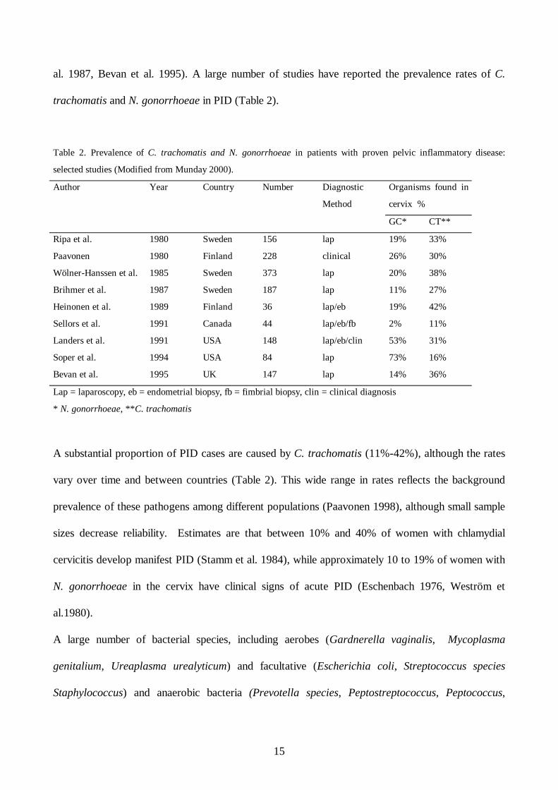
al. 1987, Bevan et al. 1995). A large number of studies have reported the prevalence rates of C.
trachomatis and N. gonorrhoeae in PID (Table 2).
Table 2. Prevalence of C. trachomatis and N. gonorrhoeae in patients with proven pelvic inflammatory disease:
selected studies (Modified from Munday 2000).
Author Year Country Number Diagnostic
Method
Organisms found in
cervix %
GC* CT**
Ripa et al. 1980 Sweden 156 lap 19% 33%
Paavonen 1980 Finland 228 clinical 26% 30%
Wölner-Hanssen et al. 1985 Sweden 373 lap 20% 38%
Brihmer et al. 1987 Sweden 187 lap 11% 27%
Heinonen et al. 1989 Finland 36 lap/eb 19% 42%
Sellors et al. 1991 Canada 44 lap/eb/fb 2% 11%
Landers et al. 1991 USA 148 lap/eb/clin 53% 31%
Soper et al. 1994 USA 84 lap 73% 16%
Bevan et al. 1995 UK 147 lap 14% 36%
Lap = laparoscopy, eb = endometrial biopsy, fb = fimbrial biopsy, clin = clinical diagnosis
* N. gonorrhoeae, **C. trachomatis
A substantial proportion of PID cases are caused by C. trachomatis (11%-42%), although the rates
vary over time and between countries (Table 2). This wide range in rates reflects the background
prevalence of these pathogens among different populations (Paavonen 1998), although small sample
sizes decrease reliability. Estimates are that between 10% and 40% of women with chlamydial
cervicitis develop manifest PID (Stamm et al. 1984), while approximately 10 to 19% of women with
N. gonorrhoeae in the cervix have clinical signs of acute PID (Eschenbach 1976, Weström et
al.1980).
A large number of bacterial species, including aerobes (Gardnerella vaginalis, Mycoplasma
genitalium, Ureaplasma urealyticum) and facultative (Escherichia coli, Streptococcus species
Staphylococcus) and anaerobic bacteria (Prevotella species, Peptostreptococcus, Peptococcus,
16
Mobiluncus species) have been isolated from the upper genital tract (UGT) of women with acute
PID (Mårdh and Weström 1970, Eschenbach et al. 1975, Sweet et al. 1980, Wasserheit et al. 1986,
Soper et al. 1994, Baveja et al. 2001). The role of these bacteria as pathogens in PID is not clearly
understood. Most appear in the normal vaginal flora (endogenous). Actinomyces, Campylobacter,
and Clostridiae are rare causes of PID. ”Endogenous” bacteria are commonly found in severe
disease, in recurrent PID, and among intrauterine device (IUD) users and older women (Eschenbach
et al. 1975, Weström 1977, Sweet et al. 1981, WHO 1987a, Soper et al. 1994). Severe PID, such as
tubo-ovarian abscess (TOA) is typically polymicrobial, with a shift from facultative to anaerobic
bacteria occurring as the infection proceeds (Eschenbach et al. 1975, Bieluch and Tally, 1983). In
mild PID, anaerobic bacteria are usually absent (Mårdh 1980).
Of women with PID UGTs have shown several bacterial vaginosis-associated (BV-) bacteria
(Eschenbach et al. 1975, 1988, Paavonen et al. 1987, Soper et al. 1994, Korn et al. 1995a, Hillier et
al. 1996). In BV, a quantitative and qualitative shift in the vaginal flora occurs due to a decrease in
the concentration of lactobacilli which causes a massive increase in the concentration of other
pathogens, e.g., Mobiluncus, Prevotella, Gardnerella vaginalis, and genital mycoplasmas. Aerobic
vaginitis (AV) is characterized by an overgrowth of virulent aerobic bacteria, e.g., Escherichia coli,
Group B streptococcus, and Enterococcus. Bacteria associated with BV or AV may ascend to the
UGT without a primary infection. In BV or AV, a massive increase in the concentration of microbial
byproducts is thought to destroy cervical host defense barriers, leading to such an ascent. Some
evidence exists as to the role of BV as a PID precursor. BV-positive women undergoing induced
abortion compared to BV-negative controls have a threefold risk of postabortion PID. This increased
rate of PID is reduced to baseline after BV treatment (Larsson et al. 1992). The causative role in
PID and TFI of BV-associated Mycoplasma genitalium has been suggested (Möller et al. 1984,
Clausen et al. 2001, Cohen et al. 2002, Simms et al. 2003), although any correlation is unclear. It is
also hypothesized that primary chlamydial or gonococcal infection, especially if untreated or when
17
treatment is delayed, is followed by an invasion of endogenous vaginal bacteria; this is seen especially
in severe PID. A rare cause of PID is Actinomyces israelii in women using an IUD. Pathogenesis of
pelvic actinomycosis is poorly understood (Lippes 1999). Recent studies show that in 20 to 30% of
PID patients, diagnostic techniques available can detect no microorganism (Munday 2000).
2.3. Pathogenesis
Even though it is well known that PID is an ascending infection, the mechanism determining the
canalicular spread of microorganisms from the lower to the upper genital tract remains poorly
understood. Evidence of this canalicular spread is provided by observations which imply that
interruption of the Fallopian tubes by cornual resection prevents salpingitis (Falk 1946). Initial
infection involves the mucosa and not the muscularis layer of the Fallopian tube (Patton 1985).
Furthermore, in women with PID, N. gonorrhoeae and C. trachomatis have been demonstrated in
epithelia of the cervix, endometrium, and Fallopian tubes (Heinonen et al. 1985, Kiviat et al. 1986).
The endocervical canal and the mucus plug are the major barriers that protect the UGT from the
vaginal flora (Rice and Schachter 1991). C. trachomatis and N. gonorrhoeae may be primary
pathogens of mucopurulent endocervicitis (Brunham et al. 1984b, Paavonen et al. 1986), and these
may break down the barriers and permit ascending infection. Damage to barriers may be sufficient to
allow other bacteria to ascend. Cervical mucus is a functional barrier which is absent during
menstruation and more penetrable by microbes during the follicular than the luteal phase (Odeblad
1968). Cervical antibodies participate in resistance against invading microorganisms; the protective
effect may result from downward flow of cervical secretion (Odeblad 1968). The functional cervical
barrier against ascending infection is lowest at the time of ovulation and when the mucus plug is
absent during menstruation (Odeblad 1968). The luteal phase of the menstrual cycle seems to protect
against ascending infection, whereas the follicular phase presents a greater risk for microbial
18
penetration because of changes in the cervical mucus (Odeblad 1968, Sweet et al. 1986). Salpingitis
often starts during menses or shortly after, suggesting that retrograde menstruation may function as a
vehicle for microorganisms (Halme et al. 1984a, Sweet et al. 1986). Thus, investigations suggest
that, for the transportation of potential pathogenic microorganisms into the upper genital tract, a
vehicle is unnecessary (Rice and Schachter 1991).
Some evidence indicates that spermatozoa may play a role in this ascending spread of microbes
(Toth et al. 1982, Wölner-Hanssen and Mårdh 1984, ). An increased risk for PID is associated with
high coital frequency (Washington et al. 1991 a). The increase in the size of the zone of ectopy seen
in young women may result in increased susceptibility to infection (Expert Committee on PID
1991). It is postulated that a cervical infection with C. trachomatis or N. gonorrhoeae or a
combination of both organisms causes an alteration in the cervicovaginal microenvironment, leading
to the overgrowth of facultative flora in the vagina and ultimately to BV. Thereafter, the original
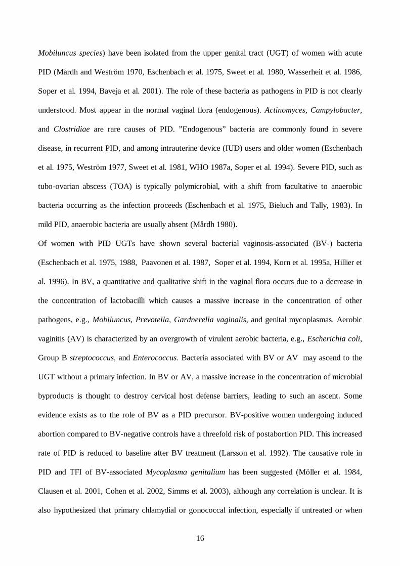
cervical pathogens, the flora causing BV, or both ascend (Wasserheit et al. 1986) (Figure 1).
19
Figure 1. Pathogenesis of pelvic inflammatory disease (reprinted from McCormack, 1994 with permission). A.
Cervical infection with C. trachomatis, N. gonorrhoeae or both organisms. B. Alteration of the cervicovaginal
microenvironment. C. Overgrowth of the vaginal facultative flora leading ultimately to BV. D. The original cervical
pathogens, the flora causing BV, or both ascend into the endometrium, Fallopian tubes, and peritoneal cavity.
Gonococcal PID is thought to extend via a direct canalicular route from the endocervix into the
endometrium and subsequently into the Fallopian tubes. This causes edema and incites an intense
polymorphonuclear leukocyte response. Gonococci attach to the microvilli of nonciliated mucosal
epithelial cells and enter these epithelial cells, resulting in cell damage and sloughing of ciliated cells
(McGee et al. 1981, Melly et al. 1981). The effect is directly toxic or cytokine-dependent. Tissue
repair and inflammatory process are initiated, resulting in tubal adhesions and scarring (Rice and
Schachter 1991).
C. trachomatis infection is associated with less severe clinical manifestations (Svensson et al. 1980).
Patients are therefore likely to present later in the disease process and to be under-represented
among hospitalized patients. In Fallopian tube organ cultures, C. trachomatis replicates within both
ciliated and non-ciliated cells (Cooper et al. 1990). Chlamydial infection induces several cytokines
20
(van Voorhis et al. 1996), which in turn can induce tissue damage even though primary salpingeal
chlamydial infection seems to cause only a mild to moderate inflammatory response and minor
permanent damage (Patton 1985). This infection also activates a humoral response, but protective
immunity appears to be short. It appears to activate a cell-mediated immune response. Repeated
exposures to chlamydia antigen in animals can induce T-cell-mediated chronic hypersensitivity
(Taylor et al. 1990) and produce extensive tubal scarring (Patton et al. 1990). These findings suggest
that tubal inflammation with resulting tissue destruction in genital chlamydia infections may be the
result of a delayed hyperimmune reaction to repeated exposures to chlamydial antigens, especially to
the 60 kD heat shock protein (CHSP-60) (Witkin et al. 1994, Eckert et al. 1997, Kinnunen et al.
2000, Kinnunen et al. 2002a, Kinnunen et al. 2003). The chronic exposure to C. trachomatis
occurring in asymptomatic women with silent chlamydial infection, in particular, may activate this
immune response. The sensitization of the immune system to chlamydial protein may lead to an
autoimmune response to the homologous human protein (HHSP-60). This autoimmune cascade may
continue even if C. trachomatis is no longer present (Witkin et al. 1994). Chronic sequelae of genital
chlamydial infection such as ectopic pregnancy and tubal infertility may be caused by the
hypersensitivity reaction to CHSP-60 (Toye et al. 1993, Brunham et al. 1992, Kinnunen et al.
2002b).
2.4. Epidemiology
PID surveillance remains problematic because of a lack of simple and accurate diagnostic tests.
Problems of case definition and diagnostic accuracy are compounded by the inaccessibility of the
female UGT to routine, large-scale diagnostic methods. A diagnostic gold standard is difficult to
formulate. PID surveillance data are also influenced by variations in case definitions and reporting
practice, the wide spectrum of the clinical manifestations of chlamydial infection, variations in health-
21
seeking behavior, and the increased management of PID in outpatient settings (Simms and
Stephenson 2000). Trends in PID cannot be inferred from genital chlamydial infection, as the data
are heavily influenced by case ascertainment bias (Simms et al. 1996). Moreover, invasive diagnostic
tests have resulted in small-scale, unrepresentative studies that have inherent selection and
participation biases. A syndromic diagnosis should be more extensively used in epidemiological
studies even if diagnostic algorithms are difficult to validate in terms of sensitivity and specificity.
Consequently, it is difficult to assess trends in PID prevalence with certainty, making comparisons
between countries almost impossible (Simms and Stephenson 2000).
A recent trend in industrialized countries is a rapid shift in the microbiological etiology of PID.
Genital gonorrheal infections have decreased, whereas genital chlamydial infections have increased –
despite large regional differences. As a consequence, the relative role of C. trachomatis in the
causation of PID has increased, whereas the role of N. gonorrhoeae has decreased. Since STIs cause
a substantial proportion of PID cases, epidemics of N. gonorrhoeae and C. trachomatis are, as seen
in Swedish surveillance data, followed by secondary PID epidemics and tertiary epidemics of ectopic
pregnancy and tubal infertility (Weström 1980). In many developed countries, gonorrhea is now a
rare disease (van der Heyden et al. 2000), whereas chlamydia rates are still high or on the rise
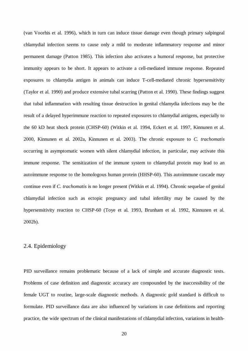
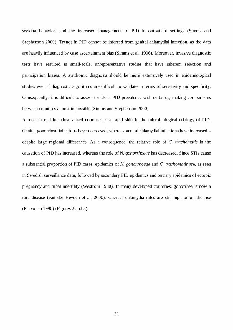
(Paavonen 1998) (Figures 2 and 3).
22
Figure 2. Reported C. trachomatis rates in the USA, 1984-1995 (from STD Surveillance, NIH 1995).
0
20
40
60
80
100
120
140
160
180
200
1984
1985
1986
1987
1988
1989
1990
1991
1992
1993
1994
1995
C. t
rach
om
atis
rat
es (i
n th
ou
san
ds)
Figure 3. Reported C. trachomatis rates in Finland, 1987-2001; data from the Finnish National Public Health Institute,
2002.
0
2000
4000
6000
8000
10000
12000
14000
16000
1987
1988
1989
1990
1991
1992
1993
1994
1995
1996
1997
1998
1999
2000
2001
In Finland, for instance, the annual number of cases of C. trachomatis detected was 8,000 in 1994
and 12,100 by the year 2001, whereas gonococcal infections decreased by two-thirds in the 1990s to
240 cases in 2001 (National Public Health Institute, 2002). This trend can be seen in a comparison of
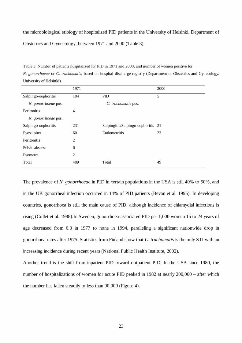
23
the microbiological etiology of hospitalized PID patients in the University of Helsinki, Department of
Obstetrics and Gynecology, between 1971 and 2000 (Table 3).
Table 3. Number of patients hospitalized for PID in 1971 and 2000, and number of women positive for
N. gonorrhoeae or C. trachomatis, based on hospital discharge registry (Department of Obstetrics and Gynecology,
University of Helsinki).
1971 2000
Salpingo-oophoritis
N. gonorrhoeae pos.
184 PID
C. trachomatis pos.
5
Peritonitis
N. gonorrhoeae pos.
4
Salpingo-oophoritis 231 Salpingitis/Salpingo-oophoritis 21
Pyosalpinx 60 Endometritis 23
Peritonitis 2
Pelvic abscess 6
Pyometra 2
Total 489 Total 49
The prevalence of N. gonorrhoeae in PID in certain populations in the USA is still 40% to 50%, and
in the UK gonorrheal infection occurred in 14% of PID patients (Bevan et al. 1995). In developing
countries, gonorrhoea is still the main cause of PID, although incidence of chlamydial infections is
rising (Collet et al. 1988).In Sweden, gonorrhoea-associated PID per 1,000 women 15 to 24 years of
age decreased from 6.3 in 1977 to none in 1994, paralleling a significant nationwide drop in
gonorrhoea rates after 1975. Statistics from Finland show that C. trachomatis is the only STI with an
increasing incidence during recent years (National Public Health Institute, 2002).
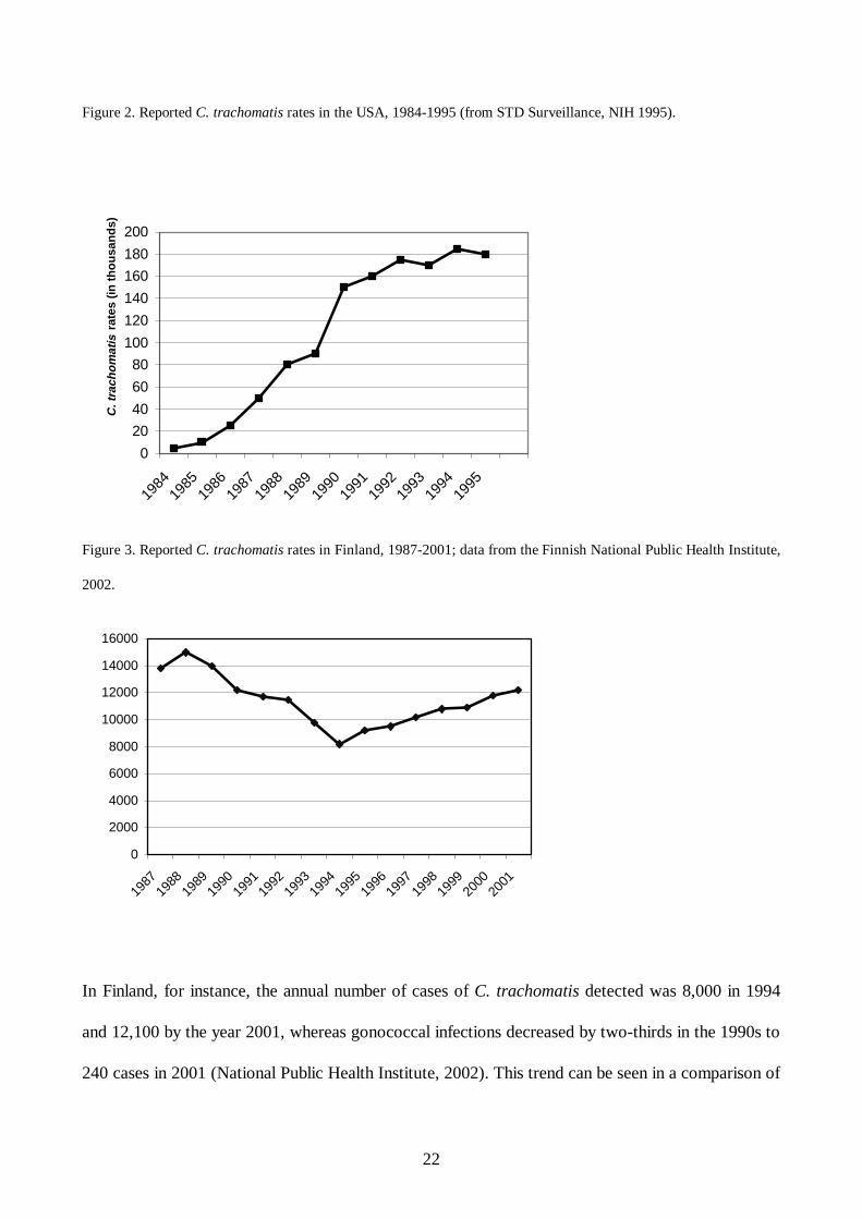
Another trend is the shift from inpatient PID toward outpatient PID. In the USA since 1980, the
number of hospitalizations of women for acute PID peaked in 1982 at nearly 200,000 – after which
the number has fallen steadily to less than 90,000 (Figure 4).
24
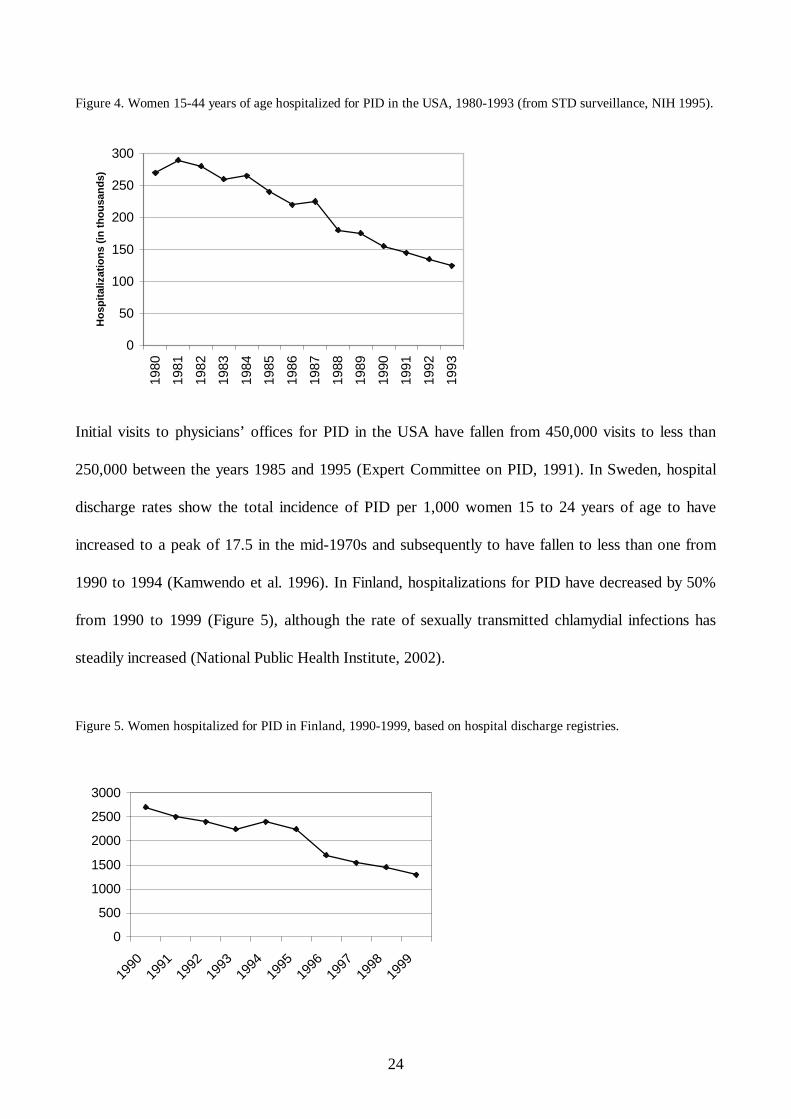
Figure 4. Women 15-44 years of age hospitalized for PID in the USA, 1980-1993 (from STD surveillance, NIH 1995).
0
50
100
150
200
250
30019
80
1981
1982
1983
1984
1985
1986
1987
1988
1989
1990
1991
1992
1993
Ho
spit
aliz
atio
ns
(in
th
ou
san
ds)
Initial visits to physicians’ offices for PID in the USA have fallen from 450,000 visits to less than
250,000 between the years 1985 and 1995 (Expert Committee on PID, 1991). In Sweden, hospital
discharge rates show the total incidence of PID per 1,000 women 15 to 24 years of age to have
increased to a peak of 17.5 in the mid-1970s and subsequently to have fallen to less than one from
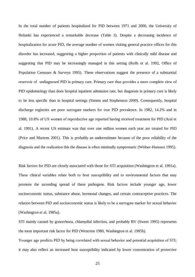
1990 to 1994 (Kamwendo et al. 1996). In Finland, hospitalizations for PID have decreased by 50%
from 1990 to 1999 (Figure 5), although the rate of sexually transmitted chlamydial infections has
steadily increased (National Public Health Institute, 2002).
Figure 5. Women hospitalized for PID in Finland, 1990-1999, based on hospital discharge registries.
0
500
1000
1500
2000
2500
3000
1990
1991
1992
1993
1994
1995
1996
1997
1998
1999
25
In the total number of patients hospitalized for PID between 1971 and 2000, the University of
Helsinki has experienced a remarkable decrease (Table 3). Despite a decreasing incidence of
hospitalization for acute PID, the average number of women visiting general practice offices for this
disorder has increased, suggesting a higher proportion of patients with clinically mild disease and
suggesting that PID may be increasingly managed in this setting (Rolfs et al. 1992, Office of
Population Censuses & Surveys 1995). These observations suggest the presence of a substantial
reservoir of undiagnosed PID in primary care. Primary care thus provides a more complete view of
PID epidemiology than does hospital inpatient admission rate, but diagnosis in primary care is likely
to be less specific than in hospital settings (Simms and Stephenson 2000). Consequently, hospital
discharge registries are poor surrogate markers for true PID prevalence. In 1982, 14.2% and in
1988, 10.8% of US women of reproductive age reported having received treatment for PID (Aral et
al. 1991). A recent US estimate was that over one million women each year are treated for PID
(Price and Martens 2001). This is probably an underestimate because of the poor reliability of the
diagnosis and the realization this the disease is often minimally symptomatic (Wölner-Hanssen 1995).
Risk factors for PID are closely associated with those for STI acquisition (Washington et al. 1991a).
These clinical variables relate both to host susceptibility and to environmental factors that may
promote the ascending spread of these pathogens. Risk factors include younger age, lower
socioeconomic status, substance abuse, hormonal changes, and certain contraceptive practices. The
relation between PID and socioeconomic status is likely to be a surrogate marker for sexual behavior
(Washington et al. 1985a).
STI mainly caused by gonorrhoea, chlamydial infection, and probably BV (Sweet 1995) represents
the most important risk factor for PID (Weström 1980, Washington et al. 1985b).
Younger age predicts PID by being correlated with sexual behavior and potential acquisition of STI;
it may also reflect an increased host susceptibility indicated by lower concentration of protective
26
chlamydial antibodies, a larger zone of cervical ectopy, and greater permeability of the cervical
mucus (Cates et al.1990, Washington and Katz 1991b). Women aged 20 to 24 have the highest
incidence of both STIs and PID, followed by teenagers (WHO 1981, Svensson et al. 1994). Overall,
75% of patients with PID are less than 25 and are sexually active women (Weström 1980, WHO
1981). Young people are behaviorally vulnerable to STI acquisition, as they generally have the
highest numbers of sexual partners and highest frequency of partner change.
The role of combined oral contraceptives (OC) in altering the risk for PID in women is complex and
incompletely understood (Weström 1980, Rubin et al. 1982, Washington et al. 1985b). Women who
use OCs may be at increased risk for developing cervical infection with C. trachomatis (Baeten et al.
2001), which may in part be due to increased prevalence of cervical ectopy (Kinghorn and Waugh
1981). On the other hand, overall OC use is associated with a reduction of 40 to 60% in the rate of
symptomatic PID and a 70% reduction in chlamydia-associated PID, but does not appear to protect
women with gonorrhoea against PID (Wölner-Hanssen et al. 1990a). Of concern is that OC use may
mask signs and symptoms of ascending infection, resulting in a greater proportion of subclinical or
“silent PID” (Svensson et al. 1984, Wölner-Hanssen 1986a, Ness et al. 1997, 2001). A 2000 meta-
analysis found in women with an IUD a relative risk for symptomatic PID of 3.3, although the
majority of these studies were not randomized controlled trials (Gareen et al. 2000). Most of the
excess risk associated with IUD use appears to be limited to the first few weeks after insertion
(Farley et al. 1992), indicating that a major determinant of PID rates associated with IUD use is the
prevalence of C. trachomatis and N. gonorrhoeae. Even if the relative risk for PID is higher in IUD
users, the absolute risk remains very low, approximately one in 1,000 (Walsh et al. 1998). A recent
Cochrane review suggests that there is no benefit in using antibiotic prophylaxis for women before
IUD insertion (Grimes and Schultz 2000). Furthermore, some evidence indicates no important effect
of IUD use on risk for tubal infertility (Grimes 2000, Hubacher et al. 2001).
27
2.5. Clinical manifestations
As a response to microorganisms spreading from the lower to the upper genital tract, the host
produces a clinical spectrum of PID including endometritis, salpingitis, pyosalpinx, tubo-ovarian
abscess, pelvic peritonitis, and perihepatitis (CDC 2002, Paavonen and Molander 2003). The clinical
picture ranges from symptomless to life-threatening disease. Mild symptoms may be missed by the
physician, and patients may seek care late, which increases the risk for tubal damage (Hillis et al
1993). On the other hand, approximately one-third of clinical PID diagnoses are false-positives, and
consequently patients with disease other than PID or with no disease at all often receive treatment
for PID.
2.5.1. Subclinical disease
Historically, low abdominal pain and palpatory tenderness of the uterus and adnexa have been
essential for suspicion of acute PID (Jacobson and Weström 1969). More than half of the women
with signs of postinfectious tubal damage such as tubal occlusion, hydrosalpinx, infertility, or ectopic
pregnancy often, however, have no history of symptomatic PID, suggesting that silent PID may lead
to permanent tubal destruction (Sellors et al. 1988, Patton et al. 1989, Wölner-Hanssen et al. 1990b,
Cates et al. 1993, Wölner-Hanssen 1995). Evidence exists of C. trachomatis infection’s being
particularly important in subclinical PID. Women with tubal damage but no history of PID are more
often chlamydia-seropositive than are women with other reasons for infertility (Brunham et al.1992,
Toye et al. 1993). Women with tubal factor infertility have often shown Chlamydia trachomatis
DNA or its antigen in their endometrium and Fallopian tubes (Henry-Suchet et al. 1981, Cleary et al.
1985). Moreover, on endometrial biopsy, up to two-thirds of women with chlamydial cervicitis and
no signs of PID have plasma cell endometritis consistent with subclinical PID (Paavonen et
28
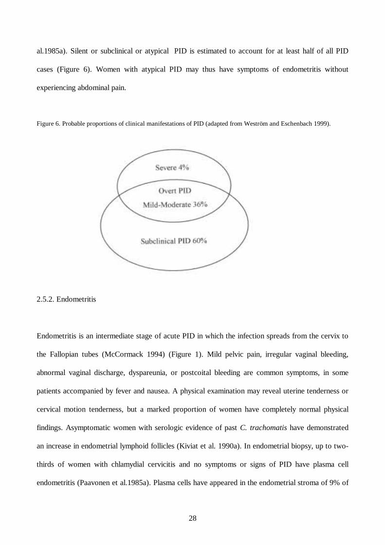
al.1985a). Silent or subclinical or atypical PID is estimated to account for at least half of all PID
cases (Figure 6). Women with atypical PID may thus have symptoms of endometritis without
experiencing abdominal pain.
Figure 6. Probable proportions of clinical manifestations of PID (adapted from Weström and Eschenbach 1999).
2.5.2. Endometritis
Endometritis is an intermediate stage of acute PID in which the infection spreads from the cervix to
the Fallopian tubes (McCormack 1994) (Figure 1). Mild pelvic pain, irregular vaginal bleeding,
abnormal vaginal discharge, dyspareunia, or postcoital bleeding are common symptoms, in some
patients accompanied by fever and nausea. A physical examination may reveal uterine tenderness or
cervical motion tenderness, but a marked proportion of women have completely normal physical
findings. Asymptomatic women with serologic evidence of past C. trachomatis have demonstrated
an increase in endometrial lymphoid follicles (Kiviat et al. 1990a). In endometrial biopsy, up to two-
thirds of women with chlamydial cervicitis and no symptoms or signs of PID have plasma cell
endometritis (Paavonen et al.1985a). Plasma cells have appeared in the endometrial stroma of 9% of
29
asymptomatic uninfected patients (Korn et al. 1995b). The only symptom of chlamydial PID is often
bleeding as a sign of endometritis (Wölner-Hanssen 1995). Fever and abnormal uterine bleeding
patterns occur in 40% of endometritis patients (Eschenbach 1980). Paavonen et al. found that history
of vaginal bleeding, presence of STI in the cervix, and antibodies to C. trachomatis or to
Mycoplasma hominis correlate with endometritis (Paavonen et al. 1985a).
The main criterion for endometritis is a histopathologic finding of inflammation in the endometrium,
which has traditionally been categorized as acute or chronic (Kiviat et al. 1990a). In Eckert’s study
of 152 women undergoing laparoscopy for suspected PID, 26 had histologic manifestations of
endometritis without laparoscopic evidence of acute salpingitis (Eckert et al. 2002). The clinical
manifestations in these 26 women with endometritis alone were generally less pronounced than in
women with definite salpingitis, and more prominent than in those without salpingitis or
endometritis. Eckert et al. (2002) concluded that the clinical and laboratory manifestations of PID
are highly correlated with its pathological findings, and endometritis emerges as a true clinical entity.
Eckert et al. found that not only gonococcal and chlamydial infection but also the first stage of the
menstrual cycle are risk factors for endometritis. Similarly, Korn et al. found that in women with
cervical N. gonorrhoea, C. trachomatis, or BV, a risk factor for endometritis is the proliferative
phase of the menstrual cycle (Korn et al. 1998). The natural history of endometritis, with or without
treatment, remains unclear. The frequency with which endometritis clears with menses, or persists
but remains limited to the uterus, or progresses into salpingitis remains unknown. In the absence of
such data, the potential for progression to salpingitis and the attendant risk for infertility would seem
to warrant aggressive antimicrobial therapy.
30
2.5.3. Mild and moderate PID
Mild and moderate PID are often used synonymously with salpingitis. Laparoscopic criteria for mild
PID were described by Jacobson and Weström in 1969: pronounced hyperemia of the tubal surface,
edema of the tubal wall, and sticky exudate on the tubal surface, while Hager et al. (1983) described
laparoscopic criteria for moderate PID, a more prominent form of salpingitis. Bilateral lower
abdominal pain is the most common presenting symptom. Patients with salpingitis are usually in good
general condition, and pain is usually slow in onset, bilateral, dull in character, and present in the
lower abdomen. Symptoms also include dyspareunia, vaginal discharge, menometrorragia, dysuria,
pain associated with menses, fever, and infrequently nausea and vomiting (WHO 2002). Examination
reveals no pelvic masses. Patients with mild PID usually visit outpatient clinics and are not
hospitalized.
Most of the laparoscopically verified cases have mild symptoms and mild physical signs. In Jacobson
and Weströms’ classical 1969 study incorporating 2,200 women, although no single symptom or sign
helped to distinguish those with salpingitis from those without, a combination of lower abdominal
pain plus cervical motion tenderness plus lower genital tract infection was apparent in 61% of
patients with and in 39% of patients without salpingitis. Peipert observed that differing clinical
symptoms and signs seen in mild versus moderate disease versus severe disease do not accurately
predict the extent of salpingitis seen at laparoscopy (Peipert 1996).
2.5.4. Severe PID
Of all patients with PID, severe PID accounts for only 3% to 4% (Jacobson and Weström 1969,
Weström 1977, WHO 1981, Eschenbach et al. 1997) (Figure 6). If the inflamed Fallopian tubes
become occluded at the fimbrial end, mucus or pus will fill the tubes, leading to an entity called
31
pyosalpinx (acute phase) or hydrosalpinx (chronic phase). If the tube does not become completely
occluded, some of the infectious pathogens spill into the pelvis and may invade the ovary at the time
of ovulation, forming a tubo-ovarian complex the anatomy of which has not yet broken down. If
treatment fails or is lacking, the acute inflammatory process may progress to its most severe phase,
resulting in a tubo-ovarian abscess (TOA). In the laparoscopic classification by Hager’s group
(1983), pyosalpinx and TOA represent severe PID. At first, usually only the ovary and tube on one
side are affected, and only at a relatively later stage does the process spread to the other adnexa, as a
result of which one can observe an ”out-of-phase” appearance of the two adnexa (Timor-Tritsch et
al. 1998). As many as 70% of TOAs can be unilateral (Landers 1996). A TOA will develop in
approximately 7% to 16% of hospitalized patients with PID, but numbers are biased because
calculations made among inpatients depend on how frequently PID patients are hospitalized for
treatment (Landers and Sweet 1983, 1985). Among those with TOA, no clearly defined risk factors
have been identifiable, and no standardized diagnostic criteria for TOA exist. An abscess seems to
develop especially in older patients with no STI (Landers 1996, Eschenbach et al. 1997). Women
presenting with a clinical diagnosis of PID and a pelvic mass may have a TOA or may have a
pyosalpinx, hydrosalpinx, tubo-ovarian complex, or other adnexal mass (Landers 1996).
Of all upper genital tract infections, severe PID represents only the tip of the iceberg. Patients often
present with symptoms such as severe lower abdominal pain, high fever, nausea, vomiting, and
purulent vaginal discharge. Pelvic examination may uncover a palpable mass, but examination may be
difficult because of severe pelvic pain. Patients with severe PID need hospitalization. Diffuse
peritonitis is extremely rare and may appear in cases of rupture of an abscess. Mortality from severe
PID is rare: a rate of only 0.29 per 100,000 women 15 to 44 years of age was reported in the USA in
1979, and mortality was usually due to rupture of an abscess, at a rate of 3% to 8% (Grimes 1986).
32
2.5.5. Perihepatitis
An extrapelvic manifestation of PID is inflammation of the liver capsule (perihepatitis or Fitz-Hugh-
Curtis syndrome) and the adjacent peritoneum. This was initially associated with gonococcal
infection when described by Curtis in 1930 and Fitz-Hugh in 1934. The Fitz-Hugh-Curtis syndrome
is characterized by violin-string adhesions between the liver and anterior abdominal wall
accompanying gross pathologic evidence of prior tubal infection. Later, perihepatitis was strongly
associated with chlamydial infection (Wölner-Hanssen et al. 1980, Wang et al. 1980, Paavonen et al.
1981). Recently, laparoscopically verified perihepatitis has been associated with elevated levels of
antibody to chlamydial HSP-60 (Money et al. 1997).
With or even without signs of pelvic infection, the acute phase may present with severe pain in the
right upper quadrant of the abdomen, thus mimicking cholecystitis, and this pain is exacerbated by
coughing or even by breathing (Wölner-Hanssen et al. 1980, Soper 2001). Diagnosis of acute
perihepatitis is established by laparoscopic visualization of an inflamed liver capsule and adjacent
peritoneum accompanied by exudate on the liver surface. The chronic phase is characterized by
violin-string adhesions between the liver surface and the anterior abdominal wall seen incidentally at
laparoscopy (Money et al. 1997).
The pathogenesis of perihepatitis is unclear. It is suggested that the main route is an extension of
tubal infection by direct peritoneal spread or by lymphatic or hematogenous spread from the pelvis to
the liver (Möller and Mårdh 1980). Perihepatitis is seen by laparoscopy in 5% to 15% of patients
with acute salpingitis, although a smaller proportion have actual clinical signs (Paavonen et al. 1981).
33
2.5.6. Periappendicitis
Acute PID may cause inflammation in adjacent structures; an example is inflammation of the
appendiceal serosa, causing periappendicitis, when the appendix is situated near the inflamed right
adnex. Periappendicitis occurs in 1% to 15% of appendices removed for acute appendicitis (Butler
1980). The majority of patients with periappendicitis are young women (Fink et al. 1990), and in
such patients PID treatment may be neglected.
2.6. Diagnosis
Clinical diagnosis of PID is imprecise. Clinical diagnosis of symptomatic PID has a PPV of 65% to
90% compared with laparoscopic diagnosis. PPV rates differ depending on clinical setting, with
higher PPV rates among sexually active young women and among patients attending STI clinics
(CDC 2002). No single finding is however, both sensitive and specific. Thus, false-positive and false-
negative diagnoses are common. Because of the difficulty of the diagnosis and the potential for
damage to the reproductive health of women even by mild or atypical PID, health-care providers
should maintain a low threshold for the diagnosis (McCormack 1994). Diagnosis and management of
other causes of lower abdominal pain are unlikely to be impaired by initiating empiric antimicrobial
therapy for PID (CDC 2002).
Lack of uniform clinical, microbiologic, or laparoscopic criteria is an obvious problem. Specimens
for etiologic diagnosis are difficult to obtain. Women who have classic clinical manifestations of PID
may well have conditions other than PID. Invasive laparoscopy is not always available, and
endometrial biopsy can remain negative because of irregular distribution of endometritis (Expert
committee on PID 1991). Thus, the wide clinical spectrum of the disease and the fact that patients
may present to a variety of clinicians will lead to major diagnostic difficulties.
34
In any fertile woman with pelvic pain, PID should be considered. Well-designed studies concerning
diagnosis of PID are few, but are essential in order to reduce over- and underdiagnosis. Improved
criteria for making diagnoses in mild disease are urgently needed. As long as the clinical diagnosis of
PID remains imprecise, focus should be on STI screening and rapid empirical use of effective
antibiotics to control the disease (Ross 2002).
2.6.1. Clinical diagnosis
Increasing concern about silent or atypical PID (Wölner-Hanssen 1995) has inspired creation of
several paradigms of clinical criteria. Such criteria have, however, never been validated in large
prospective studies (Soper et al. 1991). Jacobson and Weström, the first to evaluate signs and
symptoms of PID, found that diagnostic accuracy was improved by increasing the numbers of
positive criteria (Jacobson and Weström 1969). Weström’s group set up major and minor clinical
criteria for PID diagnosis based on their study including 2200 laparoscopically verified cases
(Weström 1983, Weström and Mårdh 1984). Hager’s group modified these criteria and did not
require abdominal pain as a major criterion, because of their knowledge of silent PID (Hager et al.
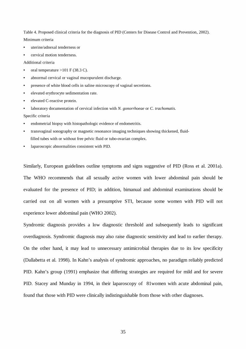
1983). The minimum criteria for the clinical syndromic diagnosis of PID recommended by the
Centers for Disease Control and Prevention (CDC) are cervical motion tenderness and uterine or
adnexal tenderness. If these minimum criteria are present, and no other cause for the illness can be
identified, empiric treatment of PID should be initiated in sexually active young women and others at
risk for STIs (CDC 2002). In patients with both pelvic tenderness and signs of lower genital tract
inflammation, PID should be considered, and treatment may be indicated based on the patient’s risk
profile. Criteria addition may serve to enhance the specificity of the minimum criteria (Table 4). CDC
guidelines suggest that more emphasis should be put on detection of lower genital tract infection
(LGTI) rather than on presence of abdominal pain.
35
Table 4. Proposed clinical criteria for the diagnosis of PID (Centers for Disease Control and Prevention, 2002).
Minimum criteria
• uterine/adnexal tenderness or
• cervical motion tenderness.
Additional criteria
• oral temperature >101 F (38.3 C).
• abnormal cervical or vaginal mucopurulent discharge.
• presence of white blood cells in saline microscopy of vaginal secretions.
• elevated erythrocyte sedimentation rate.
• elevated C-reactive protein.
• laboratory documentation of cervical infection with N. gonorrhoeae or C. trachomatis.
Specific criteria
• endometrial biopsy with histopathologic evidence of endometritis.
• transvaginal sonography or magnetic resonance imaging techniques showing thickened, fluid-
filled tubes with or without free pelvic fluid or tubo-ovarian complex.
• laparoscopic abnormalities consistent with PID.
Similarly, European guidelines outline symptoms and signs suggestive of PID (Ross et al. 2001a).
The WHO recommends that all sexually active women with lower abdominal pain should be
evaluated for the presence of PID; in addition, bimanual and abdominal examinations should be
carried out on all women with a presumptive STI, because some women with PID will not
experience lower abdominal pain (WHO 2002).
Syndromic diagnosis provides a low diagnostic threshold and subsequently leads to significant
overdiagnosis. Syndromic diagnosis may also raise diagnostic sensitivity and lead to earlier therapy.
On the other hand, it may lead to unnecessary antimicrobial therapies due to its low specificity
(Dallabetta et al. 1998). In Kahn’s analysis of syndromic approaches, no paradigm reliably predicted
PID. Kahn’s group (1991) emphasize that differing strategies are required for mild and for severe
PID. Stacey and Munday in 1994, in their laparoscopy of 81women with acute abdominal pain,
found that those with PID were clinically indistinguishable from those with other diagnoses.
36
The still ongoing PID evaluation and clinical health study (PEACH), including more than 1,500
patients, is a randomized controlled trial comparing inpatient and outpatient management of mild
and moderate PID. Its study design differs from that of many other studies which include only
hospitalized patients with severe PID; its results may therefore be more generalizable. Reports from
PEACH indicate that adnexal tenderness is a sensitive (96%) but unspecific (4%) marker of mild
PID. As expected, combining lower abdominal tenderness, adnexal tenderness, and cervical motion
tenderness reduces sensitivity but improves specificity. The two factors which best predict
endometritis are a positive bacterial result (C. trachomatis or N. gonorrhoeae) and a combination of
elevated temperature with a high white blood cell (WBC) count (Peipert et al. 2001). Jacobson and
Weström, comparing clinical and laparoscopic diagnoses of PID, found that only 66% of women
with clinically diagnosed PID actually have the condition, and that clinical criteria failed to identify
10% of laparoscopically diagnosed cases (Jacobson and Weström 1969). Since that study, multiple
studies have shown that the accuracy of the clinical diagnosis based on history and pelvic
examination is low or very low versus laparoscopy, the gold standard (Table 5).
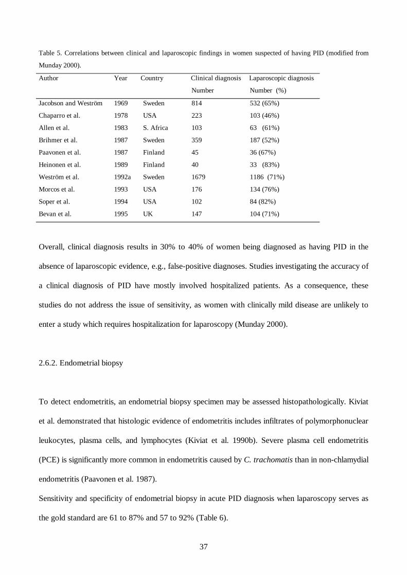
37
Table 5. Correlations between clinical and laparoscopic findings in women suspected of having PID (modified from
Munday 2000).
Author Year Country Clinical diagnosis
Number
Laparoscopic diagnosis
Number (%)
Jacobson and Weström 1969 Sweden 814 532 (65%)
Chaparro et al. 1978 USA 223 103 (46%)
Allen et al. 1983 S. Africa 103 63 (61%)
Brihmer et al. 1987 Sweden 359 187 (52%)
Paavonen et al. 1987 Finland 45 36 (67%)
Heinonen et al. 1989 Finland 40 33 (83%)
Weström et al. 1992a Sweden 1679 1186 (71%)
Morcos et al. 1993 USA 176 134 (76%)
Soper et al. 1994 USA 102 84 (82%)
Bevan et al. 1995 UK 147 104 (71%)
Overall, clinical diagnosis results in 30% to 40% of women being diagnosed as having PID in the
absence of laparoscopic evidence, e.g., false-positive diagnoses. Studies investigating the accuracy of
a clinical diagnosis of PID have mostly involved hospitalized patients. As a consequence, these
studies do not address the issue of sensitivity, as women with clinically mild disease are unlikely to
enter a study which requires hospitalization for laparoscopy (Munday 2000).
2.6.2. Endometrial biopsy
To detect endometritis, an endometrial biopsy specimen may be assessed histopathologically. Kiviat
et al. demonstrated that histologic evidence of endometritis includes infiltrates of polymorphonuclear
leukocytes, plasma cells, and lymphocytes (Kiviat et al. 1990b). Severe plasma cell endometritis
(PCE) is significantly more common in endometritis caused by C. trachomatis than in non-chlamydial
endometritis (Paavonen et al. 1987).
Sensitivity and specificity of endometrial biopsy in acute PID diagnosis when laparoscopy serves as
the gold standard are 61 to 87% and 57 to 92% (Table 6).
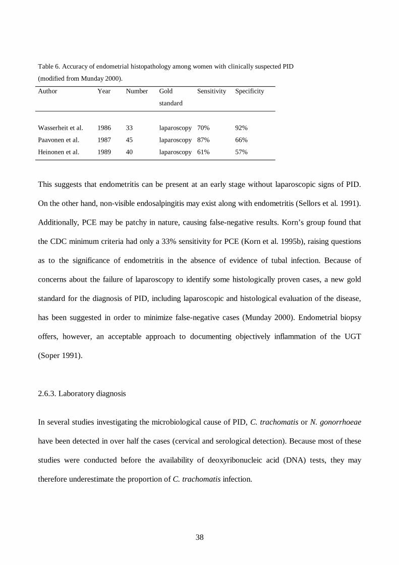
38
Table 6. Accuracy of endometrial histopathology among women with clinically suspected PID
(modified from Munday 2000).
Author Year Number
Gold
standard
Sensitivity Specificity
Wasserheit et al. 1986 33 laparoscopy 70% 92%
Paavonen et al. 1987 45 laparoscopy 87% 66%
Heinonen et al. 1989 40 laparoscopy 61% 57%
This suggests that endometritis can be present at an early stage without laparoscopic signs of PID.
On the other hand, non-visible endosalpingitis may exist along with endometritis (Sellors et al. 1991).
Additionally, PCE may be patchy in nature, causing false-negative results. Korn’s group found that
the CDC minimum criteria had only a 33% sensitivity for PCE (Korn et al. 1995b), raising questions
as to the significance of endometritis in the absence of evidence of tubal infection. Because of
concerns about the failure of laparoscopy to identify some histologically proven cases, a new gold
standard for the diagnosis of PID, including laparoscopic and histological evaluation of the disease,
has been suggested in order to minimize false-negative cases (Munday 2000). Endometrial biopsy
offers, however, an acceptable approach to documenting objectively inflammation of the UGT
(Soper 1991).
2.6.3. Laboratory diagnosis
In several studies investigating the microbiological cause of PID, C. trachomatis or N. gonorrhoeae
have been detected in over half the cases (cervical and serological detection). Because most of these
studies were conducted before the availability of deoxyribonucleic acid (DNA) tests, they may
therefore underestimate the proportion of C. trachomatis infection.
39
Cell culture has long been the gold standard for detection of C. trachomatis. Recently developed
nucleic acid amplification (NAA) tests such as polymerase chain reaction (PCR) and the ligase chain
reaction (LCR) have largely replaced cell culture and antigen tests because of much higher sensitivity
and specificity (Puolakkainen et al. 1998). PCR and LCR of first void-urine are highly effective in
detection of both symptomatic and asymptomatic chlamydial infection (Schachter et al. 1995,
Paukku et al. 1997). At best, microbiological tests may identify a cause in 80% of PID cases, and
therefore can only be a supplement in the diagnosis of PID. Moreover, not all women with pelvic
pain and positive C. trachomatis have PID at laparoscopy (Weström et al. 1992a, Bevan et al. 1995).
Identification of an organism may confirm provisional PID diagnosis, as has been seen in syndromic
diagnosis (CDC 1998).
No laboratory test is either highly sensitive or specific for PID. WBC, erythrocyte sedimentation rate
(ESR), and C-reactive protein (CRP) have served as diagnostic markers. No consistent difference in
these markers between patients with and without PID is apparent when all studies are evaluated,
although some studies have found significant differences. In laparoscopically controlled series,
elevated ESR has proven to correlate with severity of PID (Weström 1977) and with chlamydia-
associated PID (Svensson et al. 1980). An elevated CRP level is more sensitive and specific in
predicting PID than is an elevated ESR. Level of CRP also reflects disease severity (Lehtinen, et al.
1986, Miettinen et al. 1993). Changes in CRP level reflect the course of the disease better than does
ESR. In laparoscopically verified cases, elevated WBC is seen more often in PID (59%) than in non-
PID (33%) (Jacobson et al. 1969), and WBC is more often elevated in severe than in mild disease
(Weström et al. 1977). In the Peipert et al. study (1996), normal ESR and WBC in the absence of
LGTI excluded UGT infection.
Weström found a marked increase in number of inflammatory cells in wet smears of vaginal
secretions of women with PID (Weström 1983). It appears that this test may exclude the possibility
40
of PID in women with abdominal pain. In other words, women with PID almost always have signs of
LGTI, but women with LGTI do not necessarily have PID (Soper 2001).
It has been claimed that absence of LGTI will effectively exclude PID. However, limited studies are,
unfortunately, discouraging. Studies in which LGTI is defined both subjectively by naked eye
observation of vaginal discharge and objectively by microscopic analysis of vaginal or cervical smear,
show LGTI to be absent in 13% to 73% of women with PID, excluding LGTI as a single valid
predictor of PID (Munday 2000). Despite its limitations, CDC points out the importance of detection
of LGTI as a part of PID syndromic diagnosis (CDC 2002).
2.6.4. Ultrasonographic diagnosis
In 1987, Timor-Tritsch and Rottem introduced high-frequency transvaginal sonography (TVS) with
a resolution capability superior to that of transabdominal sonography (TAS); because of its close
relationship to structures, the ultrasound wave can be less attenuated (Timor-Tritsch and Rottem
1987, Timor-Tritsch et al. 1988). The diagnostic capacity of TVS quickly proved to be superior to
that of TAS (Bulas et al. 1992). Several studies have stressed that with TVS, the appearance of tubal
inflammatory disease is typical and reproducible (Tessler et al. 1989, Atri et al 1989, Timor-Tritsch
1991, Bellah et al. 1991, Timor-Tritsch 1995, Taipale et al. 1995).
Timor-Tritsch has recently introduced sonographic landmark findings for PID useful not only in
diagnosis and staging of the severity of PID but also in distinguishing PID from other pelvic
pathologies. He placed different sonographic markers of tubal disease in the context of their
pathogenesis. However, detection of mild PID is still a major problem, because of a lack of clear
sonographic signs (Timor-Tritsch et al. 1998).
Color Doppler imaging, based on mean frequency shift Doppler in detecting inflammation-induced
hyperemia results in statistically lower pulsatility indices (PI) for acute PID than those PI values for
41
the PID recovery phase. Color Doppler studies indicate that inflammation-induced hyperemia can
serve not only as an indicator of acute PID but also of recovery (Tinkanen and Kujansuu 1993,
Kupesic et al. 1995, Alatas et al. 1996, Tepper et al. 1998).
Power Doppler, a variation of conventional color Doppler imaging using the amplitude of the
Doppler signal, has become available, the greatest advantage of which is its ability to image areas of
low blood flow currently undetectable by frequency-based color Doppler techniques (Rubin et al.
1994, Hamper et al. 1997). Absolute lack of aliasing and relative angle independence are advantages
lacking in conventional color Doppler (Rubin et al. 1994, Weskott 1997). Disadvantages of power
Doppler are the lack of information about speed and direction of flow plus its high motion-sensitivity
(Kremkau 1995). The principal aim of power Doppler is to determine the position of vessels and the
presence or absence of flow. It has served in a variety of clinical applications involving the female
reproductive tract and pregnancy, such as in ovarian stromal, and in follicular and corpus luteum
perfusion, as well as for endometriotic cysts and ovarian cancer (Guerriero et al. 1999). It has proven
superior to conventional color Doppler in detecting tumor vascular distribution and ovarian
vascularity (Park et al. 1998, Tailor et al. 1998). This contrasts with the low detection rate in benign
masses with conventional color Doppler (Tekay et al. 1992). However, power Doppler should be
considered only as a secondary test, with the B-mode appearance considered first (Guerriero et al.
1999). Power Doppler has thus so far been little used in depicting hyperperfusion associated with
pelvic inflammatory processes (Papadimitriou et al. 1996).
Only a few studies have been designed to detect accuracy of PID diagnosis by TVS (Table 7).
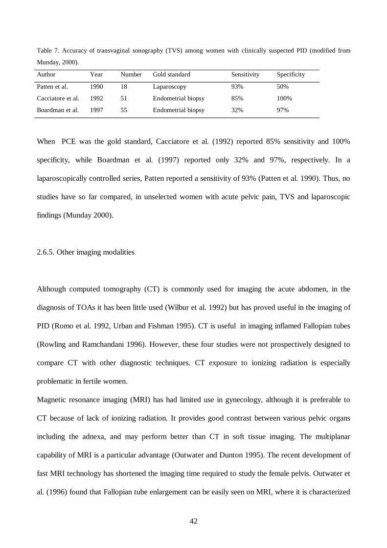
42
Table 7. Accuracy of transvaginal sonography (TVS) among women with clinically suspected PID (modified from
Munday, 2000).
Author Year Number Gold standard Sensitivity Specificity
Patten et al. 1990 18 Laparoscopy 93% 50%
Cacciatore et al. 1992 51 Endometrial biopsy 85% 100%
Boardman et al. 1997 55 Endometrial biopsy 32% 97%
When PCE was the gold standard, Cacciatore et al. (1992) reported 85% sensitivity and 100%
specificity, while Boardman et al. (1997) reported only 32% and 97%, respectively. In a
laparoscopically controlled series, Patten reported a sensitivity of 93% (Patten et al. 1990). Thus, no
studies have so far compared, in unselected women with acute pelvic pain, TVS and laparoscopic
findings (Munday 2000).
2.6.5. Other imaging modalities
Although computed tomography (CT) is commonly used for imaging the acute abdomen, in the
diagnosis of TOAs it has been little used (Wilbur et al. 1992) but has proved useful in the imaging of
PID (Romo et al. 1992, Urban and Fishman 1995). CT is useful in imaging inflamed Fallopian tubes
(Rowling and Ramchandani 1996). However, these four studies were not prospectively designed to
compare CT with other diagnostic techniques. CT exposure to ionizing radiation is especially
problematic in fertile women.
Magnetic resonance imaging (MRI) has had limited use in gynecology, although it is preferable to
CT because of lack of ionizing radiation. It provides good contrast between various pelvic organs
including the adnexa, and may perform better than CT in soft tissue imaging. The multiplanar
capability of MRI is a particular advantage (Outwater and Dunton 1995). The recent development of
fast MRI technology has shortened the imaging time required to study the female pelvis. Outwater et
al. (1996) found that Fallopian tube enlargement can be easily seen on MRI, where it is characterized
43
by tortuous folding of fluid-filled structures on T2-weighted images. Mitchell et al. in 1987,
Yamashita et al. in 1995, and Komatsu et al. in 1996 described MRI characterization of adnexal
masses. Jain et al. (1993) concluded in their study that MRI is less specific than TVS in the
assessment of pelvic masses. The role of MRI in the standard work-up of common adnexal disorders
has not yet been well described, and imaging methods such as CT and MRI are costly and not widely
available.
Mozas’s group in 1993 compared radionuclide scintigraphy with laparoscopy in 40 women with a
provisional diagnosis of PID; their sensitivity was 95% and specificity 85%.
2.6.6. Laparoscopic diagnosis
Laparoscopy allows an excellent view of pelvic structures and has historically been the gold standard
of diagnostic modalities to diagnose PID and to obtain specimens from the Fallopian tubes (Jacobson
1964, Jacobson and Weström 1969, Weström 1975, Mårdh et al. 1977). Laparoscopy also allows
study of the microbiology, pathology, and outcome of PID (Westöm 1992a). Laparoscopy is,
however, an invasive technique, it necessitates hospitalization and general anaesthesia, and is costly.
Laparoscopy can also cause complications (Härkki-Siren and Kurki 1997) and may not be practical,
especially in mild PID in the outpatient clinical settings where most PID is diagnosed. In Jacobson
and Weström’s landmark study, laparoscopy revealed salpingitis in 66%, normal pelvic anatomy in
23%, and other pelvic pathology in 12% of clinically suspected PID patients (Jacobson and Weström
1969). Laparoscopic visualization of mild, moderate, and severe PID reveals the gradual spreading
of the disease (Table 8).
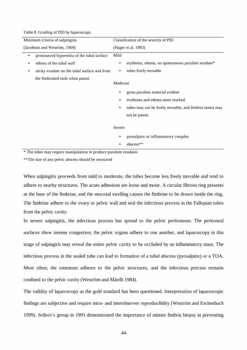
44
Table 8. Grading of PID by laparoscopy.
Minimum criteria of salpingitis
(Jacobson and Weström, 1969)
Classification of the severity of PID
(Hager et al. 1983)
• pronounced hyperemia of the tubal surface
• edema of the tubal wall
• sticky exudate on the tubal surface and from
the fimbriated ends when patent
Mild
• erythema, edema, no spontaneous purulent exudate*
• tubes freely movable
Moderate
• gross purulent material evident
• erythema and edema more marked
• tubes may not be freely movable, and fimbria stoma may
not be patent
Severe
• pyosalpinx or inflammatory complex
• abscess**
* The tubes may require manipulation to produce purulent exudates
**The size of any pelvic abscess should be measured
When salpingitis proceeds from mild to moderate, the tubes become less freely movable and tend to
adhere to nearby structures. The acute adhesions are loose and moist. A circular fibrous ring presents
at the base of the fimbriae, and the mucosal swelling causes the fimbriae to be drawn inside the ring.
The fimbriae adhere to the ovary or pelvic wall and seal the infectious process in the Fallopian tubes
from the pelvic cavity.
In severe salpingitis, the infectious process has spread to the pelvic peritoneum. The peritoneal
surfaces show intense congestion, the pelvic organs adhere to one another, and laparoscopy in this
stage of salpingitis may reveal the entire pelvic cavity to be occluded by an inflammatory mass. The
infectious process in the sealed tube can lead to formation of a tubal abscess (pyosalpinx) or a TOA.
Most often, the omentum adheres to the pelvic structures, and the infectious process remains
confined to the pelvic cavity (Weström and Mårdh 1984).
The validity of laparoscopy as the gold standard has been questioned. Interpretation of laparoscopic
findings are subjective and require intra- and interobserver reproducibility (Weström and Eschenbach
1999). Sellors’s group in 1991 demonstrated the importance of minute fimbria biopsy in preventing
45
both false-positive and false-negative laparoscopic diagnoses. Compared to fimbrial histopathologic
mini-biopsy, the sensitivity of laparoscopic diagnosis of PID was only 50% and its specificity 80, and
visual diagnosis of mild salpingitis was especially insensitive. False-negative laparoscopic results, an
acknowledged problem (Jacobson 1980, Hager et al. 1983), are possibly due to the presence of
endosalpingitis. Endometritis and endosalpingitis may not show any sign of ectosalpingitis (Sellors et
al. 1991). Inadequate criteria for visual and histopathologic diagnoses of PID may be another reason
for poor agreement between visual and histopathological assessments (Sellors et al. 1991).
Diagnostic laparoscopy still needs standardization. Henry-Suchet and Tesquier (1994) point out the
importance of a systematic diagnostic laparoscopy to achieve diagnosis and to obtain microbiological
and histological samples. Evaluation of the peritoneal exudate may document the presence of
inflammatory cells: peritoneal fluid in normal patients and in those with endometriosis contains
predominantly macrophages (Halme et al. 1984b), and acute salpingitis predominantly neutrophils
(Paavonen et al. 1985b).
Laparoscopy is particularly indicated in mild cases to confirm diagnosis and in severe cases for
treatment. Indications for laparoscopy in PID are especially strong in young women, in whom early
diagnosis is essential to preserve future fertility (Henry-Suchet and Tesquier 1994).
The diagnostic accuracy and intra- and interobserver reliability of laparoscopy has not been
extensively evaluated. When Hornstein et al. (1993) tested reproducibility of laparoscopic findings
using the revised American Fertility Society (AFS) classification system of endometriosis, intra- and
interobserver agreement were only moderate and low. Intra- and interobserver variation resulted in
endometriosis staging differences in 38% and 52 % of patients. The study documented a high degree
of variability and concluded that any future laparoscopic classification system should be validated
before adoption. Rock et al. (1995) also tested laparoscopic interobserver reproducibility of the
revised AFS endometriosis classification, with slightly better results than Hornstein’s. Bowman’s
group (1995) used the AFS scoring system for adhesions for evaluation of interobserver variability in
46
laparoscopic assessment of pelvic adhesions. Interobserver kappa statistics were low for grand total
scoring (κ 0.21), prognosis after surgery (0.32), and management recommendations (0.13),
suggesting that the AFS classification system is inappropriate for evaluating prognosis or
recommending treatment or as a means of comparison between centers. Intra- and interobserver
laparoscopic adhesion scoring tests showed variability low enough to allow observer confirmation of
videotaped laparoscopic findings, even though variability must be taken into account when clinical
standards are being established (Corson et al. 1995). In the Hornstein, Bowman, and Corson studies,
laparoscopic visual information was on videotape, while Rock used videotapes, slides, and
photographs. These few available laparoscopic observer-reliability studies clearly indicate
imperfections in laparoscopic data assessment and underline the importance of a scoring system
properly evaluated by observer-agreement studies.
Some investigators have realized the importance of systematic evaluation of laparoscopic findings for
PID (Jacobson and Weström 1969, Hager et al. 1983, Soper 1991, Henry-Suchet and Tesquier
1994). Jacobson and Weström introduced laparoscopic criteria for salpingitis, and Hager et al. in
1983 attempted to refine these criteria into a classification of severity of disease (Table 8). In their
grading system, mild salpingitis is associated with minimum visual criteria, and in addition, the tubes
are freely mobile with the ostia appearing patent. A scoring system (ASS=acute salpingitis score) for
laparoscopic findings in acute PID can determine prognosis (Henry-Suchet and Tesquier 1994).
Several criteria have been used to define PID, including tubal erythema, tubal edema, tubal fimbrial
status, mobility and patency of adnexa, and evaluation of pelvic adhesions and peritoneal fluid. In
several cases, both adnexa are not equally affected, and in defining severity, overall grade should
reflect the most severe finding (Soper 1991). Clinical severity of the disease does not predict the
severity of tubal disease (Svensson et al. 1980), especially in mild disease, in which patients often
have more severe changes in the tubes than would be expected from a benign clinical course (Kiviat
et al. 1990b). As patients with severe disease require longer-duration antibiotic therapy before
47
achieving a therapeutic response (Wasserheit et al. 1986), laparoscopic grading may play an
important role in their management. Additionally, better laparoscopic standardization of severity of
salpingitis may allow for improved capabilities of prognosis (Jacobson and Weström 1969, Henry-
Suchet and Tesquier 1994).
No standard classification system for PID diagnosis has thus been introduced nor has any scoring
system been properly evaluated for observer variation (Weström and Eschenbach 1999). Lack of
consensus in reporting laparoscopic findings concerning PID diagnosis reduces the value of
laparoscopy as the diagnostic gold standard.
2.6.7. Differential diagnosis
Approximately one-third of clinically suspected PID patients have another disease or no disease. In
the largest series of women undergoing laparoscopy for clinically suspected salpingitis, 12% had
intra-abdominal disorders other than PID (Jacobson and Weström 1969). Of clinically suspected PID
patients, 5% have surgical conditions that would be dangerous if left unrecognized (Jacobson and
Weström 1969, Weström 1977, Sweet et al. 1980). Patients with other diseases are often treated
unnecessarily with antibiotics. Differential diagnosis includes gynecologic disorders such as
endometriosis, adnexal torsion, ovarian cyst rupture, degeneration of leiomyomas, and ectopic
pregnancy (Table 9).
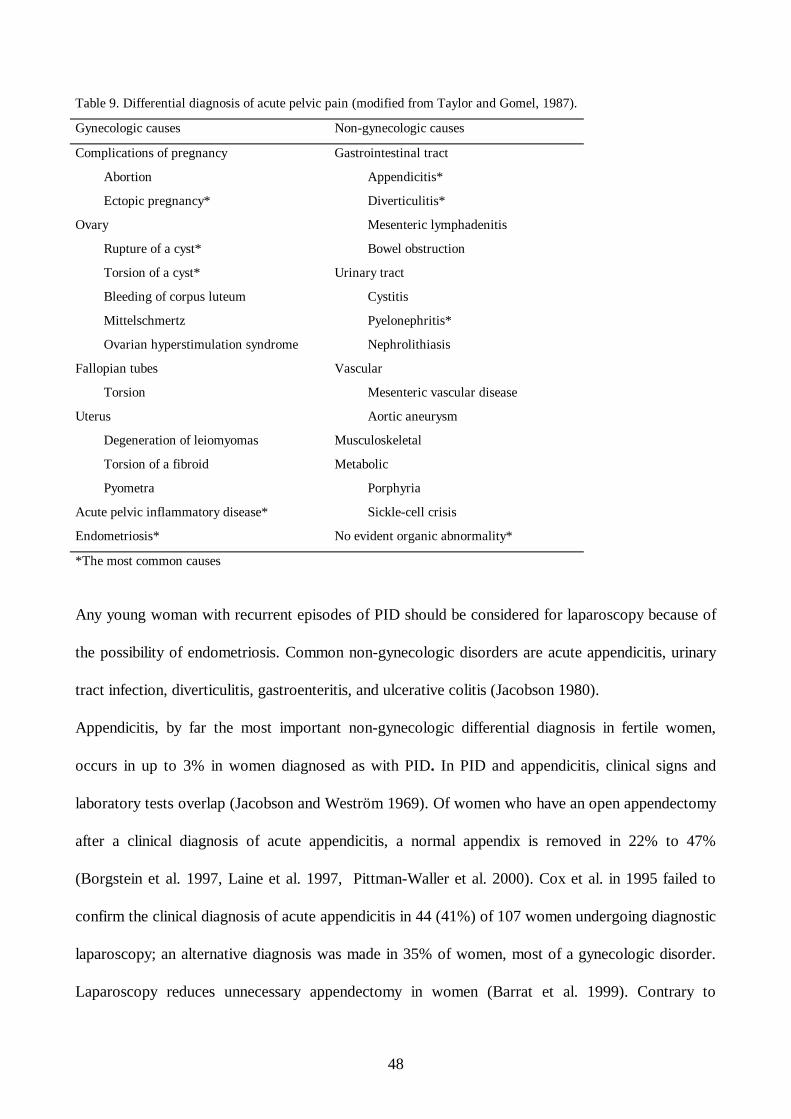
48
Table 9. Differential diagnosis of acute pelvic pain (modified from Taylor and Gomel, 1987).
Gynecologic causes Non-gynecologic causes
Complications of pregnancy
Abortion
Ectopic pregnancy*
Ovary
Rupture of a cyst*
Torsion of a cyst*
Bleeding of corpus luteum
Mittelschmertz
Ovarian hyperstimulation syndrome
Fallopian tubes
Torsion
Uterus
Degeneration of leiomyomas
Torsion of a fibroid
Pyometra
Acute pelvic inflammatory disease*
Endometriosis*
Gastrointestinal tract
Appendicitis*
Diverticulitis*
Mesenteric lymphadenitis
Bowel obstruction
Urinary tract
Cystitis
Pyelonephritis*
Nephrolithiasis
Vascular
Mesenteric vascular disease
Aortic aneurysm
Musculoskeletal
Metabolic
Porphyria
Sickle-cell crisis
No evident organic abnormality*
*The most common causes
Any young woman with recurrent episodes of PID should be considered for laparoscopy because of
the possibility of endometriosis. Common non-gynecologic disorders are acute appendicitis, urinary
tract infection, diverticulitis, gastroenteritis, and ulcerative colitis (Jacobson 1980).
Appendicitis, by far the most important non-gynecologic differential diagnosis in fertile women,
occurs in up to 3% in women diagnosed as with PID. In PID and appendicitis, clinical signs and
laboratory tests overlap (Jacobson and Weström 1969). Of women who have an open appendectomy
after a clinical diagnosis of acute appendicitis, a normal appendix is removed in 22% to 47%
(Borgstein et al. 1997, Laine et al. 1997, Pittman-Waller et al. 2000). Cox et al. in 1995 failed to
confirm the clinical diagnosis of acute appendicitis in 44 (41%) of 107 women undergoing diagnostic
laparoscopy; an alternative diagnosis was made in 35% of women, most of a gynecologic disorder.
Laparoscopy reduces unnecessary appendectomy in women (Barrat et al. 1999). Contrary to
49
expectations, the frequency of misdiagnoses leading to unnecessary appendectomy has not changed
with the introduction of new diagnostic aids such as CT, ultrasonography, laparoscopy, and clinical
scoring systems, according to a 12-year retrospective cohort study (1987-1998) including 85,790
patients, and in fact misdiagnosis of acute appendicitis has even recently increased among fertile
women (Flum et al. 2001).
Puylaert (1986) introduced transabdominal graded compression ultrasonography into the diagnosis
of acute appendicitis. Since then, several studies have used this technique (Jeffrey et al. 1987, Rioux
1992), to show sonographic diagnosis of appendicitis to be established if, in patients with right lower
quadrant pain, a blind-ending noncompressible tubular structure is visualized with anteroposterior
measurements 7 mm or greater and with thickened muscular walls (Puylaert 1986, Jeffrey et al.
1988, Puylaert 1994a). Current evidence suggests, however, a limited role for ultrasonography,
especially when clinical evidence is convincing (Roosevelt and Reynolds 1998, Lessin et al. 1999). In
a prospective multicentre study of 2,280 patients, no clinical benefit appeared from routine
ultrasonography (Franke et al.1999). Prospective studies suggest that ultrasonography may be useful
in an equivocal case in which a combination of repeated clinical assessments and compression
ultrasonography may provide additional information as to whether surgery is necessary (Rice et al.
1999, Douglas et al. 2000). The failure to visualize the appendix, whether diseased or normal,
fundamentally limits the usefulness of US (Paulson et al. 2003).
Color Doppler in the diagnosis of acute appendicitis has proven useful. Quillin et al. (1994)showed
that it can demonstrate the hyperperfusion associated with appendicitis, and with color Doppler and
gray-scale TAS combined, sensitivity is higher than with either alone. Lim et al. showed in 1996 that
color Doppler may be helpful in diagnosis of early acute appendicitis when an appendix is equivocal
in size in gray-scale TAS.
TVS has not been extensively used and appears mostly in case reports showing that it is possible to
diagnose acute appendicitis with TVS, especially in cases where TAS is negative (Puylaert 1994b,
50
Pelsang et al. 1994, Whitford and Crade 1994, Larcos 1996, Caspi et al. 1996). The criteria for acute
appendicitis seem to be useful also for TVS, though no prospective systematic evaluation of the role
of TVS exists.
2.7. Treatment
2.7.1. Conservative
Even if antimicrobial therapy is the basis of conservative therapy for PID, limited evidence exists as
to the proper antibiotic to use. Available clinical evidence leans heavily on parenteral therapy of
inpatients (Ross 2001b). Most investigators use short-term recovery as their assessment of cure,
while only long-term follow-up concerning preservation of fertility provides the outcome of concern
(Peterson et al. 1991). Assessment of the efficacy of treatment for acute PID should include: 1.
evaluation of the clinical response to acute infection, 2. elimination of N. gonorrhoeae and C.
trachomatis, if present, 3. evaluation of relapse/recurrence rates, and 4. incidence of late sequelae.
No study has evaluated all these outcome measures (Weström and Eschenbach 1999). Moreover, no
efficacy data compare parenteral with oral regimens or inpatient with outpatient treatment settings
(CDC 2002). In preliminary reports from the PEACH study, no difference appeared in outcome for
mild or moderate PID whether patients were treated as outpatients or admitted to the hospital (Ness
et al. 2002). Use of a two-week treatment period in trials reflects common clinical practice, but
whether outcomes would be similar for shorter treatment periods or be improved with longer courses
is not known. Choice of therapy is only in part aided by microbiological studies showing a wide
variety of bacteria in the Fallopian tubes but failing to determine which of these are primary
pathogens (Ross 2001b).
51
Antimicrobial coverage must provide empiric broad-spectrum coverage of likely pathogens including
C. trachomatis, N. gonorrhoeae, anaerobes, Gram-negative facultative bacteria, and streptococci.
(Peterson et al. 1991, CDC 2002).
The CDC in 2002 recommended treatment guidelines, recommending as primary outpatient oral
treatment a combination of ofloxacin 400 mg twice daily (or levofloxacin 500 mg once daily) with or
without metronidazole 500 mg twice daily orally. Alternatively, intramuscular (IM) ceftriaxone 250
mg/cefoxitin 2 g single dose plus doxycycline 100 mg twice daily with or without metronidazole 500
mg twice a day. Length of therapy should be at least 14 days. In the outpatient setting, review at 72
hours is recommended, particularly for those with a moderate or severe clinical presentation (CDC
2002).
European guidelines for PID treatment similarly recommend ofloxacin plus metronidazole twice a
day for 14 days or IM ceftriaxone/cefoxitin single dose plus doxycycline and metronidazole for 14
days (Ross 2001b). The WHO recommends as primary outpatient therapy single-dose treatment for
uncomplicated gonorrhoea (ceftriaxone) plus doxycycline 100 mg twice daily plus metronidazole 400
mg twice daily for 14 days (WHO 2002).
Only 15 to 20% of all PID patients are hospitalized, and the number is still declining (Aral et al.
1988, Rolfs et al. 1992). Severe disease (e.g., TOA), uncertain diagnosis, and unsatisfactory
response to outpatient therapy indicate hospitalization, which usually but not necessarily leads to
initiation of parenteral treatment (Table 10).
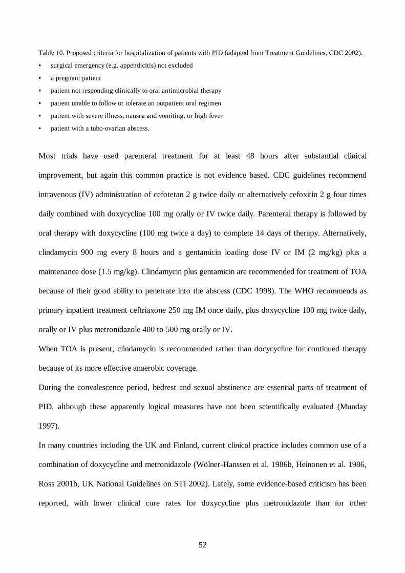
52
Table 10. Proposed criteria for hospitalization of patients with PID (adapted from Treatment Guidelines, CDC 2002).
• surgical emergency (e.g. appendicitis) not excluded
• a pregnant patient
• patient not responding clinically to oral antimicrobial therapy
• patient unable to follow or tolerate an outpatient oral regimen
• patient with severe illness, nausea and vomiting, or high fever
• patient with a tubo-ovarian abscess.
Most trials have used parenteral treatment for at least 48 hours after substantial clinical
improvement, but again this common practice is not evidence based. CDC guidelines recommend
intravenous (IV) administration of cefotetan 2 g twice daily or alternatively cefoxitin 2 g four times
daily combined with doxycycline 100 mg orally or IV twice daily. Parenteral therapy is followed by
oral therapy with doxycycline (100 mg twice a day) to complete 14 days of therapy. Alternatively,
clindamycin 900 mg every 8 hours and a gentamicin loading dose IV or IM (2 mg/kg) plus a
maintenance dose (1.5 mg/kg). Clindamycin plus gentamicin are recommended for treatment of TOA
because of their good ability to penetrate into the abscess (CDC 1998). The WHO recommends as
primary inpatient treatment ceftriaxone 250 mg IM once daily, plus doxycycline 100 mg twice daily,
orally or IV plus metronidazole 400 to 500 mg orally or IV.
When TOA is present, clindamycin is recommended rather than docycycline for continued therapy
because of its more effective anaerobic coverage.
During the convalescence period, bedrest and sexual abstinence are essential parts of treatment of
PID, although these apparently logical measures have not been scientifically evaluated (Munday
1997).
In many countries including the UK and Finland, current clinical practice includes common use of a
combination of doxycycline and metronidazole (Wölner-Hanssen et al. 1986b, Heinonen et al. 1986,
Ross 2001b, UK National Guidelines on STI 2002). Lately, some evidence-based criticism has been
reported, with lower clinical cure rates for doxycycline plus metronidazole than for other
53
antimicrobial combinations (Walker et al. 1993, 1999, Piyadigamage and Wilson 2002). Lower
success rates for doxycycline and metronidazole may be due to resistance to N. gonorrhoeae and
other facultative bacteria (Hasselqvist and Hillier 1991). Azithromycin, a macrolide antibiotic
effective against N. gonorrhoeae and C. trachomatis, has a long half-life which makes it an attractive
possibility for PID treatment in the future (Munday et al. 1997).
2.7.2. Surgical
Classically, surgery is recommended in PID if the patient does not respond to treatment, if the
diagnosis is unclear, or if another surgically treatable disease such as appendicitis is suspected (Soper
2001). Even in the 1950s and 1960s, when surgery was performed, radical extirpation of the uterus
and the adnexa was commonly performed (Widholm and Kallio 1965). However, with the
development of surgical techniques, treatment has dramatically changed. The impact of operative
laparoscopy in mild PID, e.g., acute salpingitis, is unknown. Measures of interest would be clinical
cure rates, follow-up of preservation of fertility, and incidence of ectopic pregnancies. Laparoscopic
procedures like pelvic adhesiolysis and irrigation of the pelvic cavity combined with antibiotic
therapy should be compared with antibiotic therapy alone in randomized studies.
In severe PID with TOA formation, surgical management practice has changed dramatically over
time. In the pre-antibiotic era, radical hysterectomy and adnexectomy was the method of choice to
secure clinical recovery and to avoid sequelae. Posterior colpotomy and drainage of a TOA have
been described by Rubenstein et al. in 1976, who showed high rates of morbidity after cul-de-sac
drainage of a PID abscess with a radical operation as a common end-stage procedure. Cul-de-sac
drainage is generally limited to abscesses that bulge into the vagina.
54
Ultrasonographically guided transvaginal aspiration of TOAs and pyosalpinges, when combined with
antibiotic therapy, has shown clinical cure rates of 78% to 100% (van Sonnenberg et al. 1991, Feld
et al. 1994, McGahan et al. 1996). Damage to other pelvic structures (bowel, blood vessels,
bladder) can be caused by this method, especially when the abscess formation is not directly adjacent
to the vagina. None of these studies evaluated fertility outcome after treatment.
Operative endoscopy has been used to lyse adhesions, aspirate pyosalpinges, dissect and drain
loculations of pus, and to irrigate (with saline, antiseptic, and antibiotic solutions) the pelvic and
abdominal cavities (Henry-Suchet et al. 1984, Reich and McGlynn 1987 , Mecke and Semm 1988,
Raatz 1992, DeWilde and Hesseling 1995).
Laparoscopic treatment of a pelvic abscess was first described by Dellenbach in France in 1972
(Reich and McGlynn 1987). Henry-Suchet described, in 1984, 50 cases with laparoscopic treatment
of TOA combined with intravenous (IV) antibiotics. She described for the first time the technique of
lysis of recent friable adhesions despite the presence of acute infection (Henry-Suchet et al. 1984).
Reich and McGlynn described in 1987 the treatment of 25 patients with TOA. Overall, studies with
laparoscopic treatment of TOAs combined with antimicrobials show a quick recovery in a mean 94%
of all patients (review by Henry-Suchet and Tesquier 1994) compared to 70% to 75% with
antimicrobial agents alone (Landers 1996). DeWilde and Hesseling in 1995 described laparoscopic
drainage of 20 pyosalpinges with a 100% clinical cure rate. In Henry-Suchet’s and DeWilde’s
studies, abscess formation was unilateral in 54% and 40% of cases.
Laparoscopic studies report remarkably high cure rates and shortened hospital courses compared to
those of controls. Endoscopic procedures have proven safe in acute PID. To lyse newly formed
adhesions by blunt dissection has proved easier than to lyse old adhesions. This contradicts the early
belief that surgical intervention during acute infection will result in greater injury than that done after
the infection subsides (Simpson 1909). Henry-Suchet and Tesquier (1994) point out the importance
of IV antimicrobial treatment initiated before or during laparoscopy to avoid general disseminated
55
infection. Localized dissemination can be avoided by removing purulent fluid completely. No
information is yet available, however, on the long-term reproductive outcome in women undergoing
laparoscopic procedures combined with antimicrobial treatment compared to treatment with
antimicrobial agents alone.
Because primary laparotomy is rarely indicated in acute PID and can be recommended only for
patients with peritonitis or suspicion of a ruptured TOA (Soper 2001), it is clear that hysterectomy
and adnexectomy are extremely rarely indicated in acute PID and represent surgical overtreatment
(Weström and Eschenbach 1999). Drainage of the abscess, combined with additional procedures, or
even removal of a unilateral abscess is successful in most cases, combined with antimicrobial therapy.
Most gynecologic disorders discovered in clinically suspected PID patients can be treated
laparoscopically (Porpora and Gomel 1997). However, no analyses have been performed regarding
primary and secondary recovery or cost-benefit of laparoscopic treatment of patients actually having
another surgically treatable disease (endometriosis, appendicitis, rupture of cyst, torsion etc.)
2.8. Prevention
Since at least 60% of PID cases are associated with an infection with a sexually transmitted
organism, partner notification is essential (Munday 1997). Weström showed that when partner
notification was introduced, the number of women who developed recurrent PID declined, together
with reduction in incidence of tubal damage (Weström 1994). By far the most effective way to
reduce long-term morbidity from PID is to prevent STI. In Weström’s classical Swedish longitudinal
study, an active approach to surveillance and prevention reduced long-term sequelae of PID.
Recurrent episodes of PID decreased from 21% of the total from 1960 to 1964 to 4% from 1990 to
1994 (Weström and Berger 1992b, Kamwendo et al. 1998).
56
Prevalence of C. trachomatis in asymptomatic women ranges from 1.7% to 17% depending upon
setting, context, and country (Wilson et al. 2002). The disease is asymptomatic in 75% of young
women and in at least 50% of young men (Gaydos et al. 1998). Current C. trachomatis screening
practices between countries vary widely. Sweden has an active detection policy targeting women
under 25, whereas neither Finland nor the UK has any universal screening program. The only
randomized controlled trial of screening for C. trachomatis demonstrated during one year of follow-
up that selective screening reduced subsequent risk of PID by 56% (Scholes et al. 1996), the
strongest available evidence that screening for C. trachomatis is effective. Screening has also been
shown to reduce the rate of ectopic pregnancy in Sweden (Egger et al. 1998). Several studies have
assessed the cost-effectiveness of screening for asymptomatic C.trachomatis (Honey et al 2002). The
threshold population prevalence of C. trachomatis over which the evaluations were cost-effective
ranged from 3.1% to 10.0%. All available economic evaluations agree that screening asymptomatic
women for C. trachomatis is cost-effective even in low-prevalence populations. Further studies are
needed to determine the true risk for PID following C. trachomatis, and to define true probabilities
for the consequences of C. trachomatis infection.
Tertiary prevention means the diagnosis and treatment of patients with symptomatic PID. Diagnosis
of symptomatic disease is non-sensitive, and well-designed studies on the accuracy of diagnostic tests
are few. Studies with laparoscopy, endometrial biopsy, and microbial specimens as the diagnostic
gold standard require hospitalization but may be difficult to achieve in women with mild symptoms.
Without such studies however, many women will not receive treatment for PID, and many without
PID will receive inappropriate treatment (Munday 1997, 2000). Conservative and surgical treatment
options have been validated neither in properly controlled trials nor even in long-term follow up
studies.
57
2.9. Long-term sequelae of PID
Chronic sequelae of PID are infertility, ectopic pregnancy, chronic pain, and gynecological
morbidity. The most feared complication is tubal factor infertility. The TFI proportion of all infertility
has ranged from 37% in developed countries to 85% in developing countries (WHO 1987b).
Weström’s group performed laparoscopy in 2501 women with a clinical suspicion of PID between
1960 and 1984, in a longitudinal landmark study producing essential knowledge of long-term
sequelae of PID (Weström 1975, 1980, 1994, Weström and Mårdh 1984, Weström et al. 1992a,
Weström and Berger 1992b, Weström and Eschenbach 1999). Overall, 12.1% and 0.9%,
respectively, of salpingitis patients and controls developed TFI. In Weström’s study, TFI was related
to severity of salpingitis and number of PID episodes. The rate of TFI was 0.6%, 6.2%, and 21.4%
after cases of mild, moderate, and severe salpingitis. Each PID episode roughly doubled the rate of
TFI, which became 8.0%, 19.5%, and 40.0% after one, two, and three episodes (Weström et al.
1992a, Weström 1994). In a separate analysis of 443 women from that study, Hillis showed that
women with PID who delayed seeking medical care for three or more days after the onset of
symptoms had a threefold increased risk of either ectopic pregnancy or infertility (Hillis et al. 1993).
It is likely that delaying treatment, especially in chlamydial infections, increases severity of disease
and risk for long-term sequelae (Lepine et al. 1998). Weström showed that in women with a visually
documented case of salpingitis, incidence of tubal pregnancy was sevenfold to tenfold that of control
subjects. In the Weström study, antimicrobial treatment varied greatly, which may have influenced
incidences of sequelae of PID (Weström et al. 1979).
Risk for ectopic pregnancy increases with increasing severity of PID and number of PID episodes
(Weström and Berger 1992b). In the USA, ectopic pregnancy has paralleled the increase in STIs,
and the risk has increased also with the number of episodes of salpingitis (Joesoef et al. 1991). In
58
recent decades, a significant increase has occurred in ectopic pregnancies in developed countries
(Krantz et al. 1990).
In addition to infertility and ectopic pregnancy, PID is associated with other types of morbidity.
Chronic pelvic pain following PID has occurred in 16.7% to 18.1% of patients after one or more
episodes of PID, compared to only 1.7% to 5.0% in control women (Weström 1977). Follow-up of a
cohort of women with one documented episode of PID showed a tenfold increase in admissions for
abdominal pain, an eightfold increase in hysterectomy, and a sixfold increase in endometriosis
compared to levels in control subjects (Buchan et al. 1993).
Evidence exists that silent PID caused by C. trachomatis also causes infertility and ectopic
pregnancy, due to tubal obstruction. C. trachomatis has been detected in the Fallopian tubes of
women with TFI (Patton et al. 1994). High C. trachomatis antibody titers clearly correlate with tubal
damage seen at laparoscopy (Thomas et al. 2000). Up to 70% of women who are infertile due to
obstructed Fallopian tubes have serum IgG antibodies to chlamydiae, while fewer than 25% of
women infertile for other reasons have such antibodies. The mean prevalence of serum
antichlamydial antibodies has been 60% in patients with ectopic pregnancy and 24% in control
women (Weström and Berger 1992b).
59
3. Aims of the study
The present study was undertaken to investigate diagnosis and management of patients suspected of
having acute PID. The specific aims were:
1. to evaluate MRI in the diagnosis of PID and to compare diagnostic accuracy of MRI with that of
TVS, with laparoscopy serving as the gold standard
2. to study the efficacy of acute-phase operative laparoscopy in PID and non-PID patients among
women visiting a gynecologic emergency unit for suspected acute PID
3. to assess the value of power Doppler TVS in diagnosis of acute PID and the diagnostic
performance of specific sonographic landmark findings in acute and chronic PID
4. to study diagnostic accuracy of power Doppler TVS in diagnosis of acute appendicitis and in
discrimination of acute appendicitis from PID
5. to evaluate the accuracy and intraobserver and interobserver reproducibility of laparoscopic
diagnosis of PID
60
4. Material and methods
4.1. Subjects
During 1994 to 1999, 48 women (Studies I-IV) with clinically suspected acute PID visiting the
Department of Obstetrics and Gynecology, University Hospital, Helsinki, were included. The study
was approved by the Ethics Committee of the Department of Obstetrics and Gynecology, and
informed consent was obtained from all subjects. In addition, 20 women (III) served as a control
group. Additionally, laparoscopic images of 40 patients (V) were investigated by six physicians.
Any disorders related to gynecological and abdominal surgery or disorders related to pregnancy led
to exclusion. For Studies I to IV, each women was recruited during her visit to the emergency room
of the Department of Obstetrics and Gynecology. All women included in the study were still
menstruating, and clinical PID diagnosis was based on a history of lower abdominal pain, uterine and
adnexal tenderness on bimanual examination, increased serum C-reactive protein (CRP)
concentration, a negative pregnancy test, and negative urinanalysis. Emergency room TVS was
performed on all patients.
Study I included 30 consecutive women, Study II 33 women, and Study III 30 women with
clinically suspected acute PID. The control group in Study III comprised 20 women with proven
hydrosalpinx formation (chronic PID). Study IV included 31 women with clinically suspected acute
PID. In the study design both MRI and power Doppler TVS were performed on the patients with
clinically suspected PID, and consequently Studies I to IV include to a large extent the same
patients. Of 30 patients in Study I, 19 are the same as in Studies II to IV, while 25 of 30 subjects in
Study II are also included in Studies III and IV. Studies III and IV include the same patients except
for the one additional patient in Study IV. The entire study group included 48 women with a
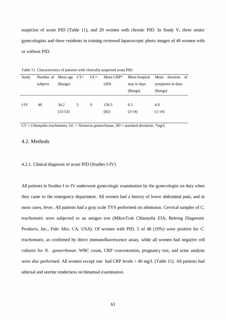
61
suspicion of acute PID (Table 11), and 20 women with chronic PID. In Study V, three senior
gynecologists and three residents in training reviewed laparoscopic photo images of 40 women with
or without PID.
Table 11. Characteristics of patients with clinically suspected acute PID.
Study
Number of
subjects
Mean age
(Range)
CT+ GC+ Mean CRP*
(SD)
Mean hospital
stay in days
(Range)
Mean duration of
symptoms in days
(Range)
I-IV
48
34.2
(15-53)
5
0
156.5
(82)
6.3
(3-14)
6.0
(1-14)
CT = Chlamydia trachomatis, GC = Neisseria gonorrhoeae, SD = standard deviation, *mg/L
4.2. Methods
4.2.1. Clinical diagnosis of acute PID (Studies I-IV)
All patients in Studies I to IV underwent gynecologic examination by the gynecologist on duty when
they came to the emergency department. All women had a history of lower abdominal pain, and in
most cases, fever. All patients had a gray scale TVS performed on admission. Cervical samples of C.
trachomatis were subjected to an antigen test (MikroTrak Chlamydia EIA; Behring Diagnostic
Products, Inc., Palo Alto, CA, USA). Of women with PID, 5 of 48 (10%) were positive for C.
trachomatis, as confirmed by direct immunofluorescence assay, while all women had negative cell
cultures for N. gonorrhoeae. WBC count, CRP concentration, pregnancy test, and urine analysis
were also performed. All women except one had CRP levels > 40 mg/L (Table 11). All patients had
adnexal and uterine tenderness on bimanual examination.
62
After the clinical diagnosis of PID, patients were hospitalized and received IV antimicrobial therapy
with doxycycline 100 mg twice daily plus metronidazole 500 mg three times daily, or cefuroxime
1500 mg three times daily plus metronidazole 500 mg three times daily. Antimicrobial therapy was
administered IV during hospitalization, followed by oral therapy. Total duration of antimicrobial
therapy was approximately 14 days.
4.2.2. Ultrasonography (Studies I-IV)
In Studies I to IV, TVS was performed in the emergency room with a 6.5 MHZ (RT-X 2000; GE
Medical Systems, Milwaukee, WI, USA) transducer. In Studies III and IV, an additional TVS was
performed by an experienced sonologist using a 5-9 MHZ broadband endovaginal transducer (ATL
HDI, Bothell, WA, USA) with color and power Doppler capabilities. Pulse repetition frequency was
500 HZ and wall filter was 100 HZ. Specific sonographic criteria were used for the diagnosis of PID
according to Timor-Tritsch et al. (1998) (Studies III and IV). PID was suspected when a pear-
shaped or retort-shaped sonolucent or particulate fluid-containing structure was detected on the
longitudinal TVS scan section. Thickness of the walls and septations was measured and was
considered thick if 5 mm or thicker. The presence or absence of incomplete septa (hyperechoic septa
that originate from one of the walls, not reaching the opposite wall), cogwheel signs (sonolucent
cogwheel-shaped structures visible in the cross-section of the thickened fluid-filled tube), or beads-
on-a-string signs (nodules sized 2 to 3 mm seen in a cross-section of the fluid-filled tube) were
recorded. Sonographic findings could be recorded only when the Fallopian tube was fluid-filled. The
extent of ovarian involvement and amount of cul-de-sac fluid was also recorded. Study IV used
sonographic landmark findings of acute appendicitis (Puylaert 1986). Acute appendicitis was
suspected when a blind-ending, non-compressible tubular structure was evident, consisting of a
63
submucosal ring with a diameter greater than 6 mm in transverse view. Specific periappendicular
changes indicating inflammation were considered a secondary sign of complicated acute appendicitis.
Differential diagnosis was performed between acute appendicitis and PID (Study IV). All
sonographic examinations were performed within 24 hours prior to laparoscopy. In Study III, a
Doppler velocity waveform was obtained from three parts of the mass. PI was measured, and the
lowest PI values were considered to represent the blood flow impedance of the mass. In Studies III
and IV, findings suggestive of hyperemia were taken as sonographic evidence of inflammation in
power Doppler examination.
4.2.3. Magnetic resonance imaging (Study I)
Within 24 hours of undergoing sonography, patients underwent MRI. The minor pelvic cavity was
examined with a 1.5-T imager (Vision; Siemens, Erlangen, Germany). Phased-array receiver coils
were used and sagittal T2-weighted turbo spinecho images obtained. The section thickness was 5
mm and the intersection gap 1 mm. The same sagittal images were obtained with a turbo inversion-
recovery sequence with three signals acquired and an acquisition time of 4 minutes, 26 seconds. T2-
weighted turbo spin-echo images were obtained in the axial oblique plain with an
acquisition time of 5 minutes 8 seconds, 5-mm thick sections and a 1-mm gap. The axial oblique
plain was selected to be perpendicular to the long axis of the uterus. The same axial oblique sections
were also obtained with a turbo short inversion time inversion recovery (STIR) sequence. Imaging
time was 21 minutes and total examination time approximately 30 minutes. The presence of a fluid-
filled tubal lumen, dilated tubes, adnexal mass, or polycystic-like ovaries and free pelvic fluid in the
cul-de-sac or the minor pelvic cavity was systematically studied. MR images were also evaluated
with regard to the morphologic appearance and signal intensity characteristics of the mass ( Jain et
al. 1993, Yamashita et al. 1995). A fluid-filled tube was diagnosed when a tortuous, fluid-filled tubal
64
lumen was identified; it had low signal intensity on T1-weighted images and high signal intensity on
T2-weighted images. A pyosalpinx was diagnosed when the tubal lumen was dilated and the walls
were thickened; it had the same signal intensity characteristics as those of a fluid-filled tube. Criteria
for tubo-ovarian abscess included an ill-defined adnexal mass with thick, irregular walls containing
fluid. An abscess usually had low signal intensity on T1-weighted images and high signal intensity on
T2-weighted images. Laparoscopy was performed within 24 hours after MRI.
4.2.4. Laparoscopy (Studies I-IV)
Laparoscopy was performed under general anesthesia by the conventional three-puncture technique
in all but one case with clinically suspected acute PID; the laparoscopic PID diagnosis was based on
commonly accepted visual findings (Studies I-IV) (Jacobson and Weström 1969, Hager et al. 1983).
Acute salpingitis was diagnosed when a hyperemic edematous Fallopian tube was visible, with no
sign of intraluminal pus. A pyosalpinx was diagnosed as an enlarged, edematous tube with partial or
total destruction of the fimbriated end. A TOA was diagnosed when the ovary and the Fallopian tube
could not be distinguished from each other, forming an adnexal complex with abscess formation.
Salpingitis was considered mild PID, whereas pyosalpinx and TOA were considered severe PID. In
patients with laparoscopically proven PID, one or more of the following procedures were performed:
lysis of pelvic adhesions, drainage and irrigation of pyosalpinges and TOAs, and extirpation of a
unilateral infectious complex. Irrigation of the pelvic cavity with 2 liters of physiologic saline was
performed in all cases. Patients with disease other than PID were treated laparoscopically when
possible (Studies I-IV). In four cases, appendectomy was performed by laparotomy for severe
infection (Study IV). All laparoscopies were documented by videotape. Unnecessary antimicrobial
therapy was ended in patients with no sign of infectious disease in laparoscopy.
65
4.2.5. Observer reproducibility (Study V)
Three senior consultants and three residents in training participated in the reproducibility study.
None of the authors was included among these observers. A total of 160 color slides, comprising
four slides made from of a suitable sample from an additional consecutive 40 patients were available
for review. Each set of four slides per case showed the pelvic structures in a systematic fashion. Any
slides which all observers considered not evaluable were excluded. All four views of each patient
were presented at the same time. This slide review was conducted during two identical sessions, two
days apart. For the second session, the same slides were shown again in random order to minimize
any recall bias based on the first review. In order to study intraobserver reproducibility, the observers
were not informed that the slides from the same cases were being shown again. Each set of four
slides was scored systematically with a predesigned case form. Six preselected components were
used for scoring: erythema of the uterus, erythema of the tubes, edema of the Fallopian tubes,
presence of tubal adhesions, fimbrial status, and characteristics of fluid in the cul-de-sac. An overall
diagnosis was made in each case. Each observer interpreted the slides independently and was
unaware of the choices of the other observers. Of the 40 patients included in the review, 9 had
histopathologically proven PID and 31 had other pelvic pathology or normal findings. The results of
senior consultants and residents were analyzed separately.
Overall diagnostic accuracy was analyzed in relation to the presence or absence of proven PID. PID
was defined as the presence or absence of salpingitis by fimbrial minibiopsy or the presence of PCE
obtained during the acute-phase laparoscopy.
Intraobserver reproducibility (agreement between two examinations by the same observer) was
analyzed for each observer based on two separate readings of the same photo images.
Overall, 240 pairs of observations were analyzed. Intraobserver reproducibility was also analyzed
separately for six preselected individual diagnostic features of PID.
66
Interobserver reproducibility (agreement between two observers reading the same set of photo
images) was analyzed from the results of the first review session. Agreement between the six
observers was tested both by overall PID diagnosis and by the individual diagnostic features of PID.
Diagnostic reproducibility was assessed in terms of accuracy and sensitivity and specificity. Intra-
and interobserver agreement was measured by kappa statistics; kappa measures agreement between
observers above what would be expected by chance alone, and is calculated as the ratio of actual to
potential agreement beyond chance (Figure 7) (Landis and Koch 1977). Kappa values above 0.75
represent excellent agreement, values between 0.40 and 0.75 moderate agreement, and values less
than 0.40 poor agreement. The p-value for a kappa estimate was used to determine whether a kappa
value differed significantly from a result obtained by chance.
Figure 7. Calculation of kappa value
4.3. Biostatistical analyses
In Study I, variables between patients with mild and severe PID were compared by x test, two-
sample t-test, or confidence interval-nonparametric test. In Study II, two radiologists interpreted MR
images independently. Interobserver reproducibility was calculated with Cohen K statistics. In Study
III, the comparisons were performed between patients with acute PID and patients with
hydrosalpinx. Comparable parameters were specific sonographic criteria for PID, pulsatility indices,
and hyperemia of the inflammatory mass. Analysis was performed by Pearsons’s chi-square test.
Standard deviation of pulsatility indices was also calculated. In Study V, intra- and interobserver
reproducibility was analyzed by kappa statistics (Landis and Koch 1977, Brennan and Silman 1992),
Kappa = Total agreement (%) - Chance agreement (%)
100% - Chance agreement (%) = 0.00 – 1.00
Actual agreement
Potential agreement

67
and 95% confidence intervals were applied to kappa measurements for intra- and interobserver
agreement ratings.
In Studies I, II, III, and V, the difference between groups was defined as p < 0.05. In Study V, a cut-
off value of 0.05 was also used to indicate significant agreement beyond chance.
68
5. Results
5.1. Diagnosis of pelvic inflammatory disease and acute appendicitis
5.1.1. MRI in diagnosis of acute PID (Study I)
In 21 (70%) patients, laparoscopy confirmed the PID diagnosis. MRI had a sensitivity of 95%, a
specificity of 89%, and an accuracy of 93% in diagnosis of PID. For TVS, the corresponding figures
were 81%, 78%, and 80%. MRI proved to be more accurate than TVS in the diagnosis of PID
(Tables 12 and 13). MRI detected 20 of 21 patients with laparoscopically proven PID, with only one
false-negative finding in a patient who had both endometrioma and salpingitis at laparoscopy. Of the
nine patients with disease other than PID in laparoscopy, one patient with tubal torsion was falsely
diagnosed by MRI as having PID. TVS diagnosed correctly 17 of 21 patients with PID including
four false-negative findings. Of the nine patients without PID, TVS diagnosed seven patients
correctly. Of the other two patients, one had endometrioma, and the other had tubal torsion at
laparoscopy, whereas at TVS both patients were believed to have tubo-ovarian abscesses.
Table 12. Diagnostic performance of MRI compared to laparoscopy in patients with clinical diagnosis of PID.
Laparoscopy
MRI PID No PID Total
PID 20 1 21
No PID 1 8 9
Total 21 9 30
Sensitivity = 95%, Specificity = 89%, Accuracy = 93%
PPV = 95%, NPV = 88%
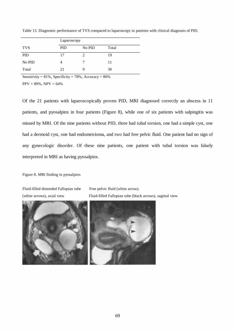
69
Table 13. Diagnostic performance of TVS compared to laparoscopy in patients with clinical diagnosis of PID.
Laparoscopy
TVS PID No PID Total
PID 17 2 19
No PID 4 7 11
Total 21 9 30
Sensitivity = 81%, Specificity = 78%, Accuracy = 80%
PPV = 89%, NPV = 64%
Of the 21 patients with laparoscopically proven PID, MRI diagnosed correctly an abscess in 11
patients, and pyosalpinx in four patients (Figure 8), while one of six patients with salpingitis was
missed by MRI. Of the nine patients without PID, three had tubal torsion, one had a simple cyst, one
had a dermoid cyst, one had endometrioma, and two had free pelvic fluid. One patient had no sign of
any gynecologic disorder. Of these nine patients, one patient with tubal torsion was falsely
interpreted in MRI as having pyosalpinx.
Figure 8. MRI finding in pyosalpinx
Fluid-filled distended Fallopian tube Free pelvic fluid (white arrow).
(white arrows), axial view Fluid-filled Fallopian tube (black arrows), sagittal view
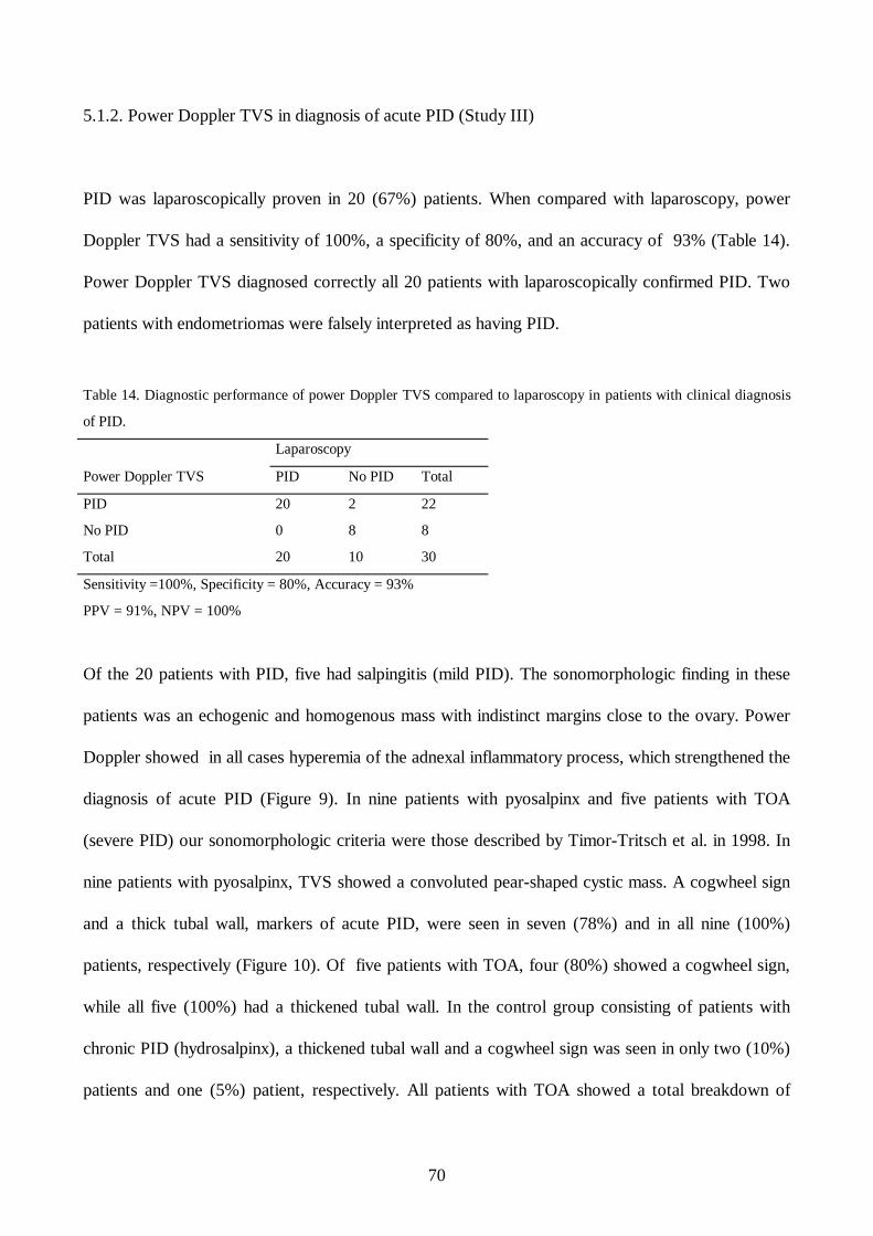
70
5.1.2. Power Doppler TVS in diagnosis of acute PID (Study III)
PID was laparoscopically proven in 20 (67%) patients. When compared with laparoscopy, power
Doppler TVS had a sensitivity of 100%, a specificity of 80%, and an accuracy of 93% (Table 14).
Power Doppler TVS diagnosed correctly all 20 patients with laparoscopically confirmed PID. Two
patients with endometriomas were falsely interpreted as having PID.
Table 14. Diagnostic performance of power Doppler TVS compared to laparoscopy in patients with clinical diagnosis
of PID.
Laparoscopy
Power Doppler TVS PID No PID Total
PID 20 2 22
No PID 0 8 8
Total 20 10 30
Sensitivity =100%, Specificity = 80%, Accuracy = 93%
PPV = 91%, NPV = 100%
Of the 20 patients with PID, five had salpingitis (mild PID). The sonomorphologic finding in these
patients was an echogenic and homogenous mass with indistinct margins close to the ovary. Power
Doppler showed in all cases hyperemia of the adnexal inflammatory process, which strengthened the
diagnosis of acute PID (Figure 9). In nine patients with pyosalpinx and five patients with TOA
(severe PID) our sonomorphologic criteria were those described by Timor-Tritsch et al. in 1998. In
nine patients with pyosalpinx, TVS showed a convoluted pear-shaped cystic mass. A cogwheel sign
and a thick tubal wall, markers of acute PID, were seen in seven (78%) and in all nine (100%)
patients, respectively (Figure 10). Of five patients with TOA, four (80%) showed a cogwheel sign,
while all five (100%) had a thickened tubal wall. In the control group consisting of patients with
chronic PID (hydrosalpinx), a thickened tubal wall and a cogwheel sign was seen in only two (10%)
patients and one (5%) patient, respectively. All patients with TOA showed a total breakdown of
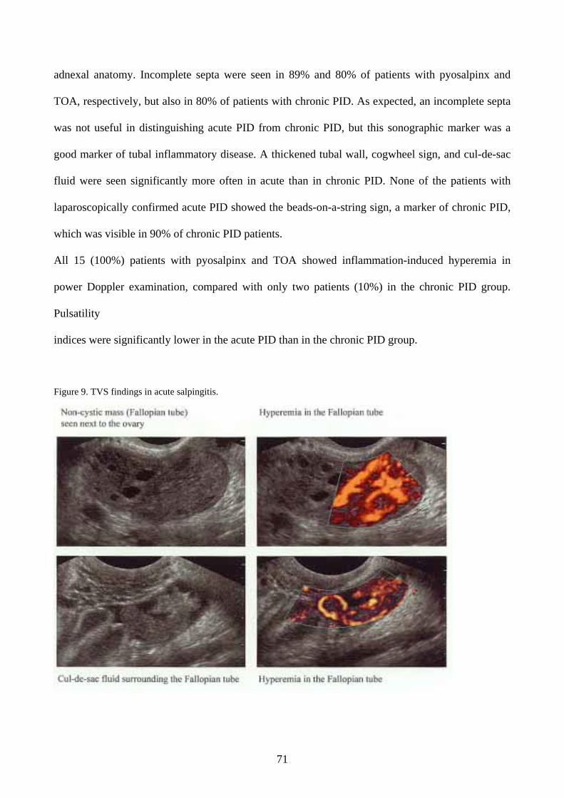
71
adnexal anatomy. Incomplete septa were seen in 89% and 80% of patients with pyosalpinx and
TOA, respectively, but also in 80% of patients with chronic PID. As expected, an incomplete septa
was not useful in distinguishing acute PID from chronic PID, but this sonographic marker was a
good marker of tubal inflammatory disease. A thickened tubal wall, cogwheel sign, and cul-de-sac
fluid were seen significantly more often in acute than in chronic PID. None of the patients with
laparoscopically confirmed acute PID showed the beads-on-a-string sign, a marker of chronic PID,
which was visible in 90% of chronic PID patients.
All 15 (100%) patients with pyosalpinx and TOA showed inflammation-induced hyperemia in
power Doppler examination, compared with only two patients (10%) in the chronic PID group.
Pulsatility
indices were significantly lower in the acute PID than in the chronic PID group.
Figure 9. TVS findings in acute salpingitis.
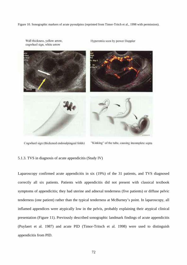
72
Figure 10. Sonographic markers of acute pyosalpinx (reprinted from Timor-Tritch et al., 1998 with permission).
5.1.3. TVS in diagnosis of acute appendicitis (Study IV)
Laparoscopy confirmed acute appendicitis in six (19%) of the 31 patients, and TVS diagnosed
correctly all six patients. Patients with appendicitis did not present with classical textbook
symptoms of appendicitis; they had uterine and adnexal tenderness (five patients) or diffuse pelvic
tenderness (one patient) rather than the typical tenderness at McBurney’s point. In laparoscopy, all
inflamed appendices were atypically low in the pelvis, probably explaining their atypical clinical
presentation (Figure 11). Previously described sonographic landmark findings of acute appendicitis
(Puylaert et al. 1987) and acute PID (Timor-Tritsch et al. 1998) were used to distinguish
appendicitis from PID.
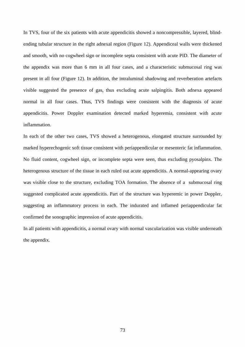
73
In TVS, four of the six patients with acute appendicitis showed a noncompressible, layered, blind-
ending tubular structure in the right adnexal region (Figure 12). Appendiceal walls were thickened
and smooth, with no cogwheel sign or incomplete septa consistent with acute PID. The diameter of
the appendix was more than 6 mm in all four cases, and a characteristic submucosal ring was
present in all four (Figure 12). In addition, the intraluminal shadowing and reverberation artefacts
visible suggested the presence of gas, thus excluding acute salpingitis. Both adnexa appeared
normal in all four cases. Thus, TVS findings were consistent with the diagnosis of acute
appendicitis. Power Doppler examination detected marked hyperemia, consistent with acute
inflammation.
In each of the other two cases, TVS showed a heterogenous, elongated structure surrounded by
marked hyperechogenic soft tissue consistent with periappendicular or mesenteric fat inflammation.
No fluid content, cogwheel sign, or incomplete septa were seen, thus excluding pyosalpinx. The
heterogenous structure of the tissue in each ruled out acute appendicitis. A normal-appearing ovary
was visible close to the structure, excluding TOA formation. The absence of a submucosal ring
suggested complicated acute appendicitis. Part of the structure was hyperemic in power Doppler,
suggesting an inflammatory process in each. The indurated and inflamed periappendicular fat
confirmed the sonographic impression of acute appendicitis.
In all patients with appendicitis, a normal ovary with normal vascularization was visible underneath
the appendix.
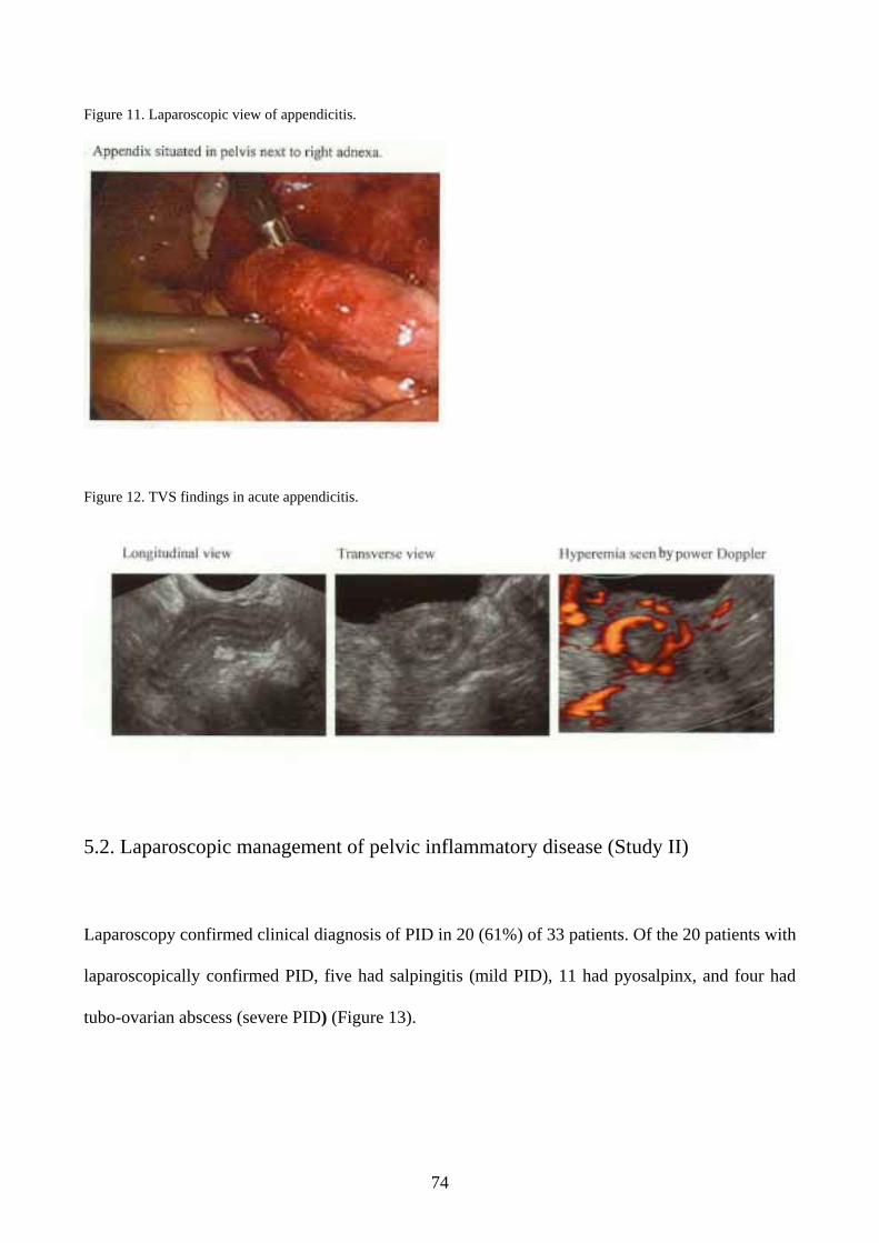
74
Figure 11. Laparoscopic view of appendicitis.
Figure 12. TVS findings in acute appendicitis.
5.2. Laparoscopic management of pelvic inflammatory disease (Study II)
Laparoscopy confirmed clinical diagnosis of PID in 20 (61%) of 33 patients. Of the 20 patients with
laparoscopically confirmed PID, five had salpingitis (mild PID), 11 had pyosalpinx, and four had
tubo-ovarian abscess (severe PID) (Figure 13).
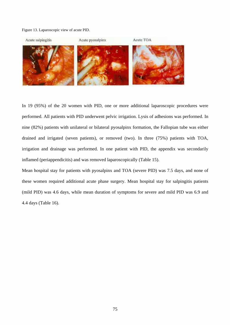
75
Figure 13. Laparoscopic view of acute PID.
In 19 (95%) of the 20 women with PID, one or more additional laparoscopic procedures were
performed. All patients with PID underwent pelvic irrigation. Lysis of adhesions was performed. In
nine (82%) patients with unilateral or bilateral pyosalpinx formation, the Fallopian tube was either
drained and irrigated (seven patients), or removed (two). In three (75%) patients with TOA,
irrigation and drainage was performed. In one patient with PID, the appendix was secondarily
inflamed (periappendicitis) and was removed laparoscopically (Table 15).
Mean hospital stay for patients with pyosalpinx and TOA (severe PID) was 7.5 days, and none of
these women required additional acute phase surgery. Mean hospital stay for salpingitis patients
(mild PID) was 4.6 days, while mean duration of symptoms for severe and mild PID was 6.9 and
4.4 days (Table 16).
76
Table 15. Laparoscopic surgery in 33 patients with clinically suspected acute PID.
PID patients N Pelvic
irrigation
Adhesiolysis Drainage and
irrigation*
Extirpation of
infectious complex
Salpingitis 5 5 5 0 0
Pyosalpinx 11 11 11 7 2
TOA 4 4 3 3 0
Total 20
*9 of 11 (82%) of pyosalpinx patients underwent drainage or extirpation,
3 of 4 (75%) of TOA patients underwent drainage
TOA=Tubo-ovarian abscess
Non PID patients N Therapeutic intervention*
Endometriosis 4 Extirpation of cyst (2)
Electrocoagulation (2)
Appendicitis 3 Appendectomy**
Adnexal torsion 3 Salpingectomy
Ruptured ovarian cyst 1 Extirpation of cyst
Normal findings 2
Total 13
*11 of 13 (85%) of non PID patients underwent therapeutic intervention
**Two patients underwent open appendectomy
Table 16. Characteristics of patients with laparoscopically proved PID. Mild PID* (N=5) Severe PID** (N=15) p value
Mean age (range) 27.8 (15-41) 39.4 (19-52) 0.025
History of past PID 0 5 NS
Nulliparous 3 6 NS
Duration of symptoms (mean, days) 4.4 6.9 NS
CRP (mean, mg/l) 136.6 145.1 NS
Hospital stay (mean, days) 4.6 7.5 0.0013
*Salpingitis, **pyosalpinx or tubo-ovarian abscess, NS=not significant
Thirteen patients (39%) with clinically suspected PID had some other disease at laparoscopy. Four patients had endometriosis, and of these two had endometriomas, one had extensive
77
endometriosis of the vesicouterine septum, and one had several endometriotic foci of the pelvic cavity. Three patients had appendicitis, three had adnexal torsion, and one had rupture of an ovarian
cyst. In two patients, no gynecological or other abnormality was visible in laparoscopy. Of 13
patients in 11(85%) a therapeutic operative intervention was performed, done laparoscopically in
nine while two with appendicitis had open appendectomy (Table 15). In all patients without PID,
antimicrobial therapy could be discontinued.
5.3. Accuracy of laparoscopic findings in pelvic inflammatory disease (Study V)
Based on laparoscopic photo images, the overall accuracy of visual diagnosis of PID by laparoscopy
was 78%, sensitivity was 27%, and specificity was 92%. Senior consultants and residents in training
performed equally well. Overall intraobserver reproducibility of laparoscopic diagnosis of PID was
only fair (kappa 0.58). Kappa coefficients for the six observers ranged from 0.26 to 0.90. Senior
consultants clearly performed better than did residents (mean kappa 0.76 vs. 0.39) even though the
difference was not significant (p=0.067).
Intraobserver reproducibility was analyzed according to features of the laparoscopic diagnosis of
PID. Results were highly variable, ranging from kappa -0.08 to 1 for individual observers. Again,
consultants performed better than residents in each of the diagnostic categories, mean kappa values
being 0.52 vs. 0.36. In assessment of tubal erythema or tubal fimbria, consultants and residents were
most inconsistent.
Overall interobserver reproducibility was only moderate (kappa 0.43), the kappa value ranging from
0.07 to 0.68. Again, the consultants performed slightly better than residents (kappa 0.48 vs. 0.38).
In an interobserver analysis of specific features of PID, experienced gynecologists performed better
than residents for each of the specific features except for interpretation of tubal edema, which
showed no difference between consultants and residents.
78
6. Discussion
6.1. Diagnosis of pelvic inflammatory disease and acute appendicitis
6.1.1. MRI in diagnosis of acute PID
The present study is the first prospective comparative trial of MRI and laparoscopy in the diagnosis
of PID. The purpose was to evaluate the usefulness of MRI in the diagnosis and to compare MRI
findings with those of laparoscopy and TVS.
We found MRI to be more accurate than TVS in the diagnosis of PID, contrary to the findings of
Jain et al. in which MRI was less specific than TVS in the assessment of pelvic masses (Jain et al.
1993). However, they used no systematic surgical intervention reference standard. We found MRI
useful not only for the diagnosis of PID but also for the detection of other disease conditions.
Only a few studies have been performed using MRI to characterize gynecologic infections (Outwater
et al. 1995, Ha et al. 1995). These studies were descriptive and not designed to address the issue of
accuracy of MRI findings. Outwater’s and Ha’s studies as well as our study point out that MRI
provides a good contrast between pelvic organs and is especially useful in soft-tissue imaging.
Outwater’s group (1995) found that the multiplanar capability of MR imaging is a particular
advantage when compared with CT. This parallells Mitchell’s finding that straight axial and coronal
planes are sub-optimal for imaging of the adnexa (Mitchell et al. 1987). We found the combination of
axial oblique and sagittal images to be most useful in differential diagnosis.
In our study, only fluid-filled Fallopian tubes (and not a normal tube) could be visualized by MRI,
confirming results of Outwater’s group from 1995. Differing appearance on T1-, T2-weighted, and
STIR images plus varying wall thickness helped in the differential diagnosis of hydrosalpinx,
79
pyosalpinx, TOA, and tubal torsion, and also endometrioma. Layering in hemorrhagic adnexal
masses (Nybergh et al. 1987) was evident in tubal torsion in our study, which distinguished this
entity from PID.
Criticism against MRI has arisen because of high costs and limited feasibility. However, MRI may
be cost-effective, if operative intervention can be avoided and adequate therapy provided early in the
disease process on the basis of MRI information (Schwartz et al. 1994). However, larger studies
among unselected women with pelvic pain are needed to test the overall utility of MRI.
6.1.2. Transvaginal sonography in diagnosis of acute PID
Power Doppler TVS was successful in the diagnosis of acute PID and also in distinguishing acute
PID and hydrosalpinx formation consistent with chronic PID. Sonographic landmark findings of
acute and chronic tubal inflammatory disease described by the Timor-Tritsch group in 1998 formed
the basis for diagnosis. First, during the acute phase, the tubal wall becomes thickened and
edematous. Secondly, purulent exudate fills the lumen, which makes the thickened endosalpingeal
folds visible (cogwheel sign). Some exudate may spill into the cul-de-sac. The progressive filling of
the tube leads to the kinking of the pyosalpinx or hydrosalpinx seen as incomplete septa in TVS.
Consequently, incomplete septa can be a marker of both acute and chronic PID. In the chronic
phase, the occluded tubal wall becomes thin and distended. Remnants of the endosalpingeal folds
may be seen as the beads-on-a-string sign which is another marker of chronic PID (Timor-Tritsch et
al. 1998). Choosing chronic PID patients as a control group gave us the possibility to identify the
specific sonographic picture of acute and chronic PID reflecting the pathological changes in the
Fallopian tube
80
Power Doppler TVS was 100% sensitive and 80% specific in our study with an overall accuracy of
93%. The diagnostic criteria of Timor-Tritsch et al. proved to be highly reproducible.
A thick tubal wall ( > 5 mm) and a cogwheel sign, both sensitive markers of acute disease, were
detected in Timor-Tritsch’s study vs. our study, in acute PID in 100% vs. 86% and in 100% vs.
79%, respectively. The thin tubal wall ( < 5 mm) and beads-on-a-string sign, markers of chronic
disease, were detected in chronic PID in 97% vs. 57% and in 90% vs. 80%, respectively. None of
our patients with acute PID presented with the beads-on-a-string sign. Cul-de-sac fluid was present
more commonly in acute PID than in chronic PID: 50% vs. 10% in the reference study compared
with 60% vs. 15% in our study. Incomplete septa, regarded as the best marker of tubal inflammatory
disease, acute or chronic, were seen in 92% vs. 85%. No other study has systematically evaluated
these landmark findings in the diagnosis of PID. A prerequisite for TVS landmark findings is that the
tube is occluded and fluid-filled, which makes walls and septa visible (Timor-Tritsch et al. 1998).
Comparable studies investigating diagnostic performance of TVS are limited. In most studies,
populations are small and study designs variable, which makes direct comparison difficult (Patten et
al. 1990, Cacciatore et al. 1992, Boardman et al. 1997). Tessler’s group (1989) diagnosed ten cases
of dilated tubes with surgery or HSG serving as the gold standards. They described a tubular, folded
structure with an echogenic wall and echoes protruding into the lumen as the basis for a hydrosalpinx
diagnosis (chronic PID), making no distinction between acute and chronic PID. Patten et al. in 1990
found, among 18 patients with clinically suspected PID, that TVS was 93% as sensitive as
laparoscopy. Diagnostic criteria included a fluid-filled thick-walled echogenic tubular structure.
Specificity could not be calculated due to the low number of women without salpingitis at
laparoscopy. Cacciatore’s group (1992) found TVS to be 85% as sensitive and just as specific in the
diagnosis of acute PID compared to values for PCE. That study group included 51 patients of which
13 had histological endometritis. However, no laparoscopy was performed. Taipale’s group (1995)
81
studied 86 patients with clinically suspected PID, and in only 37% of cases confirmed PID surgically.
In 31 % of their patients, a thick-walled ( > 5 mm) fluid-filled echogenic tube consistent with acute
PID was evident. Of the patients, 64% showed no adnexal mass, but 16% of these 55 patients
developed hydrosalpinx detected by follow-up TVS, indicating the presence of acute salpingitis
without visible TVS findings in the acute phase. Most studies suggest that TVS is helpful in the
diagnosis of moderate and severe PID, in which finding of the fluid-filled Fallopian tube is typical and
reproducible. However, none of the other studies have systematically used all the sonographic
landmark findings described by Timor-Tritsch et al. (1998). Future studies should focus on these
findings, which proved to be highly specific in our study. Data from these studies should be
evaluated with criticism. Studies incorporate highly selected patients hospitalized for symptomatic
PID. TVS has proven non-sensitive in mild disease, and studies including patients with mild
symptoms will show TVS certainly to be less than perfect. Furthermore, expectation bias by the
investigator may reduce specificity. Ideally, investigators should be blinded to all patient
characteristics.
We detected hyperemia by power Doppler examination in all 20 cases (100%) in the acute PID
group but in only two cases (10%) in the hydrosalpinx group. Pulsatility indices were significantly
lower in the acute PID group than in the hydrosalpinx group. Hyperemia seen by power Doppler in
acute PID has not been described in other studies; we found it most helpful in the detection of an
acute inflammatory process. Color Doppler studies by Kupesic et al. (1995), Alatas et al. (1996), and
Tepper et al. (1998) show consistently that acute PID causes hyperemia, leading to a decrease in
vascular resistance (low PI), and that increasing vascular resistance (high PI) reflects recovery. These
reports are in accordance with our observations.
We presented six patients with acute salpingitis without tubal occlusion. Because of lack of
intraluminal fluid, none of the specific sonographic characteristics were visible. Power Doppler was
most useful in these cases. Detection of hyperemia in the adnexal elongated non-cystic mass
82
(consistent with an inflamed Fallopian tube) suggested the presence of an inflammatory process,
confirming the diagnosis of PID. This is a new and novel finding which should be confirmed by
others.
6.1.3. Transvaginal and transabdominal sonography in diagnosis of acute appendicitis
We describe TVS findings in six women suspected of having acute PID who actually had acute
appendicitis. None of these six patients revealed, however, characteristic symptoms and signs
consistent with appendicitis, and Paulson et al. (2003) point out that when TAS fails to detect the
appendix, TVS can be most valuable: TVS may not only detect the appendix but may identify
alternative diagnoses such as PID or other acute gynecologic disorders. An interesting finding of that
study was that in women with suspected acute appendicitis, US detected in 33% other diagnoses
such as pyosalpinx and adnexal torsion, confirming the value of the transvaginal approach. The
transabdominal sonographic diagnostic criteria for acute appendicitis first described by Puylaert in
1986 and subsequently confirmed by others (Jeffrey et al. 1987, Rioux et al. 1992) proved
reproducible by transvaginal sonography in our study. All six patients had an atypical pelvic location
of the appendix near the right adnexa, which makes the differential diagnosis between appendicitis
and PID difficult. An inflamed Fallopian tube can mimic acute appendicitis, but these entities can be
distinguished by their different sonographic appearance, and TVS should always be performed if PID
is suspected (Jeffrey et al. 1994). We used earlier sonographic criteria for tubal inflammation (Timor-
Tritsch et al. 1998) to exclude acute PID. Rettenbacher et al. (2000) describes the presence of
intraluminal gas in mild, uncomplicated appendicitis. In all our cases of uncomplicated appendicitis,
TVS detected gas in the appendiceal lumen, which helped to distinguish acute appendicitis from
adnexal pathology. A common finding in complicated appendicitis is the presence of echogenic
mesenteric fat caused by edema (Jeffrey et al. 1994), which was seen in our two cases of gangrenous
83
appendicitis. It must be kept in mind that PID can cause inflammation of adjacent structures, such as
periappendicitis (Jeffrey et al. 1994), though this finding cannot be discriminated from appendicitis.
Other studies report a total of 12 cases of appendicitis diagnosed by TVS (Puylaert 1994b, Pelsang
et al. 1994, Whitford 1994, Larcos 1996, Caspi et al. 1996) and suggest that acute appendicitis
should be excluded by TVS, especially if a woman presents with equivocal findings and atypical low
pelvic pain, as was the case for our patients. In young fertile women with suspected appendicitis,
TVS should be mandatory because of the many diagnostic possibilities and high rate of misdiagnoses
(Borgstein et al. 1997). Quillin et al. (1994) showed that increased blood flow in the appendiceal
wall indicates appendicitis, confirming our finding of hyperemia in all six patients. However, it is true
that although both PID and appendicitis show inflammation-induced hyperemia, a TVS finding of
normal adnexa without increased vascularity will rule out PID.
We demonstrated that TVS is useful in the detection of an inflamed appendix and in distinguishing
acute appendicitis from PID. TVS seems to be promising in the diagnosis of acute appendicitis,
especially in women with an atypical location of the appendix in the pelvis. It is too early to
determine the overall accuracy of TVS in the diagnosis of acute appendicitis – before many open
questions have been answered. How often can the appendix be visualized by TVS? Can we rely on
TVS if the clinical diagnosis is consistent with PID? Can we perform appendectomy without
laparoscopy if TVS is consistent with appendicitis? TAS is thought to be helpful in equivocal cases
and could add information to determine whether surgery is necessary (Rice et al. 1999). TVS may
play a similar role in the future.
84
6.2. Laparoscopic management of pelvic inflammatory disease
The clinical diagnosis of PID was confirmed in 61% of our patients at laparoscopy. This finding
confirms the results of several earlier reports on the accuracy of the clinical diagnosis of PID
(Jacobson and Weström 1969, Chaparro et al. 1978, Brihmer et al. 1987, Sellors et al. 1991, Soper
et al. 1994, Bevan et al. 1995). These studies as well as ours represent, however, highly selected
populations of hospitalized patients with suspected PID, which may certainly lead to
misinterpretations when applied to patients with atypical or mild disease (Reid et al. 1995).
Of patients with laparoscopically confirmed PID, 95% underwent laparoscopic procedures.
Laparoscopy proved to be safe in this patient group, and no major complications occurred. Härkki-
Siren and Kurki in 1997 demonstrated 6.9 of 1000 (0.7%) major complications in operative
laparoscopy and pointed out that complications increased when operative laparoscopy became more
difficult. According to these findings, the extent of surgical procedures – depending on severity of
the inflammatory process – should be evaluated separately in every patient with acute PID, and only
”safe” surgery performed. Following recommendations of Henry-Suchet in her review article (Henry-
Suchet and Tesquier 1994) we started IV antimicrobial treatment before laparoscopy. This practice
is recommended for patients with PID to avoid general dissemination of the disease. In our series,
the cure rate was 100%, and no major complications occurred, suggesting that initiation of
preoperative antimicrobial therapy is reasonable.
In 10 patients with pyosalpinx or TOA, drainage and irrigation was performed. All patients
recovered well, with no additional operative interventions needed, a result in accordance with other
findings showing high clinical cure rates (94%-100%) after laparoscopic drainage of adnexal
abscesses (Henry-Suchet et al. 1984, Reich and McGlynn 1987, De Wilde and Hesseling 1995).
These studies demonstrate that radical surgery is extremely rarely needed, especially since abscess
formation is often unilateral. Compared with reports from Finland in the 1980s (Heinonen et al.
85
1986, Teisala et al. 1987, Heinonen and Miettinen 1994), our PID patients were older, had longer-
duration symptoms, and had more severe disease as measured by CRP levels, although hospital stays
were shorter. Increasing use of OCs, awareness of STIs, and decreased rate of N. gonorrhoeae may
explain the change in clinical manifestations. Our study was not designed to use microbiologic or
histopathologic sampling at laparoscopy to confirm the diagnosis of PID, even if such samples are
commonly taken in clinical practice. The laparoscopic diagnosis was based strictly on visual findings.
In contrast to many other studies (Henry-Suchet et al. 1984, Reich and McGlynn 1987, Mecke and
Semm 1988, Raatz 1992, De Wilde and Hesseling 1995), the goal of our study was not only to
diagnose and treat PID but also to record other diagnoses and to treat these patients surgically if
indicated. Overall, 11 (33%) patients had other disease, and two (6%) had no evidence of the
disease. Thus, 11 of these 13 non-PID patients underwent a therapeutic procedure, with further
elective operations, antimicrobial therapy, and hospitalization avoided. Such management is likely to
be highly cost-effective. Because most studies record approximately 30% of patients as having a
disease other than PID, future research should also focus on the management of these patients and
also perform cost-benefit analyses.
The uncontrolled study design used in this and other comparative works demonstrating operative
laparoscopy in acute PID causes an obvious methodological dilemma. No randomized, controlled
trials have apparently been performed comparing conservative antimicrobial therapy with a
combination of antimicrobial therapy and acute-phase surgical laparoscopy. In addition to clinical
cure rates, such a study should focus especially on preservation of fertility and on long-term
complications.
86
6.3. Accuracy of laparoscopic findings in pelvic inflammatory disease
The intra- and interobserver reproducibility of our laparoscopic diagnosis of PID was surprisingly
low. Overall observer reproducibility of the specific features of PID ranged from moderate to poor.
Experienced gynecologists performed consistently better than did residents in training. The overall
sensitivity of the laparoscopic diagnosis of PID in relation to histopathologically proven diagnosis
turned out also to be surprisingly low (27%).
Laparoscopy has long been recognized as the gold standard for diagnosis of PID (Jacobson and
Weström 1969, Paavonen et al. 1987, Weström et al. 1992a). Sellors et al. in 1991 raised criticism
towards laparoscopy by showing that it was nonsensitive in mild PID (salpingitis). Despite its role as
the gold standard, conflicting with doubts of its overall accuracy, reproducibility of laparoscopic
diagnosis of PID has never been properly validated. Reid et al. (1995) was one group emphasizing
the fact that reproducibility of a diagnostic test should be properly validated. The importance of the
observer as a significant source of variability in assessment has largely been neglected.
Results from other laparoscopic reproducibility studies consistently report only modest observer
agreement, confirming our results. Hornstein et al. in 1993 found only poor to moderate intra- and
interobserver reproducibility in an analysis of laparoscopic reproducibility of the
diagnosis of endometriosis. In another study (Rock et al. 1995) of interobserver reproducibility,
laparoscopic diagnosis of endometriosis also showed a high degree of variability, and still others
found only poor interobserver reproducibility when laparoscopic pelvic adhesions were assessed
(Bowman et al. 1995, Corson et al. 1995). These results and ours are a major cause for concern,
suggesting that laparoscopy as the gold standard may not be as accurate as assumed.
The low sensitivity of our laparoscopic diagnosis of PID parallels the findings by Sellors et al. (1991)
in which laparoscopy was nonsensitive in the diagnosis of mild salpingitis. Attempts have been made
to make laparoscopic criteria for PID more systematic and to create scoring systems (Hager et al.
87
1983, Soper et al. 1991, Henry-Suchet and Tesquier 1994) in order to improve diagnosis. These
scoring systems have not been properly validated but are an encouraging attempt to increase
accuracy of laparoscopic diagnosis of PID. Low accuracy implies that an extended gold standard
should be considered, including both laparoscopic and histopathologic verification of PID. One
application of our study would be to include systematic observer-agreement tests in residency
training programs.
In the interobserver study, P-values in assessment of differences between kappa values must be
interpreted with caution, as the observations (mean kappa values of each investigator) are not
independent from each other. Furthermore, prevalence of the disease may also have an effect on the
kappa value (Brennan and Silman 1992). Chance agreement may be high if prevalence is either high
or low, which then may lead to low kappa values. In our series, 9 of 40 (23%) had proven PID, and
the rest had other pelvic pathology or normal findings. Any significant systematic error is, however,
unlikely, as our results revealed no major inconsistencies and were in line with other reproducibility
studies.
88
7. Conclusion and future prospects
Clinical diagnosis of PID is difficult because of its wide range of manifestations. It is a major cause
of long-term morbidity, with an enormous economic impact. More effort should therefore be focused
on improving its diagnosis.
This study suggests that MRI is more accurate than is TVS in the diagnosis of PID when
laparoscopy serves as the gold standard. MRI is also useful in detecting any other disease processes
contributing to the clinical presentation. More studies are needed to determine the effect of
gynecologic MRI on treatment decisions and on net cost.
As shown earlier, in patients with clinically suspected PID, laparoscopy can confirm the diagnosis of
PID in only about two-thirds of the cases, although a final diagnosis was achieved in all but two of
our cases. Acute-phase surgical laparoscopy proved safe. Most women without PID underwent
some therapeutic procedure during laparoscopy which is likely to be cost-effective. In patients with
proven PID, if the infectious complex could not be removed in the acute phase, acute-phase surgical
laparoscopy allowed individual tailoring of additional elective procedures. That PID was often
predominantly unilateral means that a radical operation is not needed and would be overtreatment.
The specific sonographic landmark findings of PID described in previous studies were the basis for
the diagnosis of PID. Power Doppler TVS detected all PID cases and allowed an accurate distinction
between mild and severe PID, making it possible also to distinguish between acute and chronic PID.
Our study suggests that TVS with power Doppler can be considered the method of choice in the
bedside diagnosis of PID.
89
Specific sonographic landmark findings of both PID and appendicitis made it possible to distinguish
between these conditions. Even if hyperemia is seen by power Doppler in both diseases, a normal
adnexal structure without signs of hyperemia speaks against PID.
Observer reproducibility of laparoscopic diagnosis of PID proved to be unsatisfactory, as did overall
diagnostic accuracy when histopathologically proven PID served as the gold standard. Accuracy of
laparoscopic PID diagnosis seemed to be especially low for mild PID. Clearly, laparoscopy as a
single gold standard seems insufficient. We suggest that a better or extended gold standard
(including histopathological verification) should be used in future clinical trials. Observer-agreement
studies of laparoscopic PID diagnosis are necessary in order to develop reliable scoring systems and
to improve accuracy.
Our group of women was relatively small, so the lack of statistical power due to small subgroup
samples prohibits definitive conclusions or recommendations. Diagnostic accuracy of both MRI and
power Doppler TVS in the diagnosis of PID was, however, encouraging. Because of high costs and
the need for advanced techniques, these diagnostic tools are not feasible in all clinical settings, but
MRI and power Doppler TVS may still be cost-effective if proper therapy is provided early in the
disease course and if explorative surgery can be avoided.
The therapeutic impact of the use of TVS in diagnosis of appendicitis remains to be shown. Key
questions are whether appendectomy can be performed without diagnostic laparoscopy if TVS
findings are consistent with appendicitis, and whether surgery can be avoided if TVS is consistent
with PID in the presence of a normal appendix. How often the appendix can be visualized by TVS in
unselected populations remains unknown.
Whether acute-phase laparoscopy in the management of PID patients improves reproductive
outcome cannot be determined based on this study. A randomized controlled trial and cost-benefit
analysis are required to compare acute-phase operative laparoscopy with conventional antimicrobial
90
therapy and to answer more definitively the question whether intervention reduces long-term
complications and improves reproductive outcome.
Our women were highly preselected, including a small group of symptomatic patients hospitalized
for suspected PID, just as in most studies dealing with the clinical diagnosis of PID. One problem is
not only the high rate of false-positive diagnoses but also the high rate of false-negatives. The latter
remains unknown both in our study and comparable studies. To deal with these diagnostic dilemmas,
future studies should focus on unselected subjects, i.e., women referred for acute pelvic pain, in
order to avoid selection bias. Several studies have shown that highly symptomatic PID patients
represent only the tip of the iceberg, and that most PID patients have mild symptoms and are treated
as outpatients; this suggests that efforts should be put into studies in primary care settings. Studies
comparing inpatient vs. outpatient management of PID patients are needed, as well as studies
comparing conservative vs. operative therapy. Such studies should be prospective, randomized, and
controlled. Furthermore, investigators should be blinded to clinical data and the medical history of
the patients when evaluating new diagnostic techniques, to avoid ascertainment bias. Subject groups
should be large enough to test diagnostic accuracy, and reproducibility of diagnostic tests should also
undergo evaluation.
91
8. Summary
The main objective of this study was to evaluate new diagnostic techniques in the diagnosis of acute
PID and elucidate acute-phase operative laparoscopy as a treatment option for acute PID. Additional
objectives were to assess observer agreement and diagnostic accuracy of laparoscopic PID diagnosis.
Women hospitalized for clinically suspected acute PID were studied by MRI and power Doppler
TVS. Laparoscopy served as the gold standard. The sensitivity of MRI in the diagnosis of PID was
95%, the specificity was 89%, and the overall accuracy was 93%. For TVS, the corresponding
values were 81%, 78%, and 80%. MRI was more accurate than TVS and provided information on
the differential diagnosis of PID. Power Doppler TVS was 100% sensitive and 80% specific. Specific
sonographic landmark findings of PID and the inflammation-induced hyperemia seen by power
Doppler were the basis for the TVS diagnosis. Acute and chronic PID could be distinguished by their
differing sonographic appearance. Pulsatility indices were significantly lower in acute PID than in
chronic PID.
Acute-phase laparoscopy confirmed a diagnosis of PID in 61% of the patients. A final diagnosis was
provided by laparoscopy in 94%. Of the women without PID, 85% underwent a therapeutic
procedure which can be considered highly cost-effective. No major complications occurred. In
patients with PID, surgical laparoscopy allowed for individual tailoring of additional elective
procedures such as unilateral salpingectomy. In six patients, TVS findings were typical of acute
appendicitis.
Earlier sonographic landmark findings by TAS proved to be accurate also by TVS. TVS thus seems
to offer an opportunity to visualize the appendix in women with acute pelvic pain.
92
To test intra- and interobserver reproducibility and overall accuracy of laparoscopic diagnosis, three
senior consultants and three residents in training scored laparoscopic photo images on two separate
occasions. Histopathologically proven PID served as the gold standard. Overall accuracy of the
laparoscopic diagnosis of PID was 78%, specificity was 92%, and sensitivity was 27%, a sensitivity
which is very low, especially for mild PID. Overall intraobserver reproducibility was only fair (kappa
0.58), and interobserver reproducibility only moderate to poor (kappa 0.43). When specific
diagnostic features of PID were analyzed separately, results were no different, suggesting only
moderate to poor reproducibility. Observer agreement was clearly better among senior consultants
than among residents.
93
Acknowledgements
Research for this thesis was carried out at the Department of Obstetrics and Gynecology and
Department of Radiology, University Central Hospital of Helsinki, during the years 1994 to 1999. I
am grateful to the Heads of the Departments, Professor Olavi Ylikorkala, Professor Markku
Seppälä, and Professor Carl-Gustaf Standertskiöld-Nordenstam, and to the Administrative Head of
the Department of Obstetrics and Gynecology, Docent Maija Haukkamaa, for providing me with the
facilities to carry out this study and for their interest in my research.
I want especially to express gratitude to my supervisor, Professor Jorma Paavonen, for introducing
me to the world of gynecologic infections and for guiding me in scientific research overall. His
everlasting patience, constructive criticism, and professional attitude both as a clinician and a
scientist were invaluable. I also feel that the most important factor in the progress of this thesis was
”positive personal chemistry”.
I owe my gratitude to Docent Bruno Cacciatore for teaching me how to use transvaginal sonography
in the diagnosis of PID. His experience in ultrasound opened to me the world of power Doppler
sonography.
I wish to express my gratitude to Professor Pentti K. Heinonen and Docent Aydin Tekay as official
reviewers for their careful review of the final manuscript and for their valuable comments which have
greatly improved the text.
I want to express my sincere thanks to Docent Jari Sjöberg, whose experience in laparoscopic
surgery was essential when we planned and performed the laparoscopic procedures for patients in
our study.
I thank my collaborators in radiology, Professor Hannu Aronen and Drs Timo Tukeva and Pertti
Karjalainen for participating in the MRI study. I wish also to thank Drs Patrik Finne and John
94
Sellors, the first for his professional help with the statistical analyses, and the second, our
collaborator ”overseas,” for providing material and constructive criticism for study of the accuracy
of laparoscopic diagnosis. I also express my thanks to Dr. Luca Savelli for systematically organizing
the documented sonographic material, and to Docent Timo Paavonen, for reading the
histopathologic slides in the MRI study.
I want separately to thank Docent Juhani Toivonen, the Head of the Department of Obstetrics and
Gynecology, Jorvi Hospital, whose positive attitude toward scientific research was essential in the
progress of this thesis and made it possible for me to combine clinical work and research. His ability
always to share his time when needed is admirable.
I thank my friends and colleagues at the Departments of Obstetrics and Gynecology, University
Hospital in Helsinki and at Jorvi Hospital for creating a positive atmosphere and for encouraging me
to finish this project.
I express my special gratitude to Nina Hedkrok for her invaluable help in preparing the layout for the
pictures and tables of the manuscript and her continuous help with computer work. Her patience,
positive attitude, and ability to solve all practical problems were invaluable.
I want to thank Laila Selkinen and Leena Vaara for helping with all the arrangements concerning the
academic dissertation and Raili Alanne for her aid with the references. I owe my gratitude to Carol
Norris for her professional skills in author-editing the language of this thesis.
Finally, I reserve my warmest thanks for my wife Anne, daughters Eva and Jessica, and son Jan, who
sometimes certainly thought that some of the time spent on this could have been used otherwise!
Their love and patience reminded me that life will continue also after completing this thesis.
This study was supported by grants from the Research Funds of the Helsinki University Central
Hospital, the Academy of Finland, the K. Albin Johansson Funds, the Medical Society of Finland, the
96
References
Alatas C, Aksoy E, Akarsu C, Yakin K, Bahceci M. Hemodynamic assessment in pelvic
inflammatory disease by transvaginal color Doppler ultrasonography. Eur J Obstet Gynecol 1996;70:75-78.
Allen LA, Schoon MG. Laparoscopic diagnosis of acute pelvic inflammatory disease. Br J Obstet Gynecol 1983;90:966-968.
Aral SO, Mosher WD, Cates W. Self-reported pelvic inflammatory disease in the United States, 1988. JAMA 1991;266:2570-2573.
Atri M, Tran CN, Brer PM, Aldis AE, Kintzen GM. Accuracy of endovaginal sonography for the detection of Fallopian tubes. AJR 1989;153:523-525.
Baeten JM, Nyange PM, Richardson BA, Lavreys L, Chohan B, Martin HL, Mandaliya K, Ndinya-Achola JO, Bwayo JJ, Kreiss JK. Hormonal contraception and risk of sexually transmitted disease acquisition: Results from a prospective study. Am J Obstet Gynecol 2001;185:380-385.
Barrat C, Catheline JM, Rizk N, Champault GG. Does laparoscopy reduce the incidence of unnecessary appendicectomies? Surg Laparosc Endosc 1999;9:27-31.
Baveja, G, Saini S, Sangwan K, Arora DR. A study of pathogens in acute pelvic inflammatory disease. J Commun Dis 2001;33:121-125.
Bellah RD, Rosenberg HK. Transvaginal ultrasound in a children’s hospital: is it worthwile? Pediatr Radiol 1991;21:570-574.
Bevan CD, Johal BJ, Mumtaz G, Ridgway GL, Siddle NC. Clinical, laparoscopic and microbiological findings in acute salpingitis: report on a United Kingdom cohort. Br J Obstet Gynaecol 1995;102:407-414.
Bieluch VM, Tally FP. Pathophysiology of abscess formation. Clin Obstet Gynecol 1983;10:93-103. Boardman LA, Peipert JF, Brody JM, Cooper AS, Sung J. Endovaginal sonography for the diagnosis
of upper genital tract infection. Obstet Gynecol 1997;90:54-57. Borgstein PJ, Gordijn RV, Eijsbouts QA, Cuesta MA. Acute appendicitis – a clear-cut case in men, a
guessing game in young women: a prospective study on the role of laparoscopy. Surg Endosc 1997;11:923-927.
Bowman MC, Tin-Chiu L, Cooke ID. Inter-observer variability at laparoscopic assessment of pelvic adhesions. Hum Reprod 1995:10:155-160
Brennan P, Silman A. Statistical methods for assessing observer variability in clinical measures. BMJ 1992;304:1491-1494.
Brihmer C, Kallings I, Nord C-E, Brundin J. Salpingitis; aspects on diagnosis and etiology: a 4-year study from a Swedish capital hospital. Eur J Obstet Gynecol Reprod Biol 1987;24:211-220.
Brunham RC. Therapy for acute pelvic inflammatory disease: a critique of recent treatment trials. Am J Obstet Gynecol 1984a:148:235-240.
Brunham RC, Paavonen J, Stevens CE, Kiviat N, Kuo C-C, Critchlow CV, Holmes KK. Mucopurulent cervicitis - The ignored counterpart in women of urethritis in men. N Engl J Med 1984b;311:1-6.
Brunham RC, Peeling R, Maclean I, Kosseim ML, Paraskevas M. Chlamydia trachomatis-associated ectopic pregnancy: serologic and histologic correlates. J Infect Dis 1992;165:1076-1081.
Buchan H, Vessey M, Goldacre M, Fairweather J. Morbidity following pelvic inflammatory disease. Br J Obstet Gynaecol 1993;100:558-562.
97
Bulas DI, Ahlstrom PA, Sivit CJ, Blask AR, O’Donnell RM. Pelvic inflammatory disease in the adolecent: comparison of transabdominal and transvaginal sonographic evaluation. Radiology 1992;2:435-439.
Butler C. Surgical pathology of acute appendicitis. Human Pathol 1981;12:870-878. Cacciatore B, Leminen A, Ingman-Friberg S, Ylöstalo P, Paavonen J. Transvaginal sonographic
findings in ambulatory patients with suspected pelvic inflammatory disease. Obstet Gynecol 1992;80:912-916.
Caspi B, Weissman A, Pfefferman R, Appleman Z. Acute appendicitis: diagnosis by transvaginal sonography. Ultrasound Obstet Gynecol 1996;7:359-360.
Cates W, Rolfs RT, Aral SO. Sexually transmitted diseases, pelvic inflammatory disease, and infertility: an epidemiologic update. Epidemiol Rev 1990;12:199-220.
Cates W, Joesoef MR, Goldman MB. Atypical pelvic inflammatory disease: Can we identify clinical predictors? Am J Obstet Gynecol 1993;169:341-346.
Centers for Disease Control and Prevention. 1998 Guidelines for treatment of sexually transmitted diseases. MMWR 1998;47(RR-1):79-86.
Centers for Disease Control and Prevention. Sexually Transmitted Diseases. Treatment Guidelines 2002. Pelvic inflammatory disease. MMWR, 2002; 51(RR-6):48-76.
Chaparro MV, Ghosh S, Nashed A, Poliak A. Laparoscopy for the confirmation and prognostic evaluation of pelvic inflammatory disease. Int J Gynecol Obstet 1978;15:307-309.
Cibula D, Kuzel D, Fucikova Z, Svabik K, Zivny J. Acute exacerbation of recurrent pelvic inflammatory disease. Laparoscopic findings in 141 women with a clinical diagnosis. J Reprod Med 2001;46:49-53.
Clausen HF, Fedder J, Drasbek M, Nielsen PK, Toft B, Ingerslev HJ et al. Serological investigation of Mycoplasma genitalium in infertile women. Hum Reprod 2001;16:1866-1874.
Cleary RE, Jones RB. Recovery of Chlamydia trachomatis from the endometrium in infertile women with serum antichlamydial antibodies. Fertil Steril 1985;44:233-235.
Cohen CR, Manhart LE, Bukusi EA, Astete S, Brunham RC, Holmes KK, Sinei SK, Bwayo JJ, Totten PA. Association between Mycoplasma genitalium and acute endometritis. Lancet 2002;359:765-766.
Collet M, Reniers J, Frost E, Gass R, Yvert F, Leclerc A, Roth-Meyer C, Ivanoff B, Meheus A. Infertility in Central Africa: infection is the cause. Int J Gynecol Obstet 1988;26:423-428.
Cooper MD, Rapp J, Jeffery-Wiseman C, Barnes RC, Stephens DS. Chlamydia trachomatis infection of human fallopian tube organ cultures. J Gen Microbiol 1990;136:1109-1115.
Corson SL, Batzer FR, Gocial B, Kelly M, Gutmann JN, Maislin G. Intra-observer and inter-observer variability in scoring laparoscopic diagnosis of pelvic adhesions. Hum Reprod 1995;10:161-164
Cox MR, McCall JL, Padbury RTA, Wilson TG, Wattchow DA, Toouli J. Laparoscopic surgery in women with a clinical diagnosis of acute appendicitis. Med J Austr 1995;162:130-132.
Curtis AH. A cause of adhesions in the right upper quadrant. JAMA 1930;94:1221-1222. Dallabetta GA, Gerbase AC, Holmes KK. Problems, solutions and challenges in syndromic
management of sexually transmitted diseases. Sex Transm Dis 1998;74(suppl 1):1-11. De Wilde R, Hesseling M. Tube-preserving diagnostic operative laparoscopy in pyosalpinx.
Gynaecol Endosc 1995;4:105-108. Douglas CD, Macpherson NE, Davidson PM, Gani JS. Randomised controlled trial of
ultrasonography in diagnosis of acute appendicitis, incorporating the Alvarado score. BMJ 2000;321:919-922.
Eckert LO, Hawes SE, Wölner-Hanssen P, Money DM, Peeling RW, Brunham RC, Stevens CE, Eschenbach DA, Stamm WE. Prevalence and correlates of antibody to chlamydial heat shock
98
protein in women attending sexually transmitted disease clinics and women with confirmed pelvic inflammatory disease. J Infect Dis 1997;175:1453-1458.
Eckert LO, Hawes SE, Wölner-Hanssen P, Kiviat NB, Wasserheit JN, Paavonen J, Eschenbach DA, Holmes KK. Endometritis: the clinical-pathological syndrome. Am J Obstet Gynecol 2002;186:690-695.
Egger M, Low N, Smith GD, Lindblom B, Herrman B. Screening for chlamydial infections and the risk of ectopic pregnancy in a county in Sweden: ecological analysis. BMJ 1998;316:1776-1780.
Eilard T, Brorsson J-E, Hamark B, Forssman L. Isolation of chlamydia in acute salpingitis. Scand J Infect Dis 1976;9(suppl):82-84.
Eschenbach DA, Buchanan TM, Pollock HM, Forsyth PS, Alexander ER, Lin J-S, Wang S-P, Wentworth BB, McCormack WM, Holmes KK. Polymicrobial etiology of acute pelvic inflammatory disease. N Engl J Med 1975;293:166-171.
Eschenbach DA. Acute pelvic inflammatory disease: Etiology, risk factors and pathogenesis. Clin Obstet Gynecol 1976;19:147-169.
Eschenbach DA. Epidemiology and diagnosis of acute pelvic inflammatory disease. Obstet Gynecol 1980;55(suppl):142-152.
Eschenbach DA, Hillier SL, Critchlow C, Stevens C, DeRouen T, Holmes KK. Diagnosis and clinical manifestations of bacterial vaginosis. Am J Obstet Gynecol 1988;158:819-828.
Eschenbach DA, Wölner-Hanssen P, Hawes SE, Pavletic A, Paavonen J, Holmes KK. Acute pelvic inflammatory disease: associations of clinical and laboratory findings with laparoscopic findings. Obstet Gynecol 1997;89:184-192.
Expert Committee on Pelvic Inflammatory Disease: Pelvic inflammatory disease: Research directions in the 1990’s. Sex Transm Dis 1991;18:46-64.
Falk HC. Interpretation of the pathogenesis of pelvic infection as determined by cornual resection. Am J Obstet Gynecol 1946;52:66-73.
Farley TMM, Rosenberg MJ, Rowe PJ, Chen J-H, Meirik O. Intrauterine devices and pelvic inflammatory disease: an international perspective. Lancet 1992;339:785-788.
Feld R, Eschelman DJ, Sagerman JE, Segal S, Hovsepian DM, Sullivan KL. Treatment of pelvic abscesses and other fluid collections: efficacy of transvaginal sonographically guided aspiration and drainage. AJR 1994;163:1141-1145.
Fink AS, Kosakowski CA, Hiatt JR, Cochran AJ. Periappendicitis is a significant clinical finding. Am J Surg 1990;159:564-568.
Fitz-Hugh T. Acute gonococcic peritonitis of the right upper quadrant in women. JAMA 1934;102:2094-2096.
Flum DR, Morris A, Koepsell T, Dellinger EP. Has misdiagnosis of appendicitis decreased over time? A population-based analysis. JAMA 2001;286:1748-1753.
Franke C, Böhner H, Yang Q, Ohmann C, Röher H-D. Ultrasonography for diagnosis of acute appendicitis: results of a prospective multicenter trial. Acute abdominal pain study group. World J Surg 1999;23:141-146.
Gareen IF, Greenland S, Morgenstern H. Intrauterine devices and pelvic inflammatory disease: meta-analyses of published studies, 1974-1990. Epidemiology 2000;11:589-597.
Gaydos CA, Howell MR, Pare B, Clark KL, Ellis DA, Hendrix RM, Gaydos JC, McKee KT Jr., Quinn TC. Chlamydia trachomatis infections in female military recruits. N Engl J Med 1998;339:739-744.
Grimes DA. Deaths due to sexually transmitted diseases. JAMA 1986;255:1727-1729. Grimes DA, Schulz KF. Antibiotic prophylaxis for intrauterine contraceptive device insertion.
Cochrane Database Syst Rev 2000;(2):CDOO1327 (computer file). Grimes DA. Intrauterine device and upper-genital-tract infection. Lancet 2000;356:1013-1019.
99
Guerriero S, Ajossa S, Lai MP, Risalvato A, Paoletti MP, Melis GB. Clinical applications of colour Doppler energy imaging in the female reproductive tract and pregnancy. Hum Reprod Update 1999;5:515-529.
Ha HK, Lim GY, Cha ES, Lee HG, Ro HJ, Kim HS, Kim HH, Joo SW, Jee MK. MR imaging of tubo-ovarian abscess. Acta Radiol 1995;36:510-514.
Hager WD, Eschenbach DA, Spence MR, Sweet RL. Criteria for diagnosis and grading of salpingitis. Obstet Gynecol 1983;61:113-114
Halme J, Hammond MG, Hulka JF, Raj SG, Talbert LM.. Retrograde menstruation in healthy women and in patients with endometriosis. Obstet Gynecol 1984a;64:151-154.
Halme J, Becker S, Wing R. Accentuated cyclic activation of peritoneal macrophages in patients with endometriosis. Am J Obstet Gynecol. 1984b;148:85-90.
Hamper UM, DeJong MR, Caskey CI, Sheth S. Power Doppler imaging: clinical experience and correlation with color Doppler US and other imaging modalities. Radiographics 1997;17:499-513.
Hasselquist MB, Hillier S. Susceptibility of upper-genital tract isolates from women with pelvic inflammatory disease to ampicillin, cefpodoxime, metronidazole, and doxycycline. Sex Transm Dis 1991;18:146-149.
Heinonen PK, Teisala K, Punnonen R, Miettinen A, Lehtinen M, Paavonen J. Anatomic sites of upper genital tract infection. Obstet Gynecol 1985;66:384-390.
Heinonen PK, Teisala K, Punnonen R, Aine R, Lehtinen M, Miettinen A, Paavonen J. Treating pelvic inflammatory disease with doxycycline and metronidazole or penicillin and metronidazole. Genitourin Med 1986;62:235-239.
Heinonen PK, Teisala K, Miettinen A, Aine R, Punnonen R, Grönroos P. A comparison of ciprofloxin with doxycycline plus metronidazole in the treatment of acute pelvic inflammatory disease. Scand J Infect Dis 1989;60(suppl):66-73.
Heinonen PK, Miettinen A. Laparoscopic study on the microbiology and severity of acute pelvic inflammatory disease. Eur J Obstet Gynecol Reprod Biol 1994;57:85-89.
Henry-Suchet J, Catalan F, Loffredo V, Sanson MJ Debache C, Pigeau F, Coppin R. Chlamydia trachomatis associated with chronic inflammation in abdominal specimens from women selected for tuboplasty. Fertil Steril 1981;36:599-605.
Henry-Suchet J, Soler A, Loffredo V. Laparoscopic treatment of tuboovarian abscesses. J Reprod Med 1984;29:579-582.
Henry-Suchet J, Tesquier L. Role of laparoscopy in the management of pelvic adhesions and pelvic sepsis. Bailliere’s Clin Gynaecol 1994;8:759-772.
van der Heyden JHA, Catchpole MA, Paget WJ, Stroobant A, and the European Study Group. Trends in gonorrhoea in nine western European countries, 1991-1996 Sex Transm Inf 2000;76:110-116.
Hillier SL, Kiviat NB, Hawes SE, Hasselquist MB, Hanssen PW, Eschenbach DA, Holmes KK. Role of bacterial vaginosis-associated microorganisms in endometritis. Am J Obstet Gynecol 1996;175:435-441.
Hillis SD, Joesoef R, Marchbanks PA, Wasserheit JN, Cates W, Weström L. Delayed care of pelvic inflammatory disease as a risk factor for impaired fertility. Am J Obstet Gynecol 1993;168:1503-1509.
Holtz F. Klinische Studien über die nicht-tuberkulöse Salpingo-oophoritis. Acta Obstet Gynecol Scand 1930;10(suppl 1):3-209.
Honey E, Augood C, Templeton A, Russell I, Paavonen J, Mårdh P-A, Stary A, Stray-Pedersen B. Cost effectiveness of screening for Chlamydia trachomatis: a review of published studies. Sex Transm Infect 2002;78:406-412.
100
Hornstein MD, Gleason RE, Orav J, Haas ST, Friedman JA, Rein MS, Hill JA, Barbieri RL. The reproducibility of the revised American Fertility Society classification of endometriosis. Fertil Steril 1993;59:1015-1021
Hubacher D, Lara-Ricalde R, Taylor DJ, Guerra-Infante F, Guzmán-Rodríguez R. Use of copper intrauterine devices and the risk of tubal infertility among nulligravid women. N Engl J Med 2001;345:561-567.
Härkki-Sirén P, Kurki T. A nationwide analysis of laparoscopic complications. Obstet Gynecol 1997;89:108-112.
Jacobson L. Laparoscopy in the diagnosis of acute salpingitis. Acta Obstet Gynecol Scand 1964;43:160-174.
Jacobson L, Weström L. Objectivized diagnosis of acute pelvic inflammatory disease. Am J Obstet Gynecol 1969;105:1088-1098.
Jacobson L. Differential diagnosis of acute pelvic inflammatory disease. Am J Obstet Gynecol 1980;138:1006-1011.
Jain KA, Friedman DL, Pettinger TW, Alagappan R, Jeffrey RB, Sommer FG. Adnexal masses: comparison of specificity of endovaginal US and pelvic MR imaging. Radiology 1993;186:697-704.
Jeffrey RB, Laing FC, Lewis FR. Acute appendicitis: high-resolution real-time US findings. Radiology 1987;163:11-14.
Jeffrey RB, Laing FC, Townsend RR. Acute appendicitis: sonographic criteria based on 250 cases. Radiology 1988;167:327-329.
Jeffrey RB, Jain KA, Nghiem HV. Sonographic diagnosis of acute appendicitis: interpretive pitfalls. AJR 1994;162:55-59.
Joesoef MR, Weström L, Reynolds G, Marchbanks P, Cates W. Recurrence of ectopic pregnancy: the role of salpingitis. Am J Obstet Gynecol 1991;165:46-50.
Jones BR, Collier LH, Smith CH. Isolation of virus from inclusion blenorrhoea. Lancet 1959;i:902-905.
Kahn JG, Walker CK, Washington AE, Landers DV, Sweet RL. Diagnosing pelvic inflammatory disease. A comprehensive analysis and considerations for developing a new model. JAMA 1991;266:2594-2604.
Kamwendo F, Forslin L, Bodin L, Danielsson D. Decreasing incidences of gonorrhoea- and chlamydia-associated acute pelvic inflammatory disease. A 25-year study from an urban area of central Sweden. Sex Transm Dis 1996;23:384-391.
Kamwendo F, Forslin L, Bodin L, Danielsson D. Programmes to reduce pelvic inflammatory disease - the Swedish experience. Lancet 1998;351:25-28.
Kinghorn GR, Waugh MA. Oral contraceptive use and prevalence of infection with Chlamydia trachomatis in women. Br J Vener Dis 1981;57:259-262.
Kinnunen A, Molander P, Laurila A, Rantala I, Morrison R, Lehtinen M, Karttunen R, Tiitinen A, Paavonen J, Surcel HM. Chlamydia trachomatis reactive T lymphocytes from upper genital tract tissue specimens. Hum Reprod 2000;15:1484-1489.
Kinnunen A, Molander P, Morrison RP, Lehtinen M, Karttunen R, Tiitinen A, Paavonen J, Surcel HM. Chlamydial heat shock protein 60 specific T cells in inflamed salpingeal tissue. Fertil Steril 2002a;77:162-166.
Kinnunen A, Surcel HM, Lehtinen M, Karhukorpi J, Tiitinen A, Halttunen M, Bloigu A, Morrison RP, Karttunen R, Paavonen J. HLA DQ alleles and interleukin-10 polymorphism associated Chlamydia trachomatis-related tubal factor infertility: A case-control study. Hum Reprod 2002b;17:2073-2078.
101
Kinnunen A, Surcel HM, Halttunen M, Tiitinen A, Morrison RP, Morrison SG, Koskela P, Lehtinen M, Paavonen J. Chlamydia trachomatis heat shock protein-60 induced interferon-γ and interleukin-10 production in fertile women. Clin Exp Immunol 2003;131:299-303.
Kiviat NB, Wölner-Hanssen P, Peterson M, Wasserheit J, Stamm WE, Eschenbach DA, Paavonen J, Lingenfelter J, Bell T, Zabriskie V. Localization of Chlamydia trachomatis infection by direct immunofluorescence and culture in pelvic inflammatory disease. Am J Obstet Gynecol 1986;154:865-873.
Kiviat NB, Paavonen JA, Wölner-Hanssen P, Critchlow CW, Stamm WE, Douglas J, Eschenbach DA, Corey LA, Holmes KK. Histopathology of endocervical infection caused by Chlamydia trachomatis, herpes simplex virus, Trichomonas vaginalis, and Neisseria gonorrhoeae. Hum Pathol 1990a;21:831-837
Kiviat NB, Wölner-Hanssen P, Eschenbach DA, Wasserheit JN, Paavonen J, Bell TA, Critchlow CW, Stamm WE, Moore DE, Holmes KK. Endometrial histopathology in patients with culture-proved upper genital tract infection and laparoscopically diagnosed acute salpingitis. Am J Surg Pathol 1990b;14:167-175.
Komatsu T, Konishi I, Mandai M, Togashi K, Kawakami S, Konishi J, Mori T. Adnexal masses: transvaginal US and gadolinium-enhanced MR imaging assessment of intratumoral structure. Radiology 1996;198:109-115.
Korn AP, Bolan G, Padian N, Ohm-Smith M, Schachter J, Landers DV. Plasma cell endometritis in women with symptomatic bacterial vaginosis. Obstet Gynecol 1995a;85:387-390.
Korn AP, Hessol N, Padian N, Bolan G, Muzsnai D, Donegan E, Jonte J, Schachter J, Landers DV. Commonly used diagnostic criteria for pelvic inflammatory disease have poor sensitivity for plasma cell endometritis. Sex Transm Dis 1995b;22:335-341.
Korn AP, Hessol NA, Padian NS, Bolan GA, Donegan E, Landers DV, Schachter J. Risk factors for plasma cell endometritis among women with cervical Neisseria gonorrhoeae, cervical Chlamydia trachomatis, or bacterial vaginosis. Am J Obstet Gynecol 1998;178:987-990.
Krantz SG, Gray RH, Damewood MD, Wallach EE. Time trends in risk factors and clinical outcome of ectopic pregnancy. Fertil Steril 1990;54:42-46.
Kremkau FW. Contemporary ultrasound technology. Ultrasound Obstet Gynecol 1995;6:233-236. Kupesic S, Kurjak A, Pasalic L. Benic S. Ilijas M. The value of transvaginal color Doppler in the
assessment of pelvic inflammatory disease. Ultrasound Med Biol 1995;21:733-738. Laine S, Rantala A, Gullichsen R, Ovaska J. Laparoscopic appendectomy – is it worthwile? A
prospective randomized study in young women. Surg Endosc 1997;11:95-97. Landers DV, Sweet RL. Tubo-ovarian abscess: contemporary approach to management. Rev Infect
Dis 1983;5:876-884. Landers DV, Sweet RL. Current trends in the diagnosis and treatment of tuboovarian abscess. Am J
Obstet Gynecol 1985;151:1098-1100. Landers DV, Wölner-Hanssen P, Paavonen J, Thorpe E, Kiviat N, Ohm-Smith M, Green JR,
Schachter J, Holmes KK, Eschenbach DA, Sweet RL. Combination antimicrobial therapy in the treatment of acute pelvic inflammatory disease. Am J Obstet Gynecol 1991;164:849-858.
Landers DV. Tubo-ovarian abscess complicating pelvic inflammatory disease. In: Pelvic Inflammatory Disease. Landers DV, Sweet RL (eds), New York: Springer Verlag, 1996 pp. 94-105.
Larcos G. Detection of appendicitis by transvaginal ultrasound: a case report. Ultrasound Obstet Gynecol 1996;7:361-362.
Larsson PG, Platz-Christensen JJ, Thejls H, Forsum U, Pahlson C. Incidence of pelvic inflammatory disease after first-trimester legal abortion in women with bacterial vaginosis after treatment with metronidazole: a double-blind, randomized study. Am J Obstet Gynecol 1992;166:100-103.
102
Lehtinen M, Laine S, Heinonen PK, Teisala K, Miettinen A, Aine R, Punnonen R, Grönroos P, Paavonen J. Serum C-reactive protein determination in acute pelvic inflammatory disease. Am J Obstet Gynecol 1986;154:158-159.
Lepine LA, Hillis SD, Marchbanks PA, Joesoef MR, Peterson HB, Weström L. Severity of pelvic inflammatory disease as a predictor of the probability of live birth. Am J Obstet Gynecol 1998;178:977-981.
Lessin MS, Chan M, Catollozzi M, Gilchrist BF, Richards C, Manera L, Wallach MT, Luks FI. Selective use of ultrasonography for acute appendicitis in children. Am J Surg 1999;177:193-196.
Lim HK, Lee WJ, Kim TH, Namgung S, Lee SJ, Lim JH. Appendicitis: usefulness of color Doppler US. Radiology 1996;201:221-225.
Lippes J. Pelvic actinomycosis: a review and preliminary look at prevalence. Am J Obstet Gynecol 1999;180:265-269.
McCormack, WM. Pelvic inflammatory disease. N Engl J Med 1994;330:115-119. McGahan JP, Brown B, Jones CD, Stein M. Pelvic abscesses: transvaginal US-guided drainage with
the trocar method. Radiology 1996;200:579-581. McGee ZA, Johnson AP, Taylor-Robinson D. Pathogenic mechanisms of Neisseria
gonorrhoeae:observations on damage to human fallopian tubes in organ culture by gonococci of colony type 1 or type 4. J Infect Dis 1981;143:413-422.
Mecke H, Semm K. Pelviskopische Behandlung abszedierender Entzündungen im kleinen Becken. Geburtsh Frauenheilk 1988;48:479-482.
Melly MA, Gregg CR, McGee ZA. Studies of toxicity of Neisseria gonorrhoeae for human fallopian tube mucosa. J Infect Dis 1981;143:423-431.
Miettinen AK, Heinonen PK, Laippala P, Paavonen J. Test performance of erythrocyte sedimentation rate and C-reactive protein in assessing the severity of acute pelvic inflammatory disease. Am J Obstet Gynecol 1993;169:1143-1149.
Mitchell DG, Minz MC, Spritzer CE, Gussman D, Arger PH, Coleman BG, Axel L, Kressel HY. Adnexal masses: MR imaging observations at 1.5 T with US an CT correlation. Radiology 1987;162:319-324.
Money DM, Hawes SE, Eschenbach DA, Peeling RW, Brunham R, Wölner-Hanssen P, Stamm WE. Antibodies to the chlamydial 60 kD heat-shock protein are associated with laparoscopically confirmed perihepatitis. Am J Obstet Gynecol 1997;176:870-877.
Morcos R, Frost N, Hnat M, Petrunak A, Caldito G. Laparoscopic versus clinical diagnosis of acute pelvic inflammatory disease. J Reprod Med 1993;38:53-56.
Mozas J, Castilla JA, Alarcon, Ruiz J, Jimena P, Herruzo AJ. Diagnosis of pelvic inflammatory disease with 99 m technetium-hexamethyl-propyleneamine-oxime-labeled autologous leukocytes and pelvic radionuclide scintigraphy. Obstet Gynecol 1993;81:797-799.
Munday PE. Clinical aspects of pelvic inflammatory disease. Hum Reprod 1997;12(Suppl):121-126. Munday PE. Pelvic inflammatory disease - an evidence-based approach to diagnosis. J Infect
2000;40:31-41. Mårdh P-A, Weström L. Tubal and cervical cultures in acute salpingitis with special reference to
Mycoplasma hominis and T-strain mycoplasmas. Brit J Vener Dis 1970;46:179-186. Mårdh P-A, Ripa T, Svensson L, Weström L. Chlamydia trachomatis infection in patients with acute
salpingitis. N Engl J Med 1977;296:1377-1379. Mårdh P-A. An overview of infectious agents of salpingitis, their biology, and recent advances in
methods of detection. Am J Obstet Gynecol 1980;138:933-951. Mårdh P-A, Weström L, Colleen S, Wölner-Hanssen P. Sampling, specimen handling, and isolation
techniques in the diagnosis of chlamydial and other infections. Sex Transm Dis 1981;8:28-2850.
103
Möller BR. Mårdh PA. Experimental salpingitis in grivet monkeys by Chlamydia trachomatis. Modes of spread of infection to the fallopian tubes. Acta Pathol Microbiol Scand 1980;88:107-114.
Möller BR, Taylor-Robinson D, Furr PM. Serological evidence implicating Mycoplasma genitalium in pelvic inflammatory disease. Lancet 1984;1:1102-1103.
National Public Health Institute, annual report. 2002/5-6, p 8. Ness RB, Keder LM, Soper DE, Amortegui AJ, Gluck J, Wiesenfeld H, Sweet RL, Rice PA, Peipert
JF, Donegan SP, Kanbour-Shakir A. Oral contraception and the recognition of endometritis. Am J Obstet Gynecol 1997;176:580-585
Ness RB, Soper DE, Holley RL, Peipert J, Randall H, Sweet RL, Sondheimer SJ, Hendrix SL, Amortegui A, Trucco G, Bass DC, Kelsey SF, for the PID Evaluation Clinical Health (PEACH) Study Investigator. Hormonal and barrier contraception and risk of upper genital tract disease in the PID Evaluation and Clinical Health (PEACH) study. Am J Obstet Gynecol 2001;185:121-127.
Ness RB, Soper DE, Holley RL, Peipert J, Randall H, Sweet RL, Sondheimer SJ, Hendrix SL, Amortegui A, Trucco G, Songer T, Lave JR, Hillier SL, Bass DC, Kelsey SF. Effectiveness of inpatient and outpatient treatment strategies for women with pelvic inflammatory disease: results from the pelvic inflammatory disease evaluation and clinical health (PEACH) randomized trial. Am J Obstet Gynecol 2002;186:929-937.
Nocard E, Roux ER. Le microbe de la peripneumonie. Ann Inst Pasteur 1898;12:240-262. Noeggerath E. Latent gonorrhea, especially with regard to its influence on fertility in women. Trans
Am Gynecol Assoc 1876;1:292-305. Nyberg DA, Porter BA, Olds MO, Olson DO, Andersen R, Wesby GE. MR imaging of hemorrhagic
adnexal masses. J Comput Assist Tomogr 1987;11:664-669. Odeblad E. The functional structure of human cervical mucus. Acta Obstet Gynecol Scand 1968;47
(suppl 1):57-79. Office of Population Censuses & Surveys. Morbidity statistics from general practice: fourth national
study, 1991-1992. London: HMSO, 1995 pp. 1-382. Outwater EK, Dunton CJ. Imaging of the ovary and adnexa: clinical issues and applications of MR
imaging. Radiology 1995;194:1-18. Outwater EK, Talerman A, Dunton C. Normal adnexa uteri specimens: anatomic basis of MR
imaging features. Radiology 1996;201:751-755. Paavonen J. Chlamydia trachomatis in acute salpingitis. Am J Obstet Gynecol 1980;138:957-959. Paavonen J, Saikku P, Wang SP, von Knorring J, Aho K. Association of infection with Chlamydia
trachomatis with Fitz-Hugh-Curtis Syndrome. J Infect Dis 1981;144:176. Paavonen J, Kiviat N, Brunham RC, Stevens CE, Kuo C-C, Stamm WE, Miettinen A, Soules M,
Eschenbach DA, Holmes KK. Prevalence and manifestations of endometritis among women with cervicitis. Am J Obstet Gynecol 1985a;152:280-286.
Paavonen J, Aine R, Teisala K , Heinonen PK, Punnonen R. Comparison of endometrial biopsy and peritoneal fluid cytologic testing with laparoscopy in the diagnosis of acute pelvic inflammatory disease. Am J Obstet Gynecol 1985b;151:645-650.
Paavonen J, Critchlow CW, DeRouen T, Kiviat N, Stevens CE, Brunham RC, Stamm WE, Kuo CC, Hyde KE, Corey L, Eschenbach DA, Holmes KK. Etiology of cervical inflammation. Am J Obstet Gynecol 1986;154:556-564.
Paavonen J, Teisala K, Heinonen PK, Aine R, Laine S, Lehtinen M, Miettinen A, Punnonen R, Grönroos P. Microbiological and histopathological findings in acute pelvic inflammatory disease. Br J Obstet Gynaecol 1987;94:454-460.
Paavonen J. Pelvic Inflammatory Disease. From diagnosis to prevention. Sex Transm Dis 1998;16:747-756.
104
Paavonen J, Molander P. Pelvic Inflammatory Disease. In: Gynaecology, 3rd edition. Shaw et al. (eds), Churchill Livingstone, 2003 pp. 891-900.
Papadimitriou A, Kalagirou D, Antoniou G, Petridis N, Kalogirou O, Kalovidouris A. Power Doppler ultrasound: a potentially useful alternative in diagnosing pelvic pathologic conditions. Clin Exp Obstet Gynecol 1996;23:229-232.
Park KS, Choi BI, Won HJ, Seo JB, Kim SH, Kim TK, Han JK, Yeon KM. Intratumoral vascularity of experimentally induced VX2 carcinoma: comparison of color Doppler sonography, power Doppler sonography, and microangiography. Invest Radiol 1998;33:39-44.
Patten RM, Vincent LM, Wölner-Hanssen P, Thorpe Jr E. Pelvic inflammatory disease. Endovaginal sonography with laparoscopic correlation. J Ultrasound Med 1990;9:681-689.
Patton DL. Immunopathology and histopathology of experimental chlamydial salpingitis. Rev Infect Dis 1985;7:746-753.
Patton DL, Moore DE, Spadoni LR, Soules MR, Halbert SA, Wang S-P. A comparison of the fallopian tube’s response to overt and silent salpingitis. Obstet Gynecol 1989;73:622-630.
Patton DL, Wölner-Hanssen P, Cosgrove SJ, Holmes KK. The effects of Chlamydia trachomatis on the female reproductive tract of the Macaca nemestrina after single challenge following repeated cervical inoculations. Obstet Gynecol 1990;76:643-650.
Patton DL, Askienazy-Elbhar M, Henry-Suchet J, Campbell LA, Cappuccio A, Tannous W, Wang SP, Kuo CC. Detection of Chlamydia trachomatis in fallopian tube tissue in women with post-infectious tubal infertility. Am J Obstet Gynecol 1994;171.95-101.
Paukku M, Puolakkainen M, Apter D, Hirvonen S, Paavonen J. First-void urine testing for Chlamydia trachomatis by polymerase chain reaction in asymptomatic women. Sex Transm Dis 1997;24:343-346.
Paulson EK, Kalady MF, Pappas TN. Suspected appendicitis. N Engl J Med 2003;348:236-242. Pearce J. Pelvic inflammatory disease. BMJ 1990;300:1090-1091. Peipert JF, Boardman L, Hogan JW, Sung J, Mayer KH. Laboratory evaluation of acute upper
genital tract infection. Obstet Gynecol 1996;87:730-736. Peipert JF, Ness RB, Blume J, Soper DE, Holley R, Randall H, Sweet RL, Sondheimer SJ, Hendrix
SL, Amortegui A, Trucco G, Bass DC. Clinical predictors of endometritis in women with symptoms and signs of pelvic inflammatory disease. Am J Obstet Gynecol 2001;184;856-864.
Pelsang RE, Warnrock NG, Abu-Yousef M. Diagnosis of acute appendicitis on transvaginal ultrasonography. J Ultrasound Med 1994;13:723-725.
Peterson HB, Walker CK, Kahn JG, Washington AE, Eschenbach DA, Faro S. Pelvic inflammatory disease. Key treatment issues and options. JAMA 1991;266:2605-2611.
Piyadigamage A, Wilson JD. An audit of outpatient management of pelvic inflammatory disease. Int J STD & AIDS 2002;13:577-579.
Pittman-Waller VA, Myers JG, Stewart RM, Dent DL, Page CP, Gray GA, Pruitt BA Jr, Root HD. Appendicitis: why so complicated? Analysis of 5755 consecutive appendectomies. Am Surg 2000;66:548-554.
Porpora M, Gomel V. The role of laparoscopy in the management of pelvic pain in women of reproductive age. Fertil Steril 1997;68:765-779.
Price B, Martens M. Outpatient management of pelvic inflammatory disease. Current Women’s Health Reports 2001;1:36-40.
Puolakkainen M, Hiltunen-Back E, Reunala T, Suhonen S, Lähteenmäki P, Lehtinen M, Paavonen J. Comparison of performances of two commercially available tests, a PCR assay and ligase chain reaction test, in detection of urogenital Chlamydia trachomatis infection. J Clin Microbiol 1998;36:1489-1493.
Puylaert JBCM. Acute appendicitis: US evaluation using graded compression. Radiology 1986;158:355-360.
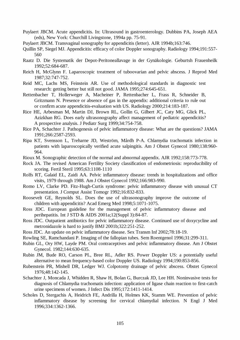
105
Puylaert JBCM. Acute appendicitis. In: Ultrasound in gastroenterology. Dubbins PA, Joseph AEA (eds), New York: Churchill Livingstone, 1994a pp. 75-91.
Puylaert JBCM. Transvaginal sonography for appendicitis (letter). AJR 1994b;163:746. Quillin SP, Siegel MJ. Appendicitis: efficacy of color Doppler sonography. Radiology 1994;191:557-
560 Raatz D. Die Systematik der Depot-Peritoneallavage in der Gynäkologie. Geburtsh Frauenheilk
1992;52:684-687. Reich H, McGlynn F. Laparoscopic treatment of tuboovarian and pelvic abscess. J Reprod Med
1987;32:747-752. Reid MC, Lachs MS, Feinstein AR. Use of methodological standards in diagnostic test
research: getting better but still not good. JAMA 1995;274:645-651. Rettenbacher T, Hollerweger A, Macheiner P, Rettenbacher L, Frass R, Schneider B,
Gritzmann N. Presence or absence of gas in the appendix: additional criteria to rule out or confirm acute appendicitis-evaluation with US. Radiology 2000;214:183-187.
Rice HE, Arbesman M, Martin DJ, Brown RL, Gollin G, Gilbert JC, Caty MG, Glick PL, Azizkhan RG. Does early ultrasonography affect management of pediatric appendicitis? A prospective analysis. J Pediatr Surg 1999;34:754-758.
Rice PA, Schachter J. Pathogenesis of pelvic inflammatory disease: What are the questions? JAMA 1991;266:2587-2593.
Ripa KT, Svensson L, Treharne JD, Weström, Mårdh P-A. Chlamydia trachomatis infection in patients with laparoscopically verified acute salpingitis. Am J Obstet Gynecol 1980;138:960-964.
Rioux M. Sonographic detection of the normal and abnormal appendix. AJR 1992;158:773-778. Rock JA. The revised American Fertility Society classification of endometriosis: reproducibility of
scoring. Fertil Steril 1995;63:1108-1110 Rolfs RT, Galaid EL, Zaidi AA. Pelvic inflammatory disease: trends in hospitalizations and office
visits, 1979 through 1988. Am J Obstet Gynecol 1992;166:983-990. Romo LV, Clarke PD. Fitz-Hugh-Curtis syndrome: pelvic inflammatory disease with unusual CT
presentation. J Comput Assist Tomogr 1992;16:832-833. Roosevelt GE, Reynolds SL. Does the use of ultrasonography improve the outcome of
children with appendicitis? Acad Emerg Med 1998;5:1071-1075. Ross JDC. European guideline for the management of pelvic inflammatory disease and
perihepatitis. Int J STD & AIDS 2001a;12(Suppl 3):84-87. Ross JDC. Outpatient antibiotics for pelvic inflammatory disease. Continued use of doxycycline and
metronidazole is hard to justify BMJ 2001b;322:251-252. Ross JDC. An update on pelvic inflammatory disease. Sex Transm Inf 2002;78:18-19. Rowling SE, Ramchandani P. Imaging of the fallopian tubes. Sem Roentgenol 1996;31:299-311. Rubin GL, Ory HW, Layde PM. Oral contraceptives and pelvic inflammatory disease. Am J Obstet
Gynecol. 1982;144:630-635. Rubin JM, Bude RO, Carson PL, Bree RL, Adler RS. Power Doppler US: a potentially useful
alternative to mean frequency-based color Doppler US. Radiology 1994;190:853-856. Rubenstein PR, Mishell DR, Ledger WJ. Colpotomy drainage of pelvic abscess. Obstet Gynecol
1976;48:142-145. Schachter J, Moncada J, Whidden R, Shaw H, Bolan G, Burczak JD, Lee HH. Noninvasive tests for
diagnosis of Chlamydia trachomatis infection: application of ligase chain reaction to first-catch urine specimens of women. J Infect Dis 1995;172:1411-1414.
Scholes D, Stergachis A, Heidrich FE, Andrilla H, Holmes KK, Stamm WE. Prevention of pelvic inflammatory disease by screening for cervical chlamydial infection. N Engl J Med 1996;334:1362-1366.
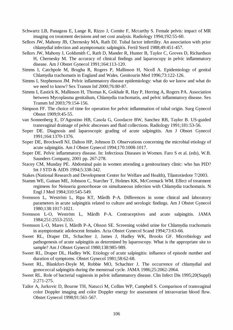
106
Schwartz LB, Panageas E, Lange R, Rizzo J, Comite F, Mccarthy S. Female pelvis: impact of MR imaging on treatment decisions and net cost analysis. Radiology 1994;192:55-60.
Sellors JW, Mahony JB, Chernesky MA, Rath DJ. Tubal factor infertility. An association with prior chlamydial infection and asymptomatic salpingitis. Fertil Steril 1988;49:451-457.
Sellors JW, Mahony J, Goldsmith C, Rath D, Mander R, Hunter B, Taylor C, Groves D, Richardson H, Chernesky M. The accuracy of clinical findings and laparoscopy in pelvic inflammatory disease. Am J Obstet Gynecol 1991;164:113-120.
Simms I, Catchpole M, Brugha R, Rogers P, Mallinson H, Nicoll A. Epidemiology of genital Chlamydia trachomatis in England and Wales. Genitourin Med 1996;73:122-126.
Simms I, Stephenson JM. Pelvic inflammatory disease epidemiology: what do we know and what do we need to know? Sex Transm Inf 2000;76:80-87.
Simms I, Eastick K, Mallinson H, Thomas K, Gokhale R, Hay P, Herring A, Rogers PA. Association between Mycoplasma genitalium, Chlamydia trachomatis, and pelvic inflammatory disease. Sex Transm Inf 2003;79:154-156.
Simpson FF. The choice of time for operation for pelvic inflammation of tubal origin. Surg Gynecol Obstet 1909;9:45-55.
van Sonnenberg E, D’Agostino HB, Casola G, Goodacre BW, Sanchez RB, Taylor B. US-guided transvaginal drainage of pelvic abscesses and fluid collections. Radiology 1991;181:53-56.
Soper DE. Diagnosis and laparoscopic grading of acute salpingitis. Am J Obstet Gynecol 1991;164:1370-1376.
Soper DE, Brockwell NJ, Dalton HP, Johnson D. Observations concerning the microbial etiology of acute salpingitis. Am J Obstet Gynecol 1994;170:1008-1017.
Soper DE. Pelvic inflammatory disease. In: Infectious Diseases in Women. Faro S et al. (eds), W.B. Saunders Company, 2001 pp. 267-278.
Stacey CM, Munday PE. Abdominal pain in women attending a genitourinary clinic: who has PID? Int J STD & AIDS 1994;5:338-342.
Stakes (National Research and Development Center for Welfare and Health), Tilastotiedote 7/2003. Stamm WE, Guinan ME, Johnson C, Starcher T, Holmes KK, McCormack WM. Effect of treatment
regimens for Neisseria gonorrhoeae on simultaneous infection with Chlamydia trachomatis. N Engl J Med 1984;310:545-549.
Svensson L, Weström L, Ripa KT, Mårdh P-A. Differences in some clinical and laboratory parameters in acute salpingitis related to culture and serologic findings. Am J Obstet Gynecol 1980;138:1017-1021.
Svensson L-O, Weström L, Mårdh P-A. Contraceptives and acute salpingitis. JAMA 1984;251:2553-2555.
Svensson L-O, Mares I, Mårdh P-A, Olsson SE. Screening voided urine for Chlamydia trachomatis in asymptomatic adolescent females. Acta Obstet Gynecol Scand 1994;73:63-66.
Sweet RL, Draper DL, Schachter J, James J, Hadley WK, Brooks GF. Microbiology and pathogenesis of acute salpingitis as determined by laparoscopy. What is the appropriate site to sample? Am J Obstet Gynecol 1980;138:985-989.
Sweet RL, Draper DL, Hadley WK. Etiology of acute salpingitis: influence of episode number and duration of symptoms. Obstet Gynecol 1981;58:62-68.
Sweet RL, Blankfort-Doyle M, Robbie MO, Schachter J. The occurrence of chlamydial and gonococcal salpingitis during the menstrual cycle. JAMA 1986;25:2062-2064.
Sweet RL. Role of bacterial vaginosis in pelvic inflammatory disease. Clin Infect Dis 1995;20(Suppl) 2:271-275.
Tailor A, Jurkovic D, Bourne TH, Natucci M, Collins WP, Campbell S. Comparison of transvaginal color Doppler imaging and color Doppler energy for assessment of intraovarian blood flow. Obstet Gynecol 1998;91:561-567.
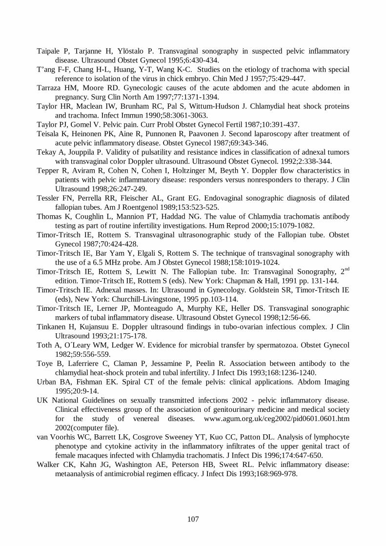
107
Taipale P, Tarjanne H, Ylöstalo P. Transvaginal sonography in suspected pelvic inflammatory disease. Ultrasound Obstet Gynecol 1995;6:430-434.
T’ang F-F, Chang H-L, Huang, Y-T, Wang K-C. Studies on the etiology of trachoma with special reference to isolation of the virus in chick embryo. Chin Med J 1957;75:429-447.
Tarraza HM, Moore RD. Gynecologic causes of the acute abdomen and the acute abdomen in pregnancy. Surg Clin North Am 1997;77:1371-1394.
Taylor HR, Maclean IW, Brunham RC, Pal S, Wittum-Hudson J. Chlamydial heat shock proteins and trachoma. Infect Immun 1990;58:3061-3063.
Taylor PJ, Gomel V. Pelvic pain. Curr Probl Obstet Gynecol Fertil 1987;10:391-437. Teisala K, Heinonen PK, Aine R, Punnonen R, Paavonen J. Second laparoscopy after treatment of
acute pelvic inflammatory disease. Obstet Gynecol 1987;69:343-346. Tekay A, Jouppila P. Validity of pulsatility and resistance indices in classification of adnexal tumors
with transvaginal color Doppler ultrasound. Ultrasound Obstet Gynecol. 1992;2:338-344. Tepper R, Aviram R, Cohen N, Cohen I, Holtzinger M, Beyth Y. Doppler flow characteristics in
patients with pelvic inflammatory disease: responders versus nonresponders to therapy. J Clin Ultrasound 1998;26:247-249.
Tessler FN, Perrella RR, Fleischer AL, Grant EG. Endovaginal sonographic diagnosis of dilated fallopian tubes. Am J Roentgenol 1989;153:523-525.
Thomas K, Coughlin L, Mannion PT, Haddad NG. The value of Chlamydia trachomatis antibody testing as part of routine infertility investigations. Hum Reprod 2000;15:1079-1082.
Timor-Tritsch IE, Rottem S. Transvaginal ultrasonographic study of the Fallopian tube. Obstet Gynecol 1987;70:424-428.
Timor-Tritsch IE, Bar Yam Y, Elgali S, Rottem S. The technique of transvaginal sonography with the use of a 6.5 MHz probe. Am J Obstet Gynecol 1988;158:1019-1024.
Timor-Tritsch IE, Rottem S, Lewitt N. The Fallopian tube. In: Transvaginal Sonography, 2nd edition. Timor-Tritsch IE, Rottem S (eds). New York: Chapman & Hall, 1991 pp. 131-144.
Timor-Tritsch IE. Adnexal masses. In: Ultrasound in Gynecology. Goldstein SR, Timor-Tritsch IE (eds), New York: Churchill-Livingstone, 1995 pp.103-114.
Timor-Tritsch IE, Lerner JP, Monteagudo A, Murphy KE, Heller DS. Transvaginal sonographic markers of tubal inflammatory disease. Ultrasound Obstet Gynecol 1998;12:56-66.
Tinkanen H, Kujansuu E. Doppler ultrasound findings in tubo-ovarian infectious complex. J Clin Ultrasound 1993;21:175-178.
Toth A, O´Leary WM, Ledger W. Evidence for microbial transfer by spermatozoa. Obstet Gynecol 1982;59:556-559.
Toye B, Laferriere C, Claman P, Jessamine P, Peelin R. Association between antibody to the chlamydial heat-shock protein and tubal infertility. J Infect Dis 1993;168:1236-1240.
Urban BA, Fishman EK. Spiral CT of the female pelvis: clinical applications. Abdom Imaging 1995;20:9-14.
UK National Guidelines on sexually transmitted infections 2002 - pelvic inflammatory disease. Clinical effectiveness group of the association of genitourinary medicine and medical society for the study of venereal diseases. www.agum.org.uk/ceg2002/pid0601.0601.htm 2002(computer file).
van Voorhis WC, Barrett LK, Cosgrove Sweeney YT, Kuo CC, Patton DL. Analysis of lymphocyte phenotype and cytokine activity in the inflammatory infiltrates of the upper genital tract of female macaques infected with Chlamydia trachomatis. J Infect Dis 1996;174:647-650.
Walker CK, Kahn JG, Washington AE, Peterson HB, Sweet RL. Pelvic inflammatory disease: metaanalysis of antimicrobial regimen efficacy. J Infect Dis 1993;168:969-978.
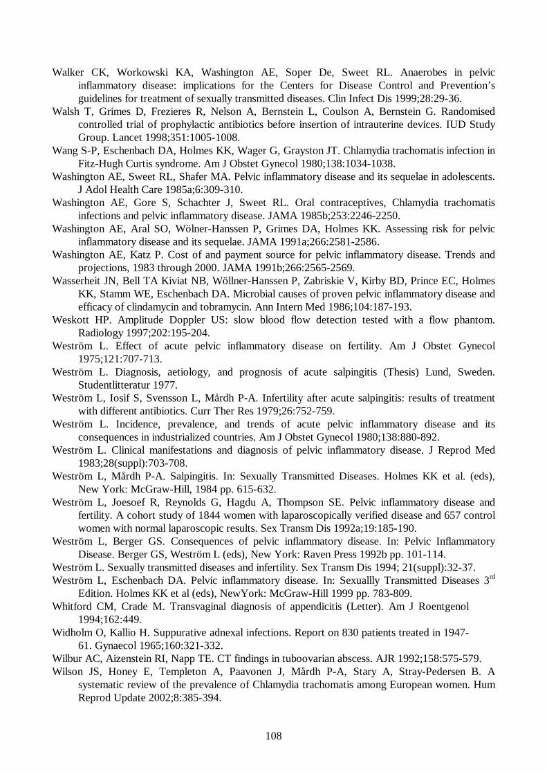
108
Walker CK, Workowski KA, Washington AE, Soper De, Sweet RL. Anaerobes in pelvic inflammatory disease: implications for the Centers for Disease Control and Prevention’s guidelines for treatment of sexually transmitted diseases. Clin Infect Dis 1999;28:29-36.
Walsh T, Grimes D, Frezieres R, Nelson A, Bernstein L, Coulson A, Bernstein G. Randomised controlled trial of prophylactic antibiotics before insertion of intrauterine devices. IUD Study Group. Lancet 1998;351:1005-1008.
Wang S-P, Eschenbach DA, Holmes KK, Wager G, Grayston JT. Chlamydia trachomatis infection in Fitz-Hugh Curtis syndrome. Am J Obstet Gynecol 1980;138:1034-1038.
Washington AE, Sweet RL, Shafer MA. Pelvic inflammatory disease and its sequelae in adolescents. J Adol Health Care 1985a;6:309-310.
Washington AE, Gore S, Schachter J, Sweet RL. Oral contraceptives, Chlamydia trachomatis infections and pelvic inflammatory disease. JAMA 1985b;253:2246-2250.
Washington AE, Aral SO, Wölner-Hanssen P, Grimes DA, Holmes KK. Assessing risk for pelvic inflammatory disease and its sequelae. JAMA 1991a;266:2581-2586.
Washington AE, Katz P. Cost of and payment source for pelvic inflammatory disease. Trends and projections, 1983 through 2000. JAMA 1991b;266:2565-2569.
Wasserheit JN, Bell TA Kiviat NB, Wöllner-Hanssen P, Zabriskie V, Kirby BD, Prince EC, Holmes KK, Stamm WE, Eschenbach DA. Microbial causes of proven pelvic inflammatory disease and efficacy of clindamycin and tobramycin. Ann Intern Med 1986;104:187-193.
Weskott HP. Amplitude Doppler US: slow blood flow detection tested with a flow phantom. Radiology 1997;202:195-204.
Weström L. Effect of acute pelvic inflammatory disease on fertility. Am J Obstet Gynecol 1975;121:707-713.
Weström L. Diagnosis, aetiology, and prognosis of acute salpingitis (Thesis) Lund, Sweden. Studentlitteratur 1977.
Weström L, Iosif S, Svensson L, Mårdh P-A. Infertility after acute salpingitis: results of treatment with different antibiotics. Curr Ther Res 1979;26:752-759.
Weström L. Incidence, prevalence, and trends of acute pelvic inflammatory disease and its consequences in industrialized countries. Am J Obstet Gynecol 1980;138:880-892.
Weström L. Clinical manifestations and diagnosis of pelvic inflammatory disease. J Reprod Med 1983;28(suppl):703-708.
Weström L, Mårdh P-A. Salpingitis. In: Sexually Transmitted Diseases. Holmes KK et al. (eds), New York: McGraw-Hill, 1984 pp. 615-632.
Weström L, Joesoef R, Reynolds G, Hagdu A, Thompson SE. Pelvic inflammatory disease and fertility. A cohort study of 1844 women with laparoscopically verified disease and 657 control women with normal laparoscopic results. Sex Transm Dis 1992a;19:185-190.
Weström L, Berger GS. Consequences of pelvic inflammatory disease. In: Pelvic Inflammatory Disease. Berger GS, Weström L (eds), New York: Raven Press 1992b pp. 101-114.
Weström L. Sexually transmitted diseases and infertility. Sex Transm Dis 1994; 21(suppl):32-37. Weström L, Eschenbach DA. Pelvic inflammatory disease. In: Sexuallly Transmitted Diseases 3rd
Edition. Holmes KK et al (eds), NewYork: McGraw-Hill 1999 pp. 783-809. Whitford CM, Crade M. Transvaginal diagnosis of appendicitis (Letter). Am J Roentgenol
1994;162:449. Widholm O, Kallio H. Suppurative adnexal infections. Report on 830 patients treated in 1947-
61. Gynaecol 1965;160:321-332. Wilbur AC, Aizenstein RI, Napp TE. CT findings in tuboovarian abscess. AJR 1992;158:575-579. Wilson JS, Honey E, Templeton A, Paavonen J, Mårdh P-A, Stary A, Stray-Pedersen B. A
systematic review of the prevalence of Chlamydia trachomatis among European women. Hum Reprod Update 2002;8:385-394.
109
Witkin SS, Jeremias J, Toth M, Ledger WJ. Proliferative response to conserved epitopes of the Chlamydia trachomatis human 60-kilodalton heat-shock proteins by lymphocytes from women with salpingitis. Am J Obstet Gynecol 1994;171:455-460.
Wittman I. The history of peritoneoscopy. In: Peritoneoscopy. North J et al. (eds). Budapest: Akademiai Kiado, 1966 pp 3-5.
World Health Organization: Pelvic inflammatory disease in nongonococcal urethritis and other selected sexually transmitted diseases of public health importance. WHO Techn Rep Ser 1981;660:89-100.
World Health Organization. Mechanisms of action, safety, and efficacy of intrauterine devices. WHO Techn Rep Ser 1987a;753:36-37.
World Health Organization. Infections, pregnancies, and infertility: Perspectives on prevention. Fertil Steril 1987b;47:964-968.
World Health Organization. Guidelines for the management of sexually transmitted infections: Lower abdominal pain 2002, pp. 26-29.
Wölner-Hanssen P, Weström L, Mårdh PA. Perihepatitis and chlamydial salpingitis. Lancet 1980;1:901-903.
Wölner-Hanssen P, Mårdh P-A. In vitro tests of the adherence of Chlamydia trachomatis to human spermatozoa. Fertil Steril 1984;42:101-107.
Wölner-Hanssen P, Svensson L, Mårdh P-A, Weström L. Laparoscopic findings and contraceptive use in women with signs and symptoms suggestive of acute salpingitis. Obstet Gynecol 1985;66:233-238.
Wölner-Hanssen P. Oral contraceptive use modifies the manifestations of pelvic inflammatory disease. Br J Obstet Gynecol 1986a;93:619-624
Wölner-Hanssen P, Eschenbach D, Paavonen J, Holmes KK. Treatment of pelvic inflammatory disease: use doxycycline with an appropriate B-lactam while we wait for better data. JAMA 1986b;256:3262-3263.
Wölner-Hanssen P, Eschenbach DA, Paavonen J, Kiviat N, Stevens CE, Critchlow C, DeRouen T, Holmes KK. Decreased risk of symptomatic chlamydial pelvic inflammatory disease associated with oral contraceptive use. JAMA 1990a;263:54-59.
Wölner-Hanssen P. Kiviat NK, Holmes KK: Atypical pelvic inflammatory disease: subacute, chronic, or subclinical upper genital tract infection in women. In: Sexually Transmitted Diseases, 2nd ed. Holmes KK, et al (eds), New York: Mcgraw-Hill, 1990b pp. 615-620.
Wölner-Hanssen P. Silent Pelvic Inflammatory Disease: is it overstated? Obstet Gynecol 1995;86:321-325.
Yamashita Y, Torashima M, Hatanaka Y, Harada M, Higashida Y, Takahashi M, Mizutani H,Tashiro H, Iwamasa J, Miyazaki K, Okamura H. Adnexal masses: accuracy of characterization with transvaginal US and precontrast and postcontrast MR imaging. Radiology 1995;194:557-565.