Embed Size (px)
Citation preview

From the Department of Orthopaedics and Traumatology,
and the Department of Pediatric Surgery, University of Helsinki;
and the Research Institute of Military Medicine, Central Military Hospital, Helsinki
FEMORAL SHAFT FRACTURES IN ADULTS:
EPIDEMIOLOGY, FRACTURE PATTERNS, NONUNIONS,
AND FATIGUE FRACTURES
A clinical study
Sari Salminen
Academic dissertation
To be presented with the permission of
The Faculty of Medicine of the University of Helsinki,
for public discussion in the Auditorium of the Töölö Hospital,
Helsinki University Central Hospital, on June 29th, 2005, at 12 o’clock noon.
Helsinki 2005
Supervised by
Docent Ole Böstman, M.D., Ph.D.Department of Orthopaedics and TraumatologyHelsinki University Central HospitalHelsinki, Finland
Docent Harri Pihlajamäki, M.D., Ph.D.Department of Surgery and Research Institute of Military MedicineCentral Military HospitalHelsinki, Finland
Reviewed by
Docent Matti U.K. Lehto, M.D., Ph.D.Coxa, Hospital for Joint ReplacementTampere, Finland
Professor Erkki Tukiainen, M.D., Ph.D.Department of Plastic SurgeryHelsinki University Central HospitalHelsinki, Finland
To be discussed with
Professor Heikki Kröger, M.D., Ph.D.Department of Orthopaedics and TraumatologyKuopio University HospitalKuopio, Finland
ISBN 952-91-8891-9 (bound)
ISBN 952-10-2523-9 (PDF)
Helsinki University Printing House
Helsinki 2005

To my family

4
5
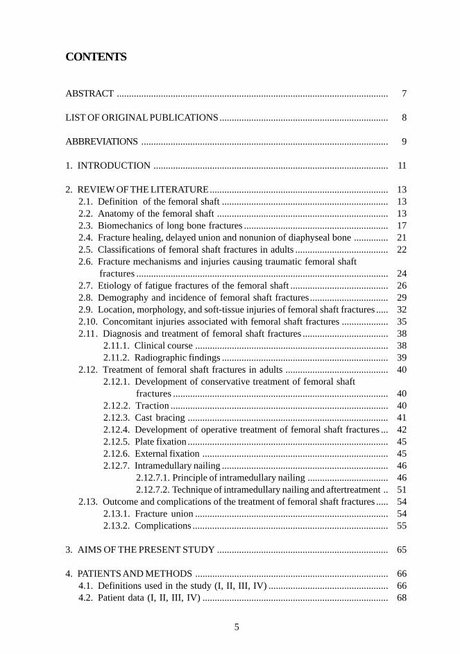
CONTENTS
ABSTRACT ............................................................................................................... 7
LIST OF ORIGINAL PUBLICATIONS..................................................................... 8
ABBREVIATIONS ..................................................................................................... 9
1. INTRODUCTION ................................................................................................ 11
2. REVIEW OF THE LITERATURE......................................................................... 132.1. Definition of the femoral shaft .................................................................... 132.2. Anatomy of the femoral shaft ...................................................................... 132.3. Biomechanics of long bone fractures ........................................................... 172.4. Fracture healing, delayed union and nonunion of diaphyseal bone .............. 212.5. Classifications of femoral shaft fractures in adults ...................................... 222.6. Fracture mechanisms and injuries causing traumatic femoral shaft
fractures ....................................................................................................... 242.7. Etiology of fatigue fractures of the femoral shaft ........................................ 262.8. Demography and incidence of femoral shaft fractures ................................ 292.9. Location, morphology, and soft-tissue injuries of femoral shaft fractures ..... 322.10. Concomitant injuries associated with femoral shaft fractures ................... 352.11. Diagnosis and treatment of femoral shaft fractures ................................... 38
2.11.1. Clinical course ............................................................................... 382.11.2. Radiographic findings .................................................................... 39
2.12. Treatment of femoral shaft fractures in adults .......................................... 402.12.1. Development of conservative treatment of femoral shaft
fractures ........................................................................................ 402.12.2. Traction ......................................................................................... 402.12.3. Cast bracing .................................................................................. 412.12.4. Development of operative treatment of femoral shaft fractures ... 422.12.5. Plate fixation .................................................................................. 452.12.6. External fixation ............................................................................ 452.12.7. Intramedullary nailing .................................................................... 46
2.12.7.1. Principle of intramedullary nailing ................................. 462.12.7.2. Technique of intramedullary nailing and aftertreatment .. 51
2.13. Outcome and complications of the treatment of femoral shaft fractures ..... 542.13.1. Fracture union ............................................................................... 542.13.2. Complications ................................................................................ 55
3. AIMS OF THE PRESENT STUDY ...................................................................... 65
4. PATIENTS AND METHODS ............................................................................... 664.1. Definitions used in the study (I, II, III, IV) ................................................. 664.2. Patient data (I, II, III, IV) ............................................................................ 68
6
4.3. Study arrangement (I, II, III, IV) ................................................................ 694.4. Fracture management (II, III, IV) ............................................................... 704.5. Follow-up time (II, II, IV) ........................................................................... 724.6. Statistical methods (I, II, III, IV) ................................................................ 72
5. RESULTS .............................................................................................................. 735.1. Demography of femoral shaft fractures in adults (I, II, III) ....................... 735.2. Injury mechanism (I, II, IV) or activity during fracture onset (III) ............ 755.3. Concomitant injuries (I, II, III, IV) .............................................................. 795.4. Fracture characteristics (I, II, III, IV) ........................................................ 795.5. Outcome and complications of fracture treatment (II, III, IV) ................... 84
6. GENERAL DISCUSSION .................................................................................... 1006.1. Incidence of femoral shaft fractures ............................................................ 1006.2. Injury season and injury mechanisms of femoral shaft fractures in adults .... 1036.3. Concomitant injuries related to femoral shaft fractures in adults ................. 1036.4. Morphology of femoral shaft fractures in adults ......................................... 1046.5. Treatment, outcome, and complications of femoral shaft fractures in
adults ............................................................................................................. 1056.6. Prevention of femoral shaft fractures .......................................................... 108
7. SUMMARY AND CONCLUSIONS ...................................................................... 1097.1. Demography of femoral shaft fractures in adults ....................................... 1097.2. Morphology of femoral shaft fractures in adults ......................................... 1097.3. Treatment of femoral shaft fractures and nonunions in adults .................... 1107.4. Prevention of femoral shaft fractures in adults ............................................ 110
ACKNOWLEDGEMENTS ........................................................................................ 113
REFERENCES ........................................................................................................... 115
ORIGINAL PUBLICATIONS .................................................................................... 147
7
ABSTRACT
The femur (thigh bone) is the longest, strongest, largest and heaviest tubular bone in thehuman body, and one of the principal load-bearing bones in the lower extremity. Femoralshaft fractures are among the most common major injuries that an orthopaedic surgeonwill be required to treat. Although demographic data of the patients have been analyzed insome epidemiologic studies, little attention has been paid to the characterization of thefracture patterns of femoral shaft fractures using morphologic classification systems.
Femoral shaft fractures are commonly thought to be primarily associated with severetrauma in young persons. Low energy violence as a cause of these fractures, especiallyamong the elderly, has been mentioned only sporadically in epidemiologic studies. Anoth-er subgroup of operatively treated femoral shaft fractures are displaced fatigue fracturesof the femoral diaphysis, which mainly occur among military trainees.
The concept of intramedullary fixation in the treatment of femoral shaft fractures hasgained wide acceptance. Delayed union or nonunion following intramedullary nailing ofthe femur has been considered an infrequent clinical problem. As a consequence of themore frequent use of intramedullary nailing for the treatment of femoral shaft fractures,an increasing number of orthopaedic surgeons will be confronted with this complication.
The present study shows that femoral shaft fractures in adults are not exclusively theresult of high energy trauma. Low energy trauma can cause 25% of the fractures. Fem-oral shaft fractures caused by low energy violence mainly occur in patients sufferingfrom a chronic disease or a condition causing osteopenia of the femur. In displacedfemoral shaft fatigue fractures, regardless of the symptoms, the diagnosis can be delayeduntil displacement. The incidences of femoral shaft fractures caused by different injuriesvary from 1.5:100 000 person-years to 9.9:100 000 person-years. Most traumatic femo-ral shaft fractures are isolated without concomitant injuries. The most common fracturetype of the femoral shaft is a non-comminuted simple AO Type A, most of which, intraumatic fractures, are purely transverse and located in the middle third of the femur.Spiral fractures that are related to low energy trauma situate in the middle third of thefemur. Displaced fatigue fractures are oblique or oblique-transverse, and located in thedistal third of the femoral shaft. Femoral shaft fractures caused by low energy trauma aremorphologically different from displaced fatigue fractures, which can also be primarilycomminuted.
Factors that predispose traumatic fresh femoral shaft fractures to nonunion after in-tramedullary nailing are related to severe fracture comminution and concomitant injuries.Reoperation of traumatic femoral shaft fractures treated with intramedullary nailing shouldbe performed within six months of the primary nailing to minimize the risk of nail break-age, if convincing signs of consolidation in progress are lacking. Exchange nailing seemsto be the method of choice for the treatment of a disturbed union. In some selected caseswith primary static interlocking nailing, dynamization alone can be considered. Bone graftingalone as a treatment of a failed union of a femoral shaft fracture cannot be recommended.
8
LIST OF ORIGINAL PUBLICATIONS
The present study is based on the following articles, referred to in the text by their Romannumerals:
I Salminen ST, Pihlajamäki HK, Avikainen VJ, Böstman OM. Population based epi-demiologic and morphologic study of femoral shaft fractures. Clin Orthop RelatRes 372:241-249, 2000
II Salminen S, Pihlajamäki H, Avikainen V, Kyrö A, Böstman O. Specific featuresassociated with femoral shaft fractures caused by low energy trauma. J Trauma43:117-122, 1997
III Salminen ST, Pihlajamäki HK, Visuri TI, Böstman OM. Displaced fatigue fracturesof the femoral shaft. Clin Orthop Relat Res 409:250-259, 2003
IV Pihlajamäki HK, Salminen ST, Böstman OM. The treatment of nonunions follow-ing intramedullary nailing of femoral shaft fractures. J Orthop Trauma 16:394-402, 2002
9
ABBREVIATIONS
ACL anterior cruciate ligamentAO Arbeitsgemeinschaft für OsteosynthesefragenAP anteroposteriorARDS adult respiratory distress syndromeASIF Association for the Study of Internal FixationBMI body mass indexC closed (intramedullary nailing)CAB chronic alcohol abusecm centimeter (SI)COPD chronic obstructive pulmonary diseaseCT computerized tomographyDCP dynamic compression plateDCS dynamic condylar screwDM diabetes mellitusF femaleG-K Grosse-KempfIAC intraoperative additional comminutionICD International Classification of DiseasesIM intramedullaryISS Injury Severity Scorekg kilogram (SI)KLR knee ligament ruptureK-S Klemm-SchellmannL leftLCL lateral collaterale ligamentLISS Less Invasive Stabilization SystemM malem meter (SI)m. musculus (muscle)m² square meter (SI)MCL medial collaterale ligamentmm millimeter (SI)mm³ cubic millimeter (SI)mmHg millimeter of mercuryMRI magnetic resonance imagingN numberND neuromuscular disordersNm NewtonmeterO open (intramedullary nailing)OA osteoarthritisOTA Orthopaedic Trauma Associationp probabilityPCL posterior cruciate ligament
10
PF previous major fractureR rightSTIR short tau inversion recoveryT2 transverse relaxationTP total prosthesis replacementV-W Vari-WallW-H Winquist and HansenWHO World Health Organization
11
1. INTRODUCTION
The femur (thigh bone) is the longest, strongest, largest and heaviest tubular bone in thehuman body (Moore 1992; Bucholz and Brumback 1996; Schatzker 1996; Platzer 2003;Whittle and Wood 2003), and one of the principal load-bearing bones in the lower extrem-ity (Whittle and Wood 2003). Femoral shaft fractures are among the most common majorinjuries that an orthopaedic surgeon will be required to treat (Gozna 1982; Whittle andWood 2003).
Femoral shaft fractures often result from high energy forces associated with possiblemultiple system injuries (Bucholz and Jones 1991; Bucholz and Brumback 1996; Whittleand Wood 2003). Fractures of the femoral diaphysis can be life-threatening on account ofan open wound, fat embolism, adult respiratory distress syndrome (ARDS) (Zalavras etal. 2005), or resultant multiple organ failure (Bucholz and Jones 1991; Bucholz and Brum-back 1996; Keel and Trentz 2005). Femoral shaft fractures can lead to a major physicalimpairment, not because of disturbed fracture healing, but rather due to fracture shorten-ing, fracture malalignment, or prolonged immobilization of the extremity by traction orcasting in an attempt to maintain the fracture length and alignment during the early phasesof healing (Bucholz and Brumback 1996). Even minor degrees of shortening and mala-lignment can eventuate in a limp and posttraumatic arthritis (Bucholz and Jones 1991;Bucholz and Brumback 1996). The art of femoral fracture care is a constant balancing ofthe often conflicting goals of anatomic alignment and early functional rehabilitation of thelimb (Bucholz and Brumback 1996).
Although most musculoskeletal injuries occur in a predictable manner, as dictated by theforces involved and the structure of the region, there are always certain fractures that areunique to each injury (Gozna 1982). Few epidemiologic studies have been published onfemoral shaft fractures. Although demographic data of the patients have been analyzed(Knowelden, Buhr, Dunbar 1964; Wong 1966; Hedlund and Lindgren 1986; Arneson et al.1988; Bengnér et al. 1990), little attention has been paid to the characterization of thefracture patterns using morphologic classification systems. Epidemiologic studies offerimportant data contributing to improved fracture treatment or better patient care. Sur-geons should have knowledge of the spectrum of fractures they treat, not only for anintrinsic educational value, but also to allow resources to be allocated on the basis ofprojected numbers of patients. The ability to predict the level of admissions to a traumaunit is useful for administrative and training purposes.
Femoral shaft fractures are commonly thought to be primarily associated with severetrauma in young persons. Low energy violence as a cause of these fractures, especiallyamong the elderly, has been mentioned only sporadically in epidemiologic studies of frac-tures of the femoral shaft (Wong 1966; Hedlund and Lindgren 1986; Arneson et al. 1988;Bengnér et al. 1990). The outcome of tibial shaft fractures caused by low energy mech-anism has been recently studied (Toivanen 2001). Another subgroup of operatively treat-ed femoral shaft fractures are displaced fatigue fractures of the femoral diaphysis. Mili-tary trainees form a relatively homogenous population to be investigated of the epidemio-
12
logic features of fatigue fractures. The trainees are usually affected by undisplaced fa-tigue (stress) fractures of the lower extremities. Displacement of a fatigue fracture in along bone is a rare but serious injury.
The treatment of femoral shaft fractures has always been a focus of interest, but may stillremain a clinical problem, and a subject of controversy. Several techniques have beendeveloped for the treatment. With the awareness of the advantages, disadvantages, andlimitations of these techniques, an orthopaedic surgeon has the opportunity to avoid pro-longed morbidity and extensive disability owing to lower extremity injuries (Bucholz andBrumback 1996; Whittle and Wood 2003). In 1963, Dencker published his thesis of 1003recent fractures of the femoral shaft in 992 patients treated at the public hospitals ofSweden during a three-year period from 1952 to 1954 with a follow-up of 4 to 8 years toanalyze the results obtained with different methods of fracture treatment (Dencker 1963).He concluded that conservative treatment with traction is the method of choice in theroutine management of femoral shaft fractures. Ten years later, as the Küntscher nailingwith reaming and a compression plate osteosynthesis had gained more popularity in frac-ture management, Kootstra introduced in the Netherlands a study of 335 consecutivefemoral shaft fractures in 329 patients with a statistical analysis of the different methodsof treatment during 1958-1969 (Kootstra 1973). Since the studies of Dencker and Koot-stra, the changes over the 30-40 years have introduced a new preferential fracture treat-ment in intramedullary nailing with extended indications, nailing types (unreamed nailingor retrograde nailing), and diverse nail materials. The concept of intramedullary fixation inthe treatment of femoral shaft fractures has gained wide acceptance, yet the the litera-ture, though abundant and comprising a lot of clinical series, shows limitations in report-ing of more or less unspecified fractures managed by one certain intramedullary nail type.In fact, the discussed issues seem seldom to have focused on the type of fracture itself,leaving the specific problematics of fractures caused by infrequent mechanisms nearlyunobserved.
Delayed union or nonunion following intramedullary nailing of the femur has been sup-posed to be an infrequent clinical problem compared with the treatment results of thelower leg (Winquist, Hansen, Clawson 1984; Thoresen et al. 1985; Brumback et al. 1988b;Christie et al. 1988; Søjberg, Eiskjaer, Møller-Larsen 1990; Brumback 1996; Bhandari etal. 2000; Herscovici et al. 2000; Tornetta and Tiburzi 2000). As a consequence of themore frequent use of intramedullary nailing for the treatment of femoral shaft fractures,an increasing number of orthopaedic surgeons will, however, be confronted with thiscomplication. Failed union of a femoral shaft fracture is a serious complication, prolong-ing patient morbidity and possibly influencing the ultimate functional recovery. Neverthe-less, the studies on the outcome of femoral shaft fractures have seldom focused onproper identification and treatment of a disturbed process of consolidation after intramed-ullary nailing of a shaft fracture of the femur. Many of the previous reports have heterog-enous patient material because the type of primary treatment before the development ofthe disturbed consolidation has varied considerably (Christensen 1973; Harper 1984; Hei-ple et al. 1985; Kempf, Grosse, Rigaut 1986, Klemm and Börner 1986; Curylo and Lind-sey 1994; Canadian Orthopaedic Trauma Society 2003). Hence, it has been difficult tocharacterize the typical features and problems of nonunion after intramedullary nailing.
13
2. REVIEW OF THE LITERATURE
2.1. Definition of the femoral shaft
The length of a tubular human femur is about one fourth of the height of a person (Thorek1962; Healey and Seybold 1969; Moore 1992). The skeletal maturity of the adult type offemoral diaphysis can be judged by the age of the patient, which usually has been 17years or older in studies concerning femoral shaft fractures in adults (Dencker 1963;Kootstra 1973), but more definitely by the closed (mature) growth plates (Platzer 2003).
The proximal end of the femur consists of the head, the neck, the greater trochanter, andthe lesser trochanter. The distal end of the femur has medial and lateral condyles. Theproximal and distal parts widen into metaphyseal subtrochanteric and supracondylar re-gions (Thorek 1962; Healey and Seybold 1969; Moore 1992; Bucholz and Brumback1996; Platzer 2003). The designation femoral shaft fracture denotes that the fracturesituates entirely on the femoral diaphysis. The definition of the diaphysis measured fromthe anteroposterior (AP) radiographs has varied (Carr and Miller 1958; Dencker 1963;Kootstra 1973; Hedlund and Lindgren 1986; Böstman et al. 1989; Canadian OrthopaedicTrauma Society 2003). The femoral shaft is 1) the portion of the bone between theproximal boundary of 4 inches (10.16 cm) from the tip of the trochanter major and thedistal boundary of 4 inches (10.16 cm) from the end of the femoral medial condyle (Carrand Miller 1958), or 2) the distance between 5 cm distal to the lesser trochanter and 6 cmproximal to the most distal point of the medial femoral condyle (Dencker 1963), or 3) thediaphyseal section between the boundaries of the lower edge of the lesser trochanter andof a line which parallels the joint space of the knee at a distance equal to the width of thecondyles (Kootstra 1973), or 4) the part of the femur between 10 cm distal to the lessertrochanter and 15 cm proximal to the knee joint line (Hedlund and Lindgren 1986), or 5)the portion of bone between a point 5 cm distal to the lesser trochanter and 8 cmproximal to the adductor tubercle (Böstman et al. 1989), or 6) the bone section betweenthe boundaries of at least 1 cm distal to the lesser trochanter and 6 cm or more proximalto the distal femoral physeal scar (Canadian Orthopaedic Trauma Society 2003).
2.2. Anatomy of the femoral shaft
The femoral shaft has a physiologic anterior curve (Thorek 1962; Dencker 1963; Koot-stra 1973), which can increase in certain pathologic conditions, such as fibrous dysplasiaor Paget’s disease (Grundy 1970; Bucholz and Brumback 1996; Whittle and Wood 2003).The external circumference of the femur is triangular exhibiting three surfaces: anterior,lateral, and medial (Thorek 1962; Healey and Seybold 1969; Kootstra 1973; Platzer 2003).The greatest cortical thickness is posteriorly, where the fascia inserts to the linea aspera,a two-lipped roughened line (Thorek 1962; Healey and Seybold 1969; Bucholz and Brum-back 1996; Platzer 2003). The medial and lateral lips of the linea aspera diverge proximal-ly and distally, the lateral lip becoming continuous proximally with the gluteal tuberosity(Thorek 1962; Platzer 2003). The medial lip extends up to the undersurface of the femo-ral neck (Platzer 2003). Lateral to this lip is a ridge, the pectineal line, descending from the
14
lesser trochanter. Both proximally and distally the femoral shaft loses its triangular formand becomes four-sided (Platzer 2003). The medullary cavity varies in diameter andshape (Thorek 1962; Healey and Seybold 1969; Kootstra 1973; Moore 1992). Slightlyproximal to the midshaft is the isthmus, where the circular medullary cavity is its narrow-est with a diameter of 8 mm to 16 mm compared with the otherwise more oval medullarycanal (Dencker 1963).
The thigh extends superficially from the inguinal ligament anteriorly and the gluteal skinfold posteriorly to the knee level (Thorek 1962; Healey and Seybold 1969). Superficialfascia contains cutaneous nerve branches from the lumbar plexus (the lateral femoralcutaneous nerve), the femoral nerve (the anterior and medial femoral cutaneous nerves),the obturator nerve (medial aspect of the thigh), and the genitofemoral nerve (the lum-boinguinal branch). The included arteries are the superficial circumflex iliac, the superfi-cial inferior epigastric, and the superficial external pudendal arteries branching from thecommon femoral artery. The great saphenous vein has the ramifications of the superficialcircumflex iliac, the superficial inferior epigastric, and the superficial external pudendalveins at the region (Thorek 1962; Healey and Seybold 1969).
On the posterior side of the femoral diaphysis attach the pectineus, adductor brevis,adductor magnus, adductor longus, and gluteus maximus muscles. From the femoralshaft originate m. vastus lateralis (upper half of the intertrochanteric line), m. vastusmedialis (medial lip of linea aspera and spiral line of femur), m. vastus intermedius (ante-rior and lateral aspect of upper two thirds of femoral shaft), the short head of m. bicepsfemoris (linea aspera and lateral supracondylar line of femur), and m. articularis genus(Thorek 1962; Healey and Seybold 1969; Kootstra 1973). The muscles of the thigh areencased by dense fibrous tissue (Healey and Seybold 1969; Kootstra 1973; Moore 1992;Bucholz and Brumback 1996). The fascia lata reinforces the lateral aspect to form distallythe iliotibial tract (Thorek 1962; Kootstra 1973; Platzer 2003), which on the lateral sideextends to the Gerdy’s tubercle of the tibia (Platzer 2003).
The thigh contains three distinct fascial compartments. The anterior compartment encas-es the knee extensor muscles (quadriceps femoris including rectus femoris, vastus inter-medius, vastus medialis, and vastus lateralis; and sartorius) innervated by the femoralnerve from the lumbar plexus L 2-4 for the quadriceps femoris and L 2-3 for the sartorius(Thorek 1962; Healey and Seybold 1969; Kootstra 1973; Hoppenfeld and deBoer 1984;Moore 1992; Platzer 2003). The rectus femoris muscle is also a weak flexor of the hip(Healey and Seybold 1969; Platzer 2003). The sartorius flexes, abducts, and mediallyrotates the thigh (Thorek 1962; Healey and Seybold 1969; Kootstra 1973; Moore 1992).The anterior compartment also includes the tensor fasciae latae, the iliacus and psoasmajor muscles, and the femoral artery and vein, femoral nerve, and lateral femoral cuta-neous nerve (Moore 1992).
The medial compartment contains the adductor muscles (gracilis, adductor longus,adductor brevis, adductor magnus, pectineus) and the obturator externus muscle, whichare supplied by the obturator nerve (Thorek 1962; Healey and Seybold 1969; Kootstra1973; Hoppenfeld and deBoer 1984; Moore 1992; Platzer 2003). The pectineus and

15
the adductor magnus muscle receive dual innervation: the former from the femoral nerveand the latter from the sciatic nerve (Kootstra 1973; Platzer 2003). The medial comp-artment also includes the deep femoral artery, obturator artery and vein, and obturatornerve.
The posterior compartment includes the flexor muscles (biceps femoris, semitendinosus,and semimembranosus), which extend the hip, and a portion of the adductor magnusmuscles, as well as branches of the deep femoral artery, sciatic nerve, and posteriorfemoral cutaneous nerve. The posterior knee flexor group is innervated by the sciaticnerve (Thorek 1962; Healey and Seybold 1969; Hoppenfeld and deBoer 1984). The bi-ceps femoris extends, adducts and laterally rotates the thigh, as well as flexes the lowerleg (Thorek 1962; Healey and Seybold 1969; Platzer 2003). The long head of the bicepsfemoris is innervated by the tibial nerve (L5-S2), and the short head receives innervationfrom the common peroneal division (S1-2) (Platzer 2003). The semimembranosus andsemitendinosus muscles also act as medial rotators of the thigh (Thorek 1962; Healey andSeybold 1969), and are innervated from the tibial nerve (L5-S2) (Platzer 2003). Theintermuscular septum between the anterior and posterior compartments is thicker thanthe septa between the medial and anterior compartments (Hoppenfeld and deBoer 1984;Bucholz and Brumback 1996; Platzer 2003). Because of the high volume of these threecompartments, compartment syndrome of the thigh is much less common than that ofthe lower leg (Bucholz and Brumback 1996).
The arterial supply of the femur is mainly derived from the deep femoral artery (a. pro-funda femoris) (Thorek 1962; Healey and Seybold 1969; Bucholz and Brumback 1996).From its branches, the lateral circumflex femoral artery, among others, supplies blood tothe extensor muscles, while other proximal branches provide vascular supply to the ad-ductor muscles, and, more distally, three perforating arteries supply the flexor muscles(Kootstra 1973). The muscular branch of the superficial femoral artery supplies blood tothe vastus medialis muscle (Kootstra 1973).
The femoral shaft has periosteal and endosteal blood supply (Laing 1953). The endostealcirculation of the femoral diaphysis is predominatly derived from a nutrient artery thatbranches from the first perforating branch of the deep femoral artery (Laing 1953; Brookes1971), enters the bone proximally and posteriorly through a nutrient foramen in the mid-dle of the diaphysis near the linea aspera, and arborizes proximally and distally (Laing1953; Bucholz and Brumback 1996). Very seldom, a second nutrient artery exists distally(Laing 1953), but no major artery enters the lower third of the shaft (Anseroff 1934;Laing 1953). Under normal physiologic conditions, the circulation is endosteal to theinner two thirds to three quarters of the cortex (Rhinelander 1968, Rhinelander et al.1968), and periosteal to the outer one quarter of the cortex (Bucholz and Brumback1996). Endosteal circulation anastomoses with the numerous small periosteal vessels thatare derived from the adjacent soft-tissues (Kootstra 1973). The periosteum is protectedfrom complete vascular disruption by an extensive collateral circulation and perpendicu-larly orientated vessels, which seldom undergo major stripping with the exception ofsevere open injuries or perioperative injuries that can possibly result in delayed fracturehealing (Kootstra 1973; Bucholz and Brumback 1996).
16
The normal blood flow is centrifugal (Brookes 1971), although some blood returns to thelarge venous sinusoids of the medullary canal. After diaphyseal fractures, the circulatorypattern is altered (Trueta and Cavadias 1955; Cavadias and Trueta 1965). In a nondis-placed fracture of the shaft, the endosteal supply can be relatively undisturbed and re-mains dominant, whereas displacement results in a complete disruption of the medullaryvessels. Proliferation of the periosteal vessels is the paramount vascular response to afracture, and the rapidly enhanced periosteal circulation is the primary source of cells andgrowth factors for healing. The medullary blood supply is eventually restored during thehealing process (Trueta and Cavadias 1955; Cavadias and Trueta 1965; Bucholz andBrumback 1996). Preservation of the muscle envelope around the fracture enhancesrevascularization of the injured bone and promotes periosteal callus formation.
Earlier studies on the blood circulation of long bone fractures treated with intramedullarynailing suggested that an intramedullary nail, when introduced into the medullary cavity,affects the intramedullary vascular system (Trueta and Cavadias 1955; Rhinelander 1974)and causes ischemia of the inner 2/3 of the cortical bone (Trueta and Cavadias 1955),which has been concerned in several studies on different nail designs (Eriksson andHovelius 1979; McMaster et al. 1980; Murti and Ring 1983; Johnson and Tencer 1990).Intramedullary reaming causes additional destruction of the endosteal circulation of along bone (Danckwardt-Lillieström 1969; Kessler et al. 1986; Klein et al. 1990; Sche-mitsch et al. 1994; Schemitsch et al. 1995). Unreamed nailing diminishes the circulationof the inner cortex by 30% (Klein et al. 1990; Schemitsch et al. 1994). Extensive reamingmay reduce the cortical blood flow by 30-70% and the total bone blood flow by up to50% (Klein et al. 1990; Grundnes and Reikerås 1993). A sixfold increase in periostealblood flow has been measured after reaming (Reichert, McCarthy, Hughes 1995).
Dislocation in femoral shaft fractures is a resultant of three forces: impinging violence,muscle action, and gravity (Kootstra 1973). As an initial fracture deformity, the proximalfragment of a fracture of the proximal third of the femoral shaft is usually abducted by m.gluteus medius and m. gluteus minimus, which both insert in the greater trochanter (Dencker1963; Bucholz and Brumback 1996), the gemelli, the obturator internus, and the quadri-ceps femoris (Healey and Seybold 1969). The proximal fragment is flexed and externallyrotated due to m. iliopsoas that inserts in the lesser trochanter (Dencker 1963; Bucholzand Brumback 1996). The distal fragment is displaced upward and medially by the ad-ductor and hamstring group of muscles (Healey and Seybold 1969). In the middle third,the proximal fragment is frequently adducted with a strong axial and varus load due to theadductor muscles (Dencker 1963; Bucholz and Brumback 1996), and flexed due to theiliopsoas muscle (Healey and Seybold 1969). The distal fragment is externally rotated bythe weight of the foot (Dencker 1963; Kootstra 1973; Bucholz and Brumback 1996), anddisplaced upward and posterior due to the adductors and hamstring muscles (Healey andSeybold 1969). The distal fragment of the supracondylar fractures is usually flexed pos-teriorly secondary to the pull of the gastrocnemius muscle (Dencker 1963; Kootstra1973; Bucholz and Brumback 1996), and can cause damage to the popliteal artery, thepopliteal vein, the tibial nerve, and the common peroneal nerve (Healey and Seybold 1969).The proximal fragment is pulled in flexion and adduction by the iliopsoas and adductormuscles (Healey and Seybold 1969). The extensors, such as m. rectus femoris, m. sarto-

17
rius, and m. gracilis, as well as the flexors, except the short head of m. biceps femorisand the tractus iliotibialis, can also cause longitudinal dislocation of the fracture frag-ments of the femoral shaft (Kootstra 1973; Bucholz and Brumback 1996). The medialangulating forces are resisted by the fascia lata (Bucholz and Brumback 1996).
2.3. Biomechanics of long bone fractures
Bone comprises organic material (mainly type I collagen) and minerals (mainly calciumhydroxyapatite), and is capable of adapting to repeated mechanical load by changing itsmicroscopic and macroscopic architectural configuration, especially in fatigue fractures.Bone remodels in response to forces to which it is subject according to the Wolff’s law(Wolff 1892). Every change in the form and function of bone or of their function alone isfollowed by certain definite changes in their internal architecture, and equally definitealteration in their external conformation, in accordance with mathematical laws (Frost1998; Frost 2004).
The effect of a force sustained in an accident depends on its magnitude, direction, andnature of load; the nature of the bone including bone microarchitecture with mineralcontent, bone density (Bentzen, Hvid, Jørgensen 1987; Rosson et al. 1991) and geomet-rical shape; and the counteraction of soft-tissues (Kootstra 1973; Brukner, Bennell, Math-eson 1999). The directions of the force are tension, compression, shear, as well as bend-ing, and torque (torsion) (Kootstra 1973). The fracturing force can be direct or indirect(rotation, axial compression, and bending without a direct impact) (Alms 1961).
Because of brittleness attributed to the mineral content (Burstein, Reilly, Martens 1976),bone breaks when deformed before other musculoskeletal materials. A fracture is a failureof the bone as a material and as a structure (Paavolainen 1979). The stress-strain behav-ior of bone is strongly dependent on the orientation of the bone microstructure withreference to the direction of loading (anisotropy). Although a complex relationship existsbetween loading patterns and mechanical properties, cortical bone is generally two timesstronger and stiffer in the longitudinal direction than in the transverse direction. Trabec-ular bone is strongest along the lamellae of the trabeculae (Bono et al. 2003).
Due to viscoelasticity of the bone and load rate (the rate at which the force is applied),approximately 43% more of torsional energy is needed to break diaphyseal bone in 50msec than to break it in 150 msec. Bones that have a larger cross-sectional area and inwhich bone tissue is distributed further away from the neutral axis will be stronger whensubject to load and, therefore, less likely to fracture. The moment of inertia (the degree towhich the shape of the material influences its strength) describing rigidity to bending(bending resistance) is greater at a distance from the neutral axis, and the polar momentof inertia describing rigidity to torsion (torsional resistance) is likewise greater at a dis-tance from the neutral axis (Gozna 1982; Brukner, Bennell, Matheson 1999). Under ten-sion and compression loads, bone strength is proportional to the bone cross-sectionalarea, and to the square of the apparent density: small reductions in bone density may beassociated with large reductions in bone strength (Gozna 1982). The strength of a tubu-lar structure is proportional to the third power of the outer diameter minus the third

18
power of the inner diameter, and with regard to stiffness, the same diameters are raised tothe fourth power (Russell et al. 1991). An increase in both the external diameter and thecortical thickness of a tubular bone will exert a great impact on its mechanical behavior(Bråten, Nordby, Terjesen 1993). For their length (longitudinal dimension), long bones ofthe lower extremity are subject to high bending moments and hence to high tensile andcompressive stresses. Any sudden change in the shape of the bone alters the distributionof stress within the structure, giving rise to stress concentration (or stress risers) that thebone attempts to compensate for by remodeling (Burstein, Reilly, Martens 1976; Gozna1982). The proximal and distal metaphyseal widenings in the subtrochanteric and supra-condylar regions of the bone result in stress concentration, which at these levels, espe-cially in the elderly, causes pathologic fractures starting at the weak metaphyseal boneand propagating into the shaft (Bucholz and Brumback 1996).
Understanding both the direction in which and the force by which a fracture is formedprovides information on lesions of the soft-tissues, and can be useful in fracture reduc-tion (Kootstra 1973). Human cortical bone offers less resistance to tensile stress at theconvex site than to compressive stress at the concave site (Kootstra 1973), even inbending (Alms 1961). In the femur, the femoral shaft fails first under tensile strain (Evans,Pedersen, Lissner 1951) that, according to cadaveric studies, is maximal on the anterola-teral aspect of the femoral shaft (Evans, Pedersen, Lissner 1951).
A bending load applied to a diaphyseal bone results in transverse fractures (Alms 1961;Gozna 1982) where the location of soft-tissue hinge is on the concave side (Gozna 1982).A normal, adult femoral shaft fractures after 250 Nm of bending movement (Kyle 1985).
Torsion (torque or twisting) causes spiral fractures with long, sharp, pointed ends, and asoft-tissue hinge on the vertical segment (Gozna 1982). The course of spiral is deter-mined by the shearing stress or tension. The spiral curves around the shaft at an angle of40º to 45º, with the long axis of the bone in a direction that would allow the portion of thebone under tension to open up (Gozna 1982). Due to the moment of inertia, a spiralfracture is common, for example, through the junction of the middle and distal one-thirdsof the tibia. In bones with pathologic lesions, minor torsional loads cause spiral fracturesthat are rarely comminuted or associated with severe soft-tissue damage (Bucholz andBrumback 1996).
Moderate axial compression combined with bending and torsion causes oblique fractures(Alms 1961; Gozna 1982) with short and blunt fracture ends without a vertical segment(Gozna 1982).
Moderate axial compression together with bending results in oblique-transverse (a trans-verse fracture with one fragment containing a protuberance or beak) or butterfly frac-tures (a bending wedge on a compression side) by simultaneous interruption of continuityin two directions. The soft-tissue hinge is on the concave side of the butterfly (Gozna1982), where compressive stresses produce an oblique fracture line due to shearing stresses(Kootstra 1973). The fracture is transverse when the oblique segment of the oblique-transverse fracture is very short (Kootstra 1973). Oblique-transverse and butterfly

19
fractures are commonly seen in the lower extremities when the thigh or calf receives alateral blow during weightbearing for instance, among pedestrians injured by automobiles(Gozna 1982).
Combinations of tension, compression, shear and torque produce a very complex stresspattern. Comminuted fractures result from a combination of a large amount of energy anda direct impingement of an abrupt force on the shaft. Here, the stresses which occur inthe bone are so great that the limit of elastic formation is exceeded several times (Kootstra1973), while the additional force is dissipated on the soft-tissues.
Bone elasticity decreases with increasing age (Kootstra 1973). Breaking strength andelasticity are, however, not the same throughout the bone (Kootstra 1973). The density ofthe cortical bone diminishes with age, especially on the anterolateral aspect of the femoralshaft (Atkinson and Weatherell 1967).
The breaking torque moment is inversely proportional to age (Hubbard 1973). The spiralfracture pattern is more pronounced with increasing age and osteoporosis (Kootstra 1973).The strength of the iliotibial tract, which is important in absorbing a bending force in thefrontal plane, diminishes with age (Pauwels 1948). Considering that the ligaments of themobile hip joint absorb torque applied to the femur (Pauwels 1948), spiral fractures arelikely to occur more frequently at a more advanced age, when hip joint mobility is re-duced and cortical bone density is altered (Kootstra 1973).
During activities like walking and running, bone is subject to a combination of loadingmodes (Burr et al. 1996; Ekenman 1998; Milgrom et al. 1998): compressive stressespredominate at heel strike, followed by high tensile stresses at push-off (Carter 1978).During physical activity, forces from ground impact and muscle contraction result inbone stress, defined as the load or force per unit area that develops on a plane surface,and in bone strain, defined as deformation of or alteration in bone dimension (Brukner,Bennell, Matheson 1999). During running, the vertical ground-reaction force has beenshown to vary from two to five times the body weight, and during jumping and landingactivities, ground-reaction forces can reach 12 times the body weight (McNitt-Gray 1991).Transient impulse forces, associated with ground-reaction forces, are propagated up-ward from the foot and undergo attenuation as they pass toward the head (Light, McLel-lan, Klenerman 1980; Wosk and Voloshin 1981). Running speed, muscle fatigue, type offoot strike, body weight, surface, terrain, and footwear influence the magnitude, propa-gation and attenuation of the impact force (Nigg and Segesser 1988; Dufek and Bates1991). When bone is loaded in vivo, contraction of muscles attached to the bone alsoinfluences the stress magnitude and distribution. In addition to muscle contraction, intactsoft-tissues substantially increase the tibial structural capacity of a rat, and the effect issimilar in normal and osteopenic bone (Nordsletten and Ekeland 1993; Nordsletten et al.1994). The calculated total force is a summation of the ground-reaction forces and themuscular forces (Scott and Winter 1990). Muscle activity partially attenuates the largebending moment and reduces the tensile and compressive stresses. Muscle contractioncan both decrease and increase the magnitude of stress applied to the bone (Brukner,Bennell, Matheson 1999).
20
Repetitive strains are essential for the maintenance of normal bone mass, but physicalactivity either increases the bone mass (Morris et al. 1997) or diminishes the bone strengthdepending on the formation of microscopic cracks within the bone (Chamay and Tschantz1972; Burr et al. 1985; Burr et al. 1990; Mori and Burr 1993). Microdamage (Rutishauerand Majno 1951; Frost 1960) due to physiological strain (Schaffler, Radin, Burr 1989)can coalesce into macrocracks eventually developing into a stress fracture, if remodelingdoes not occur (Frost 1989a; Frost 1989b). A threshold level for accumulation of micro-damage is approximately 2000 microstrain (Frost 1998), which represents the upperrange of physiological values, and above that, the relationship between strain and micro-damage becomes exponential at deformation (Frost 1989a; Frost 1989b).
Normal bone remodeling, responding to cyclic loading, is a sequential process of osteo-clastic resorption and osteoblastic new bone formation, which occurs continuously onboth periosteal and endosteal surfaces within the cortical bone and on the surface of thetrabeculae (Buckwalter et al. 1995). The main functions of remodeling are to adapt boneto mechanical loading, to prevent accumulation of microfractures or fatigue damage, andto maintain constant blood calcium levels. Remodeling with its stages of quiescence,activation, resorption, reversal, and formation results in net bone resorption, and is re-sponsible for the bone losses that accompany aging. In human bone, the metabolic turn-over rate is 0.05 mm³ of tissue every three or four months for each basic multicellularunit consisting of bone resorbing osteoclasts and bone, through matrix synthesis andmineralization, repairing osteoblasts (Frost 1989a; Frost 1989b; Frost 1991). Followingremodeling, bone requires three more months for adequate mineralization (Frost 1998).Some human studies have suggested that microdamage occurs at pre-existing sites ofaccelerated remodeling, where osteoclastic resorption weakens an area of bone and sub-jects it to higher strains before new bone is added by osteoblasts (Johnson et al. 1963).Microdamage is repaired either by direct repair (the stimulus being either a cellular mem-brane response or an electrical response in the Haversian canal cells), or by simple ran-dom remodeling of the cortex at the rate designed to keep up with the damage accumula-tion. Continued mechanical loading during a one- to two-week interval between termina-tion of the resorptive processes and commencement of bone formation (the reversalphase) can result in microdamage accumulation and the beginning of clinical symptoma-tology.
A fatigue fracture is a consequence of nonphysiologic cyclic loading of the bone. In-cipient stress osteopathy is likely, when localized pain of insidious onset worsens withprogressive training and is relieved by rest (Worthen and Yanklowitz 1978; Greaney et al.1983; Markey 1987; Jones et al. 1989; Hershman and Mailly 1990; Knapp and Garrett1997), especially if the pain is combined with a recent change in physical activity.In normal activities, skeletal loading of long bones is dominated by muscle mediatedbending forces. Repetitive bending loads produce stresses that peak on subperiostealsurfaces (Beck 2001). The repeated loading can cause microscopic damage to bonetissue with accompanying resorption by osteoclasts. This weakens the bone and triggersa remodeling response by osteoblasts. Inadequate adaptation of the bone to a mechanicalchange leads to an imbalance between bone microdamage and remodeling (Stanitski,McMaster, Scranton 1978; Jones et al. 1989; Brukner, Bennell, Matheson 1999), and

21
gradually to a fracture, which may finally result in a total displacement from repeatedapplications of a stress lower than the stress required to fracture the bone in a singleloading.
2.4. Fracture healing, delayed union, and nonunion of diaphyseal bone
Fracture healing includes phases of impaction, induction, and inflammation, soft andhard callus formation, and remodeling (Heppenstall 1980). A fractured long bone normal-ly heals by the formation of periosteal and endosteal callus. In diaphyseal fracture repair,the healing cascade attempts to bridge the fracture gap with appropriate tissue leading torestoration of the skeletal integrity and the mechnical properties of the bone. Primarybone healing is characterized by widening of the Haversian canals, formation of resorp-tion cavities and subsequent formation of new bone across the fracture gap (Lane 1914;Danis 1947). In gap healing, bone gaps are initially filled by bone with the lamellae orient-ed parallel to the fracture, and then penetrated by the osteons in a longitudinal direction(Olerud and Dankward-Lillieström 1968). The limit for direct primary osseous bridgingof the fracture gap is about 0.5 mm (Schenk and Willenegger 1977). New bone is formedboth by direct membranous ossification and by endochondral ossification. Endochondralfracture repair includes inflammatory phase, reparative phase, and remodeling phase.External callus formation includes the primary callus response and the phase of bridgingcallus (McKibbin 1978). Resorptive and formative changes in cortical bone generallyoccur in endosteal, intracortical and periosteal surfaces. In a bridging stage of the frac-ture healing process, a junction between the fracture fragments is established. In a re-modeling stage, the morphology of the fractured bone is restored (McKibbin 1978).
Fracture healing in a long bone with motion between fracture fragments after intramedul-lary nailing implies the formation of external callus tissue (Falkenberg 1961; McKibbin1978; Aro 1985). By absolute stability of plate fixation, healing is accomplished by prima-ry bone healing without external callus (Willenegger, Perren, Schenk 1971; Allgöwer andSpiegel 1979; Perren 1979) and a decrease of the torsional strength of the cortical bonelater (Paavolainen 1979).The amount of external callus in fractures intramedullary nailingdepends on the thickness of the intramedullary nail (Aro 1985). External callus ossifieswithout the intermediate cartilage stage in fractures stabilized with tight-fitting nails, whereasloose-fitting-nails result in formation of cartilage at the fracture site (Anderson, Gilmer,Tooms 1962). Persistent displacement of butterfly fragments has a deleterious effect onthe function only when the fragment is buttonholed to the quadriceps muscle or throughthe iliotibial band. Fragments dislocated more than 2 cm from the medullary canal do notcontribute to the healing of the fracture (Bucholz and Brumback 1996).
Disturbed bone healing can result from technical problems during operations, or a biolog-ical failure, or both (Frost 1989a; Frost 1989b; Robello and Aron 1992). A delayed unionis a failure of fracture repair, and may lead to nonunion (Perren 1979) where bone repairceases before a firm union has been established. Predisposing factors to nonunion havebeen 1) gap or bone loss, overdistraction (Küntscher 1965), or soft-tissue interposition atthe fracture site, 2) inadequate fracture fixation, 3) repeated manipulations injuring thefracture callus and its blood supply, 4) infection, 5) innervation impairment (Hukkanen et
22
al. 1993), or 6) periosteal stripping (Aro, Eerola, Aho 1985; Utvag, Grundnes, Reikerås1998b; Utvag, Grundnes, Reikerås 1999). The traumatic rupture of immature unitingcallus may be common in the pathogenesis of fracture nonunions (Urist, Mazet, McLean1954). According to the theory of Roux, pressure forces create bone while traction orthrust create connective tissue (Küntscher 1967). In abundant nonunion, the callus for-mation continues to increase, but does not unite the fragments by bone (Küntscher 1967).The resistance of callus to traction forces leads into tearing and crushing of the callus onthe side of traction (Küntscher 1967). In an experimental nonunion, the chondral phasewas prolonged with an abundant cartilage-specific type II collagen production in thecallus and in the interfragmentary area (Hietaniemi et al. 1998), and further, the regulationof collagen genes was altered in the early phase of the cascade (Hietaniemi 1999). Thedevelopment of pseudarthrosis has been related to mechanical instability across the frac-ture site, which prevents replacement of the cartilaginous callus by bone (Reikerås andReigstad 1985; Hulth 1989; Hietaniemi 1999).
In most nonunions, the bone ends are characterized by hypervascularization, hypertroph-ic bone formation, and high potential for union (“elephant foot”) due to insufficientfixation or premature weightbearing (Weber and Cech 1976). Other pseudarthrosis thatare viable and capable of biological reaction are the “horse hoof”, slightly hypertrophicpseudarthrosis poor in callus, and the oligotrophic pseudarthrosis without callus (Weberand Cech 1976) and with avascular fragments that have a low healing power. Pseudar-throsis that are non-viable and incapable of biological reaction include torsion wedge- ordystrophic pseudarthrosis, necrotis pseudarthrosis from comminution, defect pseudar-throsis, and atrophic pseudarthrosis (Weber and Cech 1976). Periosteal innervation isimportant for the bridging callus of fracture healing (Miller and Kasahara 1963; Aro1985). In immunopathologic and neuroimmunologic studies on nonunited diaphyseal bones,delayed union and nonunion tissue consisted of vascularized connective tissue, 5B5 fi-broblasts, CD11b macrophages, and vascular endothelial cells. Total lack of periferalinnervation was also detected (Santavirta et al. 1992).
2.5. Classifications of femoral shaft fractures in adults
No universally accepted classification scheme exists for fractures of the femoral shaft(Bucholz and Brumback 1996). Fractures caused by trauma, excluding periprostheticfractures or pathological fractures due to malignancy or osteoporosis, are categorized bysoft-tissue injury; fracture location, geometry, comminution and associated injuries (Bu-cholz and Brumback 1996).
The Tscherne and Oestern classification of closed fractures categorizes blunt soft-tissueinjuries into Grade C0 = none or negligible soft-tissue damage from indirect violence, CI= superficial abrasion caused by a fragment from within, CII = deep, skin or musclecontusion from direct trauma including impending compartment syndrome, and CIII =extensively contused skin and potentially severe muscle damage (Tscherne and Oestern1982; Oestern and Tscherne 1984).

23
The Gustilo and Anderson classification of open fractures subdivides open wounds intothree main categories: Grade I = clean puncture wound 1 cm or less; Grade II = lacera-tion less than 5 cm without contamination or extensive soft-tissue flaps, loss, avulsion, orcrush; Grade III = extensive soft-tissue damage with contamination or crush includingGrade IIIA = adequate soft-tissue coverage of bone; Grade IIIB = extensive soft-tissueloss with periosteal stripping and bone exposure; and Grade IIIC = major arterial injurypresent demanding vascular repair or reconstruction (Gustilo and Anderson 1976; Gust-ilo, Mendoza, Williams 1984; Gustilo, Merkow, Templeman 1990). Abundant soft-tissuecoverage of the femoral shaft makes Grade III, especially Grade IIIC, open fracturesrelatively uncommon compared with lower leg fractures (Bucholz and Brumback 1996).Reliability and reproducibility in using this classification may be problematic for femoralfractures as well, although this has not been studied (Brumback and Jones 1994; Bucholzand Brumback 1996).
In relation to their location, femoral shaft fractures can be categorized as proximal third,midshaft, or distal third, the latter also referred to as infraisthmal fractures (Bucholz andBrumback 1996). More detailed divisions have been used as well (Kootstra 1973).
The morphologic description by Alms is similar to other long bone fractures and classifiedaccording to the geometry of the major fracture line. The line of breakage resulting fromdirect violence is usually transverse and caused by the most common injury mechanism,bending load (Alms 1961; Gozna 1982; Bucholz and Jones 1991; Bucholz and Brumback1996). Fractures from indirect impact are usually oblique, and caused by axial compressionwith bending and torsion (Alms 1961; Gozna 1982; Bucholz and Jones 1991). Fracturesdue to muscular action are characterized as spiral (Healey and Seybold 1969), and causedby torsion load (Alms 1961; Gozna 1982; Bucholz and Jones 1991). Oblique and spiralfractures are frequently compound fractures (Healey and Seybold 1969). Axial compres-sion with bending causes an additional, oblique–transverse fracture type with a nonfrac-tured or fractured butterfly fragment (Alms 1961; Gozna 1982). Measured by the anglebetween a line perpendicular to the long axis of the femur and the main fracture line, frac-tures with an angle of less than 30 degrees are considered transverse (Müller et al. 1990).
The Arbeitsgemeinschaft für Osteosynthesefragen (AO), the Association for the Study ofInternal Fixation (ASIF) and, later, the Orthopaedic Trauma Association (OTA) haveclassified femoral shaft fractures into three main types (simple, wedge, and complex)with three main groups, and three subgroups according to the fracture location, withadditional two to five ramifications in the complex type of fractures (Müller et al. 1990;Orthopaedic Trauma Association 1996). The simple fractures are subdivided accordingto the obliquity of the single fracture line into spiral, oblique, or transverse fractures.Wedge fractures can have a spiral, bending, or fragmented configuration. Complex frac-tures include spiral and segmental fractures, and fractures with extensive comminutionover a long segment of the diaphysis. The influence of the AO/ASIF scheme on thepreferred treatment and its outcome of any given fracture is still unsolved (Bucholz andBrumback 1996). The reliability of the AO/OTA classification system in femoral fracturescaused by gunshots compared with those caused by blunt trauma has recently beencriticized due to a low interobserver agreement on fracture group (Shepherd et al. 2003).
24
The former OTA classification resembled the Gustilo classification of fracture morphol-ogy that divided femoral shaft fractures into linear, comminuted, segmental, or bonelosstypes (Gustilo 1991). Before the AO classification system was introduced, Dencker(1963) categorized a femoral shaft fracture transverse, if the angle between the fractureplane and femoral shaft was 65º-90º, and oblique, if this angle was smaller. A short ob-lique fracture had an angle of 45º-65º, and a long oblique fracture an angle < 45º. Doublefractures had two unrelated planes. In moderately or greatly comminuted fractures, theshaft was crushed over a longer distance of its length and displayed several fragments ofindeterminate shape (Dencker 1963).
The stability of diaphyseal fractures is based on the Winquist-Hansen classification of thefracture comminution (Winquist and Hansen 1980; Johnson, Johnston, Parker 1984;Winquist, Hansen, Clawson 1984): segmental fracture (double fracture of the femoralshaft), Grade I fracture (fracture with a small fragment 25% or less of the width of thefemoral shaft and not affecting the fracture stability), Grade II fracture (fracture with afragment 25% to 50% of the width of the femoral shaft), Grade III fracture (fracturewith a fragment over 50% of the width of the femoral shaft), and Grade IV fracture(fracture with circumferential comminution over a segment of bone). The degree offracture comminution has implications for the preferred form of medullary fixation andlocking of the major fracture fragments (Bucholz and Brumback 1996).
A patient can be categorized as having either an isolated femoral fracture or multipleinjuries that determine the preferred timing for fixation of fractures of the femoral shaft(Ostrum, Verghese, Santner 1993). The Injury Severity Score (ISS) is one of severalscales used to grade the severity of the multiply injured patient (Baker et al. 1974).
Classification of fatigue fractures according to Provost and Morris in 1969 categorizesfemoral shaft fractures into Group I (linear oblique radiolucency in the medial cortex ofthe proximal shaft of the femur with associated periosteal reaction), Group II (displacedspiral oblique fracture in the midshaft of the femur), and Group III (transverse fractureof the distal third of the shaft of the femur) (Provost and Morris 1969). The definitiondescribed by Hallel, Amit and Segal contains three categories: Grade I (periosteal reac-tion on one side of the cortex in one or both radiological projections, an incompletefracture), Grade II (circumferential periosteal reaction), and Grade III (a displaced frac-ture) (Hallel, Amit, Segal 1976).
2.6. Fracture mechanisms and injuries causing traumatic femoral shaftfractures
The causative violence of diaphyseal fractures can be divided into high energy injuries:motor vehicle accidents, auto-pedestrian accidents, motorcycle accidents, falls from theheight of more than three to four meters (Mosenthal et al. 1995; Demetriades et al. 2005),and gunshot wounds (Bucholz and Brumback 1996), as well as low energy injuries:slipping or stumbling at ground level, falls from the height of less than one meter, andmost sports injuries. The femur, like other long bones of the body, fractures as a result ofdirect or indirect violence or muscular action.
25
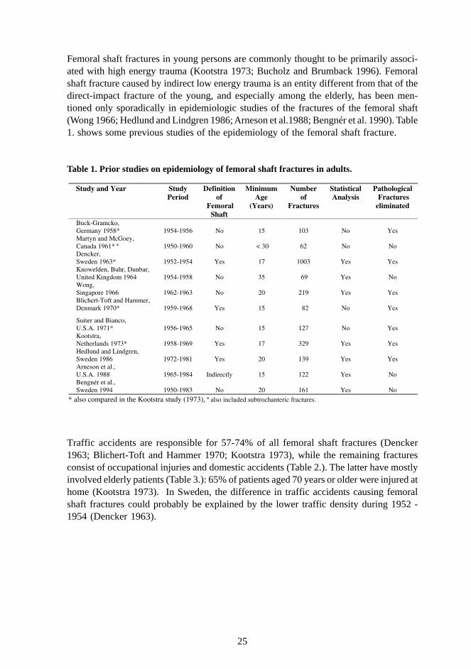
Femoral shaft fractures in young persons are commonly thought to be primarily associ-ated with high energy trauma (Kootstra 1973; Bucholz and Brumback 1996). Femoralshaft fracture caused by indirect low energy trauma is an entity different from that of thedirect-impact fracture of the young, and especially among the elderly, has been men-tioned only sporadically in epidemiologic studies of the fractures of the femoral shaft(Wong 1966; Hedlund and Lindgren 1986; Arneson et al.1988; Bengnér et al. 1990). Table1. shows some previous studies of the epidemiology of the femoral shaft fracture.
Table 1. Prior studies on epidemiology of femoral shaft fractures in adults.
Traffic accidents are responsible for 57-74% of all femoral shaft fractures (Dencker1963; Blichert-Toft and Hammer 1970; Kootstra 1973), while the remaining fracturesconsist of occupational injuries and domestic accidents (Table 2.). The latter have mostlyinvolved elderly patients (Table 3.): 65% of patients aged 70 years or older were injured athome (Kootstra 1973). In Sweden, the difference in traffic accidents causing femoralshaft fractures could probably be explained by the lower traffic density during 1952 -1954 (Dencker 1963).
Study and Year Study
Period Definition
of Femoral
Shaft
Minimum Age
(Years)
Number of
Fractures
Statistical Analysis
Pathological Fractures eliminated
Buck-Gramcko, Germany 1958*
1954-1956
No
15
103
No
Yes
Martyn and McGoey, Canada 1961* ª
1950-1960
No
< 30
62
No
No
Dencker, Sweden 1963*
1952-1954
Yes
17
1003
Yes
Yes
Knowelden, Buhr, Dunbar, United Kingdom 1964
1954-1958
No
35
69
Yes
No
Wong, Singapore 1966
1962-1963
No
20
219
Yes
Yes
Blichert-Toft and Hammer, Denmark 1970*
1959-1968
Yes
15
82
No
Yes
Suiter and Bianco, U.S.A. 1971*
1956-1965
No
15
127
No
Yes
Kootstra, Netherlands 1973*
1958-1969
Yes
17
329
Yes
Yes
Hedlund and Lindgren, Sweden 1986
1972-1981
Yes
20
139
Yes
Yes
Arneson et al., U.S.A. 1988
1965-1984
Indirectly
15
122
Yes
No
Bengnér et al., Sweden 1994
1950-1983
No
20
161
Yes
No
* also compared in the Kootstra study (1973), ª also included subtrochanteric fractures.
26
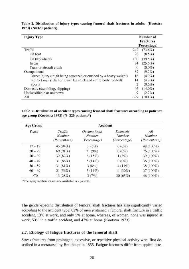
Table 2. Distribution of injury types causing femoral shaft fractures in adults (Kootstra1973) (N=329 patients).
*The injury mechanism was unclassifiable in 9 patients.
Table 3. Distribution of accident types causing femoral shaft fractures according to patient’sage group (Kootstra 1973) (N=320 patients*)
The gender-specific distribution of femoral shaft fractures has also significantly variedaccording to the accident type: 82% of men sustained a femoral shaft fracture in a trafficaccident, 13% at work, and only 5% at home, whereas, of women, none was injured atwork, 53% in a traffic accident, and 47% at home (Kootstra 1973).
2.7. Etiology of fatigue fractures of the femoral shaft
Stress fractures from prolonged, excessive, or repetitive physical activity were first de-scribed in a metatarsal by Breithaupt in 1855. Fatigue fractures differ from typical oste-
Injury Type Number of
Fractures (Percentage)
Traffic 242 (73.6%) On foot 28 (8.5%) On two wheels 130 (39.5%) In car 84 (25.6%) Train or aircraft crash 0 (0.0%) Occupational 32 (9.7%) Direct injury (thigh being squeezed or crushed by a heavy weight) 16 (4.9%)
Indirect injury (fall or lower leg stuck and entire body rotated) 14 (4.2%) Sports 2 (0.6%) Domestic (stumbling, slipping) 46 (14.0%) Unclassifiable or unknown 9 (2.7%) All 329 (100 %)
Age Group Accident
Years Traffic Occupational Domestic All Number
(Percentage) Number
(Percentage) Number
(Percentage) Number
(Percentage)
17 – 19 45 (94%) 3 (6%) 0 (0%) 48 (100%) 20 – 29 69 (91%) 7 (9%) 0 (0%) 76 (100%) 30 – 39 32 (82%) 6 (15%) 1 (3%) 39 (100%) 40 – 49 31 (86%) 5 (14%) 0 (0%) 36 (100%) 50 – 59 31 (81%) 3 (8%) 4 (11%) 38 (100%) 60 – 69 21 (56%) 5 (14%) 11 (30%) 37 (100%)
70 13 (28%) 3 (7%) 30 (65%) 46 (100%) h i j h i l ifi bl i i
27
oporotic fractures that are due to low bone density (Cummings and Melton 2002). Stressfractures of the bones are divided into two general types: a) fractures induced by cyclicalloading of normal bones with abnormal forces that are fatigue fractures, and b) thoseinduced by normal forces in abnormal bone that are insufficiency fractures (Pentecost,Murray, Brindley 1964; Daffner and Pavlov 1992; Anderson and Greenspan 1996). Insuf-ficiency fractures are seen in elderly women suffering from osteoporosis, in cases ofPaget’s disease, hyperparathyroidism, rheumatoid arthritis, diabetes, scurvy, osteomala-cia, osteogenesis imperfecta, or rickets (Pentecost, Murray, Brindley 1964; Markey 1987).Fatigue failure is a rare (Provost and Morris 1969) but increasing cause of femoral shaftfractures (Bucholz and Brumback 1996; Boden and Speer 1997). Displacement of thesestress injuries can occasionally occur (Bucholz and Brumback 1996).
The stress-fracture pathogenesis is multifactorial, it can be either intrinsic or extrinsic.Extrinsic causes have been a) training errors (excessive volume, excessive intensity;magnitude, duration and change of each strain cycle; excessive muscle fatigue, inade-quate recovery or faulty technique), and b) impact attenuation (training surfaces andconditions, footwear and other equipment).
Intrinsic factors include 1) gait mechanics related to lower extremity alignment, b) mus-cle imbalance, 2) muscle weakness, 3) lack of flexibility due to generalized muscle tight-ness, focal areas of muscle thickening, or restricted joint range of motion, 4) decreasedbone strength due to low bone mineral density (Carter et al. 1981), 5) small bone archi-tecture caused by diet and nutrition, genetics, endocrine status and hormones, exercise,or bone disease (Giladi et al. 1987a; Giladi et al. 1987b), 6) gender, 7) high body massindex (BMI), 8) body composition (Brukner and Khan 1993), 9) bone turnover includinglow bone density, elevated bone strain, and inadequate repair of microdamage, 10) lowcalcium intake associated with greater rate of bone turnover, low bone density, or inade-quate repair of microdamage, 11) caloric intake and eating disorders causing altered bodycomposition, low bone density, greater rate of bone turnover, reduced calcium absorp-tion, menstrual disturbances, and inadequate repair of microdamage, 12) hormonal fac-tors such as sex hormones, menarcheal age, menstrual disturbances, 13) genetic predis-position (low bone density, greater rate of bone remodeling, psychological traits), and 14)even psychological factors such as excessive training, nutritional intake, or eating disor-ders (Brukner, Bennell, Matheson 1999).
The structural geometry varies more than bone material properties including bone mineraldensity (Martens et al. 1981). In a prospective observational cohort study of 295 maleIsraeli military conscripts, femoral shaft stress fractures were seen more in conscriptswho had narrower tibias, a feature that may be an indication of the size of tubular bonesin general (Giladi et al. 1987b). The cross-sectional moment of inertia about the antero-posterior axis has proven to be a better indicator than the tibial width (Milgrom et al.1988). In another study, conscripts with stress fractures had a smaller tibial width, asmaller cross-sectional area, smaller moment of inertia, and a smaller modulus (Beck etal. 1996). The smaller cross-sectional dimensions were apparent in the long-bone dia-physes, not in the joint size, which suggests a specificity of the structural deficit in thefracture group and that these dimensions are influenced environmentally (Beck et al.
28
1996). This could indicate that the stress fracture group’s bones had not been sufficientlyloaded before basic training in order to develop cortices strong enough to withstand thesubsequent stresses.
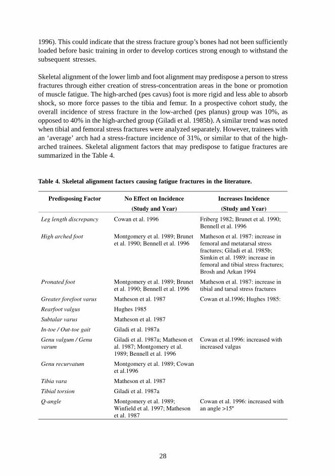
Skeletal alignment of the lower limb and foot alignment may predispose a person to stressfractures through either creation of stress-concentration areas in the bone or promotionof muscle fatigue. The high-arched (pes cavus) foot is more rigid and less able to absorbshock, so more force passes to the tibia and femur. In a prospective cohort study, theoverall incidence of stress fracture in the low-arched (pes planus) group was 10%, asopposed to 40% in the high-arched group (Giladi et al. 1985b). A similar trend was notedwhen tibial and femoral stress fractures were analyzed separately. However, trainees withan ‘average’ arch had a stress-fracture incidence of 31%, or similar to that of the high-arched trainees. Skeletal alignment factors that may predispose to fatigue fractures aresummarized in the Table 4.
Table 4. Skeletal alignment factors causing fatigue fractures in the literature.
Predisposing Factor No Effect on Incidence
(Study and Year)
Increases Incidence
(Study and Year)
Leg length discrepancy Cowan et al. 1996 Friberg 1982; Brunet et al. 1990; Bennell et al. 1996
High arched foot Montgomery et al. 1989; Brunet et al. 1990; Bennell et al. 1996
Matheson et al. 1987: increase in femoral and metatarsal stress fractures; Giladi et al. 1985b; Simkin et al. 1989: increase in femoral and tibial stress fractures; Brosh and Arkan 1994
Pronated foot Montgomery et al. 1989; Brunet et al. 1990; Bennell et al. 1996
Matheson et al. 1987: increase in tibial and tarsal stress fractures
Greater forefoot varus Matheson et al. 1987 Cowan et al.1996; Hughes 1985:
Rearfoot valgus Hughes 1985
Subtalar varus Matheson et al. 1987
In-toe / Out-toe gait Giladi et al. 1987a
Genu valgum / Genu varum
Giladi et al. 1987a; Matheson et al. 1987; Montgomery et al. 1989; Bennell et al. 1996
Cowan et al.1996: increased with increased valgus
Genu recurvatum Montgomery et al. 1989; Cowan et al.1996
Tibia vara Matheson et al. 1987
Tibial torsion Giladi et al. 1987a
Q-angle Montgomery et al. 1989; Winfield et al. 1997; Matheson et al. 1987
Cowan et al. 1996: increased with an angle >15º
29
2.8. Demography and incidence of femoral shaft fractures in adults
Femoral shaft fractures are commonly thought to be primarily associated with severetrauma in young persons. A femoral shaft fracture caused by indirect low energy traumais an entity different from that of the direct-impact fracture in the young. Low energyviolence as a cause of these fractures, especially among the elderly (Wong 1966; Hedlundand Lindgren 1986; Arneson et al. 1988; Bengnér et al. 1990). Although the incidence offractures of the shaft of the femur in the elderly is considerably lower than that of manyother fractures among aged persons (Knowelden, Buhr, Dunbar 1964), the number ofsenior citizens is increasing, and the clinicians will be more often confronted with thespecific problems associated with these fractures.
Fatigue or stress fractures mainly located in the proximal or midshaft areas usually occurin military conscripts undergoing a marked and prolonged increase in physical activity(Giladi et al. 1985a; Bucholz and Brumback 1996). The incidence of stress fractures ofthe femoral shaft in civilian population appears to be rising along with the recent emphasison physical fitness. Running accounts for most such fractures (McBryde 1975), whichhave been encountered after triathlon events and aerobic dancing as well (Clement et al.1993).
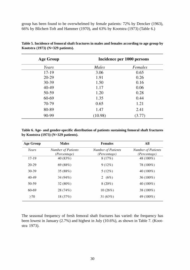
Dencker studied 1003 recent femoral shaft fractures in adults in 992 patients treated atpublic hospitals in Sweden during a three-year period of 1952 to 1954, with a follow-upof 4 to 8 years. Kootstra studied 335 traumatic femoral shaft fractures in 329 adults in theprovince of Groningen in the Netherlands during 12 years from 1958 to 1969 (Kootstra1973). Pathological fractures caused by primary and metastatic tumors were excluded inboth studies (Dencker 1963; Kootstra 1973). The frequencies of femoral shaft fracturesin adults were 334 fractures per year (Dencker 1963), and 17-36 fractures per year(Kootstra 1973). The ratio of men to women in Dencker’s series was 2.7:1, but therelation varied widely at different ages. In the Kootstra study (1973), 22.2% of the pa-tients were females (N=73), compared with 21.5% - 27% in earlier studies (Dencker1963; Blichert-Toft and Hammer 1970). The risk of sustaining a femoral shaft fracturewas greatest in men between the ages of 20 and 29 years, while in women it was greatestbetween 80 and 89 years (Dencker 1963; Kootstra 1973). In Dencker’s study, the distri-bution was fairly even in the male groups 30 to 39, 40 to 49, and 50 to 59 years, rangingbetween 86 and 122. In the female age groups 20 to 29, 30 to 39, 40 to 49 and 50 to 59years, the distribution was even, between 20 and 27. The number of fractures increasedin women over the age of 60 (Dencker 1963). In patients of 60 years or over, fractureswere twice as common in women as in men, 159 against 80. According to the study byKootstra (1973), femoral shaft fractures most frequently occurred at an early age inmales (Tables 5. and 6.). The incidence gradually diminished with increasing age, andattained its lowest value in age group 70-79, after which another increase occurred (Table5.). The marked increase in femoral shaft fracture incidence in females after 50 can onlybe explained on the basis of a constitutional factor, i.e. the hormonal change during andafter the menopause, which is associated with osteoporosis and increased fragility of thefemoral shaft. The percentage of patients aged 70 or older and sustaining a femoral shaftfracture was 14.9%-15% (Dencker 1963; Blichert-Toft and Hammer 1970). This age
30
group has been found to be overwhelmed by female patients: 72% by Dencker (1963),66% by Blichert-Toft and Hammer (1970), and 63% by Kootstra (1973) (Table 6.)
Table 5. Incidence of femoral shaft fractures in males and females according to age group byKootstra (1973) (N=329 patients).
Table 6. Age- and gender-specific distribution of patients sustaining femoral shaft fracturesby Kootstra (1973) (N=329 patients).
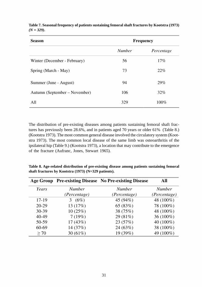
The seasonal frequency of fresh femoral shaft fractures has varied: the frequency hasbeen lowest in January (2.7%) and highest in July (10.6%), as shown in Table 7. (Koot-stra 1973).
Age Group Incidence per 1000 persons
Years Males Females 17-19 3.06 0.65 20-29 1.91 0.26 30-39 1.50 0.16 40-49 1.17 0.06 50-59 1.20 0.28 60-69 1.35 0.44 70-79 0.65 1.21 80-89 1.47 2.41 90-99 (10.98) (3.77)
Age Group Males Females All
Years Number of Patients (Percentage)
Number of Patients (Percentage)
Number of Patients (Percentage)
17-19 40 (83%) 8 (17%) 48 (100%)
20-29 69 (88%) 9 (12%) 78 (100%)
30-39 35 (88%) 5 (12%) 40 (100%)
40-49 34 (94%) 2 (6%) 36 (100%)
50-59 32 (80%) 8 (20%) 40 (100%)
60-69 28 (74%) 10 (26%) 38 (100%)
70 18 (37%) 31 (63%) 49 (100%)
31
Table 7. Seasonal frequency of patients sustaining femoral shaft fractures by Kootstra (1973)(N = 329).
The distribution of pre-existing diseases among patients sustaining femoral shaft frac-tures has previously been 28.6%, and in patients aged 70 years or older 61% (Table 8.)(Kootstra 1973). The most common general disease involved the circulatory system (Koot-stra 1973). The most common local disease of the same limb was osteoarthritis of theipsilateral hip (Table 9.) (Kootstra 1973), a location that may contribute to the emergenceof the fracture (Aufranc, Jones, Stewart 1965).
Table 8. Age-related distribution of pre-existing disease among patients sustaining femoralshaft fractures by Kootstra (1973) (N=329 patients).
Season Frequency
Number Percentage
Winter (December - February) 56 17%
Spring (March - May) 73 22%
Summer (June - August) 94 29%
Autumn (September – November) 106 32%
All 329 100%
Age Group Pre-existing Disease No Pre-existing Disease All
Years Number (Percentage)
Number (Percentage)
Number (Percentage)
17-19 3 (6%) 45 (94%) 48 (100%) 20-29 13 (17%) 65 (83%) 78 (100%) 30-39 10 (25%) 38 (75%) 48 (100%) 40-49 7 (19%) 29 (81%) 36 (100%) 50-59 17 (43%) 23 (57%) 40 (100%) 60-69 14 (37%) 24 (63%) 38 (100%)
70 30 (61%) 19 (39%) 49 (100%)
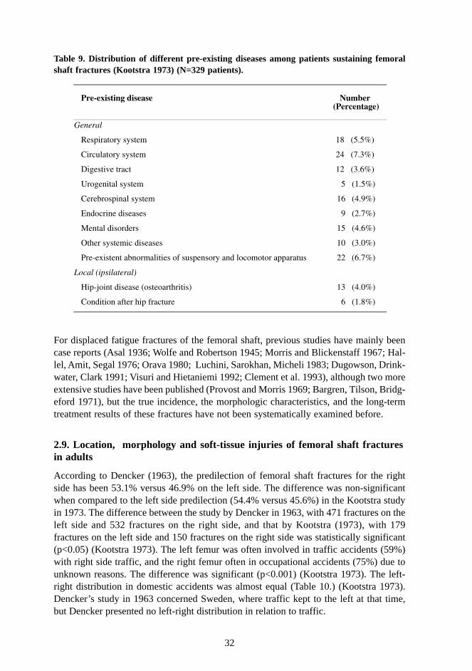
32
Table 9. Distribution of different pre-existing diseases among patients sustaining femoralshaft fractures (Kootstra 1973) (N=329 patients).
For displaced fatigue fractures of the femoral shaft, previous studies have mainly beencase reports (Asal 1936; Wolfe and Robertson 1945; Morris and Blickenstaff 1967; Hal-lel, Amit, Segal 1976; Orava 1980; Luchini, Sarokhan, Micheli 1983; Dugowson, Drink-water, Clark 1991; Visuri and Hietaniemi 1992; Clement et al. 1993), although two moreextensive studies have been published (Provost and Morris 1969; Bargren, Tilson, Bridg-eford 1971), but the true incidence, the morphologic characteristics, and the long-termtreatment results of these fractures have not been systematically examined before.
2.9. Location, morphology and soft-tissue injuries of femoral shaft fracturesin adults
According to Dencker (1963), the predilection of femoral shaft fractures for the rightside has been 53.1% versus 46.9% on the left side. The difference was non-significantwhen compared to the left side predilection (54.4% versus 45.6%) in the Kootstra studyin 1973. The difference between the study by Dencker in 1963, with 471 fractures on theleft side and 532 fractures on the right side, and that by Kootstra (1973), with 179fractures on the left side and 150 fractures on the right side was statistically significant(p<0.05) (Kootstra 1973). The left femur was often involved in traffic accidents (59%)with right side traffic, and the right femur often in occupational accidents (75%) due tounknown reasons. The difference was significant (p<0.001) (Kootstra 1973). The left-right distribution in domestic accidents was almost equal (Table 10.) (Kootstra 1973).Dencker’s study in 1963 concerned Sweden, where traffic kept to the left at that time,but Dencker presented no left-right distribution in relation to traffic.
Pre-existing disease
Number
(Percentage)
General
Respiratory system 18 (5.5%)
Circulatory system 24 (7.3%)
Digestive tract 12 (3.6%)
Urogenital system 5 (1.5%)
Cerebrospinal system 16 (4.9%)
Endocrine diseases 9 (2.7%)
Mental disorders 15 (4.6%)
Other systemic diseases 10 (3.0%)
Pre-existent abnormalities of suspensory and locomotor apparatus 22 (6.7%)
Local (ipsilateral)
Hip-joint disease (osteoarthritis) 13 (4.0%)
Condition after hip fracture 6 (1.8%)
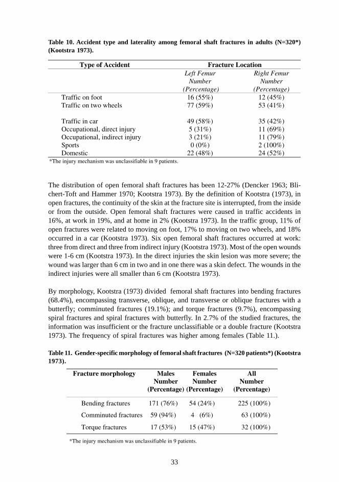
33
Table 10. Accident type and laterality among femoral shaft fractures in adults (N=320*)(Kootstra 1973).
The distribution of open femoral shaft fractures has been 12-27% (Dencker 1963; Bli-chert-Toft and Hammer 1970; Kootstra 1973). By the definition of Kootstra (1973), inopen fractures, the continuity of the skin at the fracture site is interrupted, from the insideor from the outside. Open femoral shaft fractures were caused in traffic accidents in16%, at work in 19%, and at home in 2% (Kootstra 1973). In the traffic group, 11% ofopen fractures were related to moving on foot, 17% to moving on two wheels, and 18%occurred in a car (Kootstra 1973). Six open femoral shaft fractures occurred at work:three from direct and three from indirect injury (Kootstra 1973). Most of the open woundswere 1-6 cm (Kootstra 1973). In the direct injuries the skin lesion was more severe; thewound was larger than 6 cm in two and in one there was a skin defect. The wounds in theindirect injuries were all smaller than 6 cm (Kootstra 1973).
By morphology, Kootstra (1973) divided femoral shaft fractures into bending fractures(68.4%), encompassing transverse, oblique, and transverse or oblique fractures with abutterfly; comminuted fractures (19.1%); and torque fractures (9.7%), encompassingspiral fractures and spiral fractures with butterfly. In 2.7% of the studied fractures, theinformation was insufficient or the fracture unclassifiable or a double fracture (Kootstra1973). The frequency of spiral fractures was higher among females (Table 11.).
Table 11. Gender-specific morphology of femoral shaft fractures (N=320 patients*) (Kootstra1973).
Type of Accident Fracture Location Left Femur
Number (Percentage)
Right Femur Number
(Percentage) Traffic on foot 16 (55%) 12 (45%)
Traffic on two wheels 77 (59%) 53 (41%)
Traffic in car 49 (58%) 35 (42%) Occupational, direct injury 5 (31%) 11 (69%) Occupational, indirect injury 3 (21%) 11 (79%) Sports 0 (0%) 2 (100%) Domestic 22 (48%) 24 (52%)
*The injury mechanism was unclassifiable in 9 patients.
Fracture morphology Males
Number (Percentage)
Females Number
(Percentage)
All Number
(Percentage)
Bending fractures 171 (76%) 54 (24%) 225 (100%)
Comminuted fractures 59 (94%) 4 (6%) 63 (100%)
Torque fractures 17 (53%) 15 (47%) 32 (100%)
*The injury mechanism was unclassifiable in 9 patients.

34
In the Dencker study, the most common fracture location was the middle third of thefemoral shaft (60% of the fractures) (Dencker 1963). In most cases the fracture wastransverse (32% of the fractures), which was more common fracture type (39%) in menaged 50 years or younger. Long oblique fractures were more frequent in elderly womenaged 70 years or older (55%). Different morphologic types of femoral shaft fractures areshown in Figure 1.
Figure 1. Different morphologic types of femoral shaft fractures:A. A simple transverse AO Type 32-A3.2 fracture with minimal comminution (Winquist-HansenGrade I).B. A simple oblique-transverse fracture without comminution (Winquist-Hansen Grade 0) of afemur that had a distal femoral fracture treated with a plate 16 years earlier. The AO Typedepends on the size of the transverse or oblique component of the fracture.C. A simple oblique AO Type 32-A2.2 fracture (Winquist-Hansen Grade 0).D. A slightly comminuted (Winquist-Hansen Grade I) spiral AO Type 32-A1.2 fracture.E. A spiral AO Type 32-B1.2 fracture with a bending wedge (butterfly fragment) (Winquist-Hansen Grade III).F. A comminuted irregular complex AO Type 32-C3.1 fracture of Winquist-Hansen Grade IV.G. A segmental, AO Type 32-C2.1 fracture.H. An oblique fracture of the middle third of the femoral diaphysis with minimal comminution(Winquist-Hansen Grade I) and a dislocation of the knee.
35
Torque fractures were related to domestic accidents (75%) and to pre-existing hip-jointdisease (40.6%) (Kootstra 1973). Of the 16 fractures caused by occupational directinjury, 6 fractures were comminuted. Traffic accidents caused 85% of the comminutedfractures, and the majority of these accidents were car crashes (39.2%) (Kootstra 1973).The largest percentage of open fractures was found among the comminuted fractures(32% of open fractures) (Kootstra 1973).
In motor vehicle accidents and auto-pedestrian accidents, a transverse fracture is usuallysustained because of direct impact to the shaft or indirect force transmitted through theknee, subjecting the femur to a high bending load (Bucholz and Brumback 1996). Motorcy-clists are frequently victims of a direct side impact, hence sustaining comminuted fractures(Bucholz and Brumback 1996). Gunshot wounds create spiral or comminuted fractures(Bucholz and Brumback 1996). A small caliber gunshot missile causes a comminuted frac-ture due to a direct impact of the bullet in the tubular bone of the diaphysis. In the metaphy-seal bone it causes a drill hole fracture in the distal femoral metaphysis secondary to a directimpact of the missile on the cancellous bone (Smith and Wheatley 1984).
2.10. Concomitant injuries associated with femoral shaft fractures
Concomitant injuries related to femoral shaft fractures include ipsilateral injuries of thesame lower extremity and other isolated injuries, as well as polytrauma. According toDencker’s study the prevalence of concomitant injuries has been 23% in 1963, and 45%by Kootstra in 1973.
The prevalence of general associated injuries has been 45% (Kootstra 1973) includingshock (of which 24% were related to traffic accidents). General associated injuries wererelated to traffic accidents in 57%, occupational accidents in 31%, or domestic accidentsin 4% (Kootstra 1973). A general associated injury was found in 77% of the patients whosustained a femoral shaft fracture while seated in car (Kootstra 1973). Overall, accidentssustained while seated in car comprised between 7.9% (Dencker 1963) and 25.5% (Koot-stra 1973).
Craniocerebral injury has varied from 2.7% (Dencker 1963) to 10.9% (Kootstra 1973).Facial fractures have occurred in 12.1% of cases (Dencker 1963; Kootstra 1973). Otherreported concomitant injuries are: injury in the humeroscapular area 20.0%, thoracicinjury without respiratory disturbances 6.7%, thoracic injury with respiratory distur-bances 3.6%, vertebral fracture 1.2%, upper arm or elbow injury 5.8%, forearm or handinjury 9.7%, contralateral femoral shaft fracture 1.8%, contralateral lower leg fracturefrom 1.4% (Dencker 1963) to 3.3% (Kootstra 1973) of the cases, supra- or intracondy-lar femoral fracture 1.2%, ankle or foot injury 2.4% or other injury 3.3% includingruptured auricle of the heart, ascending aorta, spleen, liver, bladder or renal contusion(Kootstra 1973).
Local injury associated with femoral shaft fracture is found in about every third case inthe traffic accident and indirect occupational injury groups. No local associated injurywas found in the direct occupational injury and domestic groups (Kootstra 1973).
36
Recognized injury of the superficial femoral artery occurs in less than 2% of patients withfracture of the femoral shaft (Isaacson, Louis, Costenbader 1975; Barr, Santer, Steven-son 1987; Kluger et al. 1994). Middiaphyseal femoral shaft fractures can be associatedwith a false aneurysm of the superficial femoral artery as an intimal flap or a pseudoaneu-rysm (Guemes et al. 1991; Kluger et al. 1994). A diagnostic arteriogram should be per-formed, followed by therapeutic embolization (Levade et al. 1983). The profunda femorisartery can be damaged in intramedullary nailing (Barnes and Broude 1985) for exampleduring the placement of the anteroposterior proximal locking screw in retrograde nailing(Coupe and Beaver 2001).
Hip dislocation accounts for 0.3%, acetabular fracture 1.5%, and femoral neck or pertro-chanteric fracture 3.4% of the femoral shaft fractures (Kootstra 1973). Ipsilateral pelvisand femoral shaft fractures form a floating hip (Wu and Shih 1993; Müller et al. 1999)and are often associated to chest trauma, other skeletal fractures or polytrauma (Müller etal. 1999). The incidence has varied from 6.7% (Kootstra 1973) to 13%-22% (Müller etal 1999). The pelvic fracture is mostly transverse or comprises posterior column or wall(Müller et al. 1999).
Femoral shaft fractures combined with ipsilateral femoral neck fractures were first de-scribed by Delaney and Street (1953). Hip fractures are detected in 1.2% (Kootstra 1973)to 2.5%-5% of patients with femoral shaft fractures (Ashby and Anderson 1977; Caseyand Chapman 1979; Swiontkowski, Hansen, Kellam 1984; Friedman and Wyman 1986;Swiontkowski 1987; Wiss, Sima, Brien 1992; Leung 2002) and are quite often missed inpolytrauma patients with femoral shaft fractures (Ostrum, Verghese, Santner 1993). Al-though about 1/4 to 1/3 of primary neck fractures are initially detected after stabilizationof the shaft fracture (Bennett, Zinar, Kilgus 1993; Riemer et al. 1993), some of the frac-tures have occurred during intramedullary nailing (Harper and Henstorf 1986; Harper andCarson 1987). Diaphyseal fracture is usually comminuted, as the trauma energy dissi-pates through shaft fracture (Wu, Shih, Chen 1993; Alho 1997; Müller et al. 1999). Hipfracture remains relatively undisplaced or minimally displaced (Bennett, Zinar, Kilgus 1993)and usually represents the Pauwels grade II type with a vertical shear (Wiss, Sima, Brien1992). The ratio between concomitant femoral neck fractures and concomitant intertro-chanteric fractures is 7:1 (Bennett, Zinar, Kilgus 1993). The femoral neck fracture is notdevoid of nonunion or malunion (Bucholz and Rathjen 1985; Bose, Corces, Anderson1992; Wiss, Sima, Brien 1992; Bennett, Zinar, Kilgus 1993).
The incidence of fracture of the ipsilateral lower leg has varied from 0.5% (Dencker1963) to 10.9% (Kootstra 1973). Ipsilateral femoral shaft and tibial shaft fractures causea Type 1 floating knee (Blake and McBryde 1975). The classification by Letts has fivesubgroups depending on whether the fractures are closed and diaphyseal (A); closed, butthe other fracture is metaphyseal (B); the other is intra-articular (C); the other is open(D); or both the fractures are open (E) (Letts, Vincent, Gouw 1986). Other classifica-tions also exist (Fraser, Hunter, Waddell 1978). Besides being caused by a high energytrauma with extensive skeletal and soft-tissue damage, floating knee is also associatedwith potentially life-threatening injuries of the head, chest and abdomen (Veith, Winquist,
37
Hansen 1984). Knee dislocation (Chen and Wang 1998) or ligament injury (Szalay, Hosk-ing, Annear 1990; van Raay, Raaymakers, Dupree 1991) can also associate with thefloating knee.
The incidence of arthroscopically verified concomitant meniscal injuries associated withipsilateral femoral shaft fractures in adults has varied from 27% (Barber et al. 1988;Vangsness et al. 1993; Blacksin, Zurlo, Levy 1998) to 38% (DeCampos et al. 1994). Inarthroscopies, lateral meniscus has accounted for 17% and medial meniscus for 11% ofthe meniscal tears which mostly are complex or radial (Vangsness et al. 1993). Accordingto a MRI study in 34 patients with closed femoral shaft fractures, the posterior horn ofthe medial meniscus is most frequently torn (Blacksin, Zurlo, Levy 1998).
Ipsilateral injury to knee ligaments has been described in 17% to 48% of femoral shaftfractures in studies that have included 30-50 patients (Ritchey, Schonholtz, Thompson1958; Shelton, Neer, Grantham 1971; Hughston et al. 1976; Nagel, Burton, Manning1977; Dunbar and Coleman 1978; Fraser, Hunter, Waddell 1978; Noyes et al. 1980; Vianoand Stalnaker 1980; Walker and Kennedy 1980; Rowntree and Getty 1981; Walker andStein 1982; Barber et al. 1988; McAndrew and Pontarelli 1988; Szalay, Hosking, Annear1990; Dickob and Mommsen 1992; DeCampos et al. 1994; Blacksin, Zurlo, Levy 1998).In a larger study including 309 patients the incidence of serious (Grade II and III) kneeligament injury was 5.3% (Moore, Patzakis, Harvey 1988). Partial ACL injury has ac-counted for 48% and total ACL injury for 5% of 40 arthroscopically examined kneesassociated with a closed femoral shaft fracture and without any previous knee injury(DeCampos et al. 1994). According to a MRI study, partial ACL tears were diagnosed in6% of all 34 knees, while total ACL ruptures were not discovered at all (Blacksin, Zurlo,Levy 1998). The medial collateral ligament was the most frequent site of ligamentousinjury (38%) followed by the posterior cruciate ligament (21%) verified by MRI (Black-sin, Zurlo, Levy 1998). Injury of MCL has been identified arthroscopically in 15% ofcases (Barber et al. 1988) and clinically in 31% of cases (Walker and Kennedy 1980).The incidence of arthroscopically identified concomitant PCL injury has varied from 7.5%(DeCampos et al. 1994) to 15% (Barber et al. 1988). The proportion of LCL injuries hasvaried from 0% (Barber et al. 1988) to 6% (Blacksin, Zurlo, Levy 1998) and 13% (Walkerand Kennedy 1980). Reports of collateral ligament injuries associated with femoral shaftfractures have been infrequent (Pedersen and Serra 1968).
Other ipsilateral knee injuries associated to a closed femoral shaft fracture and demos-trated with MRI have been knee effusion (97%), injuries of the extensor mechanism(50%) including mainly abnormal signals of the patellar tendon in 41% and tears of patel-lar or quadriceps tendons in 9% , bone bruises (22%), articular changes of the patella(12%), ipsilateral fracture of the patella from 1.7% (Dencker 1963) to 5.8% (Kootstra1973), fracture of the tibial plateau (3%), and meniscocapsular separation (3%) (Black-sin, Zurlo, Levy 1998). Articular changes or chondromalacia of the ipsilateral knee havebeen discovered in 50-53% of patients with femoral shaft fractures (Dunbar and Cole-man 1978; Vangsness et al. 1993).
38
2.11. Diagnosis of femoral shaft fractures in adults
2.11.1. Clinical course
The clinical diagnosis of the fracture of the femoral shaft is usually obvious, with pain,deformity, swelling, and shortening of the thigh (Bucholz and Brumback 1996). A thor-ough physical examination is imperative, because most fractures are a result of highspeed trauma and associated injuries are common. Orthopaedic assessment of the entirelimb should be systematic and complete. The pelvic ring and the hip are inspected fortenderness, swelling or ecchymosis may signal concomitant pelvic disruption or hip frac-ture. Since the hip cannot be moved voluntarily by the patient, palpation of the groin andbuttock is important (Chaturvedi and Sahu 1993; Riemer et al. 1993). Fullness of thebuttock with flexion and adduction of the proximal femur can denote a posterior disloca-tion of the hip (Bucholz and Brumback 1996).
The ipsilateral knee should be carefully examined before the application of skeletal trac-tion and again after intramedullary nailing due to a high incidence of ipsilateral knee injuryin patients with femoral shaft fractures (Vangsness et al. 1993). Knee ligament injurieshave been identified even in patients with an operated floating knee (Szalay, Hosking,Annear 1990; van Raay, Raaymakers, Dupree 1991). Routine arthroscopy of the ipsilater-al knee has been recommended in several studies (Dickob and Mommsen 1992; De Cam-pos et al. 1994). However, the clinical significance of concomitant complex and radialmeniscal tears remains unclear, and routine arthroscopy of the knees of all patients withfemoral fractures is unjustified (Bucholz and Brumback 1996).
Although neurovascular injuries are rarely associated with closed shaft fractures, a com-plete preoperative examination for vascular and neurologic damage is mandatory (Bu-cholz and Brumback 1996). Owing to severe pain and spasm accompanying femoralfractures, the motor strength of muscles below the knee may be diminished. A carefulpreoperative assessment of the hemodynamic stability of the patient is necessary, regard-less of the presence or absence of associated injuries (Bucholz and Brumback 1996).Distal pulses should be palpated and circulatory status evaluated (Bucholz and Brumback1996). Angiography is indicated in injuries associated with at least one “hard” symptom(pulse or neurological ischaemic deficiency) or at least two “soft” symptoms (haemato-ma, haemorrhage, hypotension, malleobrachial index smaller than 1) (Rezek 2002). Al-though few patients with isolated fractures of the femoral shaft are in hypovolemic shock(Ostrum, Verghese, Santner 1993), major blood loss more than 1200 ml into the thigh ispresent in most cases (Lieurance, Benjamin, Rappaport 1992).
In fatigue fractures, load–related pain is typical. Most runners report an increase in theirtraining during or immediately before the onset of pain, which during the first two weeksbecomes more strenuous after the training, but it may be delayed by several months(Greaney et al. 1983; Jones et al. 1989; Maitra and Johnson 1997). Symptoms, such aslimping, local pain, tenderness, swelling or pitting edema of the surrounding soft-tissues,are related to fatigue fractures (Wilson and Katz 1969; Daffner 1978; Markey 1987;Sterling et al. 1992). Local tenderness can be detected by tapping the affected bone. Toincrease the accuracy of diagnosis, several tests as a fulcrum test (Johnson, Weiss,
39
Wheeler 1994), a fist test (Milgrom et al. 1993), a hop test (Clement et al. 1993) havebeen introduced. The clinical diagnosis is still difficult and nonspecific (Sallis and Jones1991). In displaced fractures, symptoms and clinical findings resemble those of a con-ventional bone fracture.
2.11.2. Radiographic findings
Before diagnostic radiographic studies of traumatic femoral shaft fractures are performed,longitudinal traction or splinting of the extremity is applied to ensure minimal additionalsoft-tissue injury to the thigh (Bucholz and Brumback 1996). If a Thomas splint is used,the metallic ring and caliber should not obscure any part of the fracture. Nondisplacedfractures of the femoral neck are frequently missed because of the overlying shadow ofa splint or inadequate quality of the preoperative radiograph (Bucholz and Brumback1996). If the hip is externally rotated on the preoperative radiograph, there can be arotational artifact (Bucholz and Brumback 1996). In such cases, it is advisable to obtainan anteroposterior radiograph of the hip with the proximal femur internally rotated in theanesthetized patient before the operative procedure (Bucholz and Brumback 1996). Initialradiographs should include an AP view of the pelvis and AP and lateral views of the kneeand the entire femur to allow detection of longitudinal cracks and nondisplaced comminu-tion of the proximal and distal fragments (Bucholz and Brumback 1996). Lateral andoblique radiographs are recommended to rule out fractures of the femoral condyles in thecoronal plane (the Hoffa fracture) (Bucholz and Brumback 1996).
In fatigue fractures radiographic changes can be seen 2-12 weeks after the onset ofsymptoms (Sullivan et al. 1984; Matheson et al. 1987). The sensivity of radiographs is15-35% in the early stage (Daffner 1978; Greaney et al. 1983). Fatigue fractures are seenin 30-70% of cases only during the follow-up (Daffner 1978; Matheson et al. 1987). Incortical bone, a grey-looking hypodensic area seen in compact bone is related to theresorption phase of remodeling (Daffner 1978; Mulligan 1995). After that periosteal newbone forms and endosteal bone thickens (Daffner 1978; Martin and Burr 1982; Greaneyet al. 1983). A fracture, if present, can be seen as a radiolucent line in compact corticalbone and as a sclerotic line in cancellous bone (Daffner 1978; Mulligan 1995).
In most cases CT is less sensitive than conventional radiography for the detection offatigue injuries of bone. Certain fracture lines, such as longitudinal and spiral ones, can beseen more clearly with CT (Daffner 1978; Matheson et al. 1987).
Scintigraphy has long been considered to be the best diagnostic method for bone fatigueinjuries (Daffner 1978) with a sensitivity close to 100% (Stafford e al 1986; Matheson et al.1987). The scintigraphic classification according to Jones et al. (1989) has four differentgrades for fatigue fractures. False negative diagnoses have been reported, possibly becausenecrotic bone tissue is not labeled (Kanstrup 1997). An increase in uptake can be detectedas early as 6-72 hours after the onset of symptoms (Greaney et al. 1983; Matheson et al.1987; Kanstrup 1997). An oval signal, the intensity of which correlates the severity, can beseen with scintigraphy (Floyd et al 1987). Asymptomatic mild stress injuries are also visiblewith scintigraphy as clusters of increased uptake (Daffner 1978; Matheson et al. 1987).
40
MRI with specific classifications for fatigue fractures (Fredericson et al.1995; Yao et al.1998; Kiuru in 2002) is as sensitive but more specific than scintigraphy (Daffner 1978;Stafford et al. 1986; Lee and Yao 1988; Meyers and Weiner 1991; Kiuru 2002). STIR(short tau inversion recovery)- and T2 (transverse relaxation)- techniques can detectnon-specific edema (Daffner 1978; Stafford et al. 1986; Kiuru 2002), in the middle ofwhich a lower signal intensity is a sign of a fracture (Daffner 1978; Stafford et al. 1986).MRI is most accurate during the first 3 weeks (Daffner 1978; Stafford et al. 1986;Martin, Healey, Horowitz 1993). In case of uncertainty one can repeat the radiographicand MRI examinations after 3-4 weeks (Lassus et al. 2002).
2.12. Treatment of femoral shaft fractures in adults
2.12.1. Development of conservative treatment of femoral shaft fractures
The history of femoral fracture management reflects the difficulties of maintaining theanatomic alignment while encouraging early functional rehabilitation of the limb (Kesslerand Schweiberer 1989; Bucholz and Brumback 1996). Hippocrates (460-377 B.C.) treat-ed femoral shaft fractures mainly with manual reduction (Rutkow 1993). Moreover, heemphasized the importance of maintaining adequate extension in order to avoid femoralshortening (van Loon 1935; Kootstra 1973). The treatment methods have evolved overtime from the use of wooden splints in ancient civilizations (Bucholz and Brumback1996) to fabrics encased with wax (Bucholz and Brumback 1996), bandages stiffenedwith clay (Kootstra 1973) or gum (Bucholz and Brumback 1996), and fabrics hardenedwith plaster of Paris (Mathijsen 1854; Bucholz and Brumback 1996). Mechanical inade-quacies of traditional treatments to maintain the proper fracture alignment were demon-strated in the era of the skeletal radiography at the end of the 19th century (Bucholz andBrumback 1996). Charnley emphasized that if sound bony union can be secured in threemonths with conservative treatment, full recovery of the femoral shaft fracture will takeabout one year, and that in some cases early knee movement may be responsible forfibrous union (Charnley 1974). Böhler improved the systematic management and con-servative treatment of fractures (Böhler 1957). At present, femoral shaft fractures inadults are usually not treated conservatively except in underdeveloped countries, in catas-trophe medicine or war surgery (Dufour et al. 1988; Coupland 1991; Coupland 2000).
2.12.2. Traction
The skin traction method was first introduced by de Chauliac in the 14th century (vanLoon 1935; Bucholz and Brumback 1996) and modified in the 19th century by Buck (Buck1861). The major disadvantage of skin traction is the lack of means to apply sufficientforces to the limb to obtain fracture reduction (Bucholz and Brumback 1996) and to avoidpossible skin necroses. Nowadays, traction has been used as a time-consuming primarytreatment method mainly in femoral shaft fractures among children, performed as skin orskeletal traction with a percutaneous pin in the tibia or femur (Bucholz and Brumback1996).
41
Skeletal traction techniques were introduced by Steinmann in 1907 (Steinmann 1907) andby Kirschner in 1909 (Dencker 1963; Mays and Neufeld 1974; Bucholz and Brumback1996). The Thomas splint improved control of the traction forces in providing counter-action on the leg through its ring, and was used since World War I (Bucholz and Brum-back 1996). Various skeletal tractions (Thomas splint with Pearson knee-flexion piece,Braun frame, Russell traction, Perkins traction, Fisk traction, 90º-90º-traction) represent-ed the most common method of definitive treatment of femoral shaft fractures for dec-ades before the 1970s (Charnley 1974; Bucholz and Brumback 1996). The distal femurhas been successfully used for skeletal traction, as it offers a more direct longitudinal pullof the fractured femur than the proximal tibia in similar traction. Skeletal traction throughthe distal femur has associated with a higher rate of knee stiffness after fracture uniondue to, undoubtedly, scarring of the vastus medialis and vastus lateralis (Bucholz andBrumback 1996). Most skeletal traction methods require that the patient remains in thesupine position (Bucholz and Brumback 1996). The goal of skeletal traction is at first tomaintain the normal length of the limb. After 24 hours from injury, hematoma begins todevelop, and pulling the fracture to its normal length requires an increasing amount oftraction (Bucholz and Brumback 1996). With the traction, it is impossible to diminish thetraction force alone without jeopardizing the reduction stability. Gravity can not be usedto help in correcting the deformity of backward angulation, which should be corrected bytraction in the axis of the tibia (Charnley 1974). Traction of the proximal tibia is contrain-dicated in concomitant ipsilateral knee injuries (Bucholz and Brumback 1996). Trans-verse fractures are more difficult to treat with traction than oblique ones (Charnley 1974).Skeletal traction therapy was continued for a patient until significant radiographic andclinical evidence of fracture union was apparent, which usually required a minimum of 6weeks of in-hospital care (Bucholz and Brumback 1996). The aftertreatment consisted ofprogressive weightbearing with an unilateral weightbearing spica cast, (Bucholz and Brum-back 1996). The spica cast was removed 3 to 6 months after injury, and range-of-motionexercises of the ipsilateral hip and knee were started (Bucholz and Brumback 1996). TheNeufeld traction (Mays and Neufeld 1974) encases the limb and the Steinmann pin withplaster casts, while traction is applied through a roller system that permits greater earlyknee motion. The rotation of the foot can be maintained by the position of a cross-barfixed to the sole of the plaster traction unit (Charnley 1974). The 90º-90º-traction methodis not recommended for elderly patients for circulatory reasons (Miller and Welch 1978;Bucholz and Brumback 1996) and knee subluxation (Miller and Welch 1978). The resultsof traction therapy for fractures of the femoral shaft have been satisfactory (Dencker1963; Moulton, Agunwa, Hopkins 1981). Nowadays skeletal traction is used as a supple-mentary treatment method of femoral shaft fractures in adults.
2.12.3. Cast bracing
The development of cast brace systems (Mooney et al. 1970; Mooney 1974; Lesin,Mooney, Ashby 1977; Meggitt, Juett, Smith 1981; Mooney and Claudi 1984) providesambulatory treatment and prevented the hip and knee contractures that were frequentsequelae of the hip spica cast (Bucholz and Brumback 1996). In cast bracing of femo-ral shaft fractures shortening and angulation were frequent particularly in the proximaland midfemur (Sarmiento 1972). The cast brace permitted progressive weightbearing,
42
leading to gradual functional improvement in the muscles and joints and to increasingskeletal stresses that stimulate fracture healing (Bucholz and Brumback 1996). Thefracture itself controlled the loading in a cast brace of a smooth, total contact plaster ora plastic thigh cuff (Meggitt, Juett, Smith 1981) which carried 10% to 20% of the bodyweight, and resisted lateral angulation produced by the thigh musculature (Bucholz andBrumback 1996). The method was equally suitable for open fractures, fractures of thedistal third, or comminuted midshaft fractures (Connolly and King 1973; Montgomeryand Mooney 1981). Proximal shaft fractures, and simple transverse or oblique frac-tures were less amenable to cast bracing because of their high stress concentration andtendency toward angulation. Further, cast bracing was supplementary to a limited in-ternal fixation with small-diameter nonlocked intramedullary nails (Sharma et al. 1993).Cast brace was used to substitute the hip spica casting after 6 to 8 weeks as traction oras a primary treatment after 1 to 2 weeks of traction. Serial radiographs were impera-tive to check the alignment and judge the advisability of early weightbearing (Bucholzand Brumback 1996).
2.12.4. Development of operative treatment of femoral shaft fractures
Early attempts of internal fixation of femoral fractures were complicated by infectionsand implant failures (Bucholz and Brumback 1996). Lane introduced plates and screws in1905, Lambotte the fixateur externe in 1907, and Hey Groves in 1918 a massive nail thatwas used in the medullary cavity (Küntscher 1958; Bucholz and Brumback 1996). In1939, Gerhard Küntscher from Kiel, Germany presented his first cases of intramedullaryfixation of a femur using a V-shaped cross-section-designed nail (Küntscher 1940a; Künt-scher 1940b; Küntscher 1967) utilizing the principle of nailing used earlier for femoralneck fractures (Küntscher 1940a). During the World War II, Küntscher served as awartime army surgeon in Kemi, Lapland and introduced the intramedullary nailing tech-nique to Finnish surgeons (Küntscher 1948; Küntscher 1953; Lindholm 1979; Lindholm1980; Lindholm 1982; Seligson 2001). The first known Küntscher nailing on a Finnishpatient was performed in 1942 (Lindholm 1979; Lindholm 1980; Lindholm 1982). In1950s, Küntscher introduced the cloverleaf-sectioned nail and intramedullary reaming(Küntscher 1959; Küntscher 1968). The Küntscher nail was indicated for transverse orshort oblique fractures of the middle third of the femoral shaft (Blichert-Toft and Ham-mer 1970). A special Y-nail was used in pertrochanteric or subtrochanteric fractures(Küntscher 1940). Of the dynamic or elastic pins, the Rush pin was elaborated on in the1930s (Rush and Rush 1950; Rush 1968). The Küntscher nail had advantages comparedwith the Rush rod (Rush 1968) and the flexible Ender pins (Eriksson and Hovelius 1979).Although satisfactory results were obtainable for simple transverse and short obliquefractures, and for fractures with unicortical comminution, complex patterns involvinglong oblique, spiral, distal and comminuted fractures tended to shorten over the pins.Additional cerclage wiring, external fixation, cast or postoperative traction was used onrare occasions (Pankovich, Goldflies, Pearson 1979; Pankovich 1981). Despite tech-niques such as stacking multiple nails in the canal and diverging the rods in the proximaland distal fragments, the anatomic results have been poorer than those with interlockingnails. Later, improvements in the mechanical properties and design of intramedullary nails,innovations in instrumentation, modifications in the nail insertion technique, and the use
43
of fluoroscopy greatly contributed to the treatment of more complex and demandingfractures (Bucholz and Brumback 1996; Street 1996).
In the 1960s the AO-group (Arbeitsgemeinschaft für Osteosynthesefragen) of the Asso-ciation for the Study of Osteosynthesis (ASIF) developed osteosynthesis techniques con-sisting of intramedullary fixation as well as fixation by means of screws and plates, whichfurther enhanced the stability of the osteosynthesis by interfragmentary compression(Müller et al. 1991). Other methods like screws alone, bone suture, or intramedullary pinsdid not suffice to maintain the stable immobilization (Watson-Jones 1955; Böhler 1957;Dencker 1963).
Many additional refinements in the nail system have been introduced since the originalKüntscher nail, but the basic concept and system remain unchanged. Intramedullary nail-ing has been further developed by the methods of open and closed nailing, the introduc-tion of interlocking nails (Klemm and Schellman 1972; Vittali, Klemm, Schellmann 1974;Kempf, Grosse, Beck 1985; Contzen 1987), unreamed interlocking nailing techniques(Krettek et al. 1994; Kröpfl et al. 1995), and retrograde modifications in the nail insertion(Stiletto and Baacke 2001; Krupp et al. 2003). Small-diameter nails are recommended inmedullary canals smaller than 8 mm, in fractures below noncemented femoral prosthe-ses, or in fractures requiring intramedullary fixation that avoids physeal plates in youngchildren (Herscovici et al. 1992). At present, titanium nails are more frequently used infemoral shaft fractures also in adults (Im and Shin 2002).
There has been a nearly unendless debate over whether fractures of long bones should beoperated on within 24 hours or with delay. Unstabilized fractures may cause soft-tissuedamage, fat embolism, and respiratory insufficiency (Beck and Collins 1973; Rüedi andWolff 1975; Wilber and Evans 1978; Seibel et al. 1985; Wald, Shackford, Fenwick 1993).Fracture stabilization also has a positive effect on the patient’s pain, metabolism, muscletone, and body temperature, and, as a result, cerebral function (Giannoudis et al. 2002a).Early works suggested that delayed fixation resulted in more rapid fracture healing (Charnleyand Guindy 1961; Lam 1964; Smith 1964). Retrospective investigations (Riska et al.1977; Goris et al. 1982; Riska and Myllynen 1982; Johnson, Cadambi, Seibert 1985;Meek, Vivoda, Pirani 1986; Boulanger, Stephen, Brenneman 1997) demonstrated improvedsurvival of a multiply injured patient who received fracture fixation within 24 hours ofinjury resulting from a lower prevalence of sepsis due to a decrease in the rate of pulmo-nary insufficiency. Patients with a chest injury are most prone to deterioration after anintramedullary nailing procedure (Pape et al. 1993a). Higher incidences of pulmonarycomplications, and prolonged stay in the hospital or in the intensive care unit related todelayed femoral shaft fracture fixation were reported (Bone et al. 1989). In some studiesearly long bone stabilization had no effect on the outcome with regard to mortality rate,length of stay in the intensive care unit, need for mechanical ventilation, and total lengthof hospital stay in patients with a head injury (Dunham et al. 2001). The prevalence ofARDS has been significantly lower in plate fixation (p<0.001), in external fixation con-verted into intramedullary nailing (p<0.002), and in primary intramedullary nailing (p<0.003)(Pape, Giannoudis, Krettek 2002). The prevalence of early neurological deterioration was38% in a group treated with early fixation without any early neurological deterioration
44
(Martens and Ectors 1988). No difference in the long-term neurological outcome hasbeen found between the patients treated with early fixation or with delayed fixation(McKee et al. 1997). A significantly (p<0.0001) lower prevalence of perioperative neuro-logical complications has been found in patients that have undergone early definitive frac-ture fixation compared with patients treated with delayed fixation (Poole et al. 1992).Opposite results in Glasgow Coma Scale have been found in multiply injured patients withhead and femoral shaft injuries treated with fixation within 24 hours after the injury(Hofman and Goris 1991; Brundage et al. 2002). No increased risk of pulmonary orcerebral complications was demonstrated in randomized prospective investigations relat-ed to early femoral fixation in combination with head trauma (Lozman et al. 1986) and/orlong bone fractures (Poole et al. 1992).
By the 1980s, the accepted care of major fracture was early fixation within 24-48 hoursfrom injury or admission) fixation. Delayed fracture fixation was thus considered asdetrimental (Riska and Myllynen 1982; Talucci et al. 1983; Johnson, Cadambi, Seibert1985; Bone et al. 1989; Charash, Fabian, Croce 1994), which has been disputed (Rogerset al. 1994; Reynolds et al. 1995) or, especially in patients with head injury, challenging(Jaicks, Cohn, Moller 1997; Velmahos et al. 1998; Pape et al. 2000).
Nowadays, the damage control orthopaedics also concerns the treatment of femoral shaftfractures (O’Brien 2003; Harwood et al. 2005; Hildebrand et al. 2005). The femoral shaftfracture in multiply injured patient can be stabilized temporarily with an external fixation,and later treated with an intramedullary nailing within 6-8 days rather than 2-3 days due toincreased inflammatory response (p<0.0001) (Pape et al. 1999b; Pape et al. 2003). Theincreased survival of multiply injured patients by immediate fixation of long-bone frac-tures was demonstrated in the 1970s and 1980s retrospectively (Riska et al. 1977; Goriset al. 1982; Riska and Myllynen 1982; Johnson, Johnston, Parker 1984; Seibel et al. 1985;Meek, Vivoda, Pirani 1986). A randomized prospective study showed that immediatestabilization of femoral shaft fractures in the multiply injured patient resulted in a de-creased prevalence of ARDS, fewer days in the intensive care unit, and decreased hospi-tal costs (Bone et al. 1989).
Intramedullary nailing has been suggested to represent a second hit, with systemic phys-iologic effects such as neutrophil activation, elastase release, and expression of adhesionmolecules (Giannoudis et al. 1999), in a trauma patient who has already sustained the firsthit of the initial injury. Damage control orthopaedics, with staged management of themultiply injured patient with femoral shaft fractures by means of later conversion ofexternal fixation later to intramedullary nailing, has been proposed by several authors(Nowotarski et al. 2000; Scalea et al. 2000; Pape, Giannoudis, Krettek 2002; Pape et al.2002; Giannoudis 2003; Hildebrand et al. 2005). The method is used for unstable patients(Pape et al. 2002; Roberts et al. 2005) and focuses on control of hemorrhage, manage-ment of soft-tissue injury, and achievement of provisional fracture stability. Along withsevere chest (Hildebrand et al. 2005) and head trauma, haemorrhagic shock, coagulopa-thy, hypothermia and high ISS (Pape, Giannoudis, Krettek 2002), damage control ortho-paedics may also be applicable to patients with bilateral femoral fractures or femoral andtibial fractures (Zalavras et al. 2005).
45
2.12.5. Plate fixation
During the 1960s and 1970s the treatment concepts of rigid internal fixation of femoralshaft fractures followed by early limb rehabilitation gained wide acceptance because ofdissatisfactory results of nonoperative treatment (Dencker 1963; Seligson and Harman1979; Hardy 1983). Open reduction with plating was employed on nearly all fractures ofthe femoral shaft (Riska et al. 1977; Rüedi and Lüscher 1979; Sprenger 1983; Cheng,Tse, Chow 1985) or its use was limited to fractures that were amenable to intramedullarynailing, with or without cerclage wiring (Petersen and Reeder 1950; Jensen, Johansen,Morch 1977; Roberts 1977; Sprenger 1983). Multiple 90º-90º plates were applied to thefemoral diaphysis to obtain more secure fixation (Petersen and Reeder 1950; Marshall1958; Rüedi and Lüscher 1979). Evacuation of the fracture and at least partial devascu-larization of the femoral cortex were inevitable. Improvements in plate strength and de-sign permitted fracture site compression and allowed single plating of fractures of thefemoral shaft. Routine bone grafting of comminuted femoral shaft fractures with openplate fixation was recommended (Rüedi and Lüscher 1979). Plate and screws were main-ly indicated for transverse fractures and short oblique fractures, irrespective of their levelon the femoral shaft (Böhler 1957; Dencker 1963). An “indirect reduction” technique wasadvocated by the ASIF -group to obtain fracture stabilization of complex injuries withoutexcess dissection, surgical manipulation, and anatomic reduction of the comminuted frac-ture fragments (Riska et al. 1976; Mast 1989). Contraindications for plate fixation weremultiply injured patients with coagulopathy, preexisting skin infection, cardiac instability,and severe head injury with uncontrollable and fluctuating intracranial pressure measure-ments (Bucholz and Brumback 1996). The AO-technique required an extensive surgicalexposure of the lateral aspect of the femur for using a 10- to 12-hole dynamic compres-sion plate (DCP) from the lateral approach. A minimum of five screws in both the prox-imal and distal fragments were recommended (Bucholz and Brumback 1996). A minimalnumber of intrafragmental screws, preferably through the plate, were inserted to preventexcessive damage to the soft-tissues. All medial cortical defects were advised to be graft-ed (Bucholz and Brumback 1996). Perioperative antibiotics were used to minimize thechance of infection (Thompson et al. 1985). Active range of motion exercises for theknee were encouraged soon after surgery. Full weightbearing was postponed until a com-plete radiographic union was present. The delay in weightbearing for 3 to 5 months wasa major disadvantage of plating compared with closed reamed intramedullary nailing (Bu-cholz and Brumback 1996). The popularity of plating decreased since its peak in the early1970s (Gant, Shaftan, Herbsman 1970; Fisher and Hamblen 1978). The only remainingadvantage was that open plating did not require the wide spectrum of specialized operat-ing room equipment and fluoroscopy that are necessary for closed intramedullary nailing(Bucholz and Brumback 1996). Nowadays the Less Invasive Stabilization System (LISS)has been used as a percutaneous plate osteosynthesis in distal femoral shaft fractures(Schandelmaier et al. 2001; Schutz et al. 2001; Weight and Collinge 2004).
2.12.6. External fixation
At the beginning of the 20th century, Lambotte was among the first to use transfixation inthe treatment of fractures of the long bones and the method was improved by Andersonin 1933, Stader in 1937, Haynes in 1939, and Hoffmann in 1941 (Dencker 1963). Exter-

46
nal fixation using percutaneous pins inserted proximal and distal to the fracture gainedinitial popularity for the stabilization of fractures of the femoral shaft during World WarII. The lateral half-pin external fixator designs, pioneered by Wagner (Seligson and Kris-tiansen 1978), was shown to provide adequate bone fixation and stabilization for morecomplex femoral shaft injuries (Dabezies et al. 1984; DeBastiani, Aldegheri, Renzi Brivio1984; Hughes 1984; Gottschalk, Graham, Morein 1985; Chevalley, Amstutz, Bally 1992).The rigidity of femoral external fixators mainly depends on the pin diameter, which shouldbe at least 5mm (Chevalley, Amstutz, Bally 1992). The chief advantage of external fixa-tion is that exercise can be started early after the injury. The functional outcome with thismethod has been either excellent (Jackson, Jacobs, Neff 1978; Seligson and Kristiansen1978; Dabezies et al. 1984) or decreased (Jackson, Jacobs, Neff 1978; Green 1982;Alonso, Geissler, Hughes 1989). Disadvantages include difficulties in securing a goodfracture position and sufficient fixation.
External fixation of the femoral shaft has most often been used in high energy injuries inwhich rapid, rigid fracture stabilization is required because of associated injuries (Broos,Miserez, Rommens 1992). The major indication nowadays to use external fixation of thefemoral shaft is grade III open fractures excluding IIIA injuries (Bucholz and Brumback1996). Circular or small-wire external fixators (Ilizarov) used in limb lengthening andcorrection of posttraumatic deformity obstruct access to the thigh for the multiple debri-dements and dressing changes required with grade III fractures. A comparative studybetween external fixation and interlocking intramedullary fixation for closed fractures ofthe femoral shaft disclosed distinctly superior clinical results with the intramedullary tech-nique, although the severity of injuries between the two treatment groups was not identi-cal (Murphy et al. 1988). Thus, external fixation can not be indicated in the routinetreatment of closed fractures of the femoral shaft (Murphy et al. 1988). A temporaryfixation is replaced by internal fixation once the emergency situation has been resolved(Alonso, Geissler, Hughes 1989; Broos, Miserez, Rommens 1992; Bhandari et al. 2005).
2.12.7. Intramedullary nailing
2.12.7.1. Principle of intramedullary nailing
Intramedullary nailing provides a stable osteosynthesis through flexible impingement ofthe nail in the bone and represents the ideal treatment of fractures, because it requires noexternal fixation or special postoperative care (Küntscher 1958) and permits early jointmovement and weightbearing (Küntscher 1965). Intramedullary nailing delivers fewercases of malunion and fracture shortening; improved function, shorter hospitalization;earlier return to work, and generally a rapid healing of the fracture (Carr and Wingo 1973;Johnson, Johnston, Parker 1984). The nail involves longitudinal elasticity, as the Rushpin, but also elastic compressibility in cross section (Küntscher 1958). The nail is in linewith the flow of forces (Küntscher 1935) of the femur (Küntscher 1968). Küntscheremphasized the role of inflammation in stimulating callus formation in intramedullarynailing (Küntscher 1967) even without a fracture (Küntscher 1940b; Küntscher 1970).
47
Küntscher’s principles in intramedullary nailing of long bones are: 1) nailing must beperformed under fluoroscope control without direct exposure of the fracture site in orderto avoid infection, 2) the nail must be strong enough to resist the stresses caused bymuscle contraction, joint movement, and weightbearing in order to to avoid nail bendingor breakage, and 3) the nail must have sufficient elasticity to compress during insertion,and to re-expand once in place to bind the fragments firmly and thus to prevent rotationat the fracture (Küntscher 1965). The principles are valid even now: reaming should beperformed cautiously, 1 mm wider than the diameter of the nail, and the sharpness of thetip of the reamer should be adequate (Küntscher 1959).
Intramedullary nails are load-sharing devices with a minimal stress shielding (Kaartinen1993) with favourable loading conditions of the femur by gravitational, muscular, andligamentous forces (Allen et al. 1968). The centrally located isthmus allows for endostealpurchase both proximally and distally by an intramedullary rod. The load is born primarilyby the bone, which promotes fracture healing, prevents disuse osteoporosis, and reducesthe effects of stress protection minimizing the possibility of breakage of intramedullarynails (Chapman 1996). The mechanical property of the nail-bone composite has benefitscompared with plate fixation. Intramedullary nailing provides good bone stability withlimited soft-tissue exposure and dissection. The fracture callus is loaded progressively,stimulating healing and remodeling (Bucholz and Brumback 1996).
Besides vascular changes, reaming also results in thermal necrosis of the inner cortex(Schemitsch et al. 1995; Leunig and Hertel 1996) and consequent cortical porosity (Hus-by et al. 1989; Müller, Frigg, Pfister 1993; Müller et al. 1993a; Schemitsch et al. 1998). Inanimal studies, the adverse effects of rigid intramedullary nailing on cortical bone weremediated predominantly through soft-tissue trauma (Kaartinen 1993). Extensive reamingof the medullary canal, especially when combined with open reduction of the fracture,should be preferably avoided, because it widens the medullary canal and increases theporosity of the cortical bone (Kaartinen 1993). Pressure increases in the intramedullarycavity and vessels are occluded by fat embolism and bone particles (Klein et al.1990;Müller, Frigg, Pfister 1993; Müller et al. 1993b, Stürmer 1993; Wozasek et al. 1994;Leunig and Hertel 1996; Kröpfl et al. 1997). Reaming of the femoral nail results in in-creased intramedullary pressure from 70 mmHg during unreamed nailing to 200-600mmHg during reamed nailing and increased embolization (Martin et al. 1996) measuredby echocardiography of the inferior vena cava and the right atrium (Wenda et al. 1993).The effect is increased by blunt reamer tips (Müller, Rahn, Pfister 1992; Müller et al.1993a; Muller et al. 1993b; Müller et al 1993c; Müller et al 1994; Müller et al. 1998; Mülleret al. 2000).
Fat particles and bone debris are pressed extraosseally through the fracture. In tibia,intramedullary nailing has increased temporarily the pressure in the adjacent compart-ments, the perfusion of the surrounding muscles and muscle blood flow (Schemitsch,Kowalski, Swiontkowski 1996; Hupel, Aksenov, Schemitsch 1998) and the excretion ofthe urinary metabolites of prostacyclin and thromboxane (Lindström 1999). Unreamednailing, where the nail is inserted gently, avoids high pressure peaks (Heim, Schlegel,Perren 1993; Heim et al. 1995) and has been advocated as a method to reduce bone
48
marrow embolization to the lungs (Pape et al. 1993c) and the rate of infection after openfractures (Canadian Orthopaedic Trauma Society 2003). Pulmonary capillary permeabil-ity is increased by the release of various mediators due to triclycerides, and other mar-row elements (Pape et al. 1993a; Pape et al. 1993b; Wenda et al. 1993; Pape et al. 1994).Embolization may trigger the development of ARDS.
Other reported effects of reaming have been an increased consumption of coagulationfactors (Pape et al. 1998; Robinson et al. 2001) and an increased risk of infection in openfractures (Canadian Orthopaedic Trauma Society 2003). Although unreamed nailing ispreferred in the treatment of femoral shaft fractures in multiply injured patients (Pape etal. 1994; Pape et al. 1999b; Pape et al. 2002; Pape et al. 2004), reaming is in general stillrecommended (Goris et al. 1982; Winquist, Hansen, Clawson 1984; Dabezies andD’Ambrozia 1986; Lozman et al. 1986; Brumback et al. 1988a; Bråten, Terjesen, Rossvoll1995; Grover and Wiss 1995; Williams et al. 1995; Bucholz and Brumback 1996; Brum-back and Virkus 2000; Canadian Orthopaedic Trauma Society 2003).
Reaming of the canal deposits at the fracture site increases the amount of cortical bonefragments and marrow elements, which are thought to have osteoinductive properties(Kessler et al. 1986; Paley et al. 1986; Mizuno et al. 1990; Einhorn 1995; Furlong, Gian-noudis, Smith 1997; Chapman 1998). Even though no biologic effect from the reaminghas been ascertained (Reichert, MCCarthy, Hughes 1995), intensive new-bone formationaround the reaming effluent, seen both on histological sections and on radiographs, hasbeen described (Grundnes, Utvag, Reikerås 1994a; Grundnes, Utvag, Reikerås 1994b).Unreamed nailing has been associated with lower rates of fracture healing. However,good results after femoral nailing without reaming have been demonstrated as well (Kröp-fl et al. 1995; Krettek et al. 1996). Recent studies have yielded contradictory results in theuse of small diameter nails, with the average nail diameter of 10 mm, without reaming(Boyer et al. 1996; Tornetta and Tiburzi 1997; Hammacher, van Meeteren, van der Werk-en 1998; Tornetta and Tiburzi 2000, Canadian Orthopaedic Trauma Society 2003).
Intramedullary reaming enhances a greater fracture stability with a larger nail (Kessler etal. 1986; Reikerås, Skjeldal, Grogaard 1989). A tightly fit nail induces a greater periostealreaction (Grundnes and Reikerås 1994). Concerning the effect of rotational stability orstiffness (Mølster 1985; Wang et al. 1985; Mølster et al. 1987) or thickness (Anderson,Gilmer, Tooms 1962; Reikerås 1990) of the intramedullary nail on fracture healing of along bone, stress in the rat femur was reduced with stable intramedullary nails (Mølster1985). Interlocking nailing controlled femoral length and rotation without the risks oftissue devitalization, quadriceps scarring, blood loss, and infection from plate fixation.Interlocking intramedullary nail (Kempf, Grosse, Beck 1985; Bhandari et al. 2005) ena-bles treatment of the more severe degrees of comminution of the shaft. The treatmentchoice is influenced by the analysis of the type of the fracture, the nature of the fracture,and by the patient (age, functional demands), as well as the environment, and the skill ofthe surgeon (Bucholz and Brumback 1996). In simple midshaft fractures, large-diameternails that fill the medullary cavity automatically rectify the normal alignment of the femur.Similar restoration of the anatomic length and alignment is possible with comminutedfractures, and those proximal or distal to the midshaft with higher technical precision
49
(Bucholz and Brumback 1996). Only severely comminuted and distal fractures may re-quire protected weightbearing during the early stages of fracture healing. Nails allowcyclic compressive loading across the fracture site, which improves callus formation andremodeling.
Specialized instrumentation and radiographic facilities are needed in closed intramedul-lary nailing, where the fracture is reduced by external manipulation and the nail is insert-ed through the end of the bone without damage to the periosteal vasculature (Chapman1996). Because the fracture hematoma is not evacuated with closed nailing, the earlyaction of local cellular and humeral agents critical to normal fracture healing is not dis-turbed. The lesser surgical dissection indicates a lower infection rate and less quadricepsscarring (Bucholz and Brumback 1996). After closed nailing, shaft fractures predictablyheal with abundant callus formation and little or no osteopenia of the major fracturefragments. Owing to the greater diameter of the callus and the neocortex at the fracturesite, the strength of the bone may actually be greater than normal.
In open nailing, where fracture site is opened directly through the surrounding musclesto achieve reduction and insertion of the nail, the nailing is faster and does not alwaysrequire visualization by fluoroscopy, but also predisposes the fracture site to possiblenonunion by increasing the infection rate and decreasing the blood supply to the fracture(Chapman 1996).
Dynamic nailing has been recommended in simple fractures of the femoral shaft (Yamajiet al. 2002). Biomechanical studies have demonstrated that there is no significant differ-ence in the torsional rigidity or axial load to failure when one, as opposed to two, distal 6mm screw is used with slotted nails for midshaft or proximal fractures (Hajek et al.1993). Limited clinical studies confirm the adequacy of a single locking screw for mostproximal and midshaft injuries, although migration of the simple distal locking screw canoccur (Hajek et al. 1993). For comminuted fractures, a certain type of interlocking isused depending on the fracture morphology (Winquist, Hansen, Clawson 1984). Mostlocking nails have a cloverleaf cross-sectional design with one proximal hole and twodistal holes for locking of the major fracture fragments. Transfixation screws are insert-ed under radiographic control. Screws in the proximal hole or the distal holes alone yielda “dynamic” fixation for fractures with potential instability of axial compression androtation. Static locking with screws in both proximal and distal fragments is indicated infractures in which both shortening and malrotation are possible (Bucholz and Brumback1996). Locking of the fracture fragments to the nail appears to have no deleterious effecton the rapid healing that is evident after simple Küntscher nailing. Routine static lockingof all fractures eliminates postoperative loss of fixation secondary to unrecognized frac-ture comminution, which occasionally occurs after unlocked nailing (Bucholz and Brum-back 1996). The clinical outcome has been similar regardless of which interlocking nail-ing system is used (Cameron et al. 1992). While a single screw may not provide adequatestabilization of distal-third fractures (Bucholz and Brumback 1996), the Brooker-Willsdistal locking fins were equally inadequate and provided insufficient resistance to axialcompression (Bankston, Keating, Saha 1992). The use of two distal locking screws withtitanium intramedullary nails has been recommended (Im and Shin 2002).
50
The goals of interlocking nailing of femoral fractures are not so much to influence thenormal cascade of fracture healing as to provide a stable nail-bone construct during thehealing process. Interlocking nails were suited to handle severe fracture comminution,bone loss, and a proximal or distal fracture level of open fractures (Wiss et al. 1986;Johnson and Greenberg 1987). The biomechanical demands concerning the intramedul-lary nail depend on e.g. fracture location and comminution, patient size, and bone mor-phology (Bucholz and Brumback 1996). Simple transverse and short oblique midshaftfractures without associated fractures or soft-tissue injuries have been recommended tobe treated by closed interlocking nailing (Bucholz and Brumback 1996). Standard clover-leaf nails have been found to be contraindicated for type III and type IV comminutedfractures due to the risk of fracture shortening around the nail. Small-diameter nails fail tofill the canal, allowing even type I and type II comminuted fractures to telescope aroundthe nail. Before interlocking nails, standard cloverleaf nails supplemented with a minimumof two cerclage wirings of major cortical fragments were widely used for comminutedfractures (Bucholz and Brumback 1996) or for spiral or long oblique fractures (Dencker1963). The treatment resulted in slow healing observed after open intramedullary nailing,and possible disruption of the fracture hematoma by cerclage. The clinical results withcerclage wiring are satisfactory, and inferior to those of closed static intramedullary nail-ing (Wu et al. 1993). Static locking of the major fracture fragments prevents postopera-tive shortening and malrotation. The results of interlocking nailing have been good (John-son, Johnston, Parker 1984; Thoresen et al. 1985; Wu and Shih 1991).
The antegrade insertion of intramedullary nails can result in iatrogenic bursting of eitherthe proximal or distal fracture fragments. The average antecurvature of the femoral shaftin an adult has a radius of curvature of about 110 cm (Harper and Carson 1987). Inter-locking nails are prebent, but differences between the curvature of the nail and of thefemur develop stresses in the femoral canal during nail insertion if the canal is not over-reamed (Bucholz and Brumback 1996). The entrance portal should be located in line withthe longitudinal axis of the femoral canal. An anterior starting hole in front of the piri-formis fossa results in distortion of the nail and increased stress within the canal (Tencer,Sherman, Johnson 1985; Johnson, Tencer, Sherman 1987). An excessively lateral en-trance portal can cause eccentric placement of the nail and medial comminution of theproximal fracture fragment (Bucholz and Brumback 1996).
A fractured femur fixed with a standard intramedullary nail of 12 to 14 mm in diameter is50% to 70% as stiff as an intact femur in bending (Johnson et al. 1986), regardless of thenail design. With static interlocking, the nail fails at about four times body weight (John-son et al. 1986). If the load is exceeded, proximal screw cuts out through the trochanter,or the nail bends at the fracture site. The Brooker-Wills interlocking nail has failed at alower load of 1.5 times body weight by the distal fins cutting through the metaphysealbone. The torsional stiffness of the nail is much less than that of the intact femur, withslotted nails being only 3% as stiff and nonslotted nails about 50% as stiff (Bucholz andBrumback 1996). The torsional flexibility of interlocking nails does not lead to rotationalinstability of the fracture despite the spring-back effect caused by interlocking the majorfracture fragments (Bucholz and Brumback 1996).
51
The presence of a longitudinal slot, beginning at the distal end of the nail and extending toor within a few centimeters of the proximal tip, permits elastic impingement of the com-pressed nail on the endosteal surface of the bone (Bucholz and Brumback 1996). Thisminimizes malrotation and shortening of the fracture (Küntscher 1967). The slotted nailreduces the torsional stiffness by 15- to 20-fold compared with the nonslotted nail (Tenc-er et al. 1984), lowers the fatigue strength of the nail (Allen et al. 1968), and decreases theamount of metal around the locking screw holes. Thickening the wall of slotted nails canweaken the beneficial effects, but also diminish the elastic impingement of the nail on theproximal and distal fracture fragments. Close-section nonslotted nails increase the amountof metal around the screw holes lessening the risk of metal fatigue fracture. By varyingthe wall thickness, the bending stiffness of nonslotted nails can be changed to mimic thatof slotted nails, particularly in the elderly patients in whom large-diameter nails are used(Alho et al. 1992).
Fixation stability concerning compression and rotation also diminishes, when the fractureis located in a distance proximal or distal to the isthmus of the medullary canal. Fractureangulation into varus or valgus is possible during surgery in eccentric nail insertion out ofalignment with the longitudinal axis of the canal. Subtrochanteric and high proximal-thirdfractures are subjected to major muscular forces and high concentrations of tensile forc-es along the lateral cortex (Scheuba 1970). Supracondylar fractures have similar loadconcentrations in both the sagittal and frontal planes. Infraisthmal fractures include mostdistal third shaft fractures (Bucholz and Brumback 1996). Major loading of this region ofthe femur results in a higher nonunion rate with interlocking nails than seen in midshaftfractures (Bucholz and Brumback 1996).
2.12.7.2. Technique of intramedullary nailing of femoral shaft fractures and aftertreatment
Intramedullary nailing of fresh femoral shaft fractures can be performed with the patientin either the supine or lateral position on the fracture table (Acker, Murphy, D’Ambrosia1985; McFerran and Johnson 1992; Baumgärtel et al. 1994), with a traction applied throughthe proximal tibial or distal femoral pin, with the knee bent 60º and in internal rotation of10º-15º. The supine position is preferable in patients with multiple injuries, especiallywhen associated with pulmonary injury, unstable spine or pelvic fractures, or contralater-al femoral fracture, or in case of second-generation nails. The disadvantages include alimited entrance portal of the nail in the trochanteric fossa, a possibility of varus angula-tion of proximal third fractures, and a long incision needed for obese patients. Lateralposition allows an easier entrance portal, while presenting problems in rotatory malalign-ment (Bucholz and Brumback 1996). Intramedullary nailing without a fracture table (Wo-linsky et al. 1999) with the use of a distractor (Baumgärtel et al. 1994; McFerran andJohnson 1992) or manual traction (Stephen et al. 2002) has also been recommended.
From the lateral incision, extending from 1 cm distal to the tip of the greater trochantersuperiorly for 8 to 10 cm, the fascia lata and the fibers of gluteus maximus are divided inline with the skin incision. The gluteus medius and minimus are freed from the underlyinghip capsule. From the trochanteric or piriformis fossa, the awl is inserted and initialreaming is performed with the 6 to 9 mm reamers, after the bulb-tipped guide pin has
52
passed into the medullary canal of the distal fragment at the intercondylar notch. A small-diameter Küntscher nail can be placed into a previously reamed proximal fragment.
In fractures of the distal third of the shaft, the distal fragment angles posteriorly and mustbe supported in the supine position (Bucholz and Brumback 1996). In the lateral position,the distal fragment sags into the valgus position. After the length of the possible nail ismeasured, flexible reamers starting with the 9 mm end-cutting reamer are used to enlargethe medullary canal. Proceeding at 0.5 mm increments, until endosteal cortical contact ofthe reaming heads over a segment of 2 to 3cm of both the proximal and distal fragmentsis achieved, the canal is overreamed at least 0.5 mm for simple cloverleaf nails, and 1 mmfor interlocking nails, to avoid incarceration of the nail or iatrogenic comminution of thefracture (Bucholz and Brumback 1996). Distal third fractures especially require at mini-mum 1 mm to 1.5 mm overreaming of the proximal fragment. It has been documentedthat the cylindrical AO reamer design can occlude the canal resulting in a significantincrease in pulmonary artery pressure and pulmonary capillary permeability damage com-pared with other reamers (Pape et al. 1994). The 3 mm bulb-tipped guidewire is ex-changed to the 4 mm straight guide pin through a plastic medullary tube and the nail isdriven into the proximal fragment (Bucholz and Brumback 1996).
Locking screws are indicated in Winquist-Hansen Grade III and Grade IV comminutedfractures, segmental oblique and comminuted, in long spiral, in proximal or distal obliqueor comminuted fractures (Winquist, Hansen, Clawson 1984). Transverse midshaft frac-tures are considered appropriate for nailing without static locking of the major fracturefragments (Bucholz and Brumback 1996). Several targeting techniques (Granhed 1998)and hand-held targeting devices for inserting the distal locking screws have been intro-duced (Hansen and Winquist 1978; Medoff 1986; Levin, Schoen, Browner 1987; Blum-berg et al. 1990; Coetzee and van der Merwe 1992), or a free-hand technique is used(Bucholz and Brumback 1996). After closed femoral nailing, a major advantage of inter-locking nails is the relatively pain-free mobilization possible even with highly comminutedfractures (Bucholz and Brumback 1996). Dynamically locked nails with equivocal pur-chase on the unlocked fragment may need protection against malrotation until the pa-tient’s muscle tone and strength return.
Quadriceps-strengthening exercises and progressive weightbearing are instructed. Weight-bearing is delayed only in patients with very proximal or distal fractures that are at risk forimplant fatigue failure, and in patients with ipsilateral extremity injuries, such as knee liga-ment tears (Bucholz and Brumback 1996). Full range of motion of the knee can be expectedin 4 to 6 weeks after a simple nailing. Serial radiographs at 1, 3, 6, and 12 months afternailing should be obtained until fracture healing and remodeling are achieved and verified.Dynamization of the statically locked nail by removal of the screws farthest from the frac-ture has rarely been indicated, except in cases of delayed healing 3 to 4 months after injury(Brumback et al. 1988a; Brumback et al. 1988b). According to a previous report, signif-icant axial femoral shortening has resulted from dynamization (Wu 1997). Routine nailremoval is not mandatory (Miller et al. 1992). In young patients, the nail can be removed1 to 2 years after injury if fracture remodeling has proceeded normally (Bucholz andBrumback 1996). Clinically important (p=0.025) bone loss measured by volumetric
53
bone mineral density has been reported in the proximal femur of the fractured limb at thetime of intramedullary nail removal (Kröger et al. 2002). No protection in the form of castsor delayed weightbearing is necessary after nail extraction (Brumback et al. 1992b).
In the elderly patients, the osteopenic cortical bone is often comminuted despite the low-velocity mechanism of injury (Moran, Gibson, Gross 1990), and rigid intramedullaryfixation is recommended to allow full weightbearing on the leg (Hubbard 1974; Moran,Gibson, Gross 1990; Alho, Ekeland, Strømsøe 1992). On the other hand, a large, stiff nailcan create a stress riser effect in the proximal femur, and thus predispose to subsequentfemoral neck or peritrochanteric fractures (Moran, Gibson, Gross 1990).
The multiply injured patient with an open femoral fracture should be treated with imme-diate stabilization. Open fractures of the femoral shaft due to high energy accidents arefrequently accompanied by injuries to multiple organ systems (Anderson and Gustilo1980; Chapman 1980; Chapman 1986). Before the 1980s, closed intramedullary nailingwas not commonly performed for open femoral fractures (Chapman and Mahoney 1979;Lhowe and Hansen 1988; Brumback et al. 1989). Delayed intramedullary nailing 5 to 7days after wound closure, allowing the immune system to control existing fracture-sitecontamination in grade I and grade II fractures, was used (Chapman 1986). Immediatestabilization in grade I and II open injuries has been proved to be a safe technique (Win-quist, Hansen, Clawson 1984), the prevalence of infection being 2% (Winquist, Hansen,Clawson 1984; Lhowe and Hansen 1988; Brumback et al. 1989; O’Brien et al. 1991;Grosse et al. 1993). The Gustilo classification categorizes any open fracture in whichdebridement is delayed for more than 8 hours as a grade III open fracture, where imme-diate intramedullary nailing has been contraindicated (Brumback et al. 1989). Farm acci-dents, gunshot blasts and explosions are considered grade IIIB open fractures, and theyhave high rates of infection, regardless of the method of fracture fixation (Chapman1986). Grade IIIA open fracture can be safely treated with immediate intramedullarynailing, if the debridement is performed within 8 hours of injury. If the debridement isdelayed, or if a grade IIIB injury is present, then external fixation and delayed conversionof external fixation to intramedullary nailing may be used for open femoral fractures. Inpatients without multiple injuries, grade IIIA and selected grade IIIB open femoral shaftfractures can be placed in temporary skeletal traction. Delayed intramedullary nailing isperformed 5 to 7 days after soft-tissue coverage. Isolated open fractures with severecontamination require external fixation (Bucholz and Brumback 1996). Grade IIIB femo-ral shaft fractures with bone loss have also been treated successfully with primary inter-locking intramedullary nailing and additional bone grafting (Court-Brown and Browner1996; Court-Brown, Masquelet, Schenk 2002). Grade IIIC fractures require arterial le-sion repair along the attachment of external fixation. After revascularization of the limb,this can be converted to intramedullary nailing even on the day of injury. The nailing isperformed through a standard antegrade approach on a fracture table. Bone grafting isnot necessary despite the loss of small devitalized cortical fragments from the injury anddebridement (Brumback et al 1989).
The fixation of concomitant femoral neck fractures can be antegrade intramedullary nail-ing of the femoral shaft fracture combined with multiple pin or screw fixation of the
54
femoral neck fracture (Wiss, Sima, Brien 1992; Bennett, Zinar, Kilgus 1993; Leung et al.1993), or the second generation interlocking nail (femoral reconstruction nail) (Alho 1997).
The treatment methods of floating knee have varied (Blake and McBryde 1975; Karl-ström and Olerud 1977; DeLee 1979; Grana et al. 1984; Veith, Winquist, Hansen 1984;Klemm and Börner 1986; Behr et al. 1987; Schieldts et al. 1996; Lobenhoffer, Krettek,Tscherne 1997; Ostrum 2000; Arslan et al. 2003; Rios et al. 2004). At present, the mostconvenient treatment according to literature seems to be to stabilize both fractures througha single incision with retrograde intramedullary nailing of the femoral shaft and antegradenailing of the tibia (Ostrum 2000; Rios et al. 2004).
2.13. Outcome and complications of the treatment of femoral shaft fractures
2.13.1. Fracture union
The goals of all fracture management are an acceptable and lasting functional recoverywithin a reasonable time considering the severity of the injury (Böstman et al. 1989), thedegree of anatomic restoration and the duration of the bony consolidation (Stappaerts etal. 1986).
A femoral shaft fracture in adults is typically an injury of working age people (Kootstra1973). Since the median value for the union time of this fracture lies between 12 and 18weeks in most of the clinical reports, the financial and personal losses are considerableeven with an uncomplicated healing course. Moreover, delayed union can complicate thehealing of the fracture. The frequency of complications as well as the functional resultare associated with the primary severity of the injury, and the choice of the method oftreatment.
The rate of union has been 97-100% in closed fractures treated with traction (Buxton1981). With cast bracing the fracture union rate of femoral shaft fractures has been 13 to14 weeks from injury (Mooney et al. 1970; Hardy 1983; McIvor et al. 1984; Mooney andClaudi 1984; Sharma et al. 1993).
The overall fracture union rate has been 90% to 95% with plate fixation (Magerl et al.1979; Rüedi and Lüscher 1979; Sprenger 1983) and fracture union has occurred at anaverage of 17.2 weeks (Riemer et al. 1992). Most series of interlocking nailing report a97% to 100% rate of union (Warmbrod, Yelton, Weiss 1976; Klemm and Börner 1986;Wiss et al. 1986; Christie et al. 1988; Alho, Strømsøe, Ekeland 1991). Midshaft fracturesof the femur have healed clinically and radiographically within 12 to 24 weeks afterstandard closed nailing (Stambough, Hopson, Cheeks 1991; Anastopoulos et al. 1993).However, uneventful healing course without delay and an acceptable restoration of thefemur have been ascertained in 75% of patients with femoral shaft fractures treated withplate fixation or intramedullary nailing (Böstman et al. 1989). Other authors that havereported union rates approaching 100%, although some complications have been report-ed (Kempf, Grosse, Beck 1985; Johnson and Greenberg 1987; Bergman et al. 1993).
55
The mean time for femoral shaft fracture healing has been less than 16 weeks withoutreaming (Kröpfl et al. 1995; Krettek et al. 1996; Tornetta and Tiburzi 2000), but also morethan 36 weeks for 57%, compared with 18% for those that had reaming (Clatworthy etal. 1998). In other retrospective studies, no difference in nonunion or time to unionbetween reamed and nonreamed groups has emerged (Giannoudis et al. 1997; Reyndersand Broos 2000).
The union rate has been 93.3-100%, when external fixation was exchanged into reamedintramedullary nailing 7-15 days after the initial procedure (Wu and Shih 1991; Höntsch etal. 1993; Nowotarski et al. 2000).
2.13.2. Complications
Most of the patients who sustain femoral shaft fractures are young, and any complicationcan have long-term effects. Complications necessitating prolonged time for recovery andsecondary surgery imply considerable personal and financial losses, even if the ultimateradiographic and functional result would turn out satisfactory (Kootstra 1973; Böstmanet al. 1989). Historically, permanent disability has followed femoral shaft fractures in25% of the adult patients (Kootstra 1973).
Undesirable results has been related to intramedullary nailing by open technique (Kamdarand Arden 1974, Lozman et al. 1986), open nailing with cerclage wiring (Johnson, John-ston, Parker 1984; Tscherne, Haas, Krettek 1986), and open reduction and plating ofopen femoral shaft fractures (Magerl et al. 1979; Rittman et al. 1979; Rüedi and Lüscher1979; Bach and Hansen 1989; Green and Trafton 1991), or certain concomitant injuriessuch as floating knee (Hee et al. 2001).
Mortality has varied from 0.2% related to pulmonary embolism after intramedullary nail-ing or pneumonia after plate fixation of a femoral shaft fractures to 2.2% due to fatembolism during traction treatment (Dencker 1963). Bilateral femoral fractures indicatesevere systemic and local injuries (Wu and Shih 1992a) and are in polytraumatized pa-tients associated with a higher mortality rate (Copeland et al. 1998).
Respiratory failure secondary to ARDS, fat embolism (Müller, Rahn, Pfister 1992, Mülleret al. 1993a; Müller et al. 1993b; Müller et al. 1993c; Müller et al. 1994), pulmonaryembolism, or pneumonia develops in 2-3% of patients sustaining a single femoral fracturewithout other injuries (ten Duis 1997; Hofmann, Huemer, Salzer 1998), and in 10-75% ofpatients with associated injuries depending on the surgical management (Johnson, Cad-ambi, Seibert 1985; Pape et al. 1993a; Charash, Fabian, Croce 1994; Bosse et al. 1997).Bone marrow fat and inflammatory mediators are embolized to the lungs following afemoral shaft fracture due to the movement of fracture fragments (Hauser et al. 1997).The effect is decreased by fracture fixation (Riska et al. 1977; Johnson, Cadambi, Seibert1985; Lozman et al. 1986). The mechanical effect of embolic capillary obstruction isaccompanied by the biochemical effect of alveolar-capillary membrane damage by themediator-activated neutrophils (Hofmann, Huemer, Salzer 1998; Hauser et al. 1999; Rob-inson 2001; Giannoudis et al. 2002b) with increased levels of elastase and interleucin-6
56
(Giannoudis et al. 1999; Pape et al. 2000; Pape et al. 2003). Femoral fracture induces site-specific changes in T-cell immunity (Buzdon et al. 1999). Intramedullary nailing has shownto result in impairment of immune reactivity (Smith et al. 2000).
The importance of intramedullary reaming in the development of ARDS has been remark-able (Pape et al. 1995) or negligible (Bosse et al. 1997; Norris et al. 2001). Simultaneousintramedullary nailing of bilateral femoral shaft fractures has been considered a safe pro-cedure (Bonnevialle et al. 2000) or questionable (Kerr, Jackson, Atkins 1993; Giannoudiset al. 1998; Pape et al. 1999a; Giannoudis et al. 2000). The probability for respiratoryfailure increases in multiple intramedullary nailing procedures to 33%, in thoracic inju-ries (Pape et al. 1993a; Pape et al. 1993b; Pape et al. 1995; Bosse et al. 1997) to 79%, andin both to 95% (Zalavras et al. 2005). Pulmonary complications are related to the severityof injury and not the timing of surgery (Reynolds et al. 1995; Zalavras et al. 2005).
Fat embolism occurs in 2% to 23% of patients with isolated femoral shaft fractures andin a substantially higher proportion of victims of polytrauma. Floating knee has a high riskof fat embolism (13%) (Veith, Winquist, Hansen 1984; Schieldts et al. 1996).
The distribution of thromboembolic complications has varied from 5% (Dencker 1963) to10% (Suiter and Bianco 1971), and, in elderly subgroups, 25 % (Kootstra 1973). Volk-mann’s contracture can also be associated with a femoral shaft fracture (Grosz et al.1973).
Most of the problems associated with the treatment of femoral shaft fractures are bonerelated (Böstman et al. 1989). Unsatisfactory results are due to technical errors or iatro-genic complications (Böstman et al. 1989). The overall frequency of local complications,including malunions in femoral shaft fractures treated with internal fixation, has been24% (Böstman et al. 1989) ranging from 5% (Schwarzkopf, Kirschner, Ahlers 1981;Winquist, Hansen, Clawson 1984; Leighton et al. 1986) to 19% of cases (Böstman et al.1989) in intramedullary nailing with an increased (Green, Larson, Moore 1987), decreased(Leighton et al. 1986), or indifferent (Böstman et al. 1989) complication rate related toopen nailing. Reoperations have been needed in 6% of cases (Böstman et al. 1989). Inplate fixation, the rate of local complications has varied from 10% (Cheng, Tse, Chow1985; Thompson et al. 1985; Gärtner and Rudolph 1987) to 24% (Roberts 1977; Magerlet al. 1979; Rüedi and Lüscher 1979; Loomer, Meek, DeSommer 1980) and 36% of cases(Böstman et al. 1989). Reoperations had to be performed in 25% of cases (Böstman et al.1989).
Local complications have been more frequent among patients 65 years old or older(39%) probably because of decreased density of bone resulting in mechanical failures offixation and because of diminished circulation with subsequently compromised infectionresistance and tissue repair capacity in the lower extremities (Böstman et al. 1989); withsegmental comminuted fractures (38% versus 16% in the non-comminuted fracturestreated with intramedullary nailing, and 67% versus 25% in the non-comminuted frac-tures treated with a dynamic compression plate). As a result, it has been recommended toavoid intraoperative additional splitting of the femur (Böstman et al. 1989) or to perform
57
primary cancellous bone grafting of the comminuted medial femoral cortex (Sprenger1983; Müller et al. 1991) especially with the fixation with a dynamic compression plate(DCP) (Johnson and Greenberg 1987) and with the presence of concurrent lower ex-tremity fractures (Böstman et al. 1989).
Adult patients are fortunately devoid of avascular necrosis of the femoral head seen inadolescents following intramedullary nailing of the femur through the piriformis fossadue to injury to the branch of the medial circumflex artery (Mileski, Garvin, Crosby1994).
Acute compartment syndrome (Moore, Garfin, Hargens 1987) is relatively infrequent dueto the capacity of the three compartments of the thigh to accommodate substantiallyhigher blood volumes compared with those of the lower leg and the forearm. Severebleeding into one or more compartments of the thigh is necessary to elevate the compart-ment pressure above the critical level (Tarlow et al. 1986).
As a traction complication in intramedullary nailing of bilateral femoral fractures delayedperineal sloughing needing urethroplasty has been reported (Callanan, Choudhry, Smith1994).
Nerve injuries include the common peroneal nerve (due to compression against the prox-imal fibula during skeletal traction), pudendal and femoral nerves (due to compression byunpadded post of the fracture table caused by excessive traction during intramedullarynailing) (Kruger et al. 1990; Brumback et al. 1992a). The prevalence of the sciatic nerveinjury complication has been 0.4% and for the peroneal nerve 2% (Dencker 1963).
The rate of heterotopic ossification in the abductor region of the hip after reaming andintramedullary nailing of femoral shaft fractures has either been low (Küntscher 1967;Winquist, Hansen, Clawson 1984; Wiss et al. 1986; Johnson and Greenberg 1987; Wiss,Brien, Stetson 1990; Steinberg and Hubbard 1993), around the proximal tip of the nail inup to 20% of cases of closed nailing (Marks, Paley, Kellam 1988; Miller et al. 1992;Steinberg and Hubbard 1993), or as high as 68% (Brumback et al. 1990), probably due tothe procedure itself (Marks, Paley, Kellam 1988; Brumback et al. 1990; Steinberg andHubbard 1993). Range of motion of the hip has decreased (Coventry and Scanlon 1981)in 5% of cases (Marks, Paley and Kellam 1988) to 11 % of cases (Brumback et al. 1990).Küntscher suggested that heterotopic ossification was due to the prominence of the prox-imal part of the nail, significant hematoma, and some osteogenic factor which is trans-ferred via the medullary nail over a long distance (Küntscher 1967). Most authors haveagreed that the formation of heterotopic bone depends on local or systemic factors (Chalm-ers, Gray, Rush 1975). Reported local factors include transformation of local cells toosteoprogenitor cells (Puzas and Miller 1989); osteogenic reaming (Brumback et al. 1990);anatomic location (Kewalramani 1977); trauma to the gluteus muscle during reaming(Burwell 1971; Paley et al. 1986) or during a posterior or a transtrochanteric approach(Vicar and Coleman 1984); soft-tissue injury or concomitant trauma of a joint (Garland,Blum, Waters 1980); hematoma formation, foreign bodies, and immobilization. Possiblesystemic factors include head injury (Steinberg and Hubbard 1993), spinal injury, multiple
58
trauma, infection, ISS (Baker et al. 1974), or ventilator and intensive care unit days, ordelay to surgery (Steinberg and Hubbard 1973; Marks, Paley, Kellam 1988); male gender(Steinberg and Hubbard 1973); or diffuse idiopathic skeletal hyperostosis, hypertrophicosteoarthritis, and ankylosing spondylitis (Kewalramani 1977). Otherwise, no correlationbetween the degree of heterotopic ossification and the age or gender of patient, injuryseverity, presence or absence of closed intracranial injury, level or type of comminutionof fracture, timing of nailing, or type of intramedullary fixation, or proximal prominenceof nail has been reported (Brumback et al. 1990). Absence of reflex-protective mecha-nisms, presence of spasticity, or muscle injury have been proposed as causative factors(Bucholz and Brumback 1996). Different classifications of heterotopic ossification havebeen based on the distance of ossification on the ilium and the greater trochanter (Brookeret al. 1973; Marks, Paley, Kellam 1988; Brumback et al. 1990).
The overall prevalence of infection has been 3% and of osteomyelitis 0.4%. The mostcommon causative organisms for postoperative infection are Staphylococcus aureus,Staphylococcus epidermidis, and Enterococcus (Böstman et al. 1989). The prevalence ofinfection is related to the technique of fracture treatment and has earlier been 6% inoperations of closed fractures (Dencker 1963). The overall infection rate after intramed-ullary nailing has been 4% (Whittle and Wood 2003), 1.7% to 9%-12% after open in-tramedullary nail insertion (Burwell 1971; Kovacs, Richard, Miller 1973; Kamdar andArden 1974; Warmbrod, Yelton and Weiss 1976; Jenkins, Lewis, Downes 1978; Hansenand Winquist 1979; Singer and Seligson 1990) and 14% after open non-interlocking in-tramedullary nailing with additional cerclage wiring (Johnson, Johnston, Parker 1984). Ina total of 1950 femoral shaft fractures treated by open nailing, the deep infection rateaveraged 3.5% of the cases (Chapman 1996). In closed nailing, the infection rate hasbeen lower (Harper 1985; Leighton et al. 1986; Schatzker 1980; Whittaker et al. 1982),varying from 0% (Rothwell and Fitzpatrick 1978; Rothwell 1982; Johnson, Johnston,Parker 1984; Wiss et al. 1986) to 0.5% (Winquist and Hansen 1980) or to 0.8%-0.9%(King and Rush 1981; Esser, Cloke, Hart 1982; Winquist, Hansen, Clawson 1984) or to 2-5% (Dencker 1963). In an accumulated experience of 1499 closed nailing procedures offemoral shaft fractures with different nail types, the infection rate has been 0.4% (Chap-man 1996). The infection prevalence has been 9% with the traction through the distal partof femur (Dencker 1963). The rate of infection in plate fixation has varied from 0.7% to11%, mainly 2-6% (Magerl et al. 1979; Rüedi and Lüscher 1979; Böstman et al. 1989) ofthe reported cases (Evans, Pedersen, Lissner 1951; Horwitz 1972; Rüedi and Lüscher1979; Tarlow et al. 1986; Hanks, Foster, Cardea 1988; Leung et al. 1991; Moed andWatson 1995). Factors predisposing to infection can be: additional cerclage wiring inclosed nailings of comminuted fractures (Partridge and Evans 1982; Tscherne, Haas,Krettek 1986; Johnson and Greenberg 1987), and fractures with gross contamination,exposed bone, and extensive soft-tissue necrosis (Grade IIIB) (Bucholz and Brumback1996). Winquist and Hansen Grade I, II and IIIA open fractures can be safely nailed witha low risk of infection (Chapman and Mahoney 1979; Winquist, Hansen, Clawson 1984;Lhowe and Hansen 1988; Brumback et al. 1989). Osteomyelitis following femoral shaftnailing has occurred in 1.0-1.5% of cases (Strecker, Suger, Kinzl 1996). The risk ofinfection following intramedullary stabilization after external fixation has not been mark-edly greater than in definitive intramedullary nailing (Winkler et al. 1998): 0-14.3% (Wu
59
and Shih 1991; Höntsch et al. 1993; Nowotarski et al. 2000). In external fixation, pintrack infection has been detected even in 50% of the cases (Green 1982; DeBastiani,Aldegheri, Renzi Brivio 1984; Habboushe 1984; Broekhuizen 1988).
Technical errors occurred in Dencker’s study of the treatment of femoral shaft fracturesin 14% of the cases: most usually (in 5.4%) the nail was too short (Dencker 1963). Othercauses for technical errors are the use of too thin nail, impaction, nail perforation throughthe proximal or distal fragment or into the knee joint, splitting the femur, and nailing indiastasis (Dencker 1963).
Failure of plate fixation has occurred in 5% to 10% of the reported cases (Jensen,Johansen, Morch 1977; Roberts 1977; Magerl et al. 1979; Rüedi and Lüscher 1979;Sprenger 1983; Riemer et al. 1992). The incidence of mechanical failure of fixation inintramedullary nailings and plate fixations has been 7.1%, including intraoperative addi-tional splitting of the femur, osteoporotic bone in elderly patients, intraoperative iatrogenicfracture of the femoral neck in intramedullary nailing, and unstable nailing causing angu-lation (Böstman et al. 1989). Technical difficulties can be created by malalignment, ec-centric reaming, pre-existing deformity of the femur, and previous nailing (Ebraheim andPaley 1993). Some intramedullary nail types, e.g. Brooker-Wills, have shown more tech-nical intraoperative complications than others (Ebraheim and Paley 1993; Barrick andMulhern 1990; Brumback et al. 1987; Hanks, Foster, Cardea 1988; Webb 1988).
Load-shielding implants, such as plates, are prone to failure more often than load-sharingimplants, like nails (Böstman et al. 1989; Bucholz and Brumback 1996). The rate ofuninfected early loosening of the plate has been 12% in the plated femoral shaft fractures(Böstman et al. 1989). Plastic deformation (bending) and fatigue failure after intramedul-lary nailing of the femoral shaft are usually associated with small-diameter nails of 10 mmor less (Soto-Hall and McCloy 1953; Hunter 1982; Franklin et al. 1988; Laforest et al.1995; Ingman 2000; Canadian Orthopaedic Trauma Society 2003), with a nail insertionthrough a tight, narrow canal; or with loading of the leg before fracture union (Bucholzand Brumback 1996). Fatigue failure through the midportion of a standard, large-diame-ter (12 to 15 mm) nail, or a thick-walled, smaller-diameter nail, is rare. Deformation andbreakage is more common at the proximal or distal ends of interlocking nails, and over-reaming of the medullary canal with using undersized interlocking nails (Bucholz andBrumback 1996), or nails having a proximal weld between the cylindrical and slottedportions (Franklin et al. 1998). Fatigue failure, usually at the most proximal of the twodistal locking holes, can occur if fracture healing is delayed or early excessive loading ispermitted in the proximal-third fractures (Bucholz and Brumback 1996). If a fracture islocated within 5 cm of this hole, stresses are generated in the nail above its endurancelimit (Bucholz, Ross, Lawrence 1987). Once fracture healing restores 50% of the normalstiffness of the distal femur, the stresses are diminished below that of the endurance limitof the nail. Infraisthmal fractures stabilized with interlocking nailing should be managedwith delayed weightbearing until bridging callus is evident on radiographs (Bucholz andBrumback 1996). An angulated nail can be managed either by manipulation over the apexof the angle, by open transection of the nail, or using exchange nailing (Bucholz andBrumback 1996). Extraction of a bent nail after fracture healing may be impossible (Bu-

60
cholz and Brumback 1996). According to Dencker, bending of the intramedullary nailoccurred in 16% during the first two to four months after intramedullary nailing offemoral shaft fractures. The nail broke in 2% of treated femoral shaft fractures (Dencker1963).
The rate of refracture of femoral shaft fractures without relevant retrauma is 1% (Böst-man et al. 1989), or, in different populations, 2.4% - 12.5% (Hartmann and Brav 1954;Dencker 1963; Seimon 1964, Richon, Livio, Saegesser 1967, Kootstra 1973). Refracturecan occur during the early phases of callus formation at the time of hardware removal(Bucholz and Brumback 1996). More than 75% of the refracture cases occur within thefirst 12 weeks of cast immobilization of the patient, especially during quadriceps exercis-es with limited knee motion (Seimon 1964). Premature plate removal (before the mini-mum of 18 months from the time of application) has caused refractures in 13% (Breed-erveld, Patka, van Mourik 1985). The generous callus envelope that develops after closedintramedullary nailing protects the fracture site from refracture after nail removal. Re-fracture at a different level has been reported with Zickel nails (Ovadia and Chess 1988).The risk of refracture after static intramedullary nailing is negligible (Brumback et al.1992b), including a theoretic possibility of the locking screws acting as stress risers andof the static nail causing stress-shielding (Bucholz and Brumback 1996; Whittle and Wood2003). A refracture after removal of a femoral plate is a known complication in nearly10% of cases, especially if supplemental interfragmental lag screws are used for the initialinternal fixation, due to cortical atrophy under the plate and the stress riser effect on thescrew holes (Böstman 1990). According to bone density studies using computed tomog-raphy, the stress-reducing effect of static nails is small (Bråten et al. 1992). Refracturescan be successfully managed by intramedullary nailing (Böstman et al. 1989).
If clinical union of the femoral shaft fracture has not taken place within 24 weeks of theaccident, delayed union is considered to be present (Böstman et al. 1989). The rate ofdelayed union has been 1.6% (Böstman et al. 1989) and usually less than 5% (Dencker1963; Bucholz and Brumback 1996). According to Dencker, delayed union was lesscommon following traction (5%) or plate fixation (5%) than after intramedullary nailingor other open procedures as well as cerclage wiring (12-15%) in closed fractures (Dencker1963). The prevalence of delayed union has been 6% in traction and 18% in intramedul-lary nailing of open femoral shaft fractures, and 18-21% in the treatment of comminutedfractures (Dencker 1963). However, delayed union has occurred in up to 30% of caseswith traction possibly from continual distraction of the fracture site (Carr and Wingo1973). Delayed union with plate fixation has needed bone grafting in 9% of cases (Rüediand Lüscher 1979). In the study of the conservative treatment of tibial shaft fracturessevere fracture comminution, smoking, severe compounding of the fracture, female gen-der, and high age of the patient lengthened the fracture union time (Kyrö 1998).
Nonunion can be defined as pain and motion at the fracture site, and radiographic persist-ence of a radiolucent line without progressive callus formation on three sequential radio-graphs following fracture fixation (Canadian Orthopaedic Trauma Society 2003). Non-union signifies a condition which will not proceed to union without active measures(Böstman et al. 1989), and its reported frequency among femoral shaft fractures in unin-
61
fected cases has been from less than 1% (Winquist, Hansen, Clawson 1984), 1-2%(Strecker, Suger, Kinzl 1996) to 2.4% (Böstman et al. 1989) or 2-5% depending on themethod used (Dencker 1963). The reported nonunion rate has been 14% with plating(Rüedi and Lüscher 1979), 0.8% with closed intramedullary nailing (Winquist and Hansen1980), from 0% (necessitating dynamization in 0.2%) (Brumback et al. 1992b) to 4%with interlocking intramedullary nailing (Johnson, Johnston, Parker 1984), and 22% withopen noninterlocking nailing and additional cerclage wiring for comminuted fractures(Johnson, Johnston, Parker 1984), which may constitute the worst of both techniques(Whittle and Wood 2003). In open fractures, the rate of nonunion has been in 3% whenthe fracture was treated with traction and 5% with intramedullary nailing (Dencker 1963).In an accumulated experience of 1499 closed nailing procedures of femoral shaft frac-tures with different nail types, the nonunion rate has been 1% (Chapman 1996). In a totalof 1950 femoral shaft fractures treated by open nailing, the nonunion rate averaged 2.1%(Chapman 1996). The rates of nonunion and hardware failure have been higher in highproximal third and subtrochanteric fractures (Scheuba 1970). If a pseudarthrosis occursor if the nail bends or breaks, it is primarily due to an incorrect size of the nail or other-wise unstable osteosynthesis (Küntscher 1958). The relative risk of nonunion has been4.5 times greater without reaming and with the use of relatively small-diameter nails(Canadian Orthopaedic Trauma Society 2003).
Nonunion can be manifested as spontaneous breaking of the plate 4 to 8 months afterfracture treatment (Böstman et al. 1989). Deficient bone healing may arise from insuffi-cient blood supply at the fracture site, uncontrolled repetitive stress, or infection (Bucholzand Brumback 1996). Specific factors that may predispose to delayed union and nonun-ion include open fractures, extensive operative stripping of soft-tissues around the frac-ture site in plate fixation, inadequate fracture stabilization in dynamic intramedullary nail-ing, and diastasis between the fracture fragments in intramedullary nailing (Böstman et al.1989; Bucholz and Brumback 1996). Treatment options include replating with cancellousbone grafting, or simple intramedullary nailing after nonunion related to plate fixation(Böstman et al. 1989); or renailing and cancellous bone grafting in nonunions followingintramedullary nailing (Böstman et al. 1989); or an exchange reamed nailing with a largerdiameter nail (Bucholz and Brumback 1996), performed in nearly 80% without exposingthe nonunion site (Webb, Winquist, Hansen 1986); or, in cases of fracture diastasis afterinterlocking nailing, dynamization of a statically locked nail, bone grafting around thefracture gap, or exchange reamed nailing with a larger-diameter nail (Bucholz and Brum-back 1996). Slight shortening of the extremity has been possible after dynamization, evenafter 6 months of static interlocking (Brumback et al. 1988b). Autogenous bone grafting(reaming of the canal used to supplement graft taken from the posterior iliac crest or thegreater trochanter) is advisable after excision and open nailing of a synovial pseudarthro-sis, but unnecessary after nailing in most cases of hypertrophic nonunion (Bucholz andBrumback 1996). In animal studies, an extensive exchange reaming and nailing has seemedfavourable in nonunions of diaphyseal fractures (Utvag, Grundnes, Reikerås 1998c). Solidbony union rate with these treatment methods have varied from 95% (Webb, Winquist,Hansen 1986) to 100% (Böstman et al. 1989) within 7 to 18 months of the originalaccident and femoral fracture (Böstman et al. 1989).

62
A malunion has been observed in 9.8% of patients with femoral shaft fractures, and 35%of them might have another local complication as well (Böstman et al. 1989). The inci-dence of simple malunion without other local complications has been 6.3% (Böstman etal. 1989). Malunion commonly complicates treatment with traction or plaster immobiliza-tion. The rare malunion after internal fixation is caused by technical errors, such asfixation of the shaft in a malalignment (Bucholz and Brumback 1996) producing a simpletype of malunion (Böstman et al. 1989). The rate of malunion has varied from 5% withclosed intramedullary nailing (Winquist, Hansen, Clawson 1984) to 11% of the nailed and6.9% of the plated femoral shaft fractures (Böstman et al. 1989). Malunion of a femoralshaft fracture can lead to abnormal gait, limb length discrepancy, and posttraumatic ar-thritis of the knee (Bucholz and Brumback 1996). Angulatory and longitudinal malunionshave been documented more often (Winquist and Hansen 1978; Winquist and Hansen1980; Rothwell 1982; Winquist, Hansen, Clawson 1984; Harper 1985; Kempf, Gross,Beck 1985; Huckstep 1986; Tscherne, Haas, Krettek 1986; Wiss et al. 1986; Hooper andLyon 1988; Wiss, Brien, Stetson 1990; Wiss, Brien, Becker 1991; Wiss and Brien 1992;Bergman et al. 1993) than rotational (Winquist, Hansen, Clawson 1984; Harper 1985;Wiss et al. 1986; Wiss, Brien and Stetson 1990; Wiss, Brien, Becker 1991; Sharma et al.1993), which may even be undocumented (Winquist and Hansen 1978; Bergman et al.1993).
Previously, femoral shortening often followed from conservative treatment (Kootstra1973), and later, more infrequently, dynamic or simple nailing of an unstable fracturepattern in oblique or comminuted femoral shaft fractures (Rokkanen et al. 1969; Claw-son, Smith, Hansen 1971; Gillquist, Liljedahl, Rieger 1971; Rascher et al. 1972; Winquist,Hansen, Clawson 1984; Chadwick and Hayes 1988). The reported frequency of shorten-ing has been 4.1% (Böstman et al. 1989). Shortening of up to 1 to 1.5 cm is compatiblewith good function, while in 1.3% of the fractures, shortening exceeds 2 cm (Böstmanet al. 1989).
Angular malunion exceeds 15º in only 0.5% of the fractures (Böstman et al. 1989). Ananterior bow is well compensated for by hip and knee motion, and better tolerated than aposterior bow or a lateral angulation (Bucholz and Brumback 1996). The frequency ofposterior bowing has been 2.6% (Böstman et al. 1989) and valgus angulation 1.8% (Böst-man et al. 1989).
Torsional malunion usually results from conservative treatment, but can be related tooperative treatment independent of the method used, and also to simple as well as commi-nuted fractures (Ecke, Neubert, Neeb 1980; Mockwitz 1982; Wolf, Schauwecker, Tittel1984; Wissing and Spira 1986; Sennerich et al. 1992), or to the presence of additionalipsilateral injuries complicating identification of rotation (Bråten, Terjesen, Rossvoll 1993).More torsional deformity has been described after intramedullary nailing than after platefixation (Svenningsen, Nesse, Finsen 1986). In unlocked (Winquist, Hansen, Clawson1984; Hooper and Lyon 1988) or dynamically locked nailings, malrotation can occurseveral times during the treatment course (Tornetta, Ritz, Kantor 1995) due in unlockednails to the pull of the iliopsoas on the proximal fragment, operative malpositioning (Sen-nerich et al. 1992; Bråten, Terjesen, Rossvoll 1993), early postoperative rotation (Win-
63
quist, Hansen, Clawson 1984), inability to control rotation in unstable and proximal frac-ture patterns (Winquist, Hansen, Clawson 1984), and falling at home afterwards (Win-quist, Hansen, Clawson 1984). The rate of rotational malunion seems to be extremely lowafter locked intramedullary nailing (Wissing and Spira 1984; Kempf, Grosse, Beck 1985;Wissing and Spira 1986; Johnson and Greenberg 1987; Dugdale, Degnan, Turen 1992;Wiss, Brien, Stetson 1990; Alho, Strømsøe, Ekeland 1991; Tornetta, Ritz, Kantor 1995),the only exception being the fractures that are dynamically inappropriately locked (Brum-back et al. 1988a). The frequency of malrotation has been 1.8% including plate fixationand intramedullary nailing (Böstman et al. 1989). Wiss reported rotational malunion from1% to 7% (Wiss et al. 1986; Wiss, Brien, Becker 1991). The overall incidence of malro-tation exceeding 10º has been 3.6% (King and Rush 1981; Rothwell 1982; Hogh andMikkelsen 1983; Winquist, Hansen, Clawson 1984; Harper 1985; Johnson, Cadambi,Seibert 1985; Kempf, Grosse, Beck 1985; Thoresen et al. 1985; Bone and Bucholz 1986;Klemm and Börner 1986; Wiss et al. 1986; Brumback et al. 1988a and. 1988b, Christie etal. 1988, Graham and Mackie 1988; Hanks, Foster, Cardea 1988; Hooper and Lyon 1988;Murphy et al. 1988).
Malrotation of the femur is accommodated to during gait (Tornetta, Ritz, Kantor 1995).An anteversion difference of 15º or more after femoral fracture can be regarded as a truetorsional deformity (Bråten, Terjesen, Rossvoll 1992), although torsional deformity ofless than 20º will not usually be a handicap (Sudmann 1973; Sennerich et al. 1992, Bråten,Terjesen, Rossvoll 1993). Side differences exceeding 30º will cause serious problems(Bråten, Terjesen, Rossvoll 1993). Depending on the diagnostic method, which can beclinical (Kempf, Grosse, Beck 1985; Thoresen et al 1985; Wiss et al 1986; Johnson andGreenberg 1987; Alho, Strømsøe, Ekeland 1991), biplanar method on radiographs (Dun-lap et al. 1953; Rippstein 1955; König 1972; Sudmann 1973), ultrasound (Terjesen, Anda,Svenningsen 1990; Bråten, Terjesen, Rossvoll 1993), or computed tomography measure-ments (Weiner et al. 1978; Grote, Elgeti, Saure 1980; Wissing and Spira 1986; Murphy etal 1987; Høiseth, Reikerås, Fønstelein 1989, Lausten, Jørgensen, Boesen 1989; Sennerichet al. 1992), the frequency of rotational malunion of over 10º has varied from 7% (Wisset al. 1986) to 30% (Wolf, Schauwecker, Tittel 1984) or 40% (Sennerich et al. 1992;Bråten, Terjesen, Rossvoll 1993), and of over 20º from 16% (Sennerich et al. 1992) to21% (Sudmann 1973). Studied with computed tomography, the average malrotation ofthe fractured femur has been 16º (4-61º) and the median malrotation 14º in patients fullyambulatory for at least 6 months (Tornetta, Ritz, Kantor 1995). Furthermore, it has beenobserved that no significant relationship exists between a true torsional deformity (> 15º)and age or gender of the patient, fracture type or comminution, nail dimension, fracturelevel or length of follow-up (Bråten, Terjesen, Rossvoll 1993). Acute postoperative lossof rotational alignment has been reported (Winquist, Hansen, Clawson 1984; Harper 1985).A relatively high incidence has been noted with the use of unlocked intramedullary nails tostabilize comminuted fractures (Hooper and Lyon 1988). The determination of rotation ofthe femur during intramedullary nailing procedures can be difficult, particularly when thecomminuted (Winquist-Hansen III or IV ) fracture pattern does not lend itself to interdig-itation (Tornetta, Ritz, Kantor 1995). Postoperatively, better tolerated external rotationaldeformity can follow intramedullary fixation with small-diameter, unlocked nails (Bu-cholz and Brumback 1996), and on average has been more common than internal malro-
64
tation (Mockwitz 1982; Wolf, Schauwecker, Tittel 1984), although also opposite resultshave been published (Sennerich et al. 1992). Once the amount of malrotation is known,simple derotation can be performed with unlocked nails (Harper 1985; Winquist, Hansen,Clawson 1984), whereas statically locked nails (Brumback et al. 1988b) require removaland reinsertion of the distal locking screws (Thoresen et al. 1985). After bony union, theneed for corrective osteotomy must be estimated early (Winquist, Hansen, Clawson 1984),particularly in patients with associated head injuries who have been shown to heal morerapidly and with more exuberant callus (Perkins and Skirving 1987).
Traction of femoral shaft fractures has resulted in knee stiffness in up to 53% of cases(Dencker 1963; Buxton 1981; Schatzker 1996). Residual knee motion after cast bracinghas been at follow-up of less than 100º (Mooney et al. 1970; Hardy 1983; McIvor etal.1984; Mooney and Claudi 1984). According to literature, plate fixation of femoral shaftfractures has resulted in 20%-30% of patients a major residual loss of knee motion,generally attributed to excessive scarring of the quadriceps muscle (Thompson et al.1985; O’Beirne et al. 1986). In external fixation, loss of knee joint motion has been due totethering of the quadriceps muscle and related to the severity of the injuries (Jackson,Jacobs, Neff 1978; DeBastiani, Aldegheri, Renzi Brivio 1984).
Other complications with traction of femoral shaft fractures have included prolongedhospitalization, decubitus ulcers, osteoporosis, and muscle wasting (Dencker 1963; Geistand Laros 1979; Moulton, Agunwa, Hopkins 1981; Schatzker 1996).
65
3. AIMS OF THE PRESENT STUDY
The study aims at answering the following questions:
1. What are the age- and gender-specific incidence rates and morphologic fracturecharacteristics related to femoral shaft fractures of different etiology including trau-matic high energy and low energy injuries, and displaced fatigue fractures in adults?(I, II, III)
2. What are the predisposing factors to femoral shaft fractures caused by low energyinjury in adults? What is the nature of the actual cause of the problem especially inassociation with the treatment of femoral shaft fractures due to low energy injury inadults? (II)
3. What kind of symptomatology is related to displaced femoral shaft fractures in mil-itary conscripts? What is the clinical course of treatment of displaced fatigue frac-tures of the femoral shaft in military conscripts? (III)
4. Which preventive methods against femoral shaft fractures of different etiology shouldbe focused on? (I, II, III)
5. What are the factors predisposing to and the exact clinical course of patients seenwith failed femoral shaft fracture union after initial treatment with an intramedullarynail in adults? What is the effectiveness of different surgical options in the treatmentof failed union of intramedullary nailed femoral shaft fractures in adults and howshould they be assessed? (IV)
66
4. PATIENTS AND METHODS
The clinical studies were performed with the permission of the Ethics Committee of theCentral Hospital of Central Finland (I, II), the Defence Staff of the Finnish DefenceForces (III), and the Ethics Committee of the Department of Orthopaedics and Trauma-tology, Helsinki University Central Hospital (IV).
4.1. Definitions used in the study (I, II, III, IV)
The femoral diaphysis was defined as the portion of the bone located between a point 5cm distal to the lesser trochanter and a point 8 cm proximal to the adductor tubercle(Böstman et al. 1989) in all studies (I, II, III, IV). Skeletal maturity of the patients wasjudged to be present in cases of closed (mature) growth plates of the femur on theradiographs taken at the time of fracture onset (I, II, III, IV).
An injury was considered a high energy trauma if it involved a motor vehicle accident ora fall from a height of three meters or more (I, II, IV), a severe sports injury or a violentcrush (I), and a low energy trauma if the fracture resulted from slipping or stumbling atground level or falling from a height of less than one meter (I, II, IV). Bone stress injuriesincluded fatigue fractures, which occur after abnormal, repetitive stress to normal bonewith normal elastic resistance, and insufficiency fractures, which result when normalstress is exerted on abnormal bone with deficient elastic resistance (Pentecost et al. 1964;Markey 1987). A fatigue fracture was considered if a history of localized pain of insidiousonset, worsened with progressive activity and relieved by rest (Worthen and Yanklowitz1978; Greaney et al. 1983; Markey 1987; Jones et al. 1989; Hershman and Mailly 1990;Knapp and Garrett 1997; Shaffer 2001), was related to the fracture. In healthy youngadults, the following activities during the fracture onset were considered indicative of afatigue fracture: (1) ordinary physical activity such as walking, running, or bending theknee or the hip, (2) straining or struggling, (3) slipping and stumbling at ground level, or(4) a fall from a height of less than one meter (III).
Fractures of the femoral diaphysis were classified in terms of their site, configuration,degree of comminution, and soft-tissue injury (I, II, III, IV). The exact site of the centerof the main fracture line was localized into proximal, middle, or distal thirds of the femo-ral diaphysis. For true segmental fractures extending over more than 1/3 of the diaphysis,the site was determined according to the most proximal fracture line. The fracture patternwas defined according to classifications based on fracture biomechanics: spiral fracturedue to torsion load, transverse fracture due to bending load, and oblique fracture due toaxial compression with bending and torsion (Alms 1961; Gozna 1982; Bucholz and Jones1991); or more biomechanically specified with an additional oblique–transverse fracturetype with a nonfractured or fractured butterfly fragment due to axial compression andbending (Alms 1961; Gozna 1982). Fracture comminution was determined according tothe original classification by Winquist and Hansen in the low energy study (II): a segmen-tal fracture (double fracture of the femoral shaft), a Grade I fracture (fracture with asmall fragment 25% or less of the width of the femoral shaft), a Grade II fracture (frac-
67
ture with a fragment 25% to 50% of the width of the femoral shaft), a Grade III fracture(fracture with a fragment over 50% of the width of the femoral shaft), and a Grade IVfracture (fracture with circumferential comminution over a segment of bone) (Winquistand Hansen 1980; Winquist, Hansen, Clawson 1984; Bucholz and Jones 1991; Bucholzand Brumback 1996). For the epidemiologic study (I) and the studies III and IV, morespecific classification (Johnson, Johnston, Parker 1984; Johnson and Greenberg 1987)with additional Grade 0 presenting a noncomminuted fracture was used. Fracture mor-phology was classified according to the AO (Arbeitsgemeinschaft für Osteosynthesefra-gen), and the Association for the Study of Internal Fixation (Müller et al. 1990), and theOrthopaedic Trauma Association (Orthopaedic Trauma Association 1996) into three maintypes (simple, wedge and complex) with three main groups (spiral, oblique and trans-verse for simple fractures; spiral, bending or fragmented wedge for wedge fractures; andspiral, segmental, and irregular for complex fractures); and three subgroups for eachgroup based on to the fracture location with additional two to five ramifications in thecomplex type of fractures. The fracture angle was estimated between a line perpendicu-lar to the long axis of the femur and the main fracture line. Fractures with an angle of lessthan 30 degrees were considered transverse (Müller et al. 1990) (I, II, III, IV).
Concerning the displaced fatigue fractures of the femoral shaft (III), the morphologicfracture classifications were compared with the gradings previously presented in theliterature (Provost and Morris 1969; Hallel, Amit and Segal 1976).
Concerning soft-tissue injuries, closed fractures were defined as fractures without skinwound, and graded by the classification of Tscherne and Oestern: Grade C0 = none ornegligible soft-tissue damage because of indirect violence, CI = superficial abrasion causedby a fragment from within, CII = deep, skin or muscle contusion from direct traumaincluding impending compartment syndrome, CIII = extensively contused skin and pos-sible severe muscle damage (Tscherne and Oestern 1982; Oestern and Tscherne 1984)(I, II, III, IV). Open fractures (i.e. fractures with a soft-tissue damage) were classifiedby the grading of Gustilo et al. (Gustilo, Mendoza, Williams 1984; Gustilo, Merkow,Templeman 1990): Grade I: clean puncture wound 1 cm or less; Grade II: laceration lessthan 5 cm without contamination or extensive soft-tissue flaps, loss, avulsion, or crush;Grade III: extensive soft-tissue damage with contamination or crush including GradeIIIA: an adequate soft-tissue coverage of bone; Grade IIIB:extensive soft-tissue loss withperiosteal stripping and bone exposure; and Grade IIIC: major arterial injury present de-manding vascular repair (I, IV).
Significant concomitant injuries were categorized as bilateral femoral shaft fracture, oth-er diaphyseal long bone fracture, cranio-facio-cerebral injury, thoraco-abdominal injury,spinal injury, or pelvic injury (I, IV).
Complications were judged to be early or late, and general or local. Solid union wasdefined as painlessness on weightbearing, with mature bone crossing the fracture site onboth anteroposterior and lateral radiographs (Böstman et al. 1989). Delayed union wasconsidered to be present if bony union of the fracture had not occurred within 24 weeksof the injury (Böstman et al. 1989) (III). Nonunion was regarded as a condition of a
68
fracture that on three consecutive radiographs, taken at one month intervals, did notshow any progression of solid healing, and would obviously not unite without activemeasures (Böstman et al. 1989) (III, IV). This definition included patients with franknonunion, as well as those with delayed union in whom operative treatment was neces-sary in the study IV. Malunion was defined to exist unless varus or valgus angulation wasless than 7º, anterior or posterior bowing less than 10º, shortening less than 15 mm, androtational malposition not more than 10º (Böstman et al. 1989) (II, III).
In the study of displaced femoral shaft fatigue fractures (III), the body mass index (BMI)at the time of the fracture onset was calculated as body weight in kilograms divided bybody height in meters squared (kg/m2). For an adult male, younger than 30 years, a BMIof 18.5-24.9 kg/m² was considered normal (WHO 1995b). For comparison, the averageheight of a 19-year-old Finnish conscript was 178.1 cm in 1978 (Dahlström 1981).
4.2. Patient data (I, II, III, IV)
The present study comprises four clinical series. The number of patients was altogether480 with 491 femoral shaft fractures. Of the total number, 236 patients with 246 femoralshaft fractures were more precisely analyzed.
STUDY I
For the epidemiological study (I), during a 10-year period from January 1, 1985 to De-cember 31, 1994, a total of 201 consecutive fresh fractures of the femoral diaphysiswere analyzed in 192 skeletally mature patients admitted to two hospitals (Central Hospi-tal of Central Finland, Jyväskylä, and Jokilaakso Hospital, Jämsä) providing surgical carefor the population of the province of Central Finland. The median age of the patients,including 70 women (36%) and 122 men (64%), was 27 years (range, 15-92 years). Themedian age for women was 50 years (range, 15-92 years), and for men 23 years (range,15-75 years). The mean population at risk during the 10-year period consisted of the 202592 residents 15 years of age or older of a catchment area that is a semiurban, semiruralcounty. During the observation period, 50% of this population lived in towns and cities,whereas the corresponding figure in the whole country was 62% (Statistical Yearbook ofFinland 1994).
STUDY II
In the study concerning femoral shaft fractures caused by low energy injury (II), amongthe femoral shaft fractures in skeletally mature patients observed during a 10-year period,from January 1, 1985 to December 31, 1994, at the Central Hospital of Central Finland,Jyväskylä, and Jokilaakso Hospital, Jämsä, the diaphyseal fractures caused by low ener-gy injuries were further analyzed. The median age of the 50 patients with 50 femoral shaftfractures at the time of the injury was 71 years, 79 years (range, 17-92 years) for 32women (64%) and 60 years (range, 17-75 years) for 18 men (36%).
69
STUDY III
In the study of femoral shaft fatigue fractures (III), all displaced fatigue fractures of thefemoral shaft treated during a 20-year period, from January 1, 1980 to December 31,1999, at the Central Military Hospital of Finland were analyzed. The hospital provides themain surgical services for the Finnish Defence Forces. The mean population at risk peryear during the 20-year period consisted of 33 000 conscripts, 18 to 29 years of age,completing their military service (Public Information Division of the Defence Staff 2004).Among all fatigue fractures treated at the Central Military Hospital during the 20-yearperiod, there were 10 displaced diaphyseal femoral fractures in 10 previously healthymale conscripts with a median age of 19 years (range, 18-20 years).
STUDY IV
In the study examining nonunion of a femoral shaft fracture after intramedullary nailing(IV), the records of 278 patients aged 15 years or older, with 280 fresh femoral shaftfractures treated by intramedullary nailing at the Helsinki University Central Hospital (De-partment of Orthopaedics and Traumatology, Töölö Hospital) during a 7-year period,from April 1, 1989 to March 31, 1996, were retrospectively reviewed to identify a sub-group of patients with failed union. The median age of the 34 patients with 35 fractures atthe time of the injury was 31 years (range, 15 to 72 years), 34 years (range, 15-72 years)for 17 women (50%) and 30 years (range, 20-54 years) for 17 men (50%).
4.3. Study arrangement (I, II, III, IV)
For the epidemiologic (I) and the low energy related fracture study (II), annual censusdata for the population of the province of Central Finland, selected by age and gender,was obtained from the official statistics of the country (Statistical Yearbook of Finland1994). Nationwide data on the need for hospital stay owing to a femoral shaft fracturewere retrieved for the corresponding 10-year period from the National Hospital Dis-charge Register, kept by the National Research and Development Center for Welfare andHealth and covering all hospitals in Finland (I).
In all studies (I, II, III, IV), a computer search was used to identify from the hospitaldischarge register of the concerned hospital all patients with a fresh femoral shaft frac-ture registered according to the International Statistical Classification of Diseases andRelated Health Problems, version ICD-8 from 1969 to 1986 (I, II, III), version 1CD-9from 1987 to 1994 (I, II, III, IV), and version ICD-10 from 1995 on (WHO 1995a) (I, II,III, IV). For the study on failed union after intramedullary nailing, fractures treated withintramedullary nailing were retrieved from the database of surgical operations of the hos-pital (IV). The original, complete medical records, including radiographs of each patient(I, II, III), and of each patient of the subgroup (IV), were retrieved and reviewed.
Fractures within bone sections affected by metastatic or primary malignancy, local bonedisease, or areas weakened by prior surgery were categorized as pathologic, and werenot included in the morphologic analysis of the study (I, II, III, IV). Periprosthetic frac-tures were excluded (II). Traumatic fractures attributable to generalized osteopenia were
70
not considered pathologic in this context. Five patients with five pathologic femoral shaftfractures, and 39 patients with periprosthetic fractures after total hip or knee arthroplastywere excluded from the morphologic analysis of the epidemiologic study (I).
For the study of displaced fatigue fractures, patients with a history of more severe trau-ma were not included (III). The rest of the fractures were judged by previous symptomsand by possible signs of a periosteal reaction on radiographs (III). The injury season wasrecorded for the epidemiologic study (I). The injury mechanism, any possible concomintantinjury, as well as the patient’s previous health condition, including continuous medication,cigarette smoking, sports participation, possible previous injuries, fractures, or opera-tions, were registered (II, III, IV). Military training level and prior pain, or other symp-toms of the lower extremity after the beginning of the service, were recorded and evalu-ated for the study of displaced fatigue fractures (III).
Skeletal maturity, fracture morphology, and bony union of the fractures were judged onthe basis of radiographs by two reviewers (I, II, III, IV). Fractures were classified interms of their configuration, site, degree of comminution, and soft-tissue injury (I, II, III,IV). In the studies II, III, and IV, the time to bony union of the fractures was judgedclinically and radiographically. All general and local intraoperative and postoperative com-plications, as well as their consequences, were recorded (II).
4.4. Fracture management (II, III, IV)
The clinical course of the fracture treatments, with any possible intraoperative or postop-erative complications, and the ultimate results of the treatments were recorded (II, III,IV). Although the fracture treatment policies varied from time to time, the main primarytreatment method was intramedullary nailing.
STUDY II
In the study concerning femoral shaft fractures caused by low energy injuries (II), 47fractures were treated operatively. Three patients were managed nonoperatively: twopatients using plaster cast and one with skeletal traction. The reasons for refraining fromoperative procedures for these three patients were 1) tetraplegia caused by poliomyelitis,2) numerous extremity deformities resulting from rheumatoid arthritis, and 3) severeheart disease. The method of internal fixation was intramedullary nailing for 35 fractures,plate fixation for 9 fractures, and DCS fixation for 3 fractures. The intramedullary nailtypes used were Küntscher nail for 10 fractures, Grosse-Kempf interlocking nail for 21fractures (since 1987), Vari-Wall interlocking nail for 2 fractures (since 1994), and Endernailing for 2 fractures. Of the 23 interlocked nailing procedures, 17 were performed bystatic interlocking and 6 by dynamic interlocking. Open reduction and additional cerclagewiring were performed with two intramedullary nailings when the Küntscher nail wasused. The perioperative routine did not include prophylactic administration of antibiotics.
The mean duration of the hospital stay was 16 days (range, 2-87 days). After dischargefrom the hospital, the patients visited the outpatient department at 6-week intervals untilthe fracture was clinically and radiographically judged to be united.
71
STUDY III
In the study of displaced fatigue fractures of the femoral shaft (III), all fractures wereoperated on within 48 hours of the diagnosis. At surgery, all patients were given a prophy-lactic antibiotic, and low-molecular heparin was used as antithrombosis medication. Fivefractures were treated with intramedullary nailing: three with a Küntscher nail, one with astatic Grosse-Kempf interlocking nail, and one with a dynamic AO interlocking nail. Fourfractures were treated with a DCP, one of them with additional bone grafting. One frac-ture was managed with a DCS. The mean duration of the hospital stay was 12 days(range, 6-20 days). After discharge from hospital, the conscripts visited the outpatientclinic at 2- to 4-week intervals until fracture union. The aftertreatment comprised partialweightbearing on crutches, active knee mobilization, and quadriceps muscle exercises.
STUDY IV
In the study of nonunion after intramedullary nailing of femoral shaft fractures (IV), thegeneral policy of the department was to treat femoral shaft fractures operatively within24 hours of admission. The initial operations were performed by the surgeon-on-duty,usually an orthopaedic resident. Intramedullary nailing with reaming was the rule in thetreatment of closed fractures and open Gustilo Type I, II and IIIA fractures. In the wholeseries of 280 fractures, three different types of intramedullary nails were used. A conven-tional Küntscher nail was used in 76 principally transverse or short oblique fractures atthe isthmus that had a Winquist and Hansen Grade I or Grade II comminution. Interlock-ing nailing was used in 204 cases. During the early years of the study, a Klemm-Schell-mann nail was used as an interlocking nail (93 cases). From 1991 on, the AO Universalnail largely replaced the Klemm-Schellmann nail and was used in 111 fractures. The AOnail provided two holes available for proximal locking: a round hole for static locking anda slotted hole for dynamic locking. The nail type used in the primary intramedullarynailing procedure among the 34 patients with 35 fractures with failed union was a Künt-scher nail in seven, a Klemm-Schellmann nail in eleven, and an AO Universal nail inseventeen cases. The mean duration for hospital stay was 18 days (range, 6-62 days).
Surgical intervention was considered at the earliest four months after the initial intramed-ullary nailing if there was no radiographic progression of consolidation at four monthsand significant local pain on weightbearing persisted.
In the treatment of ununited fractures, nonoperative management of nonunion, such aselectrical stimulation or pulsed ultrasound, was not used. The operative treatment meth-ods used for ununited fractures included dynamization of static interlocking with or with-out autogenous extracortical bone grafting, autogenous bone grafting alone with the nailin situ, and exchange intramedullary nailing with or without autogenous bone grafting.Autogenous bone grafting was performed by inserting onlay corticocancellous bone chipsfrom the iliac crest through a lateral wound onto the fracture site. Reamed exchangenailing was performed by using a larger-diameter nail. In eight primary renailing proce-dures, a Küntscher nail was used in two, a Klemm-Schellmann nail in one, and an AOUniversal nail in five cases. The choice between the different treatment modalities and nailtypes depended on the fracture and on the disturbed union pattern, as well as the personal
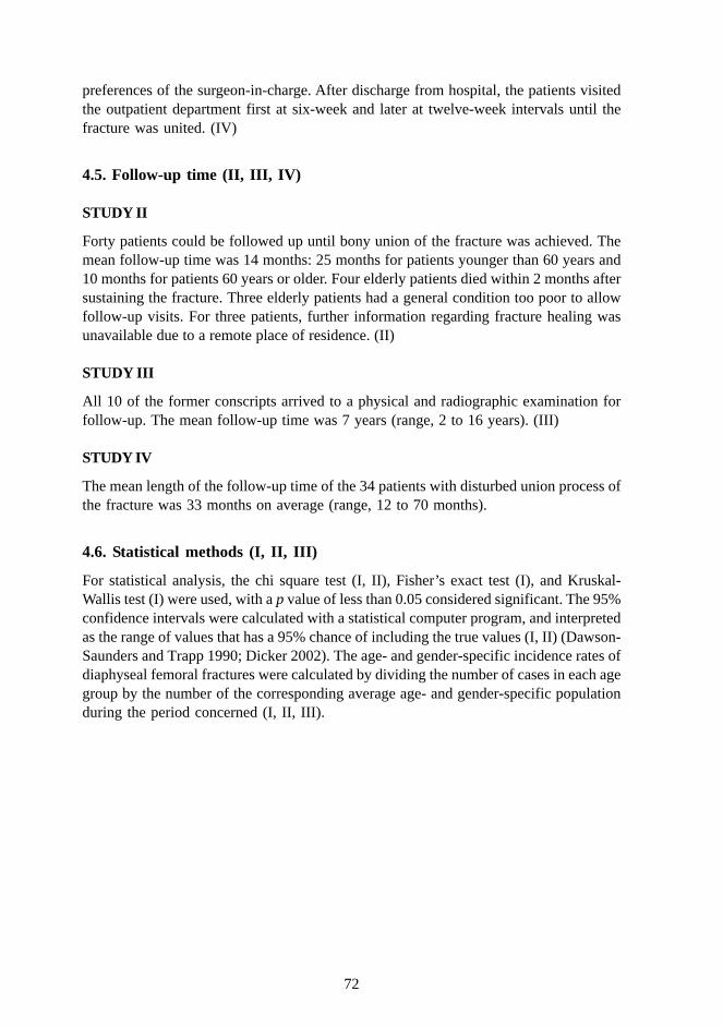
72
preferences of the surgeon-in-charge. After discharge from hospital, the patients visitedthe outpatient department first at six-week and later at twelve-week intervals until thefracture was united. (IV)
4.5. Follow-up time (II, III, IV)
STUDY II
Forty patients could be followed up until bony union of the fracture was achieved. Themean follow-up time was 14 months: 25 months for patients younger than 60 years and10 months for patients 60 years or older. Four elderly patients died within 2 months aftersustaining the fracture. Three elderly patients had a general condition too poor to allowfollow-up visits. For three patients, further information regarding fracture healing wasunavailable due to a remote place of residence. (II)
STUDY III
All 10 of the former conscripts arrived to a physical and radiographic examination forfollow-up. The mean follow-up time was 7 years (range, 2 to 16 years). (III)
STUDY IV
The mean length of the follow-up time of the 34 patients with disturbed union process ofthe fracture was 33 months on average (range, 12 to 70 months).
4.6. Statistical methods (I, II, III)
For statistical analysis, the chi square test (I, II), Fisher’s exact test (I), and Kruskal-Wallis test (I) were used, with a p value of less than 0.05 considered significant. The 95%confidence intervals were calculated with a statistical computer program, and interpretedas the range of values that has a 95% chance of including the true values (I, II) (Dawson-Saunders and Trapp 1990; Dicker 2002). The age- and gender-specific incidence rates ofdiaphyseal femoral fractures were calculated by dividing the number of cases in each agegroup by the number of the corresponding average age- and gender-specific populationduring the period concerned (I, II, III).
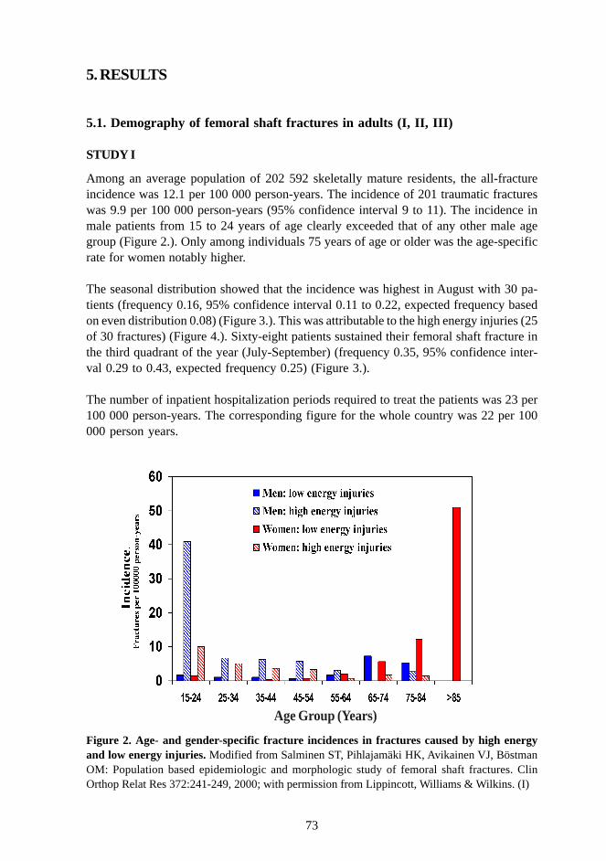
73
5. RESULTS
5.1. Demography of femoral shaft fractures in adults (I, II, III)
STUDY I
Among an average population of 202 592 skeletally mature residents, the all-fractureincidence was 12.1 per 100 000 person-years. The incidence of 201 traumatic fractureswas 9.9 per 100 000 person-years (95% confidence interval 9 to 11). The incidence inmale patients from 15 to 24 years of age clearly exceeded that of any other male agegroup (Figure 2.). Only among individuals 75 years of age or older was the age-specificrate for women notably higher.
The seasonal distribution showed that the incidence was highest in August with 30 pa-tients (frequency 0.16, 95% confidence interval 0.11 to 0.22, expected frequency basedon even distribution 0.08) (Figure 3.). This was attributable to the high energy injuries (25of 30 fractures) (Figure 4.). Sixty-eight patients sustained their femoral shaft fracture inthe third quadrant of the year (July-September) (frequency 0.35, 95% confidence inter-val 0.29 to 0.43, expected frequency 0.25) (Figure 3.).
The number of inpatient hospitalization periods required to treat the patients was 23 per100 000 person-years. The corresponding figure for the whole country was 22 per 100000 person years.
Figure 2. Age- and gender-specific fracture incidences in fractures caused by high energyand low energy injuries. Modified from Salminen ST, Pihlajamäki HK, Avikainen VJ, BöstmanOM: Population based epidemiologic and morphologic study of femoral shaft fractures. ClinOrthop Relat Res 372:241-249, 2000; with permission from Lippincott, Williams & Wilkins. (I)
Age Group (Years)
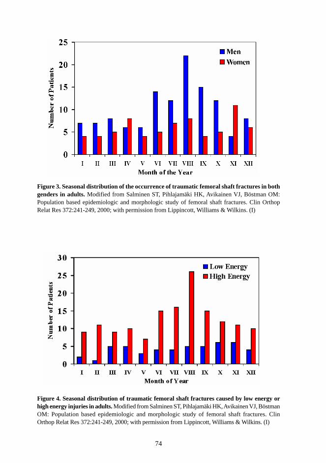
74
Figure 3. Seasonal distribution of the occurrence of traumatic femoral shaft fractures in bothgenders in adults. Modified from Salminen ST, Pihlajamäki HK, Avikainen VJ, Böstman OM:Population based epidemiologic and morphologic study of femoral shaft fractures. Clin OrthopRelat Res 372:241-249, 2000; with permission from Lippincott, Williams & Wilkins. (I)
Figure 4. Seasonal distribution of traumatic femoral shaft fractures caused by low energy orhigh energy injuries in adults. Modified from Salminen ST, Pihlajamäki HK, Avikainen VJ, BöstmanOM: Population based epidemiologic and morphologic study of femoral shaft fractures. ClinOrthop Relat Res 372:241-249, 2000; with permission from Lippincott, Williams & Wilkins. (I)
75
STUDY II
Of the 50 patients, 13 were younger than 60 years and 37 were 60 years or older. Thetotal incidence of femoral shaft fractures due to low energy injury was 2.5 per 100 000person-years. For people aged 15 to 60 years, including nine men and four women, it was0.8 per 100 000 person-years, and for people aged 60 years or older, including nine menand 28 women, it was 7.8 per 100 000 person-years. The 95% confidence interval for thefractures was 0.19 to 0.31.
STUDY III
The incidence of displaced fatigue fractures among conscripts during the 20-year periodwas 1.5 per 100 000 person-years in military service. There were no female conscriptssustaining displaced femoral shaft fatigue fractures.
The median time from the beginning of the military training to the onset of the fracturewas 53 days (range, 15-178 days). All but one conscript experienced preceding painmainly in the distal thigh on weightbearing for 1 to 6 weeks. In five men with a subse-quent fracture in the middle or the distal third of the femoral diaphysis, the pain radiatedto the knee. The first sensations of the pain were related to a combat exercise in fivepatients, and marching in five patients. Only two conscripts had sought medical attentionbecause of the preceding pain symptoms, and had been exempted from heavy militaryservice for 2 to 6 days, but radiographs had not been taken before the fracture occurred.
5.2. Injury mechanism (I, II, IV) or activity at the fracture onset (III)
STUDY I
High energy trauma caused 151 (75%) fractures, 131 (87%) of which occurred in roadtraffic accidents (Table 12.). In 47 (36%) of these cases, the automobile driver wasinjured. There were no gunshot injuries. In the low energy group (50 cases), there were37 patients 60 years of age or older. There was no increasing trend in the number of highenergy or low energy fractures during the 10-year period.
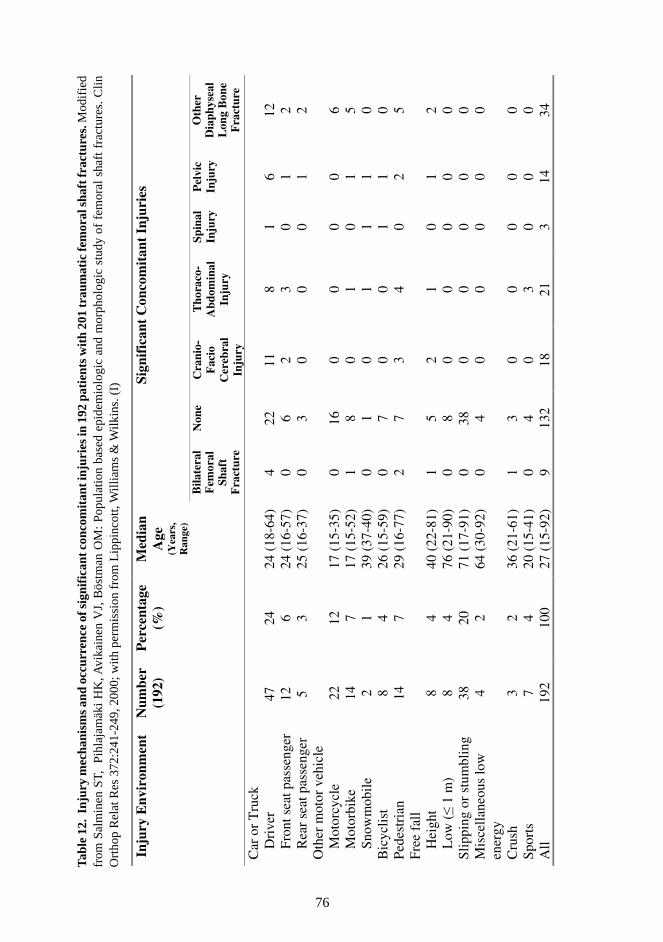
76
Tab
le 1
2. I
njur
y m
echa
nism
s an
d oc
curr
ence
of s
igni
fica
nt c
onco
mit
ant i
njur
ies
in 1
92 p
atie
nts
wit
h 20
1 tr
aum
atic
fem
oral
sha
ft fr
actu
res.
Mod
ifie
dfr
om S
alm
inen
ST,
Pi
hlaj
amäk
i H
K, A
vika
inen
VJ,
Bös
tman
OM
: Po
pula
tion
base
d ep
idem
iolo
gic
and
mor
phol
ogic
stu
dy o
f fe
mor
al s
haft
fra
ctur
es. C
linO
rtho
p R
elat
Res
372
:241
-249
, 200
0; w
ith p
erm
issi
on f
rom
Lip
pinc
ott,
Will
iam
s &
Wilk
ins.
(I)
Inju
ry E
nvir
onm
ent
Num
ber
(192
) P
erce
ntag
e (%
) M
edia
n A
ge
(Yea
rs,
Ran
ge)
Sign
ific
ant
Con
com
itan
t In
juri
es
Bila
tera
l F
emor
al
Shaf
t
Fra
ctur
e
Non
e C
rani
o-F
acio
C
ereb
ral
Inju
ry
Tho
raco
-A
bdom
inal
In
jury
Spin
al
Inju
ry
Pel
vic
Inju
ry
Oth
er
Dia
phys
eal
Lon
g B
one
Fra
ctur
e
Car
or
Tru
ck
D
rive
r 47
24
24
(18
-64)
4
22
11
8 1
6 12
Fron
t sea
t pas
seng
er
12
6 24
(16
-57)
0
6 2
3 0
1 2
R
ear
seat
pas
seng
er
5 3
25 (
16-3
7)
0 3
0 0
0 1
2 O
ther
mot
or v
ehic
le
M
otor
cycl
e 22
12
17
(15
-35)
0
16
0 0
0 0
6
Mot
orbi
ke
14
7 17
(15
-52)
1
8 0
1 0
1 5
Sn
owm
obil
e 2
1 39
(37
-40)
0
1 0
1 1
1 0
Bic
yclis
t 8
4 26
(15
-59)
0
7 0
0 1
1 0
Pede
stri
an
14
7 29
(16
-77)
2
7 3
4 0
2 5
Free
fal
l
Hei
ght
8 4
40 (
22-8
1)
1 5
2 1
0 1
2
Low
( 1
m)
8 4
76 (
21-9
0)
0 8
0 0
0 0
0 Sl
ippi
ng o
r st
umbl
ing
38
20
71 (
17-9
1)
0 38
0
0 0
0 0
Mis
cella
neou
s lo
w
ener
gy
4 2
64 (
30-9
2)
0 4
0 0
0 0
0
Cru
sh
3 2
36 (
21-6
1)
1 3
0 0
0 0
0 Sp
orts
7
4 20
(15
-41)
0
4 0
3 0
0 0
All
192
100
27 (
15-9
2)
9 13
2 18
21
3
14
34
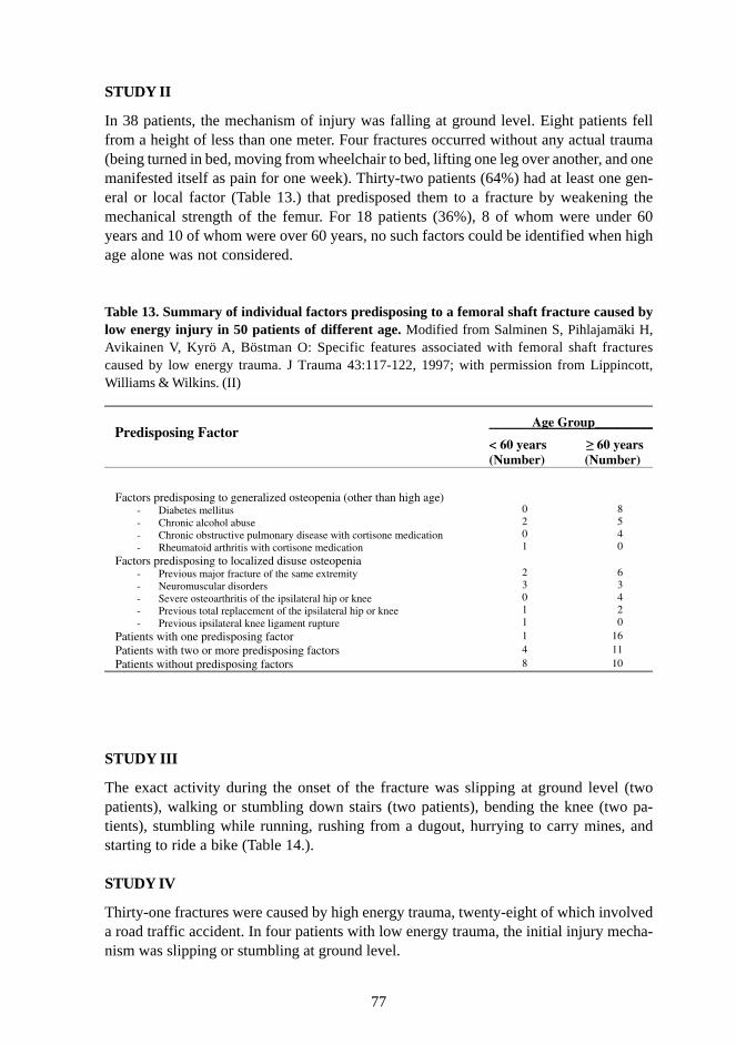
77
STUDY II
In 38 patients, the mechanism of injury was falling at ground level. Eight patients fellfrom a height of less than one meter. Four fractures occurred without any actual trauma(being turned in bed, moving from wheelchair to bed, lifting one leg over another, and onemanifested itself as pain for one week). Thirty-two patients (64%) had at least one gen-eral or local factor (Table 13.) that predisposed them to a fracture by weakening themechanical strength of the femur. For 18 patients (36%), 8 of whom were under 60years and 10 of whom were over 60 years, no such factors could be identified when highage alone was not considered.
Table 13. Summary of individual factors predisposing to a femoral shaft fracture caused bylow energy injury in 50 patients of different age. Modified from Salminen S, Pihlajamäki H,Avikainen V, Kyrö A, Böstman O: Specific features associated with femoral shaft fracturescaused by low energy trauma. J Trauma 43:117-122, 1997; with permission from Lippincott,Williams & Wilkins. (II)
STUDY III
The exact activity during the onset of the fracture was slipping at ground level (twopatients), walking or stumbling down stairs (two patients), bending the knee (two pa-tients), stumbling while running, rushing from a dugout, hurrying to carry mines, andstarting to ride a bike (Table 14.).
STUDY IV
Thirty-one fractures were caused by high energy trauma, twenty-eight of which involveda road traffic accident. In four patients with low energy trauma, the initial injury mecha-nism was slipping or stumbling at ground level.
Predisposing Factor
Age Group_________
< 60 years 60 years (Number) (Number)
Factors predisposing to generalized osteopenia (other than high age)
- Diabetes mellitus - Chronic alcohol abuse - Chronic obstructive pulmonary disease with cortisone medication - Rheumatoid arthritis with cortisone medication
0 2 0 1
8 5 4 0
Factors predisposing to localized disuse osteopenia - Previous major fracture of the same extremity - Neuromuscular disorders - Severe osteoarthritis of the ipsilateral hip or knee - Previous total replacement of the ipsilateral hip or knee - Previous ipsilateral knee ligament rupture
2 3 0 1 1
6 3 4 2 0
Patients with one predisposing factor 1 16 Patients with two or more predisposing factors 4 11 Patients without predisposing factors 8 10
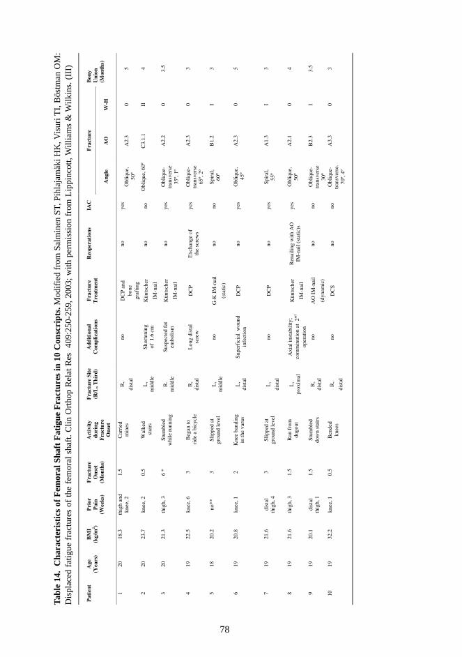
78
Tab
le 1
4. C
hara
cter
isti
cs o
f F
emor
al S
haft
Fat
igue
Fra
ctur
es in
10
Con
scri
pts.
Mod
ifie
d fr
om S
alm
inen
ST,
Pih
laja
mäk
i HK
, Vis
uri T
I, B
östm
an O
M:
Dis
plac
ed f
atig
ue f
ract
ures
of
the
fem
oral
sha
ft. C
lin O
rtho
p R
elat
Res
40
9:25
0-25
9, 2
003;
with
per
mis
sion
fro
m L
ippi
ncot
t, W
illia
ms
& W
ilkin
s. (
III)
F
ract
ure
P
atie
nt
A
ge
(Yea
rs)
B
MI
(kg/
m2 )
P
rior
P
ain
(Wee
ks)
F
ract
ure
Ons
et
(Mon
ths)
A
ctiv
ity
duri
ng
Fra
ctur
e O
nset
F
ract
ure
Site
(R/L
, Thi
rd)
A
ddit
iona
l C
ompl
icat
ions
F
ract
ure
Tre
atm
ent
R
eope
rati
ons
IA
C
A
ngle
AO
W-H
Bon
y U
nion
(M
onth
s)
1 20
18
.3
thig
h an
d kn
ee, 2
1.
5 C
arri
ed
min
es
R,
dist
al
no
DC
P a
nd
bone
gr
afti
ng
no
yes
O
bliq
ue,
50º
A
2.3
0 5
2 20
23
.7
knee
, 2
0.5
Wal
ked
s
tair
s L
, m
iddl
e S
hort
enin
g
of 1
.6 c
m
Kün
tsch
er
IM-n
ail
no
no
Obl
ique
, 60º
C
3.1.
1 II
4
3 20
21
.3
thig
h, 3
6
* S
tum
bled
w
hile
run
ning
R
. m
iddl
e Su
spec
ted
fat
em
boli
sm
Kün
tsch
er
IM-n
ail
no
yes
Obl
ique
- tr
ansv
erse
35
º, 1
º
A2.
2 0
3.5
4 19
22
.5
knee
, 6
3 B
egan
to
rid
e a
bicy
cle
R,
dist
al
Lon
g di
stal
sc
rew
D
CP
Exc
hang
e of
th
e sc
rew
s ye
s O
bliq
ue-
tran
sver
se
65º,
2º
A2.
3 0
3
5 18
20
.2
no**
3
Sli
pped
at
grou
nd le
vel
L,
mid
dle
no
G-K
IM
-nai
l
(sta
tic)
no
no
Spir
al,
60º
B1.
2 I
3
6 19
20
.8
knee
, 1
2 K
nee
band
ing
in
the
varu
s L
, di
stal
S
uper
fici
al w
ound
in
fect
ion
DC
P no
ye
s O
bliq
ue,
45º
A
2.3
0 5
7 19
21
.6
dist
al
thig
h, 4
3
Sli
pped
at
grou
nd le
vel
L,
dist
al
no
DC
P no
ye
s Sp
iral
, 55
º A
1.3
I 3
8 19
21
.6
thig
h, 3
1.
5 R
an f
rom
du
gout
L
, pr
oxim
al
Axi
al in
stab
ility
; co
mm
inut
ion
at 2
nd
oper
atio
n
Kün
tsch
er
IM-n
ail
Ren
aili
ng w
ith
AO
IM
-nai
l (st
atic
)s
yes
Obl
ique
, 50
º A
2.1
0 4
9 19
20
.1
dist
al
thig
h, 1
1.
5 S
tum
bled
do
wn
stai
rs
R,
dist
al
no
AO
IM
-nai
l
(dyn
amic
)
no
no
Obl
ique
-tr
ansv
erse
30
º
B2.
3 I
3.5
10
19
32.2
kn
ee, 1
0.
5 B
ende
d
knee
s R
,
dist
al
no
DC
S no
no
O
bliq
ue-
tran
sver
se.
70º,
4º
A3.
3 0
3
ii
ddii
li
ii
id
ih
lf
hid
hid
fh
fl
hf
id
llf
79
5.3. Concomitant injuries (I, II, III, IV)
STUDY I
Among the 60 (31%) patients who sustained significant concomitant injuries (Table 12.),34 had a second diaphyseal long bone fracture. In 14 patients, there was a concomitantfracture of the patella, of which nine were ipsilateral. All significant concomitant injurieswere associated with high energy trauma. Fifty-four of the patients with concomitantinjuries had been injured in road traffic accidents.
STUDY II
In the low energy group (II), none of the patients had significant concomitant injuries.One patient had a lesion of the femoral artery caused by the fracture.
STUDY III
In the study concerning displaced fatigue fractures of the femoral shaft (III), there wereno concomitant injuries.
STUDY IV
In the study of nonunion after intramedullary nailing (IV), 18 patients had sustained sig-nificant concomitant injuries. These included a thoracoabdominal injury in 12 cases, oth-er diaphyseal long bone fractures in six cases, a craniofacialcerebral injury in four cases,a spinal injury in two cases, and a pelvic fracture in four cases.
5.4. Fracture characteristics (I, II, III, IV)
STUDY I
There were 92 fractures in the right femur (46%) and 109 in the left femur (56%). Ninepatients had bilateral, contemporary fractures (4% of all fractures). Of the 176 closedfractures, 80 were Tscherne and Oestern Grade C II (46%), in which a contaminatedabrasion is associated with localized skin or muscle contusion from direct high energytrauma, 53 were Grade C I (30%), 34 were Grade C0 (19%), and nine were the verysevere Grade CIII (5%). Of the 25 open fractures, 14 were Gustilo Type II, six Type III,and five Type I. All six Type III open fractures were Type IIIA. The main fracture linewas in the middle third of the diaphysis in 79% of the fractures. There was a significantassociation between increasing age and a distal third location (p value 0.02). There wereeight true segmental fractures. When using biomechanical classification, the majority,155 (77%), of all fractures were transverse, oblique, or oblique-transverse (Table 15.). In93 (46% of the fractures), the angle between a line perpendicular to the long axis of thefemur and the main fracture line was less than 30 degrees (Table 15.). There was asignificant association between increasing age and occurrence of a spiral fracture (p <0.001).
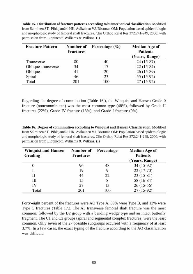
80
Table 15. Distribution of fracture patterns according to biomechanical classification. Modifiedfrom Salminen ST, Pihlajamäki HK, Avikainen VJ, Böstman OM: Population based epidemiologicand morphologic study of femoral shaft fractures. Clin Orthop Relat Res 372:241-249, 2000; withpermission from Lippincott, Williams & Wilkins. (I)
Regarding the degree of comminution (Table 16.), the Winquist and Hansen Grade 0fracture (noncomminuted) was the most common type (48%), followed by Grade IIfractures (22%), Grade IV fracture (13%), and Grade I fracture (9%).
Table 16. Degree of comminution according to Winquist and Hansen Classification. Modifiedfrom Salminen ST, Pihlajamäki HK, Avikainen VJ, Böstman OM: Population based epidemiologicand morphologic study of femoral shaft fractures. Clin Orthop Relat Res 372:241-249, 2000; withpermission from Lippincott, Williams & Wilkins. (I)
Forty-eight percent of the fractures were AO Type A, 39% were Type B, and 13% wereType C fractures (Table 17.). The A3 transverse femoral shaft fracture was the mostcommon, followed by the B2 group with a bending wedge type and an intact butterflyfragment. The C1 and C2 groups (spiral and segmental complex fractures) were the leastcommon. Only seven of the 27 possible subgroups occurred with a frequency of at least3.7%. In a few cases, the exact typing of the fracture according to the AO classificationwas difficult.
Fracture Pattern Number of Fractures
Percentage (%) Median Age of Patients
(Years, Range) Transverse 80 40 24 (15-87) Oblique-transverse 34 17 22 (15-84) Oblique 41 20 26 (15-89) Spiral 46 23 55 (15-92) Total 201 100 27 (15-92)
Winquist and Hansen Grading
Number of Fractures
Percentage Median Age of Patients
(Years, Range) 0 96 48 34 (15-92) I 19 9 22 (17-70) II 44 22 23 (15-81) III 15 8 58 (16-84) IV 27 13 26 (15-56) Total 201 100 27 (15-92)
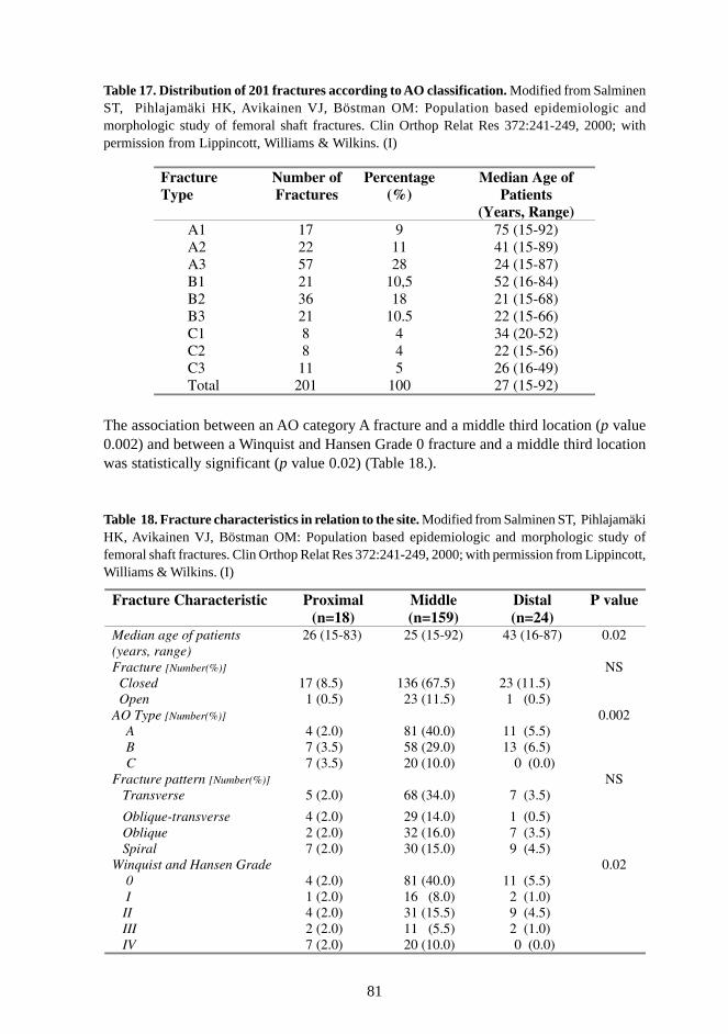
81
Table 17. Distribution of 201 fractures according to AO classification. Modified from SalminenST, Pihlajamäki HK, Avikainen VJ, Böstman OM: Population based epidemiologic andmorphologic study of femoral shaft fractures. Clin Orthop Relat Res 372:241-249, 2000; withpermission from Lippincott, Williams & Wilkins. (I)
The association between an AO category A fracture and a middle third location (p value0.002) and between a Winquist and Hansen Grade 0 fracture and a middle third locationwas statistically significant (p value 0.02) (Table 18.).
Table 18. Fracture characteristics in relation to the site. Modified from Salminen ST, PihlajamäkiHK, Avikainen VJ, Böstman OM: Population based epidemiologic and morphologic study offemoral shaft fractures. Clin Orthop Relat Res 372:241-249, 2000; with permission from Lippincott,Williams & Wilkins. (I)
Fracture Type
Number of Fractures
Percentage(%)
Median Age of Patients
(Years, Range) A1 17 9 75 (15-92) A2 22 11 41 (15-89) A3 57 28 24 (15-87) B1 21 10,5 52 (16-84) B2 36 18 21 (15-68) B3 21 10.5 22 (15-66) C1 8 4 34 (20-52) C2 8 4 22 (15-56) C3 11 5 26 (16-49) Total 201 100 27 (15-92)
Fracture Characteristic
Proximal (n=18)
Middle (n=159)
Distal (n=24)
P value
Median age of patients (years, range)
26 (15-83) 25 (15-92) 43 (16-87) 0.02
Fracture [Number(%)] Closed 17 (8.5) 136 (67.5) 23 (11.5) Open 1 (0.5) 23 (11.5) 1 (0.5)
NS
AO Type [Number(%)] A 4 (2.0) 81 (40.0) 11 (5.5) B 7 (3.5) 58 (29.0) 13 (6.5) C 7 (3.5) 20 (10.0) 0 (0.0)
0.002
Fracture pattern [Number(%)] Transverse 5 (2.0) 68 (34.0) 7 (3.5)
Oblique-transverse 4 (2.0) 29 (14.0) 1 (0.5) Oblique 2 (2.0) 32 (16.0) 7 (3.5) Spiral 7 (2.0) 30 (15.0) 9 (4.5)
NS
Winquist and Hansen Grade 0 4 (2.0) 81 (40.0) 11 (5.5) I 1 (2.0) 16 (8.0) 2 (1.0) II 4 (2.0) 31 (15.5) 9 (4.5) III 2 (2.0) 11 (5.5) 2 (1.0) IV 7 (2.0) 20 (10.0) 0 (0.0)
0.02
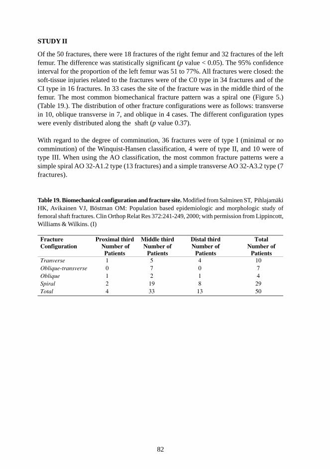
82
STUDY II
Of the 50 fractures, there were 18 fractures of the right femur and 32 fractures of the leftfemur. The difference was statistically significant (p value < 0.05). The 95% confidenceinterval for the proportion of the left femur was 51 to 77%. All fractures were closed: thesoft-tissue injuries related to the fractures were of the C0 type in 34 fractures and of theCI type in 16 fractures. In 33 cases the site of the fracture was in the middle third of thefemur. The most common biomechanical fracture pattern was a spiral one (Figure 5.)(Table 19.). The distribution of other fracture configurations were as follows: transversein 10, oblique transverse in 7, and oblique in 4 cases. The different configuration typeswere evenly distributed along the shaft (p value 0.37).
With regard to the degree of comminution, 36 fractures were of type I (minimal or nocomminution) of the Winquist-Hansen classification, 4 were of type II, and 10 were oftype III. When using the AO classification, the most common fracture patterns were asimple spiral AO 32-A1.2 type (13 fractures) and a simple transverse AO 32-A3.2 type (7fractures).
Table 19. Biomechanical configuration and fracture site. Modified from Salminen ST, PihlajamäkiHK, Avikainen VJ, Böstman OM: Population based epidemiologic and morphologic study offemoral shaft fractures. Clin Orthop Relat Res 372:241-249, 2000; with permission from Lippincott,Williams & Wilkins. (I)
Fracture Configuration
Proximal third Number of
Patients
Middle third Number of
Patients
Distal third Number of
Patients
Total Number of
Patients Tranverse 1 5 4 10 Oblique-transverse 0 7 0 7 Oblique 1 2 1 4 Spiral 2 19 8 29 Total 4 33 13 50
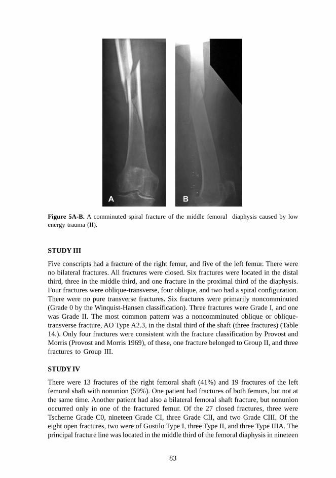
83
Figure 5A-B. A comminuted spiral fracture of the middle femoral diaphysis caused by lowenergy trauma (II).
STUDY III
Five conscripts had a fracture of the right femur, and five of the left femur. There wereno bilateral fractures. All fractures were closed. Six fractures were located in the distalthird, three in the middle third, and one fracture in the proximal third of the diaphysis.Four fractures were oblique-transverse, four oblique, and two had a spiral configuration.There were no pure transverse fractures. Six fractures were primarily noncomminuted(Grade 0 by the Winquist-Hansen classification). Three fractures were Grade I, and onewas Grade II. The most common pattern was a noncomminuted oblique or oblique-transverse fracture, AO Type A2.3, in the distal third of the shaft (three fractures) (Table14.). Only four fractures were consistent with the fracture classification by Provost andMorris (Provost and Morris 1969), of these, one fracture belonged to Group II, and threefractures to Group III.
STUDY IV
There were 13 fractures of the right femoral shaft (41%) and 19 fractures of the leftfemoral shaft with nonunion (59%). One patient had fractures of both femurs, but not atthe same time. Another patient had also a bilateral femoral shaft fracture, but nonunionoccurred only in one of the fractured femur. Of the 27 closed fractures, three wereTscherne Grade C0, nineteen Grade CI, three Grade CII, and two Grade CIII. Of theeight open fractures, two were of Gustilo Type I, three Type II, and three Type IIIA. Theprincipal fracture line was located in the middle third of the femoral diaphysis in nineteen
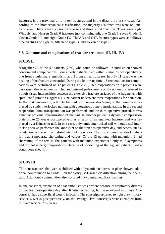
84
fractures, in the proximal third in ten fractures, and in the distal third in six cases. Ac-cording to the biomechanical classification, the majority (26 fractures) were oblique-transverse. There were six pure transverse and three spiral fractures. There were eightWinquist and Hansen Grade 0 fractures (noncomminuted), one Grade I, seven Grade II,eleven Grade III, and eight Grade IV. The AO and OTA fracture types were as follows:nine fractures of Type A, fifteen of Type B, and eleven of Type C.
5.5. Outcome and complications of fracture treatment (II, III, IV)
STUDY II
Altogether 29 of the 40 patients (73%) who could be followed up until union showedconcomitant complications. Four elderly patients died within 2 months postoperatively,one from a pulmonary embolism, and 3 from a heart disease. In only 11 cases was thehealing of the fracture uneventful. During the follow-up time, 18 reoperations for compli-cations were performed on 12 patients (Table 20.). Ten reoperations on 7 patients wereperformed due to nonunion. The predominant pathogenesis of the nonunions seemed tobe soft-tissue interposition between the extensive fracture surfaces of the fragments withspiral configuration (Figure 6.). One patient underwent three reoperations for nonunion.In the first reoperation, a Küntscher nail with severe shortening of the femur was re-placed by static interlocked nailing with autogenous bone transplantation. In the secondreoperation, bone transplantation was performed, and the third operative procedure wasaimed at proximal dynamization of the nail. In another patient, a dynamic compressionplate broke 26 weeks postoperatively as a result of an ununited fracture, and was re-placed by a Küntscher nail. In one case, a dynamic interlocked nail without distal inter-locking screws perforated the knee joint on the first postoperative day, and necessitated arereduction and insertion of distal interlocking screws. The most common mode of malun-ion was a moderate shortening and valgus. Of the 13 patients with malunion, 9 hadshortening of the femur. The patients with malunion experienced only mild symptomsand did not undergo reoperations. Because of shortening of the leg, six patients used acontinuous shoe lift.
STUDY III
The four fractures that were stabilized with a dynamic compression plate showed addi-tional comminution to Grade II on the Winquist-Hansen classification during the opera-tion. Additional comminution also occurred in two intramedullary nailings.
In one conscript, suspicion of a fat embolism was present because of respiratory distresson the first postoperative day after Küntscher nailing, but he recovered in 3 days. Oneconscript had a superficial wound infection. The conscripts returned to light duty militaryservice 6 weeks postoperatively, on the average. Two conscripts were exempted frommilitary service for 2 years.
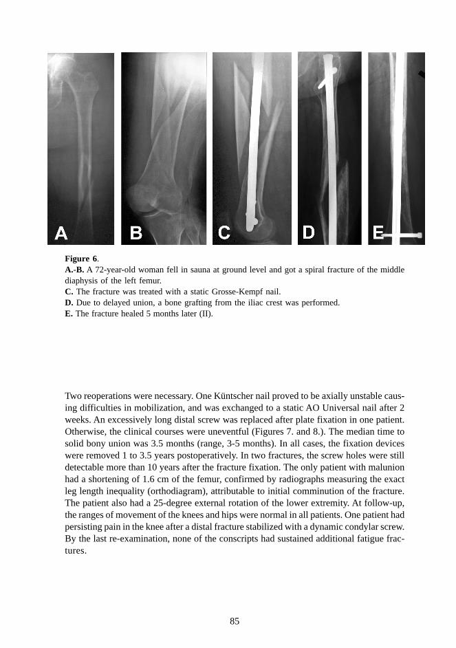
85
Two reoperations were necessary. One Küntscher nail proved to be axially unstable caus-ing difficulties in mobilization, and was exchanged to a static AO Universal nail after 2weeks. An excessively long distal screw was replaced after plate fixation in one patient.Otherwise, the clinical courses were uneventful (Figures 7. and 8.). The median time tosolid bony union was 3.5 months (range, 3-5 months). In all cases, the fixation deviceswere removed 1 to 3.5 years postoperatively. In two fractures, the screw holes were stilldetectable more than 10 years after the fracture fixation. The only patient with malunionhad a shortening of 1.6 cm of the femur, confirmed by radiographs measuring the exactleg length inequality (orthodiagram), attributable to initial comminution of the fracture.The patient also had a 25-degree external rotation of the lower extremity. At follow-up,the ranges of movement of the knees and hips were normal in all patients. One patient hadpersisting pain in the knee after a distal fracture stabilized with a dynamic condylar screw.By the last re-examination, none of the conscripts had sustained additional fatigue frac-tures.
Figure 6.A.-B. A 72-year-old woman fell in sauna at ground level and got a spiral fracture of the middlediaphysis of the left femur.C. The fracture was treated with a static Grosse-Kempf nail.D. Due to delayed union, a bone grafting from the iliac crest was performed.E. The fracture healed 5 months later (II).
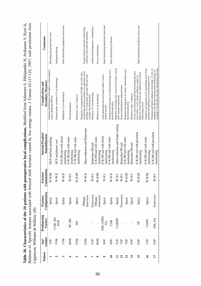
86
Tab
le 2
0. C
hara
cter
isti
cs o
f th
e 29
pat
ient
s w
ith
post
oper
ativ
e lo
cal
com
plic
atio
ns. M
odif
ied
from
Sal
min
en S
, Pi
hlaj
amäk
i H
, Avi
kain
en V
, K
yrö
A,
Bös
tman
O:
Spec
ific
fea
ture
s as
soci
ated
with
fem
oral
sha
ft f
ract
ures
cau
sed
by l
ow e
nerg
y tr
aum
a. J
Tra
uma
43:1
17-1
22,
1997
; w
ith
perm
issi
on f
rom
Lip
pinc
ott,
Will
iam
s &
Wilk
ins.
(II
)
Pat
ient
A
ge/
Gen
der
Pre
disp
osin
g F
acto
rs
Fra
ctur
e C
onfi
gura
tion
F
ract
ure
Com
min
utio
n In
tern
al F
ixat
ion/
Im
mob
iliza
tion
C
ompl
icat
ions
and
Se
cond
ary
Mea
sure
s C
omm
ents
1 35
/F
- Sp
iral
W
-H I
II
DC
P an
d bo
ne g
raft
ing
Non
unio
n an
d br
eaka
ge o
f th
e pl
ate;
re-
oste
osyn
thes
is
usin
g a
Kün
tsch
er I
M n
ail
IM n
ailin
g sh
ould
hav
e be
en u
sed
2 37
/M
CA
B, N
D,
KL
R
Spir
al
W-H
II
DC
P an
d pl
aste
r ca
st
Del
ayed
uni
on, m
alun
ion
(1.5
cm
sho
rten
ing)
In
adeq
uate
pla
ting
3 17
/M
- Sp
iral
W
-H I
I G
-K I
M n
ail w
ith d
ista
l in
terl
ocki
ng
Mal
unio
n (1
.5 c
m s
hort
enin
g)
Sta
tic
inte
rloc
king
sho
uld
have
bee
n us
ed
4 40
/M
PF, N
D
Spir
al
W-H
I
G-K
IM
nai
l with
sta
tic
inte
rloc
king
W
ound
bre
akdo
wn
5 52
/M
ND
Sp
iral
W
-H I
II
V-W
IM
nai
l wih
sta
tic
inte
rloc
king
M
alun
ion
(var
us 7
deg
rees
)
6 34
/M
- O
bliq
ue-
tran
sver
se
W-H
II
Ope
n re
duct
ion
and
Kün
tsch
er
Non
unio
n; (
1) r
eope
ratio
n: G
-K I
M n
ail w
ith s
tati
c in
terl
ocki
ng a
nd b
one
graf
ting;
(2)
reo
pera
tion:
bon
e gr
aftin
g; (
3): r
eope
ratio
n: p
roxi
mal
IM
nai
l with
ce
rcla
ge w
ire
dyna
miz
atio
n
Clo
sed
redu
ctio
n an
d st
atic
inte
rloc
king
co
uld
have
pre
vent
ed th
e no
nuni
on
7 67
/F
- T
rans
vers
e W
-H I
K
ünts
cher
IM
nai
l M
alun
ion
(2.5
cm
sho
rten
ing)
U
ndet
ecte
d in
trao
pera
tive
com
min
utio
n
8 66
/M
- O
bliq
ue-
tran
sver
se
W-H
III
G
-K I
M n
ail w
ith s
tatic
in
terl
ocki
ng
Mal
unio
n (2
cm
sho
rten
ing)
S
ever
e co
mm
inut
ion
9 89
/F
DM
, CO
PD,
OA
Sp
iral
W
-H I
G
-K I
M n
ail w
ith d
ista
l in
terl
ocki
ng
Axi
al s
hort
enin
g an
d re
curv
atum
wit
hin
2 w
eeks
; sk
elet
al tr
actio
n S
tati
c in
terl
ocki
ng s
houl
d ha
ve b
een
used
10
60/F
C
AB
Sp
iral
W
-H I
I G
-K I
M n
ail w
ith s
tatic
in
terl
ocki
ng
Non
unio
n an
d m
igra
tion
of a
dis
tal i
nter
lock
ing
scre
w;
dist
al d
ynam
izat
ion
Sta
tic
inte
rloc
king
in d
iast
asis
11
91/F
C
AB
/PF
Spir
al
W-H
I
Ope
n re
duct
ion
and
End
er n
ailin
g M
igra
tion
of a
n E
nder
nai
l; re
mov
al o
f an
End
er n
ail i
n th
ree
sepa
rate
reo
pera
tion
s
12
73/F
-
Tra
nsve
rse
W-H
I
Kün
tsch
er I
M n
ail
Dee
p w
ound
infe
ctio
n, d
elay
ed u
nion
13
79/F
-
Spir
al
W-H
III
G
-K I
M n
ail w
ith s
tatic
in
terl
ocki
ng
Non
unio
n an
d m
igra
tion
of a
dis
tal
inte
rloc
king
scr
ew;
dist
al d
ynam
izat
ion
14
92/F
-
Spir
al
W-H
I
Kün
tsch
er I
M n
ail
Mal
unio
n (v
algu
s 7
degr
ees)
15
84/F
T
P Sp
iral
W
-H I
II
G-K
IM
nai
l with
pro
xim
al
inte
rloc
king
Sev
ere
axia
l sho
rten
ing
and
nail
perf
orat
ing
the
knee
jo
int w
ithin
1 w
eek,
fin
ally
mal
unio
n (v
algu
s 10
de
gree
s); c
lose
d re
duct
ion
and
app
licat
ion
of d
ista
l in
terl
ocki
ng s
crew
s
Sta
tic
inte
rloc
king
sho
uld
have
bee
n us
ed
16
72/F
C
OPD
Sp
iral
W
-H I
II
G-K
IM
nai
l with
sta
tic
inte
rloc
king
N
onun
ion
and
mig
ratio
n of
an
inte
rloc
king
scr
ew; b
one
graf
ting
and
tight
enin
g of
an
inte
rloc
king
scr
ew
17
83/F
D
M, O
A
Tra
nsve
rse
W-H
I
G-K
IM
nai
l with
pro
xim
al
inte
rloc
king
Non
unio
n an
d fi
nall
y m
alun
ion
(sho
rten
ing
1.5
cm);
(1)
re
nail
ing:
G-K
IM
nai
l with
pro
xim
al in
terl
ocki
ng; (
2)
rena
iling
: G-K
IM
nai
l with
sta
tic in
terl
ocki
ng a
nd b
one

87
F, F
emal
e; M
, Mal
e; D
M, d
iabe
tes
mel
litu
s, C
AB
, chr
onic
alc
ohol
abu
se; C
OP
D, c
hron
ic o
bstr
ucti
ve p
ulm
onar
y di
seas
e w
ith
cort
ison
e m
edic
atio
n; P
F, p
revi
ous
maj
orfr
actu
re o
f the
sam
e ex
trem
ity;
ND
, neu
rom
uscu
lar
diso
rder
s; O
A, s
ever
e os
teoa
rthr
itis
of t
he ip
sila
tera
l hip
or
knee
; T
P, p
revi
ous
tota
l pro
sthe
sis
repl
acem
ent o
f the
ipsi
late
ral h
ip o
r kn
ee; K
LR
, pre
viou
s ip
sila
tera
l kne
e li
gam
ent r
uptu
re; W
-H, W
inqu
ist-
Han
sen
clas
sifi
cati
on; D
CP,
AO
dyn
amic
com
pres
sion
pla
te; D
CS,
AO
dyn
amic
cond
ylar
scr
ew;
IM n
ail,
intr
amed
ulla
ry n
ail;
G-K
, Gro
sse-
Kem
pf;
V-W
, Var
i-W
all.
graf
ting
18
87/F
-
Spir
al
W-H
I
G-K
IM
nai
l with
dis
tal
inte
rloc
king
M
alun
ion
(sho
rten
ing
4 cm
and
inte
rnal
rot
atio
n 10
de
gree
s)
Non
-am
bula
tory
pat
ient
, no
seco
ndar
y m
easu
res
19
71/M
-
Spir
al
W-H
I
G-K
IM
nai
l with
sta
tic
inte
rloc
king
Non
unio
n an
d m
igra
tion
of a
n in
terl
ocki
ng s
crew
, fi
nall
y m
alun
ion
(sho
rten
ing
1.5
cm);
pro
xim
al
dyna
miz
atio
n
20
61/M
D
M, N
D
Obl
ique
W
-H I
E
nder
nai
ling
Dee
p w
ound
infe
ctio
n, d
elay
ed u
nion
, fin
ally
mal
unio
n (e
xter
nal r
otat
ion
10 d
egre
es);
rem
oval
of
End
er n
ails
21
66/M
C
OPD
Sp
iral
W
-H I
G
-K I
M n
ail w
ith s
tatic
in
terl
ocki
ng
Dee
p w
ound
infe
ctio
n
22
75/M
N
D
Spir
al
W-H
I
Kün
tsch
er I
M n
ail
Mal
unio
n (s
hort
enin
g 1.
5 cm
)
23
84/F
-
Spir
al
W-H
III
G
-K I
M n
ail w
ith s
tatic
in
terl
ocki
ng
Wou
nd b
reak
dow
n, d
elay
ed u
nion
B
ecau
se o
f ol
d ag
e an
d po
or g
ener
al
cond
ition
, no
seco
ndar
y m
easu
res
wer
e pe
rfor
med
24
62/F
D
M
Tra
nsve
rse
W-H
I
DC
S L
oose
ning
of
the
plat
e pa
rt o
f D
CS
with
in 1
mon
th; (
1)
naili
ng: K
ünts
cher
IM
nai
l, w
hich
sub
sequ
ently
bro
ke;
(2)
nail
ing:
G-K
IM
nai
l wit
h st
aic
inte
rloc
king
IM
nai
ling
shou
ld h
ave
been
use
d
25
66/M
PF
O
bliq
ue-
tran
sver
se
W-H
I
V-W
IM
nai
l with
dis
tal
inte
rloc
king
D
elay
ed u
nion
26
66/F
N
D
Obl
ique
W
-H I
Pl
aste
r ca
st
Del
ayed
uni
on
IM n
ailin
g sh
ould
pos
sibl
y ha
ve b
een
perf
orm
ed d
espi
te t
etra
pleg
ia
27
88/F
PF
Sp
iral
W
-H I
D
CP
Mal
unio
n (2
.5 c
m s
hort
enin
g)
28
75/F
-
Spir
al
W-H
I
DC
P S
upra
cond
ylar
ref
ract
ure;
reo
steo
synt
hesi
s us
ing
a 95
-de
gree
con
dyla
r pl
ate
and
cerc
lage
29
79/F
D
M, P
F Sp
iral
W
-H I
D
CP
Sup
erfi
cial
wou
nd in
fect
ion
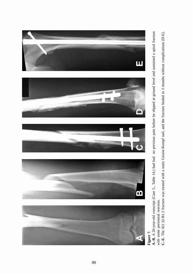
88
Fig
ure
7.A
.-B
. A 2
0-ye
ar-o
ld c
onsc
ript
(C
ase
5.,
Tabl
e 14
.) h
ad h
ad
no p
revi
ous
pain
bef
ore
he s
lippe
d at
gro
und
leve
l an
d su
stai
ned
a sp
iral
fra
ctur
ew
ith
som
e pe
rios
teal
rea
ctio
n.C
.-E
. The
AO
32-
B1.
2 fr
actu
re w
as t
reat
ed w
ith a
sta
tic G
ross
e-K
empf
nai
l, an
d th
e fr
actu
re h
eale
d in
3 m
onth
s w
ithou
t co
mpl
icat
ions
(D
-E).
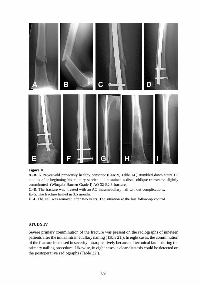
89
STUDY IV
Severe primary comminution of the fracture was present on the radiographs of nineteenpatients after the initial intramedullary nailing (Table 21.). In eight cases, the comminutionof the fracture increased in severity intraoperatively because of technical faults during theprimary nailing procedure. Likewise, in eight cases, a clear diastasis could be detected onthe postoperative radiographs (Table 22.).
Figure 8.A.-B. A 19-year-old previously healthy conscript (Case 9, Table 14.) stumbled down stairs 1.5months after beginning his military service and sustained a distal oblique-transverse slightlycomminuted (Winquist-Hansen Grade I) AO 32-B2.3 fracture.C.-D. The fracture was treated with an AO intramedullary nail without complications.E.-G. The fracture healed in 3.5 months.H.-I. The nail was removed after two years. The situation at the last follow-up control.
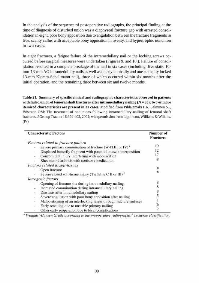
90
In the analysis of the sequence of postoperative radiographs, the principal finding at thetime of diagnosis of disturbed union was a diaphyseal fracture gap with arrested consol-idation in eight, poor bony apposition due to angulation between the fracture fragments infive, scanty callus with acceptable bony apposition in twenty, and hypertrophic nonunionin two cases.
In eight fractures, a fatigue failure of the intramedullary nail or the locking screws oc-curred before surgical measures were undertaken (Figures 9. and 10.). Failure of consol-idation resulted in a complete breakage of the nail in six cases (including five static 10-mm-13-mm AO intramedullary nails as well as one dynamically and one statically locked13-mm Klemm-Schellmann nail), three of which occurred within six months after theinitial operation, and the remaining three between six and twelve months.
Table 21. Summary of specific clinical and radiographic characteristics observed in patientswith failed union of femoral shaft fractures after intramedullary nailing (N = 35); two or moreitemized characteristics are present in 31 cases. Modified from Pihlajamäki HK, Salminen ST,Böstman OM: The treatment of nonunions following intramedullary nailing of femoral shaftfractures. J Orthop Trauma 16:394-402, 2002; with permission from Lippincott, Williams & Wilkins.(IV)
Characteristic Factors Number of
Fractures Factors related to fracture pattern
- Severe primary comminution of fracture (W-H III or IV) a - Displaced butterfly fragment with potential muscle interposition - Concomitant injury interfering with mobilization - Rheumatoid arthritis with cortisone medication
19 12 17
8
Factors related to soft-tissues - Open fracture - Severe closed soft-tissue injury (Tscherne C II or III) b
8 6
Iatrogenic factors - Opening of fracture site during intramedullary nailing - Increased comminution during intramedullary nailing - Diastasis after intramedullary nailing - Severe angulation with poor bony apposition after nailing - Malpositioning of an interlocking screw through fracture surfaces - Early renailing due to unstable primary nailing - Other early reoperation due to local complications
8 8 8 5 1 6 2
a Winquist-Hansen Grade according to the preoperative radiographs.b Tscherne classification.
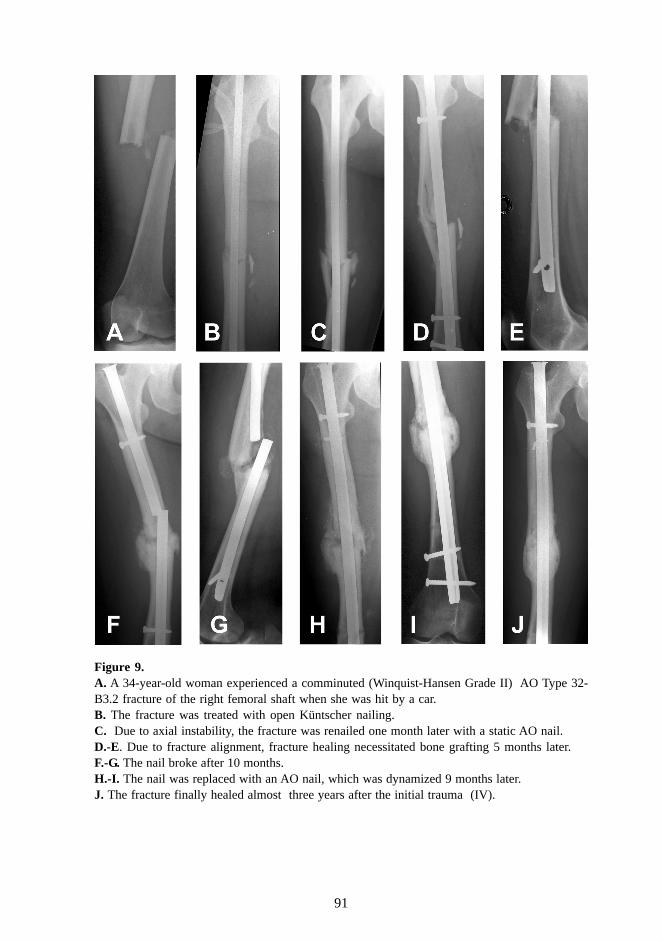
91
Figure 9.A. A 34-year-old woman experienced a comminuted (Winquist-Hansen Grade II) AO Type 32-B3.2 fracture of the right femoral shaft when she was hit by a car.B. The fracture was treated with open Küntscher nailing.C. Due to axial instability, the fracture was renailed one month later with a static AO nail.D.-E. Due to fracture alignment, fracture healing necessitated bone grafting 5 months later.F.-G. The nail broke after 10 months.H.-I. The nail was replaced with an AO nail, which was dynamized 9 months later.J. The fracture finally healed almost three years after the initial trauma (IV).
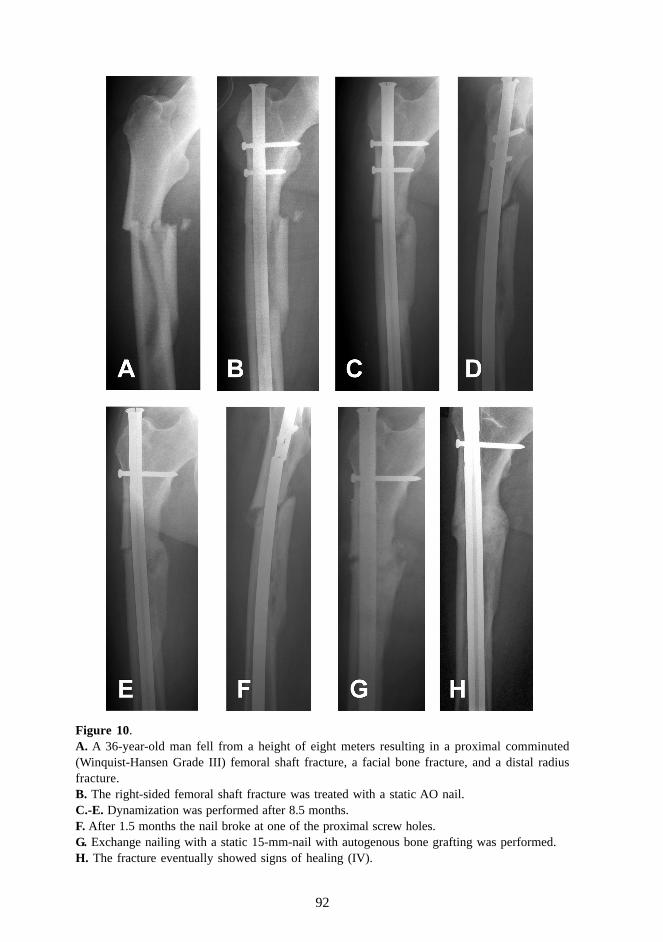
92
Figure 10.A. A 36-year-old man fell from a height of eight meters resulting in a proximal comminuted(Winquist-Hansen Grade III) femoral shaft fracture, a facial bone fracture, and a distal radiusfracture.B. The right-sided femoral shaft fracture was treated with a static AO nail.C.-E. Dynamization was performed after 8.5 months.F. After 1.5 months the nail broke at one of the proximal screw holes.G. Exchange nailing with a static 15-mm-nail with autogenous bone grafting was performed.H. The fracture eventually showed signs of healing (IV).
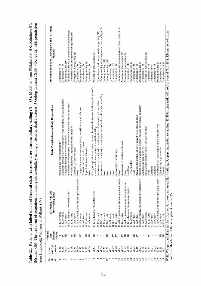
93
Tab
le 2
2.
Pat
ient
s w
ith
faile
d un
ion
of f
emor
al s
haft
fra
ctur
es a
fter
int
ram
edul
lary
nai
ling
(N =
35)
. Mod
ifie
d fr
om P
ihla
jam
äki
HK
, Sal
min
en S
T,B
östm
an O
M: T
he tr
eatm
ent o
f no
nuni
ons
follo
win
g in
tram
edul
lary
nai
ling
of f
emor
al s
haft
fra
ctur
es. J
Ort
hop
Tra
uma
16:3
94-4
02, 2
002;
with
per
mis
sion
from
Lip
pinc
ott,
Will
iam
s &
Wilk
ins.
(IV
)p
fpp
,(
)P
a- ti-
ent
Gen
der
and
Age
(Y
ears
)
Win
quis
t an
d H
anse
n
Gra
de
IM-N
ailin
g, N
ail a
nd
Inte
rloc
king
Typ
es
Ear
ly C
ompl
icat
ions
and
Ear
ly R
eope
rati
ons
Pro
cedu
re f
or F
aile
d C
onso
lidat
ion
and
Its
Tim
ing
(Mon
ths)
1 F,
45
I C
, K, d
ynam
ic
Ren
ailin
g fo
r ro
tatio
nal i
nsta
bilit
y D
ynam
izat
ion
(8)
2 F,
46
IV
C, K
, dyn
amic
Ia
trog
enic
int
raop
erat
ive
com
min
utio
n o
pen
rena
iling
for
axi
al in
stab
ility
D
ynam
izat
ion
(5)
3 M
, 28
III
C, A
O, s
tatic
Ia
trog
enic
int
raop
erat
ive
com
min
utio
n E
xcha
nge
naili
ng (
16)
4 F,
20
0 O
, K-S
, dyn
amic
, tw
o di
stal
scr
ews
Intr
aope
rati
ve c
omm
inut
ion,
a c
ross
scr
ew p
lace
d on
the
frac
ture
leve
l E
xcha
nge
nail
ing
wit
h au
toge
nous
bon
e gr
afti
ng (
4)
5 F,
18
IV
C, K
-S, s
tati
c 9º
var
us a
ngul
atio
n, s
uper
fici
al w
ound
infe
ctio
n D
ynam
izat
ion
and
auto
geno
us b
one
graf
ting
(10
) 6
F, 3
5 II
I C
, AO
, sta
tic
Dia
stas
is, 2
5º a
ntec
urva
tum
A
utog
enou
s bo
ne g
raft
ing
(8)
7 M
, 30
II
C, A
O, d
ynam
ic, o
ne d
ynam
ic p
roxi
mal
scr
ew
Non
e D
ynam
izat
ion
(5)
8 F,
37
IV
C, A
O, s
tati
c Ia
trog
nic
intr
aope
rati
ve c
omm
inut
ion,
sup
erfi
cial
wou
nd in
fect
ion
Exc
hang
e na
ilin
g (7
) 9
F, 2
7 II
O
, AO
, sta
tic
Dia
stas
is, d
elay
ed w
ould
clo
sure
D
ynam
izat
ion
(7)
10
F, 7
2 II
I O
, K, d
ynam
ic
Supe
rfic
ial w
ound
infe
ctio
n E
xcha
nge
nail
ing
(8)
11
M, 3
6 II
I C
, AO
, sta
tic
Non
e D
ynam
izat
ion
(9)
12
M, 5
3 II
I C
, K-S
, dyn
amic
, tw
o di
stal
scr
ews
9º v
algu
s an
gula
tion,
a c
ross
scr
ew w
as p
lace
d on
the
frac
ture
leve
l, re
appl
icat
ion
of a
cr
oss
scre
w in
a r
eope
ratio
n fo
r ax
ial i
nsta
bilit
y A
utog
enou
s bo
ne g
raft
ing
(7)
13
M, 4
6 0
C, K
, dyn
amic
In
trao
pera
tive
com
min
utio
n, r
enai
ling
for
axi
al a
nd r
otat
iona
l ins
tabi
lity
D
ynam
izat
ion
wit
h au
toge
nous
bon
e gr
afti
ng (
17)
14
M, 5
4 0
O, K
-S, s
tatic
In
trao
pera
tive
com
min
utio
n, r
enai
ling
beca
use
of n
ail b
reak
age
afte
r st
umbl
ing
Exc
hang
e na
iling
with
aut
ogen
ous
bone
gra
ftin
g (1
3)
15
F, 2
7 II
C
, K, d
ynam
ic
Non
e E
xcha
nge
nail
ing
(51
) 16
* F,
27
III
C, K
-S, s
tatic
N
one
Exc
hang
e na
ilin
g (
52)
17
M, 3
1 0
C, A
O, s
tati
c N
one
Dyn
amiz
atio
n (8
) 18
M
, 27
IV
O, K
-S, s
tati
c N
one
Dyn
amiz
atio
n (1
0)
19
M, 2
0 IV
C
, K-S
, sta
tic
Intr
aope
rati
ve s
hort
enin
g D
ynam
izat
ion
(5)
20
M, 2
1 II
C
, AO
, dyn
amic
, one
dyn
amic
pro
xim
al s
crew
N
one
Exc
hang
e na
ilin
g w
ith
auto
geno
us b
one
graf
ting
(14
) 21
M
, 43
IV
C, K
-S, s
tatic
In
trao
pera
tive
ben
ding
of
the
nail
A
utog
enou
s bo
ne g
raft
ing
(5)
22
F, 5
0 II
I C
, AO
, dyn
amic
, one
sta
tic p
roxi
mal
scr
ew
Non
e A
utog
enou
s bo
ne g
raft
ing
(21)
23
M
, 40
III
C, K
-S, d
ynam
ic, o
ne p
roxi
mal
scr
ew
Dia
stas
is
Dyn
amiz
atio
n (1
1)
24
F, 2
7 0
C, K
, dyn
amic
10
º re
curv
atum
E
xcha
nge
nail
ing
(9)
25
M, 2
5 II
I C
, AO
, sta
tic
Non
e D
ynam
izat
ion
(9)
26
F, 4
5 IV
C
, AO
, sta
tic
Ren
aili
ng f
or a
xial
inst
abil
ity
caus
ed b
y os
teop
orot
ic b
one
Dyn
amiz
atio
n (6
) 27
M
, 23
II
O, A
O, d
ynam
ic, o
ne d
ynam
ic p
roxi
mal
scr
ew
Rea
pplic
atio
n of
a d
ista
l cro
ss s
crew
bec
ause
of
rota
tiona
l ins
tabi
lity
Exc
hang
e na
ilin
g (9
) 28
F,
15
II
C, A
O, s
tatic
D
iast
asis
and
cor
tica
l def
ect
Dyn
amiz
atio
n (4
) 29
F,
34
II
O, K
, dyn
amic
R
enai
ling
for
axi
al in
stab
ilit
y, 2
0º a
ntec
urva
tum
A
utog
enou
s bo
ne g
raft
ing
(6)
30
F, 3
4 IV
C
, K-S
, sta
tic
Non
e D
ynam
izat
ion
(7)
31
M, 2
1 0
C, A
O, s
tati
c D
iast
asis
D
ynam
isat
ion
(6)
32
M, 2
2 0
C, A
O, s
tati
c D
iast
asis
D
ynam
izat
ion
(3)
33
F, 5
1 0
C, A
O, d
ynam
ic, o
ne d
ynam
ic p
roxi
mal
scr
ew
Prox
imal
dis
tal s
crew
hol
e is
on
the
frac
ture
leve
l E
xcha
nge
nail
ing
(9)
34
F, 2
1 II
I C
, AO
, sta
tic
Dia
stas
is a
nd c
orti
cal d
efec
t D
ynam
izat
ion
(8.5
) 35
M
, 31
III
O, K
-S, s
tatic
D
iast
asis
D
ynam
izat
ion
(5.5
)
W-H
, Win
quis
t and
Han
sen
clas
sifi
catio
n; C
, clo
sed
intr
amed
ulla
ry n
ailin
g; O
, ope
n in
tram
edul
lary
nai
ling;
K, K
ünts
cher
nai
l; A
O, A
O U
nive
rsal
Nai
l; K
-S, K
lem
m-S
chel
lman
n na
il,*
the
othe
r fe
mur
of
the
sam
e pa
tient
num
ber
15.
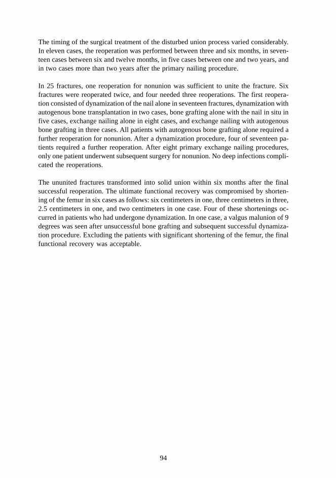
94
The timing of the surgical treatment of the disturbed union process varied considerably.In eleven cases, the reoperation was performed between three and six months, in seven-teen cases between six and twelve months, in five cases between one and two years, andin two cases more than two years after the primary nailing procedure.
In 25 fractures, one reoperation for nonunion was sufficient to unite the fracture. Sixfractures were reoperated twice, and four needed three reoperations. The first reopera-tion consisted of dynamization of the nail alone in seventeen fractures, dynamization withautogenous bone transplantation in two cases, bone grafting alone with the nail in situ infive cases, exchange nailing alone in eight cases, and exchange nailing with autogenousbone grafting in three cases. All patients with autogenous bone grafting alone required afurther reoperation for nonunion. After a dynamization procedure, four of seventeen pa-tients required a further reoperation. After eight primary exchange nailing procedures,only one patient underwent subsequent surgery for nonunion. No deep infections compli-cated the reoperations.
The ununited fractures transformed into solid union within six months after the finalsuccessful reoperation. The ultimate functional recovery was compromised by shorten-ing of the femur in six cases as follows: six centimeters in one, three centimeters in three,2.5 centimeters in one, and two centimeters in one case. Four of these shortenings oc-curred in patients who had undergone dynamization. In one case, a valgus malunion of 9degrees was seen after unsuccessful bone grafting and subsequent successful dynamiza-tion procedure. Excluding the patients with significant shortening of the femur, the finalfunctional recovery was acceptable.
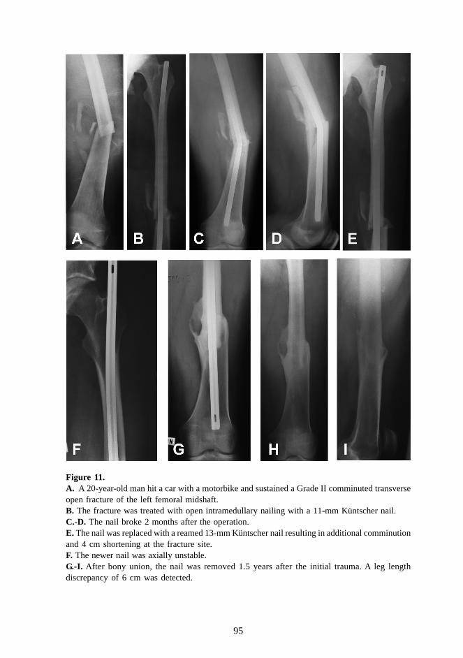
95
Figure 11.A. A 20-year-old man hit a car with a motorbike and sustained a Grade II comminuted transverseopen fracture of the left femoral midshaft.B. The fracture was treated with open intramedullary nailing with a 11-mm Küntscher nail.C.-D. The nail broke 2 months after the operation.E. The nail was replaced with a reamed 13-mm Küntscher nail resulting in additional comminutionand 4 cm shortening at the fracture site.F. The newer nail was axially unstable.G.-I. After bony union, the nail was removed 1.5 years after the initial trauma. A leg lengthdiscrepancy of 6 cm was detected.
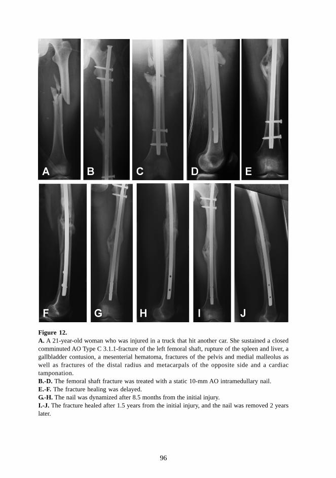
96
Figure 12.A. A 21-year-old woman who was injured in a truck that hit another car. She sustained a closedcomminuted AO Type C 3.1.1-fracture of the left femoral shaft, rupture of the spleen and liver, agallbladder contusion, a mesenterial hematoma, fractures of the pelvis and medial malleolus aswell as fractures of the distal radius and metacarpals of the opposite side and a cardiactamponation.B.-D. The femoral shaft fracture was treated with a static 10-mm AO intramedullary nail.E.-F. The fracture healing was delayed.G.-H. The nail was dynamized after 8.5 months from the initial injury.I.-J. The fracture healed after 1.5 years from the initial injury, and the nail was removed 2 yearslater.
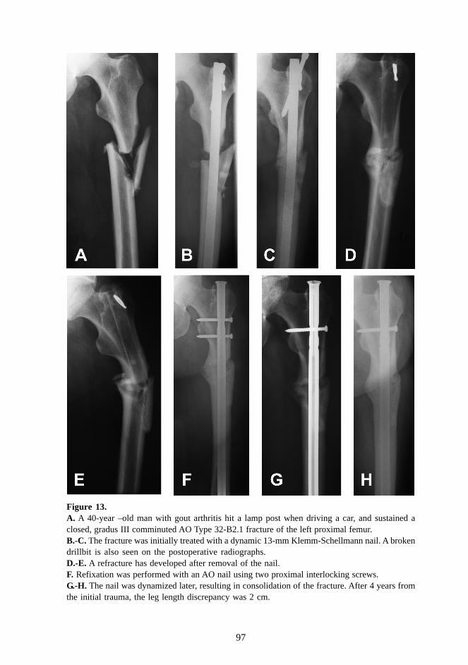
97
Figure 13.A. A 40-year –old man with gout arthritis hit a lamp post when driving a car, and sustained aclosed, gradus III comminuted AO Type 32-B2.1 fracture of the left proximal femur.B.-C. The fracture was initially treated with a dynamic 13-mm Klemm-Schellmann nail. A brokendrillbit is also seen on the postoperative radiographs.D.-E. A refracture has developed after removal of the nail.F. Refixation was performed with an AO nail using two proximal interlocking screws.G.-H. The nail was dynamized later, resulting in consolidation of the fracture. After 4 years fromthe initial trauma, the leg length discrepancy was 2 cm.
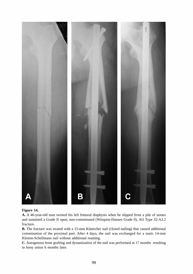
98
Figure 14.A. A 46-year-old man twisted his left femoral diaphysis when he slipped from a pile of stonesand sustained a Grade II open, non-comminuted (Winquist-Hansen Grade 0), AO Type 32-A3.2fracture.B. The fracture was treated with a 15-mm Küntscher nail (closed nailing) that caused additionalcomminution of the proximal part. After 4 days, the nail was exchanged for a static 14-mmKlemm-Schellmann nail without additional reaming.C. Autogenous bone grafting and dynamization of the nail was performed at 17 months resultingin bony union 6 months later.
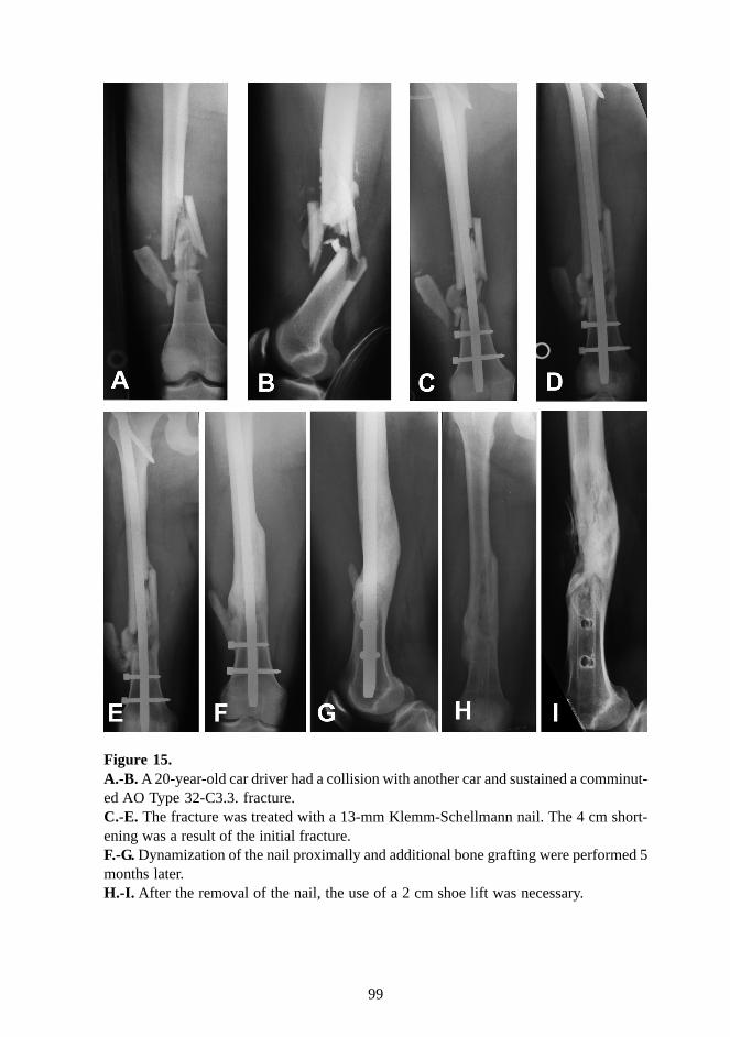
99
Figure 15.A.-B. A 20-year-old car driver had a collision with another car and sustained a comminut-ed AO Type 32-C3.3. fracture.C.-E. The fracture was treated with a 13-mm Klemm-Schellmann nail. The 4 cm short-ening was a result of the initial fracture.F.-G. Dynamization of the nail proximally and additional bone grafting were performed 5months later.H.-I. After the removal of the nail, the use of a 2 cm shoe lift was necessary.

100
6. GENERAL DISCUSSION
6.1. Incidence of femoral shaft fractures
The risk of sustaining a femoral shaft fracture is thought to vary in different populations.The peak occurrence of femoral shaft fractures in males from 15 to 24 years of age is awell known phenomenon, but the incidence figures reported vary considerably. The inci-dence in that age group, 39 per 100 000 person-years in the epidemiologic study (I), wasfound to be as high as 64.6 per 100 000 person-years in Rochester, Minnesota (122fractures during 20 years) (Arneson et al. 1988). The incidence in a male age group from20 to 29 years was 191 per 100 000 inhabitants, and in a female group 26 per 100 000inhabitants in the Netherlands (Kootstra 1973). In a Swedish population-based study(including 362 fractures during 33 years), the average annual incidences in male agegroups from 10 to 19 years and from 20 to 29 years were 14.7 and 9.2 per 100 000inhabitants in the 1980s (Bengnér et al. 1990).
In addition, the age-related features in the incidence of femoral shaft fractures appear tovary in different populations. Only one previous study, examining the mixed Asian popu-lation in Singapore, as well as the study from the Netherlands (Kootstra 1973), showed atendency similar to that observed in the epidemiologic study (I): an increase in incidencewith aging was observed only among women (Wong 1966). A few other, earlier studiesindicated that the incidence of diaphyseal femoral fractures increased with age in men andwomen. This was the case in the urban populations of Dundee and Oxford in the UnitedKingdom (Knowelden, Buhr, Dunbar 1964), of Stockholm (Hedlund and Lindgren 1986)and Malmö (Bengnér et al. 1990) in Sweden, and of Rochester, Minnesota (Arneson et al.1988). Of these, only the Rochester data showed a clear increase in distal femoral frac-tures with age, similar to the findings of the epidemiologic study (I), but the Rochestersurvey also included fractures distal to the diaphyseal section of the bone.
In the study concerning displaced femoral shaft fatigue fractures (III), the incidence inconscripts to sustain a displaced femoral shaft fatigue fracture was 1.5 per 100 000person-years in military service. Previous studies have mainly been case reports (Asal1936; Wolfe and Robertson 1945; Morris and Blickenstaff 1967; Hallel, Amit, Segal 1976;Orava 1980; Luchini, Sarokhan, Micheli 1983; Dugowson, Drinkwater, Clark 1991;Visuri and Hietaniemi 1992; Clement et al. 1993), although two more extensive studieshave been published (Provost and Morris 1969; Bargren, Tilson, Bridgeford 1971), butthe true incidence, the morphologic characteristics, and the long-term treatment resultsof these fractures have not been systematically examined before (Table 23.). For thestudy III, the age distribution of the patients was more specific: the majority of conscriptsare 19 years of age at the beginning of their basic training. In general, military service iscompleted within two years following call-up, at the age of 19 to 20. It is possible tovolunteer at 18, and deferment can be granted until the end of the year in which the manturns 28. Conscripts enter the Army, the Air Force and the Navy twice a year. Militaryservice lasts 180, 270 or 362 days. Military service on a voluntary basis for women inFinland started in 1995 (Public Information Division of the Defence Staff 2004).
101
For a military conscript, the risk of sustaining a fatigue fracture has been reported toincrease due to poor physical condition (Gilbert and Johnson 1966; Shaffer et al. 1999),low level of prior physical activity (Gilbert and Johnson 1966; Shaffer et al. 1999), lowtibial and femoral bone density (Beck et al. 1996), small diaphyseal dimensions of the tibiaand the femur (i.e. a short and thin femoral shaft) (Beck et al. 1996), malalignment andlength differences of the lower extremities (Sahi 1984; Matheson et al. 1987), poor strengthof the lower leg muscles (McBryde 1985), poor training procedures (Gilbert and Johnson1966; Greaney et al. 1983), and excessive training in specific subunits (Gilbert and John-son 1966; Hallel, Amit, Segal 1976; Meurman, Somer, Lamminen 1981; Sahi 1984; Shway-hat et al. 1994). During common conscript training, identical weight packs and otherequipment are carried, regardless of the conscript’s body stature. Provost and Morris(1969) observed that a large weight gain during the early weeks of basic training andminor prior physical activity were typical of patients sustaining a displaced femoral shaftfatigue fracture. Obese individuals may be at an increased risk for stress fractures ingeneral, although several studies on the epidemiology of stress fracture types in militaryservice failed to find an association between stress fractures and anthropometric varia-bles in either gender (Schmidt Brudvig, Gudger, Obermeyer 1983; Shaffer et al. 1999). Inone recent study, it was reported that soldiers with stress fractures detectable in scintig-raphy weighted and smoked less than control subjects (Givon et al. 2000). In the studyIII, the median body mass index of the concripts was normal, while only one conscriptwas underweight and one was obese.
Before the fracture displacement, nine conscripts experienced thigh or knee pain for 1 to6 weeks. According to Provost and Morris (1969) and Hershman et al. (Hershman, Lom-bardo, Bergfeld 1990), the pain is usually poorly localized and vague. To increase theaccuracy of diagnosis, a fulcrum test (Johnson, Weiss, Wheeler 1994), a fist test (Mil-grom et al. 1993), a hop test (Clement et al. 1993), radiographs every 2 weeks (Bargren,Tilson, Bridgeford 1971), and scintigraphy (Visuri and Hietaniemi 1992) or MRI (Kiuru2002; Lassus et al. 2002) in suspected cases have been recommended. However, dis-placement can occur even without preceding symptoms (Luchini, Sarokhan, Micheli1983), as was the case with one patient of the study III.
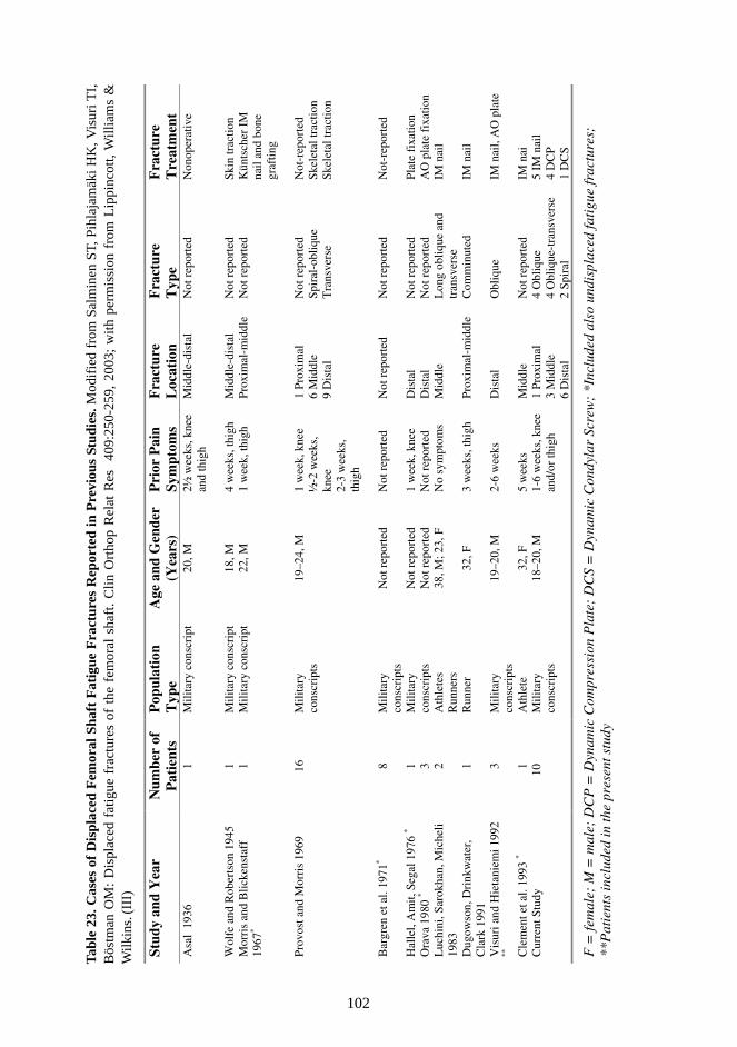
102
Tab
le 2
3. C
ases
of
Dis
plac
ed F
emor
al S
haft
Fat
igue
Fra
ctur
es R
epor
ted
in P
revi
ous
Stud
ies.
Mod
ifie
d fr
om S
alm
inen
ST,
Pih
laja
mäk
i H
K, V
isur
i TI,
Bös
tman
OM
: D
ispl
aced
fat
igue
fra
ctur
es o
f th
e fe
mor
al s
haft
. C
lin O
rtho
p R
elat
Res
40
9:25
0-25
9, 2
003;
with
per
mis
sion
fro
m L
ippi
ncot
t, W
illia
ms
&W
ilkin
s. (I
II)
Stud
y an
d Y
ear
Num
ber
of
Pat
ient
s P
opul
atio
n T
ype
Age
and
Gen
der
(Yea
rs)
Pri
or P
ain
Sym
ptom
s F
ract
ure
L
ocat
ion
Fra
ctur
e
Typ
e F
ract
ure
T
reat
men
t A
sal
1936
1
Mili
tary
con
scri
pt
20, M
2½
wee
ks, k
nee
and
thig
h M
iddl
e-di
stal
N
ot r
epor
ted
Non
oper
ativ
e
Wol
fe a
nd R
ober
tson
194
5 1
Mili
tary
con
scri
pt
18, M
4
wee
ks, t
high
M
iddl
e-di
stal
N
ot r
epor
ted
Skin
trac
tion
Mor
ris
and
Blic
kens
taff
19
67*
1 M
ilita
ry c
onsc
ript
22
, M
1 w
eek,
thig
h Pr
oxim
al-m
iddl
e N
ot r
epor
ted
Kün
tsch
er I
M
nail
and
bone
gr
aftin
g
Pr
ovos
t and
Mor
ris
1969
16
M
ilita
ry
cons
crip
ts
19–2
4, M
1
wee
k, k
nee
½-2
wee
ks,
knee
2-
3 w
eeks
, th
igh
1 Pr
oxim
al
6 M
iddl
e 9
Dis
tal
Not
rep
orte
d Sp
iral
-obl
ique
T
rans
vers
e
Not
-rep
orte
d Sk
elet
al tr
actio
n Sk
elet
al tr
actio
n
Bar
gren
et a
l. 19
71*
8 M
ilita
ry
cons
crip
ts
Not
rep
orte
d N
ot r
epor
ted
Not
rep
orte
d N
ot r
epor
ted
Not
-rep
orte
d
Hal
lel,
Am
it, S
egal
197
6 *
Ora
va 1
980
* L
uchi
ni, S
arok
han,
Mic
heli
1983
1 3 2
Mili
tary
co
nscr
ipts
A
thle
tes
Run
ners
Not
rep
orte
d N
ot r
epor
ted
38, M
; 23,
F
1 w
eek,
kne
e N
ot r
epor
ted
No
sym
ptom
s
Dis
tal
Dis
tal
Mid
dle
Not
rep
orte
d N
ot r
epor
ted
Lon
g ob
lique
and
tr
ansv
erse
Plat
e fi
xatio
n A
O p
late
fix
atio
n IM
nai
l
Dug
owso
n, D
rink
wat
er,
Cla
rk 1
991
1
Run
ner
32, F
3
wee
ks, t
high
Pr
oxim
al-m
iddl
e C
omm
inut
ed
IM n
ail
Vis
uri a
nd H
ieta
niem
i 199
2 **
3
Mili
tary
co
nscr
ipts
19
–20,
M
2-6
wee
ks
Dis
tal
Obl
ique
IM
nai
l, A
O p
late
Cle
men
t et a
l. 19
93 *
1 A
thle
te
32, F
5
wee
ks
Mid
dle
Not
rep
orte
d IM
nai
C
urre
nt S
tudy
10
Mili
tary
co
nscr
ipts
18
–20,
M
1-6
wee
ks, k
nee
and/
or th
igh
1 Pr
oxim
al
3 M
iddl
e 6
Dis
tal
4 O
bliq
ue
4 O
bliq
ue-t
rans
vers
e 2
Spir
al
5 IM
nai
l 4
DC
P 1
DC
S
F =
fem
ale;
M =
mal
e; D
CP
= D
ynam
ic C
ompr
essi
on P
late
; D
CS
= D
ynam
ic C
ondy
lar
Scre
w;
*Inc
lude
d al
so u
ndis
plac
ed fa
tigu
e fr
actu
res;
**
Pat
ient
s in
clud
ed in
the
pres
ent s
tudy
103
6.2. Injury season and injury mechanisms of femoral shaft fractures in adults
The epidemiologic study (I) showed that a third of the fractures occurred between Julyand September, suggesting a similar tendency as in the Kootstra study (1973). Based onthe data of the epidemiologic study (I), the main group sustaining high energy trauma inroad traffic accidents composed of automobile drivers.
Even if three-fourths (3/4) of the femoral shaft fractures were caused by high energytrauma, the number of low energy fractures was remarkable (I, II). The most commoncause of a diaphyseal fracture in the elderly was low energy trauma. This observation andthe excess of female patients indicate that osteoporosis is a major causative factor amongthe older age group.
Previous data regarding the specific features of femoral shaft fractures caused by lowenergy trauma are limited. In two-thirds of our patients, there was a pre-existing condi-tion likely to cause osteopenia of the femur. According to a prior study of cadaver femoraand tibia, a consistent decrease with age of all mechanical properties has been evident,with the exception of plastic modulus in femoral but not in tibial specimens (Burstein,Reilly, Martens 1976). Consequently, weakening of the mechanical strength of the femo-ra could be expected to be present in the older patients, even without any other predispos-ing factors.
It has been estimated that every third person older than 64 years falls at least once eachyear (Campbell et al. 1981; Prudham and Evans 1981; Lach et al. 1991; Forinash andMeade 2000; Carter, Kannus, Khan 2001), and that 3.5-10% of these falls can cause afracture (Baker, O’Neill, Karf 1984; Tinetti et al. 1988; Campbell et al. 1990; Forinash andMeade 2000). Using this data, the incidence of low energy femoral shaft fractures in theelderly, 8 cases per 100 000 person-years in the study II, indicates that one out of 400fractures in the elderly is a low energy femoral shaft fracture. Kannus et al. (1997)examined the trends in the incidence of fall-induced injuries in Finland for 1970-95. Theydiscovered that the number of elderly patients with fall-induced injury increased consid-erably, from 4019 in 1970 to 17 604 in 1995. The average increase was 13.5% per year.The age-adjusted incidence (per 100 000 60-year-old or older individuals) of injuriesshowed a clear increase from 1970 to 1995: 840 to 1911 in women, and 484 to 1167 inmen. In 1995, 65% of these injuries were bone fractures.
6.3. Concomitant injuries related to femoral shaft fractures in adults
As expected, the occurrence of concomitant injuries was related to high energy trauma(I). Fractures caused by low energy injuries seemed to occur without concomitant inju-ries (II), which was likewise to be expected owing to the low energy nature of the injury.In contrast, 47% of patients with femoral shaft fractures from high energy impacts havebeen reported to exhibit significant associated injuries (Taylor, Banerjee, Alpar 1994).
104
6.4. Morphology of femoral shaft fractures in adults
In concordance with a previous study (Wong 1966), most of the femoral shaft fracturesoccurred in the middle third of the femur (I). The epidemiologic study (I) showed that thepatients with spiral fractures were older than those with other types of fractures. Thestudy of the femoral shaft fractures caused by low energy injuries (II) showed that spiralfractures are typical of a low energy mechanism.
Regarding the degree of comminution, only 1/5 of the fractures represented the severelycomminuted Winquist and Hansen Grade III and Grade IV fractures (I). The originalWinquist and Hansen classification was intended for grading of comminuted fracturesonly (Winquist and Hansen 1980), but, later other authors added Grade 0 to represent anoncomminuted fracture (Johnson, Johnston, Parker 1984). However, the benefit ofseparating Grade 0 and Grade I from each other seems questionable or even meaninglessfor the treatment of the fracture.
The distribution of femoral shaft fractures according to the AO Type in the epidemiologicstudy (I): Type A 48%; Type B 39%; and Type C 13%, was close to the percentagesreported by some AO clinics (Type A 53%, Type B 34%, and Type C 13%) (Müller et al.1990). However, with 27 subgroups with additional ramifications, the AO classificationseems unnecessarily detailed and complex in classifying femoral shaft fractures. Thisobservation has been pointed out by other authors as well (Bucholz and Jones 1991;Lichtenhahn, Fernandez, Schatzker 1992; Bernstein et al. 1997). In the epidemiologicstudy (I), the use of three B types appeared to offer limited value. Moreover, it wasimpossible to elect a precise AO subtype categorization for some fractures, particularlywithin the B category.
In femoral shaft fractures caused by low energy injury (I), the most common fracturepattern was a simple spiral fracture, with minimal comminution, in the middle third of thefemoral shaft. Furthermore, in a previous series focusing on longitudinal femoral shaftfractures, 56% of the cases were found to have been caused by low energy trauma(Varjonen et al. 1990). From the radiological fracture patterns and the mechanisms ofinjury, the amount of energy involved in producing the fractures can be estimated (Alms1961). A simple spiral fracture results from a torsional low energy injury mechanism.Accordingly, comminution of the fragments was rare in the present series.
The displaced fractures were mainly located in the distal third of the femoral shaft (III).This is also in concordance with several prior reports on femoral shaft fatigue fractures(Wolfe and Robertson 1945; Provost and Morris 1969; Hallel, Amit, Segal 1976; Visuriand Hietaniemi 1992). With regard to their morphologic features, the fatigue fracturesclearly differed from femoral shaft fractures caused by low energy trauma (I). In thedistal third of the diaphysis, most of the fatigue fractures were noncomminuted obliqueor oblique-transverse types. One displaced fracture had an exceptional location in theproximal third, a site reported only once before in the literature (Provost and Morris1969). No explanatory differences in the constitution of this patient could be identified.
105
A displaced fatigue fracture can be primarily comminuted (Dugowson, Drinkwater, Clark1991), like one fracture with a Grade II comminution in the current study. The bone atthe fracture tended to be brittle, and likely to be more easily shattered during the fixationprocedure. Gentle handling of the bone during surgery is imperative also in fatigue frac-tures. (III)
6.5. Treatment, outcome and complications of femoral shaft fractures inadults
When dealing with a semiurban population similar to the one presented in the epidemio-logic study (I), extensive resource allocation for allied subspecialties, such as plasticsurgery or vascular surgery, is probably not required for femoral shaft fractures, becausesevere open fractures, Gustilo Types IIIB and IIIC, seem to be rare. Based on the epide-miologic data from this study (I), most of the femoral shaft fractures in this populationcan be treated adequately with conventional intramedullary nails, with the stability offixation and fracture alignment maintained.
In femoral shaft fractures caused by low energy injuries (II), complications occurred innearly two-thirds of our patients, and thus the complication rate associated with thesefractures must be considered high. The failures of fixation resulting in shortenings wereclearly iatrogenic complications, which could have been avoided by using static inter-locking nailing. In a previous study, 14 patients out of 133 with femoral shaft fracturessuffered a similar loss of postoperative fixation and reduction (Brumback et al. 1988b).Meticulous preoperative planning of surgical treatment of fatigue fractures is important,because errors in surgical judgement attributed to inadequate preoperative analysis of thepattern of the fracture, undetected intraoperative comminution during reaming or inser-tion of the nail, or postoperative failure to recognize an increase in comminution andinstability of the fracture were noted in that study. Undoubtedly, those conclusions arevalid for the study II as well.
In previous studies, with the high age of the patients, the complication rates recordedhave been slightly lower than in our study. In a study of patients over 65 years of age with25 subtrochanteric, 47 shaft, and 33 supracondylar femoral fractures, the overall compli-cation rate was 45% (Boyd and Wilber 1992). In that study, the local complication rate(wound infection, nonunion, malunion, and implant failure) was found to be 15%. Inanother study, 24 patients over 60 years of age with femoral shaft fractures were treatedwith locked nailing, and the total complication rate was 54% (Moran, Gibson, Gross1990). Earlier mortality rates, reported within 30-60 days of injury, have ranged from 10to 17% (Moran, Gibson, Gross 1990; Boyd and Wilber 1992). These rates approach therate of 10% of the study II. Infection after closed intramedullary nailing of the femur hasbeen previously reported to occur in less than 1% of patients (Winquist, Hansen, Clawson1984). This is considerably less than the infection rate of 10% in the study II. As for theinfection rate, a local soft-tissue lesion caused by a direct-impact injury seems to be lessimportant than the compromised general infection resistance among elderly patients withmultiple pre-existing diseases. Such patients could possibly benefit from routine perioper-ative administration of antibiotic prophylaxis.
106
Despite the low energy nature of the violence, and simple fracture configuration, unin-fected nonunion and malunion were common in the study II. Some of these complica-tions could be regarded as iatrogenic. A pre-existing general or local condition weakensthe mechanical strength of the femur and is likely to make the internal fixation of thesefractures technically very demanding. Even with an adequate and accurate preoperativeplanning and operative technique, a subsequent nonunion or malunion might be unpre-ventable among some patients with a low energy femoral shaft fracture.
Compared with the femoral shaft fractures caused by high energy trauma, the fracturesattributable to fatigue osteopathy (III) healed well, despite comminution, with a medianunion time of 3.5 months. No delayed unions or nonunions occurred. In comparison, ina recent prospective series including 83 patients with trauma treated by reamed intramed-ullary femoral nailing, the fractures united within 3.8 months, the average time to unionbeing 2.7 months (Tornetta and Tiburzi 2000). The frequency of local complications,including infection, mechanical failure, delayed union, uninfected nonunion, refracture,or simple malunion, has been 20% in traumatic femoral shaft fractures of young patients(Böstman et al. 1989). Overuse injuries present the physician a number of challenges: todetermine the diagnosis, and etiology of the injury to judge the most suitable treatment,and to ensure that the fracture does not recur (Brukner, Bennell, Matheson 1999).
In the study IV, the nonunion rate of femoral shaft fractures treated with intramedullarynailing was 12.6% (IV). When compared with this, the complication rate seems, notunexpectedly, to be lower in fatigue fractures (III).
More than one tenth of the patients of the study IV required additional surgery for failedconsolidation. The incidence of this complication was noteworthy. Two prior studies onfemoral shaft fractures also reported a nonunion rate as high as 10 percent (Kempf,Grosse, Beck 1985; Hanks, Foster, Gardea 1988), with one study even as high as 13.6percent (Eid and Deif 1980). In contrast, some authors have reported considerably lowernonunion rates, such as 0.9 percent in 520 patients with unlocked intramedullary nails(Winquist, Hansen, Clawson 1984), and 2 percent in three separate studies with 283patients (Klemm and Börner 1986), 112 patients (Wiss et al. 1986), and eighty-four pa-tients (Brumback et al. 1988b) having comminuted fractures that were treated by inter-locking nailing. In a recent prospective series including eighty-three patients treated byreamed femoral nailing, all fractures united within 115 days, the average time to unionbeing eighty days (Tornetta and Tiburzi 2000). The incidence seems to be lowest inspecialized units maintaining a strict and uniform management regimen.
The occurrence of technical faults during primary nailing can, in part, be explained by thenature of the teaching hospital. Most of the operations were performed by senior resi-dents, some of whom were still in the beginning of their learning curve for the moredemanding intramedullary nailings. Avoidance of increased comminution during intramed-ullary nailing as well as static interlocking in diastasis should be pointed out in the trainingof junior residents. A previous report also paid attention to the adverse effect of openingthe fracture site during the nailing procedure (Rokkanen, Slätis, Vankka 1969). In con-trast to several earlier reports (Curylo and Lindsey 1994; Kelly 1984; Klemm and Börner
107
1986), infection did not play an important role in the development of nonunion in thestudy IV.
In the epidemiologic study on 201 fractures (I), Type B1 fractures, which represent thefractures with a large butterfly fragment, amounted to 10.5 percent. In the study IV, alarge butterfly fragment was present in 34 percent of the nonunions. This seems toindicate a significant role of large butterfly fragments and a possible concomitant muscleinterposition in the development of failed union. In the study IV, due to the designation ofthe hospital as a referral center for the catchment area, no patients were missing from thefollow-up, and the development of truly recalcitrant nonunions was avoided as well.
The time interval between the primary nailing procedure and the correct identification ofa disturbance in the union process showed unexpected variation. In several cases, espe-cially in those with broken intramedullary nails, the delay before operative procedureswere undertaken was unacceptably long. Expectancy is seldom warranted when a diag-nosis of failed consolidation has been made. The problems associated with broken nailsand the importance of avoiding nail breakages have been stressed by some previousauthors, too (Bucholz, Ross, Lawrence 1987; Franklin et al. 1988). The conventionaldivision of the radiographic appearance of nonunion into atrophic and hypertrophic didnot seem to offer any value in the femoral shaft fractures after intramedullary nailing.
One third of the cases of the study IV required two or three reoperations before unioncould be achieved. It is difficult to compare this finding to prior documentation withvarying initial treatment of patients, including those with fractures of the subtrochantericand supracondylar regions of the femur, or even fixation of osteotomies and tibial frac-tures (Christensen 1970; Solheim and Vaage 1973; Oh et al. 1975; Okhotsky and Sou-valyan 1978; Kempf, Grosse, Rigaut 1986; Webb, Winquist, Hansen 1986; Wu and Shih1992; Curylo and Lindsey 1994; Cove et al. 1997; Ring, Barrick, Jupiter 1997; Kempf andLeung 2002). Nonunion after plate fixation requires a different policy of treatment offailed consolidation, because direct surgery on the fracture site is necessary for removalof the hardware (Webb, Winquist, Hansen 1986; Böstman et al. 1989). In light of theresults of the study IV, autogenous bone grafting from the iliac crest to the fracture sitewithout any additional measures, such as exchange intramedullary nailing or dynamiza-tion, seemed to be useless, whereas exchange intramedullary nailing was an effectivemethod without any serious complications. Bony consolidation was likewise accomplishedby dynamization, without a need for additional reoperations, in cases where the fracturegap was the principal problem. However, according to a previous report, significant axialfemoral shortening may result from dynamization (Wu 1997). The findings of the studyIV confirmed the practical value of the previously presented concept of exchange nailingwithout supplemental external bone grafting as the preferred treatment method of femoraldiaphyseal nonunion (Christensen 1970; Wu and Chen 1997; Furlong et al. 1999; Wu andChen 2002; Yu, Wu , Chen 2002). Nevertheless, in a recent series of nineteen patientswith ununited femoral shaft fracture treated by exchange nailing, nine required one ormore additional procedures before solid union was achieved, and no factors of predictivevalue could be identified (Weresh et al. 2000). Classifying nonunions into atrophic andhypertrophic was not applicable to consolidation failures after intramedullary nailing.
108
6.6. Prevention of femoral shaft fractures
Preventive measures against femoral shaft fractures should focus on the active and pas-sive protection of automobile drivers, especially young men (I), and on the preventionand treatment of osteoporosis in elderly women (I, II).
Displacement is a rare, undesirable consequence of fatigue osteopathy of the femoralshaft among young conscripts during basic military training (III). Internal fixation isrequired in all displaced femoral shaft fatigue fractures. Preventive methods should focuson an early, effective detection of these fatigue fractures to avoid fracture displacementwith a subsequent prolonged morbidity. The conclusions may be applicable beyond thecircumstances of military service, since freetime training has increased among people toa considerable degree. So far, however, only a few sports-related cases of displacedfemoral shaft fatigue fractures have been reported (Orava 1980; Luchini, Sarokhan, Micheli1983; Dugowson, Drinkwater, Clark 1990; Clement et al. 1993). The preventive methodsinclude training appropriate to individual physiology, and early recognition of fatigue oste-opathy with radiographic, scintigraphic, or preferably MRI examinations. Above all, con-scripts, even the most ambitious, should be taught to seek medical attention as soon assymptoms indicative of fatigue osteopathy emerge.
109
7. SUMMARY AND CONCLUSIONS
7.1. Demography of femoral shaft fractures in adults
Based on the data of the epidemiologic study (I), one-third of traumatic fresh femoralshaft fractures occurred during the third quadrant of the year. The distribution of malessustaining a femoral shaft fracture at that time was noteworthy. The amount of freshfemoral shaft fractures in women was highest during the fourth quadrant of the year.Even if 75% of the femoral shaft fractures were caused by high energy trauma, thenumber of low energy fractures was remarkable. The main group sustaining high energytrauma were automobile drivers in road traffic accidents. The occurrence of concomitantinjuries was, expectedly, related to high energy trauma, whereas no significant associatedinjuries were seen in patients with a low energy femoral shaft fracture. The most com-mon cause of a diaphyseal fracture in the elderly was low energy trauma. This observa-tion together with the excess of female patients indicates that osteoporosis can be a majorcausative factor among the older age group. Femoral shaft fractures caused by lowenergy violence mainly occur in patients suffering from a chronic disease or a conditioncausing osteopenia of the femur. The study (I) showed that the patients with spiral frac-tures were older than those with other types of fractures.
With regard to their incidence features, femoral shaft fractures caused by different injurymechanisms vary (I, II, III). The study of femoral shaft fractures caused by low energyinjuries (II) showed that spiral fractures are typical low energy injuries. Summarizing theresults of the study II, it appears that a fracture of the femoral shaft is only seldomcaused by low energy trauma without some predisposing factor that has weakened themechanical strength of the bone. The incidence of traumatic fractures of the adult femo-ral diaphysis was 9.9:100 000 person-years, with higher age- and gender-specific inci-dences among young men aged 15 to 24 years and elderly women 75 years old or older(I). The incidence of femoral shaft fractures caused by low energy injuries was 2.5:100000 person-years, and was higher for people aged 60 years or older (II). The incidence inconscripts to sustain a displaced femoral shaft fatigue fracture was 1.5:100 000 person-years in military service (III).
Displacement of a femoral shaft fatigue fracture can occur even without preceding symp-toms. Despite unclear knee or thigh pain for 1 to 6 weeks, femoral shaft fatigue fracturesin otherwise healthy conscripts were diagnosed only after fracture displacement.
7.2. Morphology of femoral shaft fractures in adults
The study of femoral shaft fractures caused by low energy injuries (II) showed thatspiral fractures are typical low energy injuries. Summarizing the results of the study II, itseems that a fracture of the femoral shaft is only seldom caused by low energy traumawithout some predisposing factor that has weakened the mechanical strength of the bone.
110
Regarding the morphologic features, femoral shaft fractures caused by different injurymechanisms vary (I, II, III). Most traumatic femoral shaft fractures were isolated with-out concomitant injuries (I). The most common traumatic fracture type of the femoralshaft was a simple AO Type A, non-comminuted, purely transverse, and located in themiddle third of the femur (I). The patients with spiral fractures and with fractures locatedin the distal third of the femoral shaft were older than those with other types of fractures(I). The most common fracture pattern related to low energy trauma was a simple AOType A spiral fracture, with minimal or no comminution, in the middle third of the femoralshaft (II). Femoral shaft fractures caused by low energy trauma were morphologicallydifferent from displaced fatigue fractures, which were mainly of simple AO Type A,noncomminuted oblique, or oblique-transverse, located in the distal third of the femoralshaft (III). A displaced fatigue fracture of the femoral diaphysis can be primarily commi-nuted (III).
7.3. Treatment of femoral shaft fractures in adults, and nonunions afterintramedullary nailing
Based on the data from the epidemiologic study (I), most of the femoral shaft fractures inthe community can be treated adequately with conventional intramedullary nails ratherthan interlocking nails, with the stability of fixation and fracture alignment maintained.
The complication rate in fractures caused by low energy trauma is high (II). The treat-ment of these seemingly simple fractures requires careful planning and a meticulousoperative technique (II).
Factors that predispose traumatic fresh femoral shaft fractures to nonunion after in-tramedullary nailing are related to severe fracture comminution and concomitant injuries.Without convincing signs of progressive consolidation of a femoral shaft fracture, reop-eration should be performed within six months of the primary nailing to minimize the riskof nail breakage. Exchange nailing seems to be the method of choice for the treatment ofa disturbed union. In some selected cases with primary static interlocking nailing, dy-namization alone can be considered. Bone grafting alone as a treatment of a failed union ofa femoral shaft fracture cannot be recommended (IV).
7.4. Prevention of femoral shaft fractures in adults
Based on the data of the epidemiologic study (I), preventive measures against femoralshaft fractures should generally focus on the protection of automobile drivers, especiallyyoung men, and on the effective treatment of osteoporosis in elderly women.
Furthermore, preventive methods should focus on an early, effective detection of fatiguefractures to avoid fracture displacement with subsequent prolonged morbidity, includingtraining appropriate to individual physiology, and early recognition of fatigue osteopathywith radiographic, MRI, and scintigraphic examinations. Above all, conscripts, even themost ambitious, should be taught to seek medical attention as soon as symptoms indica-tive of fatigue osteopathy emerge.
111
On the basis of the present results, the following conclusions can be drawn:
Femoral shaft fractures in adults are not exclusively results of high energy trauma. Lowenergy trauma causes 25% of the fractures (I). Femoral shaft fractures caused by lowenergy mechanism mainly occur in patients suffering from a chronic disease or a condi-tion causing osteopenia of the femur (II). Concerning displaced femoral shaft fatiguefractures, even in symptomatic fractures, the diagnosis was delayed until displacement,which can also occur without preceding symptoms (III).
With regard to their incidence and morphologic features, femoral shaft fractures causedby different injury mechanisms vary (I, II, III). The incidence of traumatic fractures ofthe adult femoral diaphysis was 9.9:100 000 person-years, with higher age- and gender-specific incidences among young men aged 15 to 24 years and elderly women 75 yearsold or older (I). The incidence of femoral shaft fractures caused by low energy injurieswas 2.5:100 000 person-years, and was higher for people aged 60 years or older (II). Theincidence in conscripts to sustain a displaced femoral shaft fatigue fracture was 1.5:100000 person-years in military service (III).
Most traumatic femoral shaft fractures are isolated without concomitant injuries (I). Themost common fracture type of the femoral shaft is a non-comminuted simple AO Type A,most of which in traumatic fractures are purely transverse and located in the middle thirdof the femur (I), in fractures related to low energy trauma spiral in the middle third of thefemur (II), and in displaced fatigue fractures oblique or oblique-transverse located in thedistal third of the femoral shaft (III). In traumatic fractures, the patients with spiralfractures, and with fractures located in the distal third of the femoral shaft, are older thanthose with other types of fractures (I). Femoral shaft fractures caused by low energytrauma are morphologically different from displaced fatigue fractures, which can also beprimarily comminuted (III).
Despite the low energy mechanism and seemingly simple morphology of fractures, treat-ment of femoral shaft fractures caused by low energy trauma is not devoid of complica-tions, and requires careful planning and a meticulous operative technique (II). In dis-placed femoral shaft fatigue fractures, gentle handling of the bone during the fracturefixation is imperative due to the extraordinary brittleness of the fracture fragments (III).Despite the additional comminution intraoperatively, the complication rate seems to below (III).
Preventive methods against traumatic femoral shaft fractures should be focused on theprotection of automobile drivers, especially young men, and on preventing low energyinjuries in elderly women (I, II). Preventive methods against femoral shaft fatigue frac-tures should focus on an early, effective detection of these fractures to avoid fracturedisplacement with a subsequent prolonged morbidity (III).
Factors that predispose traumatic fresh femoral shaft fractures to nonunion after in-tramedullary nailing are related to severe fracture comminution and concomitant injuries.Without convincing signs of consolidation in progress among traumatic femoral shaft
112
fractures treated with intramedullary nailing, reoperation should be performed within sixmonths after primary nailing to minimize the risk of nail breakage. Exchange nailing seemsto be the method of choice for the treatment of a disturbed union. In some selected cases,with primary static interlocking nailing, dynamization alone can be considered. Bone graftingalone as a treatment of a failed union of a femoral shaft fracture cannot be recommended(IV).
113
ACKNOWLEDGEMENTS
The present study was carried out at the Department of Surgery of the Central Hospital ofCentral Finland; the Department of Orthopaedics and Traumatology of the Helsinki Uni-versity Central Hospital, and at the Research Institute of Military Medicine, Central Mili-tary Hospital.
I wish to express my gratitude to Professor Seppo Santavirta, Department of Orthopae-dics and Traumatology, and Professor Risto Rintala, Department of Pediatric Surgery,providing both good facilities and the opportunity to focus on research.
I am most grateful to my supervisors, Docent Ole Böstman, M.D., Ph.D., and DocentHarri Pihlajamäki, M.D., Ph.D., for first introducing this study to me and then guiding methrough this research project. I express my sincere gratitude for their valuable adviceduring the entire work process.
I express my warmest thanks for Docent Tuomo Visuri, M.D., Ph.D. for his valuablecooperation and his expertise in fatigue fractures.
I am deeply grateful for Veikko Avikainen, M.D., Ph.D, the former Head of the Depart-ment of Orthopaedics and Traumatology of the Central Hospital of Central Finland, whoalso provided facilities for research and encouraged me in this research study. Further-more, my best thanks are due to my coauthor, Antti Kyrö, M.D., Ph.D. for his supportduring the study.
I wish to express my warmest thanks for the official reviewers of the thesis, ProfessorErkki Tukiainen, M.D., Ph.D. and Docent Matti U.K. Lehto, M.D., Ph.D. for their valu-able comments and constructive criticism on the manuscript.
I also want to thank Docent Eero Hirvensalo, M.D., Ph.D., Department of Orthopaedicsand Traumatology, and Docent Harry Lindahl, M.D., Ph.D., Department of PediatricSurgery, for the help received from the hospital organization for research. I am alsograteful to Docent Matti Mäntysaari, M.D., Ph.D., the Head of the Research Institute ofMilitary Medicine, for the possibility to use the research facilities of the Institute.
I am thankful to Pentti Kallio, M.D., Ph.D., and Docent Jari Peltonen, M.D., Ph.D., mysuperiors at the Department of Pediatric Orthopaedics of the Hospital for Children andAdolescents, for their encouragement for the completion of the study.
I also wish to thank all my colleagues during these years at the Department of Surgery ofthe Central Hospital of Central Finland, at the Department of Orthopaedics and Trauma-tology of the Töölö Hospital, and at the Department of Pediatric Surgery of the Hospitalfor Children and Adolescents. I especially want to give my warmest thanks to my closestcolleagues Päivi Salminen, M.D., Sari Pyörälä, M.D., Risto Nikku, M.D., Yrjänä Nie-tosvaara, M.D., Ph.D., Reijo Paukku, M.D., and Ilkka Helenius, M.D., Ph.D. for spur-ring me on writing.
114
I am deeply grateful to Mrs Marja Vajaranta for revising the English language of thisthesis. I heartily thank Mrs Taina Saarni and Mr Kari Kelho, for their valuable secreterialhelp and assistance for the thesis.
This study was supported financially by the Foundation for Orthopaedic and Traumato-logic Research in Finland, the Scientific Committee of National Defence, and the Bio-medicum Fonds Helsinki Foundation.
I am most grateful to my mother Julia, and my sister Saija and her family for theirencouragement and patience throughout these years. I warmly thank my friends Pia andVincent, Bernd and Ellen, Bärbl and Richard, Bettina, Kirsi, Elina, and the members of theMartha Surgeons for their support and friendship. Without Marja-Liisa, the coach, andthe members of the fencing team I would not have known how to fence through thisstudy.
Helsinki, June 2005
Sari Salminen
115
REFERENCES
Acker JH, Murphy C, D’Ambrosia R: Treatment of fractures of the femur with the Grosse-Kempfrod. Orthopedics 8:1393-1401, 1985.
Alho A, Strømsøe K, Ekeland A: Locked intramedullary nailing of femoral shaft fractures. JTrauma 31:49-59, 1991.
Alho A, Ekeland A, Strømsøe K: Femoral shaft fractures in the elderly treated with Grosse-Kempf slotted locked intramedullary nail. Ann Chir Gynaecol 81:366-371, 1992.
Alho A, Moen O, Husby T, Rønningen H, Skjeldal S: Slotted versus non-slotted locked in-tramedullary nailing for femoral shaft fractures. Arch Orthop Trauma Surg 111:91-95, 1992.
Alho A: Concurrent ipsilateral fractures of the hip and shaft of the femur. Ann Chir Gynaecol86:326-336, 1997.
Allen WC, Piotrowski G, Burstein AH, Frankel VH: Biomechanical principles of intramedullaryfixation. Clin Orthop Relat Res 60:13-20, 1968.
Allgöwer M, Spiegel PG: Internal fixation of fractures: evolution of concepts. Clin Orthop RelatRes 38:26-29, 1979.
Alms M: Fracture mechanics. J Bone Joint Surg Br 43:162-166, 1961.Alonso J, Geissler W, Hughes JL: External fixation of femoral fractures. Indications and limita-
tions. Clin Orthop Relat Res 241:83-88, 1989.Anastopoulos G, Asimakopoulos A, Exarchou E, Pantazopoulos T: Closed interlocked nailing in
comminuted and segmental femoral shaft fractures. J Trauma 35:772-775, 1993.Anderson LD, Gilmer Jr WS, Tooms RE: Experimental fractures treated with loose and tight
fitting medullary nails. Surg Forum 13:455-457, 1962.Anderson JT, Gustilo RB: Immediate internal fixation in open fractures. Orthop Clin North Am
11:569-578, 1980.Anderson MW, Greenspan A: Stress fractures. Radiology 199:1-12, 1996.Anseroff NJ: Die Arterien der langen Knochen des Menschen. Z Anat Entwicklungsgeschichte
103:793, 1934.Arneson TJ, Melton III LJ, Lewallen DG, O´Fallon WM: Epidemiology of diaphyseal and distal
femoral fractures in Rochester, Minnesota, 1965-1984. Clin Orthop Relat Res 234:188-194,1988.
Aro H, Eerola E, Aho AJ: Development of nonunions in the rat fibula after removal of periostealneural mechanoreceptors. Clin Orthop Relat Res 199:292-299, 1985.
Arslan H, Kapukaya A, Kesemenli CC, Necmioglu S, Subasi M, Coban V: The floating knee inadults: twenty-four cases of ipsilateral fractures if the femur and tibia. Acta Orthop Trau-matol Turc 37:107-112, 2003.
Asal: Überlastungsschäden am Knochensystem bei Soldaten. Arch Klin Chir 186:511-522, 1936.Ashby ME, Anderson JC: Treatment of fractures of the hip and ipsilateral femur with the Zickel
device: a report of three cases. Clin Orthop Relat Res 127:156-160, 1977.Atkinson PJ, Weatherell JA: Variation in the density of the femoral diaphysis with age. J Bone
Joint Surg Br 49:781-788, 1967.Aufranc OE, Jones WN, Stewart Jr WG: Femoral shaft fracture with ipsilateral ankylosed hip.
JAMA 192:1153-1155, 1965.Bach AW, Hansen Jr ST: Plates versus external fixation in severe open tibial shaft fractures. A
randomized trial. Clin Orthop Relat Res 241:89-94, 1989.Baker SP, O’Neill B, Haddon Jr W, Long WB: The Injury Severity Score: a method for describing
patients with multiple injuries and evaluating emergency care. J Trauma 14:187-196, 1974.
116
Baker SP, O´Neill B, Karpf RS: The Injury Fact Book. Pp.40. Lexington Books, Lexington 1984.Bankston AB, Keating EM, Saha S: The biomechanical evaluation of intramedullary nails in
distal femoral shaft fractures. Clin Orthop Relat Res 276:277-282, 1992.Barber FA, Fuller MW, Ratliff HW, Sutker AN, Britt BT: Knee injury following ipsilateral femur
fractures. Am J Knee Surg 1:71-75, 1988.Bargren JH, Tilson DH, Bridgeford OE: Prevention of displaced fatigue fractures of the femur. J
Bone Joint Surg Am 53:1115-1117, 1971.Barnes DI, Broude HB: False aneurysm of the profunda femoris artery complicating fracture of
the femoral shaft and treated by transcatheter embolization. A case report. S Afr Med J.67:824-826, 1985.
Barr H, Santer G, Stevenson I: Occult femoral artery injury in relation to fracture of the femoralshaft. J Cardiovasc Surg (Torino) 28:193-195, 1987.
Barrick EF, Mulhern PJ: Technical difficulties with the Brooker-Wills nail in acute fractures of thefemur. J Orthop Trauma 4:144-150, 1990.
Baumgärtel F, Dahlen C, Stiletto R, Gotzen L: Technique of using the AO –femoral distractor forfemoral intramedullary nailing. J Orthop Trauma 8:315-321, 1994.
Beck JP, Collins JA: Theoretical and clinical aspects of posttraumatic fat embolism syndrome.Instr Course Lect 22:38-87, 1973.
Beck TJ, Ruff CB, Mourtada FA, Shaffer RA, Maxwell-Williams K, Kao GL, Sartoris DJ, BrodineS: Dual-energy X-ray absorptiometry derived structural geometry for stress fracture pre-diction in male U.S. Marine Corps conscripts. J Bone Miner Res 11:645-653, 1996.
Beck TJ: The prediction of stress fractures. In: Burr DB, Milgrom C (eds.): Musculoskeletalfatigue and stress fractures. Pp.73-83. CRC Press, Boca Raton London New York Wash-ington D.C. 2001.
Behr JT, Apel DM, Pinzur MS, Dobozi WR, Behr MJ: Flexible intramedullary nails for ipsilateralfemoral and tibial fractures. J Trauma 27:1354-1357, 1987.
Belkin SC: Stress fractures in athletes. Orthop Clin North Am 11:735-741, 1980.Bengnér U, Ekbom T, Johnell O, Nilsson BE: Incidence of femoral and tibial shaft fractures.
Epidemiology 1950-1983 in Malmö, Sweden. Acta Orthop Scand 61:251-254, 1994.Bennell KL, Malcolm SA,Thomas SA, Reid SJ, Brukner PD, Ebeling PR, Wark JD: Risk factors
for stress fractures in track and field athletes: a 12 month prospective study. Am J SportsMed 24: 810-818, 1996.
Bennett FS, Zinar DM, Kilgus DJ: Ipsilateral hip and femoral shaft fractures. Clin Orthop RelatRes 296:168-177, 1993.
Bentzen SM, Hvid I, Jørgensen J: Mechanical strength of tibial trabecular bone evaluated by X-ray computed tomography. J Biomech 20:743-752, 1987.
Bergman M, Tornetta P, Kerina M Sandhu H, Simon G, Deysine G, Alcindor F: Femur fracturescaused by gunshots: treatment by immediate reamed intramedullary nailing. J Trauma34:783-785, 1993.
Bernstein J, Monaghan BA, Silber JS, DeLong WG: Taxonomy and treatment - a classificationof fracture classifications. J Bone Joint Surg Br 79:706-707, 1997.
Bhandari M, Guyatt GH, Tong D, Adili A, Shaughnessy SG: Reamed versus nonreamed in-tramedullary nailing of lower extremity long bone fractures: a systematic overview andmeta-analysis. J Orthop Trauma 14:2-9, 2000.
Bhandari M, Zlowodzki M, Tornetta III P, Schmidt A, Templeman DC: Intramedullary nailingfollowing external fixation in femoral and tibial shaft fractures. J Orthop Trauma 19:140-144,2005.
Blacksin MF, Zurlo JV, Levy AS: Internal derangement of the knee after ipsilateral femoral shaftfracture: MR imaging findings. Skeletal Radiol 27:434-439, 1998.
117
Blake R, McBryde A: The floating knee. South Med J 68:13-16, 1975.Blichert-Toft M, Hammer A: Treatment of fractures of the femoral shaft. Acta Orthop Scand
41:341-353, 1970.Blumberg KD, Foster WC, Blumberg JF, Adelaar RS, Deblois ME, Hussey RW, Loughran TP,
Cordea JA: A comparison of the Brooker-Wills and Russell-Taylor nails for treatment ofpatients who have fractures of the femoral shaft. J Bone Joint Surg Am 72:1019-1024, 1990.
Boden B, Speer K: Femoral stress fractures. Clin Sports Med 16:307-317, 1997.Bone L, Bucholz R: The management of fractures in the patient with multiple trauma. J Bone
Joint Surg Am 68:945-949, 1986.Bone LB, Johnson KD, Weigelt J, Scheinberg R: Early versus delayed stabilization of femoral
fractures. A prospective randomized study. J Bone Joint Surg Am 71:336-340, 1989.Bonnevialle P, Cauhepe C, Alqoh F, Bellumore Y, Rongieres M, Mansat M: Risques et résultats
de l’enclouage simultané des fractures bifémorales. Rev Chir Orthop Reparatr Appar Mot86:598-607, 2000.
Bono ES, Smolinski P, Casagranda A, Xu J: Three-dimensional trabecular alignment model.Comput Methods Biomech Biomed Engin 6:125-131, 2003.
Bose WJ, Corces A, Anderson LD: A preliminary experience with the Russell-Taylor reconstruc-tion nail for complex femoral fractures. J Trauma 32:71-76, 1992.
Bosse MJ, MacKenzie EJ Riemer BL, Brumback RJ, McCarthy ML, Burgess AR, Gens DR, YasuiY: Adult respiratory distress syndrome, penumonia, and mortality following thoracicinjury and a femoral fracture treated either with intramedullary nailing with reaming or witha plate. A comparative study. J Bone Joint Surg Am 79:799-809, 1997.
Boulanger BR, Stephen D, Brenneman FD: Thoracic trauma and early intramedullary nailing offemur fractures: are we doing harm? J Trauma 43:24-28, 1997.
Boyd Jr AD, Wilber JH: Patterns and complications of femur fractures below the hip in patentsover 65 years of age. J Orthop Trauma 6:167-174, 1992.
Boyer MI, Ribeiro E, Hu R, Powell JN: Small versus large diameter closed-section femoral nailsfor the treatment of femoral shaft fractures: is there a difference? J Trauma 41:279-282, 1996.
Breederveld RS, Patka P, Van Mourik JC: Refractures of the femoral shaft. Neth J Surg 37:114-116, 1985.
Breithaupt J: Zur Pathologie des Menschlichen Fusses. Med Ztg Berlin 36:169, 1855.Broekhuizen TH: Femoral fractures. Indications and biomechanics of external fixation. Thesis.
University of Amsterdam 1988.Brooker AF, Bowerman JW, Robinson RA, Riley Jr LH: Ectopic ossification following total hip
replacement. Incidence and a method of classification. J Bone Joint Surg Am 55:1629-1632,1973.
Brookes M: The Blood Supply of Bone. An Approach to Bone Biology. Butterworths, London1971.
Broos PL, Miserez MJ, Rommens PM: The monofixator in the primary stabilization of femoralshaft fractures in the multiply injured patients. Injury 23:525-528, 1992.
Brosh T, Arkan M: Toward early detection of the tendency to stress fractures. Clin Biomech9:111-116, 1994.
Brukner P, Khan K: Clinical sports medicine. McCraw Hill Sydney 1993.Brukner P, Bennell K, Matheson G: Stress fractures. Blackwell Science, Australia 1999.Brumback RJ: The rationales of interlocking nailing of the femur, tibia, and humerus. Clin Orthop
Relat Res 324:292-230, 1996.Brumback RJ, Handal JA, Poka A, Burgess AR, Feddis G: Radiographic analysis of the Brooker-
Wills interlocking nail in the treatment of comminuted femoral fractures. J Orthop Trauma1:120-129, 1987.
118
Brumback RJ, Reilly JP, Poka A, Lakatos RP, Bathon GH, Burgess AR: Intramedullary nailing offemoral shaft fractures. Part I: Decision-making errors with interlocking fixation. J BoneJoint Surg Am 70:1441-1452, 1988a.
Brumback RJ, Uwagie-Ero S, Lakatos RP, Poka A, Bathon GH, Burgess AR: Intramedullarynailing of femoral shaft fractures. Part II: Fracture-healing with static interlocking fixation.J Bone Joint Surg Am 70:1453-1462, 1988b.
Brumback RJ, Ellison Jr PS, Poka A, Lakatos R, Bathon GH, Burgess AR: Intramedullary nailingof open fractures of the femoral shaft. J Bone Joint Surg Am 71:1324-1331, 1989.
Brumback RJ, Wells JD, Lakatos R, Poka A, Bathon GH, Burgess AR: Heterotopic ossificationabout the hip after intramedullary nailing for fractures of the femur. J Bone Joint Surg Am72:1067-1073, 1990.
Brumback RJ, Ellison TS, Molligan H, Molligan DJ, Mahaffey S, Schmidhauser C: Pudendalnerve palsy complicating intramedullary nailing of the femur. J Bone Joint Surg Am 74:1450-1455, 1992a.
Brumback RJ, Ellison TS, Poka A, Bathon GH, Burgess AR: Intramedullary nailing of femoralshaft fractures. Part III: Long-term effects of static interlocking fixation. J Bone Joint SurgAm 74:106-112, 1992b.
Brumback RJ, Virkus WW: Intramedullary nailing of the femur: reamed versus nonreamed. J AmAcad Orthop Surg 8:83-90, 2000.
Brundage SI, McGhan R, Jurkowich GJ, Mack CD, Maier RV: Timing of femur fracture fixation:effect on outcome in patients with thoracic and head injuries. J Trauma 52:299-307, 2002.
Brunet ME, Cook SD, Brinker MR, Dickinson JA: A survey of running injuries in 1505 compet-itive and recretional runners. J Sports Med 30:307-315 ,1990
Bråten M, Terjesen T, Rossvoll I: Femoral anteversion in normal adults. Ultrasound measure-ments in 50 men and 50 women. Acta Orthop Scand 63:29-32, 1992.
Bråten M, Nordby A, Terjesen T: Bone density and geometry after locked intramedullarynailing. Computed tomography of 8 femoral fractures. Acta Orthop Scand 64:79-81, 1993.
Bråten M, Terjesen T, Rossvoll I: Torsional deformity after intramedullary nailing of femoralshaft fractures. Measurement of anteversion angles in 110 patients. J Bone Joint Surg Br75:799-803, 1993.
Bråten M, Terjesen T, Rossvoll I: Femoral shaft fractures treated by intramedullary nailing. Afollow-up study focusing on problems related to the method. Injury 26:379-383, 1995.
Bucholz RW, Rathjen K: Concomitant ipsilateral fractures of the hip and femur treated withinterlocking nails. Orthopedics 8:1402-1406, 1985.
Bucholz RW, Ross SE, Lawrence KL: Fatigue fracture of the interlocking nail in the treatment offractures of the distal part of the femoral shaft. J Bone Joint Surg Am 69:1391-1399, 1987.
Bucholz RW, Jones A: Fractures of the shaft of the femur. J Bone Joint Surg Am 73:1561-1566,1991.
Bucholz R, Brumback R: Fractures of the shaft of the femur. In: Rockwood C, Green D, BucholzR et al. (eds.): Rockwood and Green’s Fractures in Adults, 4th ed. Pp. 1827-1918, Lippin-cott-Raven, Philadelphia, 1996.
Buck G: An improved method of treating fractures of the thigh. Transact N Y Acad Med 2:232-250, 1861.
Buck-Gramcko H: Behandlungsergebnisse von Oberschenkelschaftfrakturen. Archiv Orth Un-fall Chir 50:166-181, 1958.
Buckwalter JA: Activity vs. rest in the treatment of bone, soft-tissue and joint injuries. IowaOrthop J 15:29-42, 1995.
Buckwalter JA, Glimcher MJ, Cooper RR, Recker R: Bone biology II: Formation, form, modeling,remodeling, and regulation of cell function. Instr Course Lect 45:387-399, 1996.
119
Burr DB, Martin RB, Schaffler MB, Radin EL: Bone remodeling in response to bone strengthmicroscopic cracks. In vivo fatigue microdamage. J Biomech 18:189-200, 1985.
Burr DB, Milgrom C, Boyd RD, Higgins WL, Robin G, Radin EL: Experimental stress fractures ofthe tibia. Biological and mechanical aetiology in rabbits. J Bone Joint Surg Br 72:370-375,1990.
Burr DB, Milgrom C, Fyhrie D, Forwood M, Nyska M, Finestone A, Hoshaw S, Saiag E, SimkinA: In vivo measurement of human tibial strains during vigorous activity. Bone 18:405-410,1996.
Burstein AH, Reilly DT, Martens M: Aging of bone tissue: mechanical properties. J Bone JointSurg Am 58:82-86, 1976.
Burwell HN: Internal fixation in the treatment of fractures of the femoral shaft. Injury 2:235-244,1971.
Buxton RA: The use of Perkins’ traction in the treatment of femoral shaft fractures. J Bone JointSurg Br 63:362-366, 1981.
Buzdon MM, Napolitano LM, Shi HJ, Ceresoli DM, Rauniya R, Bass BL: Femur fracture inducessite-specific changes in T-cell immunity. J Surg Res 82:201-208, 1999.
Böhler L: The treatment of fractures. Grune & Stratton, New York London 1957.Böstman O, Varjonen L, Vainionpää S, Majola A, Rokkanen P: Incidence of local complications
after intramedullary nailing and after plate fixation of femoral shaft fractures. J Trauma29:639-645, 1989.
Böstman OM: Refractures after removal of a condylar plate from the distal third of the femur. JBone Joint Surg Am 72:1013-1018, 1990.
Callanan I, Choudhry V, Smith H: Perineal sloughing as a result of pressure necrosis from thetraction post during prolonged bilateral femoral nailing. Injury 25:472, 1994.
Cameron CD, Meek RN, Blachut PA, O’Brien PJ, Pate GC: Intramedullary nailing of the femoralshaft: a prospective, randomized study. J Orthop Trauma 6:448-451, 1992.
Campbell AJ, Reinken J, Allan BC, Martinez GS: Falls in old age: a study of frequency andrelated clinical factors. Age Ageing 10:264-270, 1981.
Campbell AJ, Borrie MJ, Spears GF, Jackson SL, Brown JS, Fitzgerald JL: Circumstances andconsequences of falls experienced by a community population 70 years and over during a
prospective study. Age Ageing 19:136-141, 1990.Canadian Orthopaedic Trauma Society: Nonunion following intramedullary nailing of the femur
with and without reaming. Results of a multicenter randomized clinical trial. J Bone JointSurg Am 85:2093-2096, 2003.
Carr CR, Turnipseed D: Experiences with intramedullary fixation of compound femoral fracturesin war wounds. J Bone Joint Surg Am 35:153-171, 1953.
Carr CR, Miller RE: Management of difficult fractures of the femoral shaft. South Med J 51:642-653, 1958.
Carr CR, Wingo CH: Fractures of the femoral diaphysis. A retrospective study of the results andcosts of treatment by intramedullary nailing and by traction in a spica cast. J Bone JointSurg Am 55:690-700, 1973.
Carter DR: Anisotropic analysis of strain rosette information from cortical bone. J Biomech11:199-202, 1978.
Carter DR, Caher WE, Spengler DM, Frankel VH: Uniaxial fatigue of human cortical bone. Theinfluence of tissue physical characteristics. B Biomech 14:461-470, 1981.
Carter ND, Kannus P, Khan KM: Exercise in the prevention of falls in older people. A systematicliterature review examining the rationale and the evidence. Sports Med 31:427-438, 2001.
Casey MJ, Chapman MW: Ipsilateral concomitant fractures of the hip and femoral shaft. J BoneJoint Surg Am 61:503-509, 1979.
120
Cavadias AX, Trueta J: An experimental study of the vascular contribution to the callus offracture. Surg Gynecol Obstet 120:731-747, 1965.
Chadwick CJ, Hayes AG: The treatment of femoral shortening after intramedullary nailing. Inju-ry 19:35-38, 1988.
Chalmers J, Gray DH, Rush J: Observations on the induction of bone in soft-tissues. J BoneJoint Surg Br 57:36-45, 1975.
Chamay A, Tschantz P: Mechanical influences in bone remodeling. Experimental research onWolff’s law. J Biomech 5:173-180, 1972.
Chapman MW, Mahoney M: The role of early internal fixation in the management of openfractures. Clin Orthop Relat Res 138:120-131, 1979.
Chapman MW: The use of immediate internal fixation in open fractures. Orthop Clin North Am11:579-591, 1980.
Chapman MW: The role of intramedullary fixation in open fractures. Clin Orthop Relat Res212:26-34, 1986.
Chapman MW: The role of intramedullary nailing in fracture management. In: Browner BD (ed.):The Science and Practice of Intramedullary Nailing. Pp.27-41. Williams & Wilkins Baltimore1996.
Chapman MW: The effect of reamed and nonreamed intramedullary nailing on fracture healing.Clin Orthop Relat Res 355:S230-S238, 1998.
Charash WE, Fabian TC, Croce MA: Delayed surgical fixation of femur fractures is a risk factorfor pulmonary failure independent of thoracic trauma. J Trauma 37:667-672, 1994.
Charnley J, Guindy AS: Delayed operation in the open reduction of fractures of long bones. JBone Joint Surg Br 43:664-671, 1961.
Charnley J: The Closed Treatment of Common Fractures. 3rd ed. Pp. 166-196. Churchill Living-stone, Edinburgh and London 1974.
Chaturvedi S, Sahu SC: Ipsilateral concomitant fractures of the femoral neck and shaft. Injury24:243-246, 1993.
Chen CE, Wang JW: Floating knee with ipsilateral knee dislocation: case report. J Trauma44:735-737, 1998.
Cheng JC, Tse PY, Chow YY: The place of the dynamic compression plate in femoral shaftfractures. Injury 16:529-534, 1985.
Chevalley F, Amstutz C, Bally A: Experimental study of external fixation of femoral fractures.Mechanical properties of different kinds of fixation. Clinical implications. Rev Chir OrthopReparatrice Appar Mot 78:423-429, 1992.
Christie J, Court-Brown C, Kinninmonth AW, Howie CR: Intramedullary locking nails in themanagement of femoral shaft fractures. J Bone Joint Surg Br 70:206-210, 1988.
Christensen NO: Shortening in pseudarthrosis. Treatment with the Küntscher distractor. ActaOrthop Scand 41:363-368, 1970.
Christensen NO: Küntscher intramedullary reaming and nail fixation for non-union of fractureof the femur and the tibia. J Bone Joint Surg Br 55:312-318, 1973.
Clatworthy MG, Clark DI, Gray DH, Hardy AE: Reamed versus undreamed femoral nails. Arandomised, prospective trial. J Bone Joint Surg Br 80:485-489, 1998.
Clawson DK, Smith RF, Hansen ST: Closed intramedullary nailing of the femur. J Bone Joint SurgAm 53:681-692, 1971.
Clement DB, Ammann W, Taunton JE, Lloyd-Smith R, Jesperson D, McKay H, Goldring J,Matheson GO: Exercise-induced stress injuries to the femur. Int J Sports Med 14:347-352,1993.
Coetzee JC, van der Merwe EJ: Exposure of surgeons-in-training to radiation during intramedul-lary fixation of femoral shaft fractures. S Afr Med J 81:312-314, 1992.

121
Connolly JF, King P: Closed reduction and early cast-brace ambulation in the treatment offemoral fractures. I: An in vivo quantative analysis of immobilization in skeletal tractionand a cast brace. J Bone Joint Surg Am 55:1559-1580, 1973.
Contzen H: Development of intramedullary nailing and the interlocking nail. Aktuelle Traumatol17:250-252, 1987.
Copeland CE, Mitchell KA, Brumback RJ, Gens DR, Burgess AR: Mortality in patients withbilateral femoral fractures. J Orthop Trauma 12:315-319, 1998.
Coupe KJ, Beaver RL: Arterial injury during retrograde femoral nailing: a case report of injury toa branch of the profunda femoris artery. J Orthop Trauma 15:140-3, 2001.
Coupland RM: The Red Cross Wound Classification. P. 15. International Committee of the RedCross, Geneva 1991.
Coupland RM: War wounds of limbs. Surgical management. Butterworth Heinemann, Oxford2000.
Court-Brown CM, Browner BD: locked nailing of femoral fractures. In: Browner BD (ed.): TheScience and Practice of Intramedullary Nailing. Pp.161-182.Williams & Wilkins Baltimore1996.
Court-Brown CM, Masquelet AC, Schenk B: Open fractures. In: Kempf I, Leung KS (eds.):Practice of Intramedullary Locked Nails. Advanced Techniques and Special ApplicationsRecommended by “Association Internationale pour l’Ostéosynthèse Dynamique” (AIOD).Pp.29-56. Springer-Verlag, Berlin Heidelberg 2002.
Cove JA, Lhowe DW, Jupiter JB, Siliski JM: The management of diaphyseal nonunions. JOrthop Trauma 11:513-520, 1997.
Coventry MB, Scanlon PW: The use of radiation to discourage ectopic bone. A nine-year studyin surgery about the hip. J Bone Joint Surg Am 63:201-208, 1981.
Cowan DN, Jones BH, Frykman PN, Polly Jr DW, Harman EA, Rosenstein RM, Rosenstein MT:Lower limb morphology and risk of overuse injury among male infantry trainees. Med SciSports Exer 28:945-952, 1996.
Cummings SR and Melton III LJ: Epidemiology and outcomes of osteoporotic fractures. Lancet359:1761-1767, 2002.
Curylo LJ, Lindsey RW: Shaft nonunions: current etiology and outcome of treatment. Ortho-paedics 2:465-473, 1994.
Dabezies EJ, D’Ambrosia R, Shoji H, Norris R, Murphy G: Fractures of the femoral shaft treatedby external fixation with the Wagner device. J Bone Joint Surg Am 66:360-364, 1984.
Dabezies EJ, D’Ambrosia R: Fracture treatment for the multiply injured patient. Instr CourseLect 35:13-21, 1986.
Daffner RH: Stress fractures: current concepts. Skeletal Radiol 2:221-229, 1978.Daffner RH, Pavlov H: Stress fractures: current concepts. Am J Roentgenol 159:245-252, 1992.Dagrenant D, Kempf I: Treatment of nonunions. In: Kempf I, Leung KS (eds.): Practice of
Intramedullary Locked Nails. Advanced Techniques and Special Applications Recom-mended by “Association Internationale pour l’Ostéosynthèse Dynamique” (AIOD). Pp.57-63. Springer-Verlag, Berlin Heidelberg 2002.
Dahlström S: Body build and physique of young Finnish adults. Studies based on inducteesurveys and anthropometric measurements on 223 conscripts. Thesis. Pp. 25-54. Universi-ty of Turku, Turku 1981.
Danckwardt-Lillieström G: Reaming of the medullary cavity and its effect on diaphyseal bone. Afluorochromic, microangiographic and histologic study on the rabbit tibia and dog femur.Acta Orthop Scand 128:S1-S163, 1969.
Danis MR: Le traitement operatoire des fractures. J Internat Chir 7:311-314, 1947.Dawson-Saunders B, Trapp RG: Basic and Clinical Biostatistics. Appleton & Lange, Norwalk
1990.
122
DeBastiani G, Aldegheri R, Renzi Brivio L: The treatment of fractures with a dynamic axial fixator.J Bone Joint Surg Br 66:538-545, 1984.
DeCampos J, Vangsness Jr CT, Merritt PO, Sher J: Ipsilateral knee injury with femoral fracture.Examination under anesthesia and arthroscopic evaluation. Clin Orthop Relat Res 300:178-182, 1994.
Delaney WM, Street DM: Fracture of femoral shaft with fracture of neck of same femur; treat-ment with medullary nail for shaft and Knowles pins for neck. J Int Coll Surg 19:303-312,1953.
DeLee JC: Ipsilateral fracture of the femur and tibia treated in a quadrilateral cast brace. ClinOrthop Relat Res 142:115-122, 1979.
Demetriades D, Murray J, Brown C, Velmahos G, Salim A, Alo K, Rhee P: High-level falls: typeand severity of injuries and survival outcome according to age. J Trauma 58:342-345, 2005.
Dencker HM: Fractures of the shaft of the femur. A clinical study based on 1 003 fracturestreated in Swedish hospitals during the three-year period 1952 to 1954. Thesis. Pp.1-135.University of Gothenburg, Gothenburg 1963.
Dicker RC: Analyzing and interpreting data. In: Gregg MB (ed.): Field Epidemiology. 2nd ed. Pp.132-182. Oxford University Press, NewYork 2002.
Dickob M, Mommsen U: Damage to the knee ligament as a concomitant injury in femoral shaftfractures. Unfallchirurgie 18:218-223, 1998.
Dufek JS, Bates BT: Biomechanical factors associated with injury during landing in jump sports.Sports Med 12:326-327, 1991.
Dufour D, Kromann Jensen S, Owen Smith M, Salmela J, Stening GF, Zetterström B: Surgery ofthe victims of war. International Committee of the Red Cross, Geneva 1988.
Dugdale TW, Degnan GG, Turen CH: The use of computed tomographic scan to assess femoralmalrotation after intramedullary nailing. A case report. Clin Orthop Relat Res 279:258-263,1992.
Dugowson CE, Drinkwater BL, Clark JM: Nontraumatic femur fracture in an oligomenorrheicathlete. Med Sci Sports Exer 23:1323-1325, 1991.
Dunbar WH, Coleman SS: Occult knee injuries associated with ipsilateral femoral fractures: aprospective study. Orthop Trans 2:253, 1978.
Dunham CM, Bosse MJ, Clancy TV, Cole Jr FJ, Coles MJ, Knuth T, Luchette FA, Ostrum R,Plaisier B, Poka A, Simon RJ, EAST Practice Management Guidelines Work Group: Practiceguidelines for the optimal timing of long-bone fracture-stabilization in polytrauma pa-tients: the EAST Practice Management Guidelines Work Group. J Trauma 50:958-967, 2001.
Dunlap K, Shands Jr AR, Hollister Jr LC, Gaul Jr JS, Streit HA: A new method for determinationof torsion of the femur. J Bone Joint Surg Am 35:289-311, 1953.
Ebraheim NA, Paley KJ: Penetration of the distal femur by the distal locking device of Brooker-Wills interlocking nail. Clin Orthop Relat Res 297:218-223, 1993.
Ecke H, Neubert C, Neeb W: Analyze der Behandlungsergebnisse von 1127 Patienten mit Ober-schenkelfrakturen aus der Bundesrepublik Deutschland und der Schweiz. Unfallchirurgie6:38-43, 1980.
Eid AM, Deif AI: Aetiological fractors in non-union following Küntscher intramedullary nailingof the femur. Arch Orthop Traumat Surg 96:213-220, 1980.
Einhorn TA: Enhancement of fracture-healing. J Bone Joint Surg Am 77:940-956, 1995.Ekenman I: The stress fractures in athletes an investigation of possible predisposing factors.
Thesis. Karolinska Institutet, Stockholm 1998.Eriksson E, Hovelius L: Ender nailing in fractures of the diaphysis of the femur. J Bone Joint
Surg Am 61:1175-1181, 1979.Esser MP, Cloke JH, Hart JA: Closed Küntscher nailing: a clinical review after 20 years. Injury
13:455-459, 1982.
123
Evans FG, Pedersen HE, Lissner HR: The role of tensile stress in the mechanism of femoralfractures. J Bone Joint Surg Am 33:485-501, 1951.
Falkenberg J: An experimental study of the rate of fracture healing. Thesis. Acta Orthop ScandSuppl. 50. University of Lund 1961.
Fisher WD, Hamblen DL: Problems and pitfalls of compression fixation of long bone fractures:a review of results and complications. Injury 10:99-107, 1978.
Floyd WN Jr, Butler JE, Clanton T, Kim EE, Pjura G: Roentgenologic diagnosis of stress frac-tures and stress reactions. South Med J 80:433-9, 1987.
Forinash K, Meade DM: Trauma in the elderly. Emerg Med Serv 29:79-88, 2000.Franklin JL, Winquist RA, Benirschke SK, Hansen Jr ST: Broken intramedullary nails. J Bone
Joint Surg Am 70:1463-1471, 1988.Fraser RD, Hunter GA, Waddell JP: Ipsilateral fractures of the femur and tibia. J Bone Joint Surg
Br 60:510-515, 1978.Fredericson M, Bergman AG, Hoffman KL, Dillingham MS: Tibial stress reaction in runners.
Correlation of clinical symptoms and scintigraphy with a new magnetic resonance imaginggrading system. Am J Sports Med 23:472-481, 1995.
Friberg O: Leg length asymmetry in stress fractures: a clinical and radiological study. J SportsMed 22:485-488, 1982.
Friedman RJ, Wyman Jr ET: Ipsilateral hip and femoral shaft fractures. Clin Orthop Relat Res208:188-194, 1986.
Frost HM: Presence of microscopic cracks in vivo in bone. Bull Henry Ford Hosp 8:25, 1960.Frost HM: Some ABC’s of skeletal pathophysiology III: bone balance and the “B.BMU. Edito-
rials. Calcif Tissue Int 45:131-133, 1989a.Frost HM: The biology of fracture healing. An overview for clinicians. Part I-II. Clin Orthop
Relat Res 248:283-293, 1989b.Frost HM: Some ABC’s of skeletal pathophysiology.6. The growth / modeling / remodeling
distinction. Calcif Tissue Int 49:301-302, 1991.Frost HM: A brief review for orthopaedic surgeons: fatigue damage (microdamage) in bone (its
determinants and clinical implications). J Orthop Sci 3:272-281, 1998.Frost HM: A 2003 update of bone physiology and Wolff’s law for clinicians. Angle Orthod 74:3-
15, 2004.Furlong AJ, Giannoudis PV, Smith RM: Heterotopic ossification: a comparison between reamed
and unreamed femoral nailing. Injury 28:9-14, 1987.Furlong AJ, Giannoudis PV, DeBoer P, Matthews SJ, MacDonald DA, Smith RM: Exchange
nailing for femoral shaft aseptic non-union. Injury 30:245-249, 1999.Gant GC, Shaftan GW, Herbsman H: Experience with the ASIF compression plate in the manage-
ment of femoral shaft fractures. J Trauma 10:458-471, 1970.Garland DE, Blum CE, Waters RL: Periarticular heterotopic ossification in head-injured adults.
Incidence and location. J Bone Joint Surg Am 62:1143-1146, 1980.Geist RW, Laros GS: Symposium. Rigid internal fixation of fractures. Femoral shaft fractures:
editorial comment and comparative results. Clin Orthop Relat Res 138:5-9, 1979.Giannoudis PV, Furlong AJ, MacDonald DA, Smith RM: Reamed against unreamed nailing of
the femoral diaphysis: a retrospective study of healing time. Injury 28:15-18, 1997.Giannoudis PV, Abbott C, Stone M, Bellamy MC, Smith RM: Fatal systemic inflammatory re-
sponse syndrome following early bilateral femoral nailing. Intensive Care Med 24:641-642,1998.
Giannoudis PV, Smith RM, Bellamy MC, Morrison JF, Dickson RA, Guillou PJ: Stimulation of theinflammatory system by reamed and unreamed nailing of femoral fractures. An analysis ofthe second hit. J Bone Joint Surg Br 81:356-361, 1999.

124
Giannoudis PV, Cohen A, Hinsche A, Stratford T, Matthews SJ, Smith RM: Simultaneous bilat-eral femoral fractures: systemic complications in 14 cases. Int Orthop 24:264-267, 2000.
Giannoudis PV, Pape HC, Cohen AP, Krettek C, Smith RM: Review: Systemic effects of femoralnailing: from Küntscher to the immune reactivity era. Clin Orthop Relat Res 404:378-386,2002a.
Giannoudis PV, Veysi VT, Pape HC, Krettek C, Smith MR: When should we operate on majorfractures with severe head injuries? Am J Surg 183:261-267, 2002b.
Giannoudis PV: Surgical priorities in damage control in polytrauma. J Bone Joint Surg 85 Br: 478-483, 2003.
Giladi M, Ahronson Z, Stein M, Danon YL, Milgrom C: Unusual distribution and onset of stressfractures in soldiers. Clin Orthop Relat Res 192:142-146, 1985a.
Giladi M, Milgrom C, Stein M et al.: The low arch, a protective factor in stress fractures: aprospective study of 295 military conscripts. Orthop Rev 4:709-712, 1985b.
Giladi M, Milgrom C, Stein M, Kashtan H, Margulies J, Chisin R, Steinberg R, Kedem R, Aharon-son Z, Simkin A: External rotation of the hip. A predictor of risk for stress fractures. ClinOrthop Relat Res 216:131-134, 1987a.
Giladi M, Milgrom C, Simkin A, Stein M, Kashtan H, Margulies J, Rand N, Chisin R, Steinberg R,Aharonson Z et al: Stress fractures and tibial bone width. A risk factor. J Bone Joint SurgBr 69:326-329, 1987b.
Gilbert RS, Johnson HA: Stress fractures in military conscripts – a review of twelve years’experience. Mil Med 131:716-721, 1966.
Gillquist J, Liljedahl SO, Rieger A: Fracture of the femur diaphysis. Traction or nailing? Acta ChirScand 137:515-520, 1971.
Givon U, Friedman E, Reiner A, Vered I, Finestone A, Shemer J: Stress fractures in the IsraeliDefense Forces from 1995 to 1996. Clin Orthop Relat Res 373:227-232, 2000.
Goris RJ, Gimbrére JS, Van Niekerk JL, Schoots FJ, Booy LHD: Early osteosynthesis and pro-phylactic mechanical ventilation in the multitrauma patient. J Trauma 22:895-903, 1982.
Gottschalk FA, Graham AJ, Morein G: The management of severely comminuted fractures of thefemoral shaft, using the external fixator. Injury 16:377-381, 1985.
Gozna ER: Biomechanics of long bone injuries. In: Gozna ER, Harrington IJ (eds.): Biomechanicsof musculoskeletal injury. Pp. 1-29. Williams & Wilkins, Baltimore 1982.
Graham GP, Mackie IG: Experience with the AO locking femoral nail. Injury 19:249-253, 1988.Grana WA, Gruel J, Wedro B, Hollingsworth S: Complications of ipsilateral femur and tibia
fractures. Orthopedics 7:825-828, 1984.Granhed HP: A new technique of distal screw insertion for locked nailing. Technical note. Acta
Orthop Scand 69:320-321, 1998.Greaney RB, Gerber FH, Laughlin RL, Kmet JP, Metz CD, Kilcheski TS, Rao BR, Silverman ED:
Distribution and natural history of stress fractures in U.S. Marine Conscripts. Radiology146:339-346, 1983.
Green SA: Complications in external fixation. In Uthoff HK (ed.): Current concept in externalfixation. Pp.43-52. Springer-Verlag , Berlin 1982.
Green SA, Larson MJ, Moore TJ: Chronic sepsis following intramedullary nailing of femoralfractures. J Trauma 27:52-57, 1987.
Green A, Trafton PG: Early complications in the management of open femur fractures: a retro-spective study. J Orthop Trauma 5:51-56, 1991.
Grosse A, Christie J, Taglang G, Court-Brown C, McQueen M: Open adult femoral shaft fracturetreated by early intramedullary nailing. J Bone Joint Surg Br 75:562-565, 1993.
Grosz CR, Shaftan GW, Kottmeier PK, Herbsman H: Volkmann’s contracture and femoral shaftfractures. J Trauma 13:129-31, 1973.
125
Grote R, Elgeti H, Saure D: Bestimmung des Antetorsionswinkels am Femur mit der axialenComputertomographie. Röntgenbl 33:31-42, 1980.
Grover J, Wiss DA: A prospective study of fractures of the femoral shaft treated with a static,intramedullary, interlocking nail comparing one versus two distal screws. Orthop ClinNorth Am 26:139-142, 1995.
Grundnes O, Gjerdet NR, Utvag SE, Reikerås O: Vascular and structural changes in rat femorafollowing nailing and intramedullary occlusion. J Orthop Res 16:293-299, 1998.
Grundnes O, Reikerås O: Closed versus open medullary nailing of femoral fractures. Blood flowand healing studied in rats. Acta Orthop Scand 63:487-491, 1992.
Grundnes O, Reikerås O: Acute effects of intramedullary reaming on blood flow in rats. ActaOrthop Scand 64:203-206, 1993.
Grundnes O, Reikerås O: Nailing and occlusion of the medullary cavity. Flow and mechanicalchanges in rat femora. Acta Orthop Scand 65:175-178, 1994.
Grundnes O, Utvag SE, Reikerås O: Effects of graded reaming on fracture healing. Blood flowand healing studied in rat femurs. Acta Orthop Scand 65:32-36, 1994a.
Grundnes O, Utvag SE, Reikerås O: Restoration of bone flow following fracture and reaming inrat femora. Acta Orthop Scand 65:185-190, 1994b.
Grundy M: Fractures of the femur in Paget’s disease of bone. Their etiology and treatment. JBone Joint Surg Br 52:252-263, 1970.
Guemes FG, Minguito FJ, Alvarez M, Mateos FJ: False aneurysm of the superficial femoral arterycomplicating fracture of the femoral shaft. Rev Chir Orthop Reparatrice Appar Mot 77:575-6, 1991.
Gustilo RB: The Fracture Classification Manual. Pp.101-102. Mosby Year Book, St Louis 1991.Gustilo RB, Anderson JT: Prevention of infection in the treatment of one thousand and twenty-
five open fractures of long bones: retrospective and prospective analysis. J Bone JointSurg Am 58:453-458, 1976.
Gustilo RB, Mendoza RM, Williams DN: Problems in the management of type III (severe) openfractures: a new classification of type III open fractures. J Trauma 24:742-746, 1984.
Gustilo RB, Merkow RL, Templeman D: The management of open fractures. J Bone Joint SurgAm 72:299-304, 1990.
Gärtner J, Rudolph H: Die Femurschaftfraktur: Behandlung und Ergebnisse von 209 Frakturen.Unfallchirurgie 13:99-105, 1987.
Habboushe MP: Al-Rasheed Military Hospital external fixation system for compound missilewounds of bone. Injury 15:388-389, 1984.
Hajek PD, Bicknell Jr HR, Bronson WE, Albright JA, Saha S: The use of one compared with twodistal screws in the treatment of femoral shaft fractures with interlocking intramedullarynailing. A clinical and biomechanical analysis. J Bone Joint Surg Am 75:519-525, 1993.
Hallel T, Amit S, Segal D: Fatigue fractures of tibial and femoral shaft fractures in soldiers. ClinOrthop Relat Res 118:35-43, 1976.
Hammacher ER, van Meeteren MC, van der Werken C: Improved results in treatment of femoralshaft fractures with the unreamed femoral nail? A multicenter experience. J Trauma 45:517-521, 1998.
Hanks GA, Foster WC, Cardea JA: Treatment of femoral shaft fractures with the Brooker-Willsinterlocking intramedullary nail. Clin Orthop Relat Res 226:206-218, 1988.
Hansen ST, Winquist RA: Closed intramedullary nailing of fractures of the femoral shaft. Part II:technical considerations. Instr Course Lect 27:90-108, 1978.
Hansen ST, Winquist RA: Closed intramedullary nailing of the femur. Küntscher technique withreaming. Clin Orthop Relat Res 138:56-61, 1979.
Hardy AE: The treatment of femoral fractures by cast-brace application and early ambulation. Aprospective review of one hundred and six patients. J Bone Joint Surg Am 65:56-65, 1983.
126
Harper MC: Ununited fractures of the femur stabilized with the fluted rod. Clin Orthop 190:273-278, 1984.
Harper MG: Fractures of the femur treated by open and closed intramedullary nailing using thefluted rod. J Bone Joint Surg Am 67:699-708, 1985.
Harper MC, Henstorf J: Fractures of the femoral neck associated with technical errors in closedintramedullary nailing of the femur. J Bone Joint Surg Am 68:624-626, 1986.
Harper MC, Carson WL: Curvature of the femur and the proximal entry point for an intramedul-lary rod. Clin Orthop Relat Res 220:155-161, 1987.
Hartmann ER, Brav EA: The problem of refracture in fractures of the femoral shaft. J Bone JointSurg Am 36: 1071-1079, 1954.
Harwood PJ, Giannoudis PV, van Griensven M, Krettek C, Pape HC: Alterations in the systemicinflammatory response after early total care and damage control procedures for femoralshaft fracture in severely injured patients. J Trauma 58:446-454, 2005.
Hauser CJ, Zhou X, Joshi P, Cuchens MA, Kregor P, Devidas M, Kennedy RJ, Poole GV, HughesJL: The immune microenvironment of human fracture / soft-tissue hematomas and itsrelationship to systemic immunity. J Trauma 42:895-904, 1997.
Hauser CJ, Desai N, Fekete Z, Livingston DH, Deitch EA: Priming of neutrophil (Ca2+)i signal-ing and oxidative burst by human fracture fluids. J Trauma 47:854-858, 1999.
Healey Jr JE, Seybold WD: A Synopsis of Clinical Anatomy. Pp. 262-275. W.B. Saunders Compa-ny, Philadelphia London Toronto 1969.
Hedlund R, Lindgren U: Epidemiology of diaphyseal femoral fracture. Acta Orthop Scand57:423-427, 1986.
Hee HT, Wong HP, Low YP, Myers L: Predictors of outcome of floating knee injuries in adults.Acta Orthop Scand 72:385-394, 2001.
Heim D, Schlegel U, Perren SM: Intramedullary pressure in remaed and unreamed nailing of thefemur and tibia - an in vitro study in intact, human bones. Injury 24:S56-S63, 1993.
Heim D, Regazzoni P, Tsakiris DA, Aebi T, Schlegel U, Marbet GA, Perren SM: Intramedullarynailing and pulmonary embolism: does unreamed nailing prevent embolization? An in vivostudy in rabbits. J Trauma 38:899-906, 1995.
Heiple KG, Figgie III HE, Lacey SH, Figgie MP: Femoral shaft nonunion treated by a flutedintramedullary rod. Clin Orthop Relat Res 194:218-225, 1985.
Heppenstall RB: Fracture treatment and healing. WB Saunders Co., Philadelphia London To-ronto 1980.
Herscovici Jr D, Scott DM, Behrens F, Nelson B, Benton J: The use of Ender nails in femoralshaft fractures: what are the remainding indications? J Orthop Trauma 6:314-317, 1992.
Herscovici Jr D, Ricci WM, McAndrews P, DiPasquale T, Sanders R: Treatment of femoral shaftfracture using unreamed interlocked nails. J Orthop Trauma 14:10-14, 2000.
Hershman EB, Lombardo J, Bergfeld JA: Femoral shaft stress fractures in athletes. Clin SportsMed 9:111-119, 1990.
Hershman EB, Mailly T: Stress fractures. Clin Sports Med 9:183-214, 1990.Hey-Groves EW: Ununited fractures with special reference to gunshot injuries and the use of
bone grafting. Br J Surg 6:203, 1918.Hietaniemi K: Diaphyseal femoral fracture nonunion. An experimental study in rats. Thesis.
University of Helsinki 1999.Hietaniemi K, Lehto MUK, Paavolainen P: Major fibrillar collagens and fibronectin in an exper-
imental nonunion. An immunohistochemical study. Acta Orthop Scand 69:545-549, 1998.Hildebrand F, Giannoudis P, van Griesven M, Chawda M, Probst C, Harms O, Harwood P, Otto
K, Fehr M, Krettek C, Pape HC: Secondary effects of femoral instrumentation on pulmo-nary physiology in a standardised sheep model: what is the effect of lung contusion andreaming? Injury 36:544-555, 2005.
127
Hofman PA, Goris RJ: Timing of osteosynthesis of major fractures in patients with severe braininjury. J Trauma 31:261-263, 1991.
Hofmann S, Huemer G, Salzer M: Pathophysiology and management of the fat embolism syn-drome. Anaesthesia 53:S35-S37, 1998.
Hogh J, Mikkelsen P: Hansen-Street nailing of fractures of the femoral shaft. A study of 70consecutive cases. Injury 14:440-446, 1983.
Hooper GJ, Lyon DW: Closed unlocked nailing for comminuted femoral fractures. J Bone JointSurg Br 70:619-621, 1988.
Hoppenfeld S, deBoer P: Surgical Exposures in Orthopaedics. The Anatomic Approach. Pp. 357-387. JB Lippincott Company, Philadelphia 1984.
Horwitz T: Ipsilateral fractures of the femoral shaft and neck associated with patellar fracturecomplicated by entrapment of a major intermediate fragment within the quadriceps muscle.A report of two cases. Clin Orthop Relat Res 83:190-193, 1972.
Hubbard MJS: The fixation of experimental femoral shaft torgue fractures. Acta Orthop Scand44:55-61, 1973.
Hubbard MJS: The treatment of femoral shaft fractures in the elderly. J Bone Joint Surg Br 56:96-101, 1974.
Huckstep R: The Huckstep intramedullary compression nail. Indications, technique, and re-sults. Clin Orthop Relat Res 212:48-61, 1986.
Hughes JL: Wagner apparatus: a portable traction device for the femur. Instr Course Lect33:158-167, 1984.
Hughes LY: Biomechanical analysis of the foot and ankle for predisposition to developingstress fractures. J Orthop Sports Phys Ther 7:96-101, 1985.
Hughston JC, Andrews JR, Cross MJ, Moschi A: Classification of knee ligament instabilities,Part I. The medial compartment and cruciate ligaments. J Bone Joint Surg Am 58:159-172,1976.
Hukkanen M, Konttinen YT, Santavirta S, Paavolainen P, Gu X-H, Terenghi G, Polak JM: Rapidproliferation of calcitonin gene-related peptide-immunoreactive nerves during healing ofrat tibial fracture suggests neural involvement in bone growth and remodeling. Neuro-science 54:969-979, 1993.
Hulth A. Current concepts of fracture healing. Clin Orthop Relat Res 249:265-284, 1989.Hunter SG: Deformation of femoral intramedullary nails. A clinical study. Clin Orthop Relat Res
171:83-86, 1982.Hupel TM, Aksenov SA, Schemitsch EH: Muscle perfusion after intramedullary nailing of the
canine tibia. J Trauma 45:256-262, 1998.Husby OS, Gjerdet NR, Erichsen ES, Rykkje JM, Mølster AO: Porosity of rat femora following
intramedullary reaming and nailing. Clin Orthop Relat Res 246:305-312, 1989.Høiseth A, Reikerås O, Fønstelien E: Evaluation of three methods for measurement of femoral
neck anteversion: femoral neck anteversion, definition, measuring methods and errors.Acta Radiol 30:69-73, 1989.
Höntsch D, Weller S, Engels C, Kaiserauer S: Change in the procedure from external fixator tointramedullary nailing osteosynthesis of the femur and tibia. Aktuelle Traumatol 23:S21-S35, 1993.
Im GI, Shin SR: Treatment of femoral shaft fractures with a titanium intramedullary nail. ClinOrthop Relat Res 401:223-229, 2002.
Ingman AM: Development of small diameter intramedullary nails made from ISO 5832-9 stain-less steel. Aust N Z J Surg 70:221-225, 2000.
Isaacson J, Louis DS, Costenbader JM: Arterial injury associated with closed femoral-shaftfracture. Report of five cases. J Bone Joint Surg Am 57:1147-50, 1975.
128
Jaarsma RL, Ongkiehong BF, Grüneberg C, Verdonschot N, Duysens J, van Kampen A: Com-pensation for rotational malalignment after intramedullary nailing for femoral shaft fractur-ers. An analysis by plantar pressure measurements during gait. Injury 351270-1278, 2004.
Jackson RP, Jacobs RR, Neff JR: External skeletal fixation in severe limb trauma. J Trauma 18:201-205, 1978.
Jaicks RR, Cohn SM, Moller BA: Early fracture fixation may be deleterious after head injury. JTrauma 42:1-6, 1997.
Jenkins DH, Lewis MH, Downes EM: Simple supplementary support in Küntscher medullarynailing of the femur. Injury 9:284-286, 1978.
Jensen JS, Johansen J, Morch A: Middle third femoral fractures treated with medullary nailing orAO compression plates. Injury 8:174-181, 1977.
Johnson KD, Johnston DWC, Parker B: Comminuted femoral-shaft fractures: treatment byroller traction, cerclage wires and an intramedullary nail, or an interlocking intramedullarynail. J Bone Joint Surg Am 66:1222-1235, 1984.
Johnson KD, Cadambi A, Seibert GB: Incidence of adult respiratory distress syndrome in pa-tients with multiple musculoskeletal injuries: effect of early operative stabilization of frac-tures. J Trauma 25:375-384, 1985.
Johnson KD, Tencer AF, Blumenthal S, August A, Johnston DW: Biomechanical performanceof locked intramedullary nail systems in comminuted femoral shaft fractures. Clin OrthopRelat Res 206:151-161, 1986.
Johnson KD, Greenberg M: Comminuted femoral shaft fractures. Orthop Clin North Am 18:133-147, 1987.
Johnson KD, Tencer AF, Sherman MC: Biomechanical factors affecting fracture stability andfemoral bursting in closed intramedullary nailing of femoral fractures with illustrative casepresentations. J Orthop Trauma 1:1-11, 1987.
Johnson KD, Tencer A: Mechanics of intramedullary nails for femoral fractures. Unfallchirurg93:506-511, 1990.
Johnson AW, Weiss Jr CB, Wheeler DL: Stress fractures of the femoral shaft in athletes – morecommon than expected. A new clinical test. Am J Sports Med 22:248-256, 1994.
Jones BH, Harris JM, Vinh TN, Rubin C: Exercise-induced stress fractures and stress reactionsof bone: epidemiology, etiology, and classification. Exerc Sport Sci Rev 17:379-422, 1989.
Kaartinen E: Bone changes and osteotomy healing after rigid intramedullary nailing. An exper-imental study. Thesis. University of Helsinki, 1993.
Kamdar BA, Arden GP: Intramedullary nailing for fractures of the femoral shaft. Injury 6:7-12,1974.
Kannus P, Niemi S, Palvanen M, Parkkari J: Fall-induced injuries among elderly people. Lancet350:1174, 1997.
Kanstrup IL: Bone scintigraphy in sports medicine: a review. Scand J Med Sci Sports 7:322-330,1997.
Karlström G, Olerud S: Ipsilateral fracture of the femur and tibia. J Bone Joint Surg Am 59:240-243, 1977.
Keel M, Trentz O: Pathophysiology of polytrauma. Injury 36:691-709, 2005.Kelly PJ: Infected nonunion of the femur and tibia. Orthop Clin North Am 15:481-490, 1984.Kempf I, Grosse A, Beck G: Closed locked intramedullary nailing. Its application to comminuted
fractures of the femur. J Bone Joint Surg Am 67:709-720, 1985.Kempf I, Grosse A, Rigaut P: The treatment of noninfected pseudarthrosis of the femur and
tibia with locked intramedullary nailing. Clin Orthop Relat Res 212:142-154, 1986.Kerr PS, Jackson M, Atkins RM: Cardiac arrest during intramedullary nailing for femoral metas-
tases. J Bone Joint Surg Br 75:972-973, 1993.
129
Kessler SB, Hallfeldt KK, Perren SM, Schweiberer L: The effects of reaming and intramedullarynailing on fracture healing. Clin Orthop Relat Res 212:18-25, 1986.
Kessler SB, Schweiberer L: Progress and changes in the treatment of shaft fractures of the femurand tibia. Orthopäde 18:187-193, 1989.
Kewalramani LS: Ectopic ossification. Am J Phys Med 56:99-121, 1977.King KF, Rush J: Closed intramedullary nailing of femoral shaft fractures. A review of one
hundred and twelve cases treated by the Küntscher technique. J Bone Joint Surg Am63:1319-1323, 1981.
Kiuru M: Magnetic resonance imaging of fatigue bone stress injuries. Thesis. University ofHelsinki, 2002.
Klein MPM, Rahn BA, Frigg R, Kessler S, Perren SM: Reaming versus non-reaming in medullarynailing: Interference with cortical circulation of the canine tibia. Arch Orthop Trauma Surg109:314-316, 1990.
Klemm KW, Börner M: Interlocking nailing of complex fractures of the femur and tibia. ClinOrthop Relat Res 212:89-100, 1986.
Klemm K, Schellmann WD: Dynamische und statische Verriegelung des Marknagels. Monatss-chr Unfallheilkd Versicher Versorg Verkehrsmed 75:568-575, 1972.
Kluger Y, Gonze MD, Paul DB, DiChristina DG, Townsend RN, Raves JJ, Young JC, Diamond DL:Blunt vascular injury associated with closed mid-shaft femur fracture: a plea for concern.J Trauma 36:222-225, 1994.
Knapp TP, Garrett Jr WE: Stress fractures: general concepts. Clin Sports Med 16:339-356, 1997.Knowelden J, Buhr AJ, Dunbar O: Incidence of fractures in persons over 35 years of age. A
report to the M.R.C. Working party on fractures in the elderly. Br J Prev Soc Med 18:130-141, 1964.
König G: Eine praktische Methode zur röntgenologischen Bestimmung des Antetorsions- undKollodiaphysenwinkels. Z Orthop 110:76-82, 1972.
Kootstra G: Femoral shaft fractures in adults. A study of 329 consecutive cases with a statisticalanalysis of different methods of treatment. Thesis. University of Groningen. Van Gorcum &
Comp. B.V. Assen 1973.Kootstra G, Schipper JJ, Boontje AH, Klasen HJ, Binnendijk B: Femoral shaft fracture with injury
of the superficial femoral artery in civilian accidents. Surg Gynecol Obstet 142:399-403,1976.
Kovacs AJ, Richard LB, Miller J: Infection complicating intramedullary nailing of the fracturedfemur. Clin Orthop Relat Res 96:266-270, 1973.
Krettek C, Schulte-Eistrup S, Schandelmaier P, Rudolf J, Tscherne H: Osteosynthesis of femurshaft fractures with the unreamed AO-femur nail. Surgical technique and initial clinicalresults standard lock fixation. Unfallchirurg 97:549-567, 1994.
Krettek C, Rudolf J, Schandelmaier P, Guy P, Konemann B, Tscherne H: Unreamed intramedul-lary nailing of femoral shaft fractures: operative technique and early clinical experiencewith the standard locking option. Injury 27:233-254, 1996.
Kruger DM, Kayner DC, Hankin , Falahee MH, Kaufer H, Matthews LS, Goldstein SA: Tractionforce profiles associated with the use of a fracture table: a preliminary report. J OrthopTrauma 4:283-286, 1990.
Krupp RJ, Malkani AL, Goodin RA, Voor MJ: Optimal entry point for retrograde femoral nailing.J Orthop Trauma 17:100-105, 2003.
Kröger H, Kettunen J, Bowditch M, Joukainen J, Suomalainen O, Alhava E: Bone mineral densi-ty after the removal of intramedullary nails: a cross-sectional and longitudinal study. JOrthop Sci 7:325-330, 2002.
Kröpfl A, Berger U, Neureiter H, Hertz H, Schlag G: Intramedullary pressure and bone marrow fatintravasation in unreamed femoral nailing. J Trauma 42:946-954, 1997.
130
Küntscher G: Die Bedeutung der Darstellung des Kraftflusses im Knochen für die Chirurgie.Arch Klin Chir 182:489-551, 1935.
Küntscher G: Die Marknagelung von Knochenbrüchen. Arch Klin Chir 200:443-455, 1940a.Küntscher G: Die Marknagelung von Knochenbrüchen. Tierexperimenteller Teil. Klin Wochen-
schr 19:6-10, 1940b.Küntscher GBG: Recent advances in the field of medullary nailing. Ann Chir Gynaecol Fenn
37:115-136, 1948.Küntscher G: Zur Kritik der Marknagelung. Ann Chir Gynaecol Fenn 42:1-6, 1953.Küntscher G: The Küntscher method of intramedullary fixation. J Bone Joint Surg Am 40: 17-26,
1958.Küntscher G: Die Technik des Aufweitens der Markhöhle. Chirurg 30:28-35, 1959.Küntscher G: Intramedullary surgical technique and its place in orthopaedic surgery. My present
concept. J Bone Joint Surg Am 47:809-818, 1965.Küntscher G: Practice of Intramedullary Nailing. CC Thomas Publishing, Springfield, Illinois
1967.Küntscher G: The intramedullary nailing of fractures. Clin Orthop Relat Res 60: 5-12, 1968.Küntscher G: Das Kallus-Problem. Praktische Chirurgie, Ferdinand Enke Verlag, Stuttgart 1970.Kyle RF: Biomechanics of intramedullary nailing fracture fixation. Orthopedics 8:1356-1359,
1985.Kyrö A: management of tibial shaft fractures – factors influencing the results of treatment.
Thesis. University of Helsinki, 1998.Lach HW, Reed AT, Arfken CL, Miller JP, Paige GD, Birge SJ, Peck WA: Falls in the elderly:
reliability of a classification system. J Am Geriatr Soc 39:197-202, 1991.Laforest, Karger C, Bouslama F, Taglang G, Grosse A, Kempf I: Performances mécaniques des
clous de tibia de Grosse et Kempf non fendus de faible diamètre. Rev Chir Orthop Repar-atrice Appar Mot 80:36-43, 1995.
Laing P: The blood supply of the femoral shaft. An anatomical study. J Bone Joint Surg Br35:462-466, 1953.
Lam SJ: The place of delayed internal fixation in the treatment of fractures of the long bones. JBone Joint Surg Br 46:393-397, 1964.
Lane WA: The Operative Treatment of Fractures. Medical Publishing Company, London 1914.Lassus J, Tulikoura I, Konttinen YT, Salo J, Santavirta S: Bone stress injuries of the lower
extremity. A review. Acta Orthop Scand 73:359-368, 2002.Lausten GS, Jorgensen F, Boesen J: Measurement of anteversion of the femoral neck. J Bone
Joint Surg Br 71:237-239, 1989.Lee JK, Yao L: Stress fractures: MR imaging. Radiology 169:217-220, 1988.Leighton RK, Waddell JP, Kellam JF, Orrell KG: Open versus closed intramedullary nailing of
femoral shaft fractures. J Trauma 26: 923-926, 1986.Lesin BE, Mooney V, Ashby ME: Cast-bracing for fractures of the femur. A preliminary report of
a modified device. J Bone Joint Surg Am 59:917-923, 1977.Letts M, Vincent N, Gouw G: The “floating knee” in children. J Bone Joint Surg Br 68:442-446,
1986.Leung KS, Shen WY, So WS, Mui LT, Grosse A: Interlocking intramedullary nailing for supra-
condylar and intercondylar fractures of the distal part of the femur. J Bone Joint Surg Am73:332-340, 1991.
Leung KS, So WS, Lam TP, Leung PC: Treatment of ipsilateral femoral shaft fractures and hipfractures. Injury 24:41-45, 1993.
Leung KS: Ipsilateral hip and diaphyseal fractures. In: Kempf I, Leung KS (chief eds.): Practiceof Intramedullary Locked Nails. Advanced techniques and special applications. Pp. 3-11.Springer-Verlag , Berlin Heidelberg New York 2002.
131
Leunig M, Hertel R: Thermal necrosis after tibial reaming for intramedullary nail fixation. A reportof three cases. J Bone Joint Surg Br 78:584-587, 1996.
Levade M, Delannes B, Railhac JJ, Puget J: Deep lesion of the femoral artery and fracture of thefemoral diaphysis. Value of selective embolization. J Radiol 64:725-727, 1983.
Levin PE, Schoen Jr RW, Browner BD: Radiation exposure to the surgeon during closed inter-locking intramedullary nailing. J Bone Joint Surg Am 69:761-766, 1987.
Lhowe DW, Hansen ST: Immediate nailing of open fractures of the femoral shaft. J Bone JointSurg Am 70:812-820, 1988.
Lichtenhahn P, Fernandez DL, Schatzker J: Analyze zur ”Anwenderfreundlichkeit” der AO-Klassifikation für Frakturen. Helv Chir Acta 58:919-924, 1992.
Lieurance R, Benjamin J, Rappaport W: Blood loss transfusion in patients with isolated femurfractures. J Orthop Trauma 6:175-179, 1992.
Light LH, McLellan GE, Klenerman L: Skeletal transients on heel strike in normal walking withdifferent footwear. J Biomech 13:477-80, 1980.
Lindholm RV: Küntscher bone nailing in Finland. A review of reports from the period 1943-1976.Acta Univ Oul D42. Chir 7. University of Oulu 1979.
Lindholm RV: Küntscher bone nailing – forecast and actuality. Ann Chir Gyn 69:85-91, 1980.Lindholm RV: The bone-nailing surgeon G.B.G. Küntscher and the Finns. A historical review of
wartime collaboration and its consequences 1942-1981. Acta Univ Oul B10, Hist 5. Univer-sity of Oulu 1982.
Lindström T: Leg perfusion and release of trauma mediators in nailed tibial fractures. Thesis.Ann Univ Turkuen D331 Med-Odont, University of Turku 1999.
Lobenhoffer P, Krettek C, Tscherne H: Das komplexe Kniegelenktrauma. Orthopäde 26:1037-1045, 1997.
Loomer RL, Meek R, DeSommer F: Plating of femoral shaft fractures: the Vancouver experience.J Trauma 20:1038-1042, 1980.
Lozman J, Deno DC, Feustel PJ, Newell JC, Stratton HH, Sedransk H, Dutton R, Fortune JB, ShahDM: Pulmonary and cardiovascular consequences of immediate fixation or conservativemanagement of long-bone fractures. Arch Surg 121:992-999, 1986.
Luchini MA, Sarokhan AJ, Micheli LJ: Acute displaced femoral-shaft fractures in long-distancerunners. Two case reports. J Bone Joint Surg Am 65:689-691, 1983.
Magerl F, Wyss A, Brunner CH, Binder W: Plate osteosynthesis of femoral shaft fractures inadults: follow-up study. Clin Orthop Relat Res 138:62-73, 1979.
Maitra RS, Johnson DL: Stress fractures. Clinical history and physical examination. Clin SportsMed 16:259-274, 1997.
Markey KL: Stress fractures. Clin Sports Med 6:405-425, 1987.Marks PH, Paley D, Kellam JF: Heterotopic ossification around the hip with intramedullary
nailing of the femur. J Trauma 28:1207-1213, 1988.Marshall DV: Three-side plate fixation for fractures of the femoral and tibial shafts. J Bone Joint
Surg Am 40:323-345, 1958.Martens F, Ectors P: Priorities in the management of polytraumatised patients with head injury:
partially resolved problems. Acta Neurochir (Wien) 94:70-73, 1988.Martens M, Van Auderkerlee R, de Meester P, Mulier JC: The geometrical properties of human
femur and tibia and their importance for the mechanical behaviour of these bone struc-tures. Acta Orthop Traum Surg 98:113-120, 1981.
Martin RB, Burr DB: A hypothetical mechanism for the stimulation of osteonal remodeling byfatigue damage. J Biomech 15:137-139, 1982.
Martin SD, Healey JH, Horowitz S: Stress fracture MRI. Orthopedics 16:75-78, 1993.Martin R, Leighton RK, Petrie D, Ikejiani C, Smyth B: Effect of proximal and distal venting during
intramedullary nailing. Clin Orthop Relat Res 332:80-89, 1996.
132
Martyn JW, McGoey PF: The choice of treatment in fractures of the femoral shaft in adults. CanMed Assoc J 85:784-786, 1961.
Mast J: Planning reduction techniques in fracture surgery. Springer-Verlag New York 1989.Matheson GO, Clement DB, McKenzie DC, Taunton JE, Lloyd-Smith DR, MacIntyre JG: Stress
fractures in athletes. A study of 320 cases. Am J Sports Med 15:46-58, 1987.Mathijsen A: Du Bândage platré et de son Application dans le Traitement des Fractures. Liège
1854.Mays J, Neufeld AJ: Skeletal traction methods. Clin Orthop Relat Res 102:144-151, 1974.McAndrew MP, Pontarelli W: The long-term follow-up of ipsilateral tibial and femoral diaphy-
seal fractures. Clin Orthop Relat Res 232:190-196, 1988.McBryde Jr AM: Stress fractures in athletes. J Sports Med 3:212-217, 1975.McBryde Jr AM: Stress fractures in runners. Clin Sports Med 4: 737-752, 1985.McFerran MA, Johnson KD: Intramedullary nailing of acute femoral shaft fractures without a
fracture table: technique of using a femoral distractor. J Orthop Trauma 6:271-278, 1992.McGibbin B. The biology of fracture healing in long bones. J Bone Joint Surg Br 60: 150-162,
1978.McIvor JB, Ross P, Landry G, Davis LA: Treatment of femoral fractures with the cast brace. Can
J Surg 27: 592-594, 1984.McKee MD, Schemitsch EH, Vincent LO, Sullivan I, Yoo D: The effect of a femoral fracture on
concomitant closed head injury in patients with multiple injuries. J Trauma 42:1041-1045,1997.
McMaster WC, Prietto C, Rovner R: Closed treatment of femoral fractures with the flutedSampson intramedullary rods. Orthop Clin North Am 11:593, 1980.
McNitt-Gray JL: Kinetimatics and impulse characteristics of drop landings from three hights. IntJ Sports Biomech 7:201-223, 1991.
Medoff RJ: Insertion of the distal screws in interlocking nail fixations of femoral shaftfractures.Technical note. J Bone Joint Surg Am 68:1275-1277, 1986.
Meek RN, Vivoda EE, Pirani S: Comparison of mortality of patients with multiple injuries accord-ing to the type of fracture treatment – a retrospective age- and injury-matched series.Injury 17:2-4, 1986.
Meggitt BF, Juett DA, Smith JD: Cast-bracing for fractures of the femoral shaft. A biomechanicaland clinical study. J Bone Joint Surg Br 63:12-23, 1981.
Meurman KO, Somer K, Lamminen A: Stressfrakturen des Femurs bei Soldaten. Fortschr Rönt-genstr 134:528-532, 1981.
Meyers SP, Weiner SN: Magnetic resonance imaging features of fractures using the short tauinversion recovery (STIR) sequence: correlation with radiographic findings. Skeletal Radi-ol 20:499-507, 1991.
Mileski RA, Garvin KL, Crosby LA: Avascular necrosis of the femoral head in an adolescentfollowing intramedullary nailing of the femur. A case report. J Bone Joint Surg Am 76:1706-1708, 1994.
Miller PR, Welch MC: The hazards of tibial pin replacement in 90-90 skeletal traction. ClinOrthop Relat Res 135:97-100, 1978.
Miller R, Renwick SE, DeCoster TA, Shonnard P, Jabczenski F: Removal of intramedullary rodsafter femoral shaft fracture. J Orthop Trauma 6:460-463, 1992.
Milgrom C, Giladi M, Simkin A, Rand N, Kedem R, Kashtan H, Stein M: An analysis of thebiomechanical mechanism of tibial stress fractures among Israeli infantry conscripts. Aprospective study. Clin Orthop Relat Res 231:216-221, 1988.
Milgrom C, Finestone A, Shlamkovitch N, Eldad A, Saltzman S, Giladi M, Chisin R, Danon YL:The clinical assessment of femoral stress fractures: a comparison of two methods. MilMed 158:190-192, 1993.
133
Milgrom C, Burr D, Fyhrie D, Hoshaw S, Finestone A, Nyska M, Davidson R, Mendelson S,Giladi M, Liebergall M, Lehnert B, Voloshin A, Simkin A: A comparison of the effect ofshoes on human tibial axis strains recorded during dynamic loading. Foot Ankle Int 19:85-90, 1998.
Miller MR, Kasahara M: Observations on the innervation of human long bones. Anat Rec145:13-23, 1963.
Mizuno K, Mineo K, Tachibana T, Sumi M, Matsubara T, Hirohata K: The osteogenetic poten-tial of fracture haematoma. Subperiosteal and intramuscular transplantation of the hae-matoma. J Bone Joint Surg Br 72:822-829, 1990.
Mockwitz JJ: Korrektur von posttraumatischen Fehlstellungen im Bereich des Oberchenkelkn-ochens mit dem Verriegelungsnagel. Aktuelle Traumatol 12:303-310, 1982.
Moed BR, Watson JT: Retrograde intramedullary nailing, without reaming, of fractures of thefemoral shaft in multiply injured patients. J Bone Joint Surg Am 77:1520-1527, 1995.
Montgomery SP, Mooney V: Femur fractures: treatment with roller traction and early ambula-tion. Clin Orthop Relat Res 156:196-200, 1981.
Montgomery LC, Nelson FRT, Norton JP, Deuster FA: Orthopedic history and examination inthe etiology of overuse injuries. Med Sci Sports Exer 21:237-243, 1989.
Mooney V: Cast bracing. Clin Orthop Relat Res 102:159-166, 1974.Mooney V, Nickel VL, Harvey Jr JP, Snelson R: Cast-brace treatment for fractures of the distal
part of the femur. A prospective controlled study of one hundred and fifty patients. J BoneJoint Surg Am 52:1563-1578, 1970.
Mooney V, Claudi BF: Fractures of the shaft of the femur. In: Rockwood Jr CA, Green DP (eds.):Fractures in Adults. 2nd ed. Vol 3. pp.1357-1428. JB Lippincott, Philadelphia 1984.
Moore MR, Garfin SR, Hargens AR: Compartment syndrome of the thigh complicating surgicaltreatment of ipsilateral femur and ankle fractures. J Orthop Trauma 1:71-73, 1987.
Moore TM, Patzakis MJ, Harvey Jr JP: Ipsilateral diaphyseal femur fractures and knee ligamentinjuries. Clin Orthop Relat Res 232:182-189, 1988.
Moore KL: Clinically Oriented Anatomy. 3rd ed.Williams and Wilkins, Baltimore, 1992.Moran CG, Gibson MJ, Gross AT: Intramedullary locking nails for femoral shaft fractures in
elderly patients. J Bone Joint Surg Br 72:19-22, 1990.Mori S, Burr DB: Increased intracortical remodeling following fatigue damage. Bone 14:103,
1993.Morris JM, Blickenstaff LD: Fatigue fractures. A clinical study. Pp.157-168. Charles C Thomas
Springfield, 1967.Morris FL, Naughton GA, Gibbs JL, Carlson JS, Wark JD: Prospective ten-month exercise inter-
vention in premenarcheal girls: positive effects on bone and lean mass. J Bone Miner Res12:1453-1462, 1997.
Mosenthal AC, Livingston DH, Elcavage J, Merritt S, Stucker S: Falls: epidemiology and strat-egies for prevention. J Trauma 38:753-756, 1995.
Moulton A, Agunwa WC, Hopkins JS: Closed femoral fractures: why not simple traction? Injury13:244-248, 1981.
Müller ME, Nazarian S, Koch P, Schatzker J: The comprehensive classification of fractures oflong bones. Pp. 1-29, 128-137, Springer-Verlag, Berlin Heidelberg 1990.
Müller ME, Allgöwer M, Schneider, Willenegger H: Manual of Internal Fixation: TechniquesRecommended by the AO Group. 3rd ed. Pp.138-139. Springer-Verlag, Berlin 1991.
Müller C, Rahn BA, Pfister U: Fat embolism and fracture, a review of the literature. AktuelleTraumatol 22:104-113, 1992.
Müller C, Frigg R, Pfister U: Effect on flexible drive diameter and reamer design on the increaseof pressure in the medullary cavity during reaming. Injury 24:S40-S47, 1993.
134
Müller C, McIff T, Rahn BA, Pfister U, Weller S: Intramedullary pressure, strain on the diaphysisand increase in cortical temperature when reaming the femoral medullary cavity – a com-parison of blunt and sharp reamers. Injury 24:S22-S30, 1993a.
Müller C, McIff T, Rahn BA, Pfister U, perren SM, Weller S: Influence on the compression forceon the intramedullary pressure development in reaming of the femoral medullary cavity.Injury 24:S36-S39, 1993b.
Müller C, Rahn BA, Pfister U, Weller S: Extent of bluntness and damage to reamers from hospi-tals. Injury 24:S31-S35, 1993c.
Müller C, Rahn BA, Pfister U, Meinig RP: The incidence, pathogenesis, diagnosis, and treat-ment of fat embolism. Orthop Rev 23:107-117, 1994.
Müller CA, Schavan R, Frigg R, Perren SM, Pfister U: Intramedullary pressure increase fordifferent commercial and experimental reaming systems: an experimental investigation. JOrthop Trauma 12:540-546, 1998.
Müller EJ, Siebenrock K, Ekkernkamp A, Ganz R, Muhr G: Ipsilateral fractures of the pelvis andthe femur – floating hip? A retrospective analysis of 42 cases. Arch Orthop Trauma Surg119:179-182, 1999.
Müller CA, Baumgart F, Wahl D, Perren SM, Pfister U: Technical innovations in medullaryreaming: reamer design and intramedullary pressure increase. J Trauma 49:440-445, 2000.
Mulligan ME: The “gray cortex”: an early sign of stress fracture. Skeletal Radiol 24:201-203,1995.
Murphy SB, Simon SR, Kijewski PK, Wilkinson RH, Griscom NT: Femoral anteversion. J BoneJoint Surg Am 69:1169-1176, 1987.
Murphy CP, D’Ambrosia RD, Dabezies EJ, Acker JH, Shoji H, Chuinard RG: Complex femurfractures: treatment with the Wagner external fixation device or the Grosse-Kempf inter-locking nail. J Trauma 28:1553-1561, 1988.
Murti GS, Ring PA: Closed medullary nailing of fractures using the AO methods. Injury 14:318-323, 1983.
Mølster AO: Stability in Fracture Healing. An experimental study on intramedullary nailing ofintact and osteotomized long bones in rats. Thesis. University of Bergen 1985.
Mølster AO, Gjerdet NR, Langeland N, Levken J, Alho A: Controlled bending instability in thehealing of diaphyseal osteotomies in the rat femur. J Orthop Res 5:29-35, 1987.
Nagel DA, Burton DS, Manning J: The dashboard knee injury. Clin Orthop Relat Res 126:203-208, 1977.
Nigg BM, Segesser B: The influence of playing surfaces on the load on the locomotor systemand on football and tennis injuries. Sports Med 5:375-385, 1988.
Nordsletten L, Ekeland A: Muscle contribution to tibial fracture strength in rats. Acta OrthopScand 64:157-160, 1993.
Nordsletten L, Kaastad TS, Obrant KJ, Skjeldal S, Kirkeby OJ, Stokke O, Ekeland A: Musclecontraction increases the in vivo structural strength to the same degree in osteopenic andnormal rat tibiae. J Bone Miner Res 9:679-685, 1994.
Norris BL, Patton WC, Rudd Jr JN, Schmitt CM, Kline JA: Pulmonary dysfunction in patientswith femoral shaft fracture treated with intramedullary nailing. J Bone Joint Surg Am 83:1162-1168, 2001.
Nowotarski PJ, Turen CH, Brumback RJ, Scarboro JM: Conversion of external fixation to in-tramedullary nailing for fractures of the shaft of the femur in multiply injured patients. JBone Joint Surg Am 82:781-788, 2000.
Noyes FR, Bassett RW, Grood ES, Butler DL: Arthroscopy in acute traumatic hemarthrosis ofthe knee: incidence of anterior cruciate tears and other injuries. J Bone Joint Surg Am62:687-695, 1980.
135
O’Beirne J, O´Connell RJ, White JM, Flynn M: Fractures of the femur treated by femoral platingusing the anterolateral approach. Injury 17:387-390, 1986.
O’Brien PJ, Meek RN, Powell JN, Blachut PA: Primary intramedullary nailing of open femoralshaft fractures. J Trauma 31:113-116, 1991.
O’Brien PJ: Fracture fixation in patients having multiple injuries. Can J Surg 46:124-128, 2003.Oestern H-J, Tscherne H: Pathophysiology and classification of soft-tissue injuries associated
with fractures. In: Tscherne H, Gotzen L (eds.): Fractures with Soft-tissue Injuries. Pp.1-9.Springer Verlag, Berlin 1984.
Oh I, Nahigian SH, Rascher JJ, Farrall JP: Closed intramedullary nailing for ununited femoralshaft fractures. Clin Orthop Relat Res 106:206-215, 1975.
Okhotsky VP, Souvalyan AG: The treatment of non-union and pseudarthrosis of the long boneswith thick nails. Injury 10:92-98, 1978.
Olerud S, Dankward-Lillieström G: Fracture healing in compression osteosynthesis in the dog.J Bone Joint Surg Br 50:844-851, 1968.
Orava S: Stress fractures. Br J Sports Med 14:40-44, 1980.Orthopaedic Trauma Association. Committee for Coding and Classification: Fracture and dislo-
cation compendium. J Orthop Trauma 10 (Suppl 1):36-41, 1996.Ostrum RF, Verghese GB, Santner TJ: The lack of association between femoral shaft fractures
and hypotensive shock. J Orthop Trauma 7:338-342, 1993.Ostrum RF: Treatment of floating knee injuries through a single percutaneous approach. Clin
Orthop Relat Res 375:43-50, 2000.Ovadia DN, Chess JL: Intraoperative and postoperative subtrochanteric fracture of the femur
associated with removal of the Zickel nail. J Bone Joint Surg Am 70:239-243, 1988.Paavolainen P: Torsional strength and structural changes in tubular bone after rigid plate fixa-
tion. An experimental study on rabbits. Thesis. University of Helsinki 1979.Paley D, Young MC, Wiley AM, Fornasier VL; Jackson RW: Percutaneous bone marrow graft-
ing of fractures and bony defects. An experimental study in rabbits. Clin Orthop Relat Res208:300-312, 1986.
Pankovich AM: Adjunctive fixation in flexible intramedullary nailing of femoral fractures: astudy of twenty-six cases. Clin Orthop Relat Res 157:301-309, 1981.
Pankovich AM, Goldflies ML, Pearson RL: Closed Ender nailing of femoral shaft fractures. JBone Joint Surg Am 61:222-232, 1979.
Pape HC, Auf’m’Kolk M, Paffrath T, Regel G, Sturm J, Tsherne H: Primary intramedullary femurfixation in multiple trauma patients with associated lung contusion – a cause of posttrau-matic ARDS? J Trauma 34:540-554, 1993a.
Pape HC, Regel G, Dwenger A, Krumm K, Schweitzer G, Krettek C, Sturm JA, Tscherne H:Influences of different methods of intramedullary femoral nailing on lung function in pa-tients with multiple trauma. J Trauma 35:709-716, 1993b.
Pape HC, Regel G, Dwenger A, Sturm JA, Tscherne H: Influence of thoracic trauma and primaryfemoral intramedullary nailing on the incidence of ARDS in multiple trauma patients. Injury24:S82-S103, 1993c.
Pape HC, Dwenger A, Grotz M, Kaever V, Negatsch R, Kleemann W, Regel G, Sturm JA, TscherneH: Does the reamer type influence the degree of lung dysfunction after femoral nailingafter severe trauma? An animal study. J Orthop Trauma 8:300-309, 1994.
Pape HC, Remmers D, Regel G, Tscherne H: Pulmonary complications following intramedullarystabilization of long bones. Effect of surgical procedure, time and injury pattern. Orthopäde24:164-172, 1995.
Pape HC, Bartels M, Pohlemann T, Werner T, Von Glinski S, Baur H, Tscherne H: Coagulatoryresponse after femoral instrumentation after severe trauma in sheep. J Trauma 45:720-728,1998.
136
Pape H, Stalp M, van Griesven M, Weinberg A, Dahlweit M, Tscherne H: Optimaler Zeitpunktder Sekundäroperation bei Polytrauma. Chirurg 70:1287-1293, 1999a.
Pape HC, Bund M, Meier R, Piepenbrock S, von Glinski S, Tscherne H: Pulmonary dysfunctionfollowing primary bilateral femoral nailing – a case report. Intensive Care Med 25:547,1999b.
Pape HC, Schmidt RE, Rice J, van Griesven M, das Gupta R, Krettek C, Tscherne H: Biochemicalchanges after trauma and skeletal surgery of the lower extremity: quantification of theoperative burden. Crit Care Med 28:3441-3448, 2000.
Pape HC, van Griensven M, Rice J, Gansslen A, Hildebrand F, Zech S, Winny M, LichtinghagenR, Krettek C: Major secondary surgery in blunt trauma patients and perioperatiove cy-tokine liberation: determintaion of the clinical relevance of biochemical markers. J Trauma50:989-1000, 2001.
Pape HC, Giannoudis P, Krettek C: The timing of fracture treatment in polytrauma patients:relevance of damage control orthopaedic surgery. Review. Am J Surg 183:622-629, 2002.
Pape HC, Hildebrand F, Pertschy S, Zelle B, Garapati R, Grimme K, Krettek C: Changes in themanagement of femoral shaft fractures in polytrauma patients: from early total care todamage control orthopaedic surgery. J Trauma 53:452-462, 2002.
Pape HC, Grimme K, van Griensven M, Sott AH, Giannoudis P, Morley J, Roise O, Ellingsen E,Hildebrand F, Wiese B, Krettek C: Impact of intramedullary instrumentation versus damagecontrol for femoral fractures on immunoinflammatory parameters: prospective randomizedanalysis by the EPOF study group. J Trauma 55:7-13, 2003.
Pape HC, Hildebrand F, Pertschy S, Zelle B, Garapati R, Grimme K, Krettek C: Changes in themanagement of femoral shaft fractures in polytrauma patients: from early total care todamage control orthopaedic surgery. J Orthop Trauma 18:S13-S22, 2004.
Partridge AJ, Evans PE: The treatment of fractures of the shaft of the femur using nylon cer-clage. J Bone Joint Surg Br 64:210-214, 1982.
Pauwels F: Die Bedeutung der Bauprinzipien des Stütz- und Bewegungsapparates für die Beans-pruchung der Röhrenknochen. Z Anat Entwickl Gesch 114:129, 1948.
Pearson OM, Lieberman DE: The aging of Wolff’s “law”: ontogeny and responses to mechan-ical loading in cortical bone. Am J Phys Anthropol 39:S63-S99, 2004.
Pedersen HE, Serra JB: Injury to the collateral ligaments of the knee associated with femoralshaft fractures. Clin Orthop Relat Res 60:119-121, 1968.
Pentecost RL, Murray RA, Brindley HH: Fatigue, insufficiency, and pathologic fractures. JAMA187:1001-1004, 1964.
Perkins R, Skirving A: Callus formation and the rate of healing of femoral fractures in patientswith head injuries. J Bone Joint Surg Br 69:521-524, 1987.
Perren SM: Physical and biological aspects of fracture healing with special reference to internalfixation. Clin Orthop Relat Res 138:175-196, 1979.
Petersen LT, Reeder OS: Dual slotted plates in fixation of fractures of the femoral shaft. J BoneJoint Surg Am 32:532-541, 1950.
Platzer W: Color Atlas of Human Anatomy. Vol. I. Locomotor System. 5th ed. Thieme Verlag,Stuttgart New York 2003.
Poole GV, Miller JD, Agnew SG, Griswold JA: Lower extremity fracture fixation in head-injuredpatients. J Trauma 32:654-659, 1992.
Provost RA, Morris JM: Fatigue fracture of the femoral shaft. J Bone Joint Surg 51-A:487-498,1969.
Prudham D, Evans JG: Factors associated with falls in the elderly: a community study. AgeAgeing 10:141-146, 1981.
Public Information Division of the Defence Staff: Facts about the Finnish Defence Forces. EditaPrima Oy, Helsinki 2004.
137
Puzas JE, Miller MD et al: Pathologic bone formation. Clin Orthop Relat Res 245:269-281, 1989.Rascher JJ, Nahigian SH, Macys JR , Brown JE: Closed nailing of femoral-shaft fractures. J Bone
Joint Surg Am 54:534-544, 1972.Reichert IL, McCarthy ID, Hughes SP: The acute vascular response to intramedullary reaming
. Microscopic estimation of blood flow in the intact ovine tibia. J Bone Joint Surg Br 77:490-493, 1995.
Reikerås O, Reigstad A: Healing of stable and unstable osteotomies in rats. Arch Orthop Trau-ma Surg 104:161-163, 1985.
Reikerås O, Skjeldal S, Grogaard B: Mechanical effects of intramedullary reaming in pinnedosteotomies in rats. J Orthop Trauma 3:53-56, 1989.
Reikerås O: Healing of osteotomies under different degrees of stability in rats. J Orthop Trauma4:175-178, 1990.
Reynders P, Broos P: Healing of closed femoral fractures treated with the AO unreamed femoralnail. A comparative study with the AO reamed femoral nail. Injury 31:367-371, 2000.
Reynolds MA; Richardson JD, Spain DA, Seligson D, Wilson MA, Miller FB: Is the timig offracture fixation important for the patient with multiple trauma? Ann Surg 222:470-481,1995.
Rezek R: Indications of angiography in penetrating injuries of the extremities. Rozhl Chir 81:293-296, 2002.
Rhinelander FW: The normal microcirculation of diaphyseal cortex and its response to fracture.J Bone Joint Surg Am 50:784-800, 1968.
Rhinelander FW, Phillips R, Steel W, Beer J: Microangiography in bone healing. II. Displacedclosed fractures. J Bone Joint Surg Am 50:643-662, 1968.
Rhinelander FW: Tibial blood supply in relation to fracture healing. Clin Orthop Relat Res105:34-81, 1974.
Richon A, Livio J, Saegesser F. Les refractures après ostéosynthese par plaque à comprehen-sion. Helv Chir Acta 34:49-64, 1967.
Riemer BL, Butterfield SL, Burke III CJ, Matthews D: Immediate plate fixation of highly commi-nuted femoral diaphyseal fractures in blunt polytrauma patients. Orthopedics 15:907-916,1992.
Riemer BL, Butterfield SL, Ray RL, Daffner RA: Clandestine femoral neck fractures with ipsilat-eral diaphyseal fractures. J Orthop Trauma 7:443-449, 1993.
Ring D, Barrick WT, Jupiter JB: Recalcitrant nonunion. Clin Orthop Relat Res 340:181-189, 1997.Rios JA, Ho-Fung V, Ramirez N, Hernandez RA: Floating knee injuries treated with single-
incision technique versus traditional antegrade femur fixation: a comparative study. Am JOrthop 33:468-472, 2004.
Rippstein J: Zur Bestimmung der Antetorsion des Schenkelhalses mittels zweier Röntgenauf-nahmen. Ztschr Orthop 86:345-360, 1955.
Riska E, von Bonsdorff H, Hakkinen S, Jaroma H, Kiviluoto O, Paavilainen T: Prevention of fatembolism by early internal fixation of fractures in patients with multiple injuries. Injury8:110-116, 1976.
Riska EB, von Bonsdorff H, Hakkinen S, Jaroma H, Kiviluoto O, Paavilainen T: Primary operativefixation of long bone fractures in patients with multiple injuries. J Trauma 17:111-121, 1977.
Riska EB, Myllynen P: Fat embolism in patients with multiple injuries. J Trauma 22:891-894, 1982.Ritchey SJ, Schonholtz GJ, Thompson MS: The dashboard femoral fracture: pathomechanics,
treatment and prevention. J Bone Joint Surg Am 40:1347-1358, 1958.Rittmann WW, Schibli M, Matter P, Allgöwer M: Open fractures: long-term results in 200 con-
secutive cases. Clin Orthop Relat Res 138:132-140, 1979.Robello GT, Aron DN: Delayed and nonunion fractures. Semin Vet Med Surg Small Anim 7:98-
104, 1992.
138
Roberts JB: Management of fractures and fracture complications of the femoral shaft using theASIF compression plate. J Trauma 17:20-28, 1977.
Roberts GS, Pape HC, Jones AL, Malkani AL, Rodriguez JL, Ginannoudis PV: Damage control inorthopaedics. J Bone Joint Surg Am 87:434-449, 2005.
Robinson CM: Current concepts of respiratory insufficiency syndromes after fracture. J BoneJoint Surg Br 83:781-791, 2001.
Robinson CM, Ludlam CA, Ray DC, Swann DG, Christie J: The coagulative and cardiorespirato-ry responses to reamed intramedullary nailing of isolated fractures. J Bone Joint Surg Br83:963-973, 2001.
Rogers FB, Shackford SR, Vane DW, Kaups KL, Harris F: Prompt fixation of isolated femurfractures in a rural trauma center: a study examining the timing of fixation and resourceallocation. J Trauma 36:774-777, 1994.
Rokkanen P, Slätis P, Vankka E: Closed or open intramedullary nailing of femoral shaft fractures?A comparison with conservatively treated cases. J Bone Joint Surg Br 51:313-323, 1969.
Rosson J, Egan J, Shearer J, Monro P: Bone weakness after the removal of plates and screws.Cortical atrophy or screw holes? J Bone Joint Surg Br 73:283-286, 1991.
Rothwell AG: Closed Küntscher nailing for comminuted femoral shaft fractures. J Bone JointSurg Br 64:12-16, 1982.
Rothwell AG, Fitzpatrick CB: Closed Küntscher nailing of femoral shaft fractures. A series of 100consecutive patients. J Bone Joint Surg Br 60:504-509, 1978.
Rowntree M, Getty CJ: The knee after midshaft femoral fracture treatment: a comparison of threemethods. Injury 13:125-130, 1981.
Rüedi T, Wolff G: Prevention of post-traumatic complications through immediate therapy inpatients with multiple injuries and fractures. Helv Chir Acta 42:507-512, 1975.
Rüedi TP, Lüscher JN: Results after internal fixation of comminuted fractures of the femoralshaft with DC plates. Clin Orthop Relat Res 138:74-76, 1979.
Rush LV, Rush HL: Pitfalls and safeguards in medullary fixation of fractures with the longitudi-nal pin. Miss Doct 27:378-386, 1950.
Rush L: Dynamic intramedullary fracture – fixation of the femur. Reflections on the use of theround rod after 30 years. Clin Orthop Relat Res 60:21-27, 1968.
Russell TA, Taylor JC, LaVelle DG, Beals NB, Brymfield DL, Durham AG: Mechanical character-ization of femoral interlocking intramedullary nailing systems. J Orthop Trauma 5:332-340,1991.
Rutishauer E, Majno G: Physiopathology of bone tissue; the osteocytes and fundamentalsubstance. Bull Hosp Joint Dis 12:469-490, 1951.
Rutkow IM: Surgery. An Illustrated History. Pp. 23-29. Mosby-Year Book Inc., St Louis 1993.Sahi T: Stress fracture. Epidemiology and control. Rev Int Serv Santé Armees 57:311-313, 1984.Sallis RE, Jones K: Stress fractures in athletes: how to spot this underdiagnosed injury. Post-
grad Med 89: 185-188, 191-192, 1991.Santavirta S, Konttinen YT, Nordström D, Mäkelä A, Sorsa T, Hukkanen M, Rokkanen P: Immu-
nologic studies of nonunited fractures. Acta Orthop Scand 63:579-586, 1992.Sarmiento A: Functional bracing of tibial and femoral shaft fractures. Clin Orthop Relat Res 82:2-
13, 1972.Scalea TM, Boswell SA, Scott JD, Mitchell KA, Kramer ME, Pollak AN: External fixation as a
bridge to intramedullary nailing for patients with multiple injuries and with femur fractures:damage control orthopedics. J Trauma 48:613-623, 2000.
Scalea TM, Boswell SA, Scott JD, Mitchell KA, Kramer ME, Pollak AN: External fixation as abridge to intramedullary nailing for patients with multiple injuries and with femur fractures:damage control orthopedics. J Orthop Trauma 18:S2-S10, 2004.
139
Schaffler MB, Radin EL, Burr DB: Mechanical and morphological effects on strain rate onfatigue of compact bone. Bone 10:207-214, 1989.
Schandelmaier P, Partenheimer A, Koenemann B, Grun OA, Krettek C: Distal femoral fracturesand LISS stabilization. Injury 32 (Suppl):S55-S63, 2001.
Schatzker J: Open intramedullary nailing of the femur. Orthop Clin North Am 11:623-631, 1980.Schatzker J: Fractures of the femur. In: Schatzker J, Tile M: The rationale of operative fracture
care. 2nd ed. Pp 367-386, Springer-Verlag Berlin Heidelberg New York 1996.Schemitsch EH, Kowalski MJ, Swiontkowski MF, Senft D: Cortical bone blood flow in reamed
and unreamed locked intramedullary nailing: a fractured tibia model in sheep. J OrthopTrauma 8:373-382, 1994.
Schemitsch EH, Kowalski MJ, Swiontkowski MF, Harrington RM: Comparison of the effect onreamed and undreamed locked intramedullary nailing on blood flow in the callus andstrength of union following fracture of the sheep tibia. J Orthop Res 13:382-389, 1995.
Schemitsch EH, Kowalski MJ, Swiontkowski MF: Soft-tissue blood flow following reamed ver-sus unreamed locked intramedullary nailing: a fractured sheep tibia model. Ann Plast Surg36:70-75, 1996.
Schemitsch EH, Turchin DC, Kowalski MJ, Swiontkowski MF: Quantative assessment of boneinjury and repair after reamed and unreamed locked intramedullary nailing. J Trauma 45:250-255, 1998.
Schenk RK, Willenegger HR: Zur Histologie der primären Knochenheilung. Unfallheilkunde80:155-160, 1977.
Scheuba G: Choice of technic in osteosynthesis for trochanteric fractures. Arch Orthop Unfall-chir 68: 204-218, 1970.
Schiedts D, Mukisi M, Bouger D, Bastaraud H: Fractures de diaphyses fémorales et tibialeshomolatérales. Rev Chir Orthop Reparatrice Appar Mot 82:535-540, 1996.
Schmidt Brudvig TJ, Gudger TD, Obermeyer L: Stress fractures in 295 trainees: a one-yearstudy of incidence as related to age, sex, and race. Mil Med 148:666-667, 1983.
Schutz M, Müller M, Krettek C, Höntzsch D, Regazzoni P, Ganz R, Haas N: Minimally invasivefracture stabilization of distal femoral fractures with the LISS: a prospective multicenterstudy. Results of a clinical study with special emphasis on difficult cases. Injury32(Suppl):S48-S54, 2001.
Schwarzkopf W, Kirschner P, Ahlers J: Vergleichende klinische Untersuchungen nach Femur-schaftosteosynthesen mit Marknagelung oder Platte. Unfallheilk 153:204-205, 1981.
Scott SH, Winter DA: Internal forces of chronic running injury sites. Med Sci Sports Exerc22:357-369, 1990.
Seibel R, LaDuca J, Hassett JM, Babikian G, Mills B, Border DO, Border JR: Blunt multiple trauma(ISS36), femur traction, and the pulmonary failure- septic state. Ann Surg 202:283-295,1985.
Seimon L: Refracture of the shaft of the femur. J Bone Joint Surg Br 46:32-39, 1964.Seligson D, Kristiansen TK: Use of the Wagner apparatus in complicated fractures of the distal
femur. J Trauma 18:795-799, 1978.Seligson D, Harman K: Negative experiences with pins-in-plaster for femoral fractures. Clin
Orthop Relat Res 138:243-245, 1979.Seligson D: Observations and musings. J Orthop Trauma 15:151-152, 2001.Sennerich T, Sutter P, Ritter G, Zapf S: Computertomographische Kontrolle des Antetorsionwin-
kels nach Oberschenkelschaftfrakturen des Erwachsenen. Unfallchirurg 95:301-305, 1992.Shaffer RA, Brodine SK, Almeida SA, Williams KM, Ronaghy S: Use of simple measures of
physical activity to predict stress fractures in young men undergoing a rigorous physicaltraining program. Am J Epidemiol 149:236-242, 1999.

140
Shaffer RA: Incidence and prevalence of stress fractures in military and athletic populations. In:Burr DB, Milgrom C (eds.): Musculoskeletal fatigue and stress fractures. Pp. 1-14. CRCPress, Boca Raton London New York Washington 2001.
Sharma JG, Gupta SP, Mathur NC, Kalla R, Aseri MK, Biyani A, Arora A: Comminuted femoralshaft fractures treated by closed intramedullary nailing and functional cast bracing. JTrauma 34:786-791, 1993.
Shelton ML, Neer CS, Grantham SA: Occult knee ligament ruptures associated with fractures. JTrauma 11:853-856, 1971.
Shepherd LE, Zalavras CG, Jaki K, Shean C, Patzakis MJ: Gunshot femoral shaft fractures: is thecurrent classification system reliable? Clin Orthop Relat Res 408:101-109, 2003.
Shwayhat AF, Linenger JM, Hofherr LK, Slymen DJ, Johnson CW: Profiles of exercise historyand overuse injuries among United States Navy Sea, Air, and Land (SEAL) recruits. Am JSports Med 22:835-840, 1994.
Simkin A, Leichter I, Giladi M, Stein M, Milgrom C: Combined effect of foot arch structure andan orthotic device on stress fractures. Foot Ankle 10:25-29, 1989.
Smith JE: The results of early and delayed internal fixation of fractures of the shaft of the femur.J Bone Joint Surg Br 46:28-31, 1964.
Smith H, Wheatley K: Biomechanics of femur fractures secondary to gunshot wounds. J Trau-ma 24:970-977, 1984.
Smith RM, Giannoudis PV, Bellamy MC, Perry SL, Dickson RA, Guillou PJ: Interleucin-10 releaseand monocyte human leukocyte antigen-DR expression during femoral nailing. Clin Or-thop Relat Res 373:233-240, 2000.
Søjbjerg JO, Eiskjaer S, Møller-Larsen F: Locked nailing of comminuted and unstable fracturesof the femur. J Bone Joint Surg Br 72:23-25, 1990.
Solheim K, Vaage S: Delayed union and nonunion of fractures: clinical experience with the ASIFmethod. J Trauma 13:121-128, 1973.
Soto-Hall R, McCloy NP: Cause and treatment of angulation of femoral intramedullary nails;experimental evidence on the preferred position of the clover-leaf nail. Clin Orthop RelatRes 2:66-74, 1955.
Sprenger TR: Fractures of the shaft of the femur treated with a single AO plate. South Med J76:471-474, 1983.
Stafford SA, Rosenthal DI, Gebhardt MC, Brady TJ, Scott JA: MRI in stress fracture. AJR147:553-556, 1986.
Stambough J, Hopson C, Cheeks M: Stable and unstable fractures of the femoral shaft. OrthopRev 20:855-861, 1991.
Stanitski CL, McMaster JH, Scranton PE: On the nature of stress fractures. Am J Sports Med6:391-396, 1978.
Stappaerts KH, Broos P, Willcox et al: Factors determining quadriceps recovery following fem-oral shaft fractures. Unfallchirurg 89:121-126, 1986.
Statistical Yearbook of Finland.Volume 89 (new series), Pp.54-57, 68-71. Statistics Finland, Pain-atuskeskus Oy, Helsinki, 1994.
Steinberg GG, Hubbard C: Heterotopic ossification after femoral intramedullary rodding. J Or-thop Trauma 7:536-542, 1993.
Steinmann F: Eine neue Extension Method in der Frakturenbehandlung. Zbl Chir 34:938-942,1907.
Stephen DJ, Kreder HJ, Schemitsch EH, Conlan LB, Wild L, McKee MD: Femoral intramedullarynailing: comparison of fracture-table and manual traction. A prospective, randomized study.J Bone Joint Surg Am 84:1514-1521, 2002.
Sterling JC, Edelstein DW, Calvo RD, Webb II R: Stress fractures in the athlete: diagnosis andmanagement. Sports Med 14:336-346, 1992.
141
Stiletto RJ, Baacke M: Die minimalinvasive Technik der Femurnagelung mit dem RD-System.Unfallchirurg 104:727-732, 2001.
Strecker W, Suger G, Kinzl L: Local complications of intramedullary nailing. Orthopäde 25:274-291, 1996.
Street D: The evolution of intramedullary nailing. In: Browner BD (ed.): The Science and Prac-tice of Intramedullary Nailing. 2nd ed. Pp. 1-26. Williams & Wilkins, Baltimore 1996.
Stürmer KM: Measurement of intramedullary pressure in an animal experiment and propositionsto reduce the pressure increase. Injury 24:S7-S21, 1993.
Sudmann E: Rotational displacement after percutaneous, intramedullary osteosynthesis of fe-mur shaft fractures. Acta Orthop Scand 44:242-248, 1973.
Suiter RD, Bianco Jr AJ: Fractures of the femoral shaft. J Trauma 11:238-247, 1971.Sullivan D, Warren RF, Pavlov H, Kelman G: Stress fractures in 51 runners. Clin Orthop Relat Res
187:188-192, 1984.Svenningsen S, Nesse O, Finsen V: Intramedullary nailing versus AO-plate fixation in femoral
shaft fractures. Acta Orthop Scand 57:609-610, 1986.Swiontkowski MF, Hansen Jr ST, Kellam J: Ipsilateral fractures of the femoral neck and shaft. A
treatment protocol. J Bone Joint Surg Am 66:260-268, 1984.Swiontkowski MF: Ipsilateral femoral shaft and hip fractures. Orthop Clin North Am 18:73-84,
1987.Szalay MJ, Hosking OR, Annear P: Injury of knee ligament associated with ipsilateral femoral
shaft fractures and with ipsilateral femoral and tibial shaft fractures. Injury 21:398-400,1990.
Talucci RC, Manning J, Lampard S, Bach A, Carrico CJ: Early intramedullary nailing of femoralshaft fractures: a cause of fat embolism syndrome. Am J Surg 146:107-111, 1983.
Tarlow SD, Achterman CA, Hayhurst J, Ovadia DN: Acute compartment syndrome in the thighcomplicating fracture of the femur. A report of three cases. J Bone Joint Surg Am 68:1439-1443, 1986.
Taylor MT, Banerjee B, Alpar EK: Injuries associated with a fractured shaft of the femur. Injury25:185-187, 1994.
Tencer AF, Johnson KD; Johnston DW, Gill K: A biomechanical comparison of various meth-ods of stabilization of subtrochanteric fractures of the femur. J Orthop Res 2:297-305, 1984.
Tencer AF, Sherman ML, Johnson KD: Intramedullary rod fixation of femur fractures: parame-ters affecting fracture stability and bursting during insertion. J Biomech Eng 107:104-111,1985.
Ten Duis HJ: The fat embolism syndrome. Injury 28:77-85, 1997.Terjesen T, Anda S, Svenningsen S: Femoral anteversion in adolescents and adults measured
by ultrasound. Clin Orthop Relat Res 256:274-279, 1990.Thompson F, O’Beirne J, Gallagher J, Sheehan J, Quinlan W: Fractures of the femoral shaft
treated by plating. Injury 16:535-538, 1985.Thorek P: Anatomy in Surgery. 2nd ed. Pp. 756-786. J.P. Lippincott Company, Philadelphia
Toronto 1962.Thoresen BO, Alho A, Ekeland A, Strømsøe K, Follerås G, Haukebø A: Interlocking intramedul-
lary nailing in femoral shaft fractures. A report of forty-eight cases. J Bone Joint Surg Am67:1313-1320, 1985.
Tinetti ME, Speechley M, Ginter F: Risk factors for falls among elderly persons living in thecommunity. N J Engl Med 319:1701-1707, 1988.
Toivanen JAK: Treatment of low energy tibial shaft fractures. Plaster cast versus reamed in-tramedullary nailing. Thesis. University of Tampere, 2001.
Tornetta III P, Ritz G, Kantor A: Femoral torsion after interlocked nailing of unstable femoralfractures. J Trauma 38: 213-219, 1995.
142
Tornetta III P, Tiburzi D: The treatment of femorl shaft fractures using intramedullary interlockednails with and without intramedullary reaming: a preliminary report. J Orthop Trauma 11:89-92, 1997.
Tornetta III P, Tiburzi D: Reamed versus nonreamed anterograde femoral nailing. J OrthopTrauma 14: 15-19, 2000.
Trueta J, Cavadias AX: Vascular changes caused by the Küntscher type of nailing; an experi-mental study in the rabbit. J Bone Joint Surg Br 37:492-505, 1955.
Tscherne H, Oestern H-J: Die Klassifizierung des Weihteilschadens bei offenen und geschlos-senen Frakturen. Unfallheilkunde 85:111-115, 1982.
Tscherne H, Haas N, Krettek C: Intramedullary nailing combined with cerclage wiring in thetreatment of fractures of the femoral shaft. Clin Orthop Relat Res 212: 62-67, 1986.
Umans H, Pavlov H: Stress fractures of the lower extremity. Semin Roentgenol 24:176-193, 1994.Urist MR, Mazet Jr R, McLean FC: The pathogenesis and treatment of delayed union and non-
union; a survey of eighty-five ununited fractures of the shaft of the tibia and one hundredcontrol cases with similar injuries. J Bone Joint Surg Am 36:931-980, 1954.
Utvag SE, Grundnes O, Reikerås O: Effects of degrees of reaming on healing of segmentalfractures in rats. J Orthop Trauma 12:192-199, 1998a.
Utvag SE, Grundnes O, Reikerås O: Graded exchange reaming and nailing of non-unions. Srengthand mineralisation in rat femoral bone. Acta Orthop Trauma Surg 118:1-6, 1998b.
Utvag SE, Grundnes O, Reikerås O: Graded exchange reaming and nailing of non-unions. Strengthand mineralisation in rat femoral bone. Arch Orthop Trauma Surg 118:1-6, 1998c.
Utvag SE, Grundnes O, Reikerås O: Early muscle-periosteal lesion inhibits fracture healing inrats. Acta Orthop Scand 70:62-66, 1999.
Veith RG, Winquist RA, Hansen Jr ST: Ipsilateral fractures of the femur and tibia. A report of fifty-seven consecutive cases. J Bone Joint Surg Am 66:991-1002, 1984.
Viano DC, Stalnaker RL: Mechanics of femoral fracture. J Biomech 13:701-705, 1980.Vangsness CT, DeCampos J, Merritt PO, Wiss DA: Meniscal injury associated with femoral
shaft fractures. J Bone Joint Surg Br 75:207-209, 1993.Van Loon L: Historisch Overzicht van de Fractuurbehandeling der lange Pijpbeenderen.
Thesis.University of Leiden. N.V. Gebr. Zomer & Keunig’s Uitgevers Mij, Wageningen1935.
Van Raay JJ, Raaymakers EL, Dupree HW: Knee ligament injuries combined with ipsilateral tibialand femoral diaphyseal fractures: the “floating knee”. Arch Orthop Trauma Surg 110:75-77,1991.
Varjonen L, Majola A, Vainionpää S, Böstman O, Rokkanen P: Problems associated with longi-tudinal fractures of the femoral shaft in adults. Ann Chir Gynaecol 79:46-49, 1990.
Velmahos GC, Arroyo H, Ramicone E, Cornwell III EE, Murray JA, Asensio JA, Berne TV,Demetriades D: Timing of fracture fixation in blunt trauma patients with severe head inju-ries. Am J Surg 176:324-330, 1998.
Vicar AJ, Coleman CR: A comparison of the anterolateral, transtrochanteric, and posterior surgi-cal approaches in primary total hip arthroplasty. Clin Orthop Relat Res 188:152-159, 1984.
Visuri T, Hietaniemi K: Displaced stress fracture of the femoral shaft: a report of three cases. MilMed 157:325-327, 1992.
Vittali HP, Klemm K, Schellmann WD: The interlocking nail – an extension of the range ofindications for intramedullary stabilization. Bruns Beitr Klin Chir 221:301-308, 1974.
Wald SL, Shackford SR, Fenwick J: The effect of secondary insults on mortality and long-termdisability after severe head injury in a rural region without a trauma system. J Trauma34:377-382, 1993.
Walker DM, Kennedy JC: Occult knee ligament injuries associated with femoral shaft fractures.Am J Sports Med 8:172-174, 1980.
143
Walker SJ, Stein AH: Fractures of the femur with ipsilateral knee ligament injury. Orthop Rev11:139-141, 1982.
Wang GJ, Reger SI, Mabie KN, Richman JA, Stamp WG: Semirigid rod fixation for long bonefracture. Clin Orthop Relat Res 192:291-298, 1985.
Warmbrod JG, Yelton CL, Weiss AB: Intramedullary nailing of femoral shaft fractures. Ten years’experience. Clin Orthop Relat Res 114:282-286, 1976.
Watson-Jones R: Fractures and Joint Injuries. Vol II, 4th ed. Williams and Wilkins Co., Baltimore1955.
Webb LX: Inability to deploy the distal fixator fin of a Brooker-Wills nail. J Orthop Trauma 2:262-263, 1988.
Webb LX, Winquist RA, Hansen ST: Intramedullary nailing and reaming for delayed union andnonunion of the femoral shaft. A report of 105 consecutive cases. Clin Orthop Relat Res212:133-141, 1986.
Weber BG, Cech O: Pseudarthrosis: Pathophysiology, Biomechanics, Therapy, Results. HansHyber Publishers, Bern Stuttgart Vienna 1976.
Weight M, Collinge C: Early results of the less invasive stabilization system for mechanicallyunstable fractures of the distal femur (AO/OTA types A2, A3, C2, and C3). J OrthopTrauma 18:503-508, 2004.
Weiner DS, Cook AJ, Hoyt Jr WA, Oravec CE: Computed tomography in the measurement offemoral anteversion. Orthopaedics 1:299-306, 1978.
Wenda K, Runkel M, Degreif J, Ritter G: Pathogenesis and clinical relevance of bone marrowembolism in medullary nailing demonstrated by intraoperative echocardiography. Injury24 :S73-S81, 1993.
Weresh MJ, Hakanson R, Stover MD, Sims SH, Kellam JF, Bosse MJ: Failure of exchangereamed intramedullary nails for ununited femoral shaft fractures. J Orthop Trauma 14:335-338, 2000.
Whittaker RP, Heppenstall B, Menkowitz E, Montique F: Comparison of open vs. closed rod-ding of femurs utilizing a Sampson rod. J Trauma 22:461-468, 1982.
Whittle AP, Wood II GW: Fractures of lower extremity. In: Canale TS (ed.): Campbell’s OperativeOrthopaedics. 10th ed. Vol. 3. pp.2825-2872, Mosby, St Louis London Philadelphia SydneyToronto 2003.
Wilber MC, Evans EB: Fractures of the femoral shaft treated surgically. Comparative results ofearly and delayed operative stabilization. J Bone Joint Surg Am 60: 489-491, 1978.
Willenegger H, Perren SM, Schenk R: Primäre und sekundäre Knochenbruchheilung. Chirurg42:241-252, 1971.
Williams MM, Askins V, Hinkes EW, Zych GA: Primary reamed intramedullary nailing of openfemoral shaft fractures. Clin Orthop Relat Res 318:182-190, 1995.
Wilson ES, Katz FN: Stress fractures: an analysis of 250 consecutive cases. Radiology 92:481-486, 1969.
Winfield AC, Moore J, Bracker M, Johnson CW: Risk factors associated with stress reactions infemale Marines. Mil Med 162:698-702, 1997.
Winkler H, Hochstein P, Pfrengle S, Wentzensen A: Change in procedure to reamed intramedul-lary nail in diaphyseal femoral fractures after stabilization with external fixator. ZentralblChir 123:1239-1246, 1998.
Winquist RA, Hansen Jr ST: Segmental fractures of the femur treated by closed intramendullarynailing. J Bone Joint Surg Am 60:934-939, 1978.
Winquist RA, Hansen Jr ST: Comminuted fractures of the femoral shaft treated by intramedul-lary nailing. Orthop Clin North Am 11:633-648, 1980.
Winquist RA, Hansen Jr ST, Clawson DK: Closed intramedullary nailing of femoral fractures. Areport of five hundred and twenty cases. J Bone Joint Surg Am 66:529-539, 1984.
144
Wiss DA, Fleming CH, Matta JM, Clark D: Comminuted and rotationally unstable fractures ofthe femur treated with an interlocking nail. Clin Orthop Relat Res 212:35-47, 1986.
Wiss DA, Brien WW, Stetson WB: Interlocked nailing to treatment of segmental fractures of thefemur. J Bone Joint Surg Am 72:724-728, 1990.
Wiss DA, Brien WW, Becker Jr V: Interlocking nailing for the treatment of femoral fractures dueto gunshot wounds. J Bone Joint Surg Am 73:598-606, 1991.
Wiss DA, Sima W, Brien WW: Ipsilateral fractures of the femoral neck and shaft. J OrthopTrauma 6:159-166, 1992.
Wissing H, Spira I: Diagnostik und Operationsindikation bei Drehfehler nach Femurfraktur.Hefte Unfallheilkd 164:740-741, 1984.
Wissing H, Spira I: Die Bestimmung von Rotationsfehlern am Femur durch Computertomogra-phische Bestimmung des Antetorsionswinkels des Schenkelhalsens. Unfallchirurgie 12:1-11, 1986.
Wolf H, Schauwecker F, Tittel K: Rotationsfehler nach Marknagelung des Oberschenkels. Un-fallchirurgie 10:133-136, 1984.
Wolfe HRI, Robertson JM: Fatigue fracture of femur and tibia. Lancet 2:11-13, 1945.Wolff J: Das Gesetz der Transformation der Knochen. Verlag von August Hirschwald, Berlin
1892.Wolinsky PR, McCarthy E, Shyr Y, Johnson K: Reamed intramedullary nailing of the femur: 551
cases. J Trauma 46:392-399, 1999.Wong PCN: An epidemiological appraisal of femoral shaft fractures in a mixed Asian population
– Singapore. Singapore Med J 7:236-239, 1966.World Health Organization: International statistical classification of diseases and related health
problems. ICD-10. Finnish version. West Point Oy, Rauma 1995a.World Health Organization: Physical status: The use and interpretation of anthropometry. Re-
port of a WHO Expert Committee. WHO Technical Report Series 854, pp. 420-423, 433-452.WHO, Geneva, 1995b.
Worthen BM, Yanklowitz BA: The pathophysiology and treatment of stress fractures in militarypersonnel. J Am Podiatry Assoc 68:317-325, 1978.
Wosk J, Voloshin A: Wave attenuation in skeleton of young healthy persons. J Biomech 14:261-7, 1981.
Wozasek GE, Simon P, Redl H, Schlag G: Intramedullary pressure changes and fat intravasationduring intramedullary nailing: an experimental study in sheep. J Trauma 36:202-206, 1994.
Wu CC, Shih CH: Treatment of open femoral and tibial shaft fractures. Preliminary report onexternal fixation and secondary intramedullary nailing. J Formos Med Assoc 90:1179-85,1991.
Wu CC, Shih CH: Simultaneous bilateral femoral shaft fractures. J Trauma 32:289-293, 1992a.Wu CC, Shih CH: Treatment of 84 cases of femoral non-union. Acta Orthop Scand 63:57-60,
1992b.Wu CC, Shih CH: Femoral shaft fractures associated with unstable pelvic fractures. J Trauma
34:76-81, 1993.Wu CC, Shih CH, Ueng WN, Cheng YJ: Treatment of segmental femoral shaft fractures. Clin
Orthop Relat Res 287:224-230, 1993.Wu CC: The effect of dynamization on slowing the healing of femur shaft fractures after inter-
locking nailing. J Trauma 43:263-267, 1997.Wu CC, Chen WJ: Treatment of femoral shaft aseptic nonunions: comparison between closed
and open bone-grafting techniques. J Trauma 43:112-116, 1997.Wu CC, Chen WJ: Exchange nailing for aseptic nonunion of the femoral shaft. Int Orthop 26:80-
84, 2002.

145
Yamaji T, Ando K, Nakamura T, Washimi O, Terada N, Yamada H: Femoral shaft fracture callusformation after intramedullary nailing: a comparison of interlocking and Ender nailing. JOrthop Sci 7:472-476, 2002.
Yao L, Johnson C, Gentili A, Lee JK, Seeger LL: Stress injuries of bone: analysis of MR imagingstaging criteria. Acad Radiol 5:34-40, 1998.
Yokoyama K, Tsukamoto T, Aoki S, Wakita R, Uchino M, Noumi T, Fukushima N, Itoman M:Evaluation of functional outcome of the floating knee injury using multivariate analysis.Arch Orthop Trauma Surg 122:432-435, 2002.
Yu CW, Wu CC, Chen WJ: Aseptic nonunion of a femoral shaft treated using exchange nailing.Chang Gung Med J 25:591-598, 2002.
Zalavras C, Velmahos GC, Chan L, Demetriades D, Patzakis MJ: Risk factors for respiratoryfailure following femoral fractures: the role of multiple intramedullary nailing. Injury 36:751-757, 2005.
Notation of references according to the recommendation of the Medical Faculty of the Univer-sity of Helsinki.