Embed Size (px)
Citation preview

Axonal Plasticity Elicits Long-Term Changes inOligodendroglia and Myelinated Fibers
NINA DRØJDAHL,1 HELLE HVILSTED NIELSEN,1 JONATHAN E. GARDI,2 ANDREAS WREE,3
ALAN C. PETERSON,4 JENS RANDEL NYENGAARD,2 JO€EL EYER,5 AND BENTE FINSEN1*1Medical Biotechnology Center, University of Southern Denmark-Odense, Odense C, Denmark2Stereology and Electron Microscopy Research Laboratory and MIND Center, Arhus University, Arhus, Denmark3Institute of Anatomy, Rostock University, Rostock, Germany4Laboratory of Developmental Biology, Royal Victoria Hospital, McGill University, Montreal, Quebec, Canada5Laboratoire de Neurobiologie et Transgenese, UPRES-EA3143, INSERM Centre Hospitalier Universitaire, Angers, France
KEY WORDSmyelination; sprouting; entorhinal cortex lesion; LacZtransgenic mice; stereology
ABSTRACTAxons are linked to induction of myelination during develop-ment and to the maintenance of myelin and myelinatedtracts in the adult CNS. Currently, it is unknown whetherand how axonal plasticity in adult CNS impacts the myeli-nating cells and their precursors. In this article, we reportthat newly formed axonal sprouts are able to induce a pro-tracted myelination response in adult CNS. We show thatnewly formed axonal sprouts, induced by lesion of the ento-rhino–hippocampal perforant pathway, have the ability toinduce a myelination response in stratum radiatum and luci-dum CA3. The lesion resulted in significant recruitment ofnewly formed myelinating cells, documented by incorporationof the proliferation marker bromodeoxyuridine into chondroi-tin sulphate NG2 expressing cells in stratum radiatum andlucidum CA3 early after lesion, and the occurrence of a 28%increase in the number of oligodendrocytes, of which somehad incorporated bromodeoxyuridine, 9 weeks post-lesion.Additionally, a marked increase (41%) in myelinated fibreswas detected in silver stained sections. Interestingly, theseapparently new fibres achieved the same axon diameter asunlesioned mice but myelin thickness remained thinner thannormal, suggesting that the sprouting axons in stratum radi-atum and lucidum CA3 were not fully myelinated 9 weeks af-ter lesion. Our combined results show that sprouting axonsprovide a strong stimulus to oligodendrocyte lineage cells toengage actively in the myelination processes in the adultCNS. VVC 2009 Wiley-Liss, Inc.
INTRODUCTION
Due to a relatively poor regenerative capacity of injuredCNS axons, most studies of neuronal regeneration haveup till now focused on the inhibitory effect of myelin pro-teins on axonal growth (Kapfhammer, 1997; Koprivica etal., 2005; Lee et al., 2007; Schwab, 2004). Less attentionhas been given to the ability of the regenerating axons toinduce a myelination response (Butt and Berry, 2000),that will endow fibres with the capacity for saltatoryimpulse conduction. While neuronal regeneration refers tothe reestablishment of formerly injured neuronal connec-tions, neuronal plasticity also comprises the structural
reorganization that leads to formation of new connectionsformed by axonal sprouts (Will et al., 1985). Although,predominant during development, axonal sprouting andelongation also occurs in response to axonal loss in adultCNS (Carmichael et al., 2001; Deller and Frotscher, 1997;Noppeney et al., 2005; Steward and Vinsant, 1983), whenan injured axon may sprout and regenerate, or neighbor-ing intact axons may sprout and elongate to innervate thedeafferented target neurons (Seil, 1989).
Axons regulate myelination in the developing CNS(Barres and Raff, 1993; Burne et al., 1996; Kim et al., 2003;Trapp et al., 1997), and maintenance of myelin in the adultCNS (Polito and Reynolds, 2005). Axonal impulse conduc-tion is linked to induction of myelination (Demerens et al.,1996), and axon caliber and growth factors play a majorrole for the final thickness of the myelin sheath (Cellerinoet al., 1997; Fanarraga et al., 1998; Mihailov et al., 2004;Smith et al., 1982). Whereas, proliferation of oligodendro-cyte progenitors and their differentation into premyelinat-ing oligodendrocytes takes place in vitro in the absence ofaxons (Bansal and Pfeiffer, 1985; Knapp et al., 1987; Szu-chet et al., 1986), axons are required for formation of mye-lin sheaths (Demerens et al., 1996; Lubetzki et al., 1993;Noppeney et al., 2005; Wang et al., 2007). Similar mecha-nisms may also be responsible for the continued myelina-tion of some neocortical regions in adolescents and adults(Baumann and Pham-Dinh, 2001; Benes et al., 1994;Yakovlev and Lecours, 1966), and for the myelinationresponse, that is assumed to take place in brain regionswith injury-induced axonal sprouts.
The hippocampus is unique from a plasticity point ofview. The hippocampal dentate gyrus shows life-long
Nina Drøjdahl and Helle Hvilsted Nielsen contributed equally to this work.
Grant sponsors: The Foundation of 17.12.1981; The Faculty of Health Science-University of Southern Denmark; The Family Hede Nielsen’s Foundation; RetiredPresident Leo Nielsen and Wife Karen Margrethe Nielsen’s Foundation for BasicHealth Science; The Danish Multiple Sclerosis Society; Warwara Larsens Founda-tion; Lily Benthine Lunds Foundation; Director Ejnar Jonassons Foundation; A. J.Andersen Foundation; The Novo Nordic Foundation; The Lundbeck Foundation;The Danish MRC.
*Correspondence to: Bente Finsen, Medical Biotechnology Center, University ofSouthern Denmark-Odense, Winsloewparken 25, 2 Floor, DK-5000 Odense C,Denmark. E-mail: [email protected]
Received 9 July 2007; Accepted 29 April 2009
DOI 10.1002/glia.20897
Published online 19 May 2009 in Wiley InterScience (www.interscience.wiley.com).
GLIA 58:29–42 (2010)
VVC 2009 Wiley-Liss, Inc.

neurogenesis (Lledo et al., 2006) and evidence of sprout-ing of several intrinsic and extrinsic afferent fibre sys-tems following injury (Deller and Frotscher, 1997; Stew-ard and Vinsant, 1983). This plasticity also extends tothe CA3 pyramidal cells, that give rise to the highly col-laterized CA3-associated fibre system. Notably, theseaxons sprout after Schafferotomy or kainic acid inducedchronic epilepsi (Dinocourt et al., 2006; Siddiqui and Jo-seph, 2005). In addition, although not directly affectedby transection of the entorhino–hippocampal perforantpathway (PP), PP deafferentation leads to sprouting ofCA3-associated Timm-stained, zink-containing nervefibers within the CA3 (Drøjdahl et al., 2002; Zimmer etal., 1974). Observations of transiently increased myelinbasic protein (MBP) mRNA levels in oligodendrocytes instratum radiatum and stratum lucidum CA3 (srl-CA3)in PP deafferented hippocampus indicate that sproutingof the CA3-associated fibres might induce a myelinationresponse (Drøjdahl et al., 2004; Jensen et al., 2000b).
In this study, we investigated the capacity of the oligo-dendrocyte lineage to myelinate newly formed, axonalsprouts in the adult CNS. For this we combined the useof a transgenic oligodendroglial marker, silver-stainingof myelinated axons and design-based stereologicalmethods to estimate changes and in the length of my-elinated fibres (Larsen et al., 1998) and in oligodendro-cyte numbers (Dorph-Petersen et al., 2001; Gundersen,1986; West et al., 1991;) in the srl-CA3 of the adultmouse following PP transection. Furthermore, myelinsheath properties were evaluated by electron microscopyand oligodendrocyte formation by incorporation of thethymidine-analogue, bromodeoxyuridine into their DNAduring mitosis. The results of the study show that axo-nal lesion-induced sprouting of CA3-associated fibres isaccompanied by a significant increase in the total lengthof myelinated axons, with reduced myelin thickness, andin increased total numbers of oligodendrocytes 9 weeksafter lesion.
MATERIALS AND METHODSMice
MBP-LacZ transgenic (tg) mice expressing E. coliLacZ under the control of a 9.6 kb MBP promoter lead-ing to production of b-galactosidase (b-gal) in oligoden-drocytes were used (Foran and Peterson, 1992). Themice were on a mixed genetic background (C57BL/6 3C3H) (Foran and Peterson, 1992), but similar to C57BL/6 mice in terms of pattern of myelination and responseof NG21 cells to PP lesion (Drøjdahl et al., 2004; Nielsenet al., 2006). For the quantitative analysis young adultmale tg mice were used. In addition, young adult tgmice of mixed sexes, and young adult male C57BL/6mice obtained from Bomholtgaard A/S, Denmark, wereused for studies of the NG2 cell response and cell prolif-eration or electron microscopy. Mice were housed undera 12 h light-dark diurnal cycle with food and water adlibitum and handled in accordance with the guidelinesof The National Danish Animal Care Committee.
Surgery and Bromodeoxyuridine Injection
Axonal sprouting in the srl-CA3 of the right hippo-campus was induced by transection of the entorhino–hippocampal PP-projection (see Fig. 1) (Drøjdahl et al.,2002; Zimmer et al., 1974), performed as previouslydescribed (Drøjdahl et al., 2004). Mice used for studyingcellular proliferation following PP transection were di-vided into three groups. One group of mice, used fordetermination of the optimal interval for injection of 5-bromo-20-deoxyuridine (BrdU) received i.p. injections ofBrdU (Sigma, Denmark) in an isotonic saline solution(50 mg BrdU/kg) with 8 h intervals starting 48 h priorto sacrifice which took place either at 2, 3, 5, 7, and 10days after surgery (3–4 mice in each group). The othertwo groups of mice, that were used for quantitation ofBrdU-incorporating cells, received i.p. injections withBrdU with 8 h intervals starting from 24 to 72 h, or 48to 72 h after lesion and were allowed to survive for 7days, or for 3 days or 9 weeks (n 5 7–8 in each group)after PP transection, respectively. Additional animalswere operated and allowed to survive 9 weeks alongwith their unoperated controls (n 5 2–3 in each group)before their brains were processed for electron micros-copy. Sham operated mice were sutured after touchingof the dura with the wire knife.
Fig. 1. The hippocampal formation. (A) Schematic drawing. ThePP projection originates in the entorhinal cortex (EC) and terminatesin the molecular layer of the DG and stratum moleculare of theCA3. The line indicates the PP transection site. (B) Toluidine bluestaining 5 days after PP lesion, showing the location of the wire-knife cut in the EC (arrows). In contrast to the apparently unaf-fected stratum radiatum (sr) and stratum lucidum (sl) of CA3 (srl-CA3), there is an increased cellularity in the deafferented termina-tion fields; the outer 2/3 of the molecular layer of the DG and thestratum moleculare of the CA3. The increased cellularity in stratumlacunosum-moleculare of the CA1 is caused by the simultaneoustransection of the tempero-ammonic tract. Modified from Amaral andWitter (1995). CA1, CA3, regio superior and inferior hippocampus,respectively; DG, dentate gyrus; EC, entorhinal cortex; g, granulecell layer; Hi, hilus; ml, molecular layer; pcl, pyramidal cell layer;PP, perforant pathway projection; sl, stratum lucidum; slm, stratumlacunosum-moleculare; sm, stratum moleculare; so, stratum oriens;sr, stratum radiatum. Scale bar: 100 lm.
30 DRØJDAHL ET AL.
GLIA

Light microscopy
Tissue preparation
Mice were deeply anaesthesized with pentobarbital andperfused through the heart with 5 mL phosphate buffer(PB) (0.03 M KH2PO4, 0.12 M Na2HPO4, pH 7.4), followedby 20 mL 4% paraformaldehyde (PFA) in 0.15 M PB.Brains were post-fixed 1 1/2 h in 4% PFA, immersed in20% sucrose in 0.15 M SB at 4�C o.n., and frozen in CO2
snow. Frozen fixed brains were cut horizontally in a cryo-stat in 8 series of 30 lm (for stereological quantification)or 16 lm (for conventional cell quantification) thick sec-tions. The brains obtained from the BrdU-injected micewith 9 weeks survival were cut into 6 series of 50 lm sec-tions on a Leica VT1000S vibratome (Nussloch, Germany),as described in Wirenfeldt et al., 2003.
Toludine blue staining
For selection of mice with successful PP-lesions andguidance during delineation of the srl-CA3 sections par-allel to those used for quantification were stained withtoluidine blue using routine protocols.
Myelin silver staining
The myelin silver staining resulted in a black stainingof myelinated fibres, and was performed as previouslydescribed (Drøjdahl et al., 2002).
b-Galactosidase histochemistry
Sections were dried at RT and immersed in a freshlymade solution of substrate. This solution contained 6.67mg X-gal (5-bromo-4-chloro-3-indolyl-b-D-galactopyrano-side) (B4252, Sigma, Denmark) that was dissolved in 1mL dimethylformamide and added to a mixture of 3.1mM potassium hexacyanoferrat (II) trihydrat, 3.7 mM po-tassium hexacyanoferrat (III), and 0.1 M PB with 0.2 mMMgCl2 (Drøjdahl et al., 2004). Sections were developed at37�C until oligodendrocytes had attained a clear blue color(2 h optimized for counting). Sections were subsequentlyrinsed in 0.1 M PB for 3 3 5 min, fixed in 4% PFA in 0.1M PB for 4 h, rinsed 10 min in 0.1 M PB and 2 3 5 minin distilled water, and cover-slipped in AquatexTM (Merck,Denmark). For more intense staining sections for illustra-tion were incubated with a higher concentration of X-gal(20 mg/mL dimethylformamide).
Immunohistochemistry
CD11b/Mac-1. To validate the PP lesion, sectionswere stained for CD11b visualizing microglia/macro-phages using a rat anti-mouse-CD11b antibody (MCA711, Serotec, Norway) diluted 1:600, followed by biotinyl-ated goat anti-rat-Ig antibody (RPN 1005, Amersham,Denmark) diluted 1:200 and horseradish peroxidase con-
jugated streptavidin for enzymatic DAB developmentaccording to Drøjdahl et al. (2004).
NG2. NG2-expressing cells were visualized using theCD11b/DAB procedure but using a polyclonat rabbitanti-rat-NG2 antibody (AB5320, Chemicon Int.) (diluted1:500) followed by biotinylated goat anti-rabbit-IgG anti-body diluted 1:200 (RPN 480, Amersham, Denmark).
Oligodendrocytes and myelin. Mouse anti-rat-Rip anti-body (Developmental Studies Hybridoma Bank, Univer-sity of Iowa) recognizes 20,30-cyclic nucleotide 30-phospho-diesterase in oligodendrocytes and myelin in rats andmice (Friedman et al., 1989; Wanatabe et al., 2006). Abiotinylated antibody was kindly donated by TrevorOwens, McGill University. Staining was performed usingthe DAB procedure described by Drøjdahl et al. (2004)(dilution Rip antibody 1:50).
Myelin basic protein. The polyclonal rabbit anti-human-MBP antibody visualizes oligodendrocytes and in particu-lar myelin (A0623, Dako A/S, DK). Primary antibody wasapplied in a 1:1,000 dilution. Secondary alkaline phospha-tase (AP)-conjugated goat anti-rabbit-IgG antibody (A3812,Sigma, Denmark) was applied in a 1:100 solution, followedby rinsing, adjustment of pH to 9.5 in a Tris-HCl bufferand incubation in AP developer (Jensen et al., 1999).
Controls. Antibody specificity was verified using aninert isotype control antibody (rat IgG2b, IG-851125, Nor-dic Biosite AB, Sweden) or serum (rabbit serum, O902Dako, Denmark) in the same concentration as the pri-mary antibody, or by omission of the primary antibodyfrom the incubations.
Double-stainings
BrdU/NG2. To detect proliferating NG21 cells, sectionsfrom mice injected with BrdU were first stained for NG2(see above). Sections were thereafter rinsed in 23 SSC(300 mM NaCl, 30 mM sodium citrate, pH 8.0) for 2 3 15min at RT, incubated in a 49% solution of formamide in23 SSC for 2 h at 60�C, rinsed in 23 SSC for 2 3 5 minat 60�C, and then incubated in 2 N HCl in 0.05 M TBSfor 30 min at 37�C. Sections were then rinsed and incu-bated with monoclonal rat-anti-BrdU antibody (AB6326,Abcam Ltd., Great Britain) diluted 1:100, which wasdetected using biotinylated goat-anti-rat-Ig antibody (RPN1005, Amersham, Denmark) diluted 1:200, and AP conju-gated streptavidin (P0396, Dako A/S, Denmark) and incu-bated in an AP-developer.
BrdU/b-gal. To detect proliferating b-gal1 cells, sec-tions from mice injected with BrdU were stained for b-gal (10 mg X-gal/mL dimethylformamide) followed bystaining for BrdU as described above but leaving out thestep of inactivation of endogenous peroxidases.
b-gal/NG2 or b-gal/Rip. Sections were stained forNG2 or Rip with the CD11b/DAB procedure. After rins-ing in TBS, the sections were stained for b-gal (10 mgX-gal/ml dimethylformamide) as described above. Con-trol stainings were performed as outlined for individualimmunohistochemical stainings.
31MYELINATION AND AXONAL PLASTICITY
GLIA

Electron Microscopy
For electron microscopy animals were perfused with 5ml PB followed by 20 mL of chilled 2.5% glutaraldehyde(AppliChem GmbH, Germany) in 0,5% PFA, pH 7.4.Brains were postfixed over night and cut into 50-lm-thick sections on a Leica VT1000S vibratome. Next, tis-sue blocks containing the right hippocampal formationwere generated from mice with complete PP lesions (n 53), based on evaluation of adjacent Flouro-Jade stainedsections (Dissing-Olesen et al., 2007), and from age-matched unlesioned mice (n 5 2). The hippocampal tis-sue blocks were embedded in hydrophilic resin LowicrylK4M (Electron Microscopy Sciences, Fort Washington,PA). Semi- and ultra-thin sections representing the mid-temporal level of the hippocampus shown in Fig. 1B,were obtained by using a Leica EM FC6 cryo-ultramicro-tome. The 1-lm-thick semithin sections were stainedwith methylene blue-azur II and digitalized and the bor-ders of the srl-CA3 were marked on digitalized imagesto guide the sampling of the ultrathin sections for elec-tron microscopy. The ultrathin (75 nm) sections were col-
lected on copper grids, counterstained with 5% uranylacetate and lead citrate, and photographed using aJEOL 1003 (Peabody, MA, USA) JEM 2010 electronmicroscope operated at 80 kV.
Morphometrical Analysis
Stereological number, length, andvolume estimation
Sampling. With the use of an Olympus BH-2 micro-scope and a digital camera (Color View II), images wereprojected on a monitor where the PC based ComputerAssisted Stereological Toolbox (CAST) Grid softwaresystem version 2.3.1.5. (Olympus/Visiopharm, Denmark,modified for global spatial sampling by J. Gardi) wasused for sampling. The srl-CA3 was delineated on thescreen at the magnification of a 103 objective andcounting performed at a 1003 oil immersion lens. In thedorsal hippocampus, especially, delineation of the srl-CA3 in the silver and b-gal stained sections was facili-
Fig. 2. Low power micrographs of toluidine blue stained sectionssampled for stereological sampling within the stratum radiatum andlucidum CA3 (srl-CA3). The framed area shows the srl-CA3 at differentdorso-ventral levels of the hippocampus in 30 lm thick horisontal sec-tions from an unlesioned MBP-LacZ-tg mouse. The numbers in upperright corner indicate the dorso-ventral level of the top of the sections
relative to the surface of the brain. The sections selected for the illus-tration were adjacent to the silver and X-gal stained sections used forthe stereological quantification of fibre length and oligodendrocyte num-bers. CA1, regio superior hippocampus; CA3, regio inferior hippocam-pus; DG, dentate gyrus. Scale bar: 250 lm.
32 DRØJDAHL ET AL.
GLIA

tated by correlation with the parallel toluidine bluestained sections (see Fig. 2). The sampling was initiatedwith a random initiation point. Counting was performedby the same person (N. Drøjdahl) in the right hippocam-pus of PP lesioned mice and unlesioned controls.
Estimation of total fibre length by global spatial sam-pling. The total length of myelinated axons was esti-mated in the srl-CA3 in parallel myelin silver stained sec-tions from the MBP-LacZ tg mice. Total fibre length wasestimated by global spatial sampling, by counting inter-sections of fibres with isotropically oriented virtualplanes within a virtual box in a thick section with arbi-trary orientation (Larsen et al., 1998). Due to difficultiesin distinguishing the transition between CA3 and CA2 inthe stained sections, the stratum radiatum CA2 wasincluded in the srl-CA3. In addition during counting thesrl-CA3 was split into (1) a deep fibre dense part superfi-cial to the pyramidal cell layer, and (2) a superficial lessfibre dense part subjacent to the deafferented molecularlayer of the CA3. The total fibre length, L, of the regionwas estimated according to Larsen et al. (1998): L 5 1/hsf3 1/asf 3 1/ssf 3 d 3 2 3 RQ, where d is the plane sepa-ration distance and Q is the counted fibre-plane intersec-tions (hsf, ssf, and asf: see above). Every 16th section wasselected for counting giving 6–7 sections with the srl-CA3. Counting was performed within a box, with a heightof 10 lm (mean section thickness 14.9 lm 6 1.5 lm, 2 lmguard zone in the top, and a 2–3 lm guard zone in thebottom), a frame area (box) of 129 lm2, and a plane sepa-ration distance, d, of 9.35 lm. Counting was performed at2 different step lengths for the two sub-regions (dx, dy(deep) 5 150 lm 3 150 lm and dx, dy (superficial) 5 80.3lm 3 80.3 lm). The total fibre length of the srl-CA3 wasthe sum of the total fibre length of the 2 sub-regions.Using, this method for length estimation the resultsshould only be affected by the shrinkage of the fibresitself which is comparable between groups.
Estimation of oligodendrocyte numbers. The total num-ber of oligodendrocytes was estimated by systematic ran-dom sampling in tissue sections from the MBP-LacZ tgmice. Using an approximation of the fractionater designby Gundersen and Jensen (1987) and West et al. (1991),cells were systematic randomly sampled along the entireseptohippocampal axis of the srl-CA3. As done for thelength estimation, the stratum radiatum CA2 wasincluded in the srl-CA3. The total number of cells, N,were estimated by N 5 R Q2 3 1/hsf 3 1/asf 3 1/ssf,where Q2 is the number of cells counted, hsf is the heightsampling fraction (height of disector/section thickness),asf is the area sampling fraction (area (frame)/area (dx 3dy steps) and ssf is the section sampling fraction (Dorph-Petersen et al., 2001; West et al., 1991). Staining-inducedtissue shrinkage in the z-axis prevented placement of avirtual counting box within the tissue section, the opticaldisector, and cells were instead counted through theentire height (15.3 lm 6 1.5 lm) of the section (1/tsf 51). Total oligodendrocyte numbers might therefore beslightly underestimated due to loss of nuclei (‘‘caps’’) fromthe cut surfaces of the sections (Andersen and Gundersen,1999). With a random start, every 8th section wasselected for quantification giving 11–14 sections with CA3in each animal. The aim was to count around 150–300cells per animal using a sampling frame of 2655 lm2 andsampling steps of 90 lm 3 90 lm (dx (dy).
Cavalieri volume estimation. The sampling areas wereused to estimate the reference volumes (Gundersen andJensen, 1987), and were determined by exact delineationof the srl-CA3.
G-ratio estimation
Axonal caliber and myelin thickness were measuredon EM microphotographs of srl-CA3. The cross-sectional
Fig. 3. Myelination pattern and LacZ transgene expression in theMBP-LacZ tg mouse. (A) Tissue section obtained from an unlesionedMBP-LacZ-tg mouse and enzymatically reacted for b-gal. Deposition ofthe blue reaction product is confined to myelin and oligodendrocytes. Arelatively dense staining is seen in the stratum radiatum CA3. Myelin-ated PP fibres are seen in the molecular layer of the DG. (B) Immuno-histochemical staining showing co-localization of MBP to the b-galstained myelinated fibre tracts. (C) Silver impregnation showing the samedistribution of myelin fibres as observed in (A) and (B). (D) Double labeling
for b-gal1 and Rip1 identifying the b-gal1 cells (arrows) as mature oligo-dendrocytes. Rip antigen is located to the plasmamembrane of oligodendro-cytes and is observed as a brown rim around the blue b-gal1 cell. (E) Dou-ble labeling showing absence of co-localization of b-gal (arrowheads) toNG21 cells (asterisks). CA1, regio superior hippocampus; CA3, regio infe-rior hippocampus; DG, dentate gyrus; g, granule cell layer; ml, molecularlayer; pcl, pyramidal cell layer; sl, stratum lucidum; sr, stratum radiatum.Scale bars: 400 lm (A–C) and 30 lm (D,E). [Color figure can be viewed inthe online issue, which is available at www.interscience.wiley.com.]
33MYELINATION AND AXONAL PLASTICITY
GLIA

axonal area of myelinated axons was measured and axo-nal caliber was determined by the diameter of a circle ofarea equivalent to each axon. The G-ratio was deter-mined by dividing the diameter of the axon by the diam-eter of the fiber (axon with myelin). Measurements wereperformed with ImageJ.
Quantification of BrdU-incorporating cells
Counting of NG21, BrdU1, BrdU1NG21, b-gal1, andBrdU1b-gal1 cells 7 days after lesion was performed asdescribed in Nielsen et al. (2006) in 10 parallel sections160 lm apart, using a counting frame of 4900 lm (frac-tion of total area 61%) along the septohippocampal axisof the srl-CA3 starting 1900 lm from the surface of thebrain. In animals with 9 weeks survival counting ofBrdU1, b-gal1, and BrdU1b-gal1 was performed in 6sections 300 lm apart using the same counting criteriaas above. Results are based on counting of at least 150–200 single labeled NG21 or b-gal1 cells pr animal.
Statistics
To evaluate the precision of individual and group esti-mates (Gundersen and Osterby, 1981; Gundersen et al.,1999; Larsen et al., 1998; Wirenfeldt et al., 2003), thecoefficient of errors (CEs) of the individual estimatesand coefficient of variation (CV 5 standard deviation/mean) of the group were calculated. Results are given asmean 6 SD and analyzed using non-parametric statis-tics. P values are indicated as follows: P < 0.05: *, P <0.01: **, and P < 0.001: ***.
RESULTSb-Gal Expression in MBP-LacZ TransgenicMice Co-Localizes to Myelinated Tracts
and Oligodendrocytes
To validate the MBP-LacZ tg mice as suitable forstudies of myelin and myelinating cells stainings for b-gal were compared to the staining for MBP and themyelin silver staining. Staining localized to the samesubregions, including the srl-CA3 (Fig. 3A–C). Thestainings also confirmed previous observations of myeli-nation of the CA3-associated fibres in stratum radiatum(Berger and Frotscher, 1994; Drøjdahl et al., 2002),whereas the mossy fibres in stratum lucidum wereunmyelinated (Fig. 3C) (Blackstad and Kjaerheim,1961). Furthermore, co-localization of the oligodendro-cyte and myelin marker Rip and b-gal to the same cells(Fig. 3D) confirmed that b-gal was expressed in oligo-dendrocytes in the MBP-LacZ tg mice. At the sametime, there was no overlap in cellular expression of b-gal1 and NG2 (Fig. 3E), emphasizing that only oligo-dendrocytes expressed b-gal.
Axonal Transection Induces a StrongMicroglial Response in Stratum Moleculare
CA3 But Not in srl-CA3
Transection of the PP in the entorhinal cortex resultsin a dense anterograde axonal and terminal degenera-tion and lesion-induced microglial activation in its fieldof termination in the CA3 (Jensen et al., 1999; Mat-thews et al., 1976). This was reflected in increased
Fig. 4. Microglial response to PP lesion. CD11b staining of sectionsobtained from unlesioned control mouse (A), and PP lesioned mice 5days (B) and 9 weeks (C) post lesion. At day 5 changes in the CA3were clearly observed in the deafferented stratum moleculare (sm),whereas microglial CD11b staining in the srl-CA3 appeared unaffected.pcl, pyramidal cell layer; sl, stratum lucidum; sm, stratum moleculare;sr, stratum radiatum. Con, unlesioned control; D5, day 5 after PPlesion; W9, 9 weeks after PP lesion. Scale bar: 100 lm. [Color figurecan be viewed in the online issue, which is available at www.interscience.wiley.com.]
34 DRØJDAHL ET AL.
GLIA

microglial CD11b staining in the deafferented stratummoleculare CA3 5 days after lesion (Fig. 4A,B). Themicroglial reaction had vanished 9 weeks after lesion(Fig. 4C). In contrast to the microglial response in stra-
tum moleculare CA3, PP lesion did not elicit a visiblemicroglial reaction in the srl-CA3 at neither 5 days nor9 weeks (Fig. 4B,C). This was in accordance with previ-ous observations of absence of silver stained degenerat-
Fig. 5. Sprouting-associated increase of the total length of silverimpregnated myelinated fibres and number of b-gal1 oligodendrocytesin the srl-CA3. (A–D) Sections used for the quantification of myelinfibre length in the srl-CA3 from an unlesioned mouse (A, B) and alesioned mouse 9 weeks post lesion (C, D). (B, D) show high magnifi-cations from (A, C). An increased density of fibres was observed in thedeep layers of stratum radiatum 9 weeks post lesion. (E) Stereologicalquantification shows a marked 41% increase in the length of myelin-ated fibres in the srl-CA3 9 weeks post lesion. For additional informa-tion see Table 1. (F–I) b-gal1 oligodendrocytes in srl-CA3 of unle-sioned mice (F, G) and PP-lesioned mice 9 weeks post lesion (H, I). (G,
I) High power photomicrographs from (F, G). Compared to unlesionedmice, the number and b-gal expression of oligodendrocytes in the srl-CA3 was increased after 9 weeks (H, I). (J) Quantification showed asignificant 28% increase in the total number of b-gal1 oligodendro-cytes 9 weeks post lesion. Additional data are given in Table 2. CA1,regio superior hippocampus; CA3, regio inferior hippocampus; pcl, py-ramidal cell layer; sl, stratum lucidum; sm, stratum moleculare; sr,stratum radiatum. ** P < 0.01; Con, unlesioned control; W9, 9 weeksafter PP lesion. Scale bars: 200 lm (A, C, E, H) and 20 lm (B, D, G,I).
TABLE 1. Total Length of Myelinated Fibres in the srl-CA3
Region,srl-CA3
Numberof mice (n)
Total myelin fibrelength (m): mean (CE) CV
Total volume (mm3):mean 6 SD
Control Total 8 28.7 0.24 0.85 6 0.11Deep 24.9 (0.07) 0.26 0.65 6 0.11Superficial 3.8 (0.09) 0.26 0.20 6 0.02
9 weeks Total 9 40.4** 0.18 0.87 6 0.08Deep 35.9 (0.05)** 0.18 0.67 6 0.07Superficial 4.5 (0.08) 0.23 0.20 6 0.03
**P < 0.01 compared to unlesioned control.
35MYELINATION AND AXONAL PLASTICITY
GLIA
ing nerve fibres and terminals in the srl-CA3 (Drøjdahlet al., 2002; Jensen et al., 1999, 2000a).
Sprouting of CA3-Associated Fibres is Reflectedin Increased Total Length of Silver Stained
Myelinated Fibres
To investigate if axonal lesion-induced sprouting inthe CA3 (Drøjdahl et al., 2002; Jensen et al., 2000b;Zimmer et al., 1974) also included the myelinated CA3-associated fibres, the silver stained material from micewith 9 weeks survival post lesion was analyzed. We
observed an increased density of silver stained fibres,most pronounced in the deep fibre dense part of stratumradiatum CA3 (Fig. 5A–D). To avoid bias from shrinkageor expansion induced to the srl-CA3 due to lesion or tis-sue processing we used stereology for estimation andsubsequent comparison of the total length of myelinatedfibres in the srl-CA3 from lesioned and unlesioned mice(Fig. 5E and Table 1). Nine weeks after lesion the totallength of myelinated fibres had increased 41% (P <0.01) in the srl-CA3 of lesioned compared to unlesionedmice, most pronounced in the deep part of CA3 (Table1). CEs of individual estimates were 5–8% and CV’s ofthe group means were 18%–26% (Table 1). The changein fibre length was not due to variation in the delinea-tion of the regions as the estimates of the volumes weresimilar in the 2 groups (Table 1).
Increased Total Length of Myelinatedof Fibres Is Associated with Increased
Numbers of Oligodendrocytes
Next, the response of b-gal1 oligodendrocytes to PPlesion was analyzed. There were no visible changes of
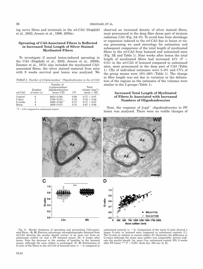
Fig. 6. Myelin thickness of sprouting and preexisting CA3-associ-ated fibres. (A, B) Electron microscopy microphotographs obtained fromsrl-CA3 showing the myelin sheath (arrow) of an axon (ax) from anunlesioned control (A) and a PP lesioned mouse (B), 9 weeks afterlesion. Note the decrease in the number of lamellae in the lesionedmouse, although the axon caliber is unchanged. (C, D) Estimations ofG-ratio of the fibres in the srl-CA3 of lesioned mice (n 5 3) compared to
unlesioned controls (n 5 2). Comparison of the mean G-ratio showed alarger G-ratio in lesioned mice compared to unlesioned controls (C).The G-ratio in relation to axonal calibre (D) illustrates the difference inG-ratios although the mean axon calibre was comparable. Arrows indi-cate the myelin sheath. Ax, axon; Con, unlesioned control; W9, 9 weeksafter PP lesion *** P < 0.001. Scale bar: 200 nm (A, B).
TABLE 2. Number of b-Galactosidase1 Oligodendrocytes in the srl-CA3
srl-CA3Number
of mice (n)
Number ofb-galactosidase1
oligodendrocytes:mean (CE) CV
Totalvolume (mm3):mean 6 SD
Control 8 6610 (0.06) 0.09 0.77 6 0.075 days 7 5760 (0.07) 0.18 0.70 6 0.049 weeks 8 8490 (0.06)** 0.17 0.77 6 0.07Sham 4 4800 (0.07) 0.15 0.67 6 0.06
**P < 0.01 compared to unlesioned control.
36 DRØJDAHL ET AL.
GLIA

the b-gal1 cells in the srl-CA3 5 days after lesion, but at9 weeks the density of oligodendrocytes and the stainingintensity of the neuropil in the srl-CA3 had increased(Fig. 5F–I). This observation was confirmed throughquantification of b-gal1 oligodendrocytes (Fig. 5J andTable 2) that showed a 28% increase in the number ofoligodendrocytes in the srl-CA3 of lesioned compared tounlesioned mice at 9 weeks (P < 0.05), but no changesin the total number of oligodendrocytes 5 days afterlesion or following sham-surgery (Table 2). The CEs ofthe individual estimates of the b-gal1 oligodendrocyteswere 6–8%, with CV values of 7–23% of the estimatedmean values (Table 2). Volumetric analysis of the srl-CA3 showed no lesion-induced changes (Table 2). Thisemphasized that the observed increase of oligodendro-cytes in the srl-CA3 was not due to different delineationat quantification.
Myelin Thickness of Sprouting andPreexisting CA3-Associated Fibres
We next asked if the sprouting axons developed thinmyelin, as in remyelination (Blakemore, 1974; Ludwinand Maitland, 1984), or if they developed thick myelinwithin the 9 weeks observation period. In order to answerthis question we investigated by electron microscopy thecaliber of transversally cut srl-CA3 fibres and axons fromlesioned and unlesioned mice. The mean axon diameter inunlesioned mice was 704.4 nm 6 259.8 (124 fibres) andwas thereby in the same range as in lesioned mice 710.2nm 6 252.7 (114 fibres). In comparison, the mean numberof myelin lamellae was smaller in lesioned mice 4.3 6 1.1(105 fibres) as compared to unlesioned mice 6.2 6 1.4 (122fibres) (P < 0.0001) (Fig. 6A,B and data not shown). Thiswas also reflected in that the fibres in the srl-CA3 of
Fig. 7. Lesion-induced changes of NG21 cells in the srl-CA3. Highpower photomicrographs from (A, D, E) are shown in the column to theright (B, D, F). (A, B) In unlesioned mice (A), the NG21 cells are arbor-ized and evenly distributed throughout the CA3. Organization of cellu-lar processes is different in the stratum lucidum, which makes thiszone appear more intensily stained (arrows). (C–F) Lesion results in atransient disintegration of the zone of NG21 cells in stratum lucidum
and transient morphological changes of NG21 cells in stratum radiatumat day 2 (C, D). The zone reappears from days 7–10 (E, F) and remains at9 weeks (not shown). pcl, pyramidal cell layer; sl, stratum lucidum; sm,stratum moleculare; sr, stratum radiatum. Con, unlesioned control; D2, 2days after PP lesion; W9, 9 weeks after PP lesion. Scale bar: 200 lm (A,C, E) and 20 lm (B, D, F). [Color figure can be viewed in the online issue,which is available at www.interscience.wiley.com.]
37MYELINATION AND AXONAL PLASTICITY
GLIA

lesioned mice had a larger G-ratio (0.83 6 0.05) thanunlesioned mice (0.78 6 0.07) (P < 0.0001) (Fig. 6C,D).Taken together, these results indicated that the sproutingfibers had obtained their final caliber, whereas the myeli-nation of the sprouting fibers remained incomplete 9weeks after lesion.
Sprouting of CA3-Associated Fibres Results inFormation of New Oligodendrocytes
To determine whether the increased number of b-gal1
oligodendrocytes might be generated from adult oligo-dendrocyte precursor cells we studied the lesion-response of the NG21 cells in the srl-CA3. In normal
mice, the process-bearing NG21 cells were evenly dis-tributed in stratum radiatum CA3, while they weremore intensely stained and densely packed in stratumlucidum CA3 (Fig. 7A,B). Two days after lesion, theNG21 cells became slightly hypertrophic and their cellprocesses in stratum lucidum were re-arranged, erasingthe border to stratum radiatum (Fig. 7C,D). The borderbetween stratum lucidum and stratum radiatum reap-peared through days 7–10 (Fig. 7E,F) when the NG21
cells had normalized their morphology throughout CA3.We have previously shown that PP lesion results in
morphological changes and proliferation of NG21 cellsand in formation of new oligodendrocytes in the dentategyrus (Nielsen et al., 2006). To investigate the origin ofthe lesion-induced increase in the number of oligoden-drocytes in the srl-CA3, PP-lesioned mice were injectedwith BrdU with 8-h intervals from 24 to 72 h and killed7 days after lesion. Quantification of the number ofNG21, BrdU1, and BrdU1NG21 cells in sections doublestained for BrdU and NG2, showed that lesion resultedin BrdU-incorporation into 21% of NG21 cells in the srl-CA3 of the ipsilateral hippocampus (Fig. 8A), which wassignificantly more than in the unlesioned, contralateralsrl-CA3, where 4% of NG21 cells had incorporated BrdU(P < 0.01, n 5 7; data not shown). Furthermore, quanti-fication of b-gal1, BrdU1, and BrdU1b-gal1 cells in dou-ble stained adjacent sections showed that 5% of b-gal1
oligodendrocytes in the ipsilateral srl-CA3 had been gen-erated through proliferation (Fig. 6B), as opposed to0.3% of oligodendrocytes in the contralateral srl-CA3 (P< 0.01, n 5 7; data not shown).
Next, to determine if the BrdU1b-gal1 cells that wereobserved 7 days after lesion, oligodendrocytes, arosefrom oligodendrocytes that had become labeled duringthe BrdU injection interval or from differentiation ofproliferating NG21 cells, additional groups of mice weresubjected to BrdU injections 48–72 h after lesion andterminated either 1 h or 9 weeks after the last injection.Quantification showed that 2.5% 6 0.5% of b-gal1 oligo-dendrocytes in the ipsilateral srl-CA3 of animals, whichhad survived for 9 weeks, had incorporated BrdU, com-pared to 0% in the contralateral srl-CA3 (Fig. 8C, Table3, P < 0.01). This was also significantly higher than thenumber of BrdU1b-gal1 oligodendrocytes in the srl-CA3(2.5% 6 0.5% vs 0.5% 6 0.5%; P < 0,01) of animals withsimilar period of BrdU-incorporation but terminated 1 hafter the last BrdU injection 3 days after lesion (Table3). In combination with the observation that 11% of the
Fig. 8. Lesion-induced proliferation of oligodendrocyte lineage cellsin the srl-CA3. (A) Double labeling for BrdU1 (bluish black) and NG21
(brown) showing a double labeled BrdU1NG21 cell (arrow) day 3 sug-gestive of proliferation. Note also a BrdU2NG21 cell (arrowhead) and aBrdU1NG22 cell (*) in the same visual field. (B, C) BrdU2b-gal1 cells(arrowheads) 3 days (B), 7 days (C), and 9 weeks (D) after lesion.Although several BrdU1 cells (*) were observed 3 days after lesion (B),double-labeled BrdU1b-gal1 cells (arrow) were not observed until 7days after lesion (C), when they were observed along with BrdU2b-gal1
(arrowheads). (D) BrdU1b-gal1 cells were also observed 9 weeks afterlesion, showing that these newly formed cells were long-lived. D3, 3days after lesion; D7, 7 days after lesion; W9, 9 weeks after lesion.Scale bar: 10 lm. [Color figure can be viewed in the online issue, whichis available at www.interscience.wiley.com.]
TABLE 3. Percentage of Double Labeled Proliferating Cells in the srl-CA3
Cell type
% BrdU-incorporating cells
Survival: 3 days; number of mice, n 5 7 Survival: 9 weeks; number of mice, n 5 6
Contralateral,mean 6 SD
Ipsilateral,mean 6 SD
Contralateral,mean 6 SD
Ipsilateral,mean 6 SD
BrdU1NG21 cells 1.1 6 0.4 11.3 6 2.3*** – –BrdU1bgal1 cells 0.3 6 0.5 0.5 6 0.5 0.0 6 0.1 2.5 6 0.5**,(**)
**P < 0.01 and ***P < 0.001 compared to the contralateral values; (**)P < 0.01 compared to the value for the ipsilateral srl-CA3, 3 days post lesion.
38 DRØJDAHL ET AL.
GLIA

NG21 cells in the ipsilateral srl-CA3 had incorporatedBrdU (Table 3), this suggested that the BrdU1b-gal1 oli-godendrocytes that were present in the srl-CA3 9 weeksafter lesion had been generated by differentiation ofNG21 cells incorporating BrdU from 48–72 h post-lesion, and not from proliferation of pre-existing oligo-dendrocytes. The data also showed that these newlygenerated oligodendrocytes were long-lived compatiblewith the increased total length of myelinated fibres 9weeks after lesion.
DISCUSSION
This is, to our knowledge, the first study that providesestimates of changes in oligodendrocyte numbers andlength of myelinated fibres induced by axonal sproutingin the adult CNS. The results of the study show thatoligodendrocyte lineage cells retain their capacity toengage in myelination processes in the adult CNS pro-vided that they are adequately stimulated by axons.Using stereological methods, we report that axonallesion-induced sprouting in the hippocampal srl-CA3(Drøjdahl et al., 2002; 2004; Zimmer, 1974) results in amarked 41% increase in the total length of myelinatedfibres, and that these fibres show the same fibre diame-ter, but thinner than normal myelin thickness, 9 weeksafter lesion. Importantly, the increase in myelinatedfibres was accompanied by a 28% increase in the num-ber of oligodendrocytes, with sequential appearance ofnewly formed NG21 cells and thereafter oligodendro-cytes in the srl-CA3. These changes might even beunderestimated since cells were quantified along theentire hippocampal axis, while the PP fibres in the mostseptal and temporal parts were unlikely to be transectedby the present single wire-knife cut.
The significant lesion-induced increase in the totallength of myelinated CA3-associated fibres provides evi-dence of a considerable plasticity of myelinated fibres,and shows that the axon is a strong regulator of oligo-dendrocyte recruitment and myelination in adult CNS.The CA3 pyramidal cells give rise to three main classesof connections; the longitudinal associational path thatextends within stratum radiatum CA3 where it connectsdifferent segments of CA3, the Schaffer collaterals pro-jecting from CA3 to stratum radiatum CA1, and thecommissural connections to the contralateral hippocam-pus (Amaral and Witter, 1995; Blackstad, 1956). Inorder words, the main afferent input to stratum radia-tum CA3 originates from the CA3 pyramidal cells them-selves. Furthermore, the cholinergic afferents to the hip-pocampus are unmyelinated (Frotscher and Leranth,1985). Taken together, this means that the majority ofthe newly formed myelinated fibres must arise fromintact axons from the ipsi- or contralateral CA3 pyrami-dal cells that are induced to sprout within the srl-CA3.High expression of growth associated proteins, such asgrowth-associated-protein-43 (GAP-43), by the CA3 py-ramidal cells, is in line with a high growth potential ofthese cells (Himi et al., 1994; Kruger et al., 1992).
The NG2 cell response within the srl-CA3 consisted oftransient morphological changes, in the form of a moder-ate hypertrophy of the cell bodies and a changed orien-tation of their processes that ablated the otherwise nor-mal border between the stratum radiatum and stratumlucidum CA3. The morphological changes of the NG21
cells in stratum lucidum can perhaps be ascribed to,that the CA3 pyramidal cells, in addition to deafferenta-tion of the distal segment of their apical dendrites, weresubjected to transsynaptic changes at the base of theirapical dendrites in the stratum lucidum (Helme-Guizonet al., 1998), due to the deafferentation of the dentategranule cells (Blackstad and Kjaerheim, 1961). Althoughthe morphological changes of the NG21 cells were not asmarked as observed in the PP-deafferented dentategyrus (Dehn et al., 2005; Nielsen et al., 2006), the incor-poration of BrdU into 21% of NG21 cells in the srl-CA3was comparable to the proliferative response in the mo-lecular layer of the dentate gyrus 7 days after PP lesion(Nielsen et al., 2006). Taken together with the observa-tion of normal numbers of oligodendrocytes in the srl-CA3 5 days after lesion, and the significantly increasednumbers of oligodendrocytes, of which some had incorpo-rated BrdU, 9 weeks after lesion, this suggested thatthe NG21 cells had received a very strong stimulus,with differentation of a small proportion of NG21 cellsinto oligodendrocytes days to weeks after lesion. Thetime course of the injury-induced axonal sprouting insrl-CA3 has not yet been established, but it is likely thatit would be as described for the PP deafferented rat den-tate gyrus where axonal elongation has been reported tobegin 4-6 days after lesion, and to be almost complete 12days after lesion (Steward and Vinsant, 1983).
The observation of a 41% increase in the total lengthof myelinated fibres in the srl-CA3, and a 28% increasein the number of oligodendrocytes, raises questionsabout (1) which cells, newly formed oligodendrocytes, ora mixture of newly formed and preexisting oligodendro-cytes, form the new myelin, and (2) the properties of thenewly formed myelin in term of myelin sheath thicknessand internode length, compared to the pre-existing mye-lin. The estimation of fibre length and number of oligo-dendrocytes in the srl-CA3 of both lesioned and unle-sioned mice allowed calculation of the average internodelength per oligodendrocyte. Calculations showed thatthe average amount of myelin produced per oligodendro-cyte in srl-CA3 of unlesioned mice was 4.3 mm myelin/oligodendrocyte (28.7 m myelin/6,610 oligodendrocytes),which was within the range reported for the rat fimbria,which encompasses the CA3 commissural fibres. In thisstructure, Suzuki and Raisman (1992) observed 20–40internodes about 150–250 lm long per oligodendrocyte,summing up to a total production of 3.0–10.0 lm of mye-lin per oligodendrocyte. If all oligodendrocytes contrib-uted equally to myelinate preexisting and added fibresin the srl-CA3, 9 weeks after lesion, the total productionof myelin would accordingly increase from 4.3 to 4.8 mmmyelin/oligodendrocyte (40.4 m myelin/8,490 oligoden-drocytes). In comparison, if the sprouting fibres weremyelinated exclusively by newly formed oligodendro-
39MYELINATION AND AXONAL PLASTICITY
GLIA

cytes these oligodendrocytes should produce 6.2 mmmyelin/oligodendrocyte (11.7 m myelin/1,880 oligoden-drocytes). Since we in both situations speak aboutincreased myelin elaboration per oligodendrocyte, thismight in itself explain the thinner myelin sheaths andthus the increased G-ratio of the myelinated fibres inthe srl-CA3 of lesioned mice 9 weeks after surgery. It isintriguing that increased G-ratio is also a feature ofmyelin generated during remyelination of demyelinatedfibres (Ludwin and Maitland, 1984). However, since thepost lesion survival time was only 9 weeks, our observa-tion of an increased G-ratio might also reflect that themyelination process was still ongoing.
In line with the induction of morphological changes ofthe NG21 cells in the srl-CA3, PP lesion has been previ-ously shown to induce a moderate microglial and astro-glial reaction in the same layers (Hailer et al., 1999).However, this response was by far exceeded by themicroglial reaction in the deafferented stratum molecu-lare CA3 (Hailer et al., 1999), and it could not be clearlydiscerned in our material. Similar to the lesion-depend-ent differences in micro- and astroglial response in thestratum moleculare CA3 and the srl-CA3, induction ofglial-derived cytokines and growth factors following PPlesion has been shown to be largely confined to the deaf-ferented stratum moleculare CA3, whereas induction islimited in the non-deafferented srl-CA3 (Gomez-Pinillaet al., 1992; Guthrie et al., 1995; Jensen et al., 2000a).This regional segregation supports the view that sprout-ing axons, rather than lesion-induced cytokines andgrowth factors are responsible for the presently observedinduction of oligodendrocyte formation and myelination.This was further substantiated by the timeprofile of theincrease in oligodendrocyte numbers, which was firstobserved at 9 weeks, and not at 5 days or at earlier timepoints, when the lesion-induced synthesis of cytokinesand growth factors in the deafferented stratum molecu-lare is highest (Fagan and Gage, 1990; Gomez-Pinilla etal., 1992; Guthrie et al., 1995; Jensen et al., 2000a).
Several studies have implicated axonal damage (Kuhl-mann et al., 2002; Trapp et al., 1998), axolemmalchanges (Sedgwick, 1997), and re-expression of develop-mentally expressed myelin-inhibitory proteins (Charleset al., 2002; Lee et al., 2007; Mi et al., 2005), in thedemyelinating pathology in patients with multiple scle-rosis and in experimental models of multiple sclerosis.Other studies have indicated that axonal surface mem-brane proteins, that are crucial to proper myelinationduring development (John and Key, 2003; Kim et al.,2003; Mihailov et al., 2004), may be induced orexpressed at insufficient levels and thereby impact onremyelinating capacity of the oligodendroglial lineageunder the same conditions. The presently obtainedresults point to signals elicited by the sprouting axon aspossible means to enhance myelin repair in the adultCNS. Thus, elucidation of the molecular similarities anddiscrepancies between developing, sprouting, anddemyelinated axons may facilitate the development ofnew strategies to improve myelin regeneration.
ACKNOWLEDGMENTS
The expert technical assistance provided by LeneJørgensen, Susanne Petersen (Medical BiotechnologyCentre, University of Southern Denmark), Inger Mar-grethe Rasmussen and Anni Petersen (Anatomy andNeurobiology, University of Southern Denmark), FraukeWinzer (Anatomy, University Rostock), Priscilla Valeraand Margaret Attiwell, McGill University Health CareCentre, and the Service Commun d’Imagerie et d’Analy-ses Microscopiques de l’Universit�e d’Angers is gratefullyacknowledged.
REFERENCES
Amaral DG, Witter MP. 1995. Hippocampal formation. In: Paxinos G,editor. The rat nervous system, 2nd ed. San Diego: Academic Press.pp 443–485.
Andersen BB, Gundersen HJ. 1999. Pronounced loss of cell nuclei andanisotropic deformation of thick sections. J Microsc 196:69–73.
Bansal R, Pheiffer SE. 1985. Developmental expression of 20, 30-cyclicnucleotide 30-phosphohydrolase in dissociated fetal rat brain culturesand rat brain. J Neurosci Res 14:21–34.
Barres BA, Raff MC. 1993. Proliferation of oligodendrocyte precursorcells depends on electrical activity in axons. Nature 361:258–260.
Baumann N, Pham-Dinh D. 2001. Biology of oligodendrocyte and myelinin the mammalian central nervous system. Physiol Rev 81:871–927.
Benes FM, Turtle M, Khan Y, Farol P. 1994. Myelination of a key relayzone in the hippocampal formation occurs in the human brain duringchildhood, adolescence, and adulthood. Arch Gen Psychiatry 51:477–484.
Bengtsson SL, Nagy Z, Skare S. Forsman L, Forssberg H, Ull�en F.2005. Extensive piano practicing has regionally specific effects onwhite matter development. Nat Neurosci 8:1148–1150.
Berger T, Frotscher M. 1994. Distribution and morphological character-istics of oligodendrocytes in the rat hippocampus in situ and in vitro:An immunocytochemical study with the monoclonal Rip antibody.J Neurocytol 23:61–74.
Blakemore WF. 1974. Pattern of remyelination in the CNS. Nature249(457):577–578.
Blackstad TW. 1956. Commisural connections of the hippocampalregion in the rat, with special reference to their mode of termination.J Comp Neurol 105:417–537.
Blackstad TW, Kjaerheim A. 1961. Special axo-dendritic synapses in thehippocampal cortex: Electron and light microscopic studies on thelayer of mossy fibres. J Comp Neurol 117:133–159.
Burne JF, Staple JK, Raff MC. 1996. Glial cells are increased propor-tionally in transgenic optic nerves with increased numbers of axons.J Neurosci 16:2064–2073.
Butt AM, Berry M. 2000. Oligodendrocytes and the control of myelina-tion in vivo: New insights from the rat anterior medullay velum.J Neurosci Res 59:477–488.
Carmichael ST, Wei L, Rovainen CM, Woolsey TA. 2001. New patternsof intracortical projections after focal cortical stroke. Neurobiol Dis8:910–922.
Cellerino A, Carroll P, Thoenen H, Barde YA. 1997. Reduced size of ret-inal ganglion cell axons and hypomyelination in mice lacking brain-derived neurotrophic factor. Mol Cell Neurosci 9:397–408.
Charles P, Reynolds R, Seilhean D, Rougon G, Aigrot MS, Niezgoda A,Zalc B, Lubetzki C. 2002. Re-expression of PSA-NCAM by demyeli-nated axons: An inhibitor of remyelination in multiple sclerosis?Brain 125:1972–1979.
Dehn D, Burbach GJ, Schafer R, Deller T. 2006. NG2 upregulation inthe denervated rat fascia dentata following unilateral entorhinal cor-tex lesion. Glia 53:491–500.
Deller T, Frotscher M. 1997. Lesion-induced plasticity of central neu-rons: Sprouting of single fibres in the rat hippocampus after unilat-eral entorhinal cortex lesion. Prog Neurobiol 53:687–727.
Demerens C, Stankoff B, Logak M, Anglade P, Allinquant B, CouraudF, Zalc B, Lubetzki C. 1996. Induction of myelination in the centralnervous system by electrical activity. Proc Natl Acad Sci USA93:9887–9892.
Dinocourt C, Gallagher SE, Thompson SM. 2006. Injury-induced axonalsprouting in the hippocampus is initiated by activation of trkB recep-tors. Eur J Neurosci 24:1857–1866.
40 DRØJDAHL ET AL.
GLIA

Dissing-Olesen L, Ladeby R, Nielsen HH, Toft-Hansen H, Dalmau I,Finsen B. 2007. Axonal lesion-induced microglial proliferation andmicroglial cluster formation in the mouse. Neurosci 149:112–122.
Dorph-Petersen KA, Nyengaard JR, Gundersen HJ. 2001. Tissueshrinkage and unbiased stereological estimation of particle numberand size. J Microsc 204:232–246.
Drøjdahl N, Hegelund IV, Poulsen FR, Wree A, Finsen B. 2002. Perfo-rant path lesioning induces sprouting of CA3-associated fibre systemsin mouse hippocampal formation. Exp Brain Res 144:79–87.
Drøjdahl N, Fenger C, Nielsen HH, Owens T, Finsen B. 2004. Dynam-ics of oligodendrocyte responses to anterograde axonal (Wallerian)and terminal degeneration in normal and TNF-transgenic mice.J Neurosci Res 75:203–217.
Fagan AM, Gage FH. 1990. Cholinergic sprouting in the hippocampus:A proposed role for IL-1. Exp Neurol 110:105–120.
Fanarraga ML, Griffiths IR, Zhao M, Duncan ID. 1998. Oligodendro-cytes are not inherently programmed to myelinate a specific size ofaxon. J Comp Neurol 399:94–100.
Foran DR, Peterson AC. 1992. Myelin acquisition in the centralnervous system of the mouse revealed by an MBP-Lac Z transgene.J Neurosci 12:4890–4897.
Franklin RJ. 2002. Why does remyelination fail in multiple sclerosis?Nat Rev Neurosci 3:705.
Friedman B, Hockfield S, Black JA, Woodruff KA, Waxman SG. 1989.In situ demonstration of mature oligodendrocytes and their processes:An immunocytochemical study with a new monoclonal antibody, rip.Glia 2:380–390.
Frotscher M, Leranth C. 1985. Cholinergic innervation of the rat hippo-campus as revealed by choline acetyltransferase immunocytochemis-try: A combined light and electron microscopic study. J Comp Neurol239:237–46.
Gomez-Pinilla F, Lee JW, Cotman CW. 1992. Basic FGF in adult ratbrain: Cellular distribution and response to entorhinal lesion andfimbria-fornix transection. J Neurosci 12:345–355.
Gundersen HJ. 1986. Stereology of arbitrary particles. A review ofunbiased number and size estimators and the presentation ofsome new ones, in memory of William R. Thompson. J Microsc143:3–45.
Gundersen HJ, Jensen EB. 1987. The efficiency of systematic samplingin stereology and its prediction. J Microsc 147:229–263.
Gundersen HJ, Jensen EB, Kieu K, Nielsen J. 1999. The efficiency ofsystematic sampling in stereology—reconsidered. J Microsc 193:199–211.
Gundersen HJ, Osterby R. 1981. Optimizing sampling efficiency of ster-eological studies in biology: Or ‘‘do more less well!’’ J Microsc 121:65–73.
Guthrie KM, Nguyen T, Gall CM. 1995. Insulin-like growth factor-1mRNA is increased in deafferented hippocampus: Spatiotemporalcorrespondence of a trophic event with axon sprouting. 352:147–160.
Hailer NP, Grampp A, Nitsch R. 1999. Proliferation of microglia andastrocytes in the dentate gyrus following entorhinal cortex lesion: Aquantitative bromodeoxyuridine-labelling study. Eur J Neurosci11:3359–3364.
Helme-Guizon A, Davis S, Israel M, Lesbats B, Mallet J, Laroche S,Hicks A. 1998. Increase in syntaxin 1B and glutamate release inmossy fibre terminals following induction of LTP in the dentategyrus: A candidate molecular mechanism underlying transsynapticplasticity. Eur J Neurosci 10:2231–2237.
Himi T, Okazaki T, Mori N. 1994. SCG10 mRNA localization in thehippocampus: Comparison with other mRNAs encoding neuronalgrowth-associated proteins (nGAPs). Brain Res 655:177–185.
Jensen MB, Hegelund IV, Lomholt ND, Finsen B, Owens T. 2000a. IFN-gamma enhances microglial reactions to hippocampal axonal degener-ation. J Neurosci 20:3612–3621.
Jensen MB, Hegelund IV, Poulsen FR, Owens T, Zimmer J, Finsen B.1999. Microglial reactivity correlates to the density and the myelina-tion of the anterogradely degenerating axons and terminals followingperforant path denervation of the mouse fascia dentata. Neurosci93:507–518.
Jensen MB, Poulsen FR, Finsen B. 2000b. Axonal sprouting regulatesmyelin basic protein gene expression in denervated mouse hippocam-pus. Int J Dev Neurosci 18:221–235.
John JA, Key B. 2003. Axon mis-targeting in the olfactory bulb dur-ing regeneration of olfactory neuroepithelium. Chem Senses28:773–779.
Kapfhammer JP. 1997. Axon sprouting in the spinal cord: Growth pro-moting and growth inhibitory mechanisms. Anat Embryol (Berl)196:417–426.
Kim JY, Sun Q, Oglesbee M, Yoon SO. 2003. The role of ErbB2 signal-ing in the onset of terminal differentiation of oligodendrocytes invivo. J Neurosci 23:5561–5571.
Koprivica V, Cho KS, Park JB, Yiu G, ATwal J, Gore B, Kim JA, Lin E,Tessier-Lavigne M, Ghen DF, He Z. 2005. EGFR activation mediatesinhibition of axon regeneration by myelin and chondroitin sulfateproteoglycans. Science 310:106–110.
Knapp PE, Skoff RP. 1987. A defect in the cell cycle of neuroglia in themyelin deficient jimpy mouse. Brain Res 432:301–306.
Kruger L, Bendotti C, Rivolta R, Samanin R. 1992. GAP-43 mRNAlocalization in the rat hippocampus CA3 field. Mol Brain Res 13:267–272.
Kuhlmann T, Lingfeld G, Bitsch A, Schuchardt J, Bruck W. 2002. Acuteaxonal damage in multiple sclerosis is most extensive in early diseasestages and decreases over time. Brain 125:2202–2212.
Larsen JO, Gundersen HJ, Nielsen J. 1998. Global spatial samplingwith isotropic virtual planes: Estimators of length density and totallength in thick, arbitrarily orientated sections. J Microsc 191:238–248.
Lee X, Yang Z, Shao Z, Rosenberg SS, Levesque M, Pepinsky RB, QuiM, Miller RH, Chang JR, Mi S. 2007. NGF regulates the expressionof axonal LINGO-1 to inhibit oligodendrocyte differentation and mye-lination. J Neurosci 27:220–225.
Lledo P-M, Alonso M, Grubb MS. 2006. Adult neurogenesis and func-tional plasticity in neuronal circuits. Nat Rev Neurosci 7:179–193.
Lubetzki C, Demerens C, Anglade P, Villarroya H, Frankfurter A, LeeVM, Zalc B. 1993. Even in culture, oligodendrocytes myelinate solelyaxons. Proc Natl Acad Sci USA 90:6820–6844.
Ludwin SK, Maitland M. 1984. Long-term remyelination fails to recon-situte normal thickness of central myelin sheaths. J Neurol Sci64(2):193–198.
Matthews DA, Cotman C, Lynch G. 1976. An electron microscopic studyof lesion-induced synaptogenesis in the dentate gyrus of the adult rat. I.Magnitude and time course of degeneration Brain Res 115:1–21.
Mi S, Miller RH, Lee X, Scott ML, Shulag-Morskay SS, Shao Z, ChangJ, Thill G, Levesque M, Zhang M, Hession C, Sah D, Trapp B, He Z,Jung V, McCoy JM, Pepinsky RB. 2005. LINGO-1 negatively regu-lates myelination by oligodendrocytes. Nat Neurosci 8:745–751.
Mihailov GV, Sereda MW, Brinkmann BG, Fischer TM, Haug B,Birchmeier C, Role L, Lai C, Schwab MH, Nave KA. 2004. Axonalneuregulin-1 regulates myelin sheath thickness. Science 304:700–703.
Nielsen HH, Ladeby R, Drøjdahl N, Peterson AC, Finsen B. 2006. Axo-nal degeneration stimulates the formation of NG21 cells and oligo-dendrocytes in the mouse. Glia 54:105–115.
Noppeney U, Friston KJ, Ashburner J, Frackowiak R, Price CJ. 2005.Early visual deprivation induces structural plasticity in gray andwhite matter. Curr Biol 15:R488–R490.
Raine CS. 1997. In: Raine CS, McFarland HF, Tourtellotte WW, editors.Multiple sclerosis: Clinical and pathogenetic basis. London: Chapman& Hall Medical. p 151–171.
Schwab ME. 2004. Nogo and axon regeneration. Curr Opin Neurobiol14:118–124.
Sedgwick ME. 1997. Pathophysiology of the demyelinated nerve fibre.In: Raine CS, McFarland HF, Tourtellotte WW, editors. Multiple scle-rosis. Clinical and pathogenetic basis, 1st ed. London: Chapman &Hall. pp 197–204.
Seil FJ. 1989. Axonal Sprouting in Response to Injury. Neural regener-ation and transplantation. In: Seil FJ, editor. Frontiers of clinicalneuroscience, Vol. 6. New York: Alan R. Liss. pp 123–135.
Siddiqui AH, Joseph SA. 2005. CA3 axonal sprouting in kainate-induced chronic epilepsy. Brain Res 1066:129–146.
Smith KJ, Blakemore WF, Murray JA, Patterson RC. 1982. Internodalmyelin volume and axon surface area. A relationship determiningmyelin thickness? J Neurol Sci 55:231–246.
Steward O, Vinsant SL. 1983. The process of reinnervation in the den-tate gyrus of the adult rat: A quantitative electron microscopic analy-sis of terminal proliferation and reactive synaptogenesis. J CompNeurol 214:370–386.
Suzuki M, Raisman G. 1992. The glial framework of central white mat-ter tracts: Segmented rows of contiguous interfascicular oligodendro-cytes and solitary astrocytes give rise to a continuous meshwork oftransverse and longitudinal processes in the adult rat fimbria. Glia6:222–235.
Szuchet S, Polak PE, Yim SH. 1986. Mature oligodendrocytes culturedin the absence of neurons recapitulate the ontogenic development ofmyelin membranes. Dev Neurosci 8:208–221.
Trapp BD, Nishiyama A, Cheng D, Macklin W. 1997. Differentiationand death of premyelinating oligodendrocytes in developing rodentbrain. J Cell Biol 137:459–468.
Trapp BD, Peterson J, Ransohoff RM, Rudick R, Mork S, Bo L. 1998.Axonal transection in the lesions of multiple sclerosis. N Engl J Med338:278–285.
Wang Z, Colognato H, Ffrench-Constant C. 2007. Contrasting effects ofmitogenic growth factors on myelination in neuron-oligodendrocyteco-cultures. Glia 55:537–545.
41MYELINATION AND AXONAL PLASTICITY
GLIA

Watanabe M, Sakurai Y, Ichinose T, Aikawa Y, Kotani M, Itoh K. 2006.Monoclonal antibody Rip specifically recognizes 20,30-cyclic nucleotide30-phosphodiesterase in oligodendrocytes. J Neurosci Res 84:525–533.
West MJ, Slomianka L, Gundersen HJ. 1991. Unbiased stereologicalestimation of the total number of neurons in the subdivisions of therat hippocampus using the optical fractionator. Anat Rec 231:482–497.
Will B, Schmitt P, Dalrymple-Alford J. 1985. Brain plasticity, learningand memory: Historical background and conceptual perspectives. In:Will B, Schmitt P, Dalrymple-Alfrod J, editors. Brain plasticity, learn-
ing and memory. Advances in behavioral biology, Vol. 28. New York:Plenum. pp 1–11.
Wirenfeldt M, Dalmau I, Finsen B. 2003. Estimation of absolute micro-glial cell numbers in mouse fascia dentata using unbiased and effi-cient cell counting principles. Glia 44:129–139.
Yakovlev PI, Lecours AR. 1966. The myelinogenic cycles of the regionalmaturation of the brain. In: Minkovski A, editor. Regional develop-ment of the brain in early life. Oxford, UK: Blackwell. pp 3–70.
Zimmer J. 1974. Long term synaptic reorganization in rat fascia den-tata deafferented at adolescent and adult stages: Observations withthe Timm method. Brain Res 76:336–342.
42 DRØJDAHL ET AL.
GLIA