Embed Size (px)
Citation preview

REVIEW
Alpha-Ketoglutarate as a Molecule with Pleiotropic Activity:Well-Known and Novel Possibilities of Therapeutic Use
Barbara Zdzisinska1• Aleksandra _Zurek1
• Martyna Kandefer-Szerszen1
Received: 21 December 2015 / Accepted: 22 February 2016 / Published online: 20 June 2016
� The Author(s) 2016. This article is published with open access at Springerlink.com
Abstract Alpha-ketoglutarate (AKG), an endogenous
intermediary metabolite in the Krebs cycle, is a molecule
involved in multiple metabolic and cellular pathways. It
functions as an energy donor, a precursor in the amino acid
biosynthesis, a signalling molecule, as well as a regulator
of epigenetic processes and cellular signalling via protein
binding. AKG is an obligatory co-substrate for 2-oxoglu-
tarate-dependent dioxygenases, which catalyse
hydroxylation reactions on various types of substrates. It
regulates the activity of prolyl-4 hydroxylase, which con-
trols the biosynthesis of collagen, a component of bone
tissue. AKG also affects the functioning of prolyl
hydroxylases, which, in turn, influences the function of the
hypoxia-inducible factor, an important transcription factor
in cancer development and progression. Additionally, it
affects the functioning of enzymes that influence epigenetic
modifications of chromatin: ten–eleven translocation
hydroxylases involved in DNA demethylation and the
Jumonji C domain containing lysine demethylases, which
are the major histone demethylases. Thus, it regulates gene
expression. The metabolic and extrametabolic function of
AKG in cells and the organism open many different fields
for therapeutic interventions for treatment of diseases. This
review presents the results of studies conducted with the
use of AKG in states of protein deficiency and oxidative
stress conditions. It also discusses current knowledge about
AKG as an immunomodulatory agent and a bone anabolic
factor. Additionally, the regulatory role of AKG and its
structural analogues in carcinogenesis as well as the results
of studies of AKG as an anticancer agent are discussed.
Keywords Alpha-ketoglutarate � Antioxidative factor �Dietary supplement � Immunomodulatory agent �Bone anabolic agent � Anticancer agent
Introduction
Metabolites, small-molecular weight molecules, are the
substrates and products of enzymatic reactions that occur
naturally within cells. Among these substances, there is
alpha-ketoglutarate (AKG; also known as 2-oxoglutarate,
2-oxopentanedioic acid), an endogenous intermediary
metabolite in the Krebs cycle (the citric acid cycle or the
tricarboxylic acid cycle, TCA) (Krebs and Johnson 1980).
For many years, AKG has been an object of interest for
researchers from various fields of science due to its
essential role in several biological processes and its broad
application scope.
AKG is an important biological molecule that plays a key
role in multiple metabolic and cellular pathways. As a Krebs
cycle metabolite, it regulates anabolic and catabolic TCA
products and substrates, thereby regulating amino acid syn-
thesis, ATP production, and reducing equivalent (NAD?/
NADH) generation, which in turn can influence reactive
oxygen species (ROS) levels (Krebs and Johnson 1980). AKG
is also an obligatory co-substrate for 2-oxoglutarate-depen-
dent dioxygenases (2-OGDDs) (McDonough et al. 2010;
Schofield and Zhang 1999), a large group of phylogenetically
conserved enzymes, which catalyse hydroxylation reactions
on various types of substrates including proteins, nucleic
acids, lipids, and metabolic intermediates. These enzymes
require the presence of Fe(II) as a cofactor as well as O2 and
& Barbara Zdzisinska
1 Department of Virology and Immunology, Institute of
Microbiology and Biotechnology, Maria Curie-Sklodowska
University, Akademicka 19, 20-033 Lublin, Poland
Arch. Immunol. Ther. Exp. (2017) 65:21–36
DOI 10.1007/s00005-016-0406-x
123
AKG as co-substrates. In the hydroxylation reaction of the
substrate, one oxygen atom from O2 is attached to a hydroxyl
group in the substrate while the other one is taken up by AKG,
which leads to decarboxylation of AKG and subsequent for-
mation of CO2 and succinate. Ascorbic acid (vitamin C) also
takes part in these reactions by inducing reduction of oxidised
Fe(IV) to Fe(II) and restoring the activity of 2-OGDD
enzymes. In humans, there are more than 60 different
2-OGDDs, and some of these enzymes play a key role in
physiologically important processes such as the hypoxic
response, fatty acid metabolism, nucleic acid repair and
modification, and epigenetic regulation (Hausinger 2004;
Rose et al. 2011). As a substrate of hydroxylases, belonging to
OGDDs, AKG exerts an impact on prolyl/aspartyl/lysyl
hydroxylations, which in turn regulates the stability of the
hypoxia-inducible factor (HIF)-1 and collagen synthesis.
Prolyl hydroxylases PHD1-3 influence the function of HIF-1
(Bruick and McKnight 2001; Epstein et al. 2001; Hirsila et al.
2003), an important transcription factor in cancer develop-
ment and progression, while prolyl-3 and prolyl-4
hydroxylases (P3H, P4H) control the biosynthesis of collagen
(Kivirikko and Pihlajaniemi 1998), a very important com-
ponent of bone tissue. AKG is also a required substrate of the
Jumonji C domain containing lysine demethylases (KDM2-
7), which are the major histone demethylases (Tsukada et al.
2006) and ten–eleven translocation hydroxylases (TET1-3)
involved in DNA demethylation, which catalyse the oxidative
decarboxylation of AKG, generating 5-hydroxy-methylcy-
tosine (5-hmC) and leading to epigenetic effects (Ito et al.
2010; Tahiliani et al. 2009). Moreover, AKG binds and reg-
ulates G protein function, because it is a ligand for the G
protein-coupled receptor (GPR99/GPR80 or OXGR1), which
acts exclusively through a Gq/11-mediated pathway (He et al.
2004). Signalling through this pathway mobilises intracellular
Ca2? (via activation of phospholipase C), which acts as a
diffusible second messenger regulating a wide range of vital
cell functions, including cellular metabolism and growth as
well as cell division and differentiation (Mizuno and Itoh
2009). In this way, AKG can also function as a signalling
molecule. The GPR99 receptor has so far been found in
kidney, placenta, testis, smooth muscles, trachea, and mast
cells (He et al. 2004; Wittenberger et al. 2002), however, its
physiological role has been recently described only in kidney,
where it regulated the acid–base balance in the kidney tubules
in an AKG-dependent manner (Tokonami et al. 2013).
Recently, it has been shown that supplementation of AKG
to adult Caenorhabditis elegans delays ageing of this
nematode. These studies revealed a novel binding protein of
AKG, namely the ATP synthase beta subunit (Chin et al.
2014). This finding suggests that regulatory networks acted
upon by AKG are more complex than it was previously
supposed.
The metabolic and extrametabolic function of AKG in
cells and the organism open many different fields for
therapeutic interventions for treatment of diseases. Meta-
bolism is closely linked with ageing. The main symptoms
of ageing, among others, are disturbances in protein
metabolism and altered bone metabolism. Abnormal pro-
tein metabolism is also observed after trauma (Engel et al.
2003), surgery (Vinnars et al. 1975), burns (Biolo et al.
2000), or infections (Askanazi et al. 1980). Given its
metabolic properties, AKG may be useful in reducing this
type of disorders. Moreover, altered metabolism is a fea-
ture of cancer cells (Jeong and Haigis 2015). Evidence
from recent years has shown that the mitochondrial genes
of Krebs cycle enzymes may function as oncogenes or
tumour suppressors by influencing different cellular pro-
cesses. Genes encoding succinate dehydrogenase (SDH)
and fumarate hydratase (FH) act as tumour suppressors
(Astuti et al. 2001; Baysal et al. 2000; Burnichon et al.
2010; Castro-Vega et al. 2014; Tomlinson et al. 2002).
Mutations thereof lead to accumulation of succinate and
fumarate, structural analogues of AKG and oncometabo-
lites, which have a tumourigenic role by inhibiting the
activity of PHD enzymes and inducing pseudo-hypoxia
(Pollard et al. 2005). In contrast, isocitrate dehydrogenase
(IDH1/2) genes (products of which catalyse the oxidative
decarboxylation of isocitrate to AKG) act as oncogenes
(Amary et al. 2011; Borger et al. 2012; Mardis et al. 2009;
Parsons et al. 2008). Mutations in these genes lead to
production of changed enzymes, which reduce AKG to
another oncometabolite—R(-)-2-hydroxyglutarate (2HG)
(Dang et al. 2009). All the oncometabolites mentioned
above modulate (inhibit) the activity of PHD, TET, and
KDM enzymes and in this way they participate in the
pathogenesis of many cancers. In this case, AKG could be
used for reactivation of 2-OGDD enzymes and reversal of
metabolic alterations (Fig. 1).
The Formation of AKG in the Krebs Cycle and itsMetabolism
AKG is a key intermediate in the TCA cycle, the energy-
producing process that occurs in cells. TCA is a cyclic
pathway of eight enzymatic reactions, which oxidises
compounds derived from glucose, fatty acids, and amino
acids in the matrix of mitochondria, leading to formation of
CO2 and reduced coenzymes (NADH and FADH2). These
coenzymes feed electrons to the respiratory chain further
used to generate ATP. In the TCA cycle, AKG is formed
from isocitrate by oxidative decarboxylation catalysed by
IDH. It can be further converted by AKG dehydrogenase to
succinyl-CoA and NADH (Krebs and Johnson 1980).
22 Arch. Immunol. Ther. Exp. (2017) 65:21–36
123
The amount of AKG produced in mitochondria depends
on the state of oxidation–reduction (redox). The advantage
of NAD? over NADH leads to oxidative decarboxylation
of AKG and formation of succinyl-CoA, while in the case
of an increased concentration of NADH and a shortage of
NAD?, reductive transamination of AKG takes place with
the participation of glutamate dehydrogenase, leading to
formation of glutamate (Owen et al. 2002). Glutamate
formed in this reaction can then, in the reaction involving
glutamine synthetase, attach another ammonium ion, which
results in formation of glutamine (Krebs 1935). AKG can
also be produced in the reaction of glutamate and pyruvate
catalysed by glutamate pyruvate transaminases (GPT1/2).
Additionally, the reversible transfer of an amino group
(NH3?) from glutamate to oxaloacetate also results in the
formation of AKG (and aspartate). This reaction is catal-
ysed by glutamate oxaloacetate transaminase, which exists
in cytoplasmic and inner-membrane mitochondrial forms,
GOT1 and GOT2, respectively (Yudkoff et al. 1994; for
review see Sookoian and Pirola 2015).
The TCA cycle metabolites can penetrate to the cyto-
plasm, where they can be used as precursors for
biosynthetic reactions. AKG can freely diffuse through
channels (such as voltage-dependent anion channels) in the
outer mitochondrial membrane, and it is transported across
the inner mitochondrial membrane through the oxoglu-
tarate carrier (OGC), also known as an oxoglutarate/malate
antiporter. The OGC plays an important role in the malate-
aspartate shuttle and the oxoglutarate-citrate (isocitrate)
shuttle (Chappell 1968; for review see Monne et al. 2013;
Palmieri et al. 1972).
Because AKG is a key intermediate in the Krebs cycle,
it is mainly found in cells (in mitochondria and cytoplasm),
but it can also be detected in small quantities (lM) in the
bloodstream (Martin et al. 1989; Rocchiccioli et al. 1984;
Wagner et al. 2010). However, in people over 40 years of
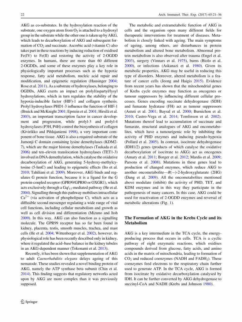
Fig. 1 Schematic representation of the pleiotropic activity of the
AKG molecule. Alpha-ketoglutarate is a precursor of glutamine
which contributes to muscle repair, prevents protein catabolism,
improves nitrogen retention, functions as an immunomodulatory
molecule, and takes part in proper function of intestines. AKG is also
involved in cell protection against oxidative stress and cyanide
poisoning. It can also influence bone strength and density and inhibit
carcinogenesis induced by oncometabolites or hypoxia by activating
enzymes from the 2-OGDD family (2-oxoglutarate-dependent dioxy-
genases). Their action involves epigenetic regulation such as histone
and DNA demethylation carried out by KDM 2–7 (Jumonji C domain
containing lysine demethylases) and TET 1–3 (10–11 translocation
hydroxylases), respectively, and non-epigenetic regulation, which
includes activation of prolyl hydroxylases: P4H (prolyl 4-hydroxy-
lase) involved in type I collagen biosynthesis and PHD2 (prolyl
hydroxylase domain-containing protein 2) responsible for hydroxy-
lation and thus inactivation of HIF-1a (hypoxia-inducible factor).
Other cofactors for the 2-OGDD enzymes are Fe2?, O2, and ascorbate
while their inhibitors known as oncometabolites include succinate,
fumarate, and 2-HG (R(-)-2-hydroxyglutarate)
Arch. Immunol. Ther. Exp. (2017) 65:21–36 23
123
age its level is gradually reduced (to the quantity of ng/ml)
(Harrison and Pierzynowski 2008). Presumably, AKG
present in blood may derive from the bacterial flora
inhabiting the intestine, as different bacteria secrete this
metabolite (Otto et al. 2011). Interestingly, physiological
increases in AKG levels have been observed in the blood of
humans after physical exercise (Brugnara et al. 2012), in
the liver of starved pigeons (Kaminsky et al. 1982), and in
starved C. elegans (Chin et al. 2014).
AKG is an important intermediate in the biosynthesis of
amino acids. Cataplerotic reactions that prevent the accu-
mulation of excess AKG in the cell are associated with
production of two amino acids: glutamate and glutamine,
which are very important for energy metabolism. These
two amino acids play a key role in many metabolic path-
ways and determine proper functioning of organs such as
kidney, intestine, liver, as well as pancreatic b cells, neu-
rons, and cells of the immune system (Newsholme et al.
2003).
In various physiological and pathological states, the
body may experience an increase in protein catabolism. In
these states, glutamine from the muscle and lung tissue is
released and becomes available to other organs (such as the
intestine or kidneys) and to immune cells. To increase its
levels in the body and thus reduce the catabolic response,
or even increase protein anabolism, a relevant clinical
nutrition therapy (enteral and parenteral) is applied (Stehle
et al. 1989). However, the use of glutamine in such a
therapy has not found widespread use in medicine, due to
the instability of aqueous solutions of this amino acid.
Nowadays glutamine supplementation in parenteral nutri-
tion is indicated primarily for patients in critical conditions
(Al Balushi et al. 2013; Stein et al. 2009). Similarly, glu-
tamate, from which glutamine can be synthesised de novo,
is not routinely used in nutritional therapy because of its
relative neurotoxicity and poor permeability across cell
membranes (Hermanussen and Tresguerres 2005). In con-
trast, as an exogenous glutamine precursor that can be used
in many deficiency states, AKG has been an object of
interest for researchers and clinicians for many years.
Many in vivo studies (Cynober et al. 1984, 1990; Dabek
et al. 2005; Filip and Pierzynowski 2008; Junghans et al.
2006; Loı et al. 2005) demonstrate that exogenous AKG
administered as a dietary supplement in the form of various
salts (ornithine, sodium, calcium) is absorbed by the
organism and can be metabolised to glutamine and gluta-
mate as well as to other amino acids (proline, arginine).
Experimental studies performed on animals have shown
that ingested AKG, glutamine, and glutamate are absorbed
in the upper small intestine and then metabolised in ente-
rocytes. During the first pass metabolism in the intestinal
mucosa, up to 95 % of glutamate, 70 % of glutamine, and
only 40 % of AKG is degraded to CO2 (Junghans et al.
2006). The remaining part of AKG may be used in various
anabolic pathways, both in the enterocytes and in periph-
eral tissues, because up to 20 % of dietary AKG appears in
the bloodstream (Filip and Pierzynowski 2008). After
absorption, this part of AKG can be used for the synthesis
of amino acids such as glutamine or proline. AKG is
rapidly removed from the bloodstream, and its half-life is
less than 5 min (Cynober et al. 1990; Dabek et al. 2005).
The circulating AKG is passed to the liver and kidney
using a sodium–potassium pump (Stoll et al. 1991; Wel-
born et al. 1998). Cell culture experiments have
demonstrated that AKG can be easily passed to fibroblasts
by simple diffusion (Aussel et al. 1996). However, the
other cell culture research suggests that the ability of AKG
to penetrate cells is relatively weak but can be increased by
the use of its esters (Koivunen et al. 2007; MacKenzie et al.
2007). AKG is fully metabolised by the body and no
excretion of the compound in the pure form with urine or
faeces is observed. Studies with the sodium salt of 14C-
labelled AKG revealed the presence of AKG carbon in
several tissues (liver, brain, skin, muscle, bone tissue)
already after 3 h of administration of the compound (Filip
and Pierzynowski 2008).
AKG and Protection of Cells Against OxidativeStress
Free radicals also known as ROS are important mediators
in cell damage and cell death processes (Sena and Chandel
2012). They are involved in a variety of pathological
conditions, attacking various cellular macromolecules.
ROS can cause peroxidation of lipids forming cell mem-
branes, structural and functional changes in proteins, or
even damage to nucleic acids. Literature data suggest that
in different cases of induced oxidative stress under in vitro
or in vivo conditions, AKG, like some other metabolites of
the Krebs cycle, has antioxidant properties (Andrae et al.
1985). AKG may participate in non-enzymatic oxidative
decarboxylation during the decomposition of hydrogen
peroxide, which is conditioned by its chemical structure
(Long and Halliwell 2011; Sokołowska et al. 1999; Vel-
vizhi et al. 2002a, b; Yamamoto and Mohanan 2003). It has
also been shown that AKG can prevent damage to mito-
chondrial DNA induced by free radicals in mouse neural
cells (Yamamoto and Mohanan 2003). Furthermore, AKG
inhibited oxidative stress induced in vivo in rats by
administration of ammonium acetate or ethanol (Velvizhi
et al. 2002a, b). Additionally, it has been detected that
AKG administration positively modulates antioxidant
levels in rats during induced hepatocarcinogenesis, restor-
ing antioxidants as well as antioxidative enzyme activity to
almost normal levels (Dakshayani et al. 2006). In another
24 Arch. Immunol. Ther. Exp. (2017) 65:21–36
123
in vivo study (Varma and Hegde 2004), AKG had a pro-
tective effect against oxidative stress-related cataract
formation induced by injection of sodium selenite to rat
pups.
One of the most interesting features of AKG is its
antagonist activity against cyanogens, i.e., materials con-
taining nitriles from which human organism can generate
free cyanide at a level that is toxic to the body. Cyanogens
may occur naturally or they can be synthetically manu-
factured; they can be found in various industrial products,
household utensils, or even in certain medications (Bhat-
tacharya et al. 2009). Cyanide (hydrocyanic acid salt) is a
rapidly acting neurotoxin, the operation of which is related
to, among others, induction of oxidative stress in neurons,
formation of ROS, and inhibition of the activity of a
number of metalloenzymes including the antioxidant
enzyme system (Ardelt et al. 1989, 1994; Muller and
Krieglstein 1995; Solomonson 1981). The action of cya-
nide in the cell leads to impaired mitochondrial activity, as
a result of which cellular respiration and energy metabo-
lism is inhibited, which finally causes lactic acidosis and
cell death (Bhattacharya et al. 2009; Hariharakrishnan et al.
2009). As has been shown, AKG acts as an antagonist of
cyanide poisoning, and its protective effect involves, e.g.,
binding cyanide by a keto group attached to the carboxylic
carbon of AKG, which leads to formation of an interme-
diate—cyanohydrin (Moore et al. 1986; Norris et al. 1990).
Furthermore, AKG prevented cyanide-induced reduction in
the level of glutathione (an important antioxidant of the
cell) and DNA fragmentation in rat thymocytes cultured
in vitro (Bhattacharya et al. 2002). Moreover, AKG is
known to have protected the brain and liver of rats from
damage caused by cyanide-induced ROS activity, and the
addition of sodium thiosulphate increased its protective
effect (Tulsawani et al. 2005). It also showed the ability to
neutralise the oxidative stress caused by cyanogens
(Bhattacharya et al. 2009).
Recent studies have shown that the AKG molecule
modulates the activity of antioxidant enzymes and sta-
bilises the oxidation–reduction homeostasis in mice of
advanced age to the level observed in young animals
(Niemiec et al. 2011).
AKG as a Complementary Factor in Statesof Protein Deficiency: Clinical and Animal Studies
As a precursor of glutamine, AKG is a molecule with high
potential in the treatment of states with increased protein
catabolism, such as recovery after trauma, severe infections
and burns, or after surgeries. In a number of independent
clinical studies, attempts have been made to use exogenous
AKG in alleviating this type of disorders by introducing
supplementation mainly with an ornithine salt of alpha-
ketoglutarate (OKG), consisting of two molecules of
ornithine and one molecule of AKG. It has been shown that
such a combination is more efficient than AKG or ornithine
alone in restoring glutamine pools in muscles (Cynober
et al. 2007). The mechanism of OKG action in the body is
still unclear, but it is probably multifactorial. OKG activity
is associated with an increased synthesis of glutamine,
proline, arginine, polyamines as well as with its capacity to
induce secretion of anabolic hormones (insulin, growth
hormone) and probably with elevated production of nitric
oxide from arginine (Cynober 2004). Many studies (Cou-
dray-Lucas et al. 2000; De Bandt et al. 1998; Donati et al.
1999; Hammarqvist et al. 1989, 1990, 1991; Wernerman
et al. 1987) showed that OKG administered orally, enter-
ally, or parenterally improved protein metabolism in
patients with chronic or acute protein deficiency. Addi-
tionally, OKG administration improved nitrogen balance of
the body and decreased protein catabolism by reducing
muscle proteolysis in patients with severe burns (Coudray-
Lucas et al. 2000; De Bandt et al. 1998; Donati et al. 1999),
after surgery (Hammarqvist et al. 1989, 1991; Wernerman
et al. 1987), trauma, or acute infections (Coudray-Lucas
et al. 2000; De Bandt et al. 1998; Donati et al. 1999;
Hammarqvist et al. 1990). In patients with burns, admin-
istration of OKG also accelerated wound healing (Coudray-
Lucas et al. 2000; De Bandt et al. 1998; Donati et al. 1999).
OKG also helped to restore metabolic balance by stimu-
lating the secretion of anabolic hormones—insulin, growth
hormone (GH), and insulin-like growth factor (IGF)-1 in
patients after trauma (Jeevanandam and Petersen 1999).
The introduction of OKG supplementation in malnourished
patients of advanced age resulted in a significant
improvement in their overall health expressed by increased
appetite and improved motor skills; it also shortened the
time of recovery after severe illnesses or surgery (Blonde-
Cynober et al. 2003; Brocker et al. 1994).
Also, many studies on animals with induced muscle
catabolism (Jeevanandam et al. 1996; Le Boucher et al.
1997; Segaud et al. 2005; Vaubourdolle et al. 1988, 1991)
showed that OKG modulated protein metabolism by
decreasing urea excretion, increasing protein synthesis in
the liver and the intestine, and by inhibiting the degradation
of myofibrils, reducing total proteolysis, and loss of glu-
tamine from muscle tissue. In addition, OKG exerted a
positive effect on the functioning of the intestinal mucosa
and contributed to its recovery after surgery (Czernichow
et al. 1997; Dumas et al. 1988; Raul et al. 1995). Studies
performed on rats have shown that OKG also improves
motor skills in healthy individuals (Moinard et al. 2004). It
is also known to protect liver cells against damage and
prevent a decrease in the activity of the cytochrome P-450
family produced in liver (Roch-Arveiller et al. 1999).
Arch. Immunol. Ther. Exp. (2017) 65:21–36 25
123
Moreover, recent studies have shown that AKG can alle-
viate intestinal mucosa injury under inflammatory
conditions and enhance protein synthesis in intestinal
epithelial cells (Hou et al. 2011; Yao et al. 2012).
AKG as an Immunomodulatory Agent
Glutamine is a known immunoenhancing nutrient in vivo
and a modulator of immune cell growth and function
in vitro (Abcouwer 2000; Andrews and Griffiths 2002;
Saito et al. 1999; Ziegler and Daignault 2000). It modulates
the function of monocytes and neutrophils (by increasing
phagocytosis and ROS intermediate production) and mac-
rophages (by enhancing cytokine production) involved in
the early, non-specific defence response (Furukawa et al.
1997, 2000; Ogle et al. 1994; Wells et al. 1999). Addi-
tionally, glutamine is also a fuel for lymphocyte functions
(Ardawi 1988) enhancing their proliferation and production
of intracellular ROS and glutathione (Chang et al. 1999a)
or cytokines (Chang et al. 1999b; Kew et al. 1999). It has
been shown that AKG, as a glutamine homologue, has
immune-enhancing properties as well, influencing both the
non-specific and the specific immune response, especially
in stress situations. Studies on rats with burn injures
demonstrated that OKG (ornithine alpha-ketoglutarate)
displays immunomodulatory properties, because it coun-
teracted the decrease in superoxide anion (O2-�) generation
in polymorphonuclear leukocytes (PMNs) from these ani-
mals (Roch-Arveiller et al. 1996). Moreover, in other
in vivo studies, OKG enhanced the intracellular production
of hydrogen peroxide (H2O2) and O2-� in PMNs and
monocytes isolated from rats with induced catabolism,
which confirmed that OKG can improve phagocyte
response during stress (Moinard et al. 1999, 2002). Addi-
tionally, it has been shown that OKG can enhance
macrophage cytotoxicity in stress situations by restoring
tumour necrosis factor-a secretion and increasing nitric
oxide production in stimulated macrophages (Moinard
et al. 1999, 2000). Also, exogenous AKG increased O2-�
and H2O2 intracellular production as well as myeloperox-
idase activity in PMNs in vitro (Muhling et al. 2010).
Furthermore, OKG counteracted thymic involution (which
is classically associated with a decreased thymocyte count)
in burn injured rats and increased tissue concentrations of
glutamine and arginine, two essential nutrients for acti-
vated immune cells (Le Boucher et al. 1999). OKG can
also stimulate various mechanisms of the immune system
to an anti-tumour response. It was observed that OKG
administered as a dietary supplement to rats with tumours
increased the cytostatic activity of macrophages and the
cytotoxic activity of natural killer cells (Robinson et al.
1999).
The Influence of AKG on Bone Tissue
In recent years, numerous papers (Dobrowolski et al. 2008;
Filip et al. 2007; Filip 2007; Harrison et al. 2004; Radzki
et al. 2012; Tatara et al. 2005, 2006, 2007) have been
published to suggest that AKG may have an anabolic effect
on bone tissue. Many in vivo studies (Harrison et al. 2004;
Tatara et al. 2006, 2007) have demonstrated that supple-
mentation of AKG or its derivatives during the animal
growth has positive effects on the development of skeleton
by improving the mechanical properties of skeletal bone.
Moreover, other in vivo studies have shown that AKG
supplementation prevented the development of osteopenia
in female ovariectomized rats (Radzki et al. 2012), in rats
after gastrectomy (Dobrowolski et al. 2008) or in model of
osteopenia induced by denervation in turkeys (Tatara et al.
2005). In a study of menopausal women, it was also
observed that administration of AKG (with Ca) inhibited
bone resorption and reduced the effects of osteopenia. In
women treated with AKG sodium salt, after 24 weeks of
treatment, a significant decrease (about 37 %) in the level
of CTX in the bloodstream was observed as well as higher
bone density of the lumbar spine in comparison with the
control group (receiving only CaCO3) (Filip et al. 2007).
The results of above studies suggest that AKG not only can
inhibit bone resorption, but can also induce reconstruction
of bone tissue in the states of osteopenia and osteoporosis.
Although the positive influence of AKG on bone min-
eral density and strength is well documented in many
in vivo studies (Dobrowolski et al. 2008; Filip et al. 2007;
Harrison et al. 2004; Radzki et al. 2012; Tatara et al.
2005, 2006, 2007), its mechanism has not been elucidated
so far. It is believed that AKG can contribute to an increase
in the body pool of amino acids necessary for synthesis of
type I collagen (proline and hydroxyproline) and thus have
a positive effect on bone quality (Harrison and Pierzy-
nowski 2008; Majamaa et al. 1987; Petersen et al. 2003).
Some of the results of studies conducted in vivo in humans
and animals (Cynober et al. 1990; Jeevanandam and
Petersen 1999; Kristensen et al. 2002; Riedel et al. 1996;
Tatara et al. 2005, 2006), where it was observed that OKG
or AKG sodium salt led to an increase in the serum levels
of proline, may be used as a support of this thesis. It is also
suggested that AKG can stimulate production of IGF-1 or
GH, which are anabolic hormones that regulate bone
modelling and remodelling. IGF-1 increases the efficiency
of mature osteoblasts by, among others, stimulating colla-
gen synthesis and inhibiting degradation thereof, while GH
stimulates proliferation of the osteoblastic cell line and
increases the expression of bone morphogenetic proteins,
which stimulate osteoblast differentiation and bone for-
mation (Giustina et al. 2008). Moukarzel et al. (1994)
26 Arch. Immunol. Ther. Exp. (2017) 65:21–36
123
showed that parenteral administration of OKG increased
the circulating plasma level of IGF-1, which was confirmed
by another study (Jeevanandam and Petersen 1999) carried
out on grown-ups who were orally supplemented with
OKG and in whose circulating blood the levels of IGF-1
and GH were increased. Furthermore, it was demonstrated
(Tatara et al. 2012) that oral administration of AKG in
combination with beta-hydroxy-beta-methylbutyrate for
pregnant sows increased the serum concentration of GH
and IGF-1 in their offspring. However, in the studies of
Harrison et al. (2004), in which a group of lambs were
supplemented with sodium salt of AKG, no increase in
IGF-1 plasma levels was observed, yet there was an
increase in their bone mineral density. In turn, other in vivo
studies (Harrison et al. 2004; Rosen et al. 1995) have
demonstrated that IGF-1 stimulates bone growth and
increases their size, but does not affect the mineral density.
These findings suggest that the positive effect of AKG on
bone tissue is probably not associated with stimulation of
IGF-1 and GH production when it is administered alone.
The positive effect of AKG on bone tissue may be due to
its important role in the biosynthesis of glutamate and
glutamine, as well as a multidirectional impact on the
synthesis of type I collagen, the main protein of the bone
matrix. AKG can participate in the metabolism of bone
collagen by various mechanisms. First of all, this
metabolite regulates the activity of enzyme P4H, as men-
tioned above, a member of the 2-OGDDs family,
commonly present in various types of cells. P4H occurs
inside the endoplasmic reticulum and controls the synthesis
of collagen by hydroxylation of proline to 4-hydroxypro-
line (Hutton et al. 1966; Kivirikko and Pihlajaniemi 1998),
which is necessary to form a triple helix of the collagen
molecule. Incomplete hydroxylation of proline in the Gly-
X-Y sequence results in incorrect formation of the collagen
triple helix. Such defective collagen molecules, contrary to
the proper ones, are degraded in the endoplasmic reticulum
instead of being secreted into the cytoplasm (Lamande and
Bateman 1999; Myllyharju 2003). AKG can also partici-
pate in the biosynthesis of collagen by increasing the pool
of proline, an amino acid that is the main component of the
collagen molecule. The primary source of proline used for
biosynthesis of collagen are the products of endogenous
and exogenous collagen degradation (Jackson et al. 1975).
Proline may also be formed by conversion of AKG to
glutamate, and then to pyrroline-5-carboxylate (P5C),
which is converted directly into proline (Smith et al. 1980).
However, this pathway is less important in acquiring a pool
of proline needed for collagen biosynthesis. It is more
important that its precursor, i.e. P5C affects the activity of
prolidase, an enzyme responsible for cleavage of dipeptides
containing C-terminal proline or hydroxyproline and
thereby creating a pool of amino acids required for the
biosynthesis of collagen (Myara et al. 1984). Karna et al.
(2001) have shown in their study that P5C indeed stimu-
lates the production of collagen through activation of
prolidase. Therefore, as a precursor of P5C, AKG can
significantly affect the metabolism of proline. The contri-
bution of AKG in the biosynthesis of collagen was
confirmed in in vitro studies, in which it stimulated the
synthesis of procollagen in human skin fibroblasts by
increasing the activity of prolidase (Son et al. 2007).
Additionally, in vivo studies (Son et al. 2007) have
demonstrated a protective effect of AKG on UVB-irradi-
ated skin, since administration thereof to the skin of mice
irradiated with UVB reduced the number of wrinkles and
decreased the degree of collagen fibre destruction in the
skin.
AKG as an Anticancer Agent
So far, not many investigations of the anticancer activity of
AKG have been carried out (Briere et al. 2005; Hou et al.
2014; MacKenzie et al. 2007; Matsumoto et al. 2006, 2009;
Robinson et al. 1999; Rzeski et al. 2012; Tennant et al.
2009). However, the results of those few available papers
suggest anti-tumour properties of AKG under both normal
and reduced oxygen levels and interference of its activity
with various mechanisms.
The Role of AKG in the Regulation of HIF-1
Activity
Recent research suggests that AKG and its structural ana-
logues (succinate, fumarate) are involved in the regulation
of signalling pathways engaged in promotion of carcino-
genesis (for review see Raimundo et al. 2011).
PHDs 1-3 which catalyse the hydroxylation of proline
residues are very important enzymes belonging to the
2-OGDD group. They are known to regulate the activity of
HIFs, which play a major role in carcinogenesis, inducing
changes in the metabolism of cancer cells (Bruick and
McKnight 2001; Epstein et al. 2001; Hirsila et al. 2003;
Jaakkola et al. 2001; Maxwell et al. 2001). The best
characterised enzyme of this group is PHD2 (prolyl
hydroxylase domain-containing protein 2), encoded by the
gene EGLN1, which is proposed to be the most important
in the hypoxic response under normoxia (Appelhoff et al.
2004; Berra et al. 2003; Schofield and Ratcliffe 2005).
AKG-dependent dioxygenases can potentially be sensitive
to the slightest changes in cellular concentrations of this
metabolite. This is related to the fact that the Michaelis
constant (Km) of AKG for many dioxygenases is similar to
the physiological concentration of this ketoacid (Clifton
et al. 2006; Loenarz and Schofield 2008). In addition, the
Arch. Immunol. Ther. Exp. (2017) 65:21–36 27
123
enzymes responsible for the formation of AKG (IDH-1, -2,
-3), and for its further processing (AKG dehydrogenase,
glutamate dehydrogenases) are strictly controlled.
During the development of solid tumours, large areas
with considerable hypoxia are formed. In response to
hypoxia, there is a change in the metabolism of cancer
cells, enabling them to survive and adapt to the changed,
highly stressful microenvironment. Metabolic changes
include, among others, intensified glycolysis, increased
glycogen synthesis, and use of glutamine (rather than
glucose) as the primary substrate for fatty acid synthesis.
This metabolic reprogramming is directed at the tran-
scriptional level by the transcription factor HIF-1, which
also induces the expression of genes regulating the process
of angiogenesis and cell proliferation (Semenza 2013).
There are two types of HIF (HIF-1 and HIF-2) in
mammalian cells. Each of them is a heterodimer consisting
of one subunit a, which is O2-labile, and one stable subunit
b. There are three known isoforms of subunit a: HIF-1a,
HIF-2a, and HIF-3a. The first one (HIF-1a) is constitu-
tively expressed in all cells, while the other two isoforms
(HIF-2a and HIF-3a) are expressed only in certain tissues
(including endothelial, lung, renal, and liver cells, as well
as the cells of the myeloid lineage). The HIF-3a subunit
functions as a dominant negative regulator of HIF-2a and
HIF-1a, because by binding to subunit b, it blocks its
binding to DNA (Kaelin and Ratcliffe 2008; Maynard et al.
2005; Wiesener et al. 2003). Under aerobic conditions,
proline residues (Pro402 and Pro564) located in the oxy-
gen-dependent degradation domain of HIF-1a are
hydroxylated by PHD2, which results in recognition of the
subunit by the VHL protein (von Hippel-Lindau tumour
suppressor), a part of the ubiquitin ligase complex E3.
Then, after poly-ubiquitination, HIF-1a is rapidly degraded
in the proteasome (Ivan et al. 2001; Kaelin 2005; Yu et al.
2001). Under the oxygen deficiency conditions or at
reduced levels of AKG or Fe2?, the hydroxylation reaction
involving PHD2 slows down or stops, resulting in HIF-1aaccumulation in the cytoplasm (Schofield and Ratcliffe
2005). After the translocation into the cell nucleus, HIF-1adimerises with a stable HIF-1b subunit and the dimer
recognises and binds to hypoxia response elements in the
genome (Kaelin 2005).
The Role of Defects in the Krebs Cycle Enzymes
in the Regulation of HIF-1 Activity
As mentioned above, under physiological conditions, HIF-
1 is activated by hypoxia. However, this factor may also be
activated in normal oxygen access, and activation of the
HIF-1-mediated response pathway in such conditions is
referred as pseudohypoxia. Stabilisation of the HIF-1asubunit occurs, e.g., in the case of a deletion or inactivating
mutation in the gene for the VHL protein, which deter-
mines the development of tumours, such as human
glioblastoma or sporadic renal cell carcinoma (Latif et al.
1993; Ohh 2006; Pugh and Ratcliffe 2003). Also, mutations
in the genes for enzymes involved in the metabolism of
AKG, which is a cofactor of PHDs, result in stabilisation of
the HIF-1a subunit. These mutations occur mainly in three
enzymes of the Krebs cycle: SDH, FH, and IDH.
SDH is a heterotetramer complex localised in mito-
chondria and composed of subunits A, B, C, and D and the
SDH5 factor involved in tetramer mounting (Hao et al.
2009). It has been demonstrated that mutations occurring in
the individual components of the complex may predispose
to the development of certain types of cancer, mainly
paragangliomas (Astuti et al. 2001; Baysal et al.
2000, 2002; Burnichon et al. 2010; Niemann and Muller
2000), pheochromocytomas (Astuti et al. 2001), renal cell
carcinoma (Ricketts et al. 2008; Vanharanta et al. 2004), T
cell acute leukaemia (Baysal 2007), and gastrointestinal
stromal tumours (Italiano et al. 2012; Janeway et al. 2011;
Stratakis and Carney 2009). FH, on the other hand, is an
active homotetramer occurring in the mitochondria and the
cytoplasm, recessive mutations in the gene of which are the
cause of development of encephalopathy and early death,
while its dominant mutations predispose to cancers such as
multiple cutaneous and uterine leiomyomas or hereditary
leiomyomatosis and renal cell cancer (Castro-Vega et al.
2014; King et al. 2006; Tomlinson et al. 2002). Defects in
the SDH and FH enzymes are the cause of accumulation of
Krebs cycle intermediates, i.e., succinate and fumarate
(structural analogues of AKG) in cells. These compounds
compete with AKG in binding to PHD2, and when bound
to the enzyme they inhibit its activity, which in turn con-
tributes to stabilisation of HIF-1a and activation of genes
that promote carcinogenesis (Briere et al. 2005; Isaacs et al.
2005; Pollard et al. 2005; Pugh and Ratcliffe 2003; Selak
et al. 2005).
Mutations in the genes of IDH1 and IDH2 may also
affect the activity of HIF-1 (Zhao et al. 2009). NADP?
dependent enzymes, IDH1 and IDH2, are homodimers,
wherein IDH1 is present in the cytoplasm and peroxisomes,
and IDH2 in cell mitochondria (Corpas et al. 1999). It has
been shown that mutations in IDH1 and IDH2 are
responsible for the development of certain cancer types,
including diffuse and anaplastic gliomas, secondary
glioblastomas, specific types of cartilaginous tumours, and
acute myeloma leukaemia (for review see Dang et al. 2010;
Schaap et al. 2013). The mutated enzymes lose their natural
ability to transform isocitrate into AKG, instead acquiring
the ability to reduce AKG to 2HG. As a result, the pool of
mobile AKG is reduced, and the cell accumulates its
structural analogue—2HG (Dang et al. 2009; Schaap et al.
2013). According to some authors’ reports, both a
28 Arch. Immunol. Ther. Exp. (2017) 65:21–36
123
reduction in the amount of AKG and an increase in the
amount of 2HG can inhibit the activity of PHD2 and
activate HIF-1 (Xu et al. 2011; Zhao et al. 2009). Some
contradictory reports suggest in turn that the R-enantiomer
of 2HG stimulates the activity of EGLN1 (PHD2), EGLN2
(PHD1), and to a lesser extent EGLN3 (PHD3) and thus
inhibit the activity of factor HIF-1. In contrast, the S-
enantiomer of 2HG has the capability of inhibiting PHDs
(Koivunen et al. 2012; Losman et al. 2013). However,
recent studies (Tarhonskaya et al. 2014) have shown that
under in vitro conditions, non-enzymatic oxidation of 2HG
to AKG may occur (involving reducing agents such as
ascorbic acid or the reduced form of L-glutathione), and the
level of AKG formed in this process is sufficient to activate
PHD2. The results of these studies may suggest a misin-
terpretation of the previous results (Koivunen et al. 2012),
but further studies are needed to determine accurately the
effect of 2HG on the activity of PHD2.
The Role of AKG and its Structural Analogues
in the Regulation of Epigenetic Processes
Chemical modifications of chromatin play an important
role in regulating the function of the genome and therefore,
in cell physiology. Information provided by epigenetic
modifications (changes made without modifying the DNA
sequence) plays a considerable part in regulating the pro-
cesses such as transcription or DNA repair and replication.
At the same time, the role of factors regulating epigenetic
processes is also very important, because changes in the
level of their expression or genomic changes in these fac-
tors may contribute to induction or maintenance of a
variety of tumours (Dawson and Kouzarides 2012). The
basic processes regulating chromatin structure and function
are DNA methylation and histone post-translational mod-
ification (acetylation, methylation, phosphorylation,
ubiquitination, biotinylation, and SUMOylation). Many
enzymes, including methyltransferases and demethylases,
are involved in these processes (Meng et al. 2015). Recent
studies indicate that AKG and its structural analogues—
succinate, fumarate, and 2HG can regulate the level of
DNA and histone methylation, as the main enzymes con-
ducting demethylation/hydroxylation reactions belong to
the family of 2-OGDD. The main demethylases of histones
are KDM2-7, which remove methyl groups from almost all
known methylation sites in histones and can also catalyse
the demethylation of three methylated lysines and arginines
(Hoffmann et al. 2012). In turn, the process of oxidative
DNA demethylation is conducted by TET1-3 hydroxylases,
which hydroxylate 5mC to 5-hmC (Ito et al. 2010; Tahil-
iani et al. 2009). Several studies (Cervera et al. 2009;
Chowdhury et al. 2011; Letouze et al. 2013; Schaap et al.
2013; Xiao et al. 2012; Xu et al. 2011) have shown that
both the Krebs cycle intermediate metabolites, succinate
and fumarate, and the R(-)2HG act as competitive inhi-
bitors of enzymes KDMs and TETs, and that the inhibitory
effect enhances methylation of DNA and histones by
methyltransferases, which in turn may increase carcino-
genesis. Nevertheless, the effect of these structural
analogues of AKG can be reversed by AKG itself.
Anti-Tumour Activity of Exogenous AKG: In Vitro
and In Vivo Studies
One of the very important elements of response to hypoxia
and HIF-1 activation is transcription of genes playing a key
role in angiogenesis. This process is crucial for the
development of solid tumours. The growing tumour tissue
needs high amounts of oxygen and nutrients that are sup-
plied by diffusion from the nearby blood vessels in the
initial stage of tumour development. As the tumour
develops and increases its size, the cells of the nearby
blood vessels start running out of oxygen, which activates
HIF-1 and initiates the process of neoangiogenesis. HIF-1
in tumour cells activates the transcription of genes for pro-
angiogenic factors such as vascular endothelial growth
factor (VEGF), PDGF-B (platelet-derived growth factor,
type B), hepatocyte growth factor, epidermal growth fac-
tor, angiopoietin-2, or placental growth factor (Sacewicz
et al. 2009). Matsumoto et al. (2006, 2009) showed that
exogenous AKG exhibited anti-tumour activity by reduc-
ing the level of the HIF-1a subunit and inhibition of
angiogenesis in hypoxic conditions. In their study, AKG
inhibited the expression of the HIF-1a subunit and the
ability to connect HIF-1 protein subunits, decreased the
activity of the Vegf gene promoter, and, consequently,
inhibited VEGF and erythropoietin production in the
Hep3B cell line. Furthermore, AKG inhibited tube for-
mation in an in vitro angiogenesis model. Those anti-
angiogenic effects of AKG were confirmed in another
in vitro study, carried out using the Lewis lung carcinoma
(LLC) cell line (Matsumoto et al. 2009). Additionally,
AKG administered alone showed anti-tumour activity and
enhanced the activity of a chemotherapeutic agent (5-flu-
orouracil, 5-FU) in vivo. In the mouse dorsal air sac assay,
AKG reduced the amount of newly formed blood vessels
caused by administration of the cancer cell line (LLC).
Also intraperitoneal administration of AKG alone or its
combination with 5-FU to mice with transplanted tumours
significantly inhibited tumour growth and angiogenesis in
tumour tissue (Matsumoto et al. 2009) which suggests the
clinical usefulness of this molecule. In turn, the results of
the experiments carried out by Briere et al. (2005) and
MacKenzie et al. (2007) suggest a possibility of application
of AKG in the treatment of neoplastic diseases in which
Krebs cycle enzyme defects cause pseudohypoxia and
Arch. Immunol. Ther. Exp. (2017) 65:21–36 29
123
activation of HIF-1 in normoxia. Briere et al. (2005) have
shown that exogenous AKG prevented translocation of
HIF-1 into the nucleus of fibroblasts with a mutation in the
gene for SDHA (succinate dehydrogenase subunit A). In
another study, the MacKenzie et al. (2007) have shown that
the competitive inhibition of PHDs by succinate or fuma-
rate may be reversed by increasing the cellular level of
AKG. However, native AKG does not easily penetrate into
cells; therefore, to increase the effectiveness of its function,
tests were carried out using cell-permeating AKG deriva-
tives, i.e. AKG esters with increased hydrophobicity—
octyl-AKG and 1-trifluoromethyl benzyl-AKG (converted
by cytoplasmic esterases to AKG). These derivatives
restored the normal activity of PHDs, thereby decreasing
the level of HIF-1a in SDH-deficient cells (MacKenzie
et al. 2007). Other in vitro studies (Tennant et al. 2009)
have shown that AKG esters (1-trifluoromethyl benzyl-
AKG, TaAKG) can restore the activity of PHDs under
hypoxic conditions, which has far-reaching effects on
tumour cells. Restoration of the PHD activity in such
conditions not only resulted in destabilisation of HIF-1a,
but also induced functional changes in cells—reversal of
the hypoxia-induced increased glycolysis process and cell
death as its consequence. In addition, it has been shown
that AKG derivatives (TaAKG and ETAKG—5-ethyl,4-1-
trifluoromethylbenzyl AKG) may also function well
in vivo. TaAKG penetrated several layers of cells in
spheroids derived from cell line HCT116 (human colon
carcinoma) and destabilised their HIF-1a subunit. More-
over, after oral administration of ETAKG to a mouse
xenograft tumour model, increased levels of AKG within
the tumour tissues were observed, as well as decreased
levels of HIF-1a, and reduced glucose metabolism. The
results of the studies mentioned above suggest that AKG in
the form of a diester can reactivate PHDs and destabilise
HIF-1a in vivo, thereby reducing the expression of genes
targeted by this factor (Tennant et al. 2009). However, it
has recently been shown that not all types of AKG esters
that penetrate the cell membrane have the same activity on
HIF-1. In the studies of Hou et al. (2014), membrane
permeable ester dimethyl AKG (DAKG), which is the
precursor of AKG, temporarily stabilized HIF-1a by inhi-
bition of PHD2. During the long-term impact of DAKG on
cells under normoxia conditions, an increase in the level of
HIF-1a and expression of its target genes was observed,
which indicates that DAKG, in contrast to AKG, promotes
the state of pseudohypoxia. The authors speculate that at a
high availability of nutrients, DAKG may have been
quickly converted to succinate or fumarate, which inhibited
the activity of PHD2. On the other hand, the activity of
PHD2 might have been inhibited by increasing intracellu-
lar levels of ROS in pseudohypoxic conditions induced by
DAKG.
Additionally, in vitro studies have shown that exogenous
AKG may affect the level of DNA methylation (Letouze
et al. 2013). In contrast to genetic mutations, DNA
methylation is a reversible process, which creates a pos-
sibility of introduction of new drugs for the treatment of
certain tumours (Rodrıguez-Paredes and Esteller 2011).
Cancer cells are often characterised by a decrease in total
DNA methylation and by hypermethylation of promoter
CpG islands, which results in transcriptional silencing of
tumour suppressor genes. This phenotype, which is char-
acterised by simultaneous multiple gene hypermethylation,
occurs, for example, in glioma, in which it is the result of
mutations in IDH1/IDH2 genes and the inhibiting activity
of 2HG on enzymes belonging to the KDM and TET
groups. Also in paraganglioma, mutations in the SDH
genes determine such a phenotype (Letouze et al. 2013). In
the studies of Letouze et al. (2013), SDH-deficient chro-
maffin cells displayed an increased 5-mC/5-hmC ratio and
histone methylation, while the addition of AKG to the
culture medium reversed the accumulation of 5mC in vitro
and consequently changed this phenotype. These results
suggest that exogenous AKG can restore the TETs enzyme
activity inhibited by succinate accumulated in cells and
restore the cellular normal phenotype.
Given the contradictory results obtained in studies using
various esters of AKG, currently, it seems to be safer to use
AKG in its native form in cancer therapy. This is supported
by the fact that recent studies have shown an antiprolifer-
ative effect of AKG on three cancer cell lines: Caco-2, HT-
29, and LS-180, representing different stages of the
development of colon adenocarcinoma (Rzeski et al. 2012).
AKG interfered in the cell cycle of the tumour cells by
increasing the expression of cyclin-dependent kinase
(CDK) inhibitors p21 Waf/CIP1 and p27 Kip1. Affecting
the cell cycle by influencing the proteins involved in its
regulation (cyclins) is a very promising feature of AKG as
a potential anticancer agent. Each stage of the cell cycle is
supported by specific cyclins forming complexes with
CDKs, which are involved in phosphorylation of certain
proteins. This facilitates maintenance of the cycle, and
consequently leads to cell division. The above-mentioned
CDK inhibitors subsequently inhibit DNA replication and
are responsible for cell cycle arrest resulting in the absence
of cell division (Meeran and Katiyar 2008; Xiong et al.
1993). The influence of AKG on the cell cycle of tumour
cells is not limited to increasing the expression of the CDK
inhibitors. It also decreased the protein level of cyclin D1
and inhibited phosphorylation of the key regulator of the
cell cycle, i.e., the Rb protein, which resulted in arresting a
large number of cells in the G1 phase, preventing them
from entering the division phase (Rzeski et al. 2012).
It should be noted that AKG in combination with
5-hydroxymethylfurfural, prepared in the form of an
30 Arch. Immunol. Ther. Exp. (2017) 65:21–36
123
infusion solution, are currently being investigated for the
treatment of patients with non-small-cell lung carcinoma,
not responding to any conventional therapy. This combi-
nation, named KARAL�, is currently in phase II clinical
study and shows great promises as cancer treatment
(Donnarumma et al. 2013).
Conclusions
Since the discovery and description of the Krebs cycle, a
number of features and functions of one of its main
metabolites, i.e. AKG have been identified so far. Yet,
many of the AKG actions are still waiting to be discovered.
However, current knowledge about this metabolite already
ensures its practical application in various fields of human
life. Today, AKG is synthesised chemically, but attempts
have been made at biotechnological production of this
metabolite by various bacteria and yeast (Otto et al. 2011).
AKG is of particular importance in industry, where it is
used as a building material in the chemical synthesis of
heterocycles. It is also a major component of new
biodegradable, chemoselective, and mechanically
adjustable elastomers with a potential application in bio-
medicine (Barrett and Yousaf 2008). However, the
properties of this molecule also allow use thereof in med-
icine. AKG is used nowadays as a component of solutions
for infusion, formulations used in wound healing, or as a
dietary supplement. It is used, for example, during cardiac
operations in order to avoid disturbances of blood flow and
pressure (Kjellman et al. 1997) or in patients after surgery
or trauma to prevent muscle breakdown. Many studies
indicate that it may be helpful in the treatment of various
diseases and solving health problems, e.g., kidney disor-
ders, bacterial and yeast infections, problems with many
organs, such as the intestines, stomach, liver, eyes, and
even during cyanide intoxication. However, the efficiency
of the therapeutic use, in some cases, is still debated.
Broadly described, the positive effect of AKG on bone
tissue suggests its potential application in prevention of
bone formation disorders, in the treatment of diseases with
progressive loss of bone mass, such as osteoporosis, or in
improving the body’s bone mass. Nevertheless, the mech-
anism of its action on bone tissue has not been elucidated
yet.
Some studies from recent years have provided a lot of
new interesting information about the role of AKG as a
molecule regulating cell function, which may allow it to be
used in the treatment of cancer. Depending on the type of
cancer, AKG could affect tumour cells by reversing their
metabolic response to hypoxia or pseudohypoxia. The
antiproliferative activity of AKG suggests in turn the
possibility of restoring oxidative phosphorylation in
tumour cells in place of aerobic glycolysis, which is
characteristic to all cancers, in states of increased avail-
ability of this metabolite to tumour cells. However,
additional studies are required to confirm this hypothesis.
Additionally, for the therapeutic efficacy of AKG, it is also
important to determine which form of supplementation
would be the most applicable—oral administration, infu-
sion, or, e.g., administration of AKG in the form of
nanomolecules whose action is aimed specifically at the
tumour cells. Not until performing additional in vitro
studies and clinical trials could we evaluate the effective-
ness of AKG as an anticancer agent then. Nevertheless,
even the results of the already existing studies suggest a
possibility of using AKG, e.g., in chemoprevention or as a
support to anti-cancer therapy.
Recent studies suggest that AKG can also regulate the
ageing process of the organism and have an influence on
prolonging the lifespan (Chin et al. 2014; Salminen et al.
2014). Research performed in the next few years will
probably give us an answer to the question whether to
expand the characteristics of AKG with another feature,
which is maintaining the longevity.
Acknowledgments This work was supported by research Grant No
2013/11/B/NZ4/04557 from State Funds for Scientific Research
National Science Centre (NCN), Poland.
Open Access This article is distributed under the terms of the
Creative Commons Attribution 4.0 International License (http://
creativecommons.org/licenses/by/4.0/), which permits unrestricted
use, distribution, and reproduction in any medium, provided you give
appropriate credit to the original author(s) and the source, provide a
link to the Creative Commons license, and indicate if changes were
made.
References
Abcouwer SF (2000) Effects of glutamine on immune cells. Nutrition
16:67–69
Al Balushi RM, Cohen J, Banks M et al (2013) The clinical role of
glutamine supplementation in patients with multiple trauma: a
narrative review. Anaesth Intensive Care 41:24–34
Amary MF, Bacsi K, Maggiani F et al (2011) IDH1 and IDH2
mutations are frequent events in central chondrosarcoma and
central and periosteal chondromas but not in other mesenchymal
tumours. J Pathol 224:334–343
Andrae U, Singh J, Ziegler-Skylakakis K (1985) Pyruvate and related
alpha-ketoacids protect mammalian cells in culture against
hydrogen peroxide-induced cytotoxicity. Toxicol Lett 28:93–98
Andrews FJ, Griffiths RD (2002) Glutamine: essential for immune
nutrition in the critically ill. Br J Nutr 87(Suppl 1):S3–S8
Appelhoff RJ, Tian YM, Raval RR et al (2004) Differential function
of the prolyl hydroxylases PHD1, PHD2, and PHD3 in the
regulation of hypoxia-inducible factor. J Biol Chem
279:38458–38465
Ardawi MS (1988) Glutamine and glucose metabolism in human
peripheral lymphocytes. Metabolism 37:99–103
Arch. Immunol. Ther. Exp. (2017) 65:21–36 31
123
Ardelt BK, Borowitz JL, Isom GE (1989) Brain lipid peroxidation and
antioxidant protectant mechanism following acute cyanide
intoxication. Toxicology 56:147–154
Ardelt BK, Borowitz JL, Maduh EU et al (1994) Cyanide induced
lipid peroxidation in different organs: subcellular distribution
and hydroperoxide generation in neuronal cells. Toxicology
89:127–137
Askanazi J, Carpentier YA, Michelsen CB et al (1980) Muscle and
plasma amino acids following injury. Influence of intercurrent
infection. Ann Surg 192:78–85
Astuti D, Latif F, Dallol A et al (2001) Gene mutations in the
succinate dehydrogenase subunit SDHB cause susceptibility to
familial pheochromocytoma and to familial paraganglioma. Am
J Hum Genet 69:49–54
Aussel C, Coudray-Lucas C, Lasnier E et al (1996) Alpha-Ketoglu-
tarate uptake in human fibroblasts. Cell Biol Int 20:359–363
Barrett DG, Yousaf MN (2008) Poly(triola-ketoglutarate) as
biodegradable, chemoselective, and mechanically tunable elas-
tomers. Macromolecules 41:6347–6352
Baysal BE (2007) A recurrent stop-codon mutation in succinate
dehydrogenase subunit B gene in normal peripheral blood and
childhood T-cell acute leukemia. PLoS One 2:e436
Baysal BE, Ferrell RE, Willett-Brozick JE et al (2000) Mutations in
SDHD, a mitochondrial complex II gene, in hereditary paragan-
glioma. Science 287:848–851
Baysal BE, Willett-Brozick JE, Lawrence EC et al (2002) Prevalence
of SDHB, SDHC, and SDHD germline mutations in clinic
patients with head and neck paragangliomas. J Med Genet
39:178–183
Berra E, Benizri E, Ginouves A et al (2003) HIF prolyl-hydroxylase 2
is the key oxygen sensor setting low steady-state levels of HIF-
1alpha in normoxia. EMBO J 22:4082–4090
Bhattacharya R, Lakshmana Rao PV, Vijayaraghavan R (2002)
In vitro and in vivo attenuation of experimental cyanide
poisoning by a-ketoglutarate. Toxicol Lett 128:185–195
Bhattacharya R, Satpute RM, Hariharakrishnan J et al (2009) Acute
toxicity of some synthetic cyanogens in rats and their response to
oral treatment with alpha-ketoglutarate. Food Chem Toxicol
47:2314–2320
Biolo G, Fleming RY, Maggi SP et al (2000) Inhibition of muscle
glutamine formation in hypercatabolic patients. Clin Sci
99:189–194
Blonde-Cynober F, Aussel C, Cynober L (2003) Use of ornithine a-
ketoglutarate in clinical nutrition of elderly patients. Nutrition
19:73–75
Borger DR, Tanabe KK, Fan KC et al (2012) Frequent mutation of
isocitrate dehydrogenase (IDH)1 and IDH2 in cholangiocarci-
noma identified through broad-based tumor genotyping.
Oncologist 17:72–79
Briere JJ, Favier J, Benit P et al (2005) Mitochondrial succinate is
instrumental for HIF1alpha nuclear translocation in SDHA-
mutant fibroblasts under normoxic conditions. Hum Mol Genet
14:3263–3269
Brocker P, Vellas B, Albarede JL et al (1994) A two-centre,
randomized, double-blind trial of ornithine oxoglutarate in 194
eldery, ambulatory, convalescent subjects. Age Ageing
23:303–306
Brugnara L, Vinaixa M, Murillo S et al (2012) Metabolomics
approach for analyzing the effects of exercise in 13 subjects with
type 1 diabetes mellitus. PLoS One 7:e40600
Bruick RK, McKnight SL (2001) A conserved family of prolyl-4-
hydroxylases that modify HIF. Science 294:1337–1340
Burnichon N, Briere JJ, Libe R et al (2010) SDHA is a tumor
suppressor gene causing paraganglioma. Hum Mol Genet
19:3011–3020
Castro-Vega LJ, Buffet A, De Cubas AA et al (2014) Germline
mutations in FH confer predisposition to malignant pheochro-
mocytomas and paragangliomas. Hum Mol Genet 23:2440–2446
Cervera AM, Bayley JP, Devilee P et al (2009) Inhibition of succinate
dehydrogenase dysregulates histone modification in mammalian
cells. Mol Cancer 8:89
Chang WK, Yang KD, Shaio MF (1999a) Lymphocyte proliferation
modulated by glutamine: involved in the endogenous redox
reaction. Clin Exp Immunol 117:482–488
Chang WK, Yang KD, Shaio MF (1999b) Effect of glutamine on Th1
and Th2 cytokine responses of human peripheral blood mononu-
clear cells. Clin Immunol 93:294–301
Chappell JB (1968) Systems used for the transport of substrates into
mitochondria. Br Med Bull 24:150–157
Chin RM, Fu X, Pai MY et al (2014) The metabolite a-ketoglutarate
extends lifespan by inhibiting ATP synthase and TOR. Nature
510:397–401
Chowdhury R, Yeoh KK, Tian YM et al (2011) The oncometabolite
2-hydroxyglutarate inhibits histone lysine demethylases. EMBO
Rep 12:463–469
Clifton IJ, McDonough MA, Ehrismann D et al (2006) Structural
studies on 2-oxoglutarate oxygenases and related double-
stranded beta-helix fold proteins. J Inorg Biochem 100:644–669
Corpas FJ, Barroso JB, Sandalio LM et al (1999) Peroxisomal NADP-
dependent isocitrate dehydrogenase. Characterization and activ-
ity regulation during natural senescence. Plant Physiol
121:921–928
Coudray-Lucas C, Le Bever H, Cynober L et al (2000) Ornithine
alpha-ketoglutarate improves wound healing in severe burn
patients: a prospective randomized double-blind trial versus
isonitrogenous control. Crit Care Med 28:1772–1776
Cynober L (2004) Ornithine alpha-ketoglutarate as a potent precursor
of arginine and nitric oxide: a new job for an old friend. J Nutr
134:2858S–2862S
Cynober L, Vaubourdolle M, Dore A et al (1984) Kinetics and
metabolic effects of orally administered ornithine alpha-ketog-
lutarate in healthy subjects fed with a standardized regimen. Am
J Clin Nutr 39:514–519
Cynober L, Coudray-Lucas C, De Bandt JP et al (1990) Action of
ornithine a-ketoglutarate, ornithine hydrochloride and calcium
a-ketoglutarate on plasma amino acid and hormonal patterns in
healthy subjects. J Am Coll Nutr 9:2–12
Cynober L, Lasnier E, Le Boucher J et al (2007) Effect of ornithine
alpha-ketoglutarate on glutamine pools in burn injury: evidence
of component interaction. Intensive Care Med 33:538–541
Czernichow B, Nsi-Emvo E, Galluser M et al (1997) Enteral
supplementation with ornithine a-ketoglutarate improves the
early adaptive response to resection. Gut 40:67–72
Dabek M, Kruszewska D, Filip R et al (2005) a-ketoglutarate (AKG)
absorption from pig intestine and plasma pharmacokinetics.
J Anim Physiol Anim Nutr 89:419–426
Dakshayani KB, Subramanian P, Manivasagam T et al (2006)
Metabolic normalization of alpha-ketoglutarate against N-ni-
trosodiethylamine-induced hepatocarcinogenesis in rats. Fundam
Clin Pharmacol 20:477–480
Dang L, White DW, Gross S et al (2009) Cancer-associated IDH1
mutations produce 2-hydroxyglutarate. Nature 462:739–744
Dang L, Jin S, Su SM (2010) IDH mutations in glioma and acute
myeloid leukemia. Trends Mol Med 16:387–397
Dawson MA, Kouzarides T (2012) Cancer epigenetics: from mech-
anism to therapy. Cell 150:12–27
De Bandt JP, Coudray-Lucas C, Lioret N et al (1998) A randomized
controlled trial of the influence of the mode of enteral ornithine
a-ketoglutarate administration in burned patients. J Nutr
128:563–569
32 Arch. Immunol. Ther. Exp. (2017) 65:21–36
123
Dobrowolski PJ, Piersiak T, Surve VV et al (2008) Dietary alpha-
ketoglutarate reduces gastrectomy-evoked loss of calvaria and
trabecular bone in female rats. Scand J Gastroent 43:551–558
Donati L, Ziegler F, Pongelli G et al (1999) Nutritional and clinical
efficacy of ornithine alpha-ketoglutarate in severe burn patients.
Clin Nutr 18:307–311
Donnarumma F, Wintersteiger R, Schober M et al (2013) Simulta-
neous quantitation of alpha-ketoglutaric acid and
5-hydroxymethylfurfural in plasma by HPLC with UV and
fluorescence detection. Anal Sci 29:1177–1182
Dumas F, De Bandt JP, Colomb V et al (1988) Enteral ornithine a-
ketoglutarate enhances intestinal adaptation to massive resection
in rats. Metabolism 47:1366–1371
Engel JM, Muhling J, Weiss S et al (2003) Low plasma glutamine
after multiple trauma: relationship with intracellular glutamine in
polymorphonuclear neutrophils during prolonged ICU stay. Acta
Anaesthesiol Scand 47:707–713
Epstein AC, Gleadle JM, McNeill LA et al (2001) C. elegans EGL-9
and mammalian homologs define a family of dioxygenases that
regulate HIF by prolyl hydroxylation. Cell 107:43–54
Filip R (2007) Alpha-ketoglutaric acid and bone tissue metabolism.
Clin Exp Med Lett 48:3–9
Filip R, Pierzynowski SG (2008) The absorption, tissue distribution
and excretion of enteraly administered alpha-ketoglutarate in
rats. J Anim Physiol Anim Nutr 92:182–189
Filip R, Pierzynowski SG, Lindegard B et al (2007) Alpha-ketoglu-
tarate decreases serum levels of C-terminal cross-linking
telopeptide of type I collagen (CTX) in postmenopausal women
with osteopenia: six-month study. Int J Vitam Nutr Res 77:89–97
Furukawa S, Saito H, Fukatsu K et al (1997) Glutamine-enhanced
bacterial killing by neutrophils from postoperative patients.
Nutrition 13:863–869
Furukawa S, Saito H, Inoue T et al (2000) Supplemental glutamine
augments phagocytosis and reactive oxygen intermediate pro-
duction by neutrophils and monocytes from postoperative
patients in vitro. Nutrition 16:323–329
Giustina A, Mazziotti G, Canalis E (2008) Growth hormone, insulin-
like growth factors, and the skeleton. Endocr Rev 29:535–559
Hammarqvist F, Wernerman J, Ali R et al (1989) Addition of
glutamine to total parenteral nutrition after elective abdominal
surgery spares free glutamine in muscle, counteracts the fall in
muscle protein synthesis, and improves nitrogen balance. Ann
Surg 209:455–461
Hammarqvist F, Wernerman J, Ali R et al (1990) Effects of an amino
acid solution enriched with either branched chain amino acids or
ornithine a-ketoglutarate on the postoperative intracellular amino
acid concentration of skeletal muscle. Br J Surg 77:214–218
Hammarqvist F, Warnerman J, Von Der Decken A et al (1991) Alpha-
ketoglutarate preserves protein synthesis and free glutamine in
skeletal muscle after surgery. Surgery 109:28–36
Hao HX, Khalimonchuk O, Schraders M et al (2009) SDH5, a gene
required for flavination of succinate dehydrogenase, is mutated
in paraganglioma. Science 325:1139–1142
Hariharakrishnan J, Satpute RM, Prasad GB et al (2009) Oxidative
stress mediated cytotoxicity of cyanide in LLC-MK2 cells and
its attenuation by alpha-ketoglutarate and N-acetyl cysteine.
Toxicol Lett 185:132–141
Harrison AP, Pierzynowski SG (2008) Biological effects of 2-oxog-
lutarate with particular emphasis on the regulation of protein,
mineral and lipid absorption/metabolism, muscle performance,
kidney function, bone formation and cancerogenesis, all viewed
from a healthy ageing perspective state of the art—review
article. J Physiol Pharmacol 59(suppl 1):91–106
Harrison AP, Tygesen MP, Sawa-Wojtanowicz B et al (2004) a-
ketoglutarate treatment early in postnatal life improves bone
density in lambs at slaughter. Bone 35:204–209
Hausinger RP (2004) FeII/alpha-ketoglutarate-dependent hydroxy-
lases and related enzymes. Crit Rev Biochem Mol Biol 39:21–68
He W, Miao FJ, Lin DC et al (2004) Citric acid cycle intermediates as
ligands for orphan G-protein-coupled receptors. Nature
429:188–193
Hermanussen M, Tresguerres JA (2005) How much glutamate is toxic
in paediatric parenteral nutrition? Acta Paediatr 94:16–19
Hirsila M, Koivunen P, Gunzler V et al (2003) Characterization of the
human prolyl 4-hydroxylases that modify the hypoxia-inducible
factor. J Biol Chem 278:30772–30780
Hoffmann I, Roatsch M, Schmitt ML et al (2012) The role of histone
demethylases in cancer therapy. Mol Oncol 6:683–703
Hou Y, Wang L, Ding B et al (2011) Alpha-ketoglutarate and
intestinal function. Front Biosci 16:1186–1196
Hou P, Kuo CY, Cheng CT et al (2014) Intermediary metabolite
precursor dimethyl-2-ketoglutarate stabilizes hypoxia-inducible
factor-1a by inhibiting prolyl-4-hydroxylase PHD2. PLoS One
9:e113865
Hutton JJ Jr, Trappel AL, Udenfriend S (1966) Requirements for
alpha-ketoglutarate, ferrous ion and ascorbate by collagen
proline hydroxylase. Biochem Biophys Res Commun
24:179–184
Isaacs JS, Jung YJ, Mole DR et al (2005) HIF overexpression
correlates with biallelic loss of fumarate hydratase in renal
cancer: novel role of fumarate in regulation of HIF stability.
Cancer Cell 8:143–153
Italiano A, Chen CL, Sung YS et al (2012) SDHA loss of function
mutations in a subset of young adult wild-type gastrointestinal
stromal tumors. BMC Cancer 12:408
Ito S, D’Alessio AC, Taranova OV et al (2010) Role of Tet proteins in
5mC to 5hmC conversion, ES-cell self-renewal and inner cell
mass specification. Nature 466:1129–1133
Ivan M, Kondo K, Yang H et al (2001) HIF alpha targeted for VHL-
mediated destruction by proline hydroxylation: implications for
O2 sensing. Science 292:464–468
Jaakkola P, Mole DR, Tian YM et al (2001) Targeting of HIF-alpha to
the von Hippel-Lindau ubiquitylation complex by O2-regulated
prolyl hydroxylation. Science 292:468–472
Jackson SH, Dennis AW, Greenberg M (1975) Iminodipeptiduria: a
genetic defect in recycling collagen; a method for determining
prolidase in erythrocytes. Can Med Assoc J 113(759):762–763
Janeway KA, Kim SY, Lodish M et al (2011) Defects in succinate
dehydrogenase in gastrointestinal stromal tumors lacking KIT
and PDGFRA mutations. Proc Natl Acad Sci USA 108:314–318
Jeevanandam M, Petersen SR (1999) Substrate fuel kinetics in
enterally fed trauma patients supplemented with ornithine alpha
ketoglutarate. Clin Nutr 18:209–217
Jeevanandam M, Holaday NJ, Petersen SR (1996) Ornithine alphake-
toglutarate (OKG) supplementation is more effective than its
components in traumatized rats. J Nutr 126:2141–2150
Jeong SM, Haigis MC (2015) Sirtuins in cancer: a balancing act
between genome stability and metabolism. Mol Cells
38:750–758
Junghans P, Derno M, Pierzynowski S et al (2006) Intraduodenal
infusion of a-ketoglutarate decreases whole body energy
expenditure in growing pigs. Clin Nutr 25:489–496
Kaelin WG Jr (2005) The von Hippel-Lindau protein, HIF hydrox-
ylation, and oxygen sensing. Biochem Biophys Res Commun
338:627–638
Kaelin WG Jr, Ratcliffe PJ (2008) Oxygen sensing by metazoans: the
central role of the HIF hydroxylase pathway. Mol Cell
30:393–402
Kaminsky YG, Kosenko EA, Kondrashova MN (1982) Metabolites of
citric acid cycle, carbohydrate and phosphorus metabolism, and
related reactions, redox and phosphorylating states of hepatic
tissue, liver mitochondria and cytosol of the pigeon, under
Arch. Immunol. Ther. Exp. (2017) 65:21–36 33
123
normal feeding and natural nocturnal fasting conditions. Comp
Biochem Physiol B 73:957–963
Karna E, Miltyk W, Wołczynski S et al (2001) The potential
mechanism for glutamine-induced collagen biosynthesis in
cultured human skin fibroblasts. Comp Biochem Physiol B:
Comp Biochem 130:23–32
Kew S, Wells SM, Yaqoob P et al (1999) Dietary glutamine enhances
murine T-lymphocyte responsiveness. J Nutr 129:1524–1531
King A, Selak MA, Gottlieb E (2006) Succinate dehydrogenase and
fumarate hydratase: linking mitochondrial dysfunction and
cancer. Oncogene 25:4675–4682
Kivirikko KI, Pihlajaniemi T (1998) Collagen hydroxylases and the
protein disulfide isomerase subunit of prolyl 4-hydroxylases.
Adv Enzymol Relat Areas Mol Biol 72:325–329
Kjellman UW, Bjork K, Ekroth R et al (1997) Addition of alpha-
ketoglutarate to blood cardioplegia improves cardioprotection.
Ann Thorac Surg 63:1625–1633
Koivunen P, Hirsila M, Remes AM et al (2007) Inhibition of hypoxia-
inducible factor (HIF) hydroxylases by citric acid cycle inter-
mediates: possible links between cell metabolism and
stabilization of HIF. J Biol Chem 282:4524–4532
Koivunen P, Lee S, Duncan CG et al (2012) Transformation by the
(R)-enantiomer of 2-hydroxyglutarate linked to EGLN activa-
tion. Nature 483:484–488
Krebs HA (1935) Metabolism of amino-acids: the synthesis of
glutamine from glutamic acid and ammonia, and the enzymic
hydrolysis of glutamine in animal tissues. Biochem J
29:1951–1969
Krebs HA, Johnson WA (1980) The role of citric acid in intermediate
metabolism in animal tissues. FEBS Lett 117 Suppl:K1–K10
(reprint from Enzymologia 1937; 4:148–156)Kristensen NB, Jungvid H, Fernandez JA et al (2002) Absorption and
metabolism of alpha-ketoglutarate in growing pigs. J Anim
Physiol Anim Nutr 86:239–245
Lamande SR, Bateman JF (1999) Procollagen folding and assembly:
the role of endoplasmic reticulum enzymes and molecular
chaperones. Semin Cell Dev Biol 10:455–464
Latif F, Tory K, Gnarra J (1993) Identification of the von Hippel-
Lindau disease tumor suppressor gene. Science 260:1317–1320
Le Boucher J, Obled C, Farges MC et al (1997) Ornithine
alphaketoglutarate modulates tissue protein metabolism in
burn-injured rats. Am J Physiol 273:E557–E563
Le Boucher J, Farges MC, Minet R et al (1999) Modulation of
immune response with ornithine a-ketoglutarate in burn injury:
an arginine or glutamine dependency? Nutrition 15:773–777
Letouze E, Martinelli C, Loriot C et al (2013) SDH mutations
establish a hypermethylator phenotype in paraganglioma. Cancer
Cell 23:739–752
Loenarz C, Schofield CJ (2008) Expanding chemical biology of
2-oxoglutarate oxygenases. Nat Chem Biol 4:152–156
Loı C, Nakib S, Neveux N et al (2005) Ornithine alpha-ketoglutarate
metabolism in the healthy rat in the postabsorptive state.
Metabolism 54:1108–1114
Long LH, Halliwell B (2011) Artefacts in cell culture: a-ketoglutarate
can scavenge hydrogen peroxide generated by ascorbate and
epigallocatechin gallate in cell culture. Biochem Bioph Res
Commun 406:20–24
Losman JA, Looper RE, Koivunen P et al (2013) (R)-2-hydroxyg-
lutarate is sufficient to promote leukemogenesis and its effects
are reversible. Science 339:1621–1625
MacKenzie ED, Selak MA, Tennant DA et al (2007) Cell-permeating
a-ketoglutarate derivatives alleviate pseudohypoxia in succinate
dehydrogenase-deficient cells. Mol Cell Biol 27:3282–3289
Majamaa K, Sasaki T, Uitto J (1987) Inhibition of prolyl hydroxy-
lation during collagen biosynthesis in human skin fibroblast
cultures by ethyl 3,4-dihydroxybenzoate. J Invest Dermatol
89:405–409
Mardis ER, Ding L, Dooling DJ et al (2009) Recurring mutations
found by sequencing an acute myeloid leukemia genome. N Engl
J Med 361:1058–1066
Martin M, Ferrier B, Baverel G (1989) Transport and utilization of
alpha-ketoglutarate by the rat kidney in vivo. Pflugers Arch
413:217–224
Matsumoto K, Imagawa S, Obara N et al (2006) 2-oxoglutarate
downregulates expression of vascular endothelial growth
factor and erythropoietin through decreasing hypoxia-in-
ducible factor-1a and inhibits angiogenesis. J Cell Physiol
209:333–340
Matsumoto K, Obara N, Ema M et al (2009) Antitumor effects of
2-oxoglutarate through inhibition of angiogenesis in a murine
tumor model. Cancer Sci 100:1639–1647
Maxwell PH, Pugh CW, Ratcliffe PJ (2001) Activation of the HIF
pathway in cancer. Curr Opin Genet Dev 11:293–299
Maynard MA, Evans AJ, Hosomi T et al (2005) Human HIF-3alpha4
is a dominant-negative regulator of HIF-1 and is down-regulated
in renal cell carcinoma. FASEB J 19:1396–1406
McDonough MA, Loenarz C, Chowdhury R et al (2010) Structural
studies on human 2-oxoglutarate dependent oxygenases. Curr
Opin Struct Biol 20:659–672
Meeran SM, Katiyar SK (2008) Cell cycle control as a basis for
cancer chemoprevention through dietary agents. Front Biosci
13:2191–2202
Meng H, Cao Y, Qin J et al (2015) DNA methylation, its mediators
and genome integrity. Int J Biol Sci 11:604–617
Mizuno N, Itoh H (2009) Functions and regulatory mechanisms of
Gq-signaling pathways. Neurosignals 17:42–54
Moinard C, Chauveau B, Walrand S et al (1999) Phagocyte functions
in stressed rats: comparison of modulation by glutamine,
arginine and ornithine 2-oxoglutarate. Clin Sci 97:59–65
Moinard C, Caldefie F, Walrand S et al (2000) Involvement of
glutamine, arginine, and polyamines in the action of ornithine
alpha-ketoglutarate on macrophage functions in stressed rats.
J Leukoc Biol 67:834–840
Moinard C, Caldefie F, Walrand S et al (2002) Effects of ornithine
2-oxoglutarate on neutrophils in stressed rats: evidence for the
involvement of nitric oxide and polyamines. Clin Sci
102:287–295
Moinard C, Dauge V, Cynober L (2004) Ornithine a-ketoglutarate
supplementation influences motor activity in healthy rats. Clin
Nutr 23:485–490
Monne M, Miniero DV, Iacobazzi V et al (2013) The mitochondrial
oxoglutarate carrier: from identification to mechanism. J Bioen-
erg Biomembr 45:1–13
Moore SJ, Norris JC, Ho IK et al (1986) The efficacy of alpha-
ketoglutaric acid in the antagonism of cyanide intoxication.
Toxicol Appl Pharmacol 82:40–44
Moukarzel AA, Goulet O, Salas JS et al (1994) Growth retardation in
children receiving long-term parenteral nutrition: effects of
ornithine and a-ketoglutarate. Am J Clin Nutr 60:408–413
Muhling J, Tussing F, Nickolaus KA et al (2010) Effects ofalpha-
ketoglutarate on neutrophil intracellular aminoand alpha-keto
acid profiles and ROS production. Amino Acids 38:167–177
Muller U, Krieglstein J (1995) Inhibitors of lipid peroxidation protect
cultured neurons against cyanide-induced injury. Brain Res
678:265–268
Myara I, Myara A, Mangeot M et al (1984) Plasma prolidase activity:
a possible index of collagen catabolism in chronic liver disease.
Clin Chem 30:211–215
Myllyharju J (2003) Prolyl 4-hydroxylases, the key enzymes of
collagen biosynthesis. Matrix Biol 22:15–24
34 Arch. Immunol. Ther. Exp. (2017) 65:21–36
123
Newsholme P, Procopio J, Ramos Lima MM et al (2003) Glutamine
and glutamate—their central role in cell metabolism and
function. Cell Biochem Funct 21:1–9
Niemann S, Muller U (2000) Mutations in SDHC cause autosomal
dominant paraganglioma, type 3. Nat Genet 26:268–270
Niemiec T, Sikorska J, Harrison A et al (2011) Alpha-ketoglutarate
stabilizes redox homeostasis and improves arterial elasticity in
aged mice. J Physiol Pharmacol 62:37–43
Norris JC, Utley WA, Hume AS (1990) Mechanism of antagonizing
cyanide induced lethality by alpha-kotoglutarate. Toxicology
62:275–283
Ogle CK, Ogle JD, Mao JX et al (1994) Effect of glutamine on
phagocytosis and bacterial killing by normal and pediatric burn
patient neutrophils. JPEN J Parenter Enteral Nutr 18:128–133
Ohh M (2006) Ubiquitin pathway in VHL cancer syndrome.
Neoplasia 8:623–629
Otto C, Yovkova V, Barth G (2011) Overproduction and secretion of
a-ketoglutarate acid by microorganisms. Appl Microbiol
Biotechnol 92:689–695
Owen OE, Kalhan SC, Hanson RW (2002) The key role of
anaplerosis and cataplerosis for citric acid cycle function.
J Biol Chem 277:30409–30412
Palmieri F, Quagliariello E, Klingenberger M (1972) Kinetics and
specificity of the oxoglutarate carrier in rat-liver mitochondria.
Eur J Biochem 29:408–416
Parsons DW, Jones S, Zhang X et al (2008) An integrated genomic
analysis of human glioblastoma multiforme. Science
321:1807–1812
Petersen L, Olewinski R, Salmon P et al (2003) Novel proline
hydroxylase activities in the pneumocandin-producing fungus
Glarealozoyensis responsible for the formation of trans 3- and
trans 4-hydroxyproline. Appl Microbiol Biotechnol 62:263–267
Pollard PJ, Briere JJ, Alam NA et al (2005) Accumulation of Krebs
cycle intermediates and over-expression of HIF1alpha in
tumours which result from germline FH and SDH mutations.
Hum Mol Genet 14:2231–2239
Pugh CW, Ratcliffe PJ (2003) The von Hippel-Lindau tumor
suppressor, hypoxia-inducible factor-1 (HIF-1) degradation,
and cancer pathogenesis. Semin Cancer Biol 13:83–89
Radzki RP, Bienko M, Pierzynowski SG (2012) Anti-osteopenic
effect of alpha-ketoglutarate sodium salt in oviarectomized rats.
J Bone Miner Metab 30:651–659
Raimundo N, Baysal BE, Shadel GS (2011) Revisiting the TCA
cycle: signaling to tumor formation. Trends Mol Med
17:641–649
Raul F, Gosse F, Galluser M et al (1995) Functional and metabolic
changes in intestinal mucosa of rats after enteral administration
of ornithine alpha-ketoglutarate salt. JPEN JPEN J Parenter
Enteral Nutr 19:145–150
Ricketts C, Woodward ER, Killick P et al (2008) Germline SDHB
mutations and familial renal cell carcinoma. J Natl Cancer Inst
100:1260–1262
Riedel E, Nundel M, Hampl H (1996) alpha-Ketoglutarate application
in hemodialysis patients improves amino acid metabolism.
Nephron 74:261–265
Robinson LE, Bussiere F, Le Boucher J et al (1999) Amino acid
nutrition and immune function in tumour-bearing rats: a
comparison of glutamine-, arginine- and ornithine 2-oxoglu-
tarate-supplemented diets. Clin Sci 97:657–669
Rocchiccioli F, Leroux JP, Cartier PH (1984) Microdetermination of
2-ketoglutaric acid in plasma and cerebrospinal fluid by capillary
gas chromatography mass spectrometry; application to pedi-
atrics. Biomed Mass Spectrom 11:24–28
Roch-Arveiller M, Tissot M, Coudray-Lucas C et al (1996)
Immunomodulatory effects of ornithine alpha-ketoglutarate in
rats with burn injuries. Arch Surg 131:718–723
Roch-Arveiller M, Fontagne J, Coudray-Lucas C et al (1999)
Ornithine a-ketoglutarate counteracts the decrease of liver
cytochrome P-450 content in burned rats. Nutrition
15:379–383
Rodrıguez-Paredes M, Esteller M (2011) Cancer epigenetics reaches
mainstream oncology. Nat Med 17:330–339
Rose NR, McDonough MA, King ON et al (2011) Inhibition of
2-oxoglutarate dependent oxygenases. Chem Soc Rev
40:4364–4397
Rosen HN, Chen V, Cittadini A et al (1995) Treatment with
growth hormone and IGF-1 in growing rats increases bone
mineral content but not bone mineral density. J Bone Miner
Res 10:1352–1358
Rzeski W, Walczak K, Juszczak M et al (2012) Alpha-ketoglutarate
(AKG) inhibits proliferation of colon adenocarcinoma cells in
normoxic conditions. Scnad J Gastroenterol 47:565–571
Sacewicz I, Wiktorska M, Wysocki T et al (2009) Mechanisms of
cancer angiogenesis (Article in Polish). Postepy Hig Med Dosw
63:159–168
Saito H, Furukawa S, Matsuda T (1999) Glutamine as an immu-
noenhancing nutrient. JPEN J Parenter Enteral Nutr 23(5
Suppl):S59–S61
Salminen A, Kaarniranta K, Hiltunen M et al (2014) Krebs cycle
dysfunction shapes epigenetic landscape of chromatin: novel
insights into mitochondrial regulation of aging process. Cell
Signal 26:1598–1603
Schaap FG, French PJ, Bovee JV (2013) Mutations in the isocitrate
dehydrogenase genes IDH1 and IDH2 in tumors. Adv Anat
Pathol 20:32–38
Schofield CJ, Ratcliffe PJ (2005) Signalling hypoxia by HIF
hydroxylases. Biochem Biophys Res Commun 338:617–626
Schofield CJ, Zhang Z (1999) Structural and mechanistic studies on
2-oxoglutarate-dependent oxygenases and related enzymes. Curr
Opin Struct Biol 9:722–731
Segaud F, Lardeux B, Alexandre-Gouabau MC et al (2005) Pretreat-
ment of starved rats with ornithine a-ketoglutarate: effects on
hepatic mRNA levels and plasma concentrations of three liver-
secreted proteins. Nutrition 21:732–739
Selak MA, Armour SM, MacKenzie ED et al (2005) Succinate links
TCA cycle dysfunction to oncogenesis by inhibiting HIF-alpha
prolyl hydroxylase. Cancer Cell 7:77–85
Semenza GL (2013) HIF-1 mediates metabolic responses to intratu-
moral hypoxia and oncogenic mutations. J Clin Invest
123:3664–3671
Sena LA, Chandel NS (2012) Physiological roles of mitochondrial
reactive oxygen species. Mol Cell 48:158–167
Smith RJ, Downing SJ, Phang JM et al (1980) Pyrroline-5-carboxy-
late synthase activity in mammalian cells. Proc Natl Acad Sci
USA 77:5221–5225
Sokołowska M, Oleszek A, Włodek L (1999) Protective effect of
alpha-keto acids on the oxidative hemolysis. Pol J Pharmacol
51:429–434
Solomonson LP (1981) Cyanide as a metabolic inhibitor. In:
Vennesland B, Conn EE, Knowles CJ, Westley J, Vissing F
(eds) Cyanide in biology. Academic Press, New York, p 11
Son ED, Choi GH, Kim H et al (2007) Alpha-ketoglutarate stimulates
procollagen production in cultured human dermal fibroblasts,
and decreases UVB-induced Wrinkle formation following topi-
cal application on the dorsal skin of hairless mice. Biol Pharm
Bull 30:1395–1399
Sookoian S, Pirola CJ (2015) Liver enzymes, metabolomics and
genome-wide association studies: from systems biology to the
personalized medicine. World J Gastroenterol 21:711–725
Stehle P, Zander J, Mertes N et al (1989) Effect of parenteral
glutamine peptide supplements on muscle glutamine loss and
nitrogen balance after major surgery. Lancet 1:231–233
Arch. Immunol. Ther. Exp. (2017) 65:21–36 35
123
Stein J, Boehles HJ, Blumenstein I, Goeters C, Schulz R (2009)
Amino acids—guidelines on parenteral nutrition, Chapter 4. Ger
Med Sci 7:Doc24
Stoll B, McNelly S, Buscher HP et al (1991) Functional hepatocyte
heterogeneity in glutamate, aspartate and alpha-ketoglutarate
uptake: a histoautoradiographical study. Hepatology 13:247–253
Stratakis CA, Carney JA (2009) The triad of paragangliomas, gastric
stromal tumours and pulmonary chondromas (Carney triad), and
the dyad of paragangliomas and gastric stromal sarcomas
(Carney–Stratakis syndrome): molecular genetics and clinical
implications. J Intern Med 266:43–52
Tahiliani M, Koh KP, Shen Y et al (2009) Conversion of 5-methyl-
cytosine to 5-hydroxymethylcytosine in mammalian DNA by
MLL partner TET1. Science 324:930–935
Tarhonskaya H, Rydzik AM, Leung IK et al (2014) Non-enzymatic
chemistry enables 2-hydroxyglutarate-mediated activation of
2-oxoglutarate oxygenases. Nat Commun 5:3423
Tatara MR, Brodzki A, Krupski W et al (2005) Effects of a-
ketoglutarate on bone homeostasis and plasma amino acids in
turkeys. Poult Sci 84:1604–1609
Tatara MR, Sliwa E, Krupski W et al (2006) Ornithine alpha-
ketoglutarate increases mineralization and mechanical properties
of tibia in turkeys. Bone 39:100–105
Tatara MR, Tygesen MP, Sawa-Wojtanowicz B et al (2007) Bone
development: the effect of short-term alpha-ketoglutarate admin-
istration on long-term mechanical properties of ribs in ram
lambs. Small Ruminant Res 67:179–183
Tatara MR, Krupski W, Tymczyna B et al (2012) Effects of combined
maternal administration with alpha-ketoglutarate (AKG) and
beta-hydroxy-methylbutyrate (HMB) on prenatal programming
of skeletal properties in the offspring. Nutr Metab 9:39
Tennant D, Frezza C, MacKenzie ED et al (2009) Reactivating HIF
prolyl hydroxylases under hypoxia results in metabolic catas-
trophe and cell death. Oncogene 28:4009–4021
Tokonami N, Morla L, Centeno G et al (2013) a-Ketoglutarate
regulates acid-base balance through an intrarenal paracrine
mechanism. J Clin Invest 123:3166–3171
Tomlinson IP, Alam NA, Rowan AJ (2002) Germline mutations in
FH predispose to dominantly inherited uterine fibroids, skin
leiomyomata and papillary renal cell cancer. Nat Genet
30:406–410
Tsukada Y, Fang J, Erdjument-Bromage H et al (2006) Histone
demethylation by a family of JmjC domain-containing proteins.
Nature 439:811–816
Tulsawani RK, Debnath M, Pant SC et al (2005) Effect of sub-acute
oral cyanide administration in rats: protective efficacy of alpha-
ketoglutarate and sodium thiosulfate. Chem-Biol Interact
156:1–12
Vanharanta S, Buchta M, McWhinney SR et al (2004) Early-onset
renal cell carcinoma as a novel extraparaganglial component of
SDHB-associated heritable paraganglioma. Am J Hum Genet
74:153–159
Varma SD, Hegde KR (2004) Effect of alpha-ketoglutarate against
selenite cataract formation. Exp Eye Res 79:913–918
Vaubourdolle M, Jardel A, Coudray-Lucas C et al (1988) Metabolism
and kinetics of parenterally administered ornithine and a-
ketoglutarate in healthy and burned animals. Clin Nutr
7:105–111
Vaubourdolle M, Coudray-Lucas C, Jardel A et al (1991) Action of
enterally administered ornithine alpha-ketoglutarate on protein
breakdown in skeletal muscle and liver of the burned rat. JPEN J
Parenter Enteral Nutr 15:517–520
Velvizhi S, Dakshayani KB, Subramanian P (2002a) Effects of alpha-
ketoglutarate on antioxidants and lipid peroxidation products in
rats treated with ammonium acetate. Nutrition 18:747–750
Velvizhi S, Nagalashmi T, Essa MM et al (2002b) Effects of alpha-
ketoglutarate on lipid peroxidation and antioxidant status during
chronic ethanol administration in Wistar rats. Pol J Pharmacol
54:231–236
Vinnars E, Bergstom J, Furst P (1975) Influence of the postoperative
state on the intracellular free amino acids in human muscle
tissue. Ann Surg 182:665–671
Wagner BM, Donnarumma F, Wintersteiger R et al (2010) Simul-
taneous quantitative determination of alpha-ketoglutaric acid and
5-hydroxymethylfurfural in human plasma by gas chromatogra-
phy-mass spectrometry. Anal Bioanal Chem 396:2629–2637
Welborn JR, Shpun S, Dantzler WH et al (1998) Effect of alpha-
ketoglutarate on organic anion transport in single rabbit renal
proximal tubules. Am J Physiol 274(1 Pt 2):F165–F174
Wells SM, Kew S, Yaqoob P et al (1999) Dietary glutamine enhances
cytokine production by murine macrophages. Nutrition
15:881–884
Wernerman J, Hammarqvist F, Von Der Decken A et al (1987)
Ornithine-alpha-ketoglutarate improves skeletal muscle protein
synthesis as assessed by ribosome analysis and nitrogen use after
surgery. Ann Surg 206:674–678
Wiesener MS, Jurgensen JS, Rosenberger C et al (2003) Widespread
hypoxia-inducible expression of HIF-2alpha in distinct cell
populations of different organs. FASEB J 17:271–293
Wittenberger T, Hellebrand S, Munck A et al (2002) GPR99, a new G
protein-coupled receptor with homology to a new subgroup of
nucleotide receptors. BMC Genom 3:17
Xiao M, Yang H, Xu W et al (2012) Inhibition of a-KG-dependent
histone and DNA demethylases by fumarate and succinate that
are accumulated in mutations of FH and SDH tumor suppressors.
Genes Dev 26:1326–1338
Xiong Y, Hannon GJ, Zhang H et al (1993) p21 is a universal
inhibitor of cyclin kinases. Nature 366:701–704
Xu W, Yang H, Liu Y et al (2011) Oncometabolite 2-hydroxyglu-
tarate is a competitive inhibitor of a-ketoglutarate-dependent
dioxygenases. Cancer Cell 19:17–30
Yamamoto H, Mohanan PV (2003) Effect of a-ketoglutarate and
oxaloacetate on brain mitochondrial DNA damage and seizures
induced by kainic acid in mice. Toxicol Lett 143:115–122
Yao K, Yin Y, Li X et al (2012) Alpha-ketoglutarate inhibits
glutamine degradation and enhances protein synthesis in intesti-
nal porcine epithelial cells. Amino Acids 42:2491–2525
Yu F, White SB, Zhao Q et al (2001) HIF-1alpha binding to VHL is
regulated by stimulus-sensitive proline hydroxylation. Proc Natl
Acad Sci USA 98:9630–9635
Yudkoff M, Nelson D, Daikhin Y et al (1994) Tricarboxylic acid
cycle in rat brain synaptosomes. Fluxes and interactions with
aspartate aminotransferase and malate/aspartate shuttle. J Biol
Chem 269:27414–27420
Zhao S, Lin Y, Xu W et al (2009) Glioma-derived mutations in IDH1
dominantly inhibit IDH1 catalytic activity and induce HIF-
1alpha. Science 324:261–265
Ziegler TR, Daignault NM (2000) Glutamine regulation of human
immune cell function. Nutrition 16:458–459
36 Arch. Immunol. Ther. Exp. (2017) 65:21–36
123