Embed Size (px)
Citation preview
71
Review
The pleiotropic role of vitamin A in regulating mucosal immunity Stitaya Sirisinha
Summary
The effect of vitamin A on mucosal immunity has never been subjected to extensive studies until recently. We started to work in this area in the early 1970s when we observed that children with protein-calorie malnutrition (PCM) often had defective mucosal immunity, judging from the incidence of respiratory tract infections and diarrhea. We reported that these children had depressed secretory IgA (sIgA) levels in their nasal wash fluids. The IgA level in specimens collected from those superimposed with some degrees of vitamin A deficiency state appeared to be more severely affected. In order to better understand the underlying mechanism associated with this condition, we started to study more detail the deficiency state using experimental vitamin A-deficient rats. From a series of experiments using this animal model, we proposed that vitamin A was needed for transport and/or secretion of sIgA across the mucosa. This conclusion was based on the observation that the secretory component of sIgA synthesized by the epithelial cells of these vitamin A deficient animals was adversely affected as compared to the control animals. From that time onward, much progress has been made by several other groups showing that other mechanisms could also influence the integrity and immune function of the mucosa. For instance, recent studies demonstrated that retinoic acid which is a biologically active form of vitamin A has an essential role in mucosal homeostasis, controlling tolerance and immunity in these non-lymphoid tissues. Such a conclusion was made possible by the availability of sophisticated new molecular biology and genetic
engineering techniques together with advances in the field of immunoregulation, e.g., the discovery of dendritic cells (DCs) and T helper cell subsets in 1980s, and the role of Toll-like receptors (TLRs) together with other innate immune regulators in controlling adaptive immune response in the early 1990s. These advances provided considerable new insights into the pleiotropic roles of vitamin A including educating mucosal DCs, differentiation of lymphocyte lineages and imprinting them with mucosal-homing properties as well as in regulating tolerance and immunity. The identification of a novel lymphocyte subpopulation, innate lymphoid cells (ILCs), at the beginning of this century has provided us with an additional insight into a new role of vitamin A in regulating homeostasis at the mucosal surface through influencing ILCs. Another new player that regulates intestinal homeostasis and mucosal immune response is microbiota whose composition is known to vary with vitamin A status. So it appears now that the role of vitamin A on mucosal immunity is far beyond regulating the adaptive Th1-Th2 cell response, but is highly pleiotropic and more complicating, e.g., polarizing the phenotype of mucosal DCs and macrophages, directing gut-homing migration of T and B cells, inducing differentiation of effector T cells and Treg subpopulation, balancing mucosal ILCs subpopulation and influencing the composition of microbiota. In this review, I will attempt to bring together these important advances to provide a comprehensive and contemporary perspective on the role of vitamin A in regulating mucosal immunity. (Asian Pac J Allergy Immunol 2015;33:71-89)
Keywords: vitamin A, vitamin A deficiency, retinoic acid, mucosal immunity, secretory IgA, mucosal immune system, microbiota
From Department of Microbiology, Faculty of Science,
Mahidol Unversity, Rama 6 Road, Bangkok, Thailand
10400
Corresponding author: Stitaya Sirisinha
E-mail: [email protected]
Asian Pac J Allergy Immunol 2015;33:71-89
72
Introduction My interest on the influence of nutrition on
immune host defense dated back to early 1970s when I started research collaboration with the group of investigators and clinicians working at the Anemia and Malnutrition Research Center of Chiang Mai University in the northern part of Thailand and the U.S. Armed Force Institute of Medical Research in Bangkok. Malnutrition, particularly kwashiorkor and marasmus, was then one of the major health problems in underdeveloped and developing countries including Thailand. Complications associated with this syndrome are infections occurring at mucosal surfaces. Malnourished individuals, particularly young children, usually have repeated and severe bacterial infections which not uncommonly end up with high fatality rate. Logically, at that time we blamed this to altered host resistance, as very little was known about the molecular mechanism of this association. It should be reminded that our interest on this subject and much of our ongoing experiments then were developped during the 1970s and early 1980s, when the disciplines and techniques like molecular biology and genetic engineering, which have great impact on the progress in medical science research, were not readily available to us. Moreover, the immune cells like dendritic cells (DCs) and their role in immunoregulation have not been fully described until mid-1980s. This is not to mention the novel information regarding innate immune system and Toll-like receptors (TLR’s) which have dominated medical science research from early 1990s onward. The importance of regulatory function of innate and adaptive immune system in controlling host mucosal immunity is made more complicated by the discovery of innate lymphoid cells (ILCs) at the beginning of this century. Results from various groups of investigators point to the fact that vitamin A plays a critical role in maintaining homeostasis in the mucosal tissues. Particularly interesting are the findings that VA and its biologically active metabolite retinoic acid (RA), in conjunction with other local environmental factors, promotes gut-associated immunity by facilitating induction of IgA-producing B cells, gut-tropic CD4+ and CD8+ T cells, Th17, T cells and ILCs. Its role in mucosal tolerance is associated with induction and mucosal imprinting of Treg cell subpopulation by migratory DCs. In this review, I will briefly highlight some of our early results on immune response in PCM children that led us to investigate
Abbreviations AAM = alternatively activated macrophage ADH = alcohol dehydrogenase AMP = antimicrobial peptide APC = antigen-presenting cell AhR = aryl hydrocarbon receptor APRIL = a proliferation-inducing ligand ASC = antibody-secreting cell BAFF = B cell-activating factor cDC = conventional DC DC = dendritic cell FDC = follicular dendritic cell FOXP3 = forkhead box P3 GATA3 = GATA-binding protein 3 GF = germfree GI = gastrointestinal HLA = human leukocyte antigen IEC = intestinal epithelial cell IFN = interferon Ig = immunoglobulin ILC = innate lymphoid cell IL = interleukin iNOS = inducible nitric oxide synthase LP = lamina propria LTi = lymphoid tissue inducer NO = nitric oxide PCM = protein-calorie malnutrition pDC = plasmacytoid DC PMN = polymorphonuclear PPAR = peroxisome proliferator-activated
receptor Pre-µDC = precursor of migratory DC PRR = pattern recognition receptor RA = retinoic acid RALDH = retinal dehydrogenase RAR = retinoic acid receptor RegIII = regenerating islet-derived protein 3 RELM = resistin-like molecule RORα = RA receptor-related orphan receptor
alpha RORt = RA receptor-related orphan receptor
gamma RXR = retinoid X receptor SC = secretory component SCFA = short-chain fatty acid sIgA = secretory immunoglobulin A TGF = transforming growth factor TLR = Toll-like receptor Treg = regulatory T cell TSLP = thymic stromal lymphopoietin VA = vitamin A VAD = vitamin A deficiency
Vitamin A regulating mucosal immunity
73
the impact of vitamin A on mucosal response in the late 1970s and early 1980s. Bringing these data together with recent reports and progress from different research groups using more recently developed and sophisticated molecular and genetic approaches, I will try to provide the readers with my view on how this micronutrient rewrites the “A-to-Z” of mucosal immune system.
Background It is well recognized that malnutrition can
interfere with the development and function of lymphoid tissues, particularly when it occurs early in life. Such a situation results in a broad spectrum of immune aberrations. Both the adaptive and innate arms of the immune system have been shown to be adversely affected by different types of nutritional deficiency state including vitamin A deficiency. The latter is known to be associated with increased incidence and severity of infections.1,2 Different degrees of deficiency states still exist in many parts of the world and vitamin A deficient children in these endemic areas are at risk of developing more frequent and more severe respiratory and diarrheal diseases. In fact, the World Health Organization has reported recently that at least 250 million children around the world still face this syndrome and severe vitamin A deficiency remains a major public health problem in many developing countries.3 Due to impaired adaptive immuny, it is not surprising that vaccination given to these malnourished individuals is not very effective, and this is the reason why in some developing countries vitamin A supplement is given at the time of vaccination.2-4 At the time when we initiated our research in the early 1970s, very little was known regarding the regulation of humoral and cell-mediated immune responses by the innate immune system, we therefore concentrated our studies on immunoglobulin synthesis and antibody production, complement system and delayed type hypersensitivity reaction in PCM children.5-7 Not unexpectedly, we found that these responses were impaired in the malnourished group compared with the well-nourished control. As a majority of the infections occurring in malnutrition often involves mucosal tissues of the respiratory and gastrointestinal tracts, it was logical to speculate that malnutrition must somehow impair the structural integrity and immune function of the mucosal (secretory) immune system (mucosal-associated lymphoid tissue ---MALT). To assess this hypothesis, we first set up the experiments to determine if the sIgA levels in the
nasal wash fluids of malnourished children was depressed. Our initial findings showed that at the time of admission to the hospital, the sIgA levels in nasal secretion was found to be significantly lower compared with the control group. On the other hand, it was surprising that the corresponding serum IgA levels in this malnourished group was significantly elevated.7 The reciprocal changes noted in these children returned to normal following nutritional therapy. Altogether, these observations suggested to us that malnutrition might interfere with the transport and/or secretion of sIgA from lamina propria to its destination in the nasal cavity. Detailed analysis of the data from individual child provided us with more interesting findings and, that is, we noted that the affected children superimposed with some degrees of vitamin A deficiency appeared to be more severely affected than those without the complication.7 However, the data from this set of experiment were collected from only a small group of patients and appeared to be insufficient to provide us with a definitive conclusion on this point. In order to get further insights into the underlying molecular mechanism of vitamin A on mucosal tissues, a larger number of children with uncomplicated vitamin A deficiency were needed. However, it was not possible to set up such a large-scale clinical study because severe uncomplicated vitamin A deficient children (e.g., vitamin A deficient without complications from concurrent deficiencies of other dietary components, inanition or secondary infection) were not readily available in sufficient number in Thailand.8,9 Therefore, we decided to replace this intended clinical study with an animal model experiment during which we could more readily induce a single uncomplicated vitamin A deficiency state.10,11
It was fortunate that during that time, the biochemistry colleagues at our institute had developed a vitamin A deficiency rat model, using a cyclic retinyl palmitate supplementation-deprivation protocol that allows a rapid induction of vitamin A deficiency with minimal, if any, secondary inanition.11 The latter complication is almost unavoidable in animals using conventional feeding protocols employed at that time by most other investigators. Data from a series of experiment using these vitamin A deficient rats allowed us to propose in early 1980s that, among other potential mechanisms, a defect of the mucosal response was associated with either an impaired transport and/or secretion of secretory component (SC) by mucosal cells.10,12 This is not entirely
Asian Pac J Allergy Immunol 2015;33:71-89
74
unexpected as it is in line with a well-known function of vitamin A in promoting differentiation of epithelial tissues and that the deficiency of this micronutrient is known to provoke epithelial cell dysfunction.2,4 Because one of the functions of vitamin A is to facilitate the glycosylation process of protein13 and the SC component of sIgA is by nature a glycoprotein, it was logical to speculate that the deficiency state may also adversely affect the synthesis of SC by mucosal epithelium which in turn interferes with the secretion of sIgA in these animals.
Physiology and metabolism of vitamin A Vitamin A is one of the micronutrients known to
be required in trace amounts in the diet of practically all vertebrate animals, as it cannot be synthesized in sufficient quantities to maintain physiological health. It is an essential dietary component for all mammals, especially during growth and development all the way back to the gestational and neonatal period.2-4,14 It is well known for its functions in vision, growth, reproduction and cellular differentiation, and in its absence, if sufficiently severe or prolonged, could be life threatening. High concentrations can have some therapeutic effects, as the vitamin A and its metabolites are known to have adjuvant activity. In fact, in some developing countries, a large dose of vitamin A supplement is given at the same time of vaccine delivery to further enhance the immune response.3,4,15 Large doses of vitamin A and/or its metabolites have been shown to have some anti-carcinogenic activity, as they can reverse the pre-neoplastic transformation of some epithelial tissues. On the other hand, overdosing can be toxic as well. Current attention on vitamin A is probably related to its crucial regulatory and effector functions on the immune system. Although its multi-faceted interactions with several immune cells have now been established in part, much is left to be learned regarding its molecular and cellular mechanisms. At the time when we initiated our studies, the underlying molecular mechanism of vitamin A was not well understood. Its metabolite (retinoic acid) has now been shown to function as regulator of gene expression.14 With regard to the immune function, vitamin A has been implicated in maintaining structural and functional integrity of the immune system, based on the fact that patients with vitamin A deficiency often have increased susceptibility to infection by different types of microbes.
Vitamin A (retinoid) is usually acquired from the diet either in the forms of retinol, retinyl esters or carotenoid. Retinol is taken up by enterocytes, metabolized to retinyl ester and stored in the liver or forms complexes with retinol binding proteins (RBP) before being transported to target tissues.14 The retinol must be oxidized to retinal by intracellular enzyme alcohol dehydrogenase (ADH) prior to being irreversibly catabolized by retinal dehydrogenase (RALDH) to its biologically active form all-trans-retinoic acid (from now referred to as RA).14,16 This bioactive metabolite can be synthesized by many cell types and tissues known to possess the RALDH enzyme necessary for such a conversion, including DCs from different tissues, e.g., gut, lungs, skin and their draining lymph nodes.16 The enzyme RALDH in mammals has many isoforms and each isoform is associated with different cell types, including intestinal epithelial cells and several gastrointestinal tract-associated immune cells like DCs from mesenteric lymph nodes (MLN) and Peyer’s patches. Many local environmental factors in the mucosa, e.g., fatty acids, aryl hydrocarbon ligands, TLR signals, GM-CSF and even vitamin A itself, can promote RA-synthesizing capacity of these cells, probably by up-regulating RALDH expression.17-20 DCs thus serve as a central role in vitamin A metabolism. After the retinal is irreversibly catabolized by the RALDH enzyme to RA, it then binds to nuclear receptors of the retinoic acid receptor (RARαβ) or retinoid X receptor (RXRαβ family.16 The complex of RA with these nuclear receptors serves as transcription factors which subsequently translocate to the nucleus and bind to DNA sequence in the promoter region of RA target genes. It has been shown further that the functions of vitamin A other than those mentioned can be acquired following binding with other complex nuclear receptors like peroxisome proliferator-activated receptor (PPAR). Several genes regulating innate and adaptive immune system are known to be under the influence of vitamin A, particularly obvious are those associated with its mucosal imprinting properties. This is of particular important in the case of selective differentiation and induction of tissue-resident DCs at both lymphoid and non-lymphoid tissues. Experimental and clinical studies of immune aberration associated with deficiency state have provided considerable new insights into the function of vitamin A in regulating immune homeostasis at mucosal sites.
Vitamin A regulating mucosal immunity
75
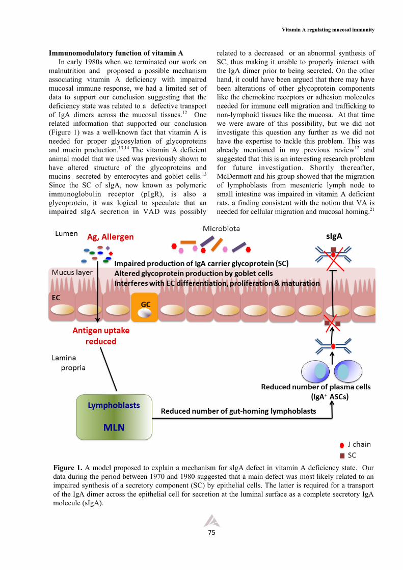
Immunomodulatory function of vitamin A In early 1980s when we terminated our work on
malnutrition and proposed a possible mechanism associating vitamin A deficiency with impaired mucosal immune response, we had a limited set of data to support our conclusion suggesting that the deficiency state was related to a defective transport of IgA dimers across the mucosal tissues.12 One related information that supported our conclusion (Figure 1) was a well-known fact that vitamin A is needed for proper glycosylation of glycoproteins and mucin production.13,14 The vitamin A deficient animal model that we used was previously shown to have altered structure of the glycoproteins and mucins secreted by enterocytes and goblet cells.13 Since the SC of sIgA, now known as polymeric immunoglobulin receptor (pIgR), is also a glycoprotein, it was logical to speculate that an impaired sIgA secretion in VAD was possibly
related to a decreased or an abnormal synthesis of SC, thus making it unable to properly interact with the IgA dimer prior to being secreted. On the other hand, it could have been argued that there may have been alterations of other glycoprotein components like the chemokine receptors or adhesion molecules needed for immune cell migration and trafficking to non-lymphoid tissues like the mucosa. At that time we were aware of this possibility, but we did not investigate this question any further as we did not have the expertise to tackle this problem. This was already mentioned in my previous review12 and suggested that this is an interesting research problem for future investigation. Shortly thereafter, McDermott and his group showed that the migration of lymphoblasts from mesenteric lymph node to small intestine was impaired in vitamin A deficient rats, a finding consistent with the notion that VA is needed for cellular migration and mucosal homing.21
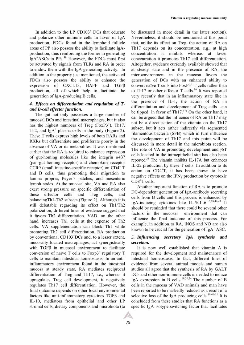
Figure 1. A model proposed to explain a mechanism for sIgA defect in vitamin A deficiency state. Our data during the period between 1970 and 1980 suggested that a main defect was most likely related to an impaired synthesis of a secretory component (SC) by epithelial cells. The latter is required for a transport of the IgA dimer across the epithelial cell for secretion at the luminal surface as a complete secretory IgA molecule (sIgA).
Asian Pac J Allergy Immunol 2015;33:71-89
76
Similarly, Kaufmann et al reported that RA is critical for the trafficking of vaccine-elicited T lymphocytes to the intestine needed for protection.22 Thus, it is now agreed that vitamin A is required to imprint gut-homing properties to T and B cells.23-26 In the present review, I briefly summarize and describe the different vitamin A-dependent steps and processes that lead to a preferential production of IgA isotype in the mucosa. Because much information in the literature is now available on the modulatory role of RA in innate immunity, I will limit my discussion to the aspects of the innate response that have important impact on the regulation of the adaptive arm.
1. Effects on antigen uptake by epithelial cells. VAD is known to interfere with proliferation and
maturation of epithelial cells, decrease the activity of brush border enzymes and induce keratinization. However, its influence on the number of goblet cells is still debatable. Detailed analysis in one recent study showed atrophy of small intestine, shortening of villi and marked increase of goblet cells with decreased mucin MUC2 gene expression but simultaneously increased MUC3 gene expression.27 In our rat model, both the goblet cells and the mucins produced declined with prolonged deficiency state.13 The composition of glycoproteins in these VAD animals were also different from those of control animals, particularly with regard to the synthesis of these high molecular weight glycoproteins. RA was reported to have suppressed the synthesis of large fucosylated glycopeptides.28 It is known that the VA status and the composition of mucins vary in different parts of GI tract and this can influence adherence of microbes and food particles to absorptive surface.29-31 If there are some structural and biochemical changes at the apical surface of these epithelial cells and M cells, e.g., degree of glycosylation and composition of the carbohydrate incorporated, this may interfere with the process of antigen production. There are recent studies showing the significance of fucosylation of the glycosylated surface in regulating intestinal symbiosis which, when disturbed, can lead to dysbiosis and increased susceptibility to intestinal pathogens.32-34 Because it was reported earlier that RA could suppress the synthesis of large fucosylated glycopeptides, it is tempting to speculate that this may also affect the fucosylation of specific high molecular weight cell-surface glycoproteins in the intestine. However, it is not known whether or not the changes just mentioned can influence antigen adherence and
uptake. Smaller numbers of Paneth cells were also noted in VAD animals and this can contribute to an overall reduction in the antimicrobial peptide defensins and RegIIIs in the inner mucus layer.27 The latter situation would allow more microbes to be in closer contact with the epithelial surface and greater chance to be up-taken by immune cells. Moreover, VAD may also impair permeability at the apical junction which can increase the chance for the microbes or allergens to come into contact and interact with the underlying immune-reactive cells, e.g., DCs, macrophages and stromal cells in the lamina propria. Other surface glycoproteins like cell adhesion molecules and microbial receptors may also be altered in VAD and this, for example, can affect the binding strength of antigen and the degree of microbial uptake which may have impact on intracellular transportation for processing and presentation.
2. Influence on antigen processing and presentation.
Until very recently, little was known about the influence of VA on antigen processing and presentation by both professional and non-professional antigen-presenting cells. However, there is now evidence showing that RA can enhance DC maturation and function including antigen processing and presenting capacity.18,25,35,36 It was demonstrated previously that VAD animals had decreased number of monocyte in spleen and bone marrow, impaired phagocytosis and aberrant mucosal immune response.16,37 The impaired intracellular killing can theoretically affect microbial degradation which may have influenced its ability to interact with HLA and transport to the cell surface for presentation to T cells. RA, in addition, has been reported to enhance upregulation of CD1d expression by APCs, thus stimulating lipid presentation, activation and proliferation of NKT cells.38 On the other hand, there are also reports using different animal models suggesting that the APCs from VA sufficient animals may also have lower expression of HLA I and HLA II, but whether or not this is to a degree that has significant impact on antigen presentation remains to be determined. The discrepancy noted among different groups of investigators is not surprising or unexpected because VA is known to be involved in protein glycosylation and its deficiency can have different effect on the synthesis of HLA, depending on the animal species used. The expression of matrix metalloproteinase 9 is also known to be altered in VAD state and this can
Vitamin A regulating mucosal immunity
77
influence migration of DCs to draining lymph nodes for lymphocyte activation. IL-1 production has also been reported to be adversely affected in VAD and this, together with possible alteration of the synthesis of other innate cytokines, can also influence the quality of the immune response.
In addition to the effect of VA /VAD on the conventional DCs like LP (GALT) DCs and splenic DCs, it is possible that it can influence another DC population now known as follicular DCs (FDC). 25,36,39 Although the FDC is known to function in germinal centers (GC) formation in the PP areas and in polarization of B cells for IgA production, not much information is available regarding its antigen processing abilities and presentation potential. Like other DCs, the FDC possesses surface, cytoplasmic and nuclear receptors that are capable of perceiving the presence of, for example, gut bacteria and local tissue factors like diet and vitamin A metabolites. In fact, it has been shown recently that direct stimulation of FDCs by bacterial products and RA can synergistically enhance the expression of chemokines CXCL13 and BAFF, and facilitates the secretion and function of TGFβ. The effect of VA on the expression of MMPs and other extracellular matrix molecules may also affect cell migration and antigen presentation and this point should be further explored in the future.
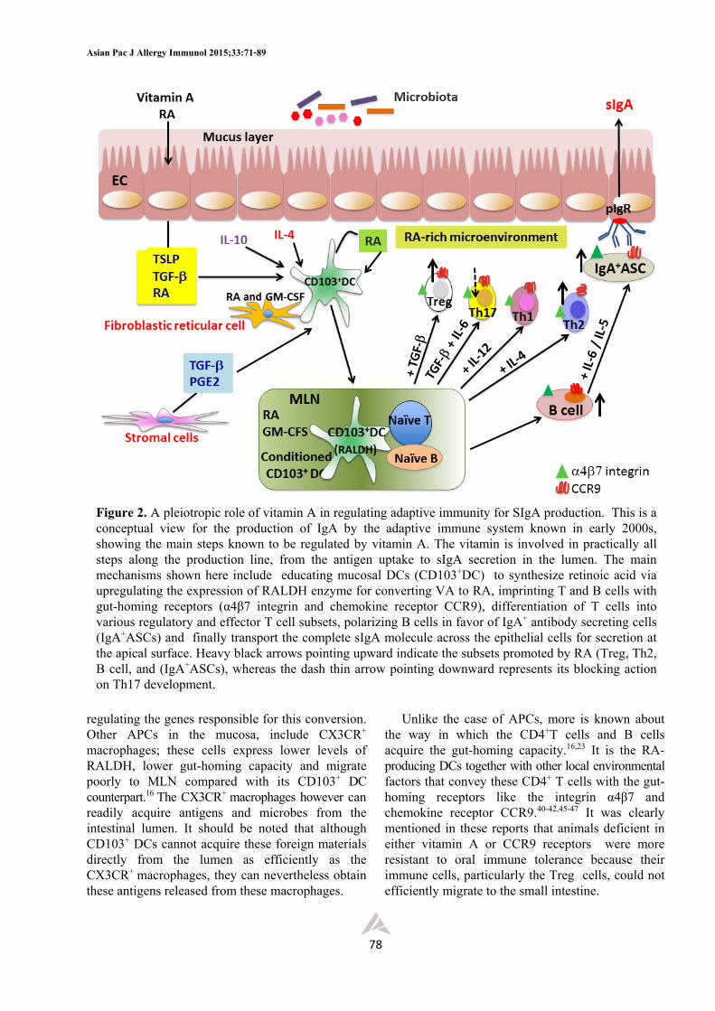
3. Influencing mucosal homing of T and B lymphocytes and DCs.
One special characteristic of mucosal immune cells is their unique mucosal-imprinting phenotype, a property required in subsequent steps in the production and secretion of IgA antibody isotype (Figure 2). Several reports agree that this special property requires the presence of RA in the mucosal environment.16,24,25,36 Various stromal cells, particularly the intestinal epithelial cells, can metabolize dietary VA to RA which contributes to RA-rich environment in mucosal tissues. The key finding on the influence of vitamin A (or RA) on the regulation of mucosal immune response was the reports from Iwata’s group, showing that RA has a central role in differentiation of DCs and that the mucosal DCs could metabolize retinol into retinoic acid. 40-42 This was subsequently followed by the findings that DC-produced RA is crucial for imprinting both T and B cells with gut-homing properties.16,43,44 RA is also known to control the generation of gut-homing migratory DC precursors (sometimes referred to as pre-µDCs) in the bone marrow and the frequency of this cell population is markedly reduced in VAD
animals.18 Under steady state, the pre-µDCs express gut-homing integrin receptor α4β7 but not the chemokine receptor CCR9. However, the latter can be up-regulated by inflammation or infection. It should be noted that the lack of lymphoid organ trafficking receptor CCR7 in these DC precursors may explain why they settle in the mucosa instead of the lymphoid organs. In the presence of RA, the pre-µDCs can also differentiate into CCR9+ plasmacytoid DCs (pDCs) and conventional DCs (cDCs) subsets that preferentially develop into intestinal CD103+cDCs. The precursors of DCs that settle in the mucosa and subsequently acquire a “conditioning” mucosal characteristic depend also on the richness of VA metabolites in the environment. These educated lamina propria DCs thereafter have the ability to convert VA to RA, as they now express the RALDH enzyme needed for the conversion.14,44 How these mucosal DCs acquire the ability to express and synthesize this enzyme in large quantity is a debatable issue. It is possible that some bone marrow CD103+ DC precursors are pre-committed to become RA producers, but, if this is not the case, they must be educated locally to acquire the ability to produce RA. Different lines of evidence point to the latter possibility. In such a case, then what are the local factors that induce this change? As these DCs are located near IECs, it is likely that a direct cell-cell interaction might be responsible for such a conversion. Another possibility is related to the mediators produced by IECs, and among these “alarmins”, the TSLP has been identified to be the principle mediator involved in this process. On the other hand, there are many other candidates that have been implicated in the induction of RALDH expression in these CD103+ DCs, and some of those that have been reported include TLR signals, Wnt/β-catenin pathway, GM-CSF and PPAR agonists.16,25,36 Recently, a unique lamina propria stromal cell has been identified and shown to possess a constitutive enzyme RALDH that can synthesize RA independently of VA.17 This cell, usually found in close proximity to mucosal DC, has markers typical of fibroblastic reticular cell. It can also synthesize GM-CSF that facilitates the synthesis of RA. However, the ability to synthesize RA by fibroblastic reticular cell requires the presence of commensal bacteria. This complex network will be discussed in more detail in the last section of this review. Incidentally, it should be mentioned that RA itself, through positive feedback, is sufficient to induce its own synthesis by up-
Asian Pac J Allergy Immunol 2015;33:71-89
78
regulating the genes responsible for this conversion. Other APCs in the mucosa, include CX3CR+
macrophages; these cells express lower levels of RALDH, lower gut-homing capacity and migrate poorly to MLN compared with its CD103+ DC counterpart.16 The CX3CR+ macrophages however can readily acquire antigens and microbes from the intestinal lumen. It should be noted that although CD103+ DCs cannot acquire these foreign materials directly from the lumen as efficiently as the CX3CR+ macrophages, they can nevertheless obtain these antigens released from these macrophages.
Unlike the case of APCs, more is known about the way in which the CD4+T cells and B cells acquire the gut-homing capacity.16,23 It is the RA-producing DCs together with other local environmental factors that convey these CD4+ T cells with the gut-homing receptors like the integrin α4β7 and chemokine receptor CCR9.40-42,45-47 It was clearly mentioned in these reports that animals deficient in either vitamin A or CCR9 receptors were more resistant to oral immune tolerance because their immune cells, particularly the Treg cells, could not efficiently migrate to the small intestine.
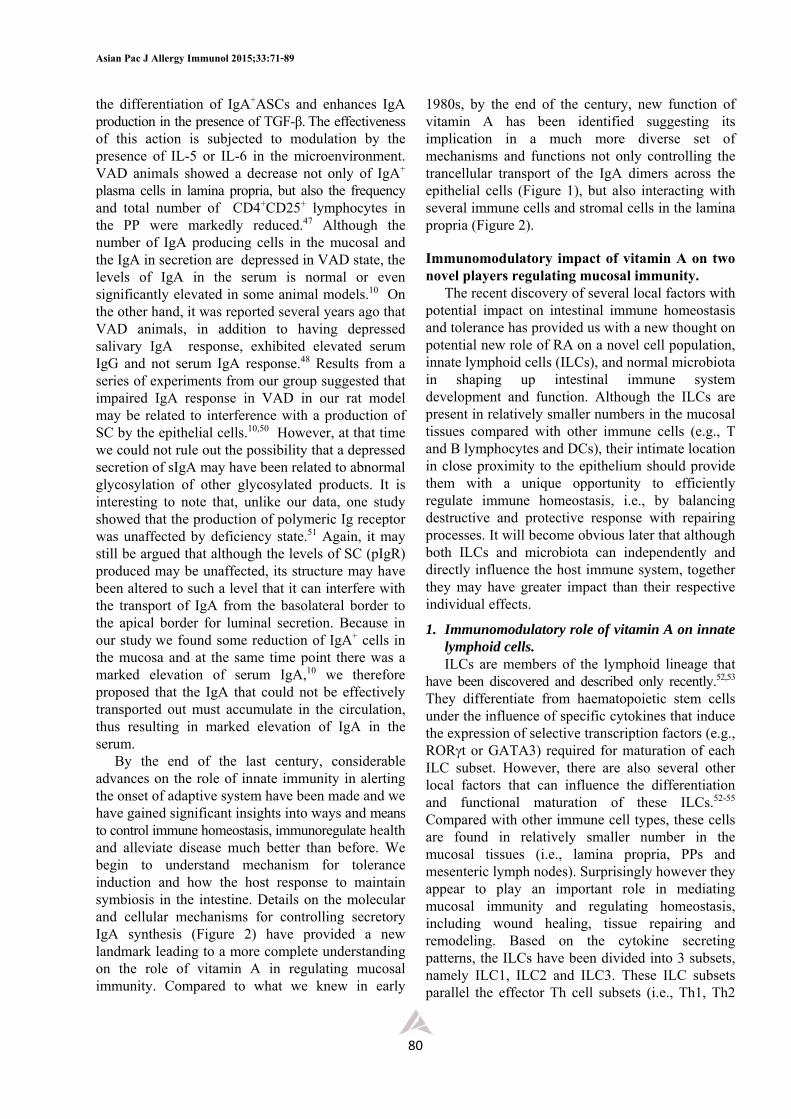
Figure 2. A pleiotropic role of vitamin A in regulating adaptive immunity for SIgA production. This is a conceptual view for the production of IgA by the adaptive immune system known in early 2000s, showing the main steps known to be regulated by vitamin A. The vitamin is involved in practically all steps along the production line, from the antigen uptake to sIgA secretion in the lumen. The main mechanisms shown here include educating mucosal DCs (CD103+DC) to synthesize retinoic acid via upregulating the expression of RALDH enzyme for converting VA to RA, imprinting T and B cells with gut-homing receptors (α4β7 integrin and chemokine receptor CCR9), differentiation of T cells into various regulatory and effector T cell subsets, polarizing B cells in favor of IgA+ antibody secreting cells (IgA+ASCs) and finally transport the complete sIgA molecule across the epithelial cells for secretion at the apical surface. Heavy black arrows pointing upward indicate the subsets promoted by RA (Treg, Th2, B cell, and (IgA+ASCs), whereas the dash thin arrow pointing downward represents its blocking action on Th17 development.
Vitamin A regulating mucosal immunity
79
In addition to the LP CD103+ DCs that educate and polarize other immune cells in favor of IgA production, FDCs found in the lymphoid follicle areas of PP also possess the ability to facilitate IgA-production, thus reinforcing the former in generating IgA+ASCs in PPs.39 However, the FDCs must first be activated by signals from TLRs and RA in order to endow them with the IgA generating activity. In addition to the property just mentioned, the activated FDCs also possess the ability to enhance the expression of CXCL13, BAFF and TGFβ production, all of which help to facilitate the generation of IgA-producing B cells.
4. Effects on differentiation and regulation of T- and B-cell effector function. The gut not only possesses a large number of mucosal DCs and intestinal macrophages, but it also has the highest numbers of Treg (FoxP3+), Th17, Th2, and IgA+ plasma cells in the body (Figure 2). These T cells express high levels of both RARs and RXRs but differentiate and proliferate poorly in the absence of VA or its metabolites. It was mentioned earlier that the RA is required to enhance expression of gut-homing molecules like the integrin α4β7 (pan-gut homing receptor) and chemokine receptor CCR9 (small intestine-specific receptor) on CD4+ T and B cells, thus promoting their migration to lamina propria, Peyer’s patches, and mesenteric lymph nodes. At the mucosal site, VA and RA also exert strong pressure on specific differentiation of these effector cells and Treg cells, and balancingTh1-Th2 subsets (Figure 2). Although it is still debatable regarding its effect on Th1/Th2 polarization, different lines of evidence suggest that it favors Th2 differentiation. VAD, on the other hand, increases Th1 cells at the expense of Th2 cells. VA supplementation can block Th1 while promoting Th2 cell differentiation. RA production by conventional CD103+DCs and, to a lesser extent, mucosally located macrophages, act synergistically with TGFβ in mucosal environment to facilitate conversion of naïve T cells to Foxp3+ regulatory T cells to maintain intestinal homeostasis. In an anti-inflammatory environment found in the intestinal mucosa at steady state, RA mediates reciprocal differentiation of Treg and Th17, i.e., whereas it upregulates Treg cell development, it negatively regulates Th17 cell differentiation. However, the final outcome depends on other local environmental factors like anti-inflammatory cytokines TGFβ and IL-10, mediators from epithelial and other LP stromal cells, dietary components and microbiota (to
be discussed in more detail in the latter section). Nevertheless, it should be mentioned at this point that, unlike its effect on Treg, the action of RA on Th17 depends on its concentration, e.g., at high concentration it inhibits whereas at lower concentration it promotes Th17 cell differentiation. Altogether, evidence currently available showed that at steady state and in the presence of RA, the microenvironment in the mucosa favors the generation of DCs with an enhanced ability to convert naïve T cells into FoxP3+ T cells rather than to Th17 or other effector T cells.16 It was reported very recently that in an inflammatory state like in the presence of IL-1, the action of RA in differentiation and development of Treg cells can be tipped in favor of Th17.47a On the other hand, it can be argued that the influence of RA on Th17 may not be a direct action of the vitamin on the Th17 subset, but it acts rather indirectly via segmented filamentous bacteria (SFB) which in turn influence the development of Th17 and this point will be discussed in more detail in the microbiota section. The role of VA in promoting development and T cells located in the intraepithelial site has also been reported.38 The vitamin inhibits IL-17A but enhances IL-22 production by these T cells. In addition to its action on CD4+T, it has been shown to have negative effects on the IFN production by cytotoxic CD8+T cells.
Another important function of RA is to promote DC-dependent generation of IgA-antibody secreting cells from B cells and this process is enhanced by IgA-inducing cytokines like IL-5/IL-6.16,19,46,47 It should be reminded that there could be several other factors in the mucosal environment that can influence the final outcome of this process. For example, in addition to RA, iNOS and NO are also known to be crucial for the generation of IgA+ ASC.
5. Influencing secretory IgA synthesis and secretion.
It is now well established that vitamin A is required for the development and maintenance of intestinal homeostasis. In fact, different lines of evidence from several animal models and human studies all agree that the synthesis of RA by GALT DCs and other non-immune cells is needed to induce IgA expression in B cells.19,20,24 The number of B cells in the mucosa of VAD animals and man have been reported to be markedly reduced as a result of a selective loss of the IgA producing cells.10,46-51 It is concluded from these studies that RA functions as a specific IgA isotype switching factor that facilitates
Asian Pac J Allergy Immunol 2015;33:71-89
80
the differentiation of IgA+ASCs and enhances IgA production in the presence of TGF-β. The effectiveness of this action is subjected to modulation by the presence of IL-5 or IL-6 in the microenvironment. VAD animals showed a decrease not only of IgA+ plasma cells in lamina propria, but also the frequency and total number of CD4+CD25+ lymphocytes in the PP were markedly reduced.47 Although the number of IgA producing cells in the mucosal and the IgA in secretion are depressed in VAD state, the levels of IgA in the serum is normal or even significantly elevated in some animal models.10 On the other hand, it was reported several years ago that VAD animals, in addition to having depressed salivary IgA response, exhibited elevated serum IgG and not serum IgA response.48 Results from a series of experiments from our group suggested that impaired IgA response in VAD in our rat model may be related to interference with a production of SC by the epithelial cells.10,50 However, at that time we could not rule out the possibility that a depressed secretion of sIgA may have been related to abnormal glycosylation of other glycosylated products. It is interesting to note that, unlike our data, one study showed that the production of polymeric Ig receptor was unaffected by deficiency state.51 Again, it may still be argued that although the levels of SC (pIgR) produced may be unaffected, its structure may have been altered to such a level that it can interfere with the transport of IgA from the basolateral border to the apical border for luminal secretion. Because in our study we found some reduction of IgA+ cells in the mucosa and at the same time point there was a marked elevation of serum IgA,10 we therefore proposed that the IgA that could not be effectively transported out must accumulate in the circulation, thus resulting in marked elevation of IgA in the serum.
By the end of the last century, considerable advances on the role of innate immunity in alerting the onset of adaptive system have been made and we have gained significant insights into ways and means to control immune homeostasis, immunoregulate health and alleviate disease much better than before. We begin to understand mechanism for tolerance induction and how the host response to maintain symbiosis in the intestine. Details on the molecular and cellular mechanisms for controlling secretory IgA synthesis (Figure 2) have provided a new landmark leading to a more complete understanding on the role of vitamin A in regulating mucosal immunity. Compared to what we knew in early
1980s, by the end of the century, new function of vitamin A has been identified suggesting its implication in a much more diverse set of mechanisms and functions not only controlling the trancellular transport of the IgA dimers across the epithelial cells (Figure 1), but also interacting with several immune cells and stromal cells in the lamina propria (Figure 2).
Immunomodulatory impact of vitamin A on two novel players regulating mucosal immunity.
The recent discovery of several local factors with potential impact on intestinal immune homeostasis and tolerance has provided us with a new thought on potential new role of RA on a novel cell population, innate lymphoid cells (ILCs), and normal microbiota in shaping up intestinal immune system development and function. Although the ILCs are present in relatively smaller numbers in the mucosal tissues compared with other immune cells (e.g., T and B lymphocytes and DCs), their intimate location in close proximity to the epithelium should provide them with a unique opportunity to efficiently regulate immune homeostasis, i.e., by balancing destructive and protective response with repairing processes. It will become obvious later that although both ILCs and microbiota can independently and directly influence the host immune system, together they may have greater impact than their respective individual effects.
1. Immunomodulatory role of vitamin A on innate lymphoid cells. ILCs are members of the lymphoid lineage that
have been discovered and described only recently.52,53 They differentiate from haematopoietic stem cells under the influence of specific cytokines that induce the expression of selective transcription factors (e.g., RORt or GATA3) required for maturation of each ILC subset. However, there are also several other local factors that can influence the differentiation and functional maturation of these ILCs.52-55 Compared with other immune cell types, these cells are found in relatively smaller number in the mucosal tissues (i.e., lamina propria, PPs and mesenteric lymph nodes). Surprisingly however they appear to play an important role in mediating mucosal immunity and regulating homeostasis, including wound healing, tissue repairing and remodeling. Based on the cytokine secreting patterns, the ILCs have been divided into 3 subsets, namely ILC1, ILC2 and ILC3. These ILC subsets parallel the effector Th cell subsets (i.e., Th1, Th2
Vitamin A regulating mucosal immunity
81
and Th17) in term of their cytokine secretion patterns. For example, ILC1s secrete IFN, ILC2s secrete IL-4, IL-5, IL-9 and IL-13, and ILC3s secrete predominantly IL-22 and IL-17. The last subset in addition expresses both membrane-associated and soluble lymphotoxin (LT) which can enhance IL-23 production by DCs. A member of the ILC3 subset known as lymphoid tissue inducer (LTi) cells is also essential for lymphoid tissue development. Although all these ILCs have no T cell antigen-binding receptors, they are reported to have MHC II, thus providing them with antigen processing and presenting potential.53 These ILCs are believed to participate in host response during a period of transition from innate to adaptive immunity and contribute to immunopathology like chronic inflammation, inflammatory diseases and allergy, and more recently shown to play a role in metabolic homeostasis.53,56 At steady state, the predominant ILC subsets in the mucosa are ILC2s and ILC3s.52,53 The former is important for protection against helminths whereas the latter is for extracellular bacteria and fungi. There is only limited set of data available on a protective function of ILC1s in the mucosa, but with the information currently available, this subset is believed to play a role in fighting against intracellular bacteria and parasites. It is of interest that the ILC2 subset has been shown recently to be associated with metabolic homeostasis, metabolic diseases and dietary stress.56 It has been identified in adipose tissues and, in the presence of IL-33, shown to protect the host against development of obesity.56 It was therefore suggested that manipulating the IL-33 – ILC2 axis may be beneficial to alleviate obesity and other metabolic diseases in humans. In the past, a defective Th2 cell function has been blamed for the increased prevalence of mucosal infections in malnutrition and vitamin A deficiency. However, with the discovery of ILCs, this concept needs modification and the effectiveness of type 2 immunity is now believed to be under the control of not only the adaptive Th2 response, but also to the cytokines produced by ILC2s.57,58 It is possible that in addition to its action on T cells, vitamin A may also influence the functional integrity of ILCs. In fact, the data recently reported by Spencer and associates57
suggested that VA regulates the balance between ILC2s and ILC3s in protecting mucosal infection and immunopathology associated with autoimmune, inflammatory and allergic diseases.
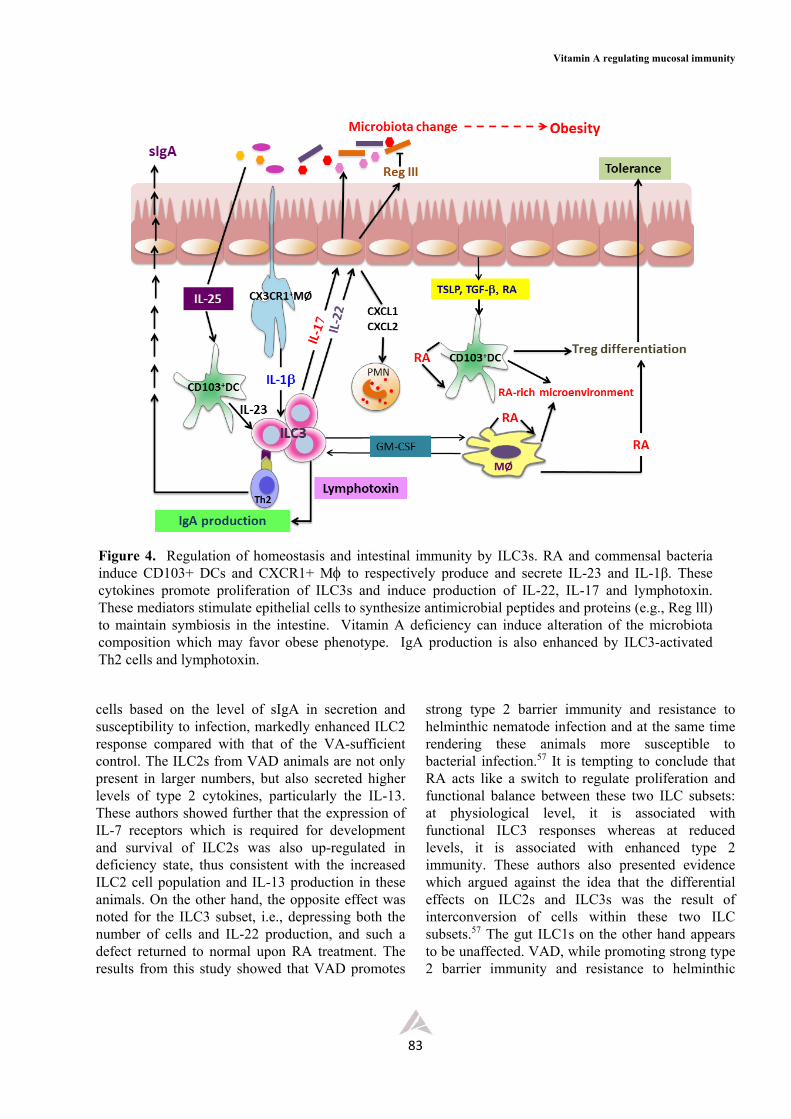
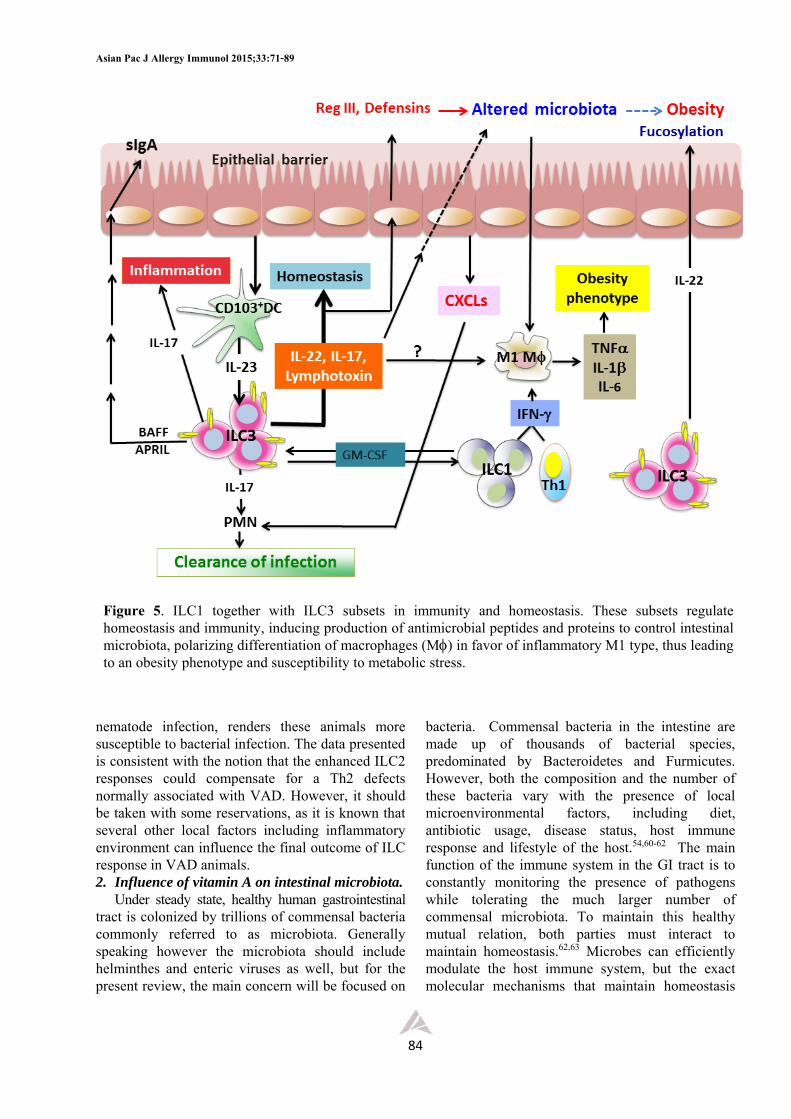
Because of their location in close proximity with the epithelium and cells of adaptive immune system, these ILCs can readily interact and crosstalk with these immune cells as well as with other stromal cells present in the mucosa and the outcomes from this interaction have been thoroughly described and discussed in several excellent reviews52,53,58 and summarized as shown in Figures 3-5. Briefly, the epithelium which receives environmental signals from, for example, diet and microbiota, as well as from endogenous innate cytokines provides signals (e.g., IL-25, IL-33 and TSLP) for the activation ofILCs and mucosal DCs in the lamina propria. The ILC2s are activated to proliferate and secrete type 2 cytokines (IL-4, IL-5, IL-9, and IL-13) and GM-CSF, whereas the DCs are activated to induce Th2 cell differentiation which eventually gives rise to classical type 2 cytokines (Figure 3). Together, the cytokines from both ILC2s and Th2 cells can interact with other immune cells like macrophages, eosinophils, mast cells and B cells. Some induce epithelial cell proliferation and secretion of antimicrobial peptides (e.g., RegIIIs and defensins) and an epidermal growth factor-like molecule known as amphiregulin, some induce goblet cell hyperplasia and secretion of mucins and RELMs, some induce smooth muscle cell hypercontractility or promote LP macrophage differentiation toward an alternatively activated phenotype (M2).53,58 The latter secretes IL-10 and, together with the anti- inflammatory cytokines produced by other somatic cells, provides an anti-inflammatory microenvironment associated with intestinal tolerance. Moreover, the IL-13 and IL-9 from ILC2s and Th2 cells can activate epithelial cells and goblet cells to produce amphiregulin and RELMs needed for tissue repair and restoring tissue integrity following damage associated with chronic and acute inflammation. Fibroblasts activated by IL-4 and IL-13 may also contribute to wound healing (Figure 3). The ILC3 subset, on the other hand, was found to be associated with inflammatory pathology and metabolic diseases like obesity (Figures 4 and 5). In response to bacterial infection, mucosal DCs are activated to produce IL-23 required for ILC3 proliferation and survival. These ILC3s subsequently secrete a number of mediators including IL-22, IL-17 and LTs. These mediators promote differentiation of the classical M1 inflammatory macrophages, and stimulate the epithelium to produce antimicrobial peptides RegIIIs and chemokine
Asian Pac J Allergy Immunol 2015;33:71-89
82
ligands CXCL1 and CXCL2. These ligands induce migration of PMNs to facilitate phagocytosis and microbial killing (Figure 4). The IL-22-induced M1 inflammatory macrophages, when activated, produce TNFα, IL-6 and IL-1 that give rise to insulin insensitive hypertrophic adipocytes associated with obesity and other metabolic disorders. This condition is facilitated by the IFN secreted from ILC1s (Figure 5).53
It was discussed in more details in the earlier sections that vitamin A and its biologically active metabolite retinoic acid are required for differentiation and maturation of mucosal DCs. These RA-activated DCs can then imprint T and B
cells with gut-homing receptors which eventually produce and secrete sIgA. Recent data based on the levels of RA in utero suggested further that dietary retinoids can also serve as key regulators of pre-natal ILCs with a lifelong impact on the size of secondary lymphoid organs in adults.59 Such an effect is probably related to its action on LTi cell maturation. RA is also known to significantly enhance the production of IL-22 by ILC3 cells which subsequently induce epithelial cells to produce the antimicrobial peptide RegIII in the colon.38 It was surprising, at least to me, to learn from a recent report by Spencer and associates57 that VAD, which is well known to have impaired Th2
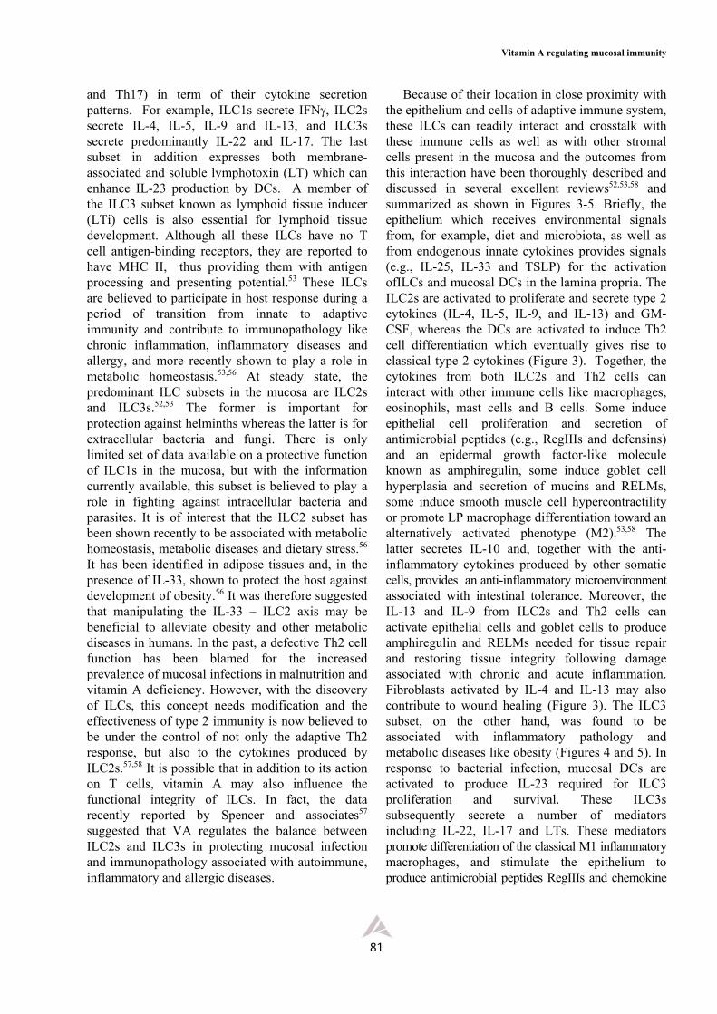
Figure 3. Innate lymphoid cells type 2 (ILC2s) regulating intestinal immunity and homeostasis. Epithelial cells release mediators (red arrow) to stimulate ILC2 proliferation and production of type 2 cytokines (IL-4, IL-5, IL-6, IL-9 and IL-13) and activate different effector pathways (e.g., AAM (M2), basophil, mast cell, eosinophil, Th2 and production of mucus and AMP). Vitamin A regulates and balances the various ILC subsets for homeostasis in the intestine. In deficiency state, there are marked increase of ILC2 cell proliferation and cytokine production, and, at the same time, the proliferation and function of ILC3 subset are suppressed
Vitamin A regulating mucosal immunity
83
cells based on the level of sIgA in secretion and susceptibility to infection, markedly enhanced ILC2 response compared with that of the VA-sufficient control. The ILC2s from VAD animals are not only present in larger numbers, but also secreted higher levels of type 2 cytokines, particularly the IL-13. These authors showed further that the expression of IL-7 receptors which is required for development and survival of ILC2s was also up-regulated in deficiency state, thus consistent with the increased ILC2 cell population and IL-13 production in these animals. On the other hand, the opposite effect was noted for the ILC3 subset, i.e., depressing both the number of cells and IL-22 production, and such a defect returned to normal upon RA treatment. The results from this study showed that VAD promotes
strong type 2 barrier immunity and resistance to helminthic nematode infection and at the same time rendering these animals more susceptible to bacterial infection.57 It is tempting to conclude that RA acts like a switch to regulate proliferation and functional balance between these two ILC subsets: at physiological level, it is associated with functional ILC3 responses whereas at reduced levels, it is associated with enhanced type 2 immunity. These authors also presented evidence which argued against the idea that the differential effects on ILC2s and ILC3s was the result of interconversion of cells within these two ILC subsets.57 The gut ILC1s on the other hand appears to be unaffected. VAD, while promoting strong type 2 barrier immunity and resistance to helminthic
Figure 4. Regulation of homeostasis and intestinal immunity by ILC3s. RA and commensal bacteria induce CD103+ DCs and CXCR1+ M to respectively produce and secrete IL-23 and IL-1β. These cytokines promote proliferation of ILC3s and induce production of IL-22, IL-17 and lymphotoxin. These mediators stimulate epithelial cells to synthesize antimicrobial peptides and proteins (e.g., Reg lll) to maintain symbiosis in the intestine. Vitamin A deficiency can induce alteration of the microbiota composition which may favor obese phenotype. IgA production is also enhanced by ILC3-activated Th2 cells and lymphotoxin.
Asian Pac J Allergy Immunol 2015;33:71-89
84
nematode infection, renders these animals more susceptible to bacterial infection. The data presented is consistent with the notion that the enhanced ILC2 responses could compensate for a Th2 defects normally associated with VAD. However, it should be taken with some reservations, as it is known that several other local factors including inflammatory environment can influence the final outcome of ILC response in VAD animals. 2. Influence of vitamin A on intestinal microbiota.
Under steady state, healthy human gastrointestinal tract is colonized by trillions of commensal bacteria commonly referred to as microbiota. Generally speaking however the microbiota should include helminthes and enteric viruses as well, but for the present review, the main concern will be focused on
bacteria. Commensal bacteria in the intestine are made up of thousands of bacterial species, predominated by Bacteroidetes and Furmicutes. However, both the composition and the number of these bacteria vary with the presence of local microenvironmental factors, including diet, antibiotic usage, disease status, host immune response and lifestyle of the host.54,60-62 The main function of the immune system in the GI tract is to constantly monitoring the presence of pathogens while tolerating the much larger number of commensal microbiota. To maintain this healthy mutual relation, both parties must interact to maintain homeostasis.62,63 Microbes can efficiently modulate the host immune system, but the exact molecular mechanisms that maintain homeostasis
Figure 5. ILC1 together with ILC3 subsets in immunity and homeostasis. These subsets regulate homeostasis and immunity, inducing production of antimicrobial peptides and proteins to control intestinal microbiota, polarizing differentiation of macrophages (M) in favor of inflammatory M1 type, thus leading to an obesity phenotype and susceptibility to metabolic stress.
Vitamin A regulating mucosal immunity
85
are not well understood. Considerable progress during the recent years has come from studies of immune development in germfree animals and in germfree animals monocolonized with specific organism. It was found that germfree animals exhibit impaired immune development characterized by immature GALT, decreased numbers of B and T lymphocytes, defective sIgA secretion, and low levels of antimicrobial peptides and proteins.54,60 Immune aberrations observed in germfree animals can be reversed by colonization with commensal bacteria. It has been shown, for example, that,
among the intestinal microbes, segmented filamentous bacteria (SFB) are required for Th17 cell differentiation.63 Although the exact molecular mechanism for this induction is not well understood, it has been proposed that these bacteria induce IL-25 expression by epithelial cells which interferes with macrophage production of IL-23 that can in turn suppress Th17 cell development. Another possibility is related to the effect of diet including vitamin A (to be discussed below). In addition to exerting direct influence on host immune system, the commensal bacteria can also make use of their metabolites to
Figure 6. ILC3s enhance fucosylation of epithelial cells. Following receiving signal from CD103+DC, activated ILC3 together with lymphotoxin (LTα1β2) upregulate the expression of IL-22 receptor on the epithelial surface (blue rectangle). The activated ILC3s thereafter receive a signal from segmented filamentous bacteria (SFB) and secrete IL-22. Binding of IL-22 to its receptor (IL-22R) on the basal surface of the epithelium activates the mechanism that upregulates the production of fucosyltransferase 2 (Fut2) enzyme (thick solid red arrow) which adds fucose subunit (green triangle) to the glycosylated surface of the epithelial cells. Commensal bacteria possess fucosidase enzyme that can cleave fucose from the fucosylated carbohydrate and use it for energy source. Most pathogens, on the other hand, lack this enzyme and therefore cannot successfully compete with the commensals for survival.
Asian Pac J Allergy Immunol 2015;33:71-89
86
indirectly modulate host response.64-66 An example for the latter situation is the metabolism of dietary fibers by commensal bacteria to short-chain fatty acids (SCFAs) which favor differentiation of CD4+ naïve Th cell into Treg. The SCFAs produced provide the host with anti-inflammatory environment, most likely by depressing the production of pro-inflammatory cytokines by macrophages. Such an influence has been linked to metabolic homeostasis. The pleiotropic interaction between bacterial metabolites like SCFAs and host may provide answer to the key question in immunology on how the mucosal immune system discriminates between commensals and pathogens.
The composition of commensal bacteria in the GI tract varies with the types of diet consumed, regulating both the composition and the total number of aerobic and anaerobic bacteria in different parts of GI tract.29,57,65 While micronutrients like vitamins and trace elements were thought to have relatively minor impact on the microbiota, recent reports have shown this to be inaccurate, as the signals from these micronutrients may be amplified first by inducing IECs and other stromal cells to secrete mediators which may give stronger signals and impact on the luminal microbes.65,66 A few years ago Cha and associates observed that VAD alters structural integrity of small intestine of mice, with increased numbers of goblet cells and mucin production, but with fewer numbers of Paneth cells.27 These investigators demonstrated further that both the mucins and antimicrobial peptides and proteins produced by these cells have specific effect on microbiota of these animals and speculated that the alteration of the microbiota like the ratio between aerobic and anaerobic bacteria associated with VAD diet might be crucial for proliferation and differentiation of immune cells, particularly the Th17 cells. It appears therefore that VAD can alter the phenotype and function of epithelial cells by, for example, enhancing mucin production by goblet cells and reducing the levels of antimicrobial peptides which subsequently alter the number and composition of commensal bacteria in the intestinal lumen. Alteration of the microbiota leads to a condition known as dysbiosis which can disrupt the normal process of immune cell development and intestinal tolerance and homeostasis and tissue repair. Considerable attention is now focused into this research area and recently there are many excellent and extensive reviews on the complex and dynamic nature of host-microbiota interaction.54,60-67
In addition to its effect via Th17 proliferation and differentiation, the microbiota possesses many other potential pathways to enhance sIgA production. For example, studies using GF animals showed that commensal bacteria could influence early B cell development in the intestine and promote IgA class switching via T cell-independent pathway.67 There is evidence suggesting that commensal bacteria stimulate IEC and mononuclear phagocytes in the intestine to produce BAFF, APRIL and TGFβ which favor IgA class switching and promote survival of IgA+ ASCs.54,60 Commensal bacteria may also influence isotype switching indirectly by first interacting with CD103+DCs or FDCs. RA and RAR agonists have been reported to facilitate commensals to induce IgA class switching and can also directly or indirectly stimulate this process via action on ILC3s.68 The latter not only has membrane-associated lymphotoxin but also secretes soluble LT which promotes T cell homing to gut and enhance both T cell-dependent and T cell-independent IgA response. Evidence is available showing that commensals can also influence the fate of Foxp3+ Treg cells.
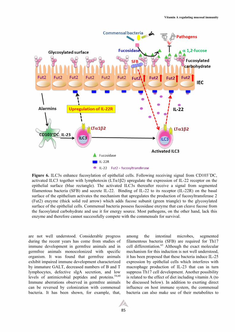
Independent reports from different groups of investigators recently provided us with a new set of data which supports a critically important principle regarding the regulation of intestinal symbiosis by ILC3s (Figure 6). These studies demonstrated the role of ILC3s in the synthesis of fucosylated glycoproteins at the apical surface of enterocytes. The latter can serve as a food source for commensal bacteria which possess fucosidase enzymes that cleave the terminal fucose sugar residue from the fucosylated proteins and use the cleaved product for energy source.32-34 Most pathogens, on the other hand, do not normally possess this enzyme and so cannot compete with the normal flora for the limited source of energy for survival. It was shown that the ILC3s, and probably together with IECs and CD103+DCs, are involved in this regulatory network following receiving unknown signal from the microbiota. The ILC3s, activated by a microbiota-independent signal(s) in the previous step, receive an additional signal from segmented filamentous bacteria (SFB) to produce and secrete IL-22. The latter binds to its receptor (IL-22R) on the surface of epithelium to upregulate the synthesis of fucosyltransferase 2 (Fut2, Figure 6). The enzyme is needed to add the fucose residue to the terminal end of the glycoprotein on the apical surface of epithelium.32 It should be recalled that vitamin A deficiency can alter the structure of high molecular weight
Vitamin A regulating mucosal immunity
87
fucosylated glycoproteins secreted by goblet cells,13 and one could therefore speculate further that the deficiency may have adverse impact on the fucosylation process in vivo which may lead to dysbiosis and immunopathology. Polymorphisms of the enzyme fucosyltransferase 2 has been reported to be associated with the onset of multiple infections and inflammatory diseases as well as metabolic syndrome in the humans.32, Earlier Spencer and associates reported that ILC3s and cytokines produced were markedly depressed in VAD,57 and this could further impair the ILC3 fucosylation function. This, together with the report by Cha and associates,27 clearly demonstrate the new role of vitamin A in modulating mucosal immunity, i.e., via modulating gut microbiota.
Altogether the data regarding ILCs and microbiota have now clearly added a new dimension to the function of vitamin A in regulating immune homeostasis in the intestine and provided us with new insights on novel approaches to manage diseases associated with dysbiosis condition. For instance, the use prebiotics and probiotics to alleviate diseases by regulating intestinal homeostasis appear particularly simple and promising.
Conclusions Local host factors like RA and cytokines together
with gut microbiota distinctively and efficiently regulate differentiation, migration and effector functions of immune cells, particularly T cells, with a final outcome of maintaining tolerance and strong type 2 mucosal immunity. Data currently available show how vitamin A deficiency promotes the type 2 barrier immunity in the face of collapsing intestinal Th1, Th17 and ILC3. RA serves as a “trafficking-receptor” switch to convey gut-homing properties of immune cells: inducing gut-homing receptors while suppressing lymph node or non-gut tissue-homing receptors. RA also co-operates with microbiota and their metabolites to maintain homeostasis in the immune system. They cross-regulate the generation of FoxP3+ Treg cells and IL-10+ T cells. RA promotes the former but microbiota and their metabolites promote the latter cell types. The potential significance of vitamin A on the epithelial cells should also be taken into consideration, as it continuously interacts with immune cells underneath and with environmental factors like microbiota, pathogens, antibiotics and other dietary constituents in the luminal surface. During the second half of the last century, a large number of investigators have
contributed to the progress in this research area and should be considered as having rewritten the “A-to-Z” concept of mucosal immune system, confirming the pleiotropic role of vitamin A in developing and regulating tolerance and homeostasis. A clearer understanding on how the different environmental factors regulate mucosal immune response provide us with a chance to rethink the framework and design novel approaches for the intervention strategies to alleviate diseases and to correct pathological conditions associated with, for example, intestinal dysbiosis and aberrant inflammatory reaction in the intestine. Lastly, a more complete understanding on the link between microbiota, diet, immune response and metabolic syndrome will provide us with new therapeutic approaches in controlling and managing diseases such as obesity and other lifestyle-associated diseases.
Acknowledgments I would like to express my appreciation to Dr.
Matsayapan Pudla (Faculty of Dentistry, Mahidol University) for her assistance with some of the illustrations used in this review.
References 1. Schaible UE, Kaufmann SHE. Malnutrition and infection: complex
mechanisms and global impacts. PloS Med. 2007;4:e115.
2. Nauss KM. Influence of vitamin A status on the immune system.
In: Bauernfeind JC, editor. Vitamin A deficiency and its control.
New York: Academic Press; 1986. p. 207-43.
3. World Health Organization. Global prevalence of vitamin A
deficiency in populations at risk 1995-2005: WHO Global database
on vitamin A deficiency. Geneva: World Health Organization;
2009.
4. Sommer A. Vitamin A deficiency and clinical disease: an historical
overview. J Nutr. 2008;138:1835-9.
5. Sirisinha S, Suskind R, Edelman R, Charupatana C, Olson RE.
Complement and C3-proactivator levels in children with protein-
calorie malnutrition and effect of nutritional treatment. Lancet.
1973;1:1016-20.
6. Edelman R, Suskind , Olson RE, Sirisinha S. Mechanisms of
defective cutaneous hypersensitivity in children with protein-calorie
malnutrition. Lancet. 1973;1:506-9.
7. Sirisinha S, Suskind R, Edelman R, Asvapaka C, Olson RE.
Secretory and serum IgA in children with protein-calorie
malnutrition. Pediatrics. 1975; 55:66-70.
8. Udomkesmalee E, Dhanamitta S, Sirisinha S, Charoenkiatkul S,
Tuntipopipat S, Banjong O, et al. Effect of vitamin A and zinc
supplementation on the nutriture of children in northeast Thailand.
Am J Clin Nutr. 1992;56:50-7.
9. Kramer TR, Udomkesmalee E, Dhanamitta S, Sirisinha S,
Charoenkiatkul S, Tuntipopipat S, et al. Lymphocyte
Asian Pac J Allergy Immunol 2015;33:71-89
88
responsiveness of children supplemented with vitamin A and zinc.
Am J Clin Nutr. 1993; 58:566-70.
10. Sirisinha S, Darip MD, Moonkarndi P, Ongsakul M, Lamb AJ.
Impaired local immune response in vitamin A-deficient rats. Clin
Exp Immunol. 1980;40:127-35.
11. Lamb AJ, Apiwatanaporn P, Olson JA. Induction of rapid,
synchronous vitamin A deficiency in the rat. J Nutr.
1974;104:1140-8.
12. Sirisinha S. Impairment of secretory immune system in vitamin A
deficiency state. J Sc Soc Thailand .1988;14:161-80.
13. Rojanapo W, Olson JA, Lamb AJ. Biochemical and immunological
characterization and the synthesis of rat intestinal glycoproteins
following the induction of rapid synchronous vitamin A deficiency.
Biochim Biophys Acta. 1980;633:386-99.
14. D’Ambrosio DN, Clugston RD, Blaner WS. Vitamin A
metabolism: an update. Nutrients. 2011;3:63-103.
15. Villamor E, Fawzi WW. Effects of vitamin A supplementation on
immune responses and correlation with clinical outcomes. Clin
Microbiol Rev. 2005;18:446-64.
16. Mora JR, Iwata M, von Andrian VH. Vitamin effects on the
immune system: vitamins A and D take centre stage. Nat Rev
Immunol. 2008;8:685-98.
17. Vicente-Suarez I, Larange A, Reardon C, Matho M, Feau S,
Chodaczek G, et al. Unique lamina propria stromal cells imprint the
functional phenotype of mucosal dendritic cells. Mucosal Immunol.
2015;8:141-51.
18. Klebanoff CA, Spencer SP, Torabi-Parizi P, Grainger JR,
Roychoudhuri R, Ji Y, et al. Retinoic acid controls the homeostasis
of pre-cDC-derived splenic and intestinal dendritic cells. J Exp
Med. 2013;210:1961-76.
19. Zeng R, Oderup C, Yuan R, Lee M, Habtezion A, Hadeiba H, et al.
Retinoic acid regulates development of a gut homing precursor for
intestinal dendritic cells. Mucosal Immunol. 2013;6:847-56.
20. Rudraraju R, Jones BG, Surman SL, Sealy RE, Thomas PG,
Hurwitz JL. Respiratory tract epithelial cells express retinaldehyde
dehydrogenase ALDH1A and enhance IgA production by
stimulated B cells in the presence of vitamin A. PLoS ONE.
2014;9:e86554.
21. McDermott MR, Mark DA, Befus AD, Baliga BS, Suskind RM,
Bienenstock J. Impaired intestinal localization of mesenteric
lymphoblasts associated with vitamin A deficiency and protein-
calorie malnutrition. Immunology. 1982;45:1-5.
22. Kaufman DR, De Calisto J, Simmons NL, Cruz AN, Villablanca
EJ, Mora JR, et al. Vitamin A deficiency impairs vaccine-elicited
gastrointestinal immunity. J Immunol. 2011;187:1877-83
23. Strober W. Vitamin A rewrites the ABCs of oral tolerance. Mucosal
Immunol. 2008;1:92-5.
24. Ross AC, Chen Q, Ma Y. Vitamin A and retinoic acid in the
regulation of B-cell development and antibody production. Vitam
Horm. 2011;86:103-6.
25. Hall JA, Grainger JR, Spencer SP, Belkaid Y. The role of retinoic
acid in tolerance and immunity. Immunity. 2011;35:13-22.
26. Raverdeau M, Mills KHG. Modulation of T cell and innate immune
responses by retinoic acid. J Immunol. 2014;192:2953-8.
27. Cha H-R, Chang S-Y, Chang J-H, Kim J-O, Yang J-Y, Kim C-H, et
al. Downregulation of Th17 cells in the small intestine by
disruption of gut flora in the absence of retinoic acid. J Immunol.
2010; 184:6799-8606.
28. Amos B, Lotan D, Lotan R. Increased fucosylation of high-
molecular-weight glycoproteins accompanies retinoic acid-induced
differentiation of F-9 embryonal carcinoma cells. Int J Cancer.
1990;46:86-94.
29. Mowat AM, Agace WW. Regional specialization within the
intestinal immune system. Nat Rev Immunol. 2014;14:667-85.
30. Peterson LW, Artis D. Intestinal epithelial cells: regulators of
barrier function and immune homeostasis. Nat Rev Immunol.
2014;14:141-53.
31. Spencer SP, Belkaid Y. Dietary and commensal derived nutrients:
shaping mucosal and systemic immunity. Curr Opin Immunol.
2012;24:379-84.
32. Goto Y, Obata T, Kunisawa J, Sato S, Ivanov II, Lamichhane A, et
al. Innate lymphoid cells regulate intestinal epithelial cell
glycosylation. Science. 2014;345;1254009.
33. Pickard JM, Maurice CF, Kinnebrew MA, Abt MC, Schenten D,
Golovkina TV, et al. Rapid fucosylation of intestinal epithelium
sustains host-commensal symbiosis in sickness. Nature.
2014;514:638-41.
34. Pham TA, Clare S, Goulding D, Arasteh JM, Stares MD, Browne
HP, et al. Epithelial IL-22RA-mediated fucosylation promotes
intestine colonization resistance to an opportunistic pathogen. Cell
Host Microbe. 2014;16:504-16.
35. Geissmann F, Revy P, Brousse N, Lepelletier Y, Folli C, Durandy
A, et al. Retinoids regulate survival and antigen presentation by
immature dendritic cells. J Exp Med. 2003;198:623-34.
36. Cassani B, Villablanca EJ, De Calisto J, Wang S, Mora JR. Vitamin
A and immune regulation: role of retinoic acid in gut-associated
dendritic cell education, immune protection and tolerance. Mol
Aspects Med. 2012;33:63-76.
37. Ongsakul M, Sirisinha S, Lamb AJ. Impaired blood clearance of
bacteria and phagocytic activity in vitamin A-deficient rats. Proc
Soc Exp Biol Med. 1985;178:204-8.
38. Mielke LA, Jones SA, Raverdeau M, Higgs R, Stefanska A, Groom
JR, et al. Retinoic acid expression associates with enhanced IL-22
production by T cells and innate lymphoid cells and attenuation
of intestinal inflammation. J Exp Med. 2013;210:1117-24.
39. Suzuki K, Maruya M, Kawamoto S, Sitnik K, Kitamura H, Agace
WW, et al. The sensing of environmental stimuli by follicular
dendritic cells promotes immunoglobulin A generation in the gut.
Immunity. 2010;33:71-83.
40. Iwata M, Ishima Y, Kagechika H. Retinoic acids exert direct effects
on T cells to suppress Th1 development and enhance Th2
development via retinoic acid receptors. Int Immunol.
2003;15:1017-25.
Vitamin A regulating mucosal immunity
89
41. Iwata M, Hirakiyama A, Ishima Y, Kagechika H, Kato C, Song SY.
Retinoic acid imprints gut-homing specificity on T cells. Immunity.
2004;21:527-38.
42. Mora JR, Iwata M, Eksteen B, Song SY, Junt T, Senman B, et al.
Generation of gut-homing IgA-secreting B cells by intestinal
dendritic cells. Science. 2006;314:1157-60.
43. Beijer MR, Kraal G, den Haan JMM. Vitamin A and dendritic cell
differentiation. Immunology. 2013; 142:39-45.
44. Pino-Lagos K, Benson MJ, Noelle RJ. Retinoic acid in the immune
system. Ann N Y Acad Sc. 2008;1143:170-87.
45. Liu X, Li Y, Wang Y, Wang Q, Li X, Bi Y, et al. Gestational
vitamin A deficiency reduces the intestinal immune response by
decreasing the number of immune cells in rat offspring. Nutrition.
2014;30:350-7.
46. Seo G-Y, Jang Y-S, Kim H-A, Lee M-R, Park M-H, Park S-K, et
al. Retinoic acid, acting as a highly specific IgA isotype switch
factor, cooperates with TGF-β1 to enhance the overall IgA
response. J Leuk Biol. 2013;94:325-35.
47.
Bjersing JL, Telemo E, Dahlgren U, Hanson LA. Loss of ileal IgA+
plasma cells and of CD+ lymphocytes in ileal Peyer’s patches of
vitamin A deficient rats. Clin Exp Immunol. 2002;130:404-8.
47a.Basu R, Whitley SK, Bhaumik S, Zindl CL, Schoeb TR, Benveniste
EN, et al. IL-1 signaling modulates activation of STAT
transcription factors to antagonize retinoic acid signaling and
control the TH17 cell-iTreg cell balance. Nat Immunol.
2015;16:286-289.
48. Stephensen CB, Moldoveanu Z, Gangopadhyay NN. Vitamin A
deficiency diminishes the salivary IgA response and enhances the
serum IgG response to influenza A virus infection in BALB/c mice.
J Nutr. 1996;126:94-102.
49. Wiedermann U, Hanson LA, Holmgren J, Kahu H, Dahlgren UI.
Impaired mucosal antibody response to cholera toxin in vitamin A
deficient rats immunized with oral cholera vaccine. Infect Immun.
1993;61:3952-7.
50. Puengtomwatanakul S, Sirisinha S. Impaired biliary secretion of
immunoglobulin A in vitamin A deficient rats. Proc Soc Exp Biol
Med. 1986;182:437-42.
Reference 51-68 are available online