Embed Size (px)
Citation preview
1
Alpha-ketoglutarate, an endogenous metabolite, extends lifespan and compresses
morbidity in aging mice
Azar Asadi Shahmirzadi1,2, Daniel Edgar1, Chen-Yu Liao1, Yueh-Mei Hsu1, Mark Lucanic1, Arash
Asadi Shahmirzadi, Christopher Wiley1, Rebeccah Riley1, Brian Kaplowitz1, Garbo Gan1, Chisaka
Kuehnemann1, Dipa Bhaumik1, Judith Campisi1, Brian K Kennedy1,3,4,* and Gordon J. Lithgow1*
1The Buck Institute for Research on Aging
8001 Redwood Blvd.
Novato, CA 94945.
2USC Leonard Davis School of Gerontology, University of Southern California, 3715 MClintock
Ave., Los Angeles, CA90191
3Department of Biochemistry and Physiology, Yong Loo School Lin School of Medicine,
National University of Singapore, 10 Medical Dr., Singapore 117597
4Centre for Healthy Ageing, National University Health System, Singapore, 1E Kent Ridge Rd.
Singapore, 119228
*Corresponding authors: [email protected] & [email protected]
certified by peer review) is the author/funder. All rights reserved. No reuse allowed without permission. The copyright holder for this preprint (which was notthis version posted October 4, 2019. . https://doi.org/10.1101/779157doi: bioRxiv preprint
2
Abstract
The decline in early life mortality since the 1950s has resulted in dramatic demographic shift
towards aged population. Aging manifests as a decline in health, multiple organ dysfunction and
increased vulnerability to diseases, which degrades quality of life. A verity of genetic and
pharmacological interventions, mostly from non-vertebrate models, have been identified that can
enhance lifespan. Whether these interventions extend healthspan, the disease free and functional
period of life, has only sometimes been tested and is often a matter of debate. Human aging
indices have been developed to assess elements of functional decline with aging (e.g. sarcopenia,
cognitive function). However, corresponding comprehensive indices in mice are seldom applied
to aging studies. To probe the relationship between healthspan and lifespan extension in
mammals, we performed a series of longitudinal, clinically-relevant healthspan measurements.
Metabolism and aging are tightly connected and specific perturbations of nutrient-sensing
pathways can enhance longevity in laboratory animals. Here we show that alpha-ketoglutarate
(delivered in the form of a Calcium salt, CaAKG), a key metabolite in tricarboxylic (TCA) cycle
that is reported to extend lifespan in worms , can significantly extend lifespan and healthspan in
mice. AKG is involved in various fundamental processes including collagen synthesis and
epigenetic changes. Due to its broad roles in multiple biological processes, AKG has been a
subject of interest for researchers in various fields. AKG also influences several age-related
processes, including stem cell proliferation and osteoporosis. To determine its role in mammalian
aging, we administered CaAKG in 18 months old mice and determined its effect on the onset of
frailty and survival, discovering that the metabolite promotes longer, healthier life associated
with a decrease in levels of inflammatory factors. Interestingly the reduction in frailty was more
dramatic than the increase in lifespan, leading us to propose that CaAKG compresses morbidity.
certified by peer review) is the author/funder. All rights reserved. No reuse allowed without permission. The copyright holder for this preprint (which was notthis version posted October 4, 2019. . https://doi.org/10.1101/779157doi: bioRxiv preprint
3
Introduction
The decline in early life mortality since the 1950s has resulted in dramatic demographic shift
towards an aged population. Aging manifests as a decline in health, multiple organ dysfunction
and increased vulnerability to diseases, all of which degrade quality of life. A verity of genetic
and pharmacological interventions, developed mostly from non-vertebrate model organisms,
have been identified that enhance lifespan [1-4]. Whether these interventions extend healthspan,
the disease free and functional period of life, has only sometimes been tested and is often a
matter of debate [5-7]. Human aging indices have been developed to assess elements of
functional decline with aging (e.g. sarcopenia, cognitive function). However, corresponding
comprehensive indices in mice are seldom applied to aging studies [8]. To probe the relationship
between healthspan and lifespan extension in mammals, we performed a series of longitudinal,
clinically-relevant healthspan measurements.
Metabolism and aging are tightly connected and specific perturbations of nutrient-sensing
pathways can enhance longevity in laboratory animals [9-11]. Here we show that alpha-
ketoglutarate (delivered in the form of a calcium salt, CaAKG), a key metabolite in tricarboxylic
acid (TCA) cycle that is reported to extend lifespan in worms [12], can significantly extend
lifespan and healthspan in mice. AKG is involved in various fundamental processes, including
central metabolism, collagen synthesis [13] and epigenetic regulation [14, 15]. Due to its broad
roles in multiple biological processes, AKG has been a subject of interest for researchers in
various fields [16]. AKG also influences several age-related processes, including stem cell
proliferation [17, 18] and osteoporosis [19]. To study its role in mammalian aging, animals
received sustained CaAKG treatment starting at 18 months of age and determined its effect on
certified by peer review) is the author/funder. All rights reserved. No reuse allowed without permission. The copyright holder for this preprint (which was notthis version posted October 4, 2019. . https://doi.org/10.1101/779157doi: bioRxiv preprint
4
survival, as well as the onset of frailty. We find that CaAKG promotes longer, healthier life
associated with a decrease in levels of inflammatory factors. Strikingly, the reduction in frailty
was more dramatic than the increase in lifespan, leading us to propose that CaAKG compresses
period of morbidity.
Results
C57BL/6 mice were fed regular chow until they were started on a diet containing CaAKG at 540
days of life (Extended Diagram 1). We assessed two cohorts of mice each consisting of 45± 2
females and 45± 2 males (total of 182 animals), allowing us to check for reproducibility, one
of the challenges in all areas of biology [20]. Here we report dietary supplemented 2% CaAKG
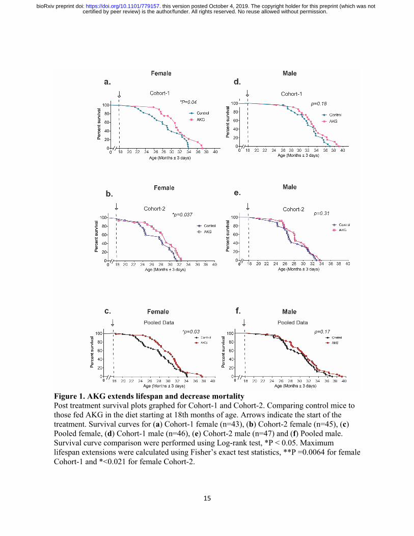
(w/w) increases survival in two independent cohorts of aged mice (Fig. 1). In the first cohort of
female mice, median lifespan and survival (age at 90th percentile mortality) were significantly
extended by 16.6% and 19.7% from inception of CaAKG feeding. These findings have been
repeated in the second cohort of mice: the female median lifespan and survival were significantly
extended by 10.5% and 8% (Fig. 1a, b and Extended Table 1). Although, improved survival for
males was not significant in both cohorts (Fig. 1d, e), median lifespan was extended for 9.6%
and 12.8% from inception of treatment, respectively (Extended Table 1).
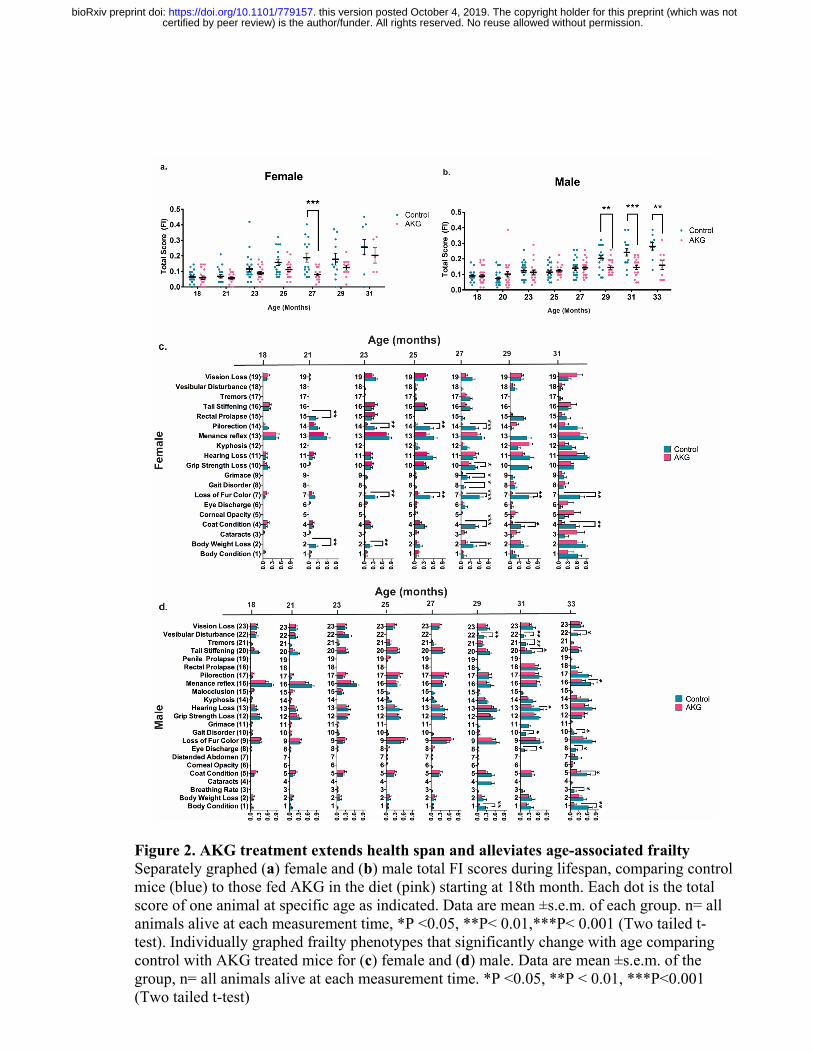
In order to assess healthspan, we applied measurements that are based on a clinically relevant
frailty index [8, 21]. The frailty index (FI) consists of 31 phenotypes that are indicators of age-
associated health deterioration, with each phenotype scored on a 0, 0.5 or 1 scale, based on its
severity. Body weight and surface temperature were collected and converted to the same scoring
scale (Extended Table 1). All scorings were conducted in a blinded manner. The 31-metrics
share many characteristics of the human frailty indices and have been reported to progress
similarly with aging in mice and humans [22]. Measurements were repeated approximately every
certified by peer review) is the author/funder. All rights reserved. No reuse allowed without permission. The copyright holder for this preprint (which was notthis version posted October 4, 2019. . https://doi.org/10.1101/779157doi: bioRxiv preprint
5
eight weeks, providing us with eight and seven sets of data respectively for male and female
groups. To establish a baseline clinical assessment, we collected our dataset right before the start
of the treatment at 18th months (Fig. 2a-d). The total frailty score, which is the sum of 31 frailty
phenotypes, indicates the state of increased vulnerability to adverse health outcomes, which is
comparable to morbidity. In both female and male animals, CaAKG decreases incidence and
severity of different aging phenotypes and postpones frailty (Fig. 2a, b). Females show
significant health benefits after 9 months of CaAKG supplementation (P<0.001, Fig. 2a). After
11 months of treatment, male animals start to show significant health improvement comparing to
control animals which persisted until the last measurement at 33 months of age (P<0.01, P<0.05,
Fig. 2b).
Since mice in our aging study were older than previously published papers on frailty [8], we first
tested whether any individual frailty indicators show significant changes upon aging in our study.
Our results show that aging significantly increased the incidence of 19 and 23 frailty phenotypes
in female and male respectively (Two-way ANOVA, Extended Table 2). We reported the effects
of CaAKG treatment on these phenotypes at different time points (Fig. 2c, d). CaAKG treatment
significantly decreased the severity of multiple aging phenotypes in females including
piloerection, grimace (pain assessment), loss of fur color, poor coat condition, grip strength loss,
gait disorder, rectal prolapse and body weight loss (Fig. 2c). In males, grip strength loss, gait
disorder, balance loss (vestibular disturbance), grimace, loss of menace reflex, hearing loss, eye
discharge, poor coat condition, poor body condition, tail stiffening and abnormal breathing rate
were all decreased (Fig. 2d). The improvement in health of both sexes was most prominent
around the median life of the animal. Among the frailty measures affected by CaAKG, protection
certified by peer review) is the author/funder. All rights reserved. No reuse allowed without permission. The copyright holder for this preprint (which was notthis version posted October 4, 2019. . https://doi.org/10.1101/779157doi: bioRxiv preprint
6
from age-related changes in female coat color was particularly prominent. CaAKG treatment
could reverse age-dependent hair graying in first cohort, however in the second cohort of
females, CaAKG treatment only prevented the hair graying (Extended Fig. 4). Gait improvement
in CaAkG treated females and males close to median life is consistent with increased locomotion
activity, as scored using metabolic cages (Extended Fig. 5). Interestingly, despite increased
locomotion, the levels of oxygen consumption, carbon dioxide production and energy
expenditure were significantly lower in the CaAKG treated group. The decrease in carbon
dioxide production was preserved later in life (Extended Fig. 6). Advanced age-related weight
loss has been reported in old human and rodents, which has been associated with morbidity [23].
Data from the second cohort of mice confirms that CaAKG administration leads to body weight
preservation in male mice (P value<0.001, Extended Fig. 1b, d). Not all phenotypes were
improved by CaAKG. For instance, treated mice failed to perform better in a treadmill
exhaustion test and showed no cardiac functional improvement, as determined using
echocardiography. Importantly, however, we did not detect any significant adverse changes
associated with CaAKG treatment (Extended Fig. 7).
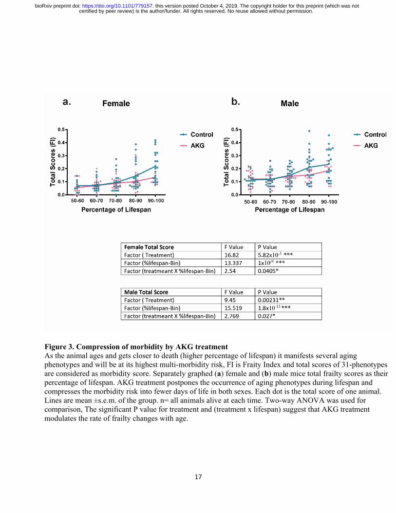
Since the age for onset of age-related phenotypes can be quite heterogeneous in mammals, we
plotted frailty datasets not only as a function of chronological time, but also in proportion to the
lifespan of each mouse by binning scores within ten percentile (e.g. scores collected between
60% and 70% of the animals’ lifespans were binned and plotted together (Fig 3). This allowed us
to align assessments with respect to the biologic age of the animal. Our findings show that AKG
treatment decreases the proportion of life in which the animal is frail and vulnerable to adverse
health incomes (determined as the area under the frailty curve and calculated at a 52% reduction
certified by peer review) is the author/funder. All rights reserved. No reuse allowed without permission. The copyright holder for this preprint (which was notthis version posted October 4, 2019. . https://doi.org/10.1101/779157doi: bioRxiv preprint
7
for females and 40% for males, Fig. 3 a, b, P<0.01). Our results also suggest that CaAKG
modulates the rate of frailty changes with age (Fig. 3, Table). This improvement in healthy days
of life is disproportionately larger than the increase in lifespan. Through the extensive frailty data
collection, we have therefore been able to demonstrate that an intervention by a natural product
results in properties consistent with morbidity compression.
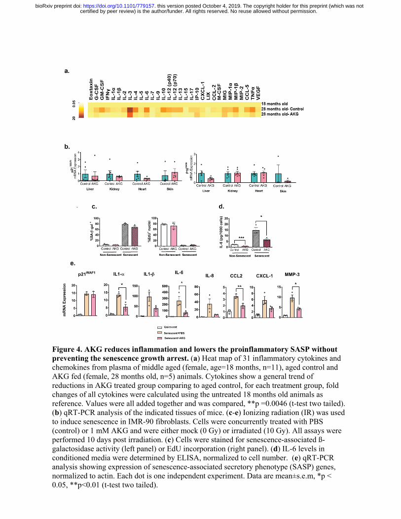
To gain better mechanistic insight on the beneficial effects of AKG, we initiated AKG treatment
in a new cohort of female mice at 18 months of age. The reported beneficial effect of AKG on
lifespan in C. elegans was associated with reduced TOR signaling [12]. However, in mice we did
not detect any decrease in mTORC1 signaling upon three months of AKG treatment (Extended
Fig. 6). Age-associated diseases are accompanied by chronic inflammation, which is generally
linked to an age-associated functional decline in mice and humans [24]. We measured levels of
31 inflammatory cytokines in the serum of aged female mice (28 months old). In untreated mice,
the levels of most cytokines increase; however, CaAKG fed animals were largely refractory to
these changes (Fig. 4a). There is a general trend of suppression for all the cytokines assayed in
plasma of CaAKG treated animals ( Fig. 4a, P<0.01). The reduction in inflammation is
consistent with previous findings in the intestine of young pigs receiving AKG supplemented
diet [25].
Studies have shown the accumulation of senescent cells in different tissues of old mice [26].
These cells can contribute to age-associated chronic inflammation by acquiring senescence-
associated secretory phenotype (SASP) and their removal can extend mouse lifespan [27]. We
looked into senescent markers and did not detect any significant changes for senescent markers
(Fig. 4b). We performed cell culture studies to explore possible effects of NaAKG in primary
certified by peer review) is the author/funder. All rights reserved. No reuse allowed without permission. The copyright holder for this preprint (which was notthis version posted October 4, 2019. . https://doi.org/10.1101/779157doi: bioRxiv preprint
8
fibroblasts. While no changes in the senescence formation response to ionizing radiation were
observed; however, consistent to in-vivo findings we detect a significant reduction in a range of
inflammatory cytokines, indicating that AKG can alter the SASP and reduce inflammation
without effecting the formation of senescent cells (Extended Fig. 4c-e). Specifically, we found a
reduction of IL-1b, IL-6, CCL2 and MMP3 without any changes in ß-gal and p21 (Fig. 4d, e).
Discussion
Recent aging research has been based on the principle that there are underlying biological
processes driving aging and that intervening in those processes will increase health and function,
while delaying associated co-morbidities. Here we demonstrate that AKG, a key metabolite in
TCA cycle, has longevity effects consistent with compressed morbidity in a long-term study of
mouse aging. It is important that CaAKG administration started at 18 months of age still had
robust effects. This is valuable since human clinical studies are likely to be initiated at a similar
relative time point during aging. If translated to humans, this effect would be an ideal outcome,
extending lifespan but more importantly reducing the debilitating period of functional decline
and disease management that plague many aging individuals. Interestingly, in humans plasma
AKG levels decline 10 folds between the ages of 40 and 80 [28]. The molecule is not available in
the human diet, making direct supplementation the only feasible route to restore levels. AKG has
been used in human clinical studies linked to diseases without associated adverse effects [29-31].
More studies will be needed to determine whether chronic AKG consumption can affect other
parameters of aging. Given its GRAS status and human safety record, our findings point to a
potential safe human intervention that may impact important elements of aging and improve
quality of life in the elderly population.
certified by peer review) is the author/funder. All rights reserved. No reuse allowed without permission. The copyright holder for this preprint (which was notthis version posted October 4, 2019. . https://doi.org/10.1101/779157doi: bioRxiv preprint
9
Material and methods
Animal Housing and Diet
All mice were housed on a 12-h light/dark cycle and kept at 20–22 °C. Two independent cohorts
of C57BL6/J mice were purchased from Jackson Laboratories at 14 months of age. Animals
were aged on a regular mouse diet (Teklad Irradiated 18% protein and 6% fat diet-2918) which is
very similar to their diet at Jax 5K52 (18-19% protein and 6-7% fat) prior to arrival, until they
reached 18th month of age when treatment started. AKG treated animals were subjected to a
lifelong 2% (w/w) AKG supplement on 2918 diet while Control group were kept on standard-
2918 diet. Pure calcium 2-oxoglutarate was purchased from Carbosynth Company and
homogeneously mixed during manufacturing of the 2918 diet prior to irradiation and pelleting.
Mice were housed in groups (5 per cage at a maximum) and aggressive male mice were isolated
to prevent fighting. All lifespan and healthspan experiments were started around 18th month of
age. Mice were inspected daily, and treated for non-life threatening conditions as directed by the
veterinary staff. The only conditions that received treatment were dermatitis and prolapse
(topical solution three times per week). Total of 8 mice in control group and 3 mice in AKG
group were treated for dermatitis and prolapse. The principal endpoint of the health span and
lifespan study was natural death. The Buck Institute is a AAALAC accredited facility. Each
room contains sentinel mice (CD-1 females- 1 cage/75-100 cages). Health screening is done 4
times per year at 3 months intervals. Diagnostics consist of serological screening and fecal and
fur analysis for internal and external parasites.
Survival
certified by peer review) is the author/funder. All rights reserved. No reuse allowed without permission. The copyright holder for this preprint (which was notthis version posted October 4, 2019. . https://doi.org/10.1101/779157doi: bioRxiv preprint
10
The principle endpoint of health span and lifespan study was natural death. We recorded the age at
which mice were found dead or selected for euthanasia (a procedure for mice deemed unlikely to survive for
the next 48 hours and are in enormous discomfort). The criteria for euthanasia was based on an
independent assessment by a veterinarian, according to AAALAC guidelines and only where the
condition of the animal was considered [3]. Severe lethargy, rapid weight loss (over two
weeks>20%), severe distended abdomen and body condition score with signs of pain (grimace),
inability to move despite the stimuli, severe ulcer or bleeding tumor, severe temperature loss
with abnormal breathing rate. Animals found dead or euthanized were necropsied for pathology
score. No invasive measurements were performed on this population, n=180 animals (two
cohorts of 90 animals). A sacrificed group were purchased at 14th months of age, baselined and
grouped the following week. The mice, n=12 were either receiving Teklad-2918 or 2% w/w
AKG supplemented 2918. Animals were sacrificed and tissues were collected after 3 months of
treatment. The Institutional Animal Care and Use Committee (IACUC) at the Buck Institute
approved all animal experimental procedures, housing and diets for Research on Aging. Food
intake and body weight were measured on a biweekly and bimonthly basis for the duration of the
study.
Baselining and grouping of the animals
Mammals age heterogeneously and the 18 months old mice already manifest some age-
associated deterioration of health phenotypes. All the animals were scored before grouping and
all the 31 scores were applied to assign animals into different groups. A balanced partitioning of
mice was done: for any given mouse in any given group, there are similar mice in all other
groups. This allows any outcome of the study to be more related to experiments or the treatment
rather than the inherent property of a group.
Aging Index (frailty Index)
certified by peer review) is the author/funder. All rights reserved. No reuse allowed without permission. The copyright holder for this preprint (which was notthis version posted October 4, 2019. . https://doi.org/10.1101/779157doi: bioRxiv preprint
11
One can find the complete protocol as published before by Whitehead et al., 2014. For the
subjective properties of the assessments, all measurements were completely blinded.
These assessments indicate age-associated deterioration of health and include evaluation of the
animal musculoskeletal system, the vestibulocochlear/auditory systems, the ocular and nasal
systems, the digestive system, the urogenital system, the respiratory system, signs of discomfort,
body weight and body surface temperature. 0 is assigned if no sign of frailty is observed and the
animal is healthy for that phenotype. A moderate phenotype and a severe phenotype will be
scored 0.5 and 1 respectively. Loss of temperature and weight were scored using standard
deviation for our study (Extended Table 2). We should note that the recessive Cdh23 allele (ahl)
in C57BL/6J strain, when homozygous, leads to increased susceptibility to age related hearing
loss. Hearing loss was one of the most abundance aging phenotype in our data set.
Statistical analyses
Python Software was used to extract all the healthspan data and create files compatible with R
software for analysis. Data were analyzed using R, GraphPad Prism 7 and OASIS 2 software.
Log-rank (Mantel–Cox) tests were used to analyze Kaplan–Meier curves, and a Fisher`s exact
test was performed for maximum lifespan analysis (at 90% survival). Two-tailed Student’s t-tests
were used for analyses of scoring at each time point between control and AKG treated group.
Two-way analysis of variance (ANOVA) with Bonferroni post hoc correction was used for
analysis between morbidity curves. The area under curve (AUC) of mortality graphs were
measured baselining at 18 months of age. The changes in AUC were used to calculate the percent
compression of morbidity.
Inflammatory cytokines and chemokines
certified by peer review) is the author/funder. All rights reserved. No reuse allowed without permission. The copyright holder for this preprint (which was notthis version posted October 4, 2019. . https://doi.org/10.1101/779157doi: bioRxiv preprint
12
Blood were from the jugular vein of young (18 months old), aged control and AKG fed (29
months old) animals. The samples were sent to Eve technologies (Calgary, Alberta, Canada) for
measurement of soluble cytokines and chemokines in serum using multiplex lase bead array
technology (MD31).
Cell culture
IMR-90 fetal lung fibroblasts were obtained from ATCC and were cultured at 37° C in 3%
O2and 5% CO2. Dulbecco's modified Eagle's media (DMEM) supplemented with 10% fetal
bovine serum and streptomycin/penicillin were used. Media was changed every 2 days during the
experiment. For damage-induced senescence, cells were irradiated with doses of either 0 or 10
Gy of ionizing radiation (IR). Cells were concurrently treated with PBS (control) or 1 mM AKG
for 10 days, changing media every 2 days. All assays were performed 10 days post irradiation.
EdU (5-ethynyl-2´-deoxyuridine) staining Proliferation Kit (iFluor 488) ab219801 was used to
detect cell proliferation. Cells were stained for the senescence-associated β-gal (SA-β-gal)
marker as described [3]. Non-senescent cells (having undergone fewer than 35 population
doublings) were made quiescent by washing with PBS and incubating in DMEM containing
0.2% serum for 4 day. Cultures that had > 80% SA-β-gal positive cells and ≤ 4% EdU positive
cells were considered senescent.
ELISA
Conditioned media were prepared by washing cells 3 times in PBS and incubating them in
serum-free DMEM containing penicillin/streptomycin for 24 h. Conditioned media were
removed and cells were trypsinized for cell counts. The conditioned media were then centrifuged
to remove cellular debris, and supernatants were used for ELISA. IL-6 ELISAs were performed
certified by peer review) is the author/funder. All rights reserved. No reuse allowed without permission. The copyright holder for this preprint (which was notthis version posted October 4, 2019. . https://doi.org/10.1101/779157doi: bioRxiv preprint
13
using kits and procedures from R&D (#D06050). The resultant data were normalized to cell
number.
RT PCR
For cell culture experiments, RNA was isolated using ISOLATE II RNA mini kit (Bioline #BIO-
52073). RNA quality and quantity were assessed using NanoDropTM 1000 Spectrophotometer
measures (Thermo Scientific). Total cDNAs were synthesized from 500ng of RNA using random
primers and iScript RT reagents following the manufacturer's protocol Superscript II (Invitrogen,
Carlsbad, USA). Gene expression was measured from cDNA using the Roche Universal Probe
Library system (Indianapolis, IN, USA). All values were normalized to beta-actin.
For in-vivo study tissues were collected form 12 animals described as sacrificed group. Tissues
were homogenized in 1 ml Invitrogen TRIzol™ Reagent using metal beads combined with high-
speed shaking (Tissuelyser Qiagen at 20 Hrtz, for 6 min). Skin samples were crushed with pistol
and liquid nitrogen prior to homogenizing step. The chloroform extraction and
ethanol precipitation were performed on homogenized tissues to extract RNA. The RNA quality
and quantity were assessed and cDNA were synthesized as described. Gene expression was
quantified by real-time quantitative PCR using the Roche Universal Probe Library system
(Indianapolis, IN, USA). The primer sets (0.1 μM) were as follows: 1) p16 F:5'-
AACTCTTTCGGTCGTACCCC-3' and R: 5'-TCCTCGCAGTTCGAATCTG -3' with Custom
designed probe : 5'-/56-FAM/AGG TGA TGA/ZEN/TGATGGGCAACGTTCAC/3IABkFQ -3'.
2) p21 R: 5'-TTTGCTCCTGTGCGGAAC -3' and F:5'-TTGCCAGCAGAATAAAAGGTG -3'
with probe #9. Transcript levels were normalized to Beta-glucuronidase (GUSB) as an
endogenous control.
Extended Data
certified by peer review) is the author/funder. All rights reserved. No reuse allowed without permission. The copyright holder for this preprint (which was notthis version posted October 4, 2019. . https://doi.org/10.1101/779157doi: bioRxiv preprint
14
Extended Data include eight figures and can be found here.
Unpublished data can be obtained by contacting the corresponding authors.
Data availability
All other data are available from the corresponding authors on reasonable request.
Acknowledgements
This work was supported by The Weldon Foundation and also by Ponce de Leon Health.
certified by peer review) is the author/funder. All rights reserved. No reuse allowed without permission. The copyright holder for this preprint (which was notthis version posted October 4, 2019. . https://doi.org/10.1101/779157doi: bioRxiv preprint
15
Figure 1. AKG extends lifespan and decrease mortality Post treatment survival plots graphed for Cohort-1 and Cohort-2. Comparing control mice to those fed AKG in the diet starting at 18th months of age. Arrows indicate the start of the treatment. Survival curves for (a) Cohort-1 female (n=43), (b) Cohort-2 female (n=45), (c) Pooled female, (d) Cohort-1 male (n=46), (e) Cohort-2 male (n=47) and (f) Pooled male. Survival curve comparison were performed using Log-rank test, *P < 0.05. Maximum lifespan extensions were calculated using Fisher’s exact test statistics, **P =0.0064 for female Cohort-1 and *<0.021 for female Cohort-2.
certified by peer review) is the author/funder. All rights reserved. No reuse allowed without permission. The copyright holder for this preprint (which was notthis version posted October 4, 2019. . https://doi.org/10.1101/779157doi: bioRxiv preprint
16
Figure 2. AKG treatment extends health span and alleviates age-associated frailty Separately graphed (a) female and (b) male total FI scores during lifespan, comparing control mice (blue) to those fed AKG in the diet (pink) starting at 18th month. Each dot is the total score of one animal at specific age as indicated. Data are mean ±s.e.m. of each group. n= all animals alive at each measurement time, *P <0.05, **P< 0.01,***P< 0.001 (Two tailed t-test). Individually graphed frailty phenotypes that significantly change with age comparing control with AKG treated mice for (c) female and (d) male. Data are mean ±s.e.m. of the group, n= all animals alive at each measurement time. *P <0.05, **P < 0.01, ***P<0.001 (Two tailed t-test)
certified by peer review) is the author/funder. All rights reserved. No reuse allowed without permission. The copyright holder for this preprint (which was notthis version posted October 4, 2019. . https://doi.org/10.1101/779157doi: bioRxiv preprint
17
Figure 3. Compression of morbidity by AKG treatment As the animal ages and gets closer to death (higher percentage of lifespan) it manifests several aging phenotypes and will be at its highest multi-morbidity risk, FI is Fraity Index and total scores of 31-phenotypes are considered as morbidity score. Separately graphed (a) female and (b) male mice total frailty scores as their percentage of lifespan. AKG treatment postpones the occurrence of aging phenotypes during lifespan and compresses the morbidity risk into fewer days of life in both sexes. Each dot is the total score of one animal. Lines are mean ±s.e.m. of the group. n= all animals alive at each time. Two-way ANOVA was used for comparison, The significant P value for treatment and (treatment x lifespan) suggest that AKG treatment modulates the rate of frailty changes with age.
certified by peer review) is the author/funder. All rights reserved. No reuse allowed without permission. The copyright holder for this preprint (which was notthis version posted October 4, 2019. . https://doi.org/10.1101/779157doi: bioRxiv preprint
18
Figure 4. AKG reduces inflammation and lowers the proinflammatory SASP without preventing the senescence growth arrest. (a) Heat map of 31 inflammatory cytokines and chemokines from plasma of middle aged (female, age=18 months, n=11), aged control and AKG fed (female, 28 months old, n=5) animals. Cytokines show a general trend of reductions in AKG treated group comparing to aged control, for each treatment group, fold changes of all cytokines were calculated using the untreated 18 months old animals as reference. Values were all added together and was compared, **p =0.0046 (t-test two tailed). (b) qRT-PCR analysis of the indicated tissues of mice. (c-e) Ionizing radiation (IR) was used to induce senescence in IMR-90 fibroblasts. Cells were concurrently treated with PBS (control) or 1 mM AKG and were either mock (0 Gy) or irradiated (10 Gy). All assays were performed 10 days post irradiation. (c) Cells were stained for senescence-associated ß-galactosidase activity (left panel) or EdU incorporation (right panel). (d) IL-6 levels in conditioned media were determined by ELISA, normalized to cell number. (e) qRT-PCR analysis showing expression of senescence-associated secretory phenotype (SASP) genes, normalized to actin. Each dot is one independent experiment. Data are mean±s.e.m, *p < 0.05, **p<0.01 (t-test two tailed).
certified by peer review) is the author/funder. All rights reserved. No reuse allowed without permission. The copyright holder for this preprint (which was notthis version posted October 4, 2019. . https://doi.org/10.1101/779157doi: bioRxiv preprint
19
1. Kenyon, C., et al., A C. elegans mutant that lives twice as long as wild type. Nature,
1993. 366(6454): p. 461-4. 2. Wood, J.G., et al., Sirtuin activators mimic caloric restriction and delay ageing in
metazoans. Nature, 2004. 430(7000): p. 686-9. 3. Harrison, D.E., et al., Rapamycin fed late in life extends lifespan in genetically
heterogeneous mice. Nature, 2009. 460(7253): p. 392-5. 4. Friedman, D.B. and T.E. Johnson, A mutation in the age-1 gene in Caenorhabditis
elegans lengthens life and reduces hermaphrodite fertility. Genetics, 1988. 118(1): p. 75-86.
5. Bansal, A., et al., Uncoupling lifespan and healthspan in Caenorhabditis elegans longevity mutants. Proc Natl Acad Sci U S A, 2015. 112(3): p. E277-86.
6. Hahm, J.H., et al., C. elegans maximum velocity correlates with healthspan and is maintained in worms with an insulin receptor mutation. Nat Commun, 2015. 6: p. 8919.
7. Hansen, M. and B.K. Kennedy, Does Longer Lifespan Mean Longer Healthspan? Trends Cell Biol, 2016. 26(8): p. 565-568.
8. Whitehead, J.C., et al., A clinical frailty index in aging mice: comparisons with frailty index data in humans. J Gerontol A Biol Sci Med Sci, 2014. 69(6): p. 621-32.
9. Kaeberlein, M., et al., Regulation of yeast replicative life span by TOR and Sch9 in response to nutrients. Science, 2005. 310(5751): p. 1193-6.
10. Lin, S.J., P.A. Defossez, and L. Guarente, Requirement of NAD and SIR2 for life-span extension by calorie restriction in Saccharomyces cerevisiae. Science, 2000. 289(5487): p. 2126-8.
11. Selman, C., et al., Ribosomal protein S6 kinase 1 signaling regulates mammalian life span. Science, 2009. 326(5949): p. 140-4.
12. Chin, R.M., et al., The metabolite alpha-ketoglutarate extends lifespan by inhibiting ATP synthase and TOR. Nature, 2014. 510(7505): p. 397-401.
13. Myllyharju, J., Prolyl 4-hydroxylases, the key enzymes of collagen biosynthesis. Matrix Biol, 2003. 22(1): p. 15-24.
14. Tsukada, Y., et al., Histone demethylation by a family of JmjC domain-containing proteins. Nature, 2006. 439(7078): p. 811-6.
15. Ito, S., et al., Role of Tet proteins in 5mC to 5hmC conversion, ES-cell self-renewal and inner cell mass specification. Nature, 2010. 466(7310): p. 1129-33.
16. Zdzisinska, B., A. Zurek, and M. Kandefer-Szerszen, Alpha-Ketoglutarate as a Molecule with Pleiotropic Activity: Well-Known and Novel Possibilities of Therapeutic Use. Arch Immunol Ther Exp (Warsz), 2017. 65(1): p. 21-36.
17. Song, J., et al., alpha-Ketoglutarate Promotes Pancreatic Progenitor-Like Cell Proliferation. Int J Mol Sci, 2018. 19(4).
certified by peer review) is the author/funder. All rights reserved. No reuse allowed without permission. The copyright holder for this preprint (which was notthis version posted October 4, 2019. . https://doi.org/10.1101/779157doi: bioRxiv preprint
20
18. Carey, B.W., et al., Intracellular alpha-ketoglutarate maintains the pluripotency of embryonic stem cells. Nature, 2015. 518(7539): p. 413-6.
19. Dobrowolski, P.J., et al., Dietary alpha-ketoglutarate reduces gastrectomy-evoked loss of calvaria and trabecular bone in female rats. Scand J Gastroenterol, 2008. 43(5): p. 551-8.
20. Reality check on reproducibility. Nature, 2016. 533(7604): p. 437. 21. Searle, S.D., et al., A standard procedure for creating a frailty index. BMC Geriatr, 2008.
8: p. 24. 22. Kane, A.E., et al., Animal models of frailty: current applications in clinical research.
Clin Interv Aging, 2016. 11: p. 1519-1529. 23. Alley, D.E., et al., Changes in weight at the end of life: characterizing weight loss by time
to death in a cohort study of older men. Am J Epidemiol, 2010. 172(5): p. 558-65. 24. Chung, H.Y., et al., Molecular inflammation: underpinnings of aging and age-related
diseases. Ageing Res Rev, 2009. 8(1): p. 18-30. 25. He, L., et al., Administration of alpha-ketoglutarate improves epithelial restitution under
stress injury in early-weaning piglets. Oncotarget, 2017. 8(54): p. 91965-91978. 26. van Deursen, J.M., The role of senescent cells in ageing. Nature, 2014. 509(7501): p.
439-46. 27. Baker, D.J., et al., Clearance of p16Ink4a-positive senescent cells delays ageing-
associated disorders. Nature, 2011. 479(7372): p. 232-6. 28. Harrison, A.P. and S.G. Pierzynowski, Biological effects of 2-oxoglutarate with
particular emphasis on the regulation of protein, mineral and lipid absorption/metabolism, muscle performance, kidney function, bone formation and cancerogenesis, all viewed from a healthy ageing perspective state of the art--review article. J Physiol Pharmacol, 2008. 59 Suppl 1: p. 91-106.
29. Riedel, E., M. Nundel, and H. Hampl, alpha-Ketoglutarate application in hemodialysis patients improves amino acid metabolism. Nephron, 1996. 74(2): p. 261-5.
30. Filip, R.S., et al., Alpha-ketoglutarate decreases serum levels of C-terminal cross-linking telopeptide of type I collagen (CTX) in postmenopausal women with osteopenia: six-month study. Int J Vitam Nutr Res, 2007. 77(2): p. 89-97.
31. Jeevanandam, M. and S.R. Petersen, Substrate fuel kinetics in enterally fed trauma patients supplemented with ornithine alpha ketoglutarate. Clin Nutr, 1999
certified by peer review) is the author/funder. All rights reserved. No reuse allowed without permission. The copyright holder for this preprint (which was notthis version posted October 4, 2019. . https://doi.org/10.1101/779157doi: bioRxiv preprint