Embed Size (px)
Citation preview
Syddansk Universitet
Pleiotropic effects of cancer cells' secreted factors on human stromal (mesenchymal)stem cellsAl-toub, Mashael; Almusa, Abdulaziz; Almajed, Mohammed; Al-Nbaheen, May; Kassem,Moustapha; Aldahmash, Abdullah; Alajez, NehadPublished in:Stem Cell Research & Therapy
DOI:10.1186/scrt325
Publication date:2013
Document versionSubmitted manuscript
Citation for pulished version (APA):Al-toub, M., Almusa, A., Almajed, M., Al-Nbaheen, M., Kassem, M., Aldahmash, A., & Alajez, N. (2013).Pleiotropic effects of cancer cells' secreted factors on human stromal (mesenchymal) stem cells. Stem CellResearch & Therapy, 4(114), [24405819]. DOI: 10.1186/scrt325
General rightsCopyright and moral rights for the publications made accessible in the public portal are retained by the authors and/or other copyright ownersand it is a condition of accessing publications that users recognise and abide by the legal requirements associated with these rights.
• Users may download and print one copy of any publication from the public portal for the purpose of private study or research. • You may not further distribute the material or use it for any profit-making activity or commercial gain • You may freely distribute the URL identifying the publication in the public portal ?
Take down policyIf you believe that this document breaches copyright please contact us providing details, and we will remove access to the work immediatelyand investigate your claim.
Download date: 13. Feb. 2017
Pleiotropic effects of cancer cells’ secreted factorson human stromal (mesenchymal) stem cellsAl-toub et al.
Al-toub et al. Stem Cell Research & Therapy 2013, 4:114http://stemcellres.com/content/4/5/114

RESEARCH Open Access
Pleiotropic effects of cancer cells’ secreted factorson human stromal (mesenchymal) stem cellsMashael Al-toub1, Abdulaziz Almusa1, Mohammed Almajed1, May Al-Nbaheen1,2, Moustapha Kassem1,3,Abdullah Aldahmash1,3 and Nehad M Alajez1*
See related commentary by Lam, http://stemcellres.com/content/4/6/138
Abstract
Introduction: Studying cancer tumors’ microenvironment may reveal a novel role in driving cancer progressionand metastasis. The biological interaction between stromal (mesenchymal) stem cells (MSCs) and cancer cellsremains incompletely understood. Herein, we investigated the effects of tumor cells’ secreted factors as representedby a panel of human cancer cell lines (breast (MCF7 and MDA-MB-231); prostate (PC-3); lung (NCI-H522); colon(HT-29) and head & neck (FaDu)) on the biological characteristics of MSCs.
Methods: Morphological changes were assessed using fluorescence microscopy. Changes in gene expression wereassessed using Agilent microarray and qRT-PCR. GeneSpring 12.1 and DAVID tools were used for bioinformatic andsignaling pathway analyses. Cell migration was assessed using a transwell migration system. SB-431542, PF-573228and PD98059 were used to inhibit transforming growth factor β (TGFβ), focal adhesion kinase (FAK), and mitogenactivated protein kinase kinase (MAPKK) pathways, respectively. Interleukin-1β (IL1β) was measured using ELISA.
Results: MSCs exposed to secreted factors present in conditioned media (CM) from FaDu, MDA-MB-231, PC-3 andNCI-H522, but not from MCF7 and HT-29, developed an elongated, spindle-shaped morphology with bipolarprocesses. In association with phenotypic changes, genome-wide gene expression and bioinformatics analysisrevealed an enhanced pro-inflammatory response of those MSCs. Pharmacological inhibitions of FAK and MAPKKseverely impaired the pro-inflammatory response of MSCs to tumor CM (approximately 80% to 99%, and 55% to88% inhibition, respectively), while inhibition of the TGFβ pathway was found to promote the pro-inflammatoryresponse (approximately 3-fold increase). In addition, bioinformatics and pathway analysis of gene expression datafrom tumor cell lines combined with experimental validation revealed tumor-derived IL1β as one mediator of thepro-inflammatory phenotype observed in MSCs exposed to tumor CM.MSCs exhibited significant tropism toward secreted factors from the aforementioned tumor cell lines, while bothnormal and MSCs exposed to tumor CM were capable of attracting human peripheral blood mononuclear cells(PBMCs).
Conclusions: Our data revealed tumor-derived IL1β as one mediator of the pro-inflammatory response in MSCsexposed to tumor CM, which was found to be positively regulated by FAK and MAPK signaling and negativelyregulated by TGFβ signaling. Thus, our data support a model where MSCs could promote cancer progressionthrough becoming pro-inflammatory cells within the cancer stroma.
* Correspondence: [email protected] Cell Unit, Department of Anatomy, College of Medicine, King SaudUniversity, Riyadh 11461, Kingdom of Saudi ArabiaFull list of author information is available at the end of the article
© 2013 Al-toub et al.; licensee BioMed Central Ltd. This is an Open Access article distributed under the terms of the CreativeCommons Attribution License (http://creativecommons.org/licenses/by/2.0), which permits unrestricted use, distribution, andreproduction in any medium, provided the original work is properly cited.
Al-toub et al. Stem Cell Research & Therapy 2013, 4:114http://stemcellres.com/content/4/5/114
IntroductionStromal (mesenchymal) stem cells (MSCs), also referredto as stromal cells, are multipotent cells which are presentwithin the stroma of bone marrow and probably otherorgans and capable of differentiating into the three canon-ical lineages: osteoblasts, adipocytes and chondrocytes [1].Aside from their differentiation potential, MSCs are alsocapable of migrating to injured tissues and contributingto tissue regeneration [2-4]. Emerging data suggest thatMSCs possess immunomodulatory and regenerative prop-erties as they can secrete a large number of growth factorsand immune active molecules [5] that can improve tissuesurvival and suppress the activity of various immune cells,such as alloantigen activated T and B lymphocytes [6,7].Moreover, MSCs can secrete a large number of paracrinefactors, including chemoattractants for endothelial cells,monocytes and macrophages [8]. Several recent studieshave reported that bone marrow MSCs migrate to thestromal compartment of tumors [9,10] and that a dynamicinteraction between tumor cells and MSCs exists resem-bling what has been reported during inflammation and,thus, ‘tumors are wounds that never heal’ [11].Over the past several years, a significant amount of
research has emerged documenting a role for MSCs inpromoting epithelial-to-mesenchymal transition (ETM) andaccelerating tumor growth and metastasis [9,12-14]. Inaddition, MSCs are being introduced into therapy for anumber of clinical indications and there is a concern ofpossible promoting effects on tumor growth by MSCs[15]. On the other hand, several other studies reported thatMSCs exert tumor suppressive effects [16-18]. Therefore,understanding the settings under which MSCs exertpromoting versus inhibitory effects on tumor growth andmetastasis is currently under intensive investigation.Given this complex interplay between MSCs and tumor
cells, the goal of this study was to assess the cellular andmolecular changes in MSCs in response to secreted factorspresent in conditioned media (CM) from a panel of humantumor cell lines covering a spectrum of human cancers(breast, prostate, lung, colon, and head and neck). Inte-grated analysis of phenotypic changes, gene expressionand bioinformatics revealed a pro-inflammatory re-sponse of MSCs when exposed to CM of several tumorcell lines. Interestingly, the biological responses ofMSCs were not identical. MSCs responded mainly totumor cell lines which express high levels of IL1β. Weidentified tumor-derived IL1β as the prominent cyto-kine responsible for induction of inflammatory responsein MSCs and signaling via focal adhesion kinase (FAK)and, to lesser extent, mitogen activated protein kinasekinase (MAPKK), as key positive regulators of an in-flammatory response, while transforming growth factorβ (TGFβ) signaling was found to inhibit the response ofMSCs to tumor CM. Our data further support a model
where MSCs could drive tumorigenicity through inductionof inflammation.
MethodsEthics statementExperiments performed in this study do not need ethicscommittee approval.
Cell cultureTumor cell lines used in this study (breast, MCF7 andMDA-MB-231; prostate, PC-3; lung, NCI-H522; headand neck, FaDu; and colon, HT-29) have been describedpreviously [19-23]. The human telomerized hMSC-TERT-GFP cell line was developed by Dr Kassem, Odense,Denmark [24,25]. All cell lines were maintained in (D)MEM4.5g/L glucose (Invitrogen Corp., Carlsbad, CA, USA) andsupplemented with 10% fetal bovine serum, 1% NEAA, 1%L-glutamine, 100 mg/L penicillin and 100 mg/L strepto-mycin at 37°C and 5% CO2. For TGFβ inhibition experi-ments, MSC were cultured as described above andwere exposed to MDA-MB-231 CM in the presence of10 μM SB-431542 (Sigma, St. Louis, MO, USA). Controlwells were treated with dimethyl sulfoxide (DMSO). CMplus SB-431542 or vehicle (DMSO, Sigma) was changedevery three to four days for the duration of the experiment.Recombinant human IL1β and IL6 were purchased fromInvitrogen. FAK inhibitor (PF-573228) and mitogen activatedprotein kinase kinase (MAPKK) inhibitor (PD98059) werepurchase from Sigma and were reconstituted in DMSO.
Collection of tumor cell lines conditioned mediaThe tumor cell lines, MCF7, HT-29, MDA-MB-231, PC-3,NCI-H522 and FaDu were seeded in six-well plates at1 × 106/well (4 ml total) in (D)MEM supplemented with10% fetal bovine serum (FBS), 1% NEAA and 1% penicillin/streptomycin and incubated at 37°C and 5% CO2. Forty-eight hours later (cells were approximately 90% confluent),CM from the tumor cell lines were collected and spundown at 300 × g for 10 minutes to remove any cellularcontent and debris. In some experiments, CM was passedthrough a 0.45 μM filter to remove any remaining cellularcontent and debris. The hMSC-TERT-GFP cells were thenseeded in 24-well plates at 8 × 10⁴/ml in the collected CM(80% tumor CM + 20% fresh medium). The MSCs wereexposed to fresh CM every two to three days for theduration of the experiment.
Quantification of secreted IL1β using ELISAQuantification of secreted IL1β from tumor cell lines orfrom MSCs exposed to tumor CM was done using theLEGEND MAX™ Human IL-1β ELISA Kit (BiolegenedInc., San Diego, CA, USA) according to the manufacturer’srecommendations. CM from tumor cell lines were collectedas described above and stored at −80°C for the ELISA. To
Al-toub et al. Stem Cell Research & Therapy 2013, 4:114 Page 2 of 18http://stemcellres.com/content/4/5/114
measure secreted IL1β from control MSCs or MSCsexposed to tumor CM, MSCs were exposed to MCF7or FaDu CM for seven days. Subsequently, the cellswere washed three times with PBS and fresh culturemedium was added. CM was collected for the ELISA72 hours later.
Fluorescence microscopyMicroscopy was performed on the indicated days usinga Nikon® ECLIPSE Ti-U inverted fluorescence micro-scope. Cells were either imaged directly or were washedwith 1x PBS, followed by staining with Hoechst 33342(10 μg/ml) in PBS for 10 minutes at 37°C.
Microarray experimentHuman MSCs were exposed to FaDu tumor CM asdescribed above. On day 7, when the spindle-shapephenotype was usually observed, the cells from three differ-ent replicates were harvested and RNA was extracted usingthe Roche MagNA Pure automated nucleic acid purificationsystem (Roche Diagnostics GmbH, Mannheim, Germany).RNA quantity and quality were measured using theNanoDrop 2000 spectrophotometer (Thermo Scientific,Wilmington, DE, USA). Control RNA was collected fromthe same batch of MSCs exposed to normal medium.Extracted RNA was labeled and then hybridized to theAgilent Human GE 4x44K v2 Microarray chip (AgilentTechnologies, Santa Carla, CA, USA). All microarray ex-periments were conducted at the Microarray Core Facility(Stem Cell Unit, King Saud University College of Medicine,Riyadh, Saudi Arabia). Data analyses were conducted usingGeneSpring X software (Agilent Technologies) and theDAVID bioinformatic tool as described previously [26].Microarray data were deposited in the Gene ExpressionOmnibus (GEO) database (accession number GSE50722).
Quantitative real-time polymerase chain reactionThe expression of a panel of genes identified from themicroarray experiment in MSCs exposed to tumor CMfrom FaDu, MCF7, MDA-MB-231, PC-3 and NCI-H522was performed using the StepOne Plus PCR system(Applied Biosystems Inc, Foster City, CA, USA); theprimers used are listed in Table 1. Briefly, RNA wasextracted using the Roche MagNA Pure automated nucleicacid purification system (Roche Diagnostics GmbH).cDNA was generated using a High-Capacity cDNA Re-verse Transcription Kit (Applied Biosystems Inc). Thereal-time PCR reaction was run using Fast SYBR®Green Master Mix (Applied Biosystems Inc). The rela-tive fold change in RNA expression was calculatedusing the 2−ΔΔCt method, where the average of ΔCtvalues for the amplicon of interest were normalized tothat of an endogenous gene (GAPDH), compared withcontrol specimens [27].
Table 1 Primer sequences used for qRT-PCR
No. Name Sequence
1 CCL3
F 5’ AAGGACACGGGCAGCAGACA 3’
R 5’ AGCAGCAAGTGATGCAGAGAACTGG 3’
2 CCL5
F 5’ TACATTGCCCGCCCACTGCC 3’
R 5’ TCGGGTGACAAAGACGACTGCT 3’
3 CCL8
F 5’ GGGACTTGCTCAGCCAGATTCAGT 3’
R 5’ CAGCACAGACCTCCTTGCCCC 3’
4 CXCL2
F 5’ GGGGTTCGCCGTTCTCGGA 3’
R 5’ TGCGAGGAGGAGAGCTGGCAA 3’
5 CXCL3
F 5’ CGCCCAAACCGAAGTCATAGCCA 3’
R 5’ TGGTAAGGGCAGGGACCACCC 3’
6 CXCL5
F 5’ GTTGAGAGAGCTGCGTTGCGT 3’
R 5’ TCAGGGAGGCTACCACTTCCACC 3’
7 CXCL6
F 5’ GGTAAACTGCAGGTGTTCCCCGC 3’
R 5’ CCCGTTCTTCAGGGAGGCTACCA 3’
8 IL6
F 5’ CGAGCCCACCGGGAACGAAA 3’
R 5’ GGACCGAAGGCGCTTGTGGAG 3’
9 IL1B
F 5’ AGGCACAAGGCACAACAGGCT 3’
R 5’ TGGCTGCTTCAGACACTTGAGCAAT 3’
10 IGF2
F 5’ GCTCTGCCCCGTCGCACATT 3’
R 5’ TTGGTGTCTGGAAGCCGGCGA 3’
11 EHF
F 5’ GGCATGGGGTTGCCGGAGAG 3’
R 5’ CTGGAAACATTGCACGTGGAGTAGC 3’
12 CSF2
F 5’ GACCTCCAGGAGCCGACCTGC 3’
R 5’ AGTTTCCGGGGTTGGAGGGCA 3’
13 SAA1
F 5’ GGCTTTTGATGGGGCTCGGGA 3’
R 5’ CCCCCAGGTCCCCTTTTGGC 3’
14 MMP12
F 5’ TGCCCGTGGAGCTCATGGAGAC 3’
R 5’ TGTGCATCCCCTCCAATGCCAG 3’
Al-toub et al. Stem Cell Research & Therapy 2013, 4:114 Page 3 of 18http://stemcellres.com/content/4/5/114

In vitro angiogenesis assayAn in vitro angiogenesis assay was conducted as we de-scribed previously [28]. MSCs were seeded in a 24-wellplate at 8 × 10⁴/well in normal or CM from FaDu or MDA-MB-231 cell lines. On day 10, a 24-well plate was preparedfor the matrigel assay by adding 250 μl of chilled Matrigel®(10 mg/mL, Basement Membrane Matrix, BD Biosciences,San Diego, CA, USA) for each well, and then the plate wasincubated at 37°C for 30 minutes. MSCs exposed to CM orcontrol were trypsinized and cultured in 24-well plates pre-coated with Matrigel® at 1 × 10⁵ in 500 μl of media. Imageswere taken at 2 hours and 72 hours using a Nikon®ECLIPSE Ti-U inverted fluorescence microscope.
Adipogenic and osteoblastic differentiationMSCs were seeded in a 24-well plate at 8 × 10⁴/well innormal or CM from FaDu or MDA-MB-231 cell lines.On day 10, cells were switched to adipogenic ((D)MEMsupplemented with 10% FBS, 10% horse serum (Sigma), 1%penicillin/streptomycin, 100 nM dexamethasone, 0.45 mMisobutyl methyl xanthine ((IBMX) (Sigma)), 3 μg/mLinsulin (Sigma) and 1 μM rosiglitazone ((BRL49653)(Novo Nordisk, Bagsvaerd, Denmark)) or osteogenic ((D)MEM containing 10% FBS, 1% penicillin/streptomycin,50 μg/mL L-ascorbic acid (Wako Chemicals GmbH,Neuss, Germany), 10 mM β-glycerophosphate (Sigma), and10 nM calcitriol ((1α,25-dihydroxy vitamin D3) (Sigma)),10 nM dexamethasone (Sigma)) differentiation mediumas we previously described [28]. Medium was changedevery three days. On day 6, adipocytic and osteoblasticdifferentiation was measured using Oil-Red-O and alkalinephosphatase (ALP) staining, respectively.
Transwell cell migration assayOn the day of the experiment, tumor cells were trypsinizedand counted using an automated cell counter (Vi-Cell XRcell viability analyzer, Beckman Coulter Inc, Fullerton, CA,USA). Subsequently, 4 × 10⁵ cells were seeded in 2 ml oflow serum (D)MEM ((D)MEM + 1% FBS, 1% NEAA, 1%penicillin/streptomycin) in the lower chamber of a 12-welltranswell migration system (BD Biosciences). Twenty fourhours later, 1 × 10⁵ hMSC were re-suspended in 1 ml oflow serum (D)MEM in the upper chamber. MSC migrationtoward (D)MEM supplemented with 1% FBS was used as anegative control. Twenty four hours later, inserts wereremoved, and cells on the upper surface were scrapedusing a cotton swap, and, subsequently, were fixed with4% Paraformaldehyde (PFA) for 20 minutes, followedby H & E staining. Stained inserts were subsequentlycut and mounted on microscope slides. Digital slideswere taken using a digital microscope and eight (1600 ×1000 mcM2) fields were counted from each insert. Forleukocyte migration, MSCs were exposed to tumor CMfor seven days. Subsequently, wells were washed and
fresh (D)MEM + 0.5% BSA was added. CM from controlMSCs ((D)MEM + 0.5% BSA) or MSCs exposed to FaDuCM ((D)MEM + 0.5% BSA) was collected 72 hours laterand used in the migration experiment. Human peripheralblood mononuclear cells (PBMCs) (1 × 10⁵) were seeded inthe upper chamber, while control medium or MSC CM wasplaced in the lower chamber. Two hours later, images of mi-grating cells were taken using a Zeiss inverted microscope.
Statistical analysisStatistical analyses and graphing were performed usingMicrosoft® excel 2007 and Graphpad Prism 6.0 software(Graphpad® software, San Diego, CA, USA). P values werecalculated using the two-tailed t-test. Correlative analyseswere done using Pearson’s correlation using Graphpadprism 6.0.
ResultsEffects of conditioned media on MSCs morphology andgene expressionInitially, we assessed the effect of CM from a FaDu tumorcell line on MSC morphology. We observed a strikingdifference in the shape of MSCs following five to sevendays exposure to FaDu CM compared to control MSCculture (Figure 1a). MSCs exposed to FaDu CM exhibited aspindle-shaped morphology and were more elongated withbipolar processes compared to the larger control MSCswith flattened morphology.This striking finding led us to hypothesize that secreted
factors from FaDu tumor cells mediated biological changesin MSC phenotype and gene expression. To identifiy thosegenetic changes, we conducted global gene expression ana-lysis of MSCs exposed to FaDu CM compared to controlMSCs cultures. Microarray data and pathway analyses of theupregulated genes revealed significant enrichment for genesinvolved in inflammatory response-related cytokines andchemokines, for example, IL1β, CSF2, CSF3, IL6, CXCL2,CXCL1, IL13 and IL1α, as well as metalloproteinases(Figure 1b, c, and Additional file 1: Table S1).
Effects of CM from tumor cell lines on MSC morphologyand gene expression is cell line-dependentWe subsequently sought to determine if secreted factorsfrom other tumor cell lines exert similar phenotypic andgene expression changes on MSCs to those seen withFaDu. MSCs were exposed to CM collected from a panelof human cancer cell lines (MCF7 and MDA-MB-231(breast), PC-3 (prostate), NCI-H522 (lung) and HT-29(colon)). Changes in morphology were evaluated on days1, 2, and 7. Interestingly, MSCs exposed to all cell lines,except MCF7 and HT-29 CM, exhibited marked changesin appearance compared to control cells (Figure 2). MSCsexposed to PC-3 developed spindle shape morphology,with bipolar cellular projections at day 7 and MSCs
Al-toub et al. Stem Cell Research & Therapy 2013, 4:114 Page 4 of 18http://stemcellres.com/content/4/5/114
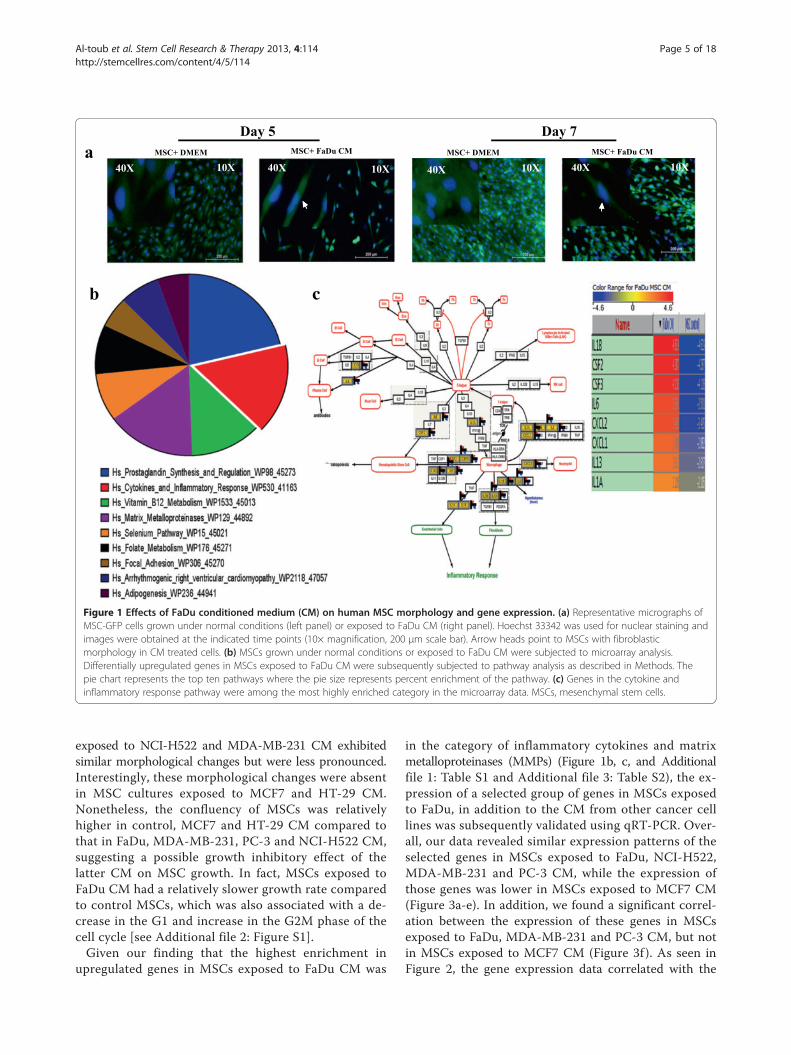
exposed to NCI-H522 and MDA-MB-231 CM exhibitedsimilar morphological changes but were less pronounced.Interestingly, these morphological changes were absentin MSC cultures exposed to MCF7 and HT-29 CM.Nonetheless, the confluency of MSCs was relativelyhigher in control, MCF7 and HT-29 CM compared tothat in FaDu, MDA-MB-231, PC-3 and NCI-H522 CM,suggesting a possible growth inhibitory effect of thelatter CM on MSC growth. In fact, MSCs exposed toFaDu CM had a relatively slower growth rate comparedto control MSCs, which was also associated with a de-crease in the G1 and increase in the G2M phase of thecell cycle [see Additional file 2: Figure S1].Given our finding that the highest enrichment in
upregulated genes in MSCs exposed to FaDu CM was
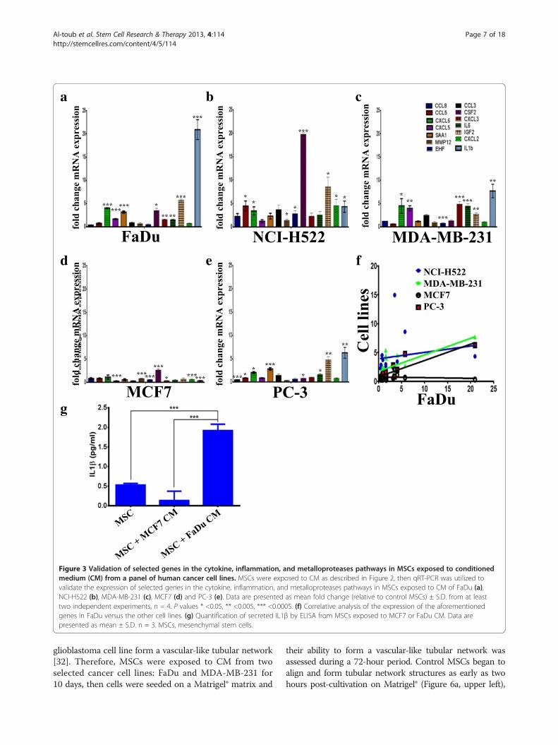
in the category of inflammatory cytokines and matrixmetalloproteinases (MMPs) (Figure 1b, c, and Additionalfile 1: Table S1 and Additional file 3: Table S2), the ex-pression of a selected group of genes in MSCs exposedto FaDu, in addition to the CM from other cancer celllines was subsequently validated using qRT-PCR. Over-all, our data revealed similar expression patterns of theselected genes in MSCs exposed to FaDu, NCI-H522,MDA-MB-231 and PC-3 CM, while the expression ofthose genes was lower in MSCs exposed to MCF7 CM(Figure 3a-e). In addition, we found a significant correl-ation between the expression of these genes in MSCsexposed to FaDu, MDA-MB-231 and PC-3 CM, but notin MSCs exposed to MCF7 CM (Figure 3f ). As seen inFigure 2, the gene expression data correlated with the
Figure 1 Effects of FaDu conditioned medium (CM) on human MSC morphology and gene expression. (a) Representative micrographs ofMSC-GFP cells grown under normal conditions (left panel) or exposed to FaDu CM (right panel). Hoechst 33342 was used for nuclear staining andimages were obtained at the indicated time points (10× magnification, 200 μm scale bar). Arrow heads point to MSCs with fibroblasticmorphology in CM treated cells. (b) MSCs grown under normal conditions or exposed to FaDu CM were subjected to microarray analysis.Differentially upregulated genes in MSCs exposed to FaDu CM were subsequently subjected to pathway analysis as described in Methods. Thepie chart represents the top ten pathways where the pie size represents percent enrichment of the pathway. (c) Genes in the cytokine andinflammatory response pathway were among the most highly enriched category in the microarray data. MSCs, mesenchymal stem cells.
Al-toub et al. Stem Cell Research & Therapy 2013, 4:114 Page 5 of 18http://stemcellres.com/content/4/5/114

observed phenotypic changes. MSCs exposed to FaDuCM secreted a significant amount of IL1β, compared tocontrol MSCs or MSCs exposed to MCF7 CM (Figure 3g),which is concordant with the qRT-PCR data.
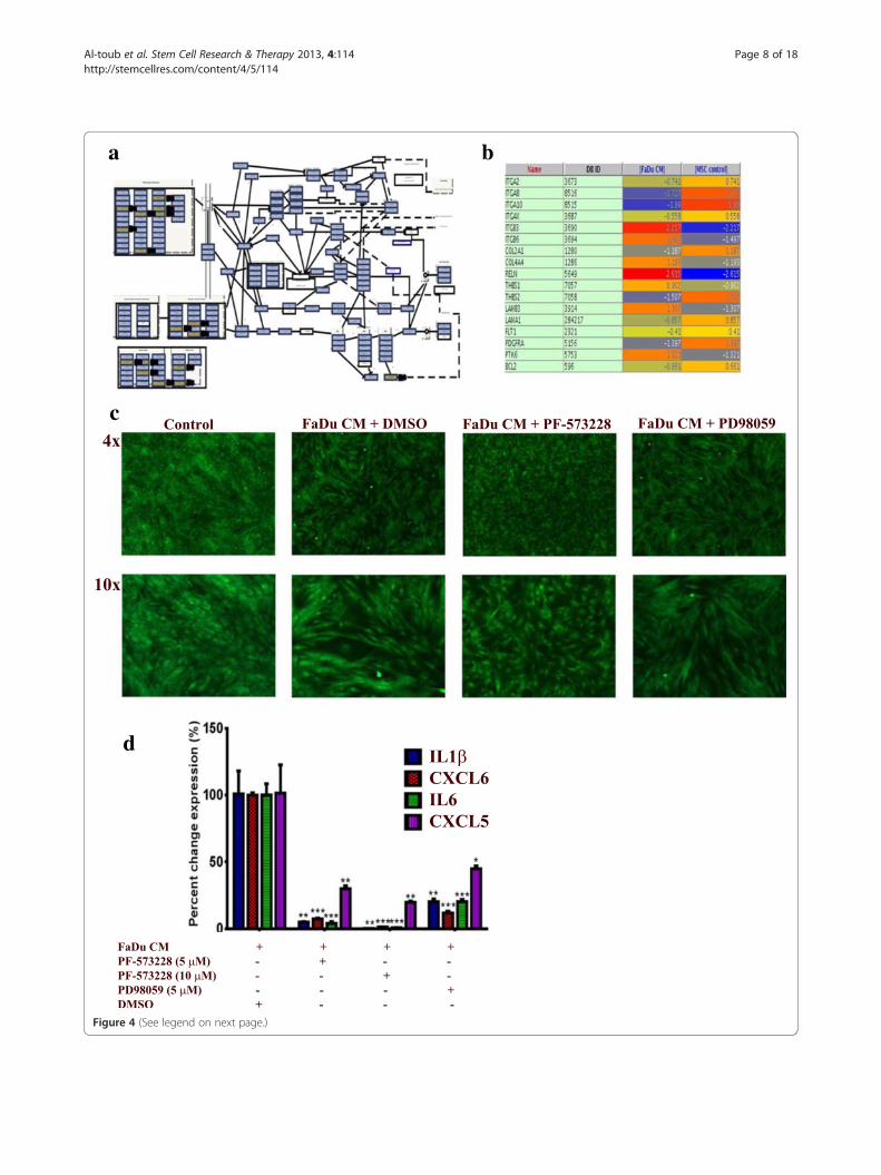
Pro-inflammatory response of MSCs exposed to FaDu CM ismediated mainly through focal adhesion kinase signalingPathway analysis of differentially expressed genes in MSCsexposed to FaDu CM revealed multiple enriched pathways.Among those, FAK (P = 2.1 × 10 -5) and, to lesser extent,MAPK (P = 0.03) were very prominent [see Additionalfile 1: Table S1]. Differentially expressed genes in theFAK pathway are shown in Figure 4a and b. To assesswhether FAK and MAPK pathways are indeed involvedin regulating the pro-inflammatory response of MSCsexposed to tumor CM, MSCs were exposed to control orFaDu CM in the presence of PF-573228 (FAK inhibitor),PD98059 (MAPKK inhibitor) or DMSO. On day 5, cellswere monitored for phenotypic changes. As shown inFigure 4c, FAK inhibitor almost completely inhibited thepro-inflammatory phenotype, while MAPKK inhibitorhad a less pronounced effect. qRT-PCR analysis of apanel of pro-inflammatory cytokines (IL1β, CXCL6, IL6and CXCL5) revealed drastic inhibition of the expressionof those cytokines in the presence of FAK inhibitor in adose dependent manner (Figure 4d). MAPKK inhibitoralso significantly inhibited the pro-inflammatory responsein MSCs exposed to FaDu CM, but less than that seenwith the FAK inhibitor (Figure 4d).
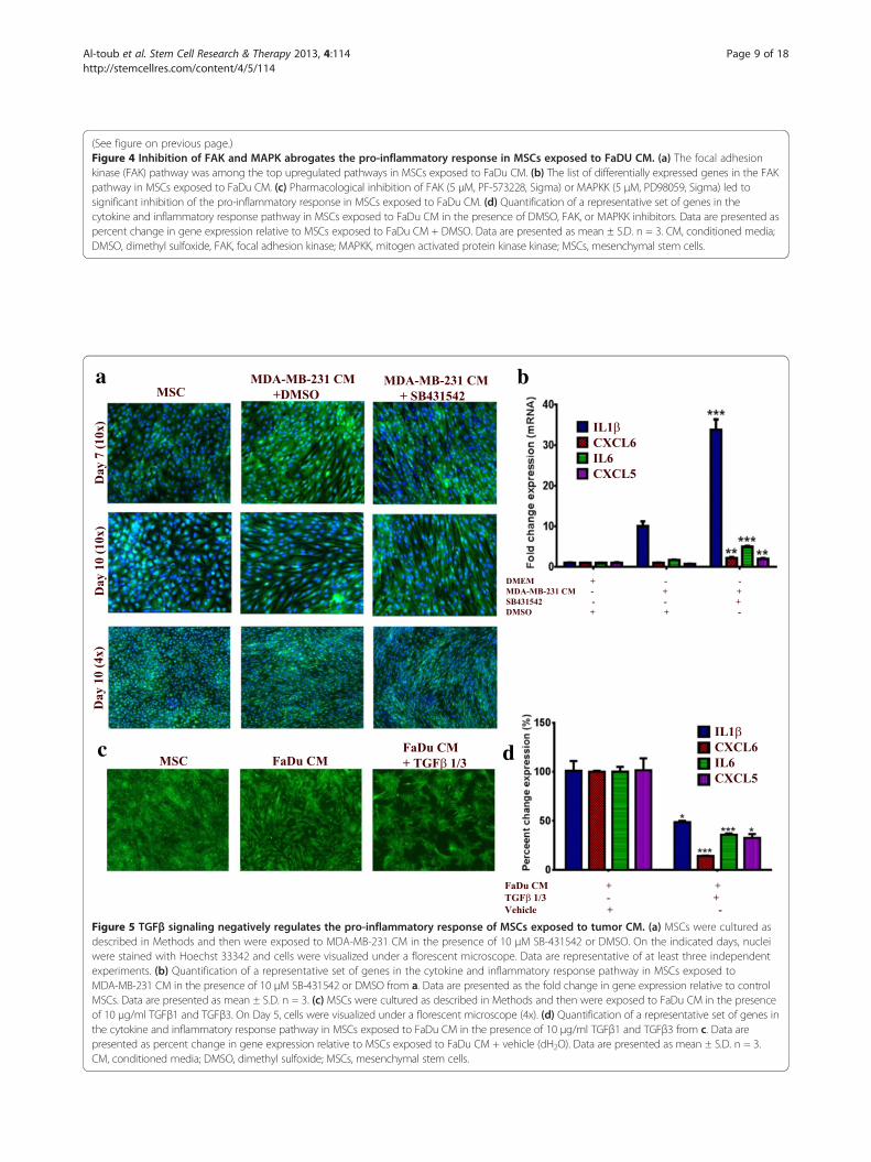
Signaling via TGFβ negatively regulates the pro-inflammatoryresponse of MSCs to FaDu CMGiven its critical role in tumorigenicity and in regulatingthe differentiation of MSCs [29-31], we hypothesizedthat changes in TGFβ signaling could potentially regulatethe observed changes in the phenotype of MSCs. Interest-ingly, pharmacological inhibition of the TGFβ receptorkinase using SB-431542 (10 μM) in MSCs in the presenceof MDA-MB-231 CM (this cell line was selected becauseit has the highest expression of TGFβ among all cell linesused in this study, data not shown) led to significantenhancement in the characteristic morphology of MSCs(Figure 5a). Concordant with that, the expression of thepro-inflammatory cytokine panel was significantly increasedin the presence of SB-431542 compared to control DMSO(>3-fold), Figure 5b. On the other hand, treating MSCs withrecombinant TGFβ1 and TGFβ3 in the presence of FaDuCM (this cell line was selected for this experiment since itinduced the strongest phenotype and has low TGFβ expres-sion compared to MDA-MB-231) led to significant inhib-ition of the pro-inflammatory phenotype at the cellular andmolecular levels (Figure 5c and d). Therefore, our data indi-cate an inhibitory role for TGFβ signaling on mediating theobserved changes in the MSCs phenotype.
MSCs exposed to tumor CM have diminished multilineagedifferentiation potentialRecent study using an in vitro angiogenesis assay hasindicated that human MSCs exposed to CM from a
Figure 2 Comparative analysis of morphological changes in MSCs exposed to conditioned medium (CM) from a panel of humancancer cell lines. MSCs were grown under normal conditions ((D)MEM) or were exposed to CM from the indicated cancer cell lines(FaDu, MCF7, MDA-MB-231, PC-3, NCI-H522 and HT-29) and, subsequently, images were obtained on days1, 2 and 7. Representativemicrographs from at least three independent experiments are shown. All images were taken using 10x magnification. MSCs,mesenchymal stem cells.
Al-toub et al. Stem Cell Research & Therapy 2013, 4:114 Page 6 of 18http://stemcellres.com/content/4/5/114
glioblastoma cell line form a vascular-like tubular network[32]. Therefore, MSCs were exposed to CM from twoselected cancer cell lines: FaDu and MDA-MB-231 for10 days, then cells were seeded on a Matrigel® matrix and
their ability to form a vascular-like tubular network wasassessed during a 72-hour period. Control MSCs began toalign and form tubular network structures as early as twohours post-cultivation on Matrigel® (Figure 6a, upper left),
a b c
d e f
g
Figure 3 Validation of selected genes in the cytokine, inflammation, and metalloproteases pathways in MSCs exposed to conditionedmedium (CM) from a panel of human cancer cell lines. MSCs were exposed to CM as described in Figure 2, then qRT-PCR was utilized tovalidate the expression of selected genes in the cytokine, inflammation, and metalloproteases pathways in MSCs exposed to CM of FaDu (a),NCI-H522 (b), MDA-MB-231 (c), MCF7 (d) and PC-3 (e). Data are presented as mean fold change (relative to control MSCs) ± S.D. from at leasttwo independent experiments, n = 4. P values * <0.05, ** <0.005, *** <0.0005. (f) Correlative analysis of the expression of the aforementionedgenes in FaDu versus the other cell lines. (g) Quantification of secreted IL1β by ELISA from MSCs exposed to MCF7 or FaDu CM. Data arepresented as mean ± S.D. n = 3. MSCs, mesenchymal stem cells.
Al-toub et al. Stem Cell Research & Therapy 2013, 4:114 Page 7 of 18http://stemcellres.com/content/4/5/114
c
d
ba
Figure 4 (See legend on next page.)
Al-toub et al. Stem Cell Research & Therapy 2013, 4:114 Page 8 of 18http://stemcellres.com/content/4/5/114
(See figure on previous page.)Figure 4 Inhibition of FAK and MAPK abrogates the pro-inflammatory response in MSCs exposed to FaDU CM. (a) The focal adhesionkinase (FAK) pathway was among the top upregulated pathways in MSCs exposed to FaDu CM. (b) The list of differentially expressed genes in the FAKpathway in MSCs exposed to FaDu CM. (c) Pharmacological inhibition of FAK (5 μM, PF-573228, Sigma) or MAPKK (5 μM, PD98059, Sigma) led tosignificant inhibition of the pro-inflammatory response in MSCs exposed to FaDu CM. (d) Quantification of a representative set of genes in thecytokine and inflammatory response pathway in MSCs exposed to FaDu CM in the presence of DMSO, FAK, or MAPKK inhibitors. Data are presented aspercent change in gene expression relative to MSCs exposed to FaDu CM + DMSO. Data are presented as mean ± S.D. n = 3. CM, conditioned media;DMSO, dimethyl sulfoxide, FAK, focal adhesion kinase; MAPKK, mitogen activated protein kinase kinase; MSCs, mesenchymal stem cells.
c d
ba
Figure 5 TGFβ signaling negatively regulates the pro-inflammatory response of MSCs exposed to tumor CM. (a) MSCs were cultured asdescribed in Methods and then were exposed to MDA-MB-231 CM in the presence of 10 μM SB-431542 or DMSO. On the indicated days, nucleiwere stained with Hoechst 33342 and cells were visualized under a florescent microscope. Data are representative of at least three independentexperiments. (b) Quantification of a representative set of genes in the cytokine and inflammatory response pathway in MSCs exposed toMDA-MB-231 CM in the presence of 10 μM SB-431542 or DMSO from a. Data are presented as the fold change in gene expression relative to controlMSCs. Data are presented as mean ± S.D. n = 3. (c) MSCs were cultured as described in Methods and then were exposed to FaDu CM in the presenceof 10 μg/ml TGFβ1 and TGFβ3. On Day 5, cells were visualized under a florescent microscope (4x). (d) Quantification of a representative set of genes inthe cytokine and inflammatory response pathway in MSCs exposed to FaDu CM in the presence of 10 μg/ml TGFβ1 and TGFβ3 from c. Data arepresented as percent change in gene expression relative to MSCs exposed to FaDu CM + vehicle (dH2O). Data are presented as mean ± S.D. n = 3.CM, conditioned media; DMSO, dimethyl sulfoxide; MSCs, mesenchymal stem cells.
Al-toub et al. Stem Cell Research & Therapy 2013, 4:114 Page 9 of 18http://stemcellres.com/content/4/5/114

cb
a
Figure 6 (See legend on next page.)
Al-toub et al. Stem Cell Research & Therapy 2013, 4:114 Page 10 of 18http://stemcellres.com/content/4/5/114
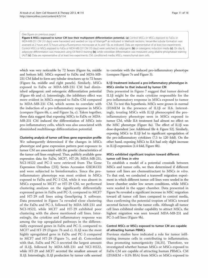
which was very noticeable by 72 hours (Figure 6a, middleand bottom left). MSCs exposed to FaDu and MDA-MB-231 CM failed to form any tubular structures up to 72 hours(Figure 6a, middle and right panels). Similarly, MSCsexposed to FaDu or MDA-MB-231 CM had dimin-ished adipogenic and osteogenic differentiation potential(Figure 6b and c). Interestingly, the inhibitory effect wasmore evident in MSCs exposed to FaDu CM comparedto MDA-MB-231 CM, which seems to correlate withthe induction of a pro-inflammatory response in MSCs(compare Figure 6b, c, and Figure 3a, c). Taken together,these data suggest that exposing MSCs to FaDu or MDA-MB-231 CM induced the differentiation of MSCs intopro-inflammatory cells, which was also associated withdiminished multilineage differentiation potential.
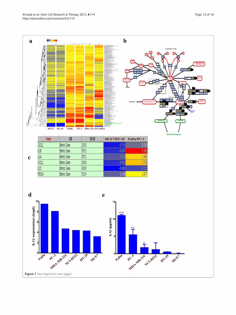
Clustering analysis of tumor cell lines gene expression profileWe subsequently determined if the changes in MSCsphenotype and gene expression pattern post exposure totumor CM are associated with the genetic characteristics ofthe tumor cell lines employed. Thus, publicly available geneexpression data for FaDu, MCF7, HT-29, MDA-MB-231,NCI-H522 and PC-3 were retrieved from The GeneExpression Omnibus ([33]; Series Accession GSE36133)and were subjected to bioinformatics. Since the pro-inflammatory phenotype was most evident in MSCsexposed to FaDu and PC-3 CM, while it was absent inMSCs exposed to MCF7 or HT-29 CM, we performedclustering analyses on the significantly differentiallyexpressed genes in FaDu and PC-3, compared to MCF7and HT-29 cell lines using GeneSpring X software.Data presented in Figure 7a revealed close clusteringof the FaDu and PC-3, followed by MDA-MB-231 andNCI-H522, while MCF7 and HT-29 exhibited poorclustering with the above mentioned cell lines. Inter-estingly, the cytokine and inflammatory response wasamong the top upregulated pathways in the differen-tially expressed genes in FaDu and PC-3, compared toMCF7 and HT-29 (Figure 7b and c). IL1β was the mosthighly upregulated gene in FaDu and PC-3 comparedto MCF7 and HT-29 (Figure 7c, and d). Concordantwith that, FaDu and PC-3 secreted the largest amountof IL1β, followed by MDA-MB-231 and NCI-H522,while HT-29 and MCF7 secreted the smallest amount ofIL1β. Interestingly, IL1β production by tumor cells seemed
to correlate with the induced pro-inflammatory phenotype(compare Figure 7e and Figure 3).
IL1β treatment induced a pro-inflammatory phenotype inMSCs similar to that induced by tumor CMData presented in Figure 7 suggest that tumor derivedIL1β might be the main cytokine responsible for thepro-inflammatory response in MSCs exposed to tumorCM. To test this hypothesis, MSCs were grown in normal(D)MEM in the presence of IL1β or IL6. Interest-ingly, treating MSCs with IL1β phenocopied the pro-inflammatory phenotype seen in MSCs exposed totumor CM, while IL6 treatment had almost no effect onthe MSC phenotype (Figure 8a). The effect of IL1β wasdose-dependent [see Additional file 4: Figure S2]. Similarly,exposing MSCs to IL1β led to significant upregulation ofthe pro-inflammatory cytokines (7.5 to 120 fold). On theother hand, exposing MSCs to IL6 had only slight increasein IL1β expression (1.4 fold, Figure 8b).
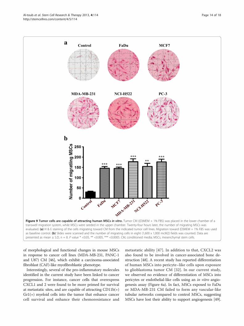
MSCs exhibited significant tropism toward differenttumor cell lines in vitroTo establish a model of a potential crosstalk betweenMSCs and tumor cells, we then determined if differenttumor cell lines are chemoattractant to MSCs in vitro.To that end, we conducted a transwell migration experi-ment in which different tumor cell lines were seeded in thelower chamber under low serum conditions, while MSCswere seeded in the upper chamber. Data presented inFigure 9a revealed a significant increase in MSC migrationtoward all tumor cell lines compared to control media,thus confirming the potential tropism of MSCs towardsecreted factors from the tumor cells. Although all tumorcell lines exhibited similar capability to attract MSCs, thehighest migration was seen toward MDA-MB-231 andPC-3 cell lines (Figure 9b).
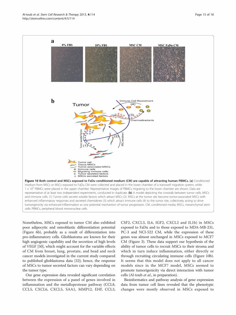
Control MSCs or MSCs exposed to tumor CM are capableof attracting human PBMCsPrevious studies have indicated a role for tumor infil-trating immune cells in contributing to inflammation,thus promoting tumorigenicity [34,35]. Therefore, weinvestigated whether human MSCs or MSCs exposed toFaDu CM are capable of attracting human PBMCs. CM((D)MEM + 0.5% BSA) from MSCs or MSCs exposed to
(See figure on previous page.)Figure 6 MSCs exposed to tumor CM lose their multipotent differentiation potential. (a) Control MSCs or MSCs exposed to FaDu orMDA-MB-231 CM (10 days) were harvested and seeded on top of Matrigel® as indicated in Methods sections. Vessel-like tubular formation wasassessed at 2 hours and 72 hours using a fluorescence microscope at 4x and 10x, as indicated. Data are representative of at least two experiments.Control MSCs or MSCs exposed to FaDu or MDA-MB-231 CM (10 days) were switched to adipogenic (b) or osteogenic induction media (c). On day 6,adipocyte differentiation was measured using Oil-Red-O staining (b), while osteoblast differentiation was measured using alkaline phosphatase staining(ALP) (c). Data are representative of at least two experiments. CM, conditioned media; MSCs, mesenchymal stem cells.
Al-toub et al. Stem Cell Research & Therapy 2013, 4:114 Page 11 of 18http://stemcellres.com/content/4/5/114
c
d e
ba
Figure 7 (See legend on next page.)
Al-toub et al. Stem Cell Research & Therapy 2013, 4:114 Page 12 of 18http://stemcellres.com/content/4/5/114

FaDu CM were collected and placed in the lower chamberin a transwell migration system, while 1 × 10⁵ humanPBMCs were seeded in the upper chamber. As shown inFigure 10a, a significant increase in PBMC migration to-ward MSCs or MSCs exposed to FaDu CM was observed.
DiscussionFor several decades, the molecular changes within tumorcells were studied in order to understand factors responsiblefor promoting tumor progression and metastasis, whilelittle attention was paid to the possible contributory roleof tumor microenvironment. Recent evidence suggeststhat the tumor microenvironment, which is composed ofa very complex network of extracellular matrix (ECM)proteins and many cell types, such as endothelial cells,stromal (mesenchymal) stem cells, pericytes, fibroblasts
and immune cells, plays a critical role in tumor progressionand metastasis [36,37]. Among these components, MSCshave been the focus of intensive investigation [9,17,38-45].In the present report, we examined the crosstalk between
tumor cells and MSCs and we investigated the effect(s)of tumor secreted factors on MSCs at the cellular andmolecular levels. As surrogates for malignant tumors,we employed a number of well characterized cancer celllines. We reported that secreted factors from FaDu cellsled to significant morphological and genetic changes inMSCs with enhanced expression of pro-inflammatorycytokines, and similar responses were also observed whenadditional tumor cell lines were evaluated. However, theseeffects were not universal for all malignant cell lines. Forexample, MCF7 and HT-29 did not exert these effects.Our findings corroborate recent findings of the presence
(See figure on previous page.)Figure 7 Cluster and pathway analysis of basal gene expression in FaDu, NCI-H522, MDA-MB-231, MCF7, PC-3 and HT-29 tumor celllines. (a) Clustering analysis of the tumor cell lines indicated close clustering for FaDu and PC-3, followed by MDA-MB-231 and NCI-H522, whileMCF7 and HT-29 did not cluster readily with the group. Clustering analyses were performed on differentially expressed genes in FaDu and PC-3relative to MCF7 and HT-29. (b) Cytokine and inflammatory response pathway was among the top enriched pathways in differentially expressedgenes between FaDu and PC-3 relative to MCF7 and HT-29. (c) Genes in the cytokine and inflammatory response pathways from b and theirexpression levels are shown. (d) mRNA expression level of IL1β in different tumor cell lines from the microarray data. (e) Quantification ofsecreted IL1β by ELISA from different tumor CM. Data are presented as mean ± S.D. n = 3. CM, conditioned media.
b
a
Figure 8 IL1β treatment induced a pro-inflammatory response in MSCs. (a) MSCs were cultured in normal (D)MEM in the presence ofrecombinant IL1β (10 and 50 ng/ml), recombinant IL6 (50 ng/ml), both IL1β and IL6 (50 ng/ml each) or in the presence of vehicle control (dH2O).Images were taken on day 4 and 7 (4x). (b) Quantification of a representative set of genes in the cytokine and inflammatory response pathway inMSCs exposed to different cytokines from (a). Data are presented as fold change in gene expression relative to MSCs exposed to vehicle. Dataare presented as mean ± S.D., n = 3. MSCs, mesenchymal stem cells.
Al-toub et al. Stem Cell Research & Therapy 2013, 4:114 Page 13 of 18http://stemcellres.com/content/4/5/114
of morphological and functional changes in mouse MSCsin response to cancer cell lines (MDA-MB-231, PANC-1and U87) CM [46], which exhibit a carcinoma-associatedfibroblast (CAF)-like myofibroblastic phenotype.Interestingly, several of the pro-inflammatory molecules
identified in the current study have been linked to cancerprogression. For instance, cancer cells that overexpressCXCL1 and 2 were found to be more primed for survivalat metastatic sites, and are capable of attracting CD11b(+)Gr1(+) myeloid cells into the tumor that enhance cancercell survival and enhance their chemoresistance and
metastatic ability [47]. In addition to that, CXCL2 wasalso found to be involved in cancer-associated bone de-struction [48]. A recent study has reported differentiationof human MSCs into pericyte–like cells upon exposureto glioblastoma tumor CM [32]. In our current study,we observed no evidence of differentiation of MSCs intopericytes or endothelial-like cells using an in vitro angio-genesis assay (Figure 6a). In fact, MSCs exposed to FaDuor MDA-MB-231 CM failed to form any vascular-liketubular networks compared to control MSCs, suggestingMSCs have lost their ability to support angiogenesis [49].
a
b
Figure 9 Tumor cells are capable of attracting human MSCs in vitro. Tumor CM ((D)MEM + 1% FBS) was placed in the lower chamber of atranswell migration system, while MSCs were seeded in the upper chamber. Twenty-four hours later, the number of migrating MSCs wasevaluated. (a) H & E staining of the cells migrating toward CM from the indicated tumor cell lines. Migration toward (D)MEM + 1% FBS was usedas baseline control. (b) Slides were scanned and the number of migrating cells in eight (1,600 x 1,000 mcM2) fields was counted. Data arepresented as mean ± S.D, n = 8. P value * <0.05, ** <0.005, *** <0.0005. CM, conditioned media; MSCs, mesenchymal stem cells.
Al-toub et al. Stem Cell Research & Therapy 2013, 4:114 Page 14 of 18http://stemcellres.com/content/4/5/114
Nonetheless, MSCs exposed to tumor CM also exhibitedpoor adipocytic and osteoblastic differentiation potential(Figure 6b), probably as a result of differentiation intopro-inflammatory cells. Glioblastoma are known for theirhigh angiogenic capability and the secretion of high levelsof VEGF [50], which might account for the variable effectsof CM from breast, lung, prostate, and head and neckcancer models investigated in the current study comparedto published glioblastoma data [32]; hence, the responseof MSCs to tumor secreted factors can vary depending onthe tumor type.Our gene expression data revealed significant correlation
between the expression of a panel of genes involved ininflammation and the metalloprotease pathway (CCL8,CCL5, CXCL6, CXCL5, SAA1, MMP12, EHF, CCL3,
CSF2, CXCL3, IL6, IGF2, CXCL2 and IL1b) in MSCsexposed to FaDu and to those exposed to MDA-MB-231,PC-3 and NCI-522 CM, while the expression of thesegenes was almost unchanged in MSCs exposed to MCF7CM (Figure 3). These data support our hypothesis of theability of tumor cells to recruit MSCs to their stroma andwhich in turn induce inflammation, either directly orthrough recruiting circulating immune cells (Figure 10b).It seems that this model does not apply to all cancermodels since in the MCF7 model, MSCs seemed topromote tumorigenicity via direct interaction with tumorcells (Al-toub et al., in preparation).Bioinformatics and pathway analysis of gene expression
data from tumor cell lines revealed that the phenotypicchanges were mostly observed in MSCs exposed to
a
b
Figure 10 Both control and MSCs exposed to FaDu conditioned medium (CM) are capable of attracting human PBMCs. (a) Conditionedmedium from MSCs or MSCs exposed to FaDu CM were collected and placed in the lower chamber of a transwell migration system, while1 x 105 PBMCs were placed in the upper chamber. Representative images of PBMCs migrating to the lower chamber are shown. Data arerepresentative of at least two independent experiments, conducted in duplicate. (b) A model depicting the crosstalk between tumor cells, MSCsand immune cells. (1) Tumor cells secrete soluble factors which attract MSCs (2). MSCs at the tumor site become tumor-associated MSCs withenhanced inflammatory responses and secreted chemokines (3) which attract immune cells (4) to the tumor site, collectively acting to drivetumorigenicity via enhanced inflammation as one potential mechanism of tumor progression. CM, conditioned media; MSCs, mesenchymal stemcells; PBMCs, peripheral blood mononuclear cells.
Al-toub et al. Stem Cell Research & Therapy 2013, 4:114 Page 15 of 18http://stemcellres.com/content/4/5/114

CM from cell lines with a pro-inflammatory nature(such as, FaDu and PC-3, Figure 7c). Indeed our investiga-tion has identified tumor-derived IL1β to be the primarydriver of the pro-inflammatory phenotype observed inMSCs exposed to tumor CM, whereas treating MSCs withrecombinant IL1β mimicked the effects of tumor CM atthe cellular and molecular level (Figures 7d-e and 8a-b).Nonetheless, we also identified signaling via FAK and,
to lesser extent, MAPK to be critical for the induction ofthe observed phenotype (Figure 4). In contrast, pharma-cological inhibition of TGFβ signaling in MSCs led tosubstantial enhancement in the observed changes in pheno-type and gene expression in MSCs exposed to MDA-MB-231 CM (Figure 5a and b), which was also associated witha slight increase in cell proliferation [see Additional file 5:Figure S3]. Treating MSCs with recombinant TGFβ1 andTGFβ3 in the presence of FaDu CM led to significantinhibition of the observed phenotype at the cellular andmolecular level (Figure 5c and d), which further implicatedTGFβ signaling in negatively regulating MSC differen-tiation in response to tumor CM. Thus, our findingscorroborate previous studies suggesting a role for theTGFβ signaling pathway in regulating mesenchymalstem cell differentiation [31].
ConclusionsOur data support an evolving hypothesis that cancer cellssecrete a large number of factors regulating biologicalcharacteristics of MSCs and transforming MSCs into pro-inflammatory cells. We identified tumor-derived IL1β as onepotential mediator of the observed phenotype. Nonetheless,we also identified FAK and MAPK signaling to regulate posi-tively, while TGFβ signaling was found to negatively regulatethe response of MSCs to tumor CM. Taken together, ourdata support a model where MSCs contribute to tumorigen-icity through their pro-inflammatory phenotype induced bycancer cell-derived factors, such as IL1β (Figure 10b).
Additional files
Additional file 1: Table S1. Pathway analysis of upregulated genes inMSCs exposed to FaDu CM.
Additional file 2: Figure S1. Effect of FaDu CM on MSC cell growthand cell cycle.
Additional file 3: Table S2. Functional annotation and clusteringanalysis of genes upregulated (10 fold) in MSCs exposed to FaDu CM.
Additional file 4: Figure S2. Dose dependent effect of IL1β on MSCphenotype.
Additional file 5: Figure S3. SB-431542 promotes the growth of MSCs inthe presence of MDA-MB-231 CM. MSCs were grown in MDA-MB-231 CM inthe presence of SB-431542 or DMSO. Cell viability was measured on days 3, 7,and 10 using alamar blue assay. Data are presented as mean ± S.D., n = 9.
AbbreviationsALP: Alkaline phosphatase; BSA: Bovine serum albumin; CCL3: Chemokine(C-C motif) ligand 3; CCL5: Chemokine (C-C motif) ligand 5; CCL8: Chemokine
(C-C motif) ligand 8; CM: Conditioned medium; CSF2: Colony stimulatingfactor 2; CSF3: Colony stimulating factor 3; CXCL1: Chemokine (C-X-C motif)ligand 1; CXCL2: Chemokine (C-X-C motif) ligand 2; CXCL3: Chemokine(C-X-C motif) ligand 3; CXCL5: Chemokine (C-X-C motif) ligand 5;CXCL6: Chemokine (C-X-C motif) ligand 6; (D)MEM: (D)ulbecco’s modifiedEagle’s medium; DMSO: Dimethyl sulfoxide; EHF: Ets homologous factor;ELISA: Enzyme-linked immunosorbent assay; EMT: Epithelial mesenchymaltransition; FAK: Focal adhesion kinase; FBS: Fetal bovine serum;GAPDH: Glyceraldehyde 3-phosphate dehydrogenase; H & E: Hematoxylinand eosin; hMSC-TERT-GFP: Human mesenchymal stem cell-telomerized-green fluorescence protein; IGF2: Insulin-like growth factor 2; IL13: Interleukin13; IL1A: Interleukin 1, Alpha; IL1B: Interleukin 1, Beta; IL6: Interleukin 6;MAPK: Mitogen activated protein kinase; MMP12: Matrix metallopeptidase 12;MSCs: Mesenchymal stem cells; NEAA: Non-essential amino acids;PBMCs: Peripheral blood mononuclear cells; PBS: Phosphate-buffered saline;qRT-PCR: Quantitative real-time reverse-transcription PCR; SAA1: Serumamyloid A1; TGF-beta: Transforming growth factor beta; VEGF: Vascularendothelial growth factor A.
Competing interestsThe authors declare that they have no competing interests.
Author’ contributionsM Al-toub performed the experiments and participated in preparing themanuscript; A Almusa, M Almajid performed the experiments; M Al-Nbaheencharacterized MSCs phenotype; M Kassem, A Aldahamsh participated instudy design, interpretation of data and preparation of the manuscript. MKassem characterized and provided hMSC-TERT-GFP cell lines. NM Alajez wasresponsible for obtaining funding, study design, data interpretation,bioinformatics analysis and preparation of the manuscript. All authors readand approved the final manuscript.
AcknowledgementsThis work was supported by grant No. 11-MED1582-02 from the NationalPlan for Sciences and Technology Program, King Saud University, kingdom ofSaudi Arabia. We thank Dr Abdulkarim Alhetheel (King Saud University) forproviding the PBMCs. We thank Radhakrishnan Vishnubalaji and DanaHamam (Stem Cell Unit, King Saud University) for their assistance with thein vitro angiogenesis, and the Oil Red O/ALP staining, respectively. We thankDr Amer Mahmood (Stem Cell Unit, King Saud University) for his assistancewith TGFβ inhibition experiments.
Author details1Stem Cell Unit, Department of Anatomy, College of Medicine, King SaudUniversity, Riyadh 11461, Kingdom of Saudi Arabia. 2Saudi ElectronicUniversity, Riyadh, Saudi Arabia. 3KMEB, Department of Endocrinology,University of Southern Denmark, Odense, Denmark.
Received: 24 February 2013 Revised: 26 February 2013Accepted: 12 September 2013 Published: 17 September 2013
References1. Dominici M, Le Blanc K, Mueller I, Slaper-Cortenbach I, Marini F, Krause D,
Deans R, Keating A, Prockop D, Horwitz E: Minimal criteria for definingmultipotent mesenchymal stromal cells, The International Society forCellular Therapy position statement. Cytotherapy 2006, 8:315–317.
2. Granero-Molto F, Weis JA, Miga MI, Landis B, Myers TJ, O’Rear L, LongobardiL, Jansen ED, Mortlock DP, Spagnoli A: Regenerative effects oftransplanted mesenchymal stem cells in fracture healing. Stem Cells 2009,27:1887–1898.
3. Chen J, Li Y, Wang L, Lu M, Zhang X, Chopp M: Therapeutic benefit ofintracerebral transplantation of bone marrow stromal cells after cerebralischemia in rats. J Neurol Sci 2001, 189:49–57.
4. Wu GD, Nolta JA, Jin YS, Barr ML, Yu H, Starnes VA, Cramer DV: Migration ofmesenchymal stem cells to heart allografts during chronic rejection.Transplantation 2003, 75:679–685.
5. Kristensen LP, Chen L, Nielsen MO, Qanie DW, Kratchmarova I, Kassem M,Andersen JS: Temporal profiling and pulsed SILAC labeling identify novelsecreted proteins during ex vivo osteoblast differentiation of humanstromal stem cells. Mol Cell Proteomics 2012, 11:989–1007.
Al-toub et al. Stem Cell Research & Therapy 2013, 4:114 Page 16 of 18http://stemcellres.com/content/4/5/114

6. Chamberlain G, Fox J, Ashton B, Middleton J: Concise review:mesenchymal stem cells: their phenotype, differentiation capacity,immunological features, and potential for homing. Stem Cells 2007,25:2739–2749.
7. Phinney DG, Prockop DJ: Concise review: mesenchymal stem/multipotentstromal cells: the state of transdifferentiation and modes of tissuerepair–current views. Stem Cells 2007, 25:2896–2902.
8. Kinnaird T, Stabile E, Burnett MS, Lee CW, Barr S, Fuchs S, Epstein SE:Marrow-derived stromal cells express genes encoding a broad spectrumof arteriogenic cytokines and promote in vitro and in vivo arteriogenesisthrough paracrine mechanisms. Circ Res 2004, 94:678–685.
9. Karnoub AE, Dash AB, Vo AP, Sullivan A, Brooks MW, Bell GW, Richardson AL,Polyak K, Tubo R, Weinberg RA: Mesenchymal stem cells within tumourstroma promote breast cancer metastasis. Nature 2007, 449:557–563.
10. Dwyer RM, Potter-Beirne SM, Harrington KA, Lowery AJ, Hennessy E, MurphyJM, Barry FP, O’Brien T, Kerin MJ: Monocyte chemotactic protein-1secreted by primary breast tumors stimulates migration of mesenchymalstem cells. Clin Cancer Res 2007, 13:5020–5027.
11. Dvorak HF: Tumors: wounds that do not heal. Similarities between tumorstroma generation and wound healing. N Engl J Med 1986, 315:1650–1659.
12. Erez N, Truitt M, Olson P, Arron ST, Hanahan D: Cancer-associated fibroblastsare activated in incipient neoplasia to orchestrate tumor-promotinginflammation in an NF-kappaB-dependent manner. Cancer Cell 2010,17:135–147.
13. Navab R, Strumpf D, Bandarchi B, Zhu CQ, Pintilie M, Ramnarine VR,Ibrahimov E, Radulovich N, Leung L, Barczyk M, Panchal D, To C, Yun JJ, DerS, Shepherd FA, Jurisica I, Tsao MS: Prognostic gene-expression signatureof carcinoma-associated fibroblasts in non-small cell lung cancer. ProcNatl Acad Sci U S A 2011, 108:7160–7165.
14. Quante M, Tu SP, Tomita H, Gonda T, Wang SS, Takashi S, Baik GH, ShibataW, Diprete B, Betz KS, Friedman R, Varro A, Tycko B, Wang TC: Bonemarrow-derived myofibroblasts contribute to the mesenchymal stemcell niche and promote tumor growth. Cancer Cell 2011, 19:257–272.
15. Lepperdinger G, Brunauer R, Jamnig A, Laschober G, Kassem M: Controversialissue: is it safe to employ mesenchymal stem cells in cell-based therapies?Exp Gerontol 2008, 43:1018–1023.
16. Khakoo AY, Pati S, Anderson SA, Reid W, Elshal MF, Rovira II, Nguyen AT, MalideD, Combs CA, Hall G, Zhang J, Raffeld M, Rogers TB, Stetler-Stevenson W, FrankJA, Reitz M, Finkel T: Human mesenchymal stem cells exert potentantitumorigenic effects in a model of Kaposi’s sarcoma. J Exp Med 2006,203:1235–1247.
17. Nakamizo A, Marini F, Amano T, Khan A, Studeny M, Gumin J, Chen J,Hentschel S, Vecil G, Dembinski J, Andreeff M, Lang FF: Human bonemarrow-derived mesenchymal stem cells in the treatment of gliomas.Cancer Res 2005, 65:3307–3318.
18. Zhu Y, Sun Z, Han Q, Liao L, Wang J, Bian C, Li J, Yan X, Liu Y, Shao C, ZhaoRC: Human mesenchymal stem cells inhibit cancer cell proliferation bysecreting DKK-1. Leukemia 2009, 23:925–933.
19. Shabahang M, Buras RR, Davoodi F, Schumaker LM, Nauta RJ, Uskokovic MR,Brenner RV, Evans SR: Growth inhibition of HT-29 human colon cancercells by analogues of 1,25-dihydroxyvitamin D3. Cancer Res 1994,54:4057–4064.
20. Alajez NM, Shi W, Wong D, Lenarduzzi M, Waldron J, Weinreb I, Liu FF:Lin28b promotes head and neck cancer progression via modulationof the insulin-like growth factor survival pathway. Oncotarget 2012,3:1641–1652.
21. Shi W, Gerster K, Alajez NM, Tsang J, Waldron L, Pintilie M, Hui AB, Sykes J,P’ng C, Miller N, McCready D, Fyles A, Liu FF: MicroRNA-301 mediatesproliferation and invasion in human breast cancer. Cancer Res 2011,71:2926–2937.
22. Alajez NM, Mocanu JD, Krushel T, Bell JC, Liu FF: Enhanced vesicularstomatitis virus (VSVDelta51) targeting of head and neck cancer incombination with radiation therapy or ZD6126 vascular disruptingagent. Cancer Cell Int 2012, 12:27.
23. Banks-Schlegel SP, Gazdar AF, Harris CC: Intermediate filament andcross-linked envelope expression in human lung tumor cell lines.Cancer Res 1985, 45:1187–1197.
24. Simonsen JL, Rosada C, Serakinci N, Justesen J, Stenderup K, Rattan SI,Jensen TG, Kassem M: Telomerase expression extends the proliferativelife-span and maintains the osteogenic potential of human bonemarrow stromal cells. Nat Biotechnol 2002, 20:592–596.
25. Bentzon JF, Stenderup K, Hansen FD, Schroder HD, Abdallah BM, Jensen TG,Kassem M: Tissue distribution and engraftment of human mesenchymalstem cells immortalized by human telomerase reverse transcriptasegene. Biochem Biophys Res Commun 2005, 330:633–640.
26. Alajez NM, Shi W, Hui AB, Yue S, Ng R, Lo KW, Bastianutto C, O’Sullivan B,Gullane P, Liu FF: Targeted depletion of BMI1 sensitizes tumor cells toP53-mediated apoptosis in response to radiation therapy. Cell DeathDiffer 2009, 16:1469–1479.
27. Alajez N, Shi W, Hui A, Bruce J, Lenarduzzi M, Ito E, Yue S, O’Sullivan B, Liu F:Enhancer of Zeste homolog 2 (EZH2) is overexpressed in recurrentnasopharyngeal carcinoma and is regulated by miR-26a, miR-101, andmiR-98. Cell Death Dis 2010, 1:e85.
28. Vishnubalaji R, Manikandan M, Al-Nbaheen M, Kadalmani B, Aldahmash A,Alajez NM: In vitro differentiation of human skin-derived multipotentstromal cells into putative endothelial-like cells. BMC Dev Biol 2012, 12:7.
29. Grunert S, Jechlinger M, Beug H: Diverse cellular and molecularmechanisms contribute to epithelial plasticity and metastasis. Nat RevMol Cell Biol 2003, 4:657–665.
30. Ganapathy V, Ge R, Grazioli A, Xie W, Banach-Petrosky W, Kang Y, Lonning S,McPherson J, Yingling JM, Biswas S, Mundy GR, Reiss M: Targeting theTransforming Growth Factor-beta pathway inhibits human basal-likebreast cancer metastasis. Mol Cancer 2010, 9:122.
31. Roelen BA, Dijke P: Controlling mesenchymal stem cell differentiation byTGFBeta family members. J Orthop Sci 2003, 8:740–748.
32. Birnbaum T, Hildebrandt J, Nuebling G, Sostak P, Straube A:Glioblastoma-dependent differentiation and angiogenic potential ofhuman mesenchymal stem cells in vitro. J Neurooncol 2011, 105:57–65.
33. Gene Expression Omnibus. http://www.ncbi.nlm.nih.gov/geo.34. Grivennikov SI, Greten FR, Karin M: Immunity, inflammation, and cancer.
Cell 2010, 140:883–899.35. Kuraishy A, Karin M, Grivennikov SI: Tumor promotion via injury- and
death-induced inflammation. Immunity 2011, 35:467–477.36. Albini A, Sporn MB: The tumour microenvironment as a target for
chemoprevention. Nat Rev Cancer 2007, 7:139–147.37. Chaffer CL, Weinberg RA: A perspective on cancer cell metastasis. Science
2011, 331:1559–1564.38. Liu S, Ginestier C, Ou SJ, Clouthier SG, Patel SH, Monville F, Korkaya H, Heath
A, Dutcher J, Kleer CG, Jung Y, Dontu G, Taichman R, Wicha MS: Breastcancer stem cells are regulated by mesenchymal stem cells throughcytokine networks. Cancer Res 2011, 71:614–624.
39. Goldstein RH, Reagan MR, Anderson K, Kaplan DL, Rosenblatt M: Humanbone marrow-derived MSCs can home to orthotopic breast cancertumors and promote bone metastasis. Cancer Res 2010, 70:10044–10050.
40. Studeny M, Marini FC, Dembinski JL, Zompetta C, Cabreira-Hansen M,Bekele BN, Champlin RE, Andreeff M: Mesenchymal stem cells: potentialprecursors for tumor stroma and targeted-delivery vehicles foranticancer agents. J Natl Cancer Inst 2004, 96:1593–1603.
41. Loebinger MR, Kyrtatos PG, Turmaine M, Price AN, Pankhurst Q, Lythgoe MF,Janes SM: Magnetic resonance imaging of mesenchymal stem cellshoming to pulmonary metastases using biocompatible magneticnanoparticles. Cancer Res 2009, 69:8862–8867.
42. Komarova S, Roth J, Alvarez R, Curiel DT, Pereboeva L: Targeting ofmesenchymal stem cells to ovarian tumors via an artificial receptor.J Ovarian Res 2010, 3:12.
43. Djouad F, Plence P, Bony C, Tropel P, Apparailly F, Sany J, Noel D, JorgensenC: Immunosuppressive effect of mesenchymal stem cells favors tumorgrowth in allogeneic animals. Blood 2003, 102:3837–3844.
44. Brune JC, Tormin A, Johansson MC, Rissler P, Brosjo O, Lofvenberg R, vonSteyern FV, Mertens F, Rydholm A, Scheding S: Mesenchymal stromal cellsfrom primary osteosarcoma are non-malignant and strikingly similar totheir bone marrow counterparts. Int J Cancer 2011, 129:319–330.
45. Hung SC, Deng WP, Yang WK, Liu RS, Lee CC, Su TC, Lin RJ, Yang DM,Chang CW, Chen WH, Wei HJ, Gelovani JG: Mesenchymal stem celltargeting of microscopic tumors and tumor stroma developmentmonitored by noninvasive in vivo positron emission tomographyimaging. Clin Cancer Res 2005, 11:7749–7756.
46. McGrail DJ, Ghosh D, Quach ND, Dawson MR: Differential mechanicalresponse of mesenchymal stem cells and fibroblasts to tumor-secretedsoluble factors. PLoS One 2012, 7:e33248.
47. Acharyya S, Oskarsson T, Vanharanta S, Malladi S, Kim J, Morris PG, Manova-Todorova K, Leversha M, Hogg N, Seshan VE, Norton L, Brogi E, Massagué J:
Al-toub et al. Stem Cell Research & Therapy 2013, 4:114 Page 17 of 18http://stemcellres.com/content/4/5/114
A CXCL1 paracrine network links cancer chemoresistance andmetastasis. Cell 2012, 150:165–178.
48. Oue E, Lee JW, Sakamoto K, Iimura T, Aoki K, Kayamori K, Michi Y, YamashiroM, Harada K, Amagasa T, Yamaguchi A: CXCL2 synthesized by oralsquamous cell carcinoma is involved in cancer-associated bonedestruction. Biochem Biophys Res Commun 2012, 424:456–461.
49. Burns JS, Kristiansen M, Kristensen LP, Larsen KH, Nielsen MO, ChristiansenH, Nehlin J, Andersen JS, Kassem M: Decellularized matrix fromtumorigenic human mesenchymal stem cells promotesneovascularization with galectin-1 dependent endothelial interaction.PLoS One 2011, 6:e21888.
50. Robles Irizarry L, Hambardzumyan D, Nakano I, Gladson CL, Ahluwalia MS:Therapeutic targeting of VEGF in the treatment of glioblastoma.Expert Opin Ther Targets 2012, 16:973–984.
doi:10.1186/scrt325Cite this article as: Al-toub et al.: Pleiotropic effects of cancer cells’secreted factors on human stromal (mesenchymal) stem cells. Stem CellResearch & Therapy 2013 4:114.
Submit your next manuscript to BioMed Centraland take full advantage of:
• Convenient online submission
• Thorough peer review
• No space constraints or color figure charges
• Immediate publication on acceptance
• Inclusion in PubMed, CAS, Scopus and Google Scholar
• Research which is freely available for redistribution
Submit your manuscript at www.biomedcentral.com/submit
Al-toub et al. Stem Cell Research & Therapy 2013, 4:114 Page 18 of 18http://stemcellres.com/content/4/5/114