Embed Size (px)
Citation preview
AIHA – The Laboratory
Perspective on Testing Tom Bullock
Joint UK NEQAS (BTLP) & BBTS BBT SIG Annual
Meeting
20th November 2018
Auto Immune Haemolytic
Anaemia (AIHA) • BSH guideline (Hill et al. 2017):
“AIHA is a decompensated acquired haemolysis caused by the
host’s immune system acting against its own red cell antigens”
• Incidence of approximately 1:100,000 p.a.
• Idiopathic, or secondary to associated disorders / drugs (50/50 split)
• Manifests as a variety of types:
– Warm type (IgG or sometimes IgA)
– Cold type (IgM / C3d)
– Mixed type (IgG / C3d)
Hill, Q.A., Stamps, R., Massey, E., Grainger, J.D., Provan, D., Hill, A. and British Society for Haematology, 2017. The diagnosis and
management of primary autoimmune haemolytic anaemia. British journal of haematology, 176(3), pp.395-411.
Auto Immune Haemolytic
Anaemia (AIHA)
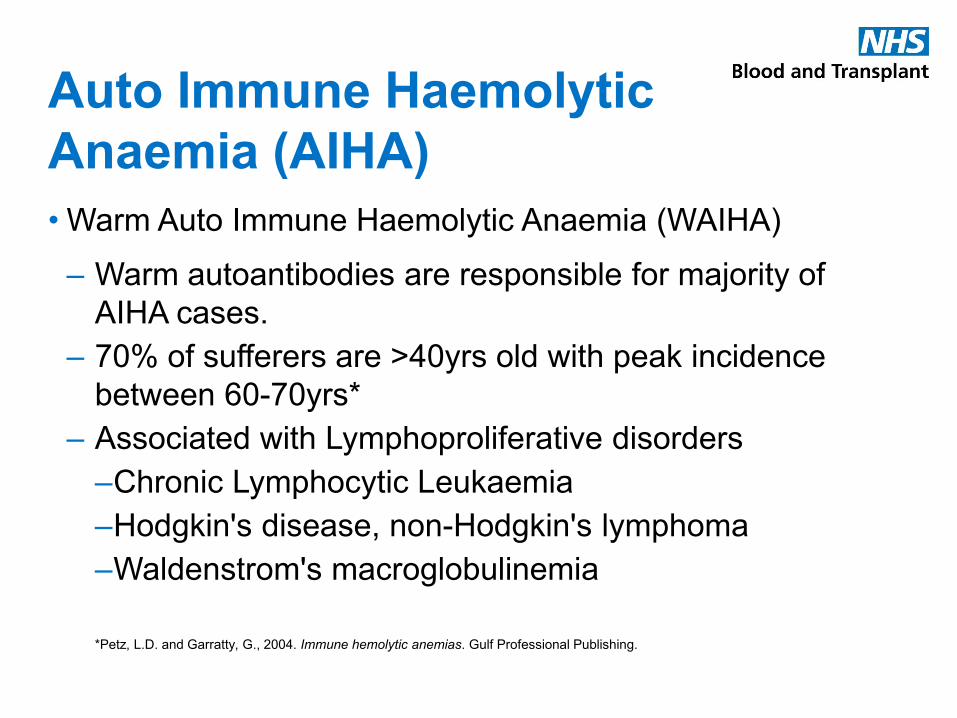
• Warm Auto Immune Haemolytic Anaemia (WAIHA)
– Warm autoantibodies are responsible for majority of
AIHA cases.
– 70% of sufferers are >40yrs old with peak incidence
between 60-70yrs*
– Associated with Lymphoproliferative disorders
–Chronic Lymphocytic Leukaemia
–Hodgkin's disease, non-Hodgkin's lymphoma
–Waldenstrom's macroglobulinemia
*Petz, L.D. and Garratty, G., 2004. Immune hemolytic anemias. Gulf Professional Publishing.
Auto Immune Haemolytic
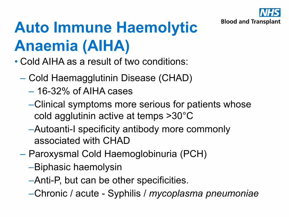
Anaemia (AIHA) • Cold AIHA as a result of two conditions:
– Cold Haemagglutinin Disease (CHAD)
– 16-32% of AIHA cases
–Clinical symptoms more serious for patients whose
cold agglutinin active at temps >30°C
–Autoanti-I specificity antibody more commonly
associated with CHAD
– Paroxysmal Cold Haemoglobinuria (PCH)
–Biphasic haemolysin
–Anti-P, but can be other specificities.
–Chronic / acute - Syphilis / mycoplasma pneumoniae
Auto Immune Haemolytic
Anaemia (AIHA)
• Mixed type AIHA
• Combination of IgG / C3d reactivity
• Rare <5% of AIHA
• Associated conditions:
– Systemic Lupus Erythematosus (SLE)
– Lymphoma
Auto Immune Haemolytic
Anaemia (AIHA)
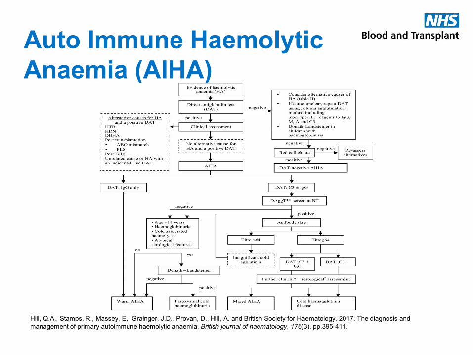
Hill, Q.A., Stamps, R., Massey, E., Grainger, J.D., Provan, D., Hill, A. and British Society for Haematology, 2017. The diagnosis and
management of primary autoimmune haemolytic anaemia. British journal of haematology, 176(3), pp.395-411.
Importance of a clinical history
• Age – AIHA more common in older people, but not exclusive
• Ethnic origin – Could there be an inherited abnormality of RBC
• Diagnosis – Is the AIHA secondary to underlying disease?
• Medications – is it drug mediated? e.g. pencillin
• Brisk haemolysis - How fast is it falling?
• Symptomatic?
• Urgency?
Get the full
picture!
Laboratory testing –
Haematology / Biochemistry
• Tests look for haematological and biochemical indicators of
haemolysis
– Reticulocyte count - increased
– Lactate dehydrogenase (LDH) – may be normal or increased
– Bilirubin – increased
– Haptoglobin – reduced
– Blood film – spherocytes, agglutination or polychromasia
– Urinalysis/dipstick test positive for blood but urine microscopy
negative for red cells - if haemolysis is intravascular
haemoglobinuria rather than haematuria
Laboratory testing - Transfusion • Testing aims to differentiate between what type of AIHA is present, to
group the patient and identify any underlying clinically significant
alloantibodies, in order to provide suitable units for transfusion.
• Results seen in ABO / D grouping / phenotyping and antibody
screening can include:
– Anomalous ABO reverse group due to cold-reacting (IgM)
autoantibody
– Unable to phenotype due to autoantibody (IgG) coating of cells –
false positive reactions
– Panagglutinating antibody, various strengths, temp ranges
depending on type of AIHA
– Positive autologous control (patient’s cells Vs patient’s plasma)
• Testing can be complex and time-consuming, therefore can delay
supply of suitable units for the patient.

Serological Toolkit
Pre-warming
Warm washing
Temperature
Techniques
Reagents
Alloadsorption
Autoadsorption Genotyping
Titration
DAT
Temperature
• Antibodies have different optimal thermal ranges.
• Cold-reacting antibodies are not generally considered to be clinically
significant unless they react above 30°C.
• Testing following pre-warming and warm washing of a sample may
help to remove and cold-reacting autoantibody coating the patient’s
RBC and allow phenotyping.
• Pre-warming plasma may also remove the reactivity of any cold
reacting autoantibody so that panagglutination is not seen in IAT tests
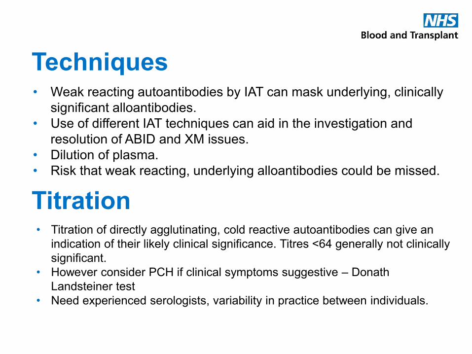
Titration
• Weak reacting autoantibodies by IAT can mask underlying, clinically
significant alloantibodies.
• Use of different IAT techniques can aid in the investigation and
resolution of ABID and XM issues.
• Dilution of plasma.
• Risk that weak reacting, underlying alloantibodies could be missed.
Techniques
• Titration of directly agglutinating, cold reactive autoantibodies can give an
indication of their likely clinical significance. Titres <64 generally not clinically
significant.
• However consider PCH if clinical symptoms suggestive – Donath
Landsteiner test
• Need experienced serologists, variability in practice between individuals.
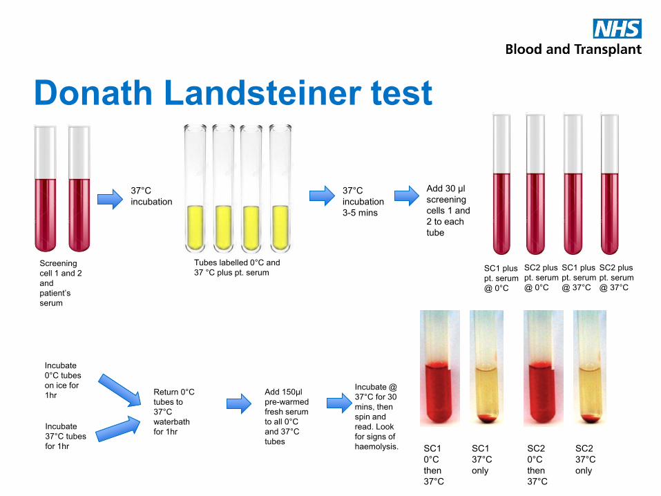
Donath Landsteiner test
Screening
cell 1 and 2
and
patient’s
serum
37°C
incubation
SC1 plus
pt. serum
@ 0°C
37°C
incubation
3-5 mins
Add 30 µl
screening
cells 1 and
2 to each
tube
Tubes labelled 0°C and
37 °C plus pt. serum SC2 plus
pt. serum
@ 0°C
SC1 plus
pt. serum
@ 37°C
SC2 plus
pt. serum
@ 37°C
Incubate
0°C tubes
on ice for
1hr
Incubate
37°C tubes
for 1hr
Return 0°C
tubes to
37°C
waterbath
for 1hr
Add 150µl
pre-warmed
fresh serum
to all 0°C
and 37°C
tubes SC1
0°C
then
37°C
SC1
37°C
only
SC2
0°C
then
37°C
SC2
37°C
only
Incubate @
37°C for 30
mins, then
spin and
read. Look
for signs of
haemolysis.
• For patients with WAIHA it may be possible to use directly agglutinating
reagents to obtain a phenotype
- Not all antisera available as directly agglutinating reagents
- If patient has been transfused in past 3 months phenotyping result
not reliable due to presence of transfused cells
- Genotyping
• Chloroquine Diphosphate treatment can be used to remove bound
antibody and phenotype
- Antigens destroyed by CDP treatment include
- Availability of genotyping has led to the decline of CDP treatment in
many reference labs
• 0.01M DTT treatment used to abolish activity of cold-reacting IgM
antibodies to permit phenotyping, typically only for ABO and Rh.
Reagents
Direct Antiglobulin Test (DAT)
• Direct Antiglobulin Test (DAT) looks for what is coating the cells in vivo
• Must include monospecific testing for IgG and C3d as a minimum.
– Warm type (IgG or sometimes IgA)
– Cold type (IgM / C3d)
– Mixed type (IgG / C3d)
• Take clinical history into account when interpreting results
– Recent upper respiratory tract / viral infection - PCH
– Malignant disorders – CLL, Lymphoma
– Post transplant
– Transfusion history
• Positive DAT does not necessarily indicate AIHA
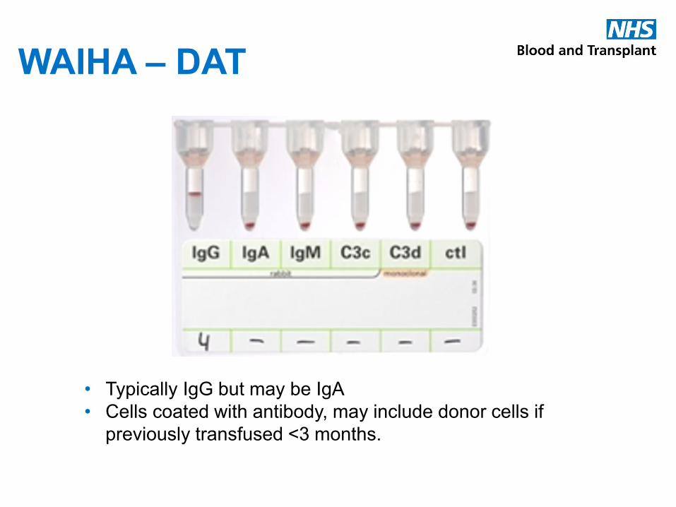
WAIHA – DAT
• Typically IgG but may be IgA
• Cells coated with antibody, may include donor cells if
previously transfused <3 months.
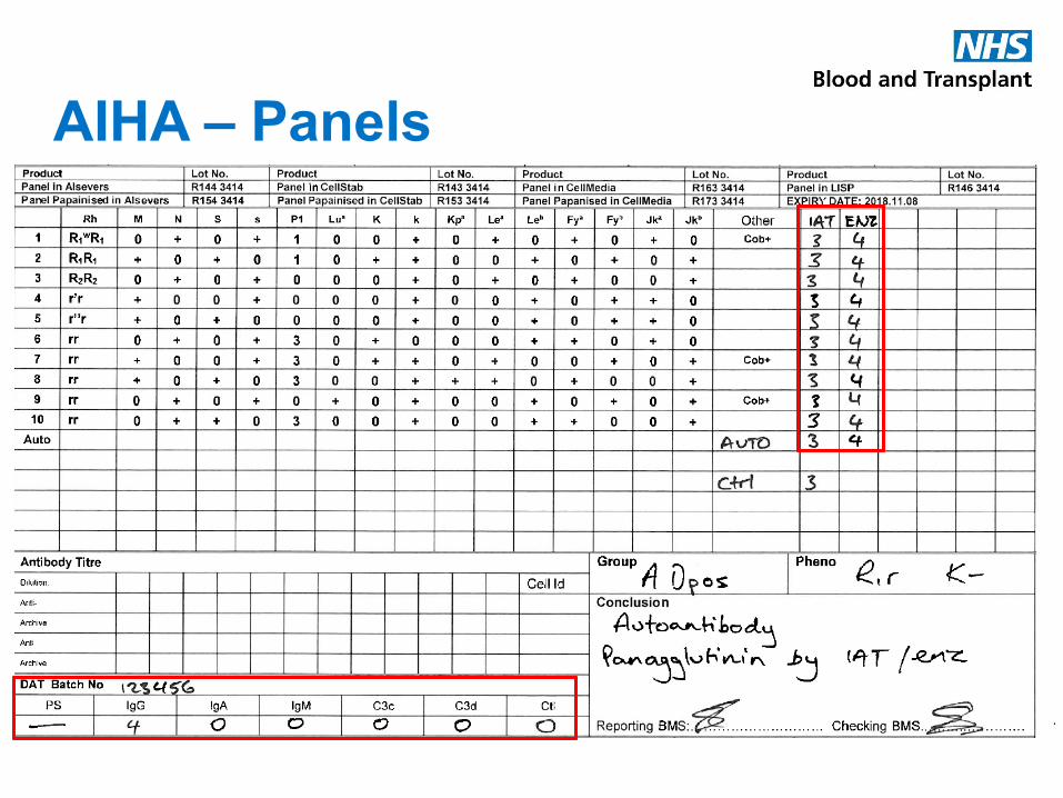
AIHA – Panels
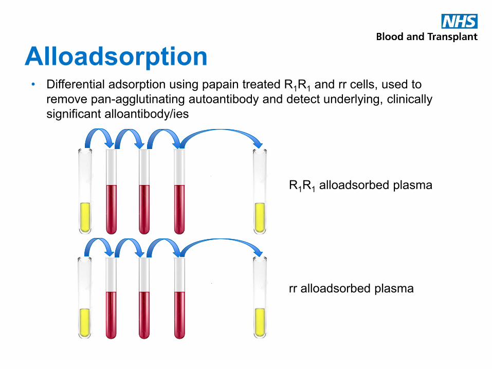
Alloadsorption • Differential adsorption using papain treated R1R1 and rr cells, used to
remove pan-agglutinating autoantibody and detect underlying, clinically
significant alloantibody/ies
R1R1 alloadsorbed plasma
rr alloadsorbed plasma
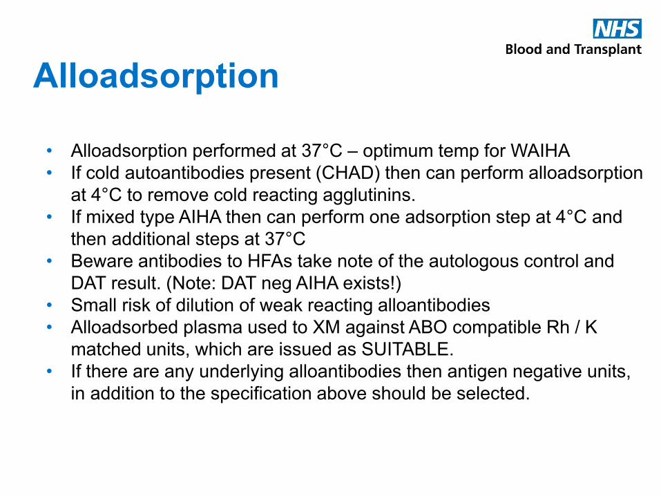
Alloadsorption
• Alloadsorption performed at 37°C – optimum temp for WAIHA
• If cold autoantibodies present (CHAD) then can perform alloadsorption
at 4°C to remove cold reacting agglutinins.
• If mixed type AIHA then can perform one adsorption step at 4°C and
then additional steps at 37°C
• Beware antibodies to HFAs take note of the autologous control and
DAT result. (Note: DAT neg AIHA exists!)
• Small risk of dilution of weak reacting alloantibodies
• Alloadsorbed plasma used to XM against ABO compatible Rh / K
matched units, which are issued as SUITABLE.
• If there are any underlying alloantibodies then antigen negative units,
in addition to the specification above should be selected.

AIHA – Panels
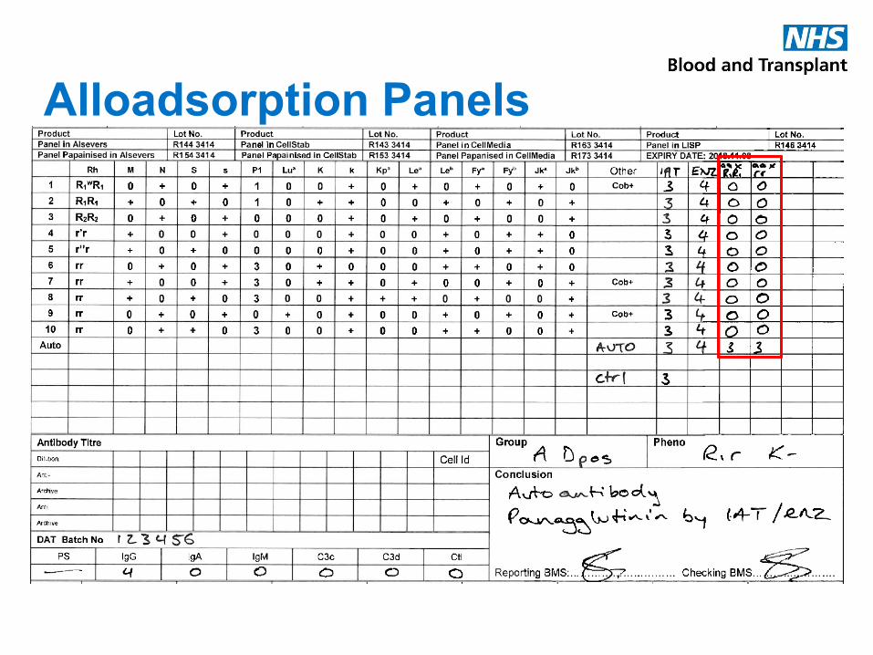
Alloadsorption Panels
Alloadsorption Panels
negative
Autoadsorption • It is possible to perform autoadsorption, but in WAIHA cases
this is not always the best option because:
- Autoantibody coating the RBC may have a blocking effect
• Use a mixture of 1% Papain / 0.2M DTT (ZZAP treatment) to
dissociate bound autoantibody prior to autoadsorption.
- ZZAP treatment removes Kell system, MNSs, Fya and Fyb
antigens
• Not possible to perform autoadsorption if the patient has
been transfused in the past 3 months – risk of adsorbing
alloabs
• In addition, quite often these patients have a low Hb due to
their clinical condition and therefore a low HCT, which does
not allow for sufficient cells to undertake autoadsorption.
Genotyping
• Genotyping can be useful in the following circumstances:
– DAT+ cases when extended typing is required to inform blood selection or alloantibody exclusion, especially where alloadsorption has been unsuccessful
– Previously transfused patients when extended typing is required to inform exclusion of additional specificities
NOTE: Fully genotyped matched blood is rarely required, does not significantly reduce alloimmunisation beyond full Rh / K matching / matching for antigens where there is an associated alloantibody and is not sustainable for large cohorts of the UK population.
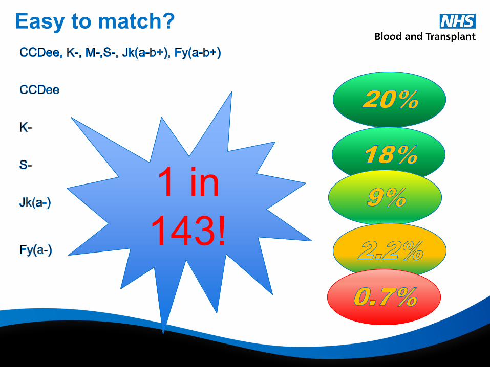
Easy to match?
1 in
143!
1 in
143!
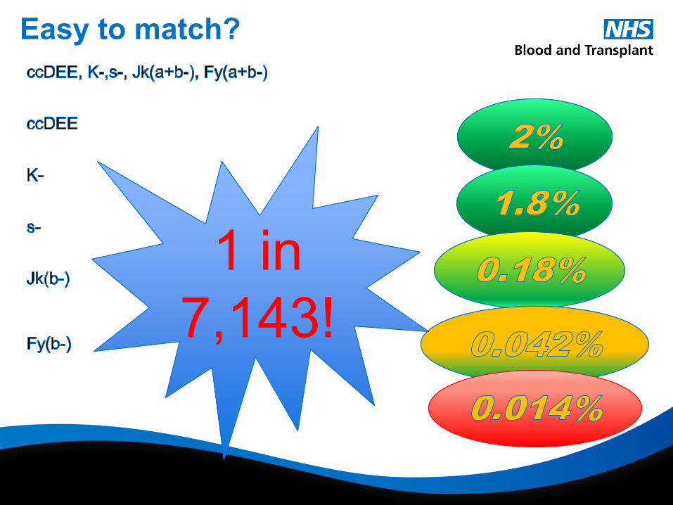
Easy to match?
1 in
7,143!
1 in
7,143!
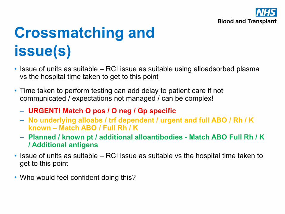
Crossmatching and
issue(s) • Issue of units as suitable – RCI issue as suitable using alloadsorbed plasma
vs the hospital time taken to get to this point
• Time taken to perform testing can add delay to patient care if not communicated / expectations not managed / can be complex!
– URGENT! Match O pos / O neg / Gp specific
– No underlying alloabs / trf dependent / urgent and full ABO / Rh / K known – Match ABO / Full Rh / K
– Planned / known pt / additional alloantibodies - Match ABO Full Rh / K / Additional antigens
• Issue of units as suitable – RCI issue as suitable vs the hospital time taken to get to this point
• Who would feel confident doing this?
Communication and
collaboration
• Ensure best quality care and treatment – collaboration and communication between scientists and clinical staff
• Let clinical staff know where blood provision may be delayed because of a complex serological picture
• Advise on blood provision in the interim, should blood be required sooner
• Advise on blood provision in an emergency
• Ensure staff know what to do in each scenario when a panagglutinin is present
Thank you