Embed Size (px)
Citation preview
Anatomy of Larynx‘the organ of voice’
Presenter : Kanato T
INTRODUCTION:
The larynx is an air passage, a sphincter and an organ of phonation.
Generation of intrathrocic pressure for coughing and lifting.
Extends from the tongue to the trachea It is mobile on deglutition Understanding of basic laryngeal anatomy is
must for all ENT surgeon for Surgery & route of cancer spread.

Larynx(lar´inks)- ‘the organ of voice’
General Description Embryology and development Cartilages Laryngeal joints Ligaments & Muscles ( Extrinsic and Intrinsic) Mucous membrane Cavity of larynx Spaces Nerve supply Blood supply & Lymphatic drainage Comparative anatomy ( infant Vs adult)
General Description.
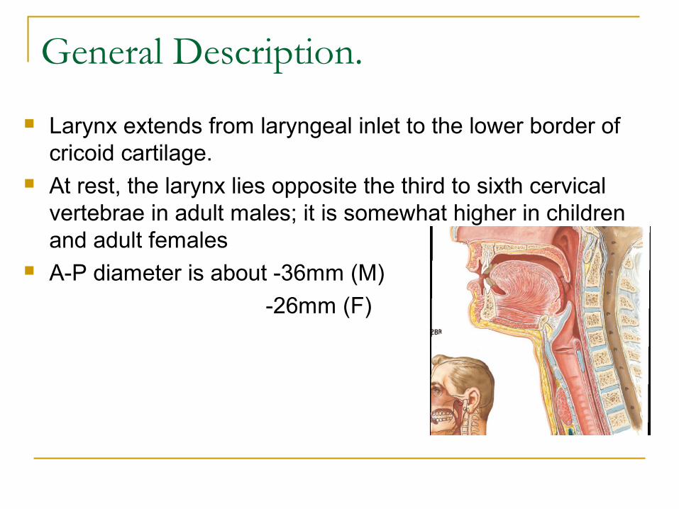
Larynx extends from laryngeal inlet to the lower border of cricoid cartilage.
At rest, the larynx lies opposite the third to sixth cervical vertebrae in adult males; it is somewhat higher in children and adult females
A-P diameter is about -36mm (M)
-26mm (F)
Laryngeal framework.
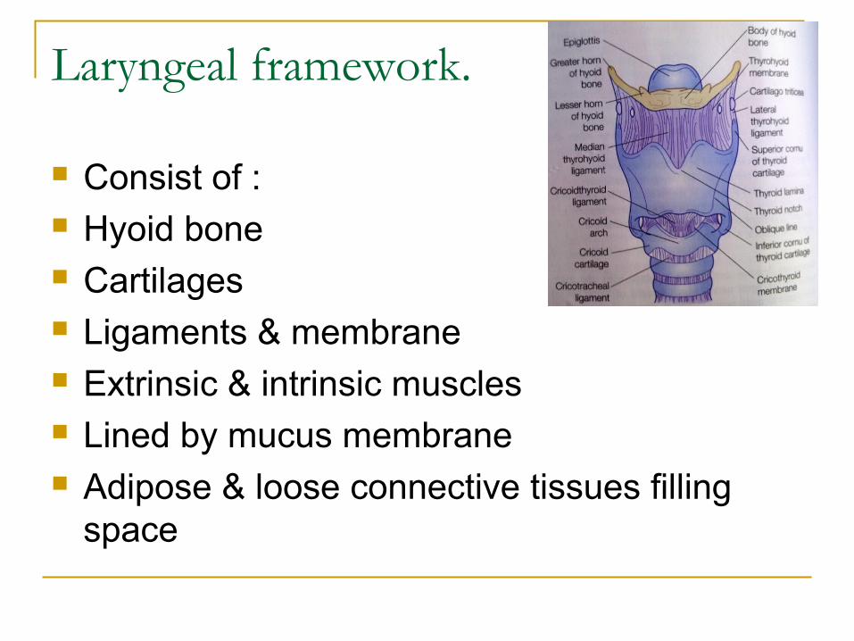
Consist of : Hyoid bone Cartilages Ligaments & membrane Extrinsic & intrinsic muscles Lined by mucus membrane Adipose & loose connective tissues filling
space

General principles of development The development of the larynx can be divided into
prenatal and postnatal stages.
At birth, the larynx is located high in the neck between the C1 and C4 vertebrae, allowing concurrent breathing or vocalization and deglutition.
By age 2 years, the larynx descends inferiorly; by age 6 years, it reaches the adult position between C4 and C7 vertebrae. This new position provides a greater range of phonation (because of the wider supraglottic pharynx) at the expense of losing this separation of function, i.e., deglutition and breathing.
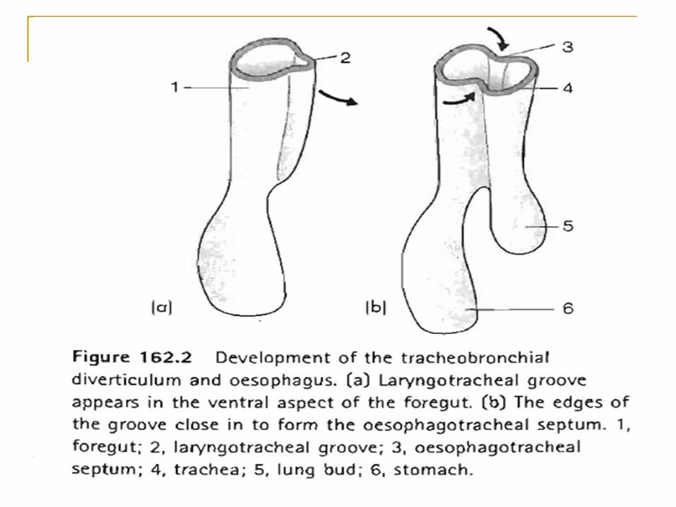
Embryology The larynx develops from the endodermal lining and the
adjacent mesenchyme of the foregut between the fourth and sixth branchial arches.
At 20 days' gestation, the foregut is first identifiable with a ventral laryngotracheal groove. It continues to deepen until its lateral edges fuse.
Trachea becomes separated from the esophagus by the tracheoesophageal septum with a persistent slit like opening into the pharynx
This fusion occurs in the caudal-to-cranial direction, and incomplete fusion results in development of persistent communication between the larynx or trachea and the esophagus
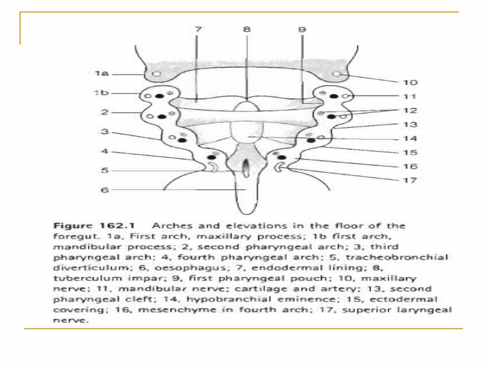
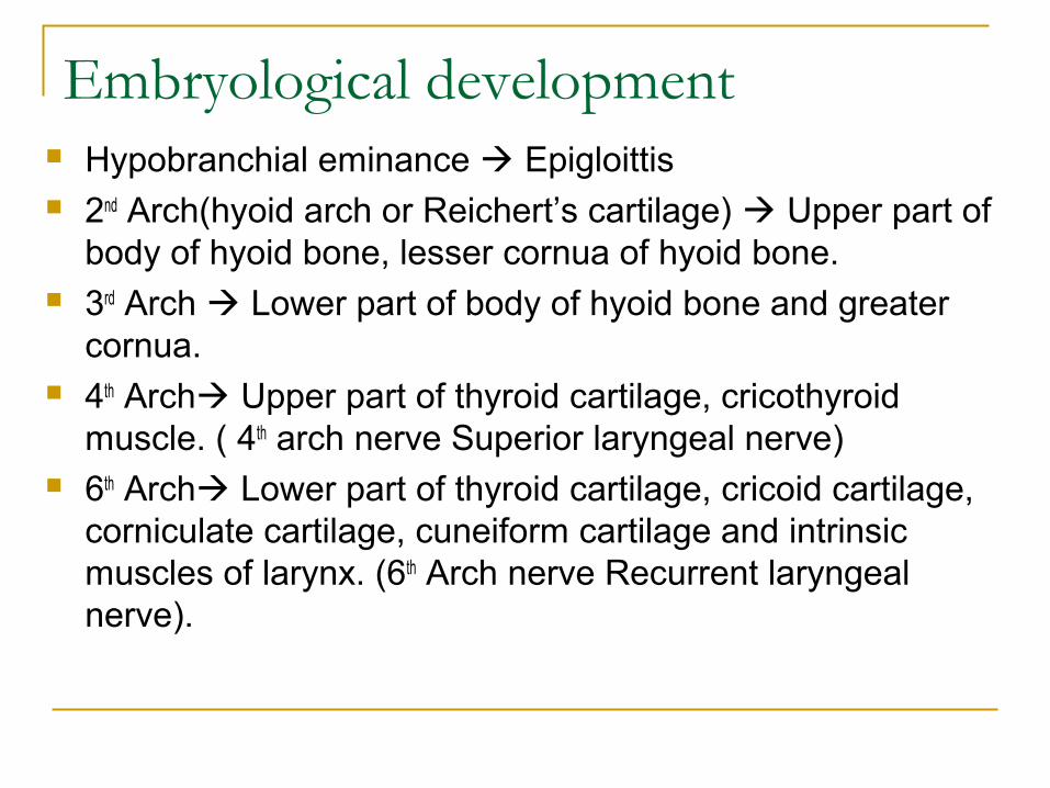
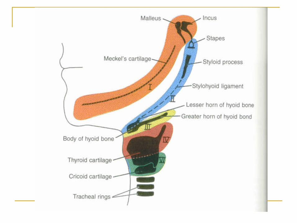
Embryological development Hypobranchial eminance Epigloittis 2nd Arch(hyoid arch or Reichert’s cartilage) Upper part of
body of hyoid bone, lesser cornua of hyoid bone. 3rd Arch Lower part of body of hyoid bone and greater
cornua. 4th Arch Upper part of thyroid cartilage, cricothyroid
muscle. ( 4th arch nerve Superior laryngeal nerve) 6th Arch Lower part of thyroid cartilage, cricoid cartilage,
corniculate cartilage, cuneiform cartilage and intrinsic muscles of larynx. (6th Arch nerve Recurrent laryngeal nerve).
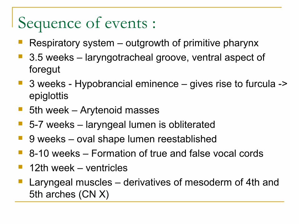
Sequence of events : Respiratory system – outgrowth of primitive pharynx 3.5 weeks – laryngotracheal groove, ventral aspect of
foregut 3 weeks - Hypobrancial eminence – gives rise to furcula ->
epiglottis 5th week – Arytenoid masses 5-7 weeks – laryngeal lumen is obliterated 9 weeks – oval shape lumen reestablished 8-10 weeks – Formation of true and false vocal cords 12th week – ventricles Laryngeal muscles – derivatives of mesoderm of 4th and
5th arches (CN X)
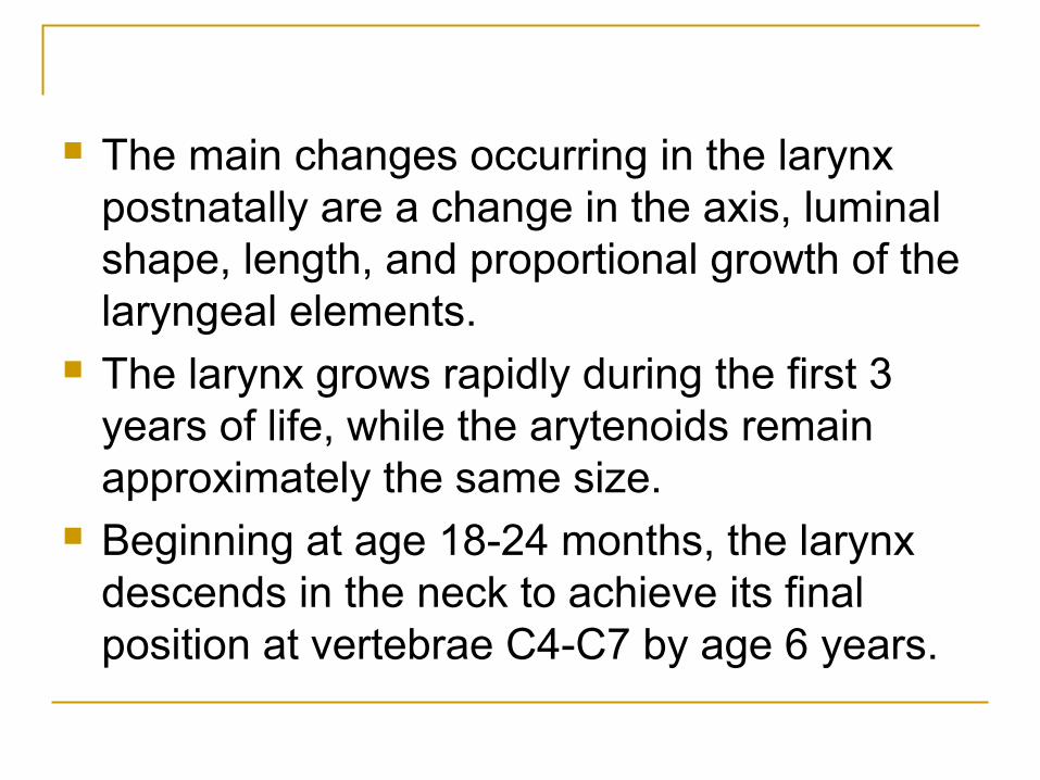
The main changes occurring in the larynx postnatally are a change in the axis, luminal shape, length, and proportional growth of the laryngeal elements.
The larynx grows rapidly during the first 3 years of life, while the arytenoids remain approximately the same size.
Beginning at age 18-24 months, the larynx descends in the neck to achieve its final position at vertebrae C4-C7 by age 6 years.
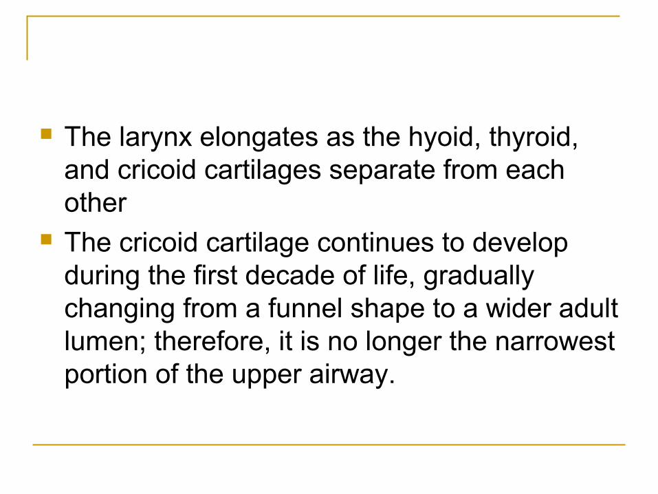
The larynx elongates as the hyoid, thyroid, and cricoid cartilages separate from each other
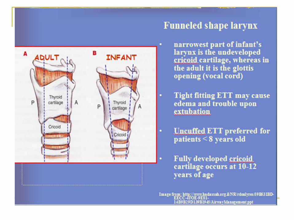
The cricoid cartilage continues to develop during the first decade of life, gradually changing from a funnel shape to a wider adult lumen; therefore, it is no longer the narrowest portion of the upper airway.
Congenital Anomalies
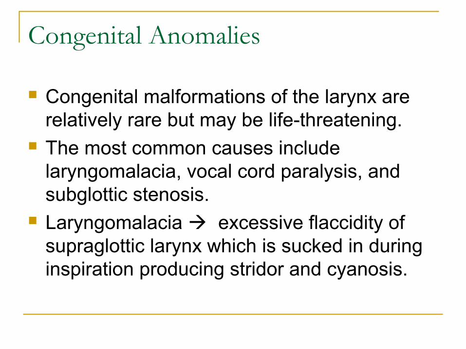
Congenital malformations of the larynx are relatively rare but may be life-threatening.
The most common causes include laryngomalacia, vocal cord paralysis, and subglottic stenosis.
Laryngomalacia excessive flaccidity of supraglottic larynx which is sucked in during inspiration producing stridor and cyanosis.
Congenital Anomalies Laryngeal atresia occurs if the endolarynx fails
to recanalize. Immediate tracheotomy is required for survival.
Laryngeal webs occur when the epithelium partially fails to resorb. A weblike mass may appear at the glottic level, often with significant subglottic extension.
Subglottic stenosis is a deformity in the development of the normal cricoid cartilage (sixth branchial arch).
Laryngotracheal cleft results from a failure to form the tracheoesophageal septum.
Hyoid bone
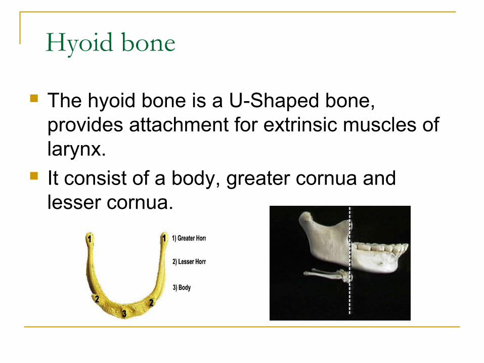
The hyoid bone is a U-Shaped bone, provides attachment for extrinsic muscles of larynx.
It consist of a body, greater cornua and lesser cornua.
Laryngeal Cartilages
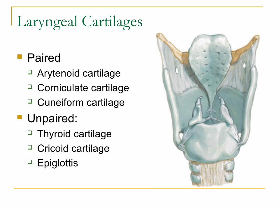
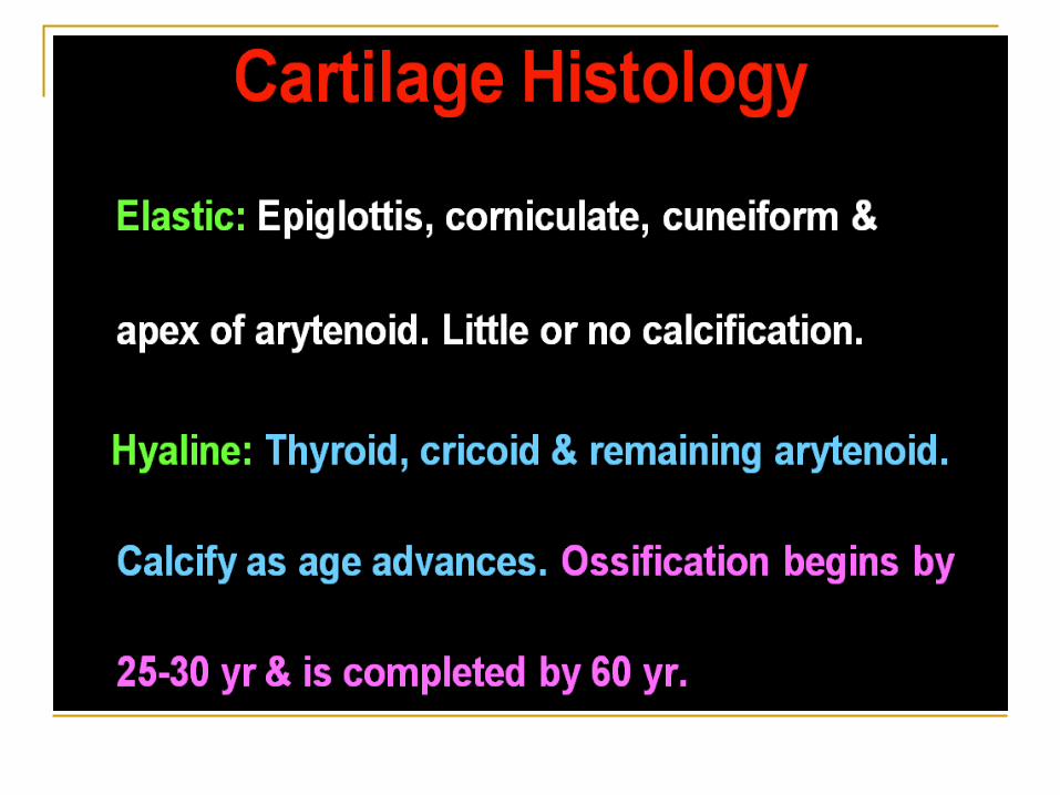
Paired Arytenoid cartilage Corniculate cartilage Cuneiform cartilage
Unpaired: Thyroid cartilage Cricoid cartilage Epiglottis
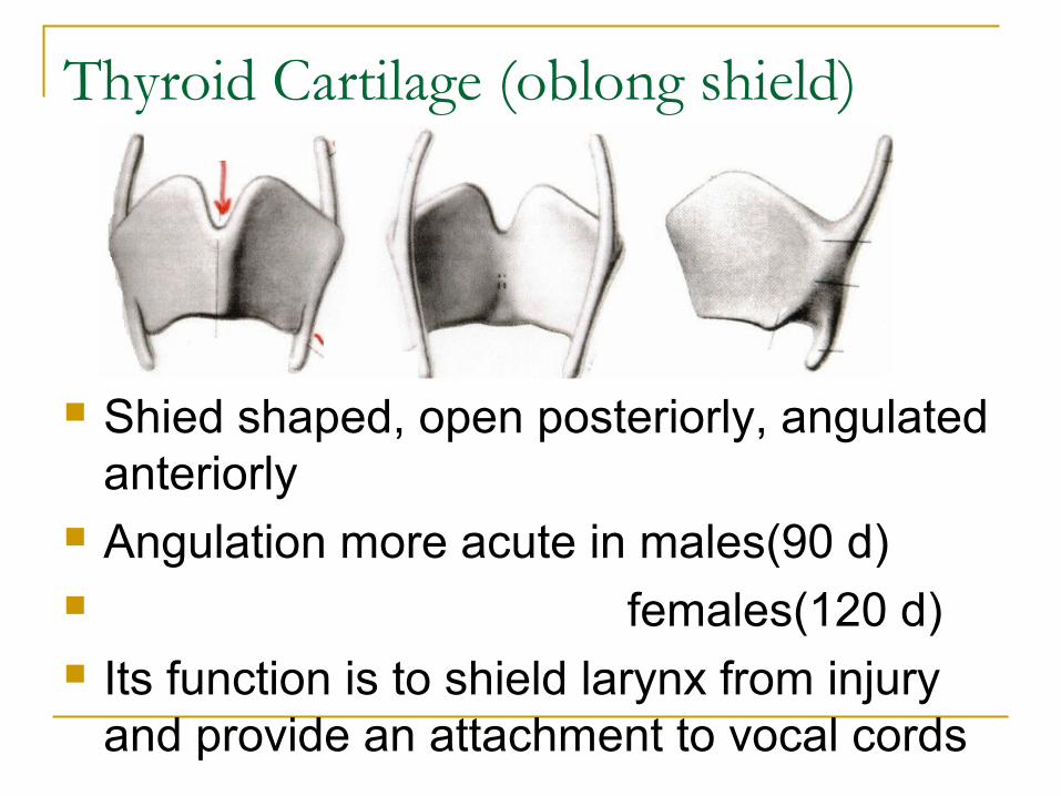
Thyroid Cartilage (oblong shield)
Shied shaped, open posteriorly, angulated anteriorly
Angulation more acute in males(90 d) females(120 d) Its function is to shield larynx from injury
and provide an attachment to vocal cords
This cartilage has 2 alae/wing which meet anteriorly, they form a depression called the THYROID NOTCH before meeting at the protruberance of the Adam’s apple or laryngeal prominence.
Posterior border of each lamina prolonged above and below to formed superior & inferior cornu
Superior cornu-Lateral thyroid ligament attached Inferior cornu- Articulate with cricoid cartilage Ossifies at 20-30 years of age, begins in the
inferior margin and progress cranially
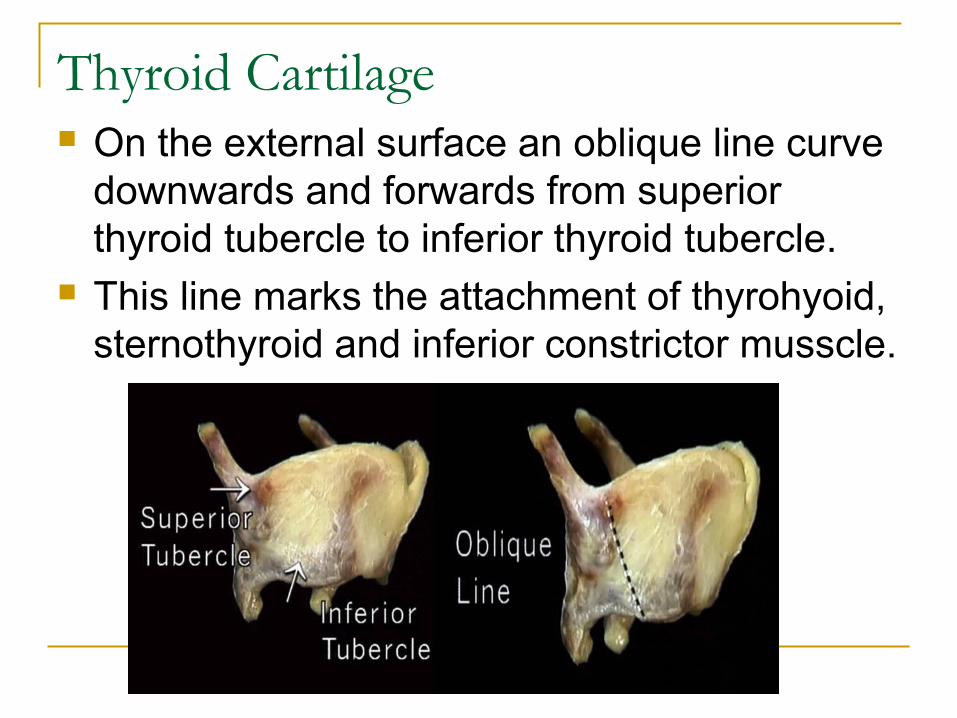
Thyroid Cartilage On the external surface an oblique line curve
downwards and forwards from superior thyroid tubercle to inferior thyroid tubercle.
This line marks the attachment of thyrohyoid, sternothyroid and inferior constrictor musscle.
Inner aspect of thyroid cartilage
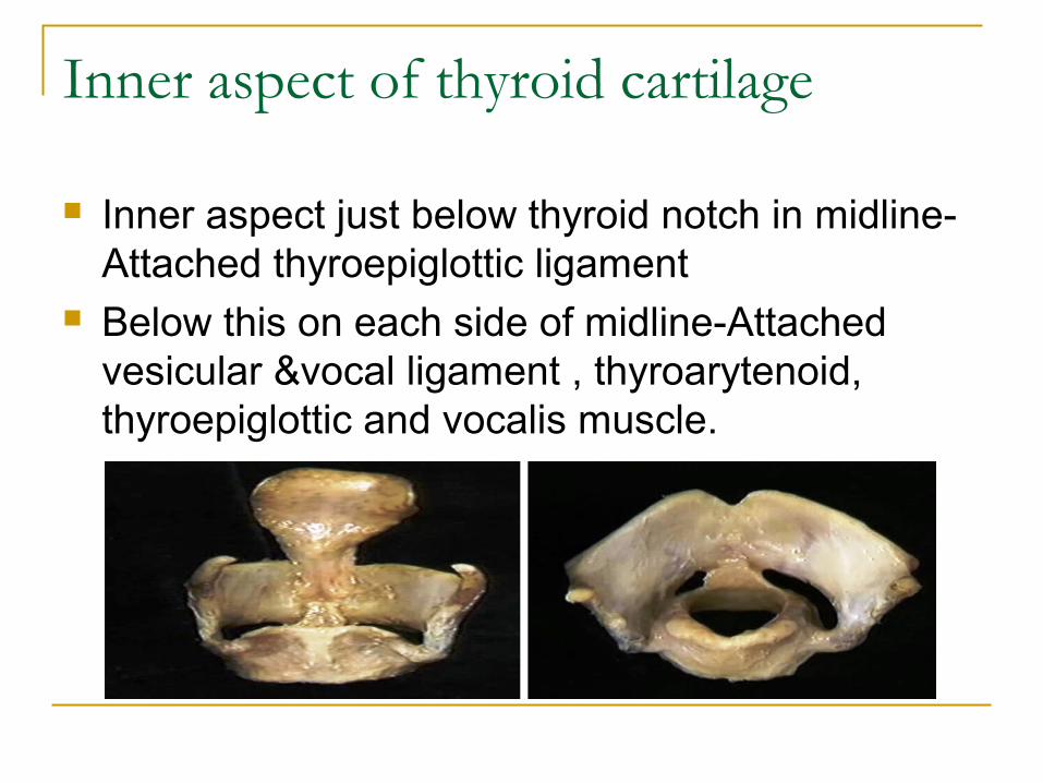
Inner aspect just below thyroid notch in midline- Attached thyroepiglottic ligament
Below this on each side of midline-Attached vesicular &vocal ligament , thyroarytenoid, thyroepiglottic and vocalis muscle.
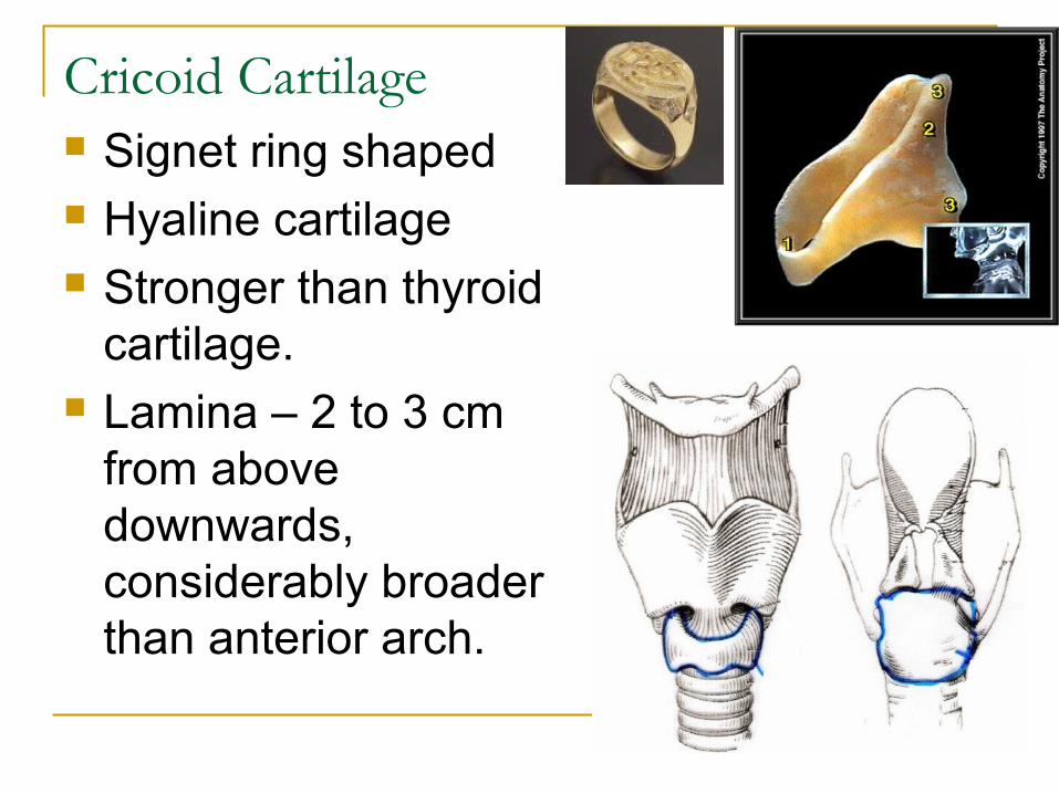
Cricoid Cartilage Signet ring shaped Hyaline cartilage Stronger than thyroid
cartilage. Lamina – 2 to 3 cm
from above downwards, considerably broader than anterior arch.
5.Lamina – flat portion of the ring located posteriorly and extends upward to form the POSTERIOR border of the larynxLevel: Adult: C6-C7 Children: C3-C4Posterolaterally, cricoid articulates w/ Inferior cornu of the thyroid cartilage, which forms true synovial joints (permit a ROCKING action of the cricoid cartilage on the thyroid cartilage and a slight anteroposterior SLIDING motion (cricoid cart. Supports the 2 arytenoid cartilages on posterosuperior aspect)
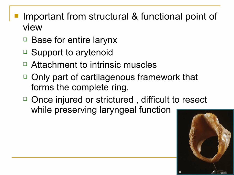
Important from structural & functional point of view Base for entire larynx Support to arytenoid Attachment to intrinsic muscles Only part of cartilagenous framework that
forms the complete ring. Once injured or strictured , difficult to resect
while preserving laryngeal function
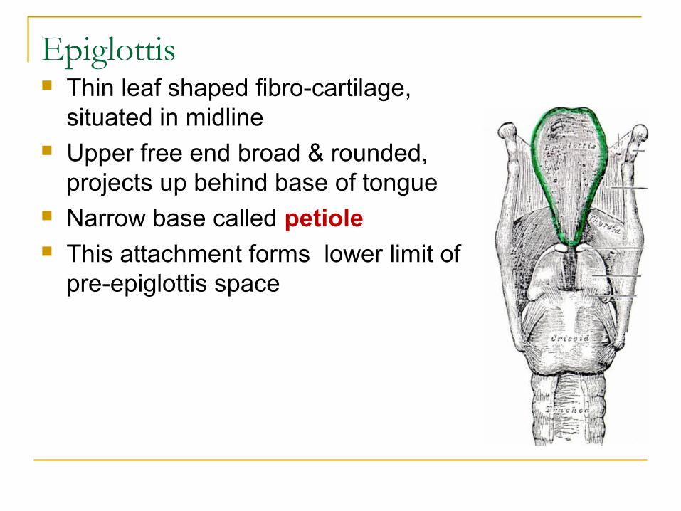
Epiglottis Thin leaf shaped fibro-cartilage,
situated in midline Upper free end broad & rounded,
projects up behind base of tongue Narrow base called petiole This attachment forms lower limit of
pre-epiglottis space
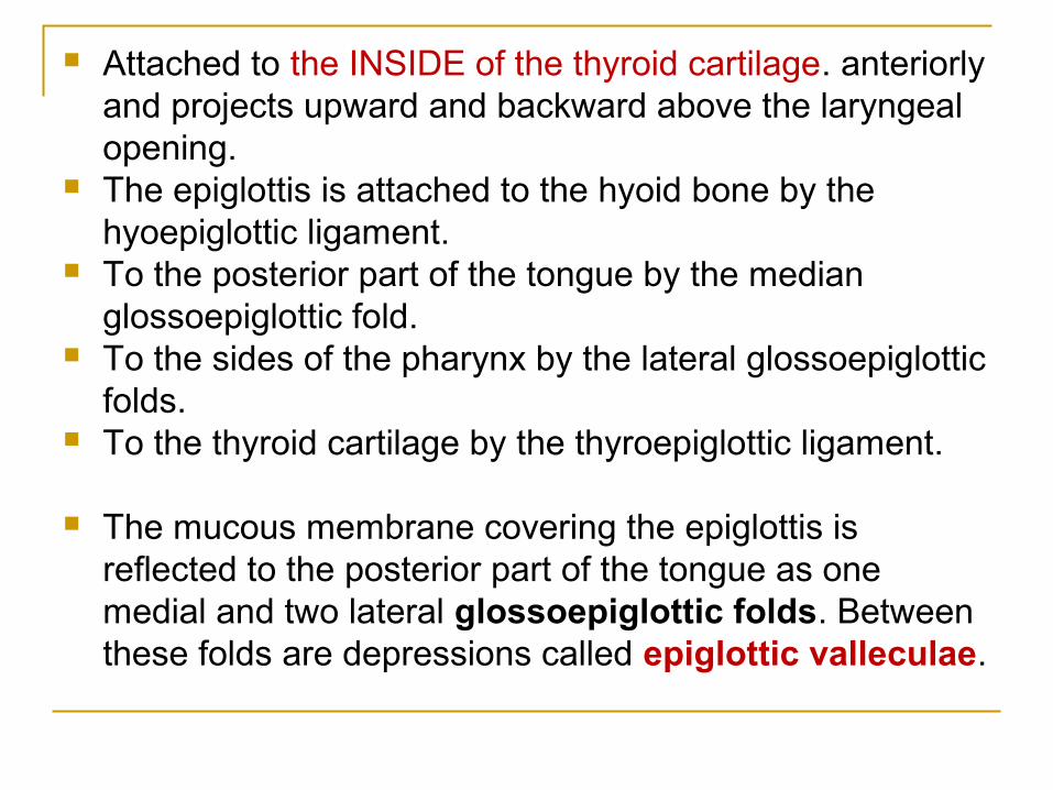
Attached to the INSIDE of the thyroid cartilage. anteriorly and projects upward and backward above the laryngeal opening.
The epiglottis is attached to the hyoid bone by the hyoepiglottic ligament.
To the posterior part of the tongue by the median glossoepiglottic fold.
To the sides of the pharynx by the lateral glossoepiglottic folds.
To the thyroid cartilage by the thyroepiglottic ligament.
The mucous membrane covering the epiglottis is reflected to the posterior part of the tongue as one medial and two lateral glossoepiglottic folds. Between these folds are depressions called epiglottic valleculae.
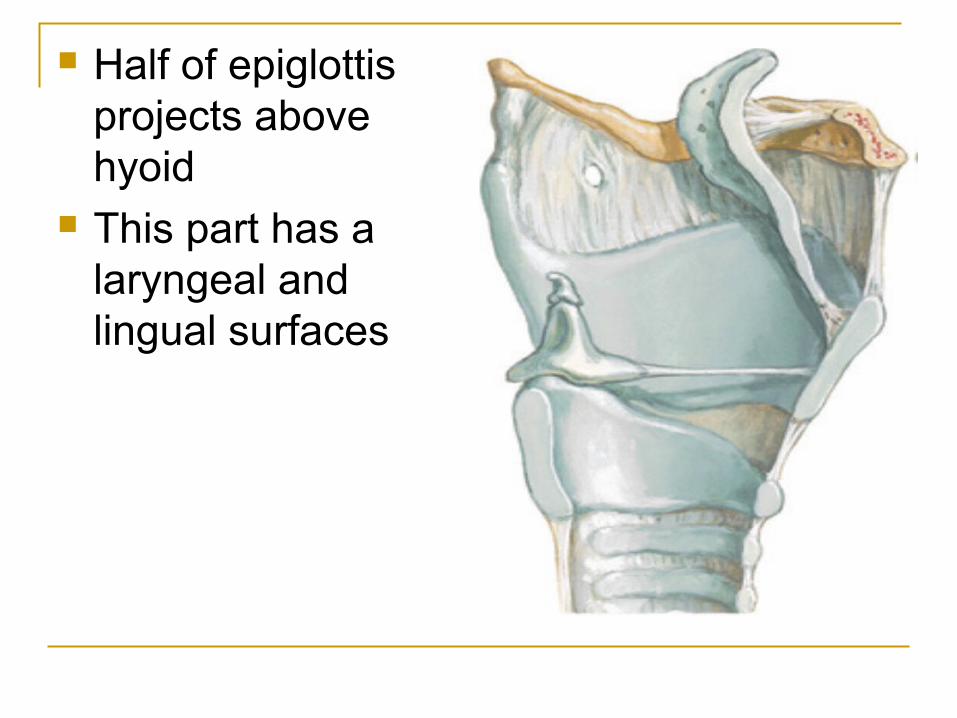
Half of epiglottis projects above hyoid
This part has a laryngeal and lingual surfaces
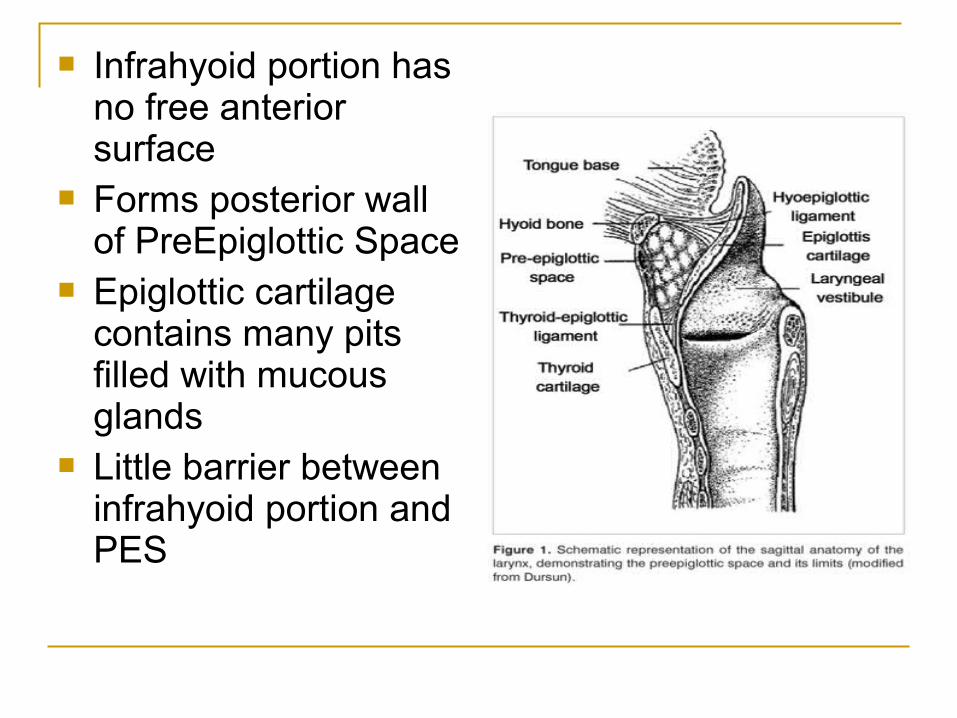
Infrahyoid portion has no free anterior surface
Forms posterior wall of PreEpiglottic Space
Epiglottic cartilage contains many pits filled with mucous glands
Little barrier between infrahyoid portion and PES
Applied anatomy
Most of mucosal surface of supraglottic region covers epiglottis thus majority of supraglottic tumour are epiglottic
Epiglottic cartilage contain pits lacunae filled with mucous gland thus providing less cartilaginous barrier between infrahyoid portion of epiglottis & pre-epiglottic space (Tendency of spread more in infrahyoid tumor)
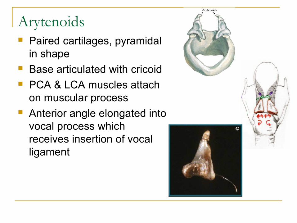
Arytenoids Paired cartilages, pyramidal
in shape Base articulated with cricoid PCA & LCA muscles attach
on muscular process Anterior angle elongated into
vocal process which receives insertion of vocal ligament
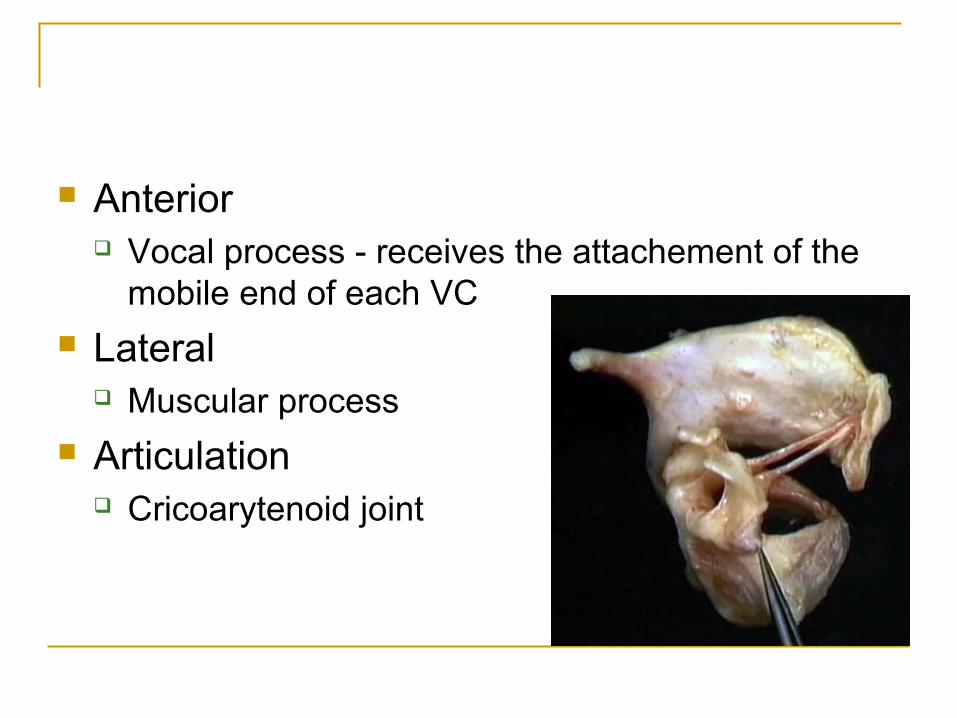
Anterior Vocal process - receives the attachement of the
mobile end of each VC
Lateral Muscular process
Articulation Cricoarytenoid joint
Corniculate Cartilages
Fibroelastic Cartilages of Santorini Small cartilages above the arytenoid and in
the aryepiglottic folds
Cuneiform Cartilages
Firboelastic cartilages Cartilages of Wrisberg Elongated pieces of small yellow elastic
cartilage in the aryepiglottic folds
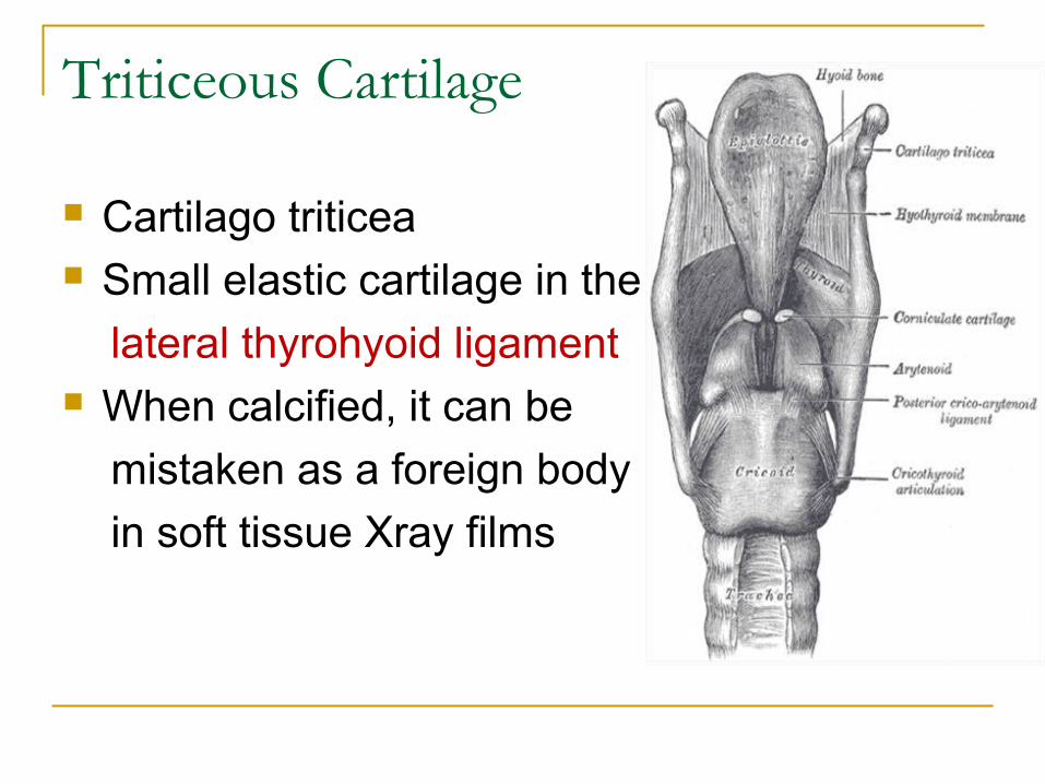
Triticeous Cartilage
Cartilago triticea Small elastic cartilage in the
lateral thyrohyoid ligament When calcified, it can be
mistaken as a foreign body
in soft tissue Xray films
Laryngeal Joints
Cricothyroid Joint Between inferior
cornu of the thyroid cartilage and facet on the cricoid cartilage at the junction of the arch and lamina
Two movements: Rotation Gliding
Cricoarytenoid Joint Between the base of
the arytenoid cartilage and the facet on the upper border of the lamina of the cricoid cartilage
Two movements: Rotation Gliding
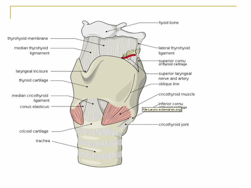
Ligament & membrane of larynx Extrinsic ligaments/Membranes: Connect
laryngeal cartilages to hyoid bone above & trachea below
Thyrohyhoid membrane Cricothyroid membrane Cricotracheal membrane Hyoepigloittic ligament Intrinsic ligaments/Membranes: Connect
laryngeal cartilage together, Forming internal framework of larynx
Crico-vocal membrane (Conus elasticus) Quadrangular membrane
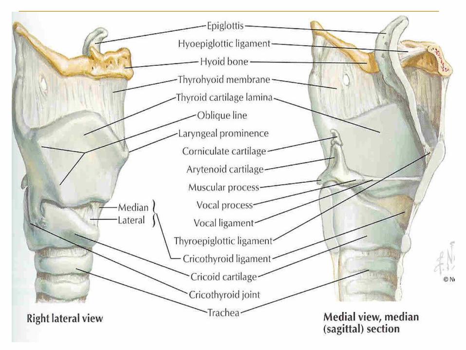
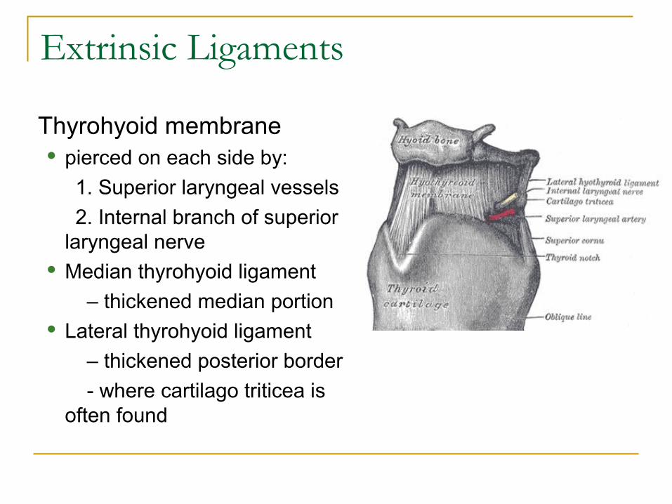
Extrinsic Ligaments
Thyrohyoid membrane pierced on each side by:
1. Superior laryngeal vessels
2. Internal branch of superior laryngeal nerve
Median thyrohyoid ligament
– thickened median portion Lateral thyrohyoid ligament
– thickened posterior border
- where cartilago triticea is often found
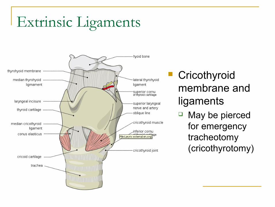
Extrinsic Ligaments
Cricothyroid membrane and ligaments May be pierced
for emergency tracheotomy (cricothyrotomy)
Extrinsic Ligaments
Cricotracheal Ligament Attaches the cricoid cartilage to the first attached
ring
Hyoepiglottis It connects the epiglottic cartilage to hyoid bone.
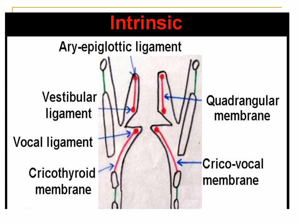
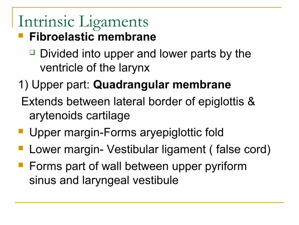
Intrinsic Ligaments Fibroelastic membrane
Divided into upper and lower parts by the ventricle of the larynx
1) Upper part: Quadrangular membrane
Extends between lateral border of epiglottis & arytenoids cartilage
Upper margin-Forms aryepiglottic fold Lower margin- Vestibular ligament ( false cord) Forms part of wall between upper pyriform
sinus and laryngeal vestibule
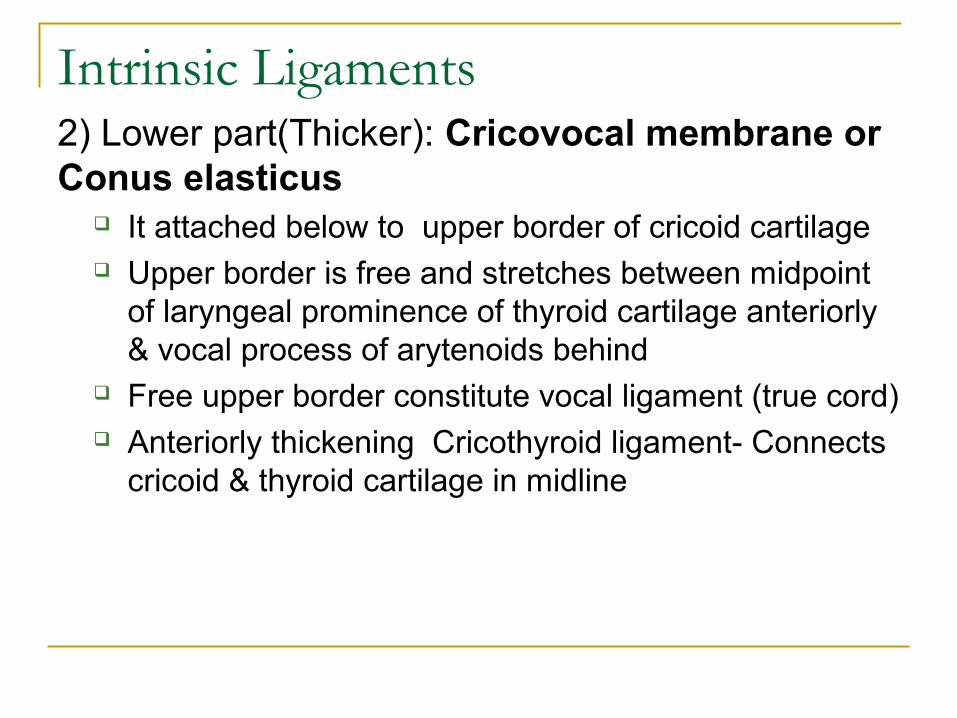
Intrinsic Ligaments2) Lower part(Thicker): Cricovocal membrane or Conus elasticus
It attached below to upper border of cricoid cartilage Upper border is free and stretches between midpoint
of laryngeal prominence of thyroid cartilage anteriorly & vocal process of arytenoids behind
Free upper border constitute vocal ligament (true cord) Anteriorly thickening Cricothyroid ligament- Connects
cricoid & thyroid cartilage in midline
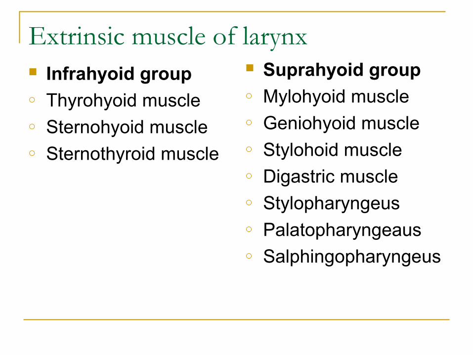
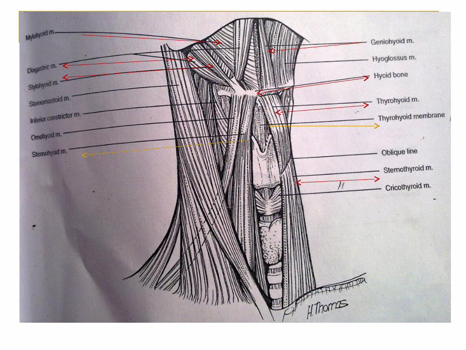
Extrinsic muscle of larynx Infrahyoid groupo Thyrohyoid muscleo Sternohyoid muscleo Sternothyroid muscle
Suprahyoid groupo Mylohyoid muscleo Geniohyoid muscleo Stylohoid muscle o Digastric muscleo Stylopharyngeuso Palatopharyngeauso Salphingopharyngeus
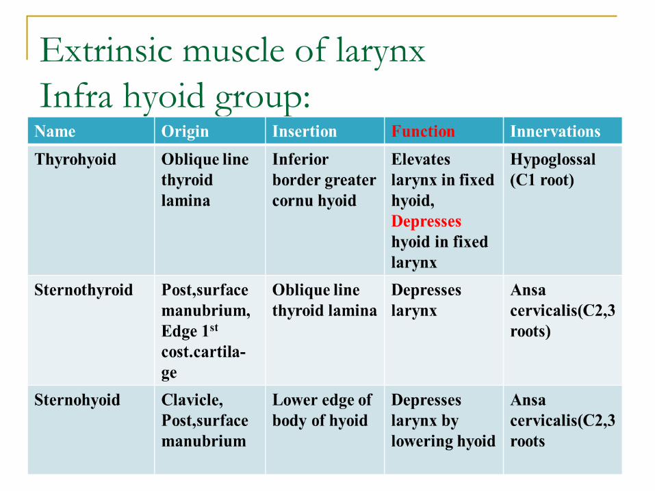
Extrinsic muscle of larynxInfra hyoid group:
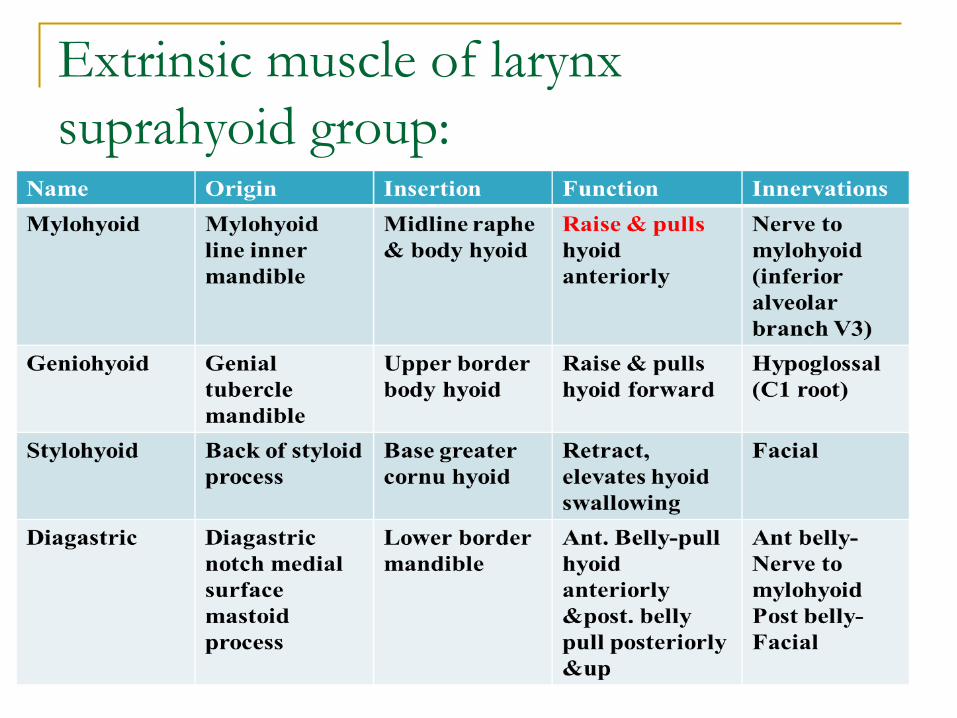
Extrinsic muscle of larynxsuprahyoid group:
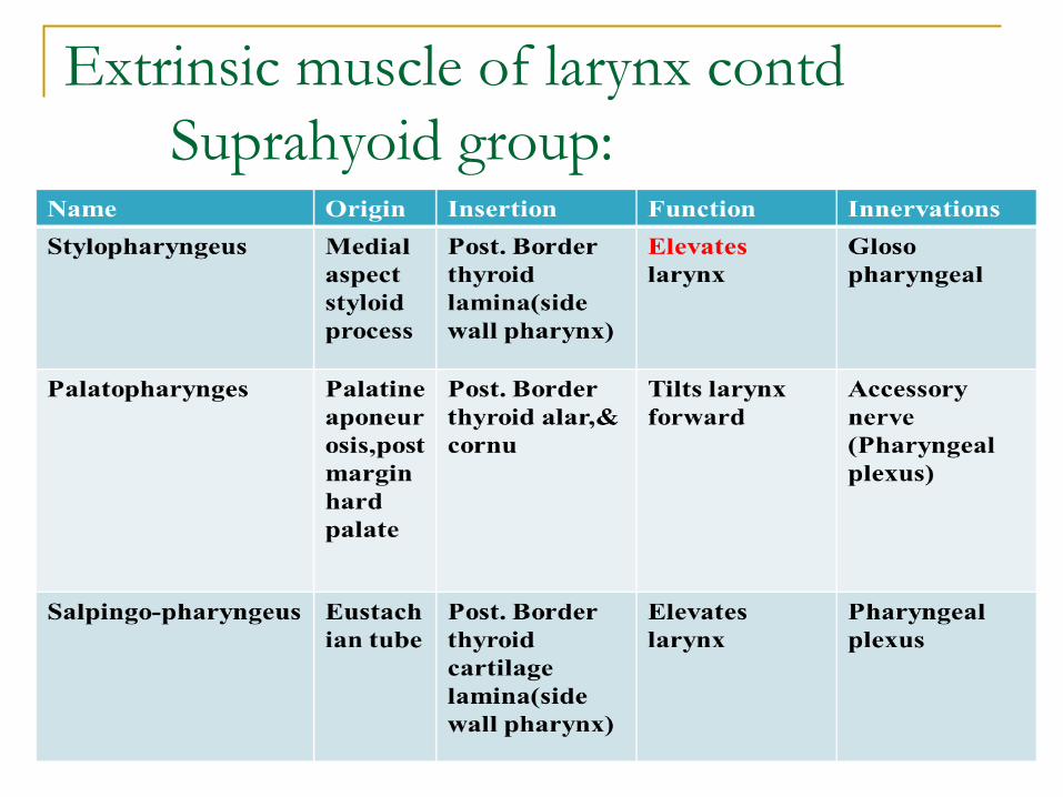
Extrinsic muscle of larynx contdSuprahyoid group:
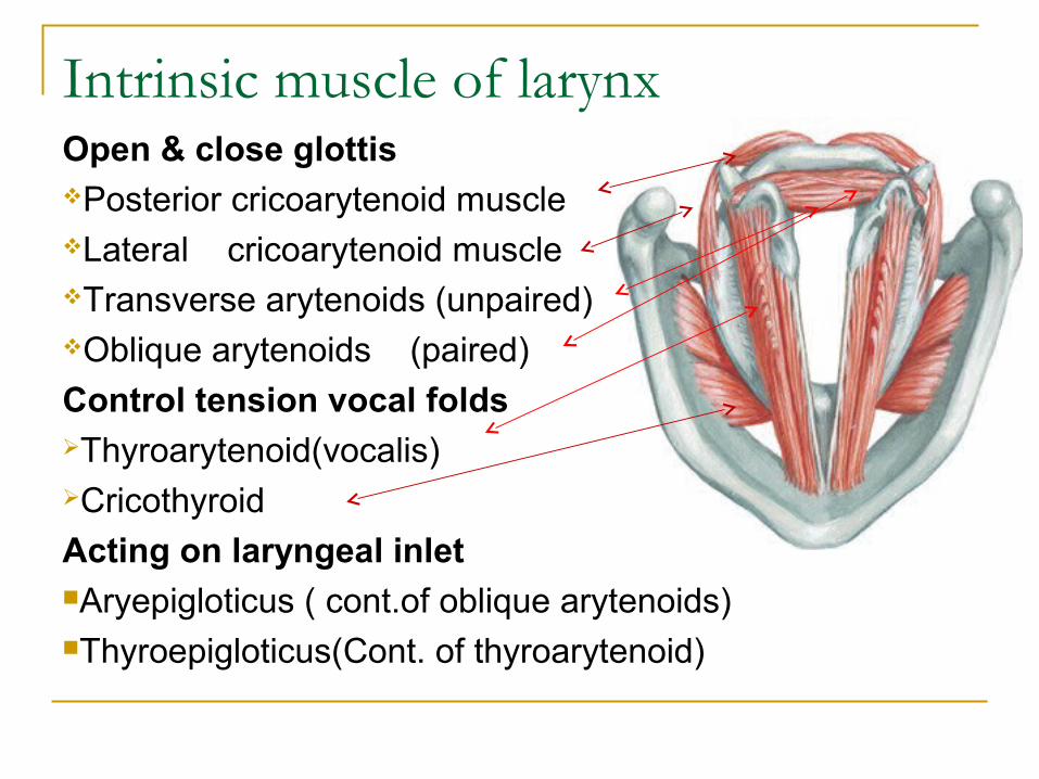
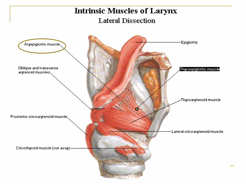
Intrinsic muscle of larynxOpen & close glottisPosterior cricoarytenoid muscleLateral cricoarytenoid muscleTransverse arytenoids (unpaired)Oblique arytenoids (paired)
Control tension vocal foldsThyroarytenoid(vocalis)Cricothyroid
Acting on laryngeal inletAryepigloticus ( cont.of oblique arytenoids)Thyroepigloticus(Cont. of thyroarytenoid)
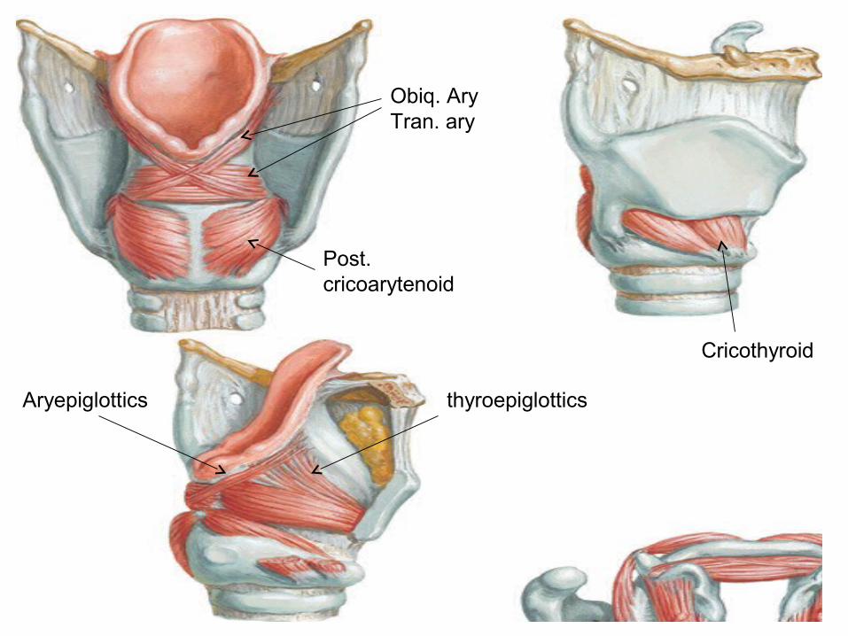
Aryepiglottics thyroepiglottics
Obiq. AryTran. ary
Cricothyroid
Post. cricoarytenoid
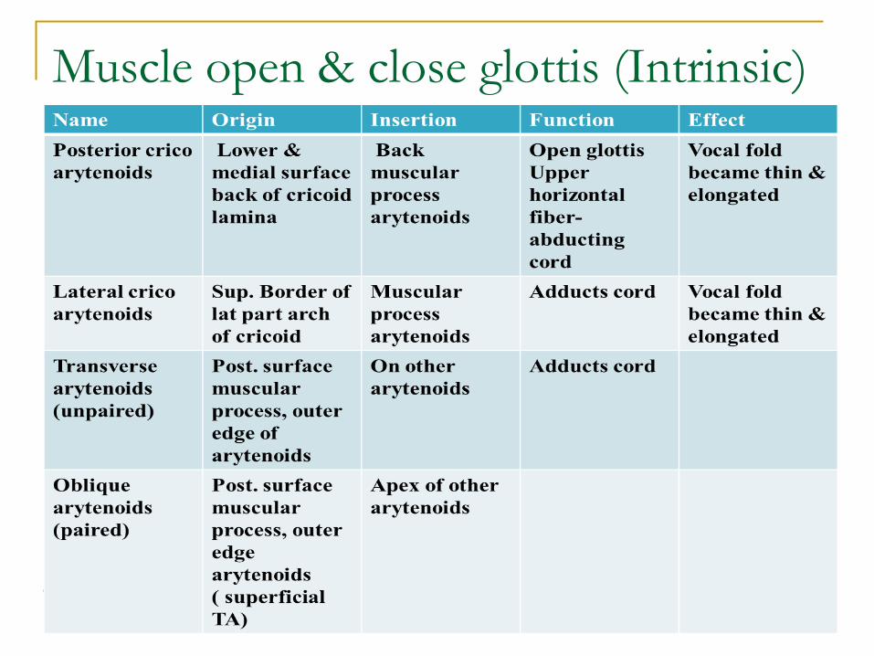
Muscle open & close glottis (Intrinsic)
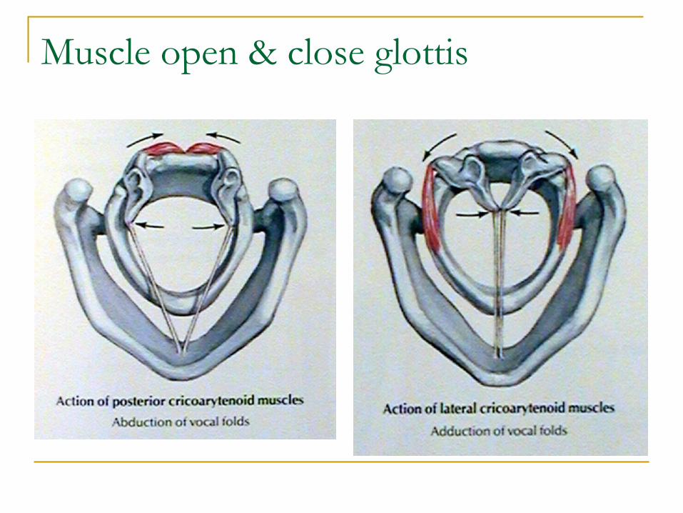
Muscle open & close glottis
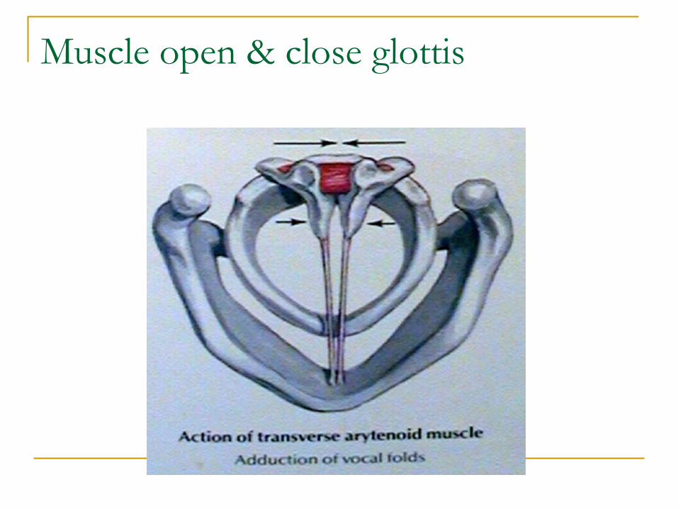
Muscle open & close glottis
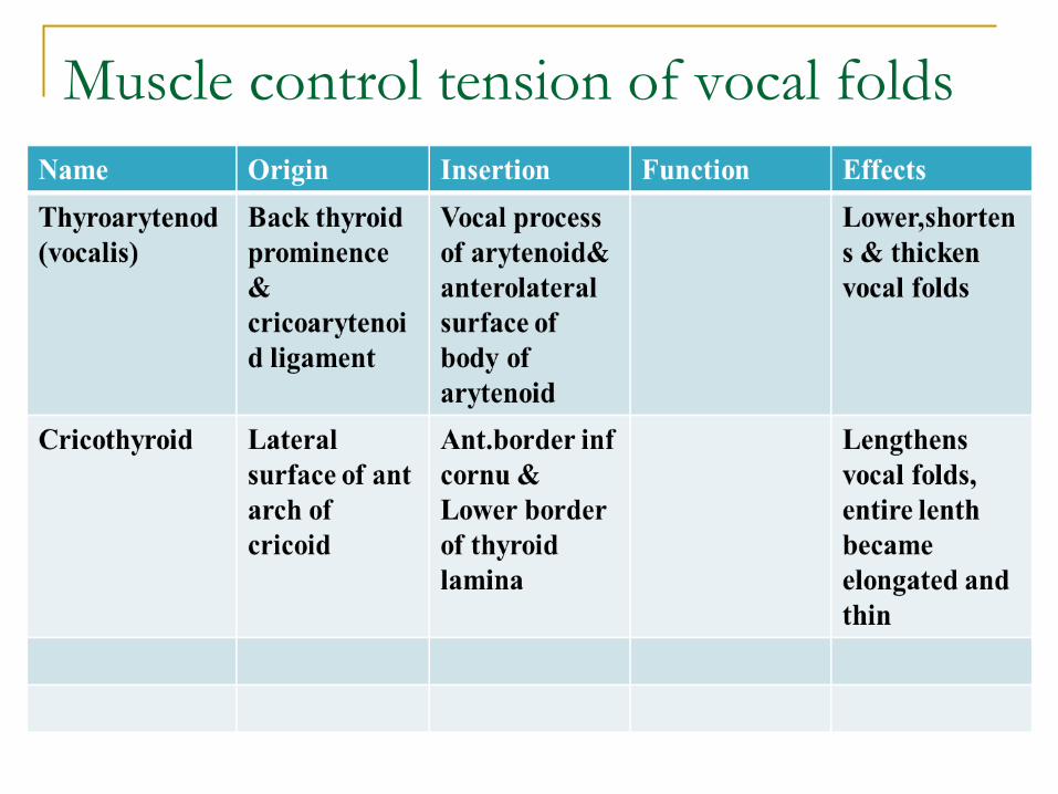
Muscle control tension of vocal folds
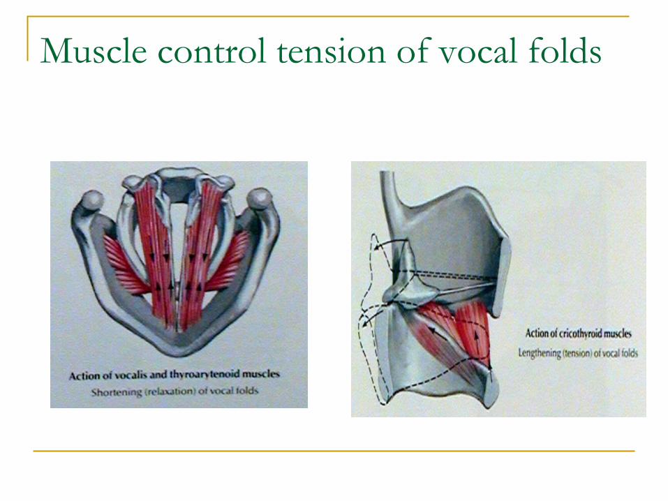
Muscle control tension of vocal folds
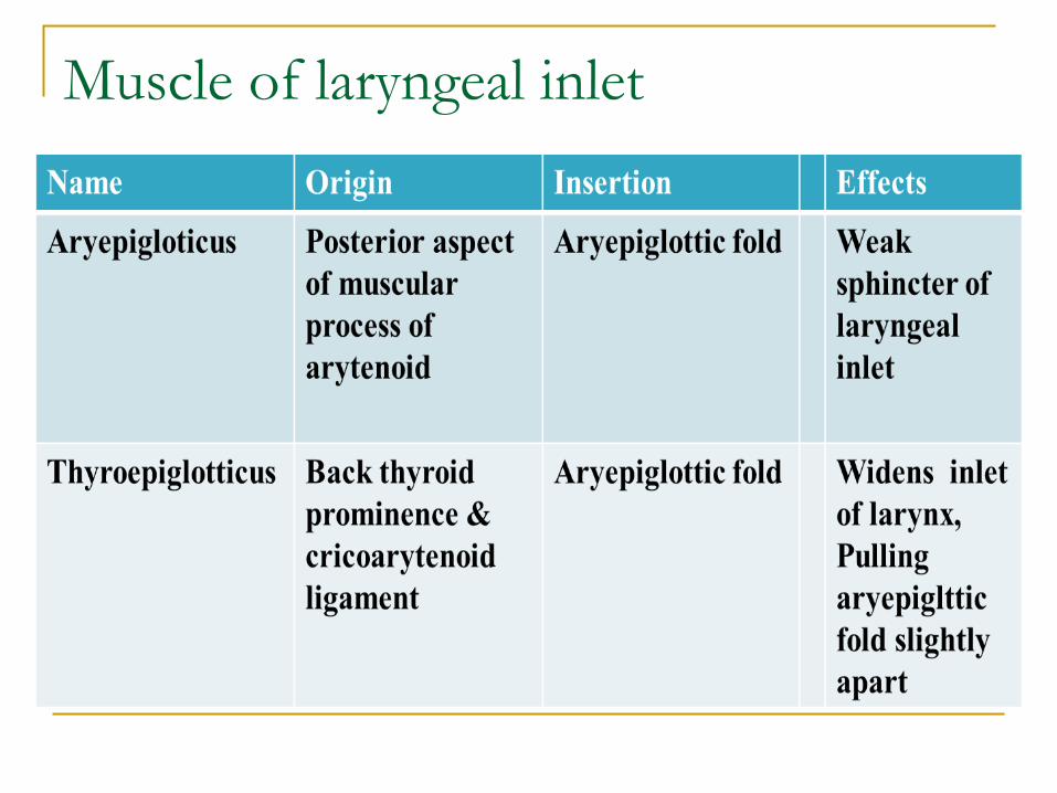
Muscle of laryngeal inlet
Mucous membrane of larynx:Lined by pseudo stratified ciliated columnar Closely attached over posterior surface of epiglottis, corniculate & cuneiform, vocal ligament, elsewhere loosely attached (Oedema)Mucous gland are freely distributed throughout Vocal folds do not poses any glands (lubricated from saccules)
Non keratinizing stratified sqamous epithelium:Upper half of posterior surface of epiglottis Upper half of eryepiglotic fold posterior glottis, vocal folds.
Cavity of the Larynx
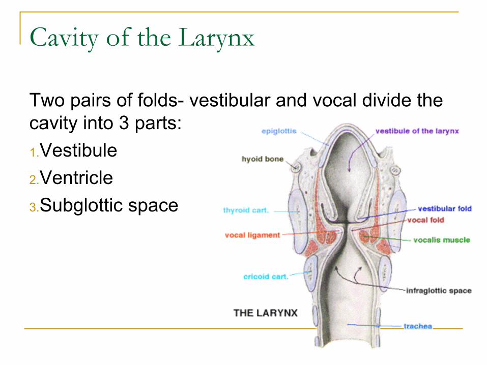
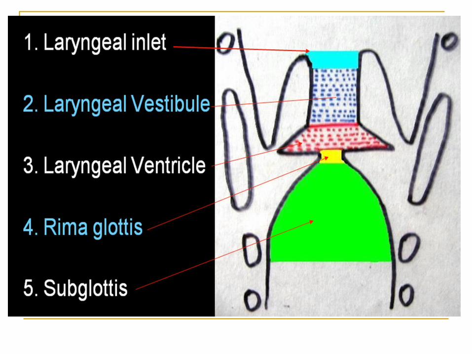
Two pairs of folds- vestibular and vocal divide the cavity into 3 parts:
1.Vestibule
2.Ventricle
3.Subglottic space
Cavity of the Larynx
Vestibule – boundaries: Anterior: posterior surface of epiglottis Posterior: interval between arytenoid
cartilages Lateral: inner surface of aryepiglottic folds
and upper surfaces of the false cord
Cavity of the Larynx cont..
Ventricle( sinus of Larynx) Deep elliptical space between vestibular and
vocal fold. Saccule – conical pouch at anterior part of
the ventricle, lies bet. Inner surface of thyroid cartilage and false cord; has numerous mucous glands open into the surface of its lining mucosa for lubricating the vocal cords.
Cavity of the Larynx cont…
Glottis (rima glottidis) – space between free margin of the true VC,
opening/aperture Posterior glottic chink in adult: 18-19mm;
New born: 4mm; total glottic chink in a newborn: 14mm2
Cavity of the Larynx cont..
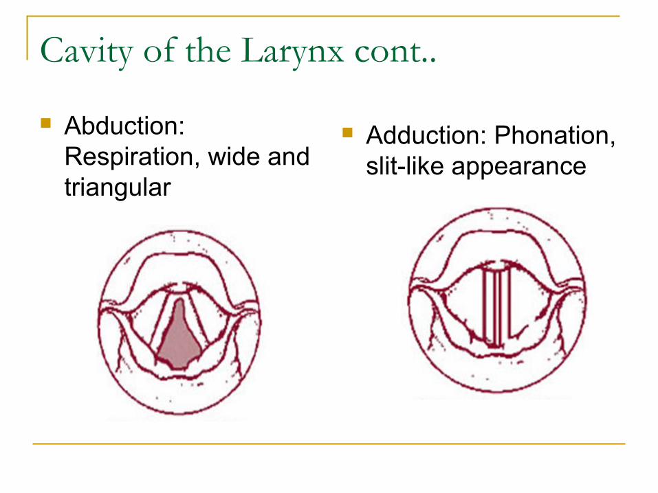
Abduction: Respiration, wide and triangular
Adduction: Phonation, slit-like appearance
Cavity of the Larynx cont..
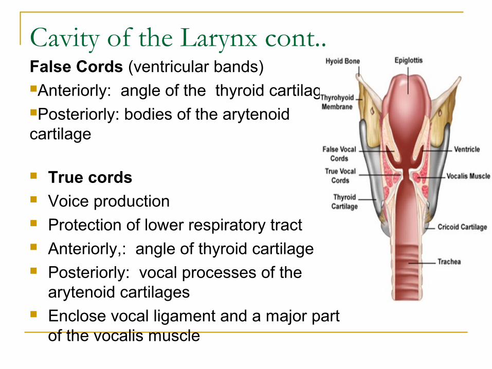
True cords Voice production Protection of lower respiratory tract Anteriorly,: angle of thyroid cartilage Posteriorly: vocal processes of the
arytenoid cartilages Enclose vocal ligament and a major part
of the vocalis muscle
False Cords (ventricular bands)Anteriorly: angle of the thyroid cartilagePosteriorly: bodies of the arytenoid cartilage
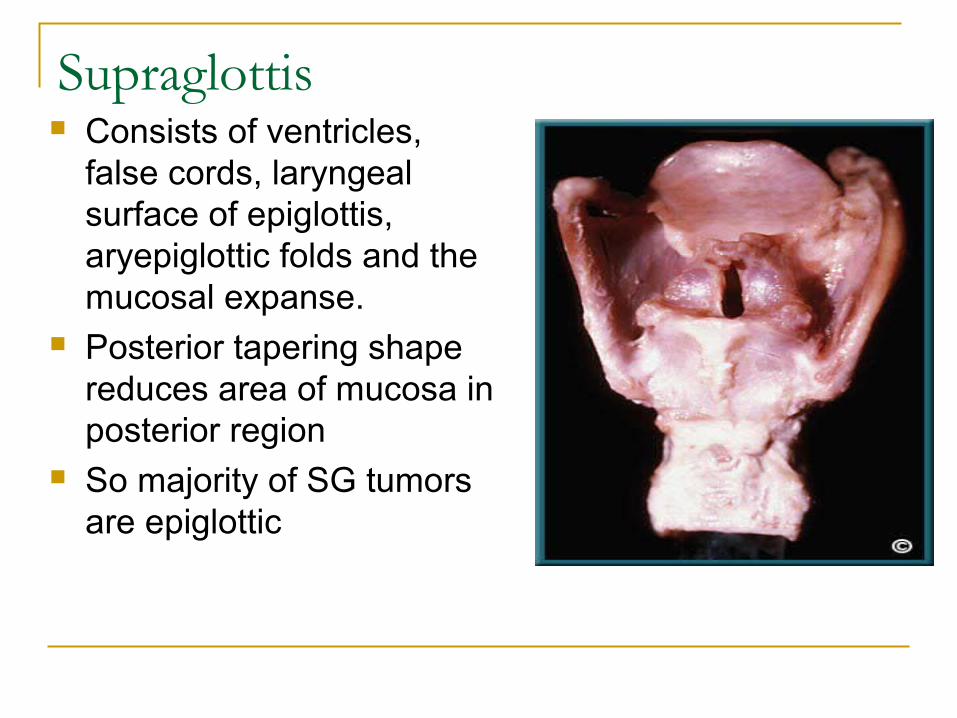
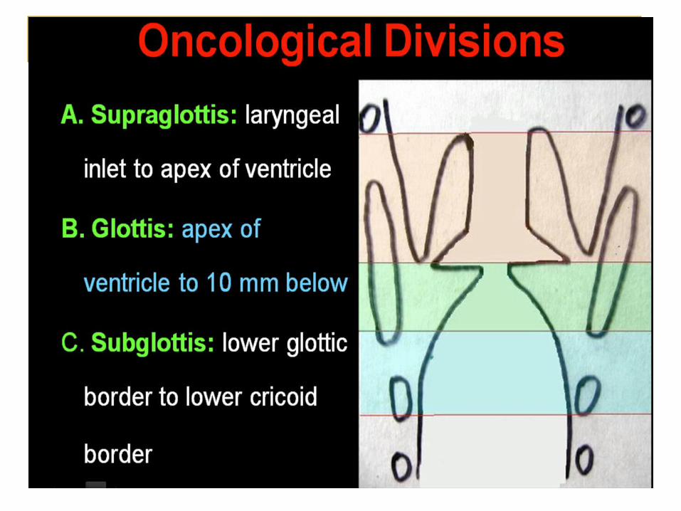
Supraglottis Consists of ventricles,
false cords, laryngeal surface of epiglottis, aryepiglottic folds and the mucosal expanse.
Posterior tapering shape reduces area of mucosa in posterior region
So majority of SG tumors are epiglottic
Applied anatomy Inferior limit of supraglottis is Clinically- imaginary horizontal plane passing
through the apex of Laryngeal ventricle. Anatomically - superior arcuate line where the
squamous epithelium and respiratory epithelium meet.
The Marginal Zone comprises of Suprahyoid epiglottis and Aryepiglottic fold(There is lack of embryologic separation from adjacent hypopharynx
Early lympathic spreads because of rich vascularity and lymphatics.
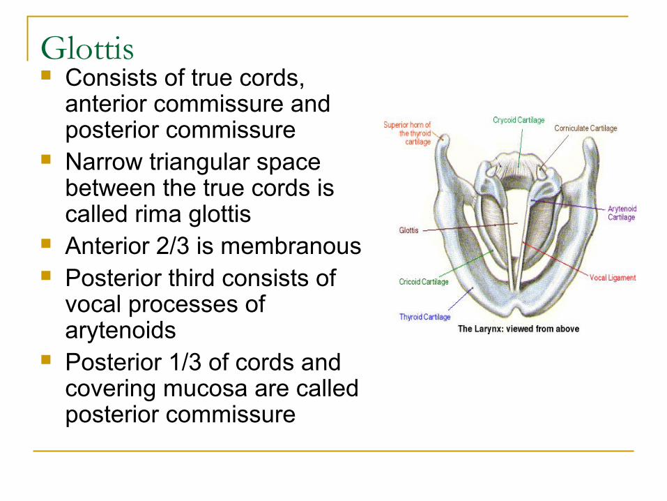
Glottis Consists of true cords,
anterior commissure and posterior commissure
Narrow triangular space between the true cords is called rima glottis
Anterior 2/3 is membranous Posterior third consists of
vocal processes of arytenoids
Posterior 1/3 of cords and covering mucosa are called posterior commissure
Applied Anatomy Anterior commissure is directly attached to
the thyroid cartilage by Broyle’s ligament without intervening inner perichondrium.
Lesion at the anterior commissure can invade the thyroid cartilage early because of absence of inner perichondrium.
Since Broyle's ligament contains blood vessels and lymphatics, it represents a potential route for the escape of malignant tumours from the larynx.
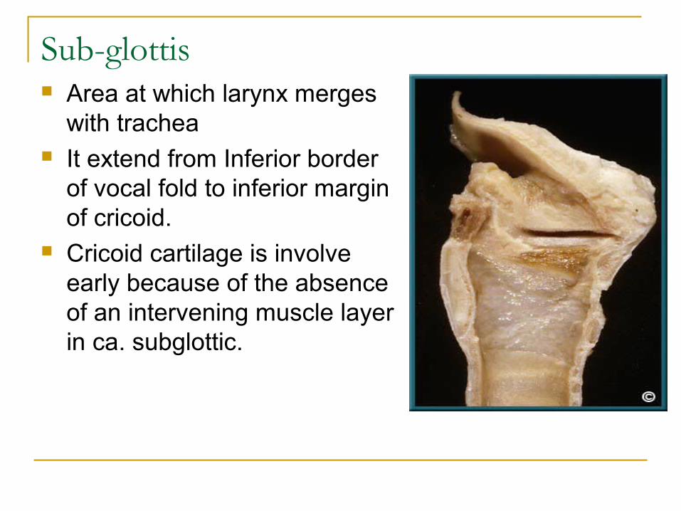
Sub-glottis Area at which larynx merges
with trachea It extend from Inferior border
of vocal fold to inferior margin of cricoid.
Cricoid cartilage is involve early because of the absence of an intervening muscle layer in ca. subglottic.
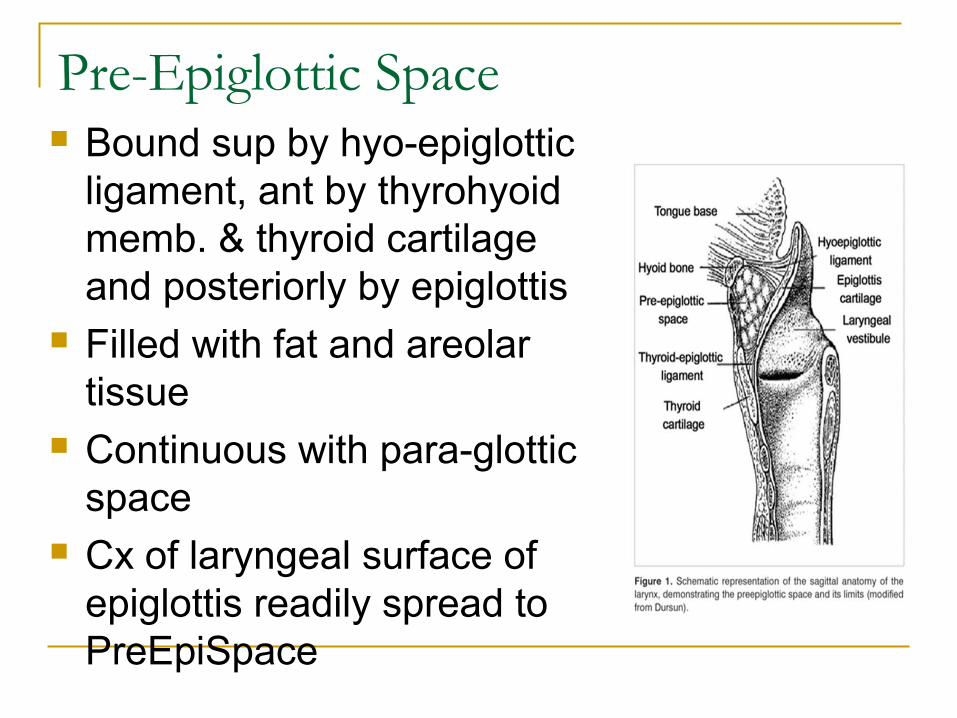
Pre-Epiglottic Space Bound sup by hyo-epiglottic
ligament, ant by thyrohyoid memb. & thyroid cartilage and posteriorly by epiglottis
Filled with fat and areolar tissue
Continuous with para-glottic space
Cx of laryngeal surface of epiglottis readily spread to PreEpiSpace
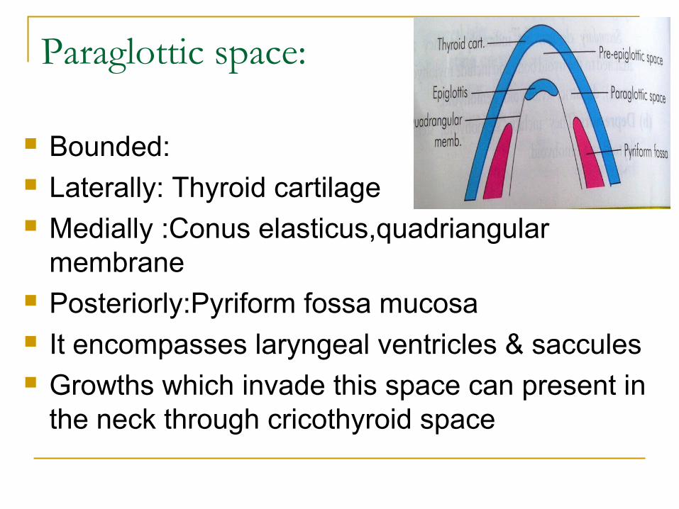
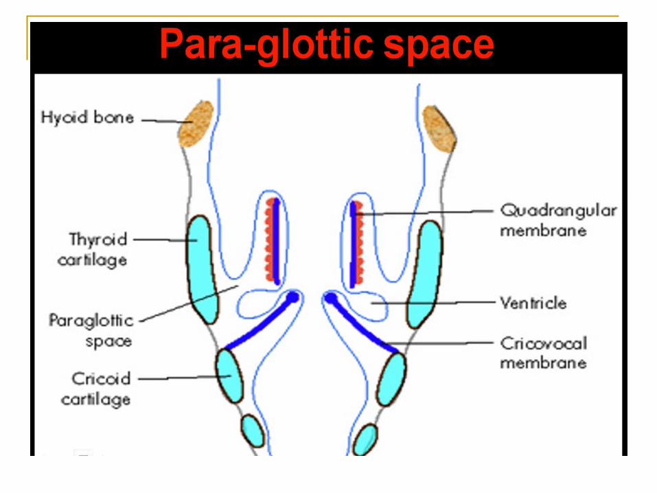
Paraglottic space:
Bounded: Laterally: Thyroid cartilage Medially :Conus elasticus,quadriangular
membrane Posteriorly:Pyriform fossa mucosa It encompasses laryngeal ventricles & saccules Growths which invade this space can present in
the neck through cricothyroid space
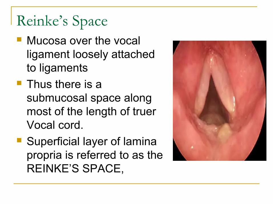
Reinke’s Space Mucosa over the vocal
ligament loosely attached to ligaments
Thus there is a submucosal space along most of the length of truer Vocal cord.
Superficial layer of lamina propria is referred to as the REINKE’S SPACE,
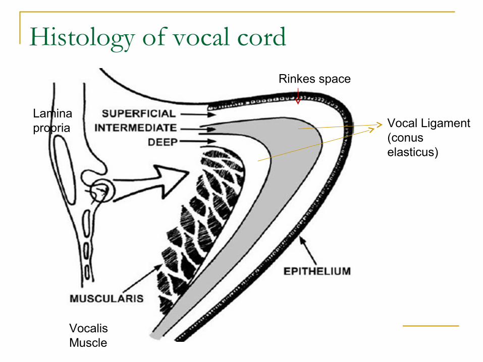
Histology of vocal cordRinkes space
Vocal Ligament(conus elasticus)
Vocalis Muscle
Lamina propria
Applied anatomy Blood vessels and lymphatics are almost
absent in Reinke’s space preventing early spread of cancer.
It is this layer that vibrates the most during phonation.
Accumulation of fluid under epithelium of true vocal cord(Reinke’s space) is called Reinke’s oedema.
Nerve supply contd.. Sup. Laryngeal N- Inf ganglion vagus & superior cervical sympathetic. Descend behind ICA At greater horn- Divide small external & larger internal branch External branch – Motor to Cricothyroid Internal branch- Pierce thyrohyoid membrane. Divide-Two sensory & secretomotor Upper- pharynx,epiglottis,valeculla,vestibule Lower- Aryepiglottic fold, mucous membrane up to vocal cords Internal branch- caries Afferent fibers from neuromuscular & stretch
receptor Sup. Laryngeal nerve end by anastomoses with RLN (Galens
anastomoses)
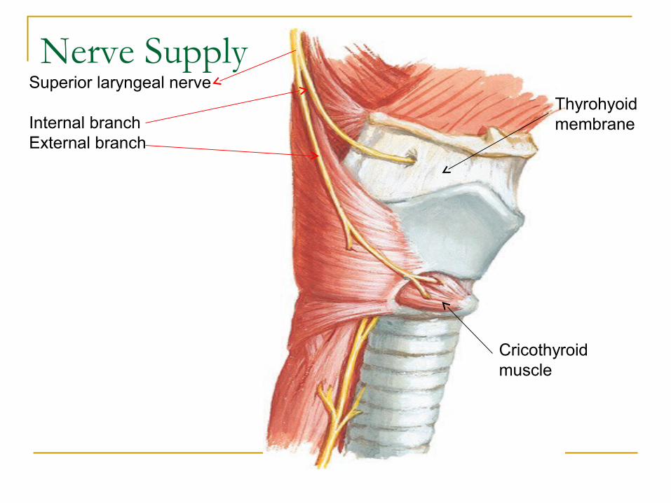
Nerve SupplySuperior laryngeal nerve
Internal branchExternal branch
Cricothyroid muscle
Thyrohyoid membrane

Internal branchInternal branch of superior laryngeal nerve
Sensory branches
Recurrent laryngeal nerve
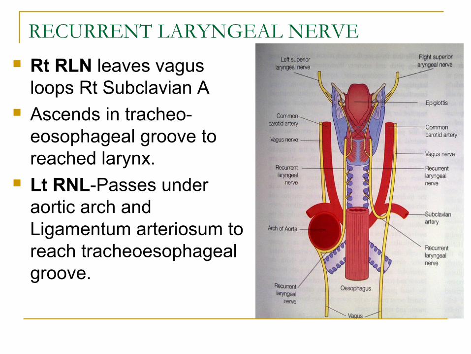
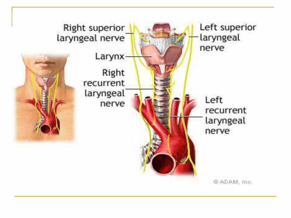
RECURRENT LARYNGEAL NERVE
Rt RLN leaves vagus loops Rt Subclavian A
Ascends in tracheo-eosophageal groove to reached larynx.
Lt RNL-Passes under aortic arch and Ligamentum arteriosum to reach tracheoesophageal groove.
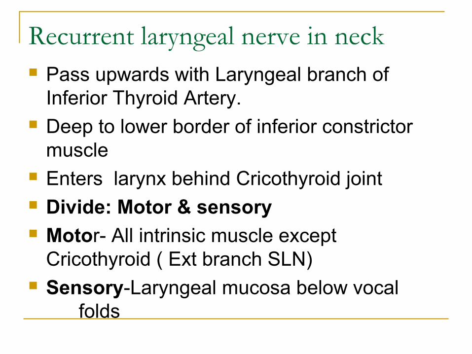
Recurrent laryngeal nerve in neck Pass upwards with Laryngeal branch of
Inferior Thyroid Artery. Deep to lower border of inferior constrictor
muscle Enters larynx behind Cricothyroid joint Divide: Motor & sensory Motor- All intrinsic muscle except
Cricothyroid ( Ext branch SLN) Sensory-Laryngeal mucosa below vocal
folds
Laryngeal innervations -Applied anatomyInternal laryngeal nerve:Lies in medial wall of pyriform sinus mucosa
Tropical anesthesia and Pain in ca pyriform sinus
Damage to the internal laryngeal nerve produce anesthesia in supraglottic part of larynx so that FB can readily enter it (Breaking the reflex arc)Damage to external laryngeal nerve cause some weakness of phonation due to loss of tightening effect of the cricothyroid on the vocal cord.
Laryngeal innervations -Applied anatomy
Recurrent laryngeal nerve: Left RLN- More liable to injury (extensive
course) Variable relation between RLN & ITA- RLN may cross in front/behind/between
artery Right RLN more variable location whereas
Left RLN more likely posterior to artery.
Semon’s law- In gradual progressive lesions affecting the recurrent laryngeal nerve resulting in palsy, abductors are affected first then the adductors.
On the other hand, in functional paralysis of larynx, the adductors are the first to be paralysed.
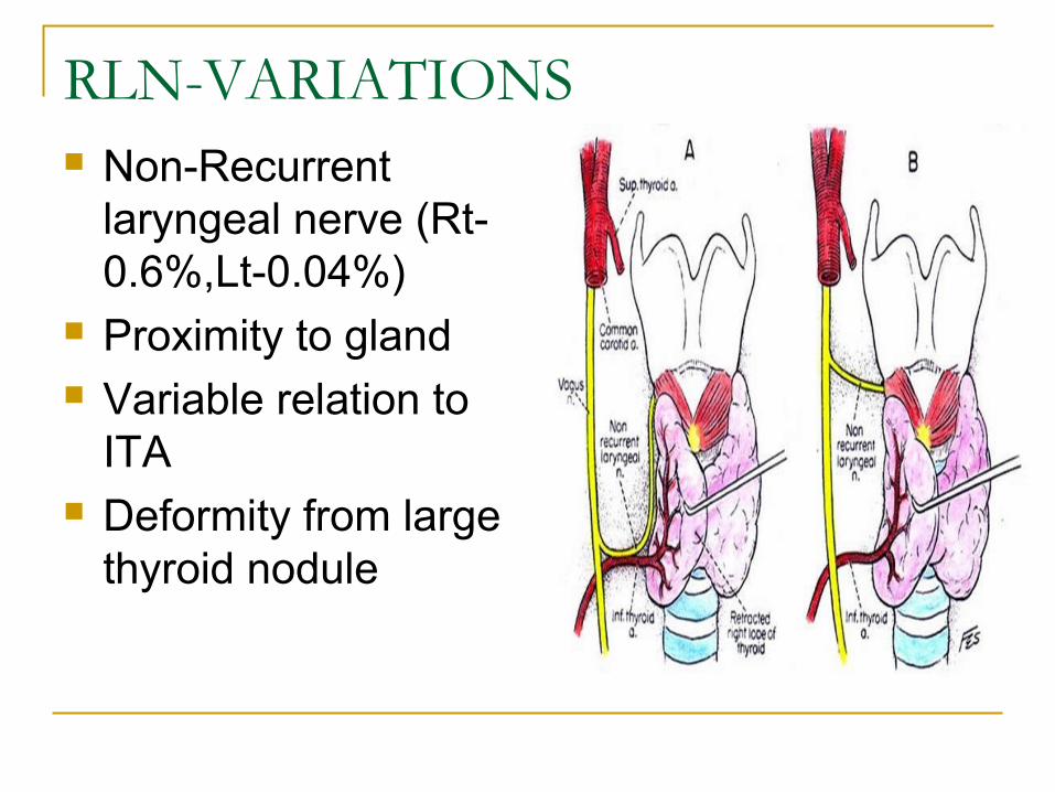
RLN-VARIATIONS Non-Recurrent
laryngeal nerve (Rt-0.6%,Lt-0.04%)
Proximity to gland Variable relation to
ITA Deformity from large
thyroid nodule
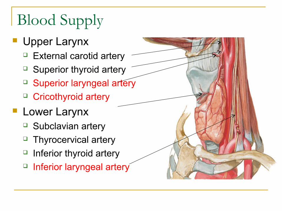
Blood Supply Upper Larynx
External carotid artery Superior thyroid artery Superior laryngeal artery Cricothyroid artery
Lower Larynx Subclavian artery Thyrocervical artery Inferior thyroid artery Inferior laryngeal artery
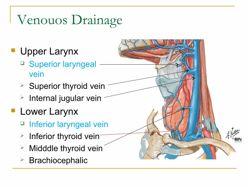
Venouos Drainage
Upper Larynx Superior laryngeal
vein Superior thyroid vein Internal jugular vein
Lower Larynx Inferior laryngeal vein Inferior thyroid vein Midddle thyroid vein Brachiocephalic
Lymphatic Drainage Upper & lower group by vocal folds Above vocal folds- Vessels that accompanying superior laryngeal
vein pierce thyrohyoid membrane to drain into Upper deep
cervical node Below vocal folds- Lower deep cervical chain through Pre-laryngeal(Delphian) & pre-tracheal nodes No lymphatic in vocal folds
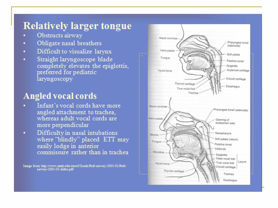
Infant Larynx Positioned high in the neck- this allows the
epiglottis to meet soft palate and makes nasopharyngeal channel for nasal breathing during sucking.
Laryngeal cartilage are softer ,easily displaced, easily irritable
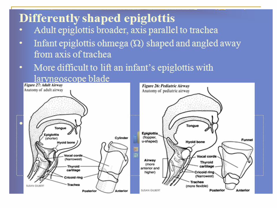
Epoigloittis- longer, narrower, tubular; hence mentioned as omega shaped.
Thyroid cartilage is flat, cricoid cartilage is smaller then size of glottis making subglottis the narrowest part.
Aryoepiglottic folds are disproportionately large.
Arytenoids are more prominent Mucous membrane and connective tissue are
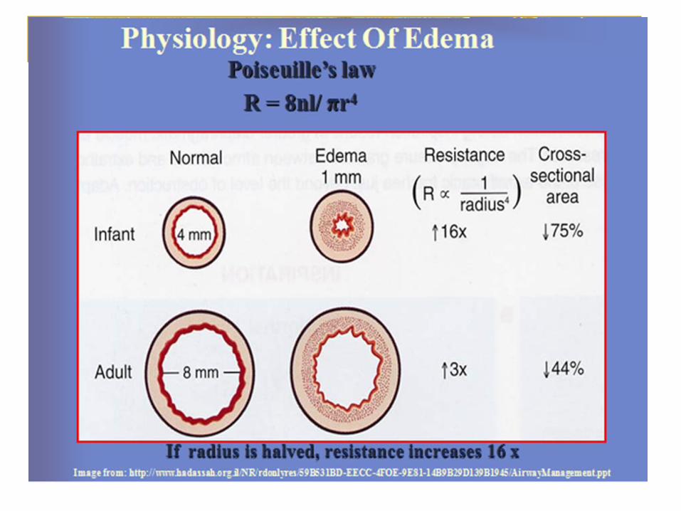
loosely attached and easily undergo oedematous changes.
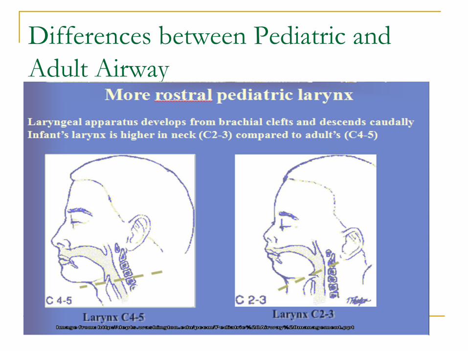
Differences between Pediatric and Adult Airway