Embed Size (px)
Citation preview

Basic Human Anatomy Lesson 7: Respiratory System Page 1
Basic Human Anatomy
Lesson 7: Respiratory System
Welcome to Lesson 7 of the Basic Human Anatomy Course. Today, we’ll be
studying the Human Respiratory System.
I have 4 goals for you in this lesson:
1. Define respiration, external respiration, internal respiration and breathing. 2. Identify the main subdivisions of the respiratory system and their functions. 3. Describe the external nose, nasal chambers, pharynx, larynx, trachea,
bronchi, alveoli, lungs, and pleural cavities. 4. Describe breathing and breathing mechanisms.
INTRODUCTION
a. Respiration. Respiration is the exchange of gases between the atmosphere and
the cells of the body. It is a physiological process. There are two types of
respiration--external and internal. External respiration is the exchange of gases
between the air in the lungs and blood. Internal respiration is the exchange of
gases between the blood and the individual cells of the body.
b. Breathing. Breathing is the process that moves air into and out of the lungs. It
is a mechanical process. There are two types of breathing in humans—costal
(thoracic) and diaphragmatic (abdominal). In costal breathing, the major structure
causing the movement of the air is the rib cage. In diaphragmatic breathing,
interaction between the diaphragm and the abdominal wall causes the air to
move into and out of the lungs.

Basic Human Anatomy Lesson 7: Respiratory System Page 2
COMPONENTS AND SUBDIVISIONS OF THE HUMAN RESPIRATORY SYSTEM
See figure 7-1 for an illustration of the human respiratory system.
a. Components. The components of the human respiratory system consist of air
passageways and two lungs. Air moves from the outside of the body into tiny sacs
in the lungs called alveoli (pronounced al-VE-oh-lie).
b. Main Subdivisions. The main subdivisions of the respiratory system may be
identified by their relationship to the voice box or larynx. Thus, the main
subdivisions are as listed in table 7-1.

Basic Human Anatomy Lesson 7: Respiratory System Page 3
Figure 7-1. The human respiratory system.

Basic Human Anatomy Lesson 7: Respiratory System Page 4
SUBDIVISION FUNCTION
SUPRALARYNGEAL STRUCTURES (su-prah-lah-RIN-je-al)
Cleanse, warm, moisten, and test inflowing air
LARYNX (voice box) (LARE-inks)
Controls the volume of inflowing air; produces
selected pitch(vibration frequency) in the
moving column of air
INFRALARYNGEAL STRUCTURES (in-frah-lah-RIN-je-al)
Distribute air to the alveoli of the lung where
the actual external respiration takes place
Table 7-1. The main subdivisions of the respiratory system.
SUPRALARYNGEAL STRUCTURES
Figure 7-2. Supralaryngeal structures.

Basic Human Anatomy Lesson 7: Respiratory System Page 5
a. External Nose. The external nose is the portion projecting from the face. It is
supported primarily by cartilages. It has a midline divider called the nasal septum,
which extends from the internal nose. Paired openings (nostrils) lead to paired
paces (vestibules). Guard hairs in the nostrils filter inflowing air.
b. Nasal Chambers (Internal Nose). Behind each vestibule of the external nose is
a nasal chamber. The two nasal chambers together form the internal nose. These
chambers too are separated by the nasal septum.
(1) Mucoperiosteum. The walls of the nasal chambers are lined with a thick
mucous-type membrane known as the mucoperiosteum. It has a ciliated
epithelial surface and a rich blood supply, which provides warmth and
moisture. At times, it may become quite swollen.
CILIATED = provided with cilia (hairlike projections which move fluids to the rear)
(2) Conchae. The lateral wall of each chamber has three scroll- like
extensions into the nasal chamber which help to increase the surface area
exposed to the inflowing air. These scroll-like extensions are known as
conchae.
CONCHA (pronounced KON-kah) = sea shell
CONCHA (singular), CONCHAE (plural)
(3) Olfactory epithelium. The sense of smell is due to special nerve endings
located in the upper areas of the nasal chambers. The epithelium
containing the sensory endings is known as the olfactory epithelium.
(4) Paranasal sinuses. There are air "cells" or cavities in the skull known as
paranasal sinuses. The paranasal sinuses are connected with the nasal

Basic Human Anatomy Lesson 7: Respiratory System Page 6
chambers and are lined with the same ciliated mucoperiosteum. Thus,
these sinuses are extensions of the nasal chambers into the skull bones. For
this reason, they are known as paranasal sinuses.
c. Pharynx. The pharynx (FAIR-inks) is the common posterior space for the
respiratory and digestive systems.
(1) Nasopharynx. That portion of the pharynx specifically related to the
respiratory system is the nasopharynx. It is the portion of the pharynx
above the soft palate. The two posterior openings (nares) of the nasal
chambers lead into the single space of the nasopharynx. The auditory
(eustachian) tubes also open into the nasopharynx. The auditory tubes
connect the nasopharynx with the middle ears (to equalize the pressure
between the outside and inside of the eardrum). Lying in the upper
posterior wall of the nasopharynx are the pharyngeal tonsils (adenoids).
The soft palate floor of the nasopharynx is a trapdoor which closes off the
upper respiratory passageways during swallowing.
(2) Oropharynx. The portion of the pharynx closely related to the digestive
system is the oropharynx. It is the portion of the pharynx below the soft
palate and above the upper edge of the epiglottis. (The epiglottis is the flap
that prevents food from entering the larynx (discussed below) during
swallowing.)
(3) Laryngopharynx. That portion of the pharynx which is common to the
respiratory and digestive systems is the laryngopharynx. It is the portion of
the pharynx below the upper edge of the epiglottis. Thus, the digestive and
respiratory systems lead into it from above and lead off from it below.
Basic Human Anatomy Lesson 7: Respiratory System Page 7
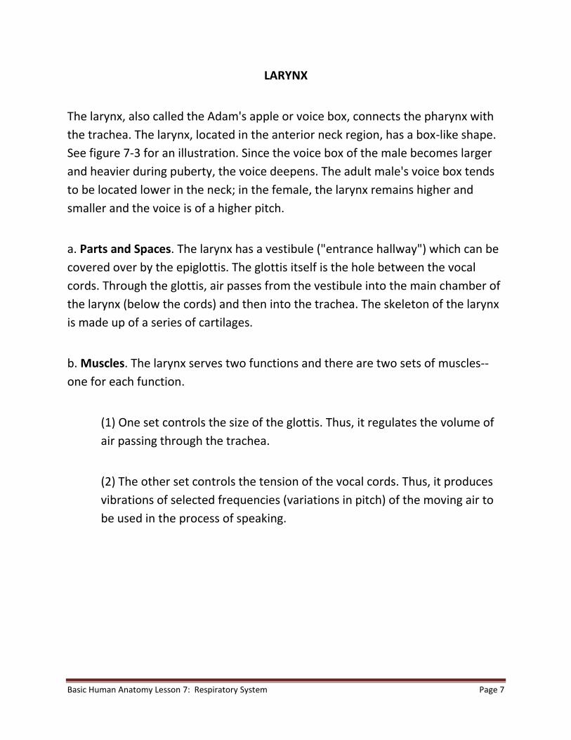
LARYNX
The larynx, also called the Adam's apple or voice box, connects the pharynx with
the trachea. The larynx, located in the anterior neck region, has a box-like shape.
See figure 7-3 for an illustration. Since the voice box of the male becomes larger
and heavier during puberty, the voice deepens. The adult male's voice box tends
to be located lower in the neck; in the female, the larynx remains higher and
smaller and the voice is of a higher pitch.
a. Parts and Spaces. The larynx has a vestibule ("entrance hallway") which can be
covered over by the epiglottis. The glottis itself is the hole between the vocal
cords. Through the glottis, air passes from the vestibule into the main chamber of
the larynx (below the cords) and then into the trachea. The skeleton of the larynx
is made up of a series of cartilages.
b. Muscles. The larynx serves two functions and there are two sets of muscles--
one for each function.
(1) One set controls the size of the glottis. Thus, it regulates the volume of
air passing through the trachea.
(2) The other set controls the tension of the vocal cords. Thus, it produces
vibrations of selected frequencies (variations in pitch) of the moving air to
be used in the process of speaking.

Basic Human Anatomy Lesson 7: Respiratory System Page 8
Figure 7-3. The larynx.

Basic Human Anatomy Lesson 7: Respiratory System Page 9
INFRALARYNGEAL STRUCTURES
a. Trachea and Bronchi. The respiratory tree (figure 7-4) is the set of tubular
structures which carry the air from the larynx to the alveoli of the lungs. Looking
at a person UPSIDE DOWN, the trachea is the trunk of the tree and the bronchi
are the branches. These tubular parts are held open (made patent) by rings of
cartilage. Their lining is ciliated to remove mucus and other materials that get into
the passageway.
Figure 7-4. Infralaryngeal structures ("respiratory tree").

Basic Human Anatomy Lesson 7: Respiratory System Page 10
b. Alveoli. The alveoli (alveolus, singular) are tiny spherical (balloon-like) sacs
which are connected to the larger tubes of the lungs by tiny tubes known as
alveolar ducts and bronchioles. The alveoli are so small that there are billions in
the adult lungs. This very small size produces a maximum surface area through
which external respiration takes place. External respiration is the actual exchange
of gases between the air in the alveolar spaces and the adjacent blood capillaries
through their walls.
c. Lungs. A lung is an individual organ composed of tubular structures and alveoli
bound together by fibrous connective tissue (FCT). In the human, there are two
lungs--right and left. Each lung is supplied by a primary or mainstem bronchus
leading off of the trachea. The right lung is larger in volume than the left lung. The
left lung must leave room for the heart. The right lung is divided into three
pulmonary lobes (upper, middle, and lower) and 10 bronchopulmonary segments
(2 + 3 + 5). The left lung is divided into two pulmonary lobes (upper and lower)
and eight bronchopulmonary segments (4 + 4). A pulmonary lobe is a major
subdivision of a lung marked by fissures (deep folds). Each lobe is further
partitioned into bronchopulmonary segments. Each lobe is supplied by a
secondary or lobar bronchus. Each segment is supplied by a tertiary or segmental
bronchus, a branch of the lobar bronchus.
d. Pleural Cavities. See Lesson 3 to review a description of pleural cavities. That
Lesson indicates that each serous cavity has inner and outer membranes. In the
case of the lungs, the inner membrane is known as the visceral pleura which very
closely covers the surface of the lungs. The outer membrane is known as the
parietal pleura, forming the outer wall of the cavity. The pleural cavities are the
potential spaces between the inner and outer membranes. The pleural cavities
allow the lungs to move freely with a minimum of friction during the expansion
and contraction of breathing.

Basic Human Anatomy Lesson 7: Respiratory System Page 11
BREATHING AND BREATHING MECHANISMS IN HUMANS
INTRODUCTION
a. Boyle's law tells us that as the volume (V) of a gas-filled container increases, the
pressure (P) inside decreases; as the volume (V) of a closed container decreases,
the pressure (P) inside increases. When two connected spaces of air have
different pressures, the air moves from the space with greater pressure to the
one with lesser pressure. In regard to breathing, we can consider the air pressure
around the human body to be constant. The pressure inside the lungs may be
greater or less than the pressure outside the body. Thus, a greater internal
pressure causes air to flow out; a greater external pressure causes air to flow in.
b. We can compare the human trunk to a hollow cylinder. This cylinder is divided
into upper and lower cavities by the diaphragm. The upper is the thoracic cavity
and is essentially gas-filled. The lower is the abdominopelvic cavity and is
essentially water-filled.
COSTAL (THORACIC) BREATHING
a. Inhalation. Muscles attached to the thoracic cage raise the rib cage. A typical
rib might be compared to a bucket handle, attached at one end to the sternum
(breastbone) and at the other end to the vertebral column. The "bucket handle" is
lifted by the overall movement upward and outward of the rib cage. These
movements increase the thoracic diameters from right to left (transverse) and
from front to back (A-P). Thus, the intrathoracic volume increases. Recalling
Boyle's law, the increase in volume leads to a decrease in pressure. The air
pressure outside the body then forces air into the lungs and inflates them.

Basic Human Anatomy Lesson 7: Respiratory System Page 12
b. Exhalation. The rib cage movements and pressure relationships are reversed
for exhalation. Thus, intrathoracic volume decreases. The intrathoracic pressure
increases and forces air outside the body.
DIAPHRAGMATIC (ABDOMINAL) BREATHING
The diaphragm is a thin, but strong, dome-shaped muscular membrane that
separates the abdominal and thoracic cavities. The abdominal wall is elastic in
nature. The abdominal cavity is filled with soft, watery tissues.
a. Inhalation. As the diaphragm contracts, the dome flattens and the diaphragm
descends. This increases the depth (vertical diameter) of the thoracic cavity and
thus increases its volume. This decreases air pressure within the thoracic cavity.
The greater air pressure outside the body then forces air into the lungs.
b. Exhalation. As the diaphragm relaxes, the elastic abdominal wall forces the
diaphragm back up by pushing the watery tissues of the abdomen against the
underside of the relaxed diaphragm. The dome extends upward. The process of
inhalation is thus reversed.
Introduction to Basic Human Anatomy is a distance learning product that is based on the Correspondence Subcourse MD0006 of the U.S. Army Medical Department Center and School. This presentation was produced by the Brookside Associates, Ltd., which is privately-held and not connected to any governmental agency. The views expressed here are those of the authors, and unless otherwise noted, do not necessarily reflect the views of the Brookside Associates, Ltd., any governmental agencies or private organizations. This presentation is unclassified, and © 2009, with all rights reserved.