Embed Size (px)
Citation preview
Anatomy of the LARYNX
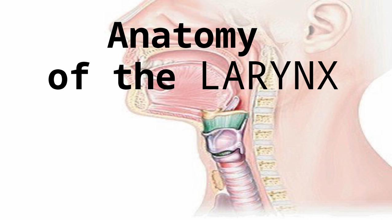
Shape & Site
• A 2-inch-long, tube shaped organ.
• open into the laryngeal part of the pharynx above and is continuous with the trachea below
• It projects forwards in the median region of the neck extending from the root of tongue to the trachea ( from the middle of c3 vertebra to the lower border of c6 vertebra)
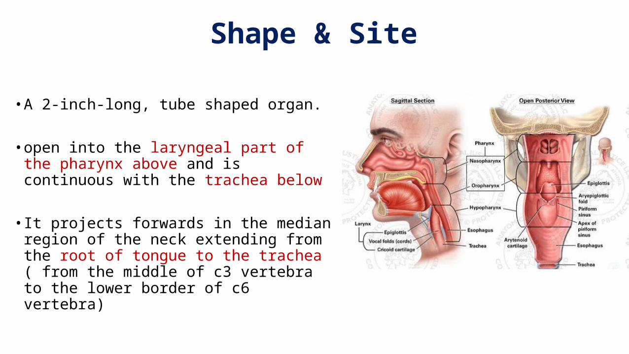
Function
• The larynx is the portion of the respiratory tract containing the vocal cords
• The larynx functions in:1. Deglutition (swallowing) protecting the trachea
against food aspiration2. respiration (breathing)3. Phonation (voice production) commonly called
the voice box
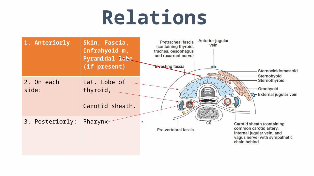
Relations1. Anteriorly Skin, Fascia,
Infrahyoid m, Pyramidal lobe(if present)
2. On each side: Lat. Lobe of thyroid,
Carotid sheath.
3. Posteriorly: Pharynx
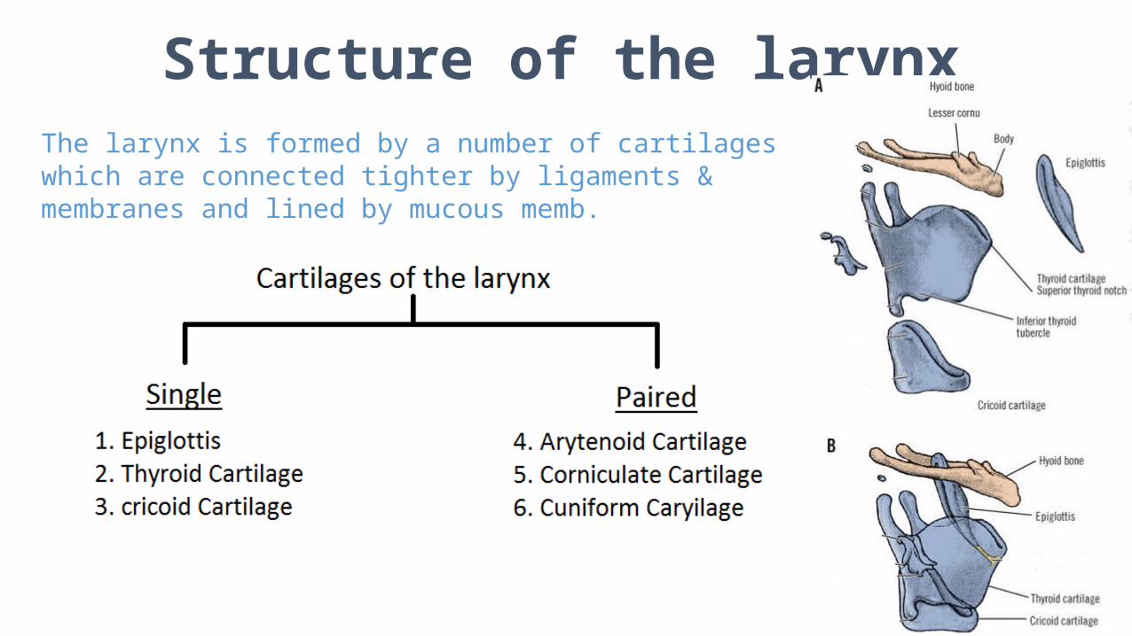
Structure of the larynxThe larynx is formed by a number of cartilages which are connected tighter by ligaments & membranes and lined by mucous memb.
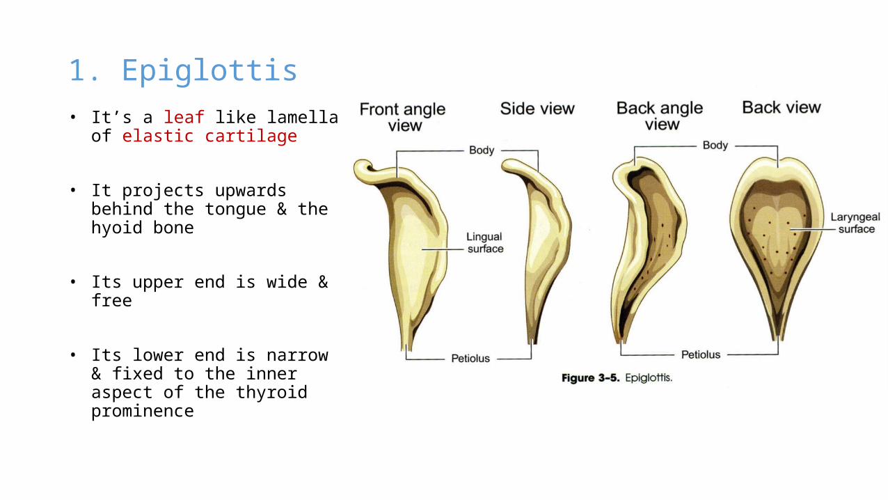
1. Epiglottis
• It’s a leaf like lamella of elastic cartilage
• It projects upwards behind the tongue & the hyoid bone
• Its upper end is wide & free
• Its lower end is narrow & fixed to the inner aspect of the thyroid prominence
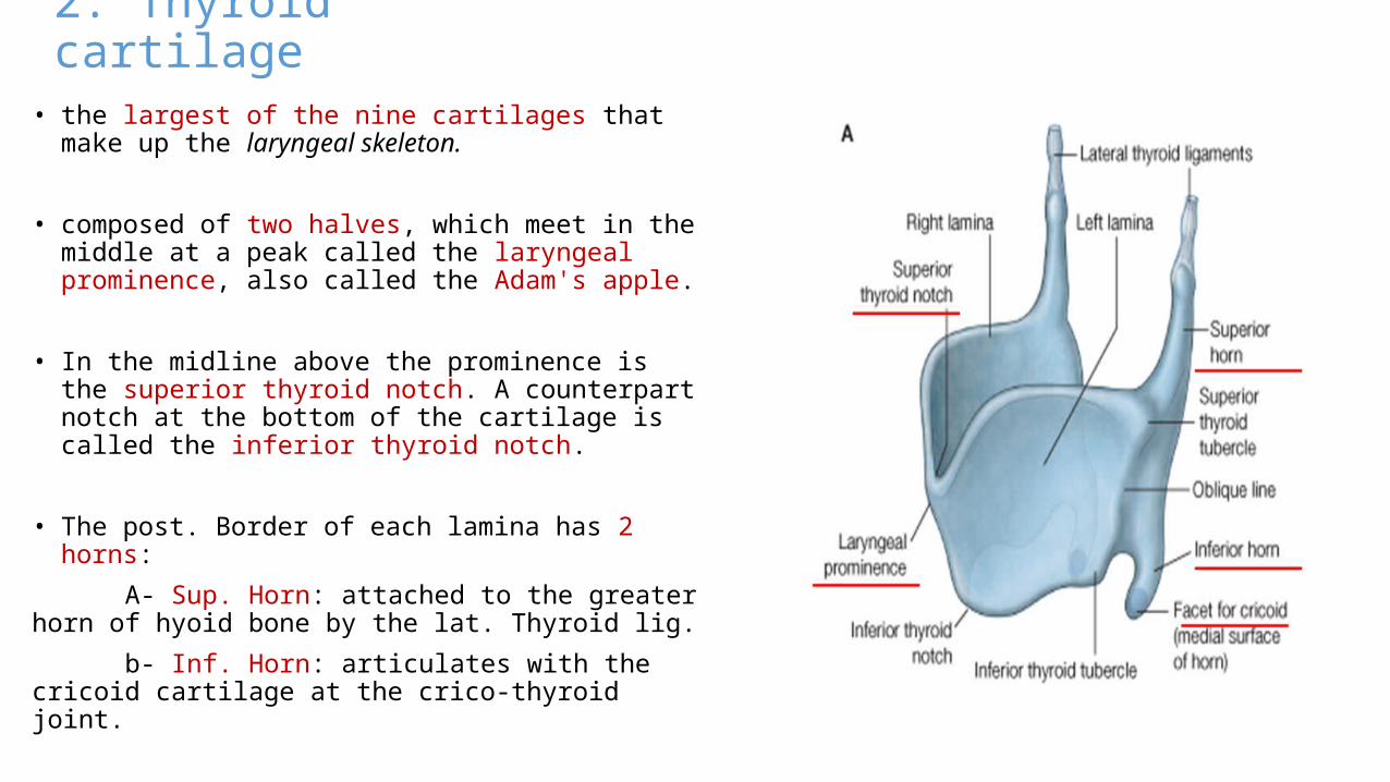
2. Thyroid cartilage• the largest of the nine cartilages that make up the
laryngeal skeleton.
• composed of two halves, which meet in the middle at a peak called the laryngeal prominence, also called the Adam's apple.
• In the midline above the prominence is the superior thyroid notch. A counterpart notch at the bottom of the cartilage is called the inferior thyroid notch.
• The post. Border of each lamina has 2 horns:
A- Sup. Horn: attached to the greater horn of hyoid bone by the lat. Thyroid lig.
b- Inf. Horn: articulates with the cricoid cartilage at the crico-thyroid joint.
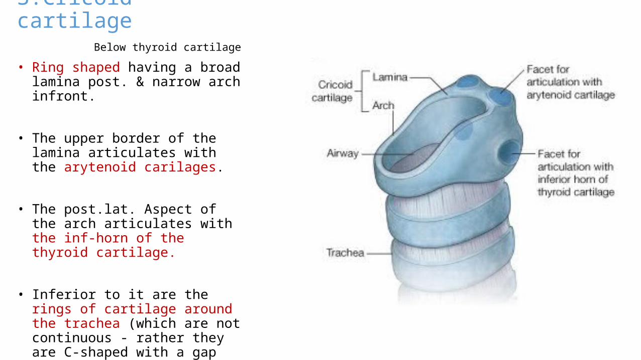
3.Cricoid cartilage Below thyroid cartilage • Ring shaped having a broad lamina
post. & narrow arch infront.
• The upper border of the lamina articulates with the arytenoid carilages.
• The post.lat. Aspect of the arch articulates with the inf-horn of the thyroid cartilage.
• Inferior to it are the rings of cartilage around the trachea (which are not continuous - rather they are C-shaped with a gap posteriorly)
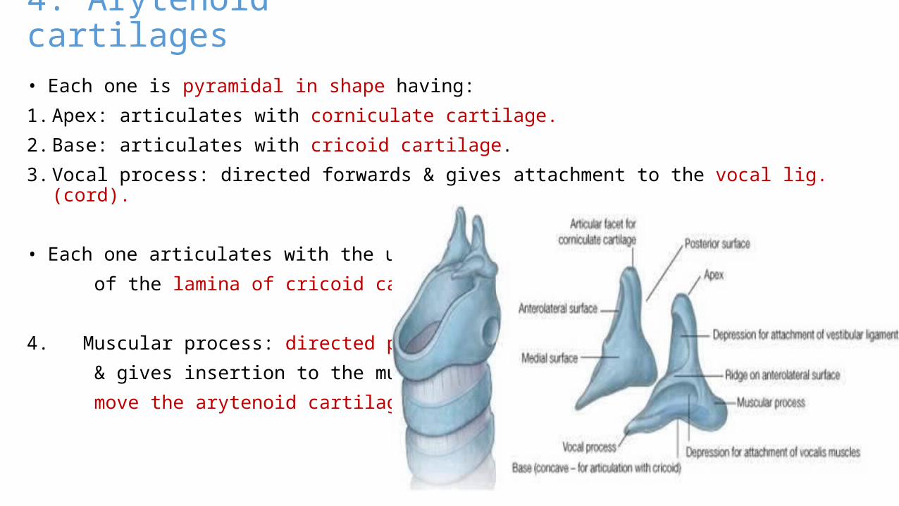
4. Arytenoid cartilages• Each one is pyramidal in shape having:
1. Apex: articulates with corniculate cartilage.
2. Base: articulates with cricoid cartilage.
3. Vocal process: directed forwards & gives attachment to the vocal lig. (cord).
• Each one articulates with the upper border
of the lamina of cricoid cartilage.
4. Muscular process: directed posterolat.
& gives insertion to the muscles which
move the arytenoid cartilage.
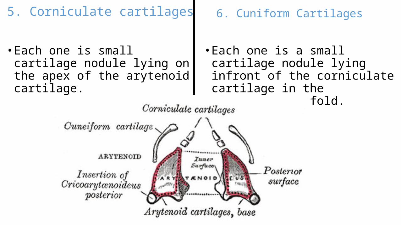
5. Corniculate cartilages
• Each one is small cartilage nodule lying on the apex of the arytenoid cartilage.
6. Cuniform Cartilages
• Each one is a small cartilage nodule lying infront of the corniculate cartilage in the oryepiglottic fold.
Membranes of the larynx
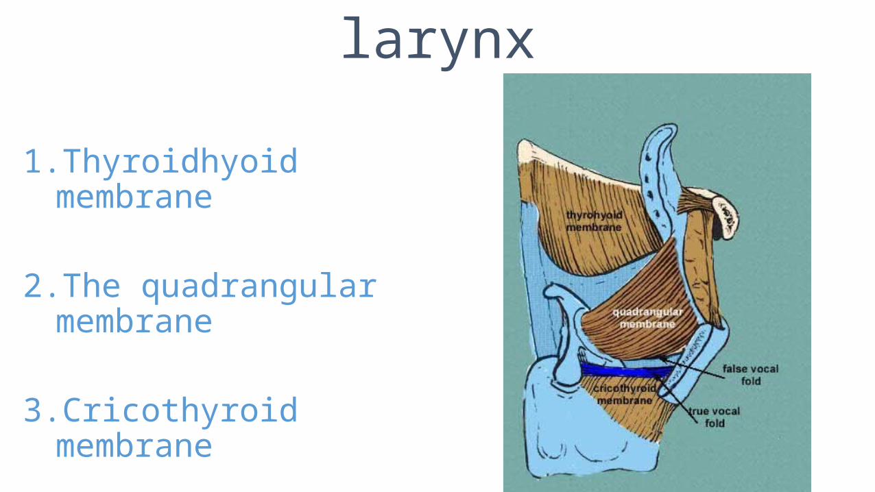
1. Thyroidhyoid membrane
2. The quadrangular membrane
3. Cricothyroid membrane
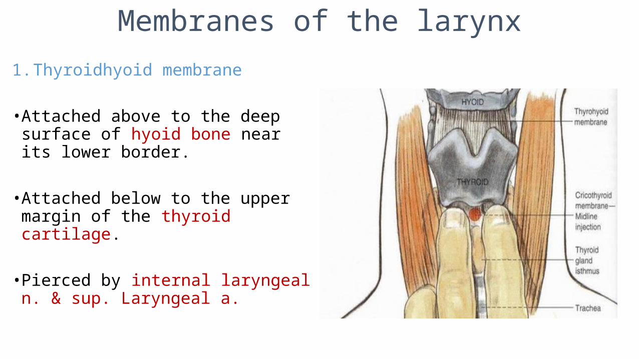
Membranes of the larynx
1. Thyroidhyoid membrane
• Attached above to the deep surface of hyoid bone near its lower border.
• Attached below to the upper margin of the thyroid cartilage.
• Pierced by internal laryngeal n. & sup. Laryngeal a.
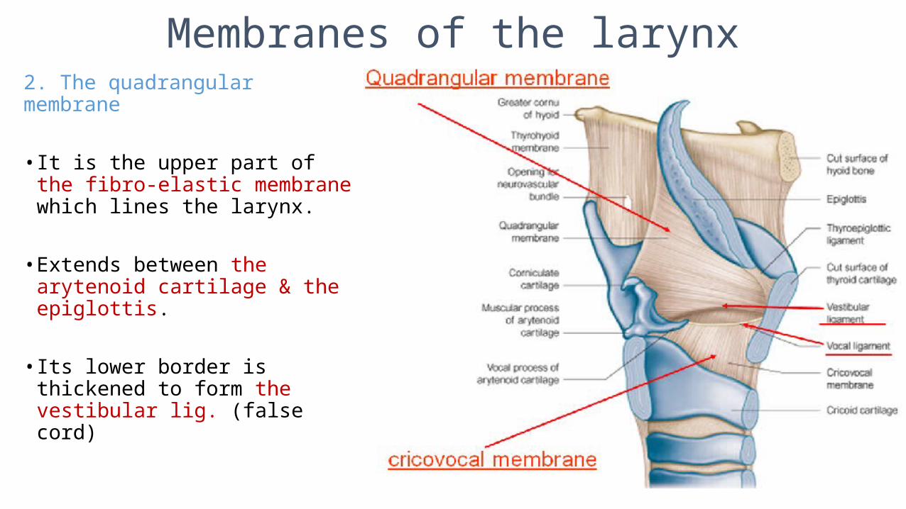
Membranes of the larynx2. The quadrangular membrane
• It is the upper part of the fibro-elastic membrane which lines the larynx.
• Extends between the arytenoid cartilage & the epiglottis.
• Its lower border is thickened to form the vestibular lig. (false cord)
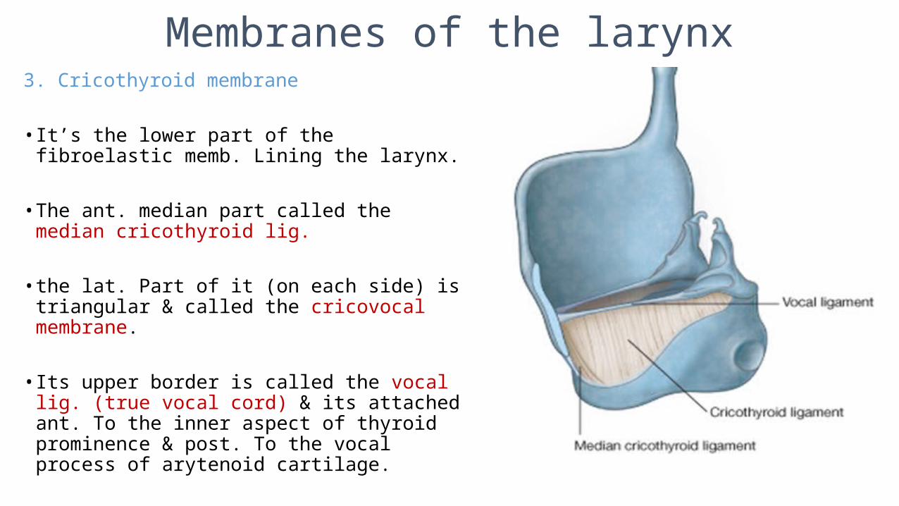
Membranes of the larynx3. Cricothyroid membrane
• It’s the lower part of the fibroelastic memb. Lining the larynx.
• The ant. median part called the median cricothyroid lig.
• the lat. Part of it (on each side) is triangular & called the cricovocal membrane.
• Its upper border is called the vocal lig. (true vocal cord) & its attached ant. To the inner aspect of thyroid prominence & post. To the vocal process of arytenoid cartilage.
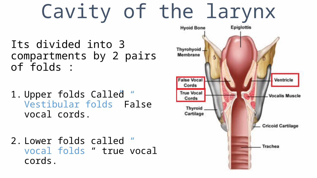
Cavity of the larynx
Its divided into 3 compartments by 2 pairs of folds :
1. Upper folds Called “ Vestibular folds” False vocal cords.
2. Lower folds called “ vocal folds “ true vocal cords.
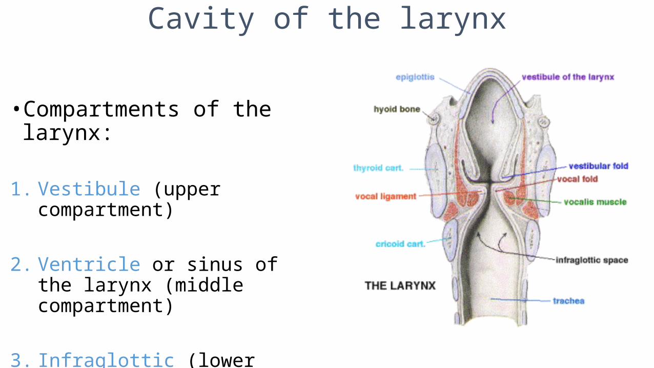
Cavity of the larynx
• Compartments of the larynx:
1. Vestibule (upper compartment)
2. Ventricle or sinus of the larynx (middle compartment)
3. Infraglottic (lower compartment)
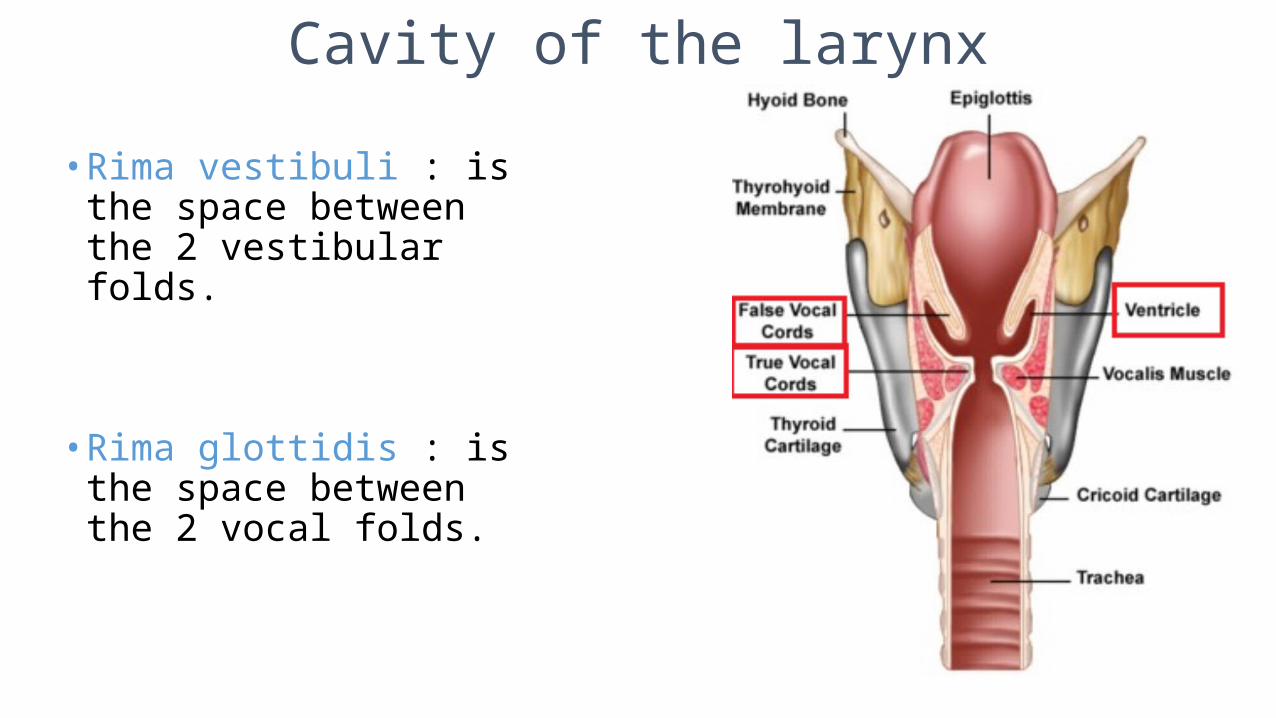
Cavity of the larynx
• Rima vestibuli : is the space between the 2 vestibular folds.
• Rima glottidis : is the space between the 2 vocal folds.
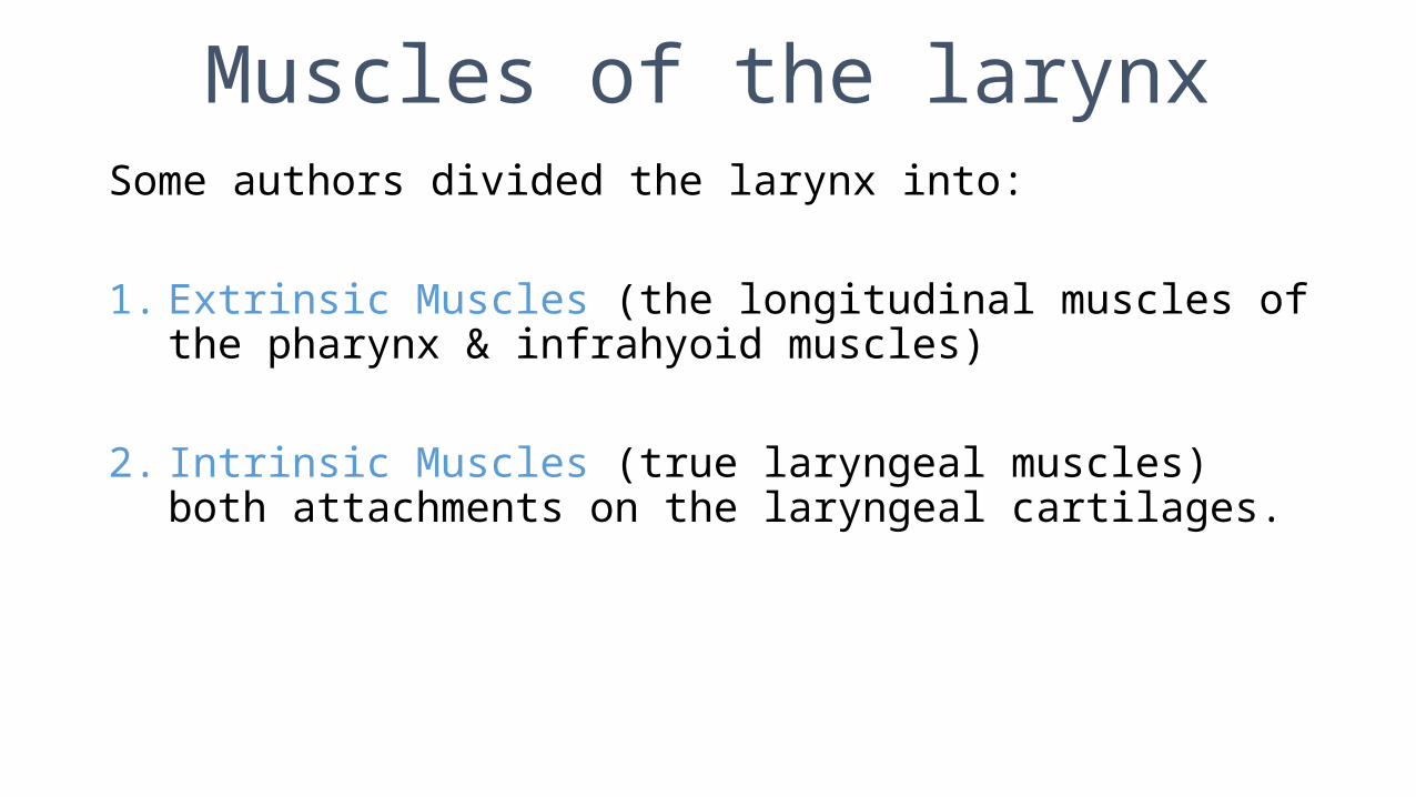
Muscles of the larynxSome authors divided the larynx into:
1. Extrinsic Muscles (the longitudinal muscles of the pharynx & infrahyoid muscles)
2. Intrinsic Muscles (true laryngeal muscles) both attachments on the laryngeal cartilages.
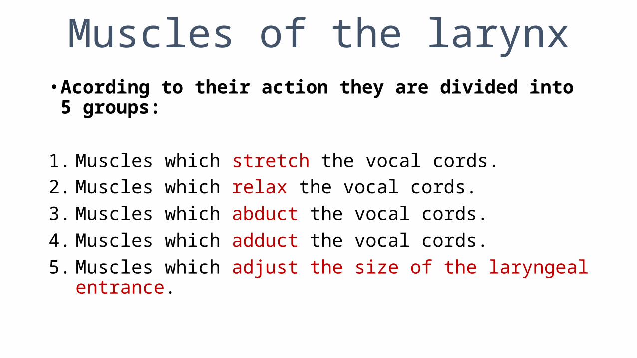
Muscles of the larynx• Acording to their action they are divided into 5 groups:
1. Muscles which stretch the vocal cords.2. Muscles which relax the vocal cords.3. Muscles which abduct the vocal cords.4. Muscles which adduct the vocal cords.5. Muscles which adjust the size of the laryngeal entrance.
Muscles of the larynx!
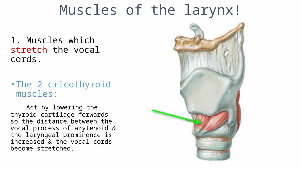
1. Muscles which stretch the vocal cords.
• The 2 cricothyroid muscles: Act by lowering the thyroid cartilage forwards so the distance between the vocal process of arytenoid & the laryngeal prominence is increased & the vocal cords become stretched.
Muscles of the larynx
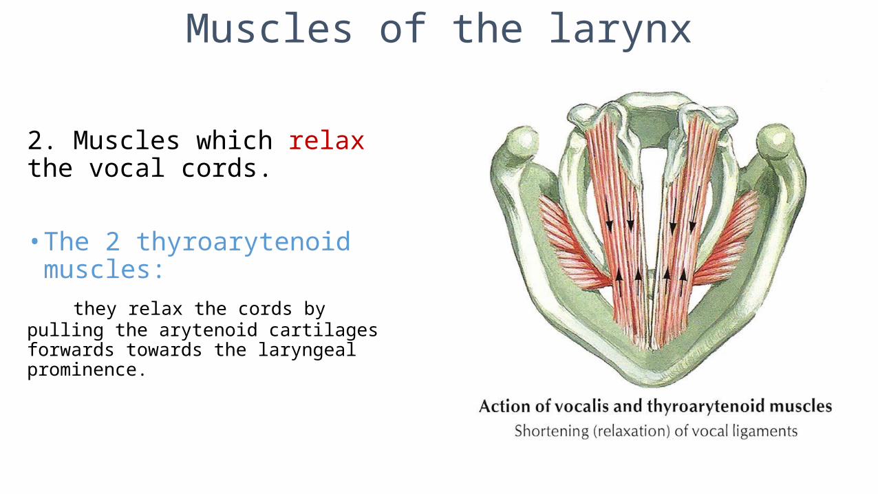
2. Muscles which relax the vocal cords.
• The 2 thyroarytenoid muscles: they relax the cords by pulling the arytenoid cartilages forwards towards the laryngeal prominence.
Muscles of the larynx
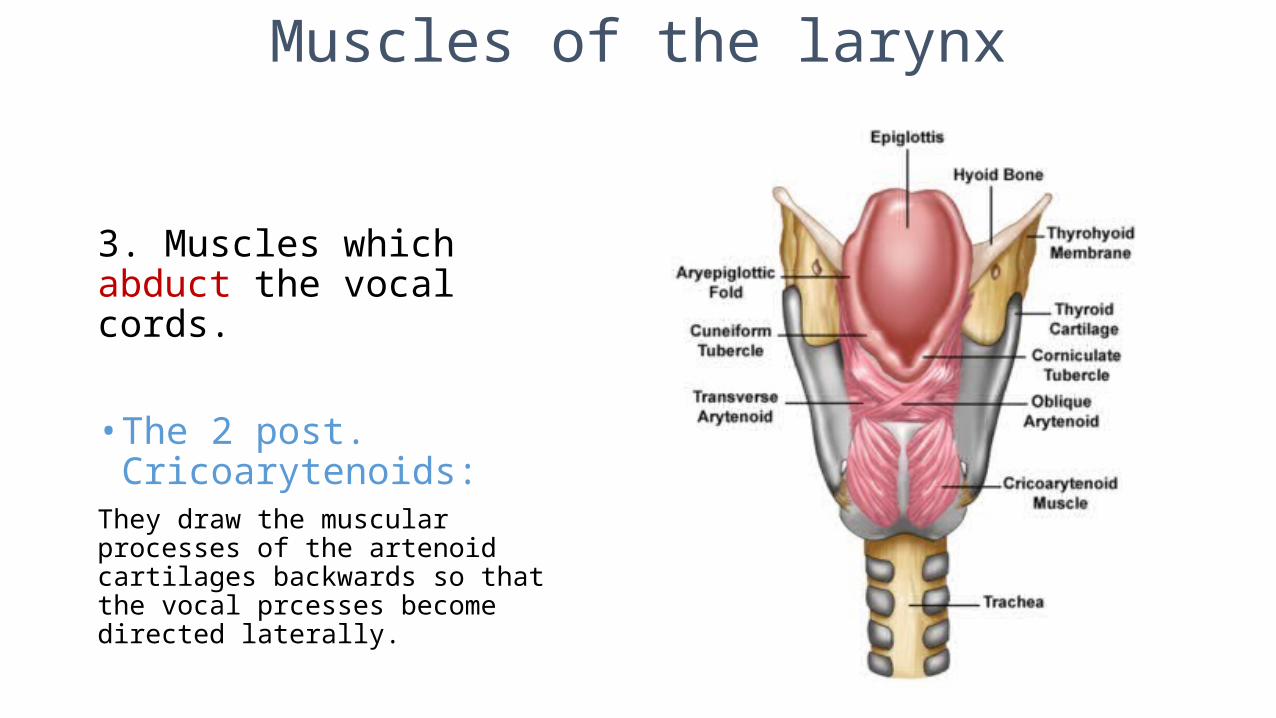
3. Muscles which abduct the vocal cords.
• The 2 post. Cricoarytenoids:They draw the muscular processes of the artenoid cartilages backwards so that the vocal prcesses become directed laterally.
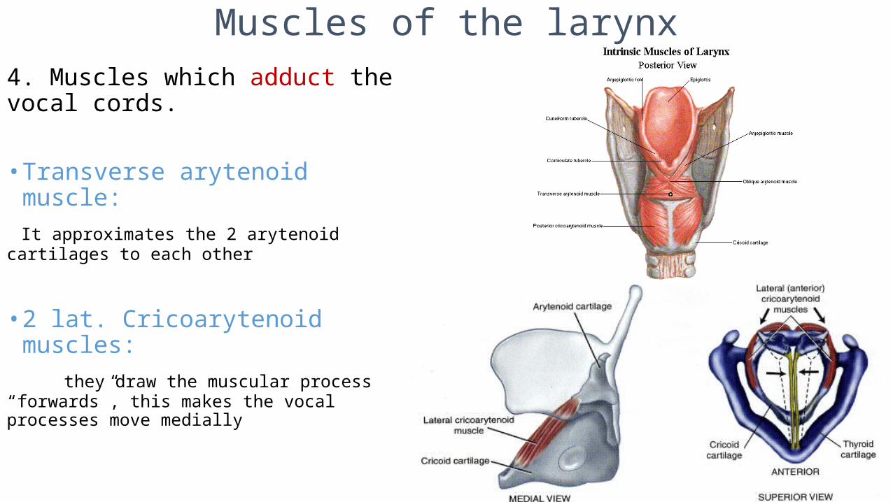
Muscles of the larynx4. Muscles which adduct the vocal cords.
• Transverse arytenoid muscle: It approximates the 2 arytenoid cartilages to each other
• 2 lat. Cricoarytenoid muscles: they draw the muscular process “forwards”, this makes the vocal processes move medially
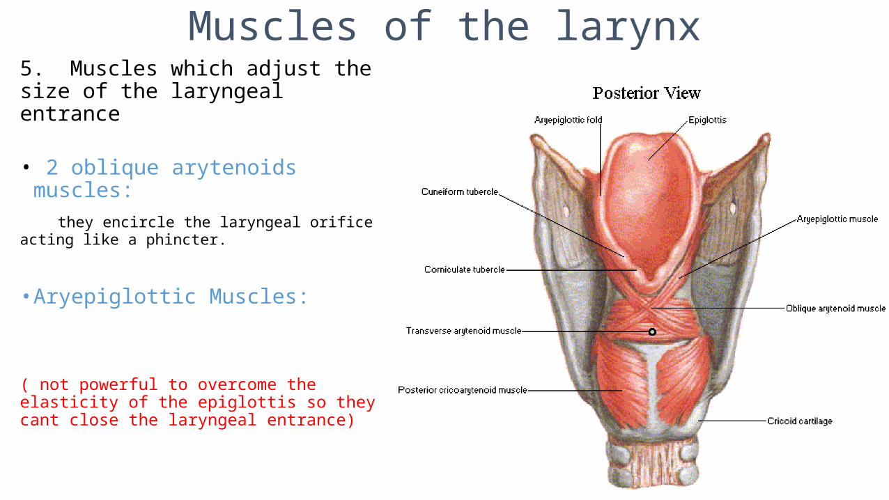
Muscles of the larynx5. Muscles which adjust the size of the laryngeal entrance
• 2 oblique arytenoids muscles: they encircle the laryngeal orifice acting like a phincter.
• Aryepiglottic Muscles:
( not powerful to overcome the elasticity of the epiglottis so they cant close the laryngeal entrance)

Arterial supply of the larynx
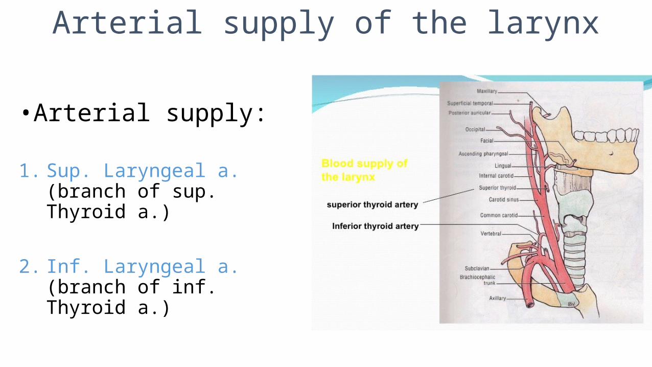
•Arterial supply:
1. Sup. Laryngeal a. (branch of sup. Thyroid a.)
2. Inf. Laryngeal a. (branch of inf. Thyroid a.)
Venous drainage of the larynx
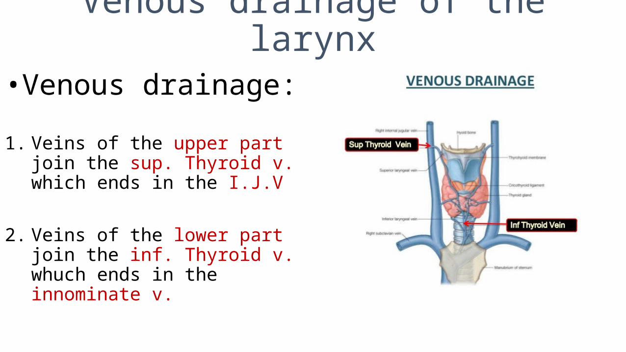
•Venous drainage:
1. Veins of the upper part join the sup. Thyroid v. which ends in the I.J.V
2. Veins of the lower part join the inf. Thyroid v. whuch ends in the innominate v.
Lymphatic drainage of the larynx
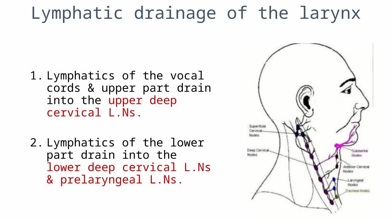
1. Lymphatics of the vocal cords & upper part drain into the upper deep cervical L.Ns.
2. Lymphatics of the lower part drain into the lower deep cervical L.Ns & prelaryngeal L.Ns.
Nerve supply of the larynx
1. Motor supply of the laryngeal muscles
2. Sensory supply of the mucous membrane
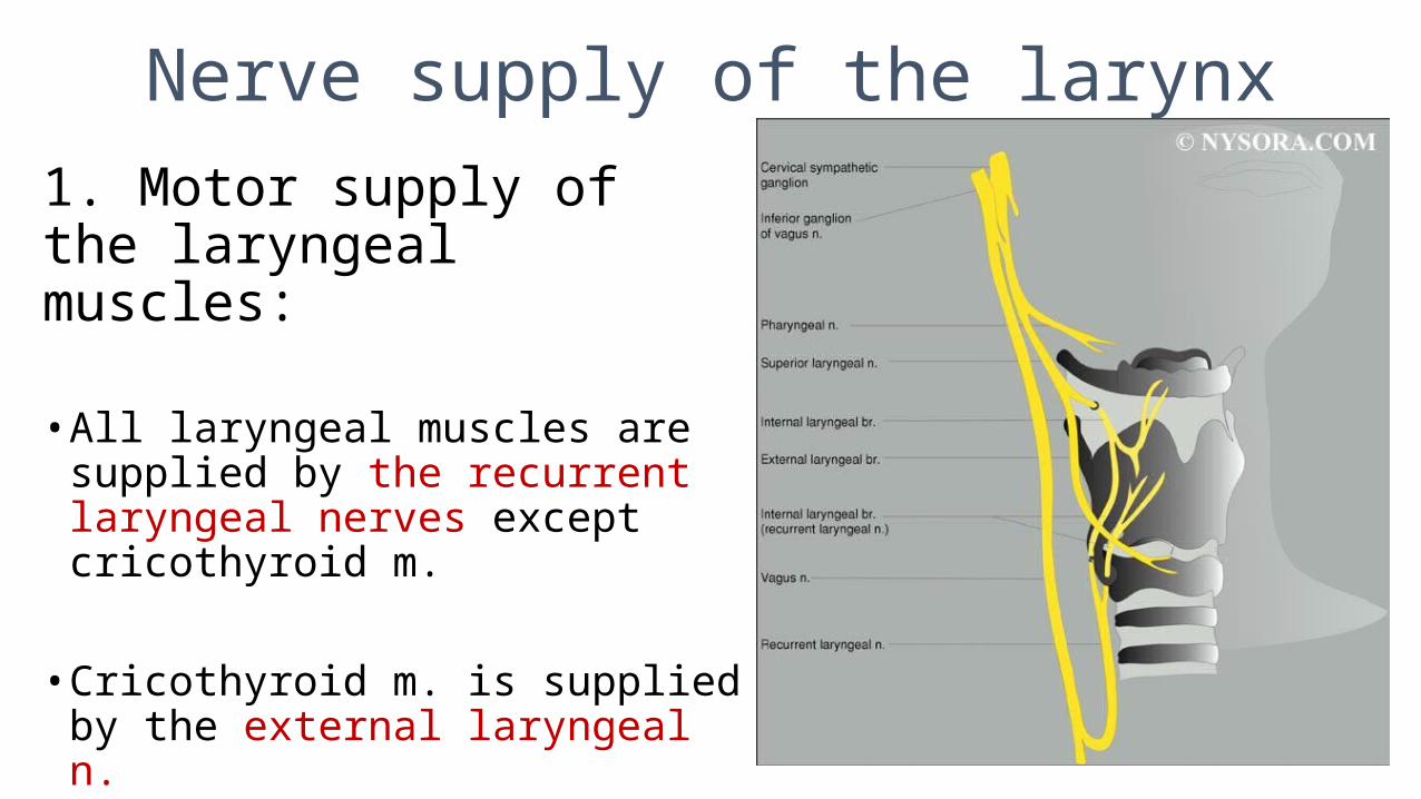
Nerve supply of the larynx1. Motor supply of the laryngeal muscles:
• All laryngeal muscles are supplied by the recurrent laryngeal nerves except cricothyroid m.
• Cricothyroid m. is supplied by the external laryngeal n.
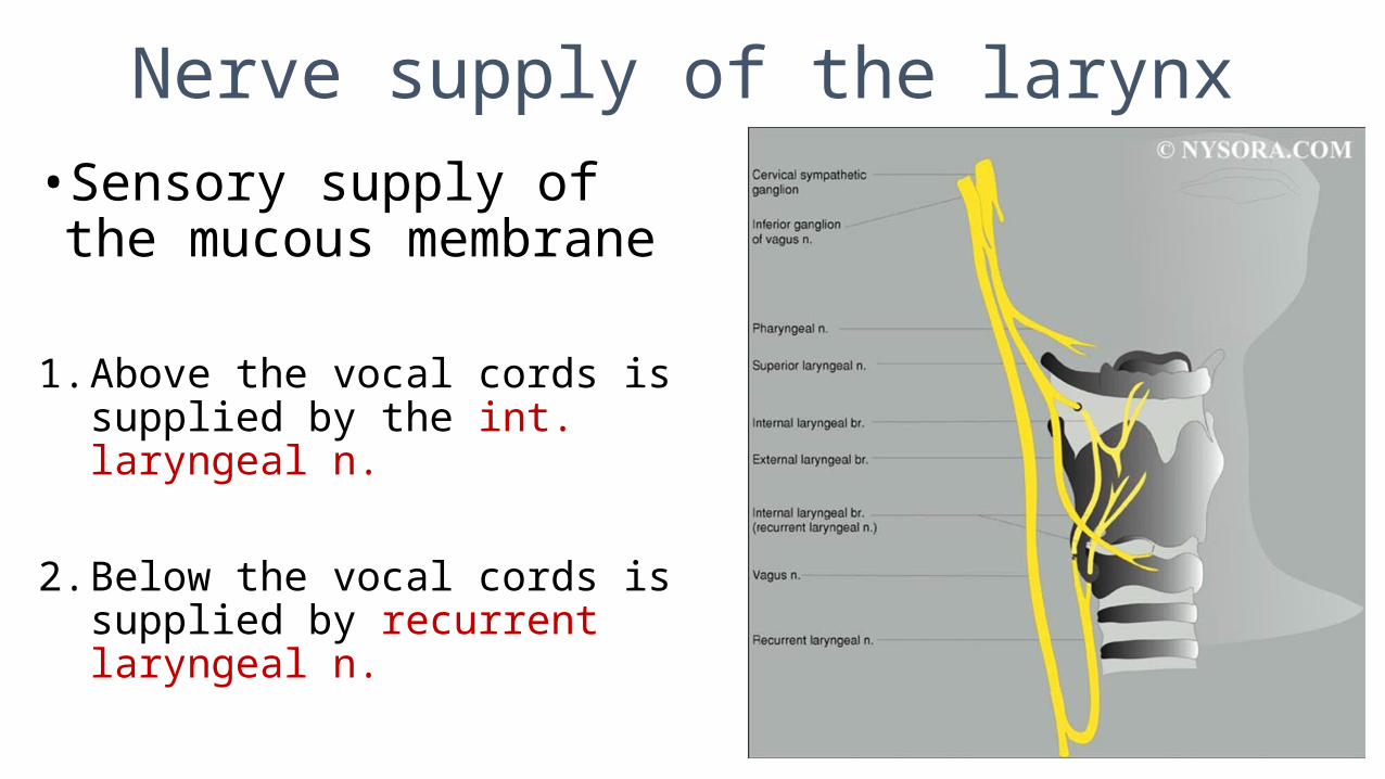
Nerve supply of the larynx• Sensory supply of the mucous
membrane
1. Above the vocal cords is supplied by the int. laryngeal n.
2. Below the vocal cords is supplied by recurrent laryngeal n.