Embed Size (px)
DESCRIPTION
Embryology ,Anatomy and physiology of Larynx. Prepaired by: Dr. Hiwa As’ad Abdulkareem. Sources : Scott-browns of otolaryngology , head and neck surgery . Synopsis of otolaryngology . www.drtbalu.com www.wekipedia.com. Embryology of larynx. Development of larynx : - PowerPoint PPT Presentation
Citation preview
Embryology ,Anatomy and physiology of Larynx
Prepaired by: Dr. Hiwa As’ad
Abdulkareem
• Sources :• Scott-browns of otolaryngology , head and
neck surgery .• Synopsis of otolaryngology .• www.drtbalu.com• www.wekipedia.com
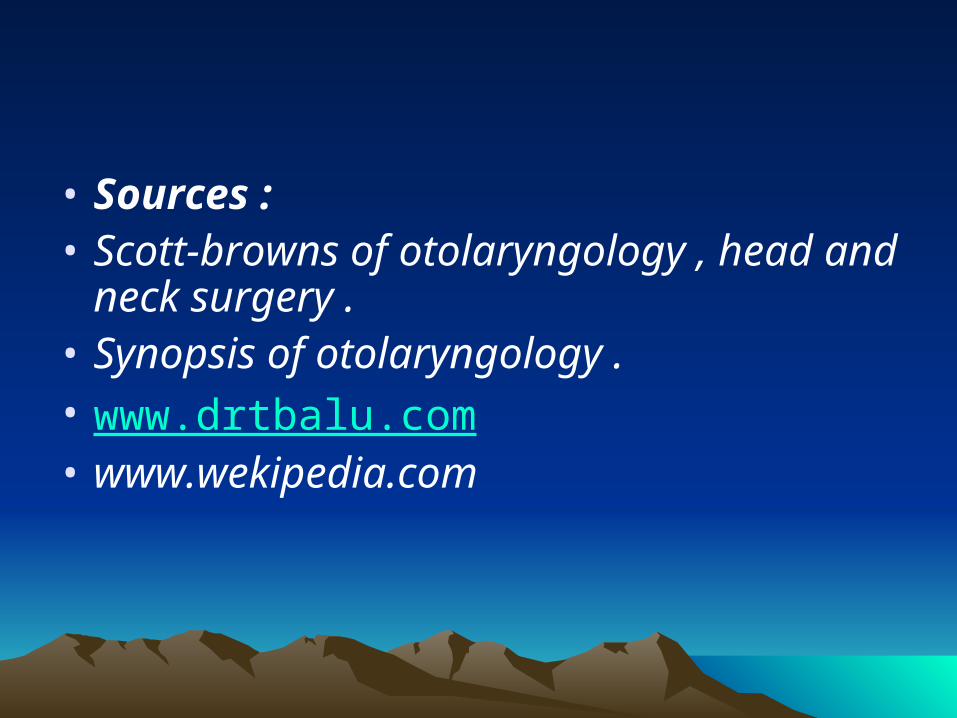
Embryology of larynxEmbryology of larynx Development of larynx :
•during the 4th week of intra uterine life.
•starts in the form of laryngotracheal groove in the ventral wall of the pharynx.
•The groove gradually deepens and its edges fuse to form a septum, this septum separates the laryngotracheal tube from the pharynx and oesophagus.
•The process of this fusion starts caudally and extend cranially.
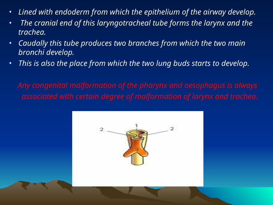
• Lined with endoderm from which the epithelium of the airway develop.• The cranial end of this laryngotracheal tube forms the larynx and the
trachea. • Caudally this tube produces two branches from which the two main
bronchi develop. • This is also the place from which the two lung buds starts to develop.
Any congenital malformation of the pharynx and oesophagus is always
associated with certain degree of malformation of larynx and trachea.
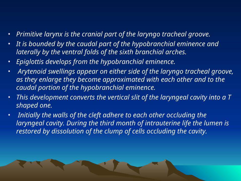
• Primitive larynx is the cranial part of the laryngo tracheal groove. • It is bounded by the caudal part of the hypobranchial eminence and
laterally by the ventral folds of the sixth branchial arches. • Epiglottis develops from the hypobranchial eminence.• Arytenoid swellings appear on either side of the laryngo tracheal groove,
as they enlarge they become approximated with each other and to the caudal portion of the hypobranchial eminence.
• This development converts the vertical slit of the laryngeal cavity into a T shaped one.
• Initially the walls of the cleft adhere to each other occluding the laryngeal cavity. During the third month of intrauterine life the lumen is restored by dissolution of the clump of cells occluding the cavity.
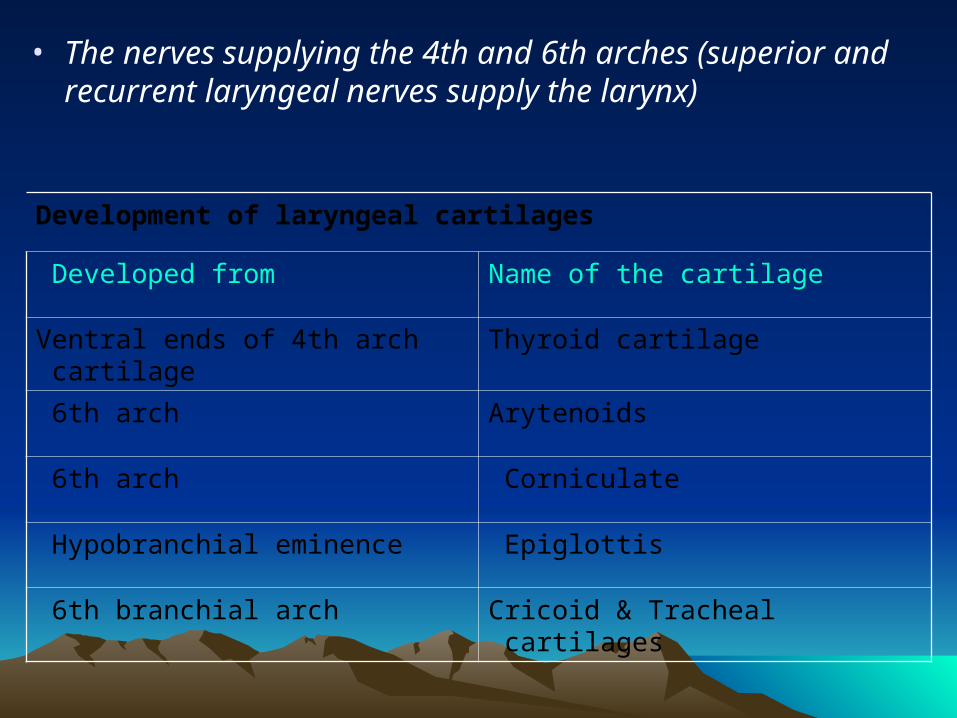
• The nerves supplying the 4th and 6th arches (superior and recurrent laryngeal nerves supply the larynx)
Development of laryngeal cartilages
Name of the cartilage Developed from
Thyroid cartilage Ventral ends of 4th arch cartilage
Arytenoids 6th arch
Corniculate 6th arch
Epiglottis Hypobranchial eminence
Cricoid & Tracheal cartilages 6th branchial arch
ANATOMY OF THE LARYNXANATOMY OF THE LARYNXIntroduction:•Situated above the trachea. • Extends from the laryngeal inlet to the inferior border of the cricoid cartilage .•Opposite the third to sixth cervical vertebrae, being a little higher in women than in men. The infantile larynx: •Smaller than the adult compared to body size •More funnel shaped.•Its narrowest part is at the junction of the subglottic larynx with the trachea and even slight swelling in this area may result in marked airway obstruction .• Cartilages are much softer and collapse more easily on forced inspiration. •The larynx starts high up under the tongue in early life and with age assumes an increasingly lower position in the neck .
• The size of the larynx is almost the same in boys and girls till puberty. After puberty the antero posterior diameter of the larynx virtually doubles in males .
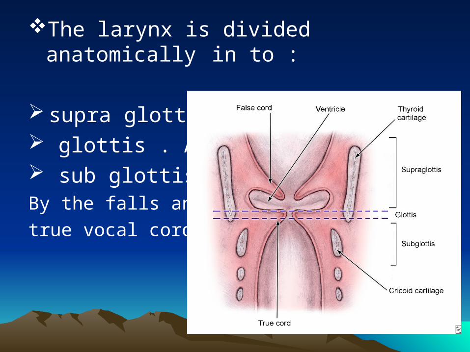
The larynx is divided anatomically in to :
supra glottis . glottis . And sub glottis .By the falls and
true vocal cords.
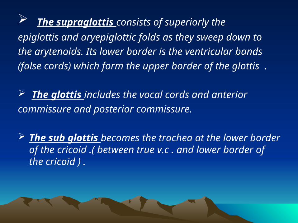
The supraglottis consists of superiorly the
epiglottis and aryepiglottic folds as they sweep down to
the arytenoids. Its lower border is the ventricular bands
(false cords) which form the upper border of the glottis .
The glottis includes the vocal cords and anterior
commissure and posterior commissure.
The sub glottis becomes the trachea at the lower border of the cricoid .( between true v.c . and lower border of the cricoid ) .
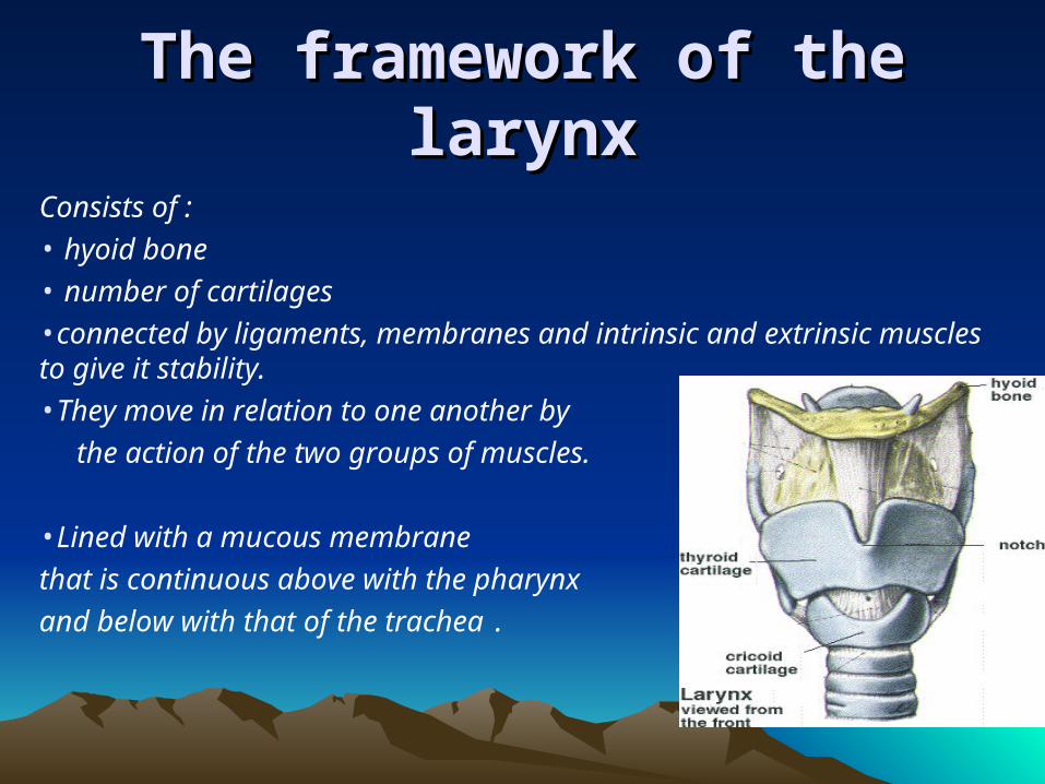
The framework of the larynxThe framework of the larynx
Consists of : • hyoid bone• number of cartilages •connected by ligaments, membranes and intrinsic and extrinsic muscles to give it stability.•They move in relation to one another by
the action of the two groups of muscles.
•Lined with a mucous membrane
that is continuous above with the pharynx
and below with that of the trachea .
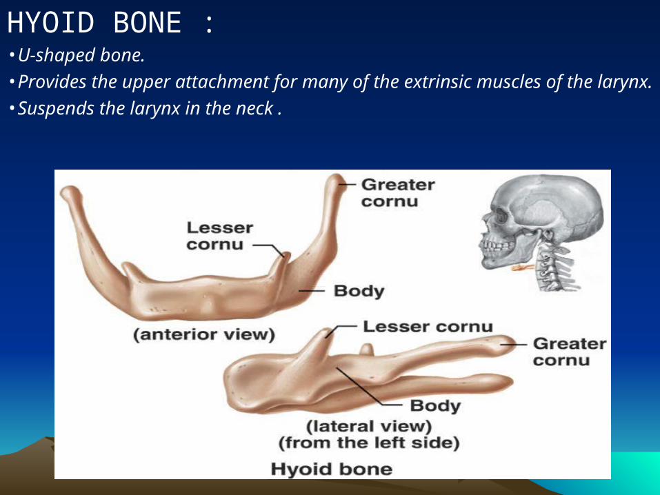
HYOID BONE : •U-shaped bone. •Provides the upper attachment for many of the extrinsic muscles of the larynx.•Suspends the larynx in the neck .
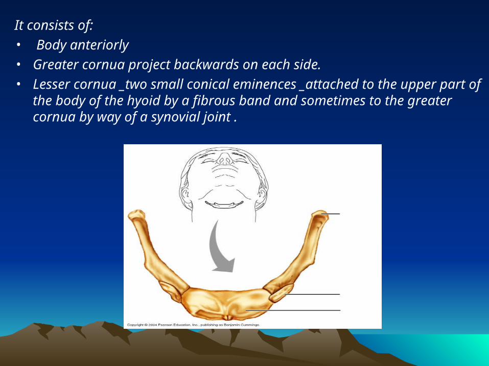
It consists of:• Body anteriorly • Greater cornua project backwards on each side. • Lesser cornua _two small conical eminences _attached to the upper part
of the body of the hyoid by a fibrous band and sometimes to the greater cornua by way of a synovial joint .
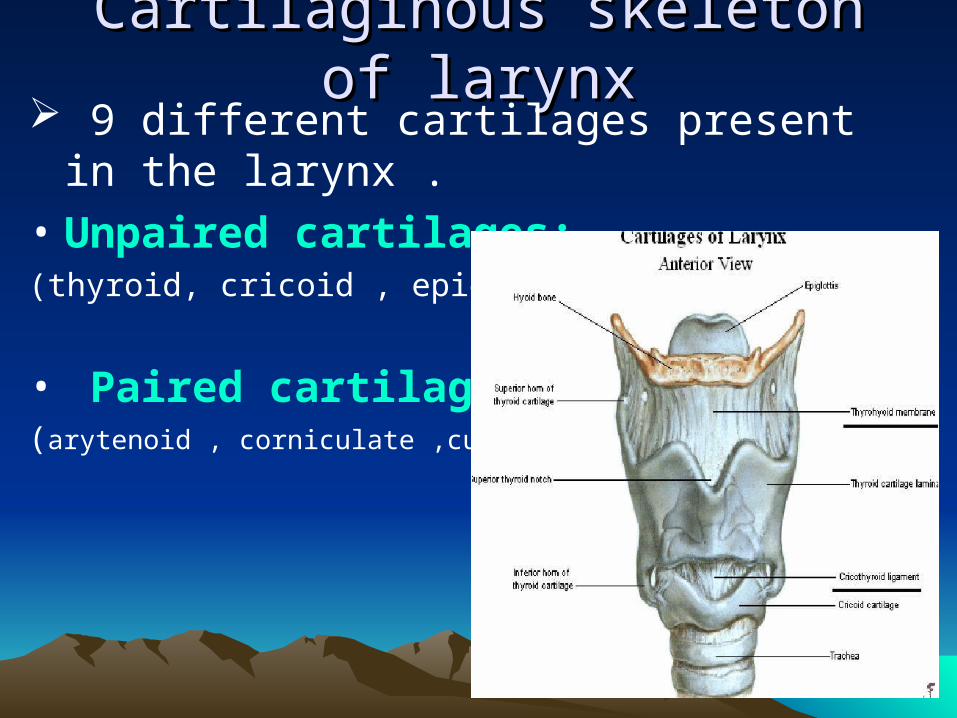
Cartilaginous skeleton of larynxCartilaginous skeleton of larynx 9 different cartilages present in the larynx .
• Unpaired cartilages:(thyroid, cricoid , epiglottis)
• Paired cartilages:(arytenoid , corniculate ,cuneiform)
THYROID CARTILAGE:
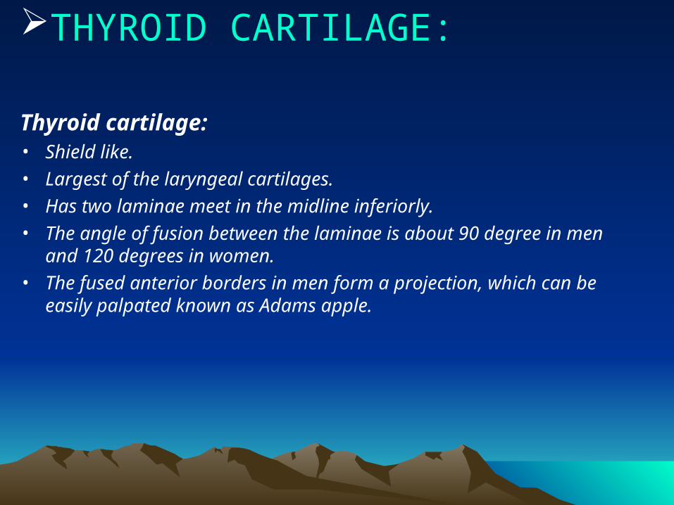
Thyroid cartilage: • Shield like.• Largest of the laryngeal cartilages. • Has two laminae meet in the midline inferiorly. • The angle of fusion between the laminae is about 90 degree in men
and 120 degrees in women. • The fused anterior borders in men form a projection, which can be
easily palpated known as Adams apple.
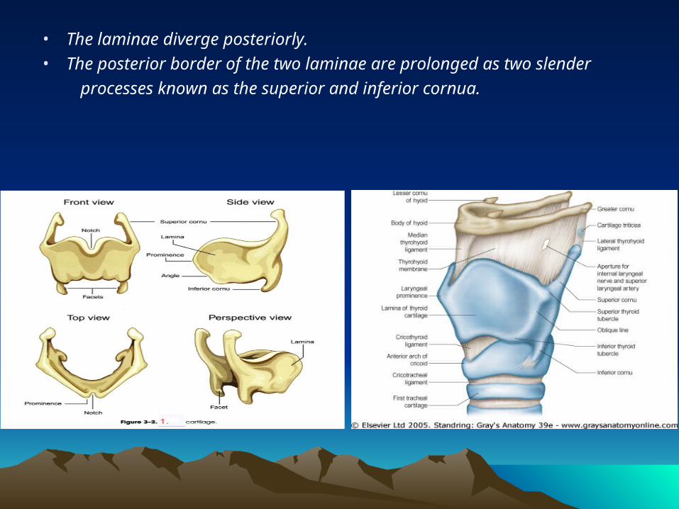
• The laminae diverge posteriorly.
• The posterior border of the two laminae are prolonged as two slender
processes known as the superior and inferior cornua.
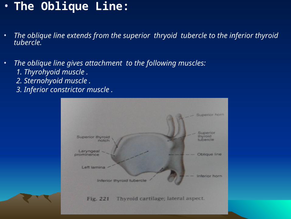
• The Oblique Line:
• The oblique line extends from the superior thryoid tubercle to the inferior thyroid tubercle.
• The oblique line gives attachment to the following muscles: 1. Thyrohyoid muscle . 2. Sternohyoid muscle . 3. Inferior constrictor muscle .
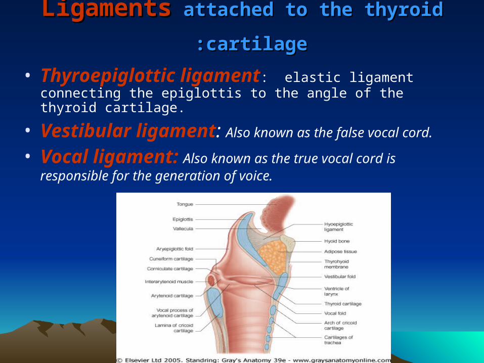
LigamentsLigaments attached to the thyroid cartilage attached to the thyroid cartilage::
• Thyroepiglottic ligament: elastic ligament connecting the epiglottis to the angle of the thyroid cartilage.
• Vestibular ligament: Also known as the false vocal cord.
• Vocal ligament: Also known as the true vocal cord is responsible for the generation of voice.
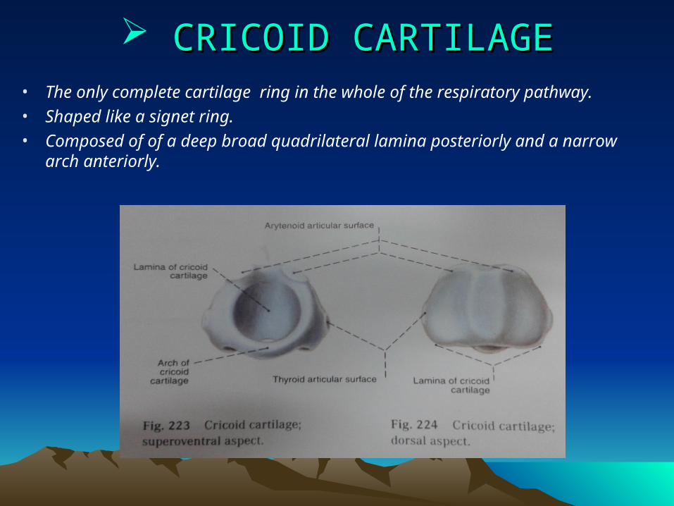
CRICOID CARTILAGECRICOID CARTILAGE• The only complete cartilage ring in the whole of the respiratory pathway.
• Shaped like a signet ring.
• Composed of of a deep broad quadrilateral lamina posteriorly and a narrow arch anteriorly.
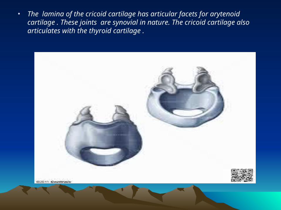
• The lamina of the cricoid cartilage has articular facets for arytenoid cartilage . These joints are synovial in nature. The cricoid cartilage also articulates with the thyroid cartilage .
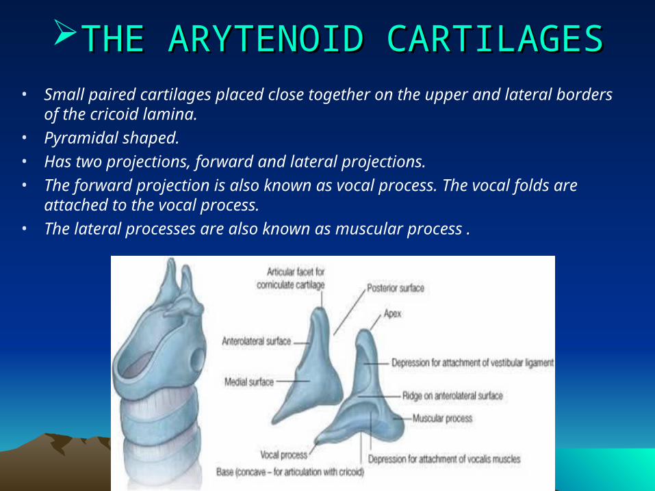
THE ARYTENOID CARTILAGESTHE ARYTENOID CARTILAGES• Small paired cartilages placed close together on the upper and lateral borders of
the cricoid lamina.
• Pyramidal shaped.
• Has two projections, forward and lateral projections.
• The forward projection is also known as vocal process. The vocal folds are attached to the vocal process.
• The lateral processes are also known as muscular process .
• The apex of this cartilage curves backwards and articulates with corniculate cartilages. Aryepiglottic folds are attached to these cartilages.
CORNICULATE AND CUNEIFORM CARTILAGESCORNICULATE AND CUNEIFORM CARTILAGES
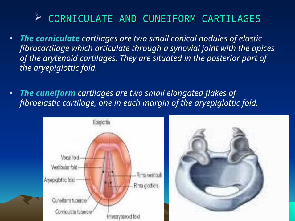
• The corniculate cartilages are two small conical nodules of elastic fibrocartilage which articulate through a synovial joint with the apices of the arytenoid cartilages. They are situated in the posterior part of the aryepiglottic fold.
• The cuneiform cartilages are two small elongated flakes of fibroelastic cartilage, one in each margin of the aryepiglottic fold.
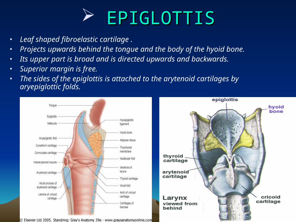
EPIGLOTTISEPIGLOTTIS• Leaf shaped fibroelastic cartilage .• Projects upwards behind the tongue and the body of the hyoid bone. • Its upper part is broad and is directed upwards and backwards. • Superior margin is free. • The sides of the epiglottis is attached to the arytenoid cartilages by
aryepiglottic folds.
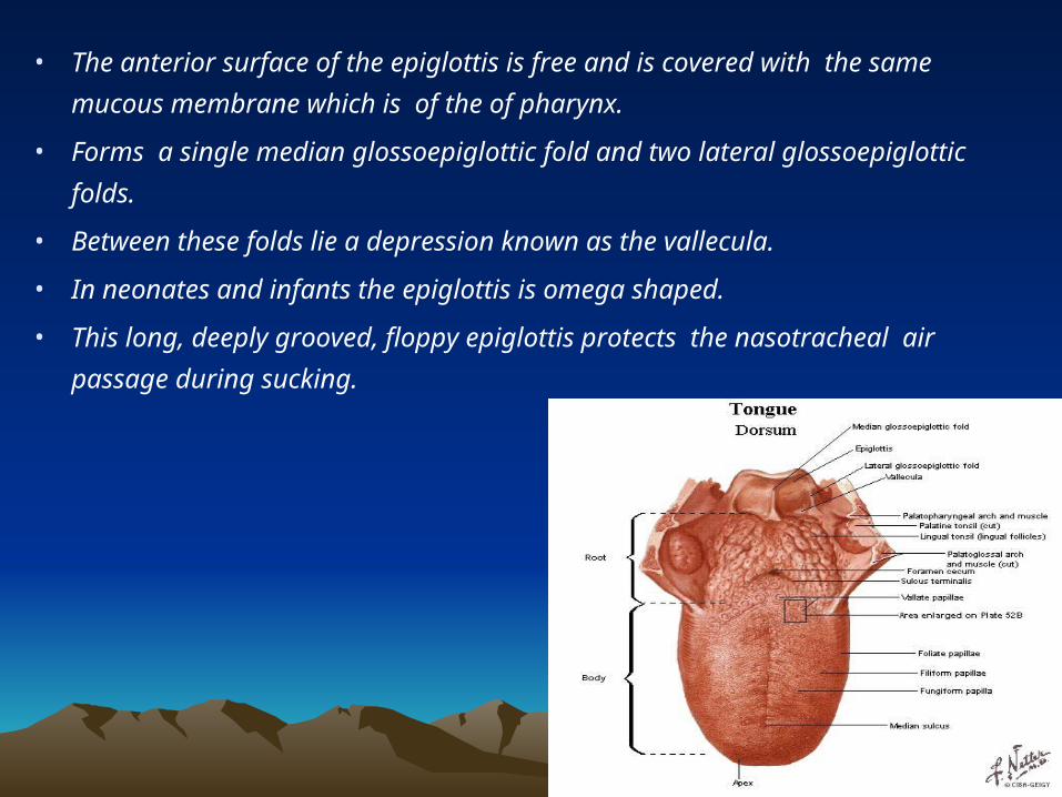
• The anterior surface of the epiglottis is free and is covered with the same
mucous membrane which is of the of pharynx.
• Forms a single median glossoepiglottic fold and two lateral glossoepiglottic
folds.
• Between these folds lie a depression known as the vallecula.
• In neonates and infants the epiglottis is omega shaped.
• This long, deeply grooved, floppy epiglottis protects the nasotracheal air
passage during sucking.
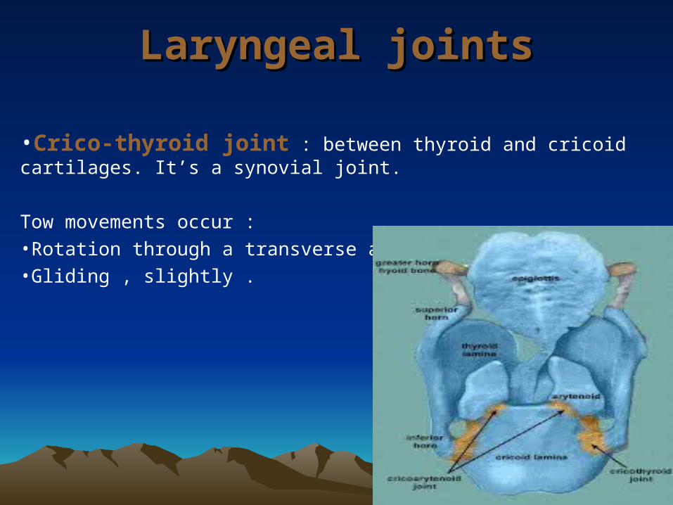
Laryngeal jointsLaryngeal joints
•Crico-thyroid joint : between thyroid and cricoid cartilages. It’s a synovial joint.
Tow movements occur :•Rotation through a transverse axis .•Gliding , slightly .
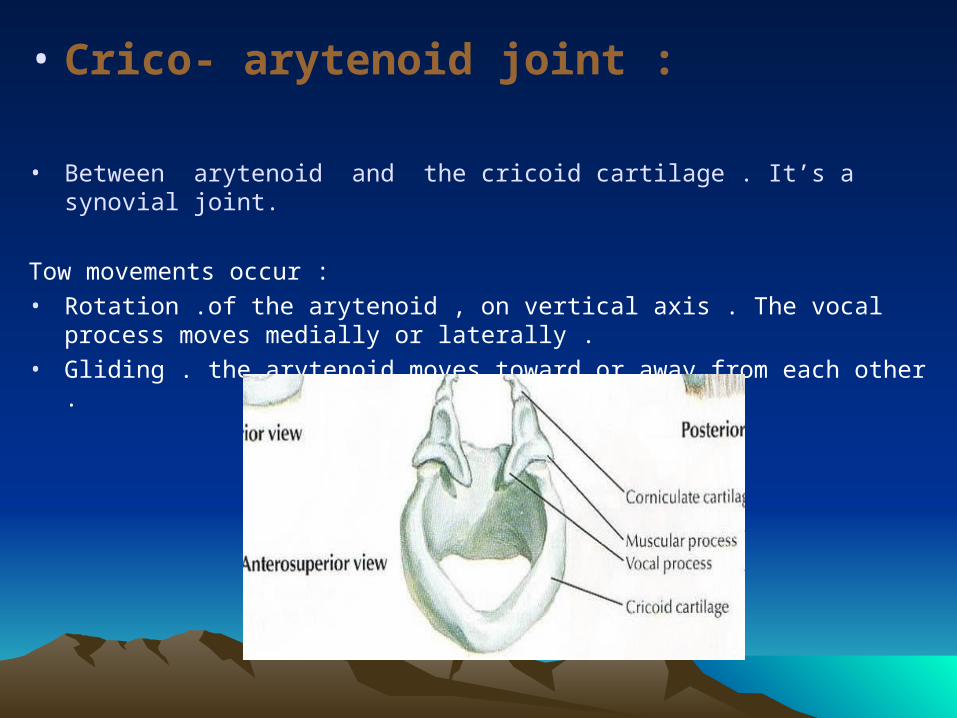
• Crico- arytenoid joint :
• Between arytenoid and the cricoid cartilage . It’s a synovial joint.
Tow movements occur :
• Rotation .of the arytenoid , on vertical axis . The vocal process moves medially or laterally .
• Gliding . the arytenoid moves toward or away from each other .
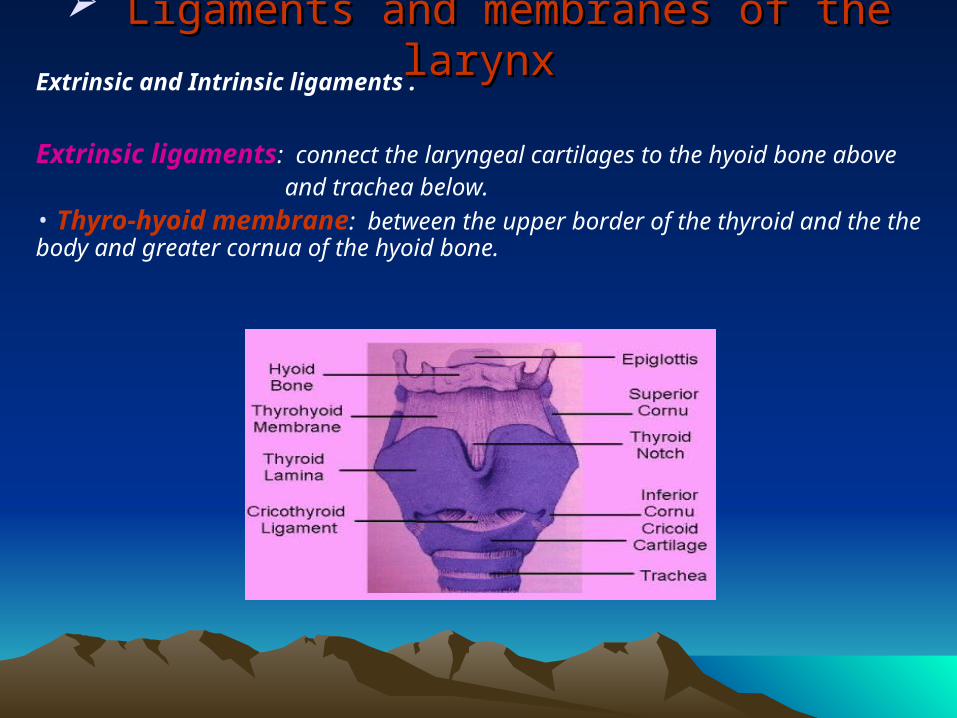
Ligaments and membranes of the larynxLigaments and membranes of the larynxExtrinsic and Intrinsic ligaments .
Extrinsic ligaments: connect the laryngeal cartilages to the hyoid bone above and trachea below.• Thyro-hyoid membrane: between the upper border of the thyroid and the the body and greater cornua of the hyoid bone.
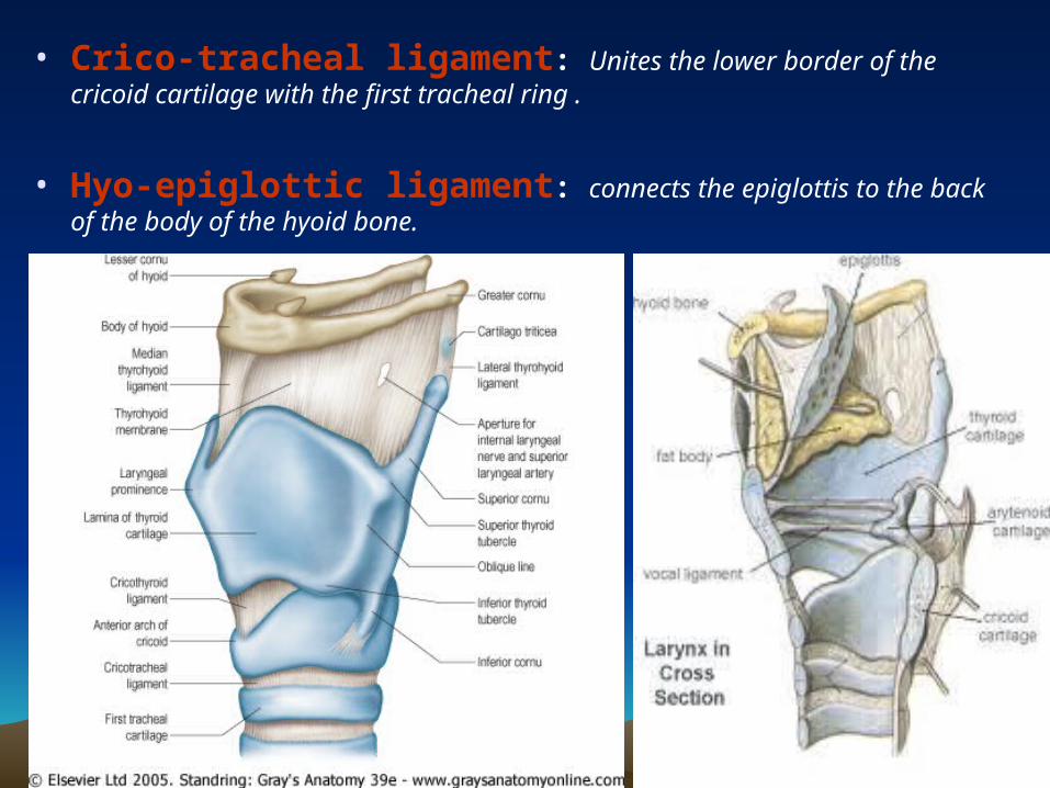
• Crico-tracheal ligament: Unites the lower border of the cricoid cartilage with the first tracheal ring .
• Hyo-epiglottic ligament: connects the epiglottis to the back of the body of the hyoid bone.
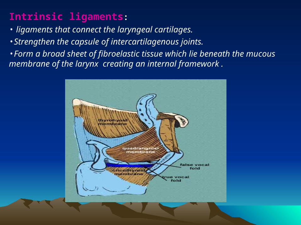
Intrinsic ligaments:
• ligaments that connect the laryngeal cartilages. •Strengthen the capsule of intercartilagenous joints. •Form a broad sheet of fibroelastic tissue which lie beneath the mucous membrane of the larynx creating an internal framework .
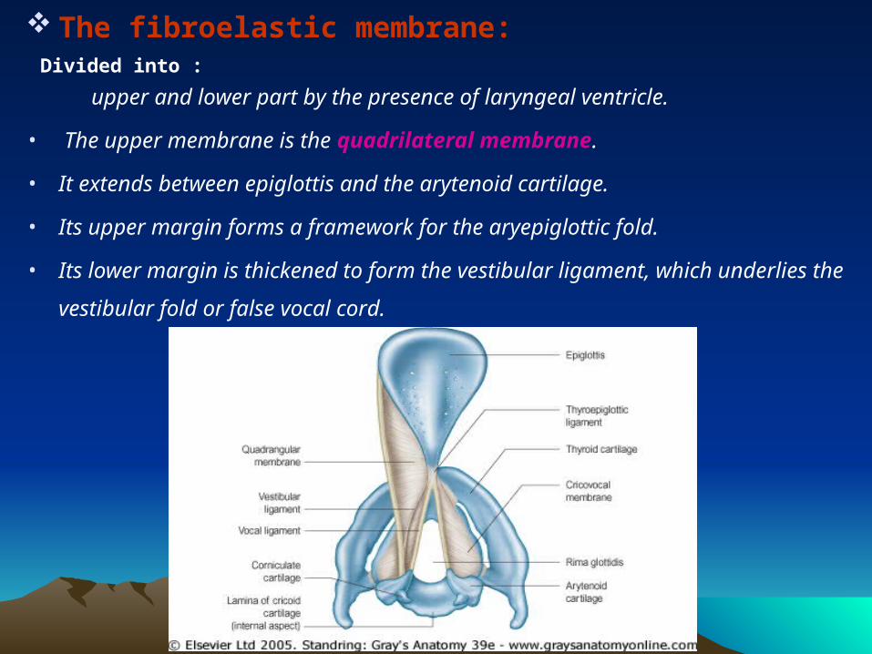
The fibroelastic membrane: Divided into :
upper and lower part by the presence of laryngeal ventricle.
• The upper membrane is the quadrilateral membrane.
• It extends between epiglottis and the arytenoid cartilage.
• Its upper margin forms a framework for the aryepiglottic fold.
• Its lower margin is thickened to form the vestibular ligament, which underlies the
vestibular fold or false vocal cord.

The lower part is a thicker membrane, containing many elastic fibers. It is also known as cricovocal ligament or cricothryoid ligament or conus elasticus. •Below it is attached to the upper border of the cricoid cartilage.•Above it is stretched between the midpoint of thyroid cartilage anteriorly and the vocal process of the arytenoid behind. •The free upper border of this membrane forms the vocal cord.
Muscles of the larynxMuscles of the larynx
The extrinsic muscles .
intrinsic muscles .
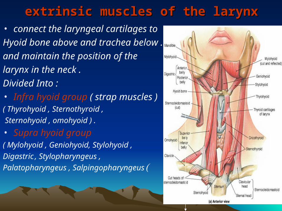
extrinsic muscles of the larynxextrinsic muscles of the larynx• connect the laryngeal cartilages to
Hyoid bone above and trachea below .
and maintain the position of the
larynx in the neck .
Divided Into :• Infra hyoid group ( strap muscles )( Thyrohyoid , Sternothyroid ,
Sternohyoid , omohyoid ) .
• Supra hyoid group
( Mylohyoid , Geniohyoid, Stylohyoid ,
Digastric , Stylopharyngeus ,
Palatopharyngeus , Salpingopharyngeus (
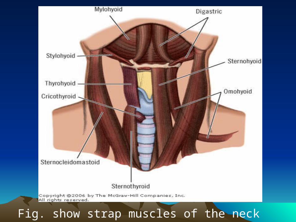
Fig. show strap muscles of the neck
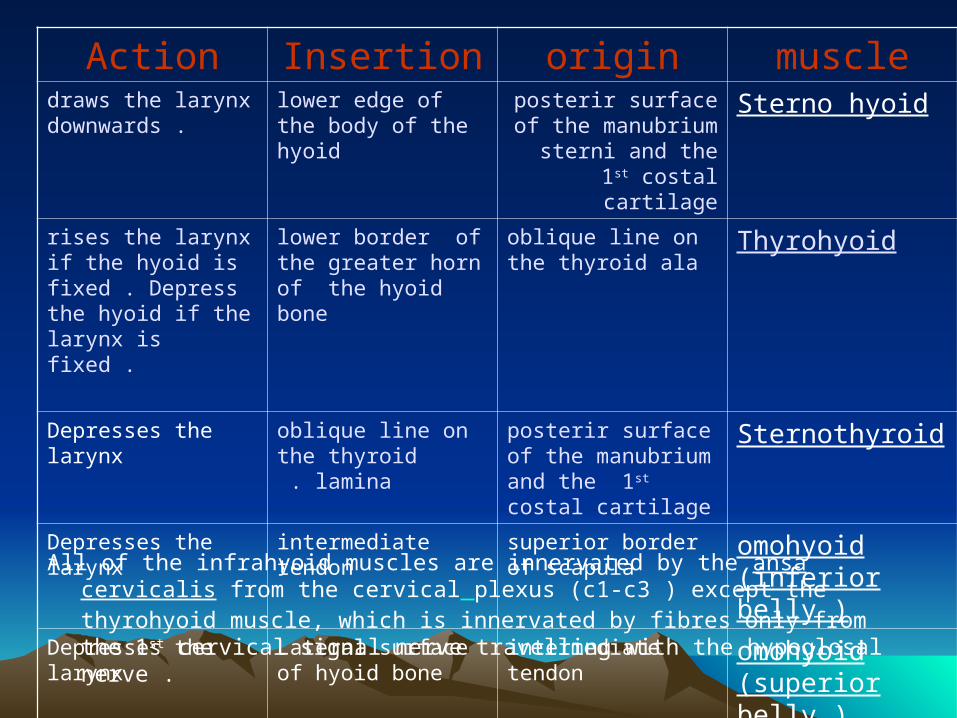
All of the infrahyoid muscles are innervated by the ansa cervicalis from the cervical plexus (c1-c3 ) except the thyrohyoid muscle, which is innervated by fibres only from the 1st cervical signal nerve travelling with the hypoglosal nerve .
muscleoriginInsertionActionSterno hyoidposterir surface of the
manubrium sterni and the 1st costal cartilage
lower edge of the body of the hyoid
draws the larynx downwards .
Thyrohyoidoblique line on the thyroid ala
lower border of the greater horn of the hyoid bone
rises the larynx if the hyoid is fixed . Depress the hyoid if the larynx is fixed .
Sternothyroidposterir surface of the manubrium and the 1st costal cartilage
oblique line on the thyroid lamina .
Depresses the larynx
omohyoid (inferior belly )
superior border of scapula
intermediate tendonDepresses the larynx
omohyoid (superior belly )
intermediate tendonLateral surface of hyoid bone
Depresses the larynx
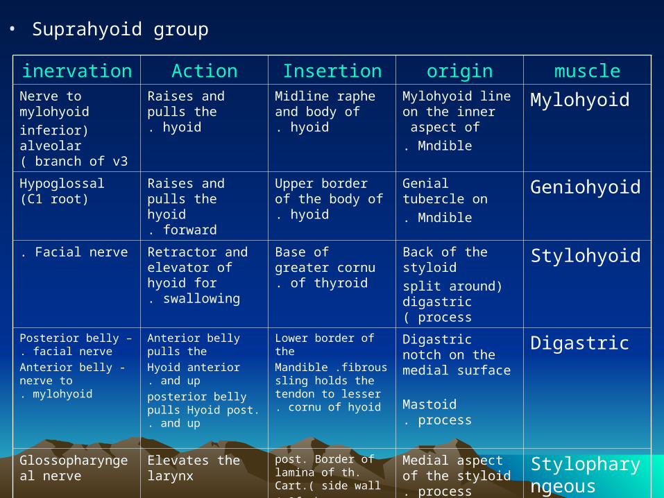
• Suprahyoid group
muscleoriginInsertionActioninervation
MylohyoidMylohyoid line on the inner aspect of
Mndible.
Midline raphe and body of hyoid.
Raises and pulls the hyoid.
Nerve to mylohyoid
)inferior alveolar branch of v3(
GeniohyoidGenial tubercle on
Mndible.
Upper border of the body of hyoid.
Raises and pulls the hyoid forward.
Hypoglossal (C1 root)
StylohyoidBack of the styloid
)split around digastric process(
Base of greater cornu of thyroid.
Retractor and elevator of hyoid for
swallowing.
Facial nerve.
DigastricDigastric notch on the medial surface
Mastoid process.
Lower border of the
Mandible .fibrous sling holds the tendon to
lesser cornu of hyoid.
Anterior belly pulls the
Hyoid anterior and up.
posterior belly pulls Hyoid post. and up.
Posterior belly – facial nerve.
Anterior belly - nerve to mylohyoid.
Stylopharyngeous
Medial aspect of the styloid
process.
post. Border of lamina of th. Cart.( side wall
Of pharynx(
Elevates the larynxGlossopharyngeal nerve
Palatopharyngeus
Palatine aponeurosis
And post. Margin of palat.
post . Border of thyroid alar and cornu.
Helps tilts the larynx forward.
Accessory nerve
pharyngeal plexus((
Salpingopharyngeus
Eustachian tubepost . Border of thyroid cart. .( side wall of pharynx )
Elevates the larynxPharyngeal plexus
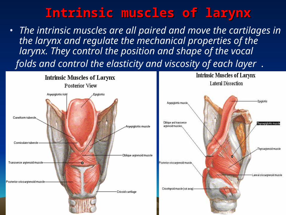
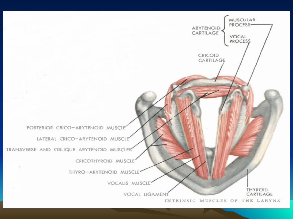
Intrinsic muscles of larynxIntrinsic muscles of larynx • The intrinsic muscles are all paired and move the cartilages in
the larynx and regulate the mechanical properties of the larynx. They control the position and shape of the vocal
folds and control the elasticity and viscosity of each layer .
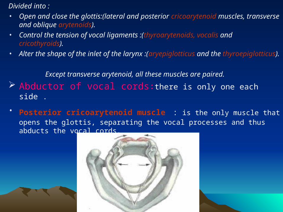
Divided into :
• Open and close the glottis:(lateral and posterior cricoarytenoid muscles, transverse and oblique arytenoids).
• Control the tension of vocal ligaments :(thyroarytenoids, vocalis and cricothyroids).
• Alter the shape of the inlet of the larynx :(aryepiglotticus and the thyroepiglotticus).
Except transverse arytenoid, all these muscles are paired.
Abductor of vocal cords : there is only one each side .
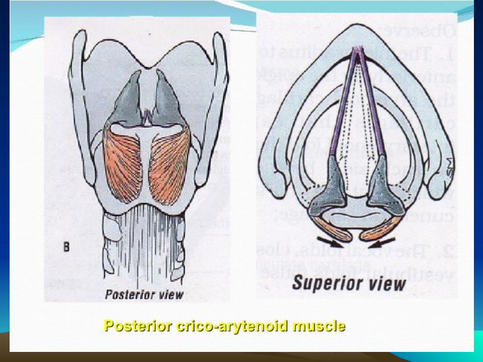
• Posterior cricoarytenoid muscle : is the only muscle that opens the glottis, separating the vocal processes and thus abducts the vocal cords.
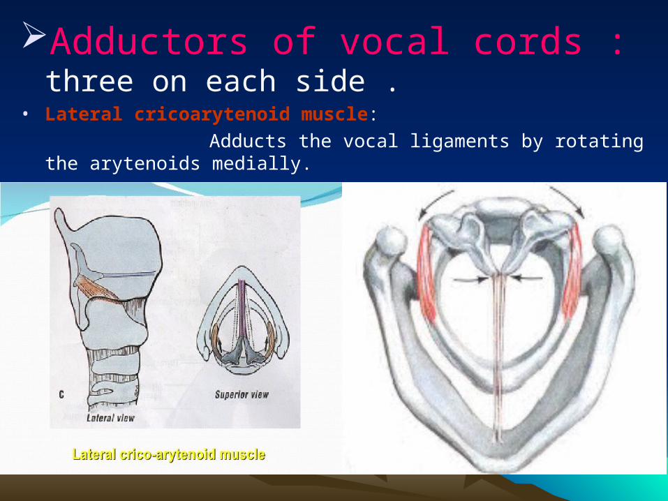
Adductors of vocal cords : three on each side .
• Lateral cricoarytenoid muscle:
Adducts the vocal ligaments by rotating the arytenoids medially.
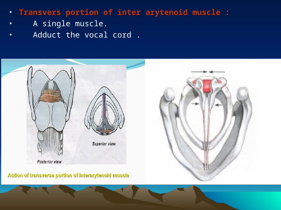
• Transvers portion of inter arytenoid muscle :• A single muscle.• Adduct the vocal cord .
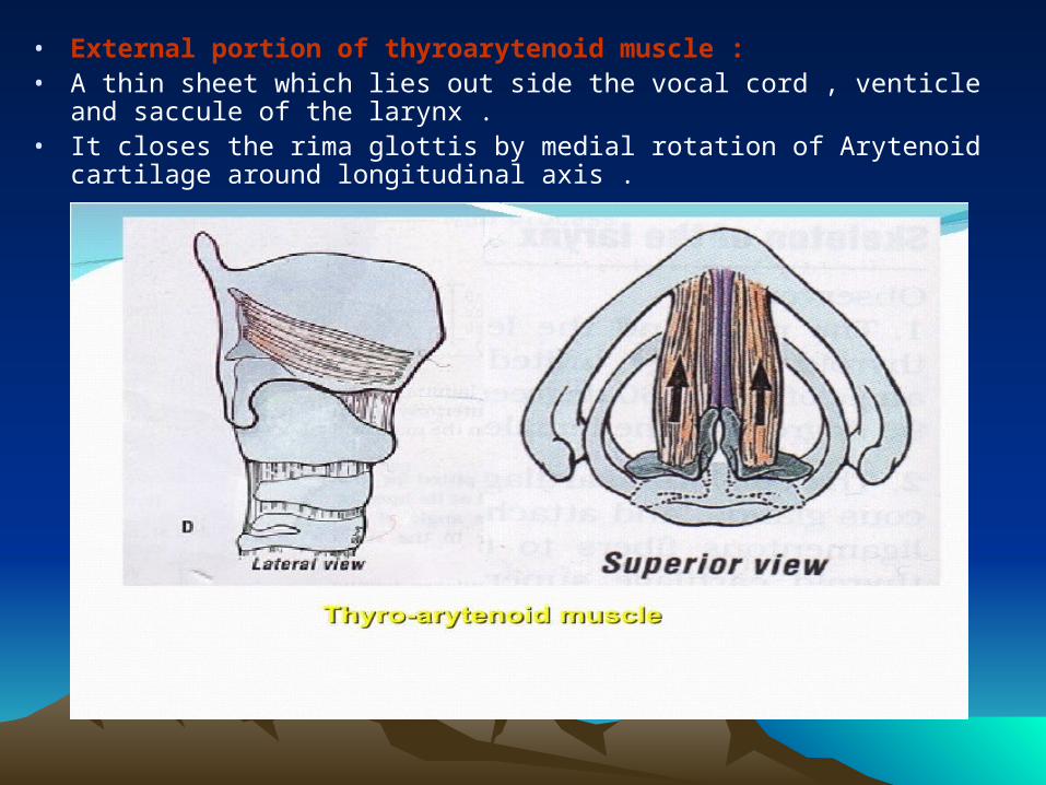
• External portion of thyroarytenoid muscle : • A thin sheet which lies out side the vocal cord , venticle and saccule of the larynx . • It closes the rima glottis by medial rotation of Arytenoid cartilage around
longitudinal axis .
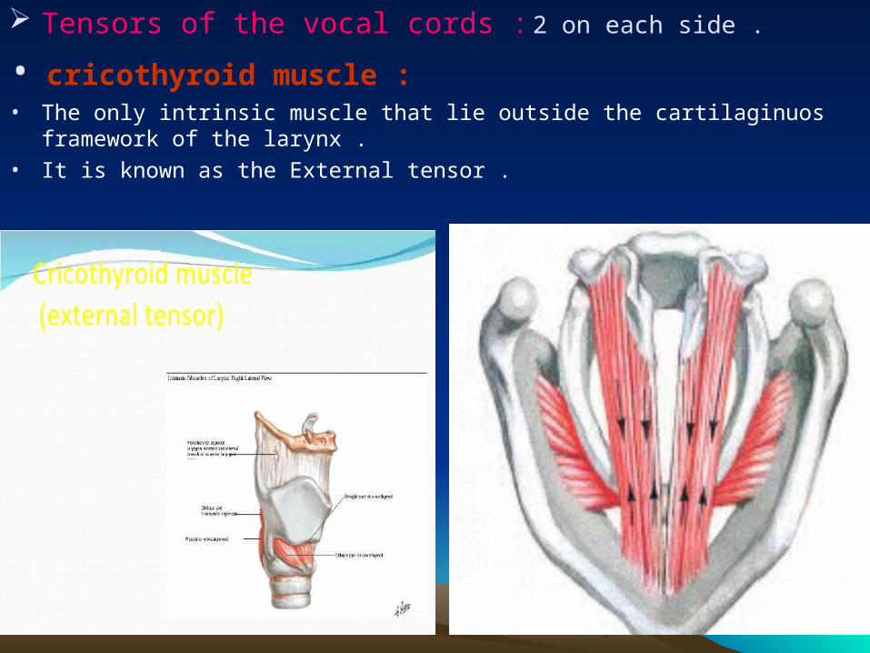
Tensors of the vocal cords : 2 on each side .
• cricothyroid muscle : • The only intrinsic muscle that lie outside the cartilaginuos framework of the larynx . • It is known as the External tensor .
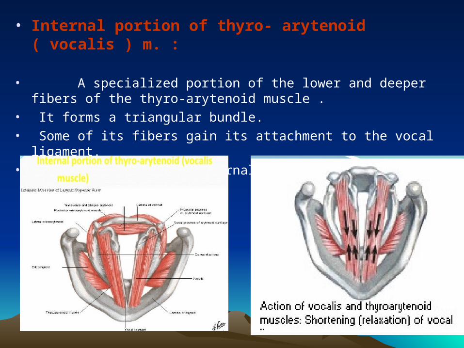
• Internal portion of thyro- arytenoid ( vocalis ) m. :
• A specialized portion of the lower and deeper fibers of the thyro-arytenoid muscle .
• It forms a triangular bundle.• Some of its fibers gain its attachment to the vocal ligament. • It is known as the internal tensor .
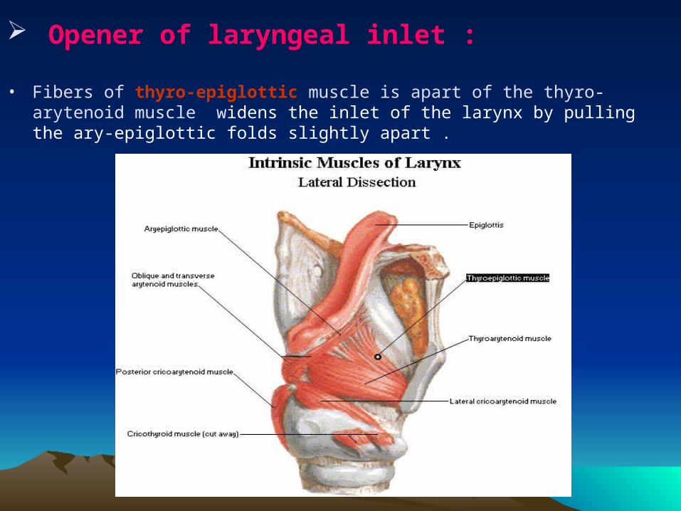
Opener of laryngeal inlet :
• Fibers of thyro-epiglottic muscle is apart of the thyro-arytenoid muscle widens the inlet of the larynx by pulling the ary-epiglottic folds slightly apart .
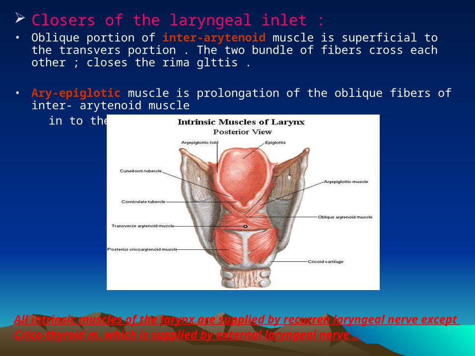
Closers of the laryngeal inlet : • Oblique portion of inter-arytenoid muscle is superficial to the transvers portion .
The two bundle of fibers cross each other ; closes the rima glttis .
• Ary-epiglotic muscle is prolongation of the oblique fibers of inter- arytenoid muscle in to the ary- epiglotic fold .
All intrinsic muscles of the larynx are supplied by recurren laryngeal nerve except Crico-thyroid m. which is supplied by external laryngeal nerve ..
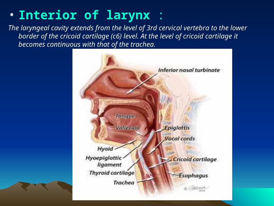
• Interior of larynx :The laryngeal cavity extends from the level of 3rd cervical vertebra to the lower
border of the cricoid cartilage (c6) level. At the level of cricoid cartilage it becomes continuous with that of the trachea.
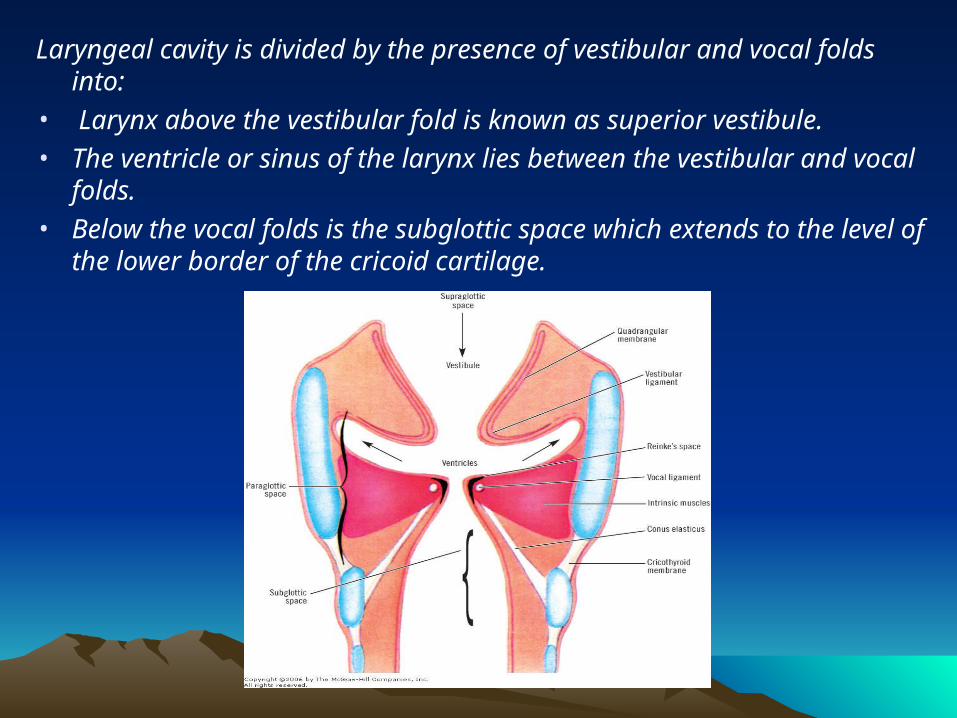
Laryngeal cavity is divided by the presence of vestibular and vocal folds into:
• Larynx above the vestibular fold is known as superior vestibule. • The ventricle or sinus of the larynx lies between the vestibular and vocal
folds. • Below the vocal folds is the subglottic space which extends to the level
of the lower border of the cricoid cartilage.
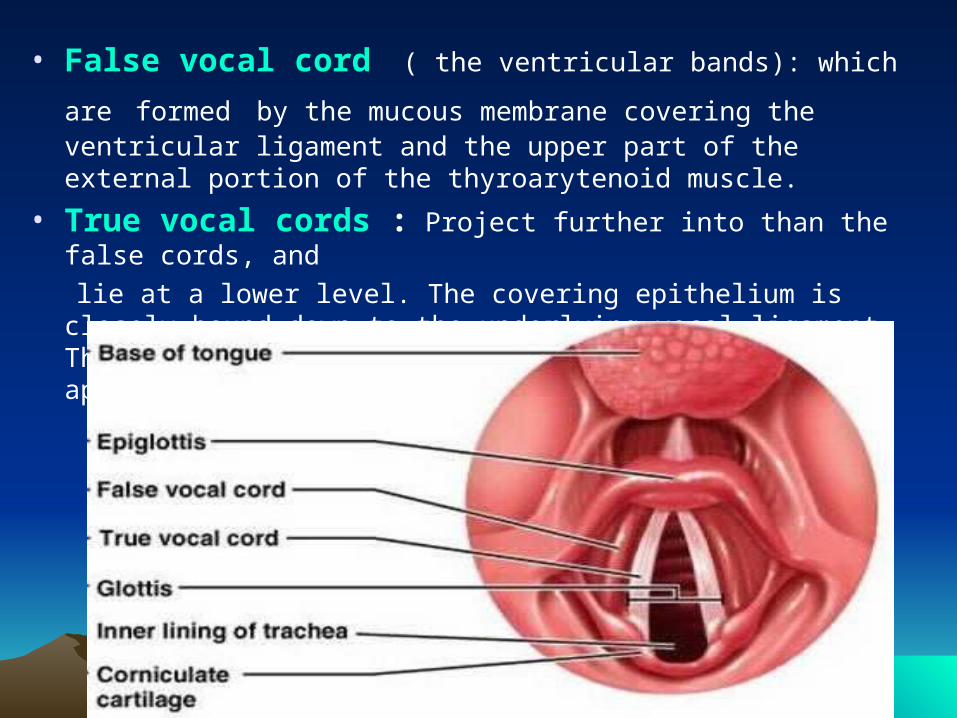
• False vocal cord ( the ventricular bands): which are formed by the mucous membrane covering the ventricular ligament and the upper part of the external portion of the thyroarytenoid muscle.
• True vocal cords : Project further into than the false cords, and
lie at a lower level. The covering epithelium is closely bound down to the underlying vocal ligament. The blood supply is poor, hence the pearly white appearance of the vocal cords.
Histologically the vocal fold:
contains 5 layers:•Layer 1 (squamous epithelial lining): It is very thin .
•Layer 2 (Superfical layer of the lamina propria =Reinke's space ): composed of loose fibers and matrix.. This layer contains only minimal elastic and collagenous fibers and offers least resistance to vibration. The integrity of this layer is vital for proper phonatory function.
•Layer 3 (Intermediate layer of lamina propria): a higher concentration of elastic and collagenous fibres. Provides protection to the vocal folds from mechanical damage.
• Layer 4 (Deep layer of lamina propria): a dense collection of elastic and collagenous fibers.
Along with the intermediate layer constitute the vocal ligament.
•Layer 5 (vocalis muscle) : portion of thyro _arytenoid muscle.
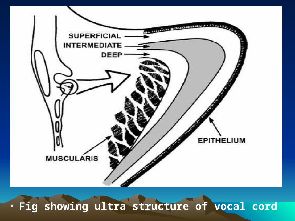
• Fig showing ultra structure of vocal cord
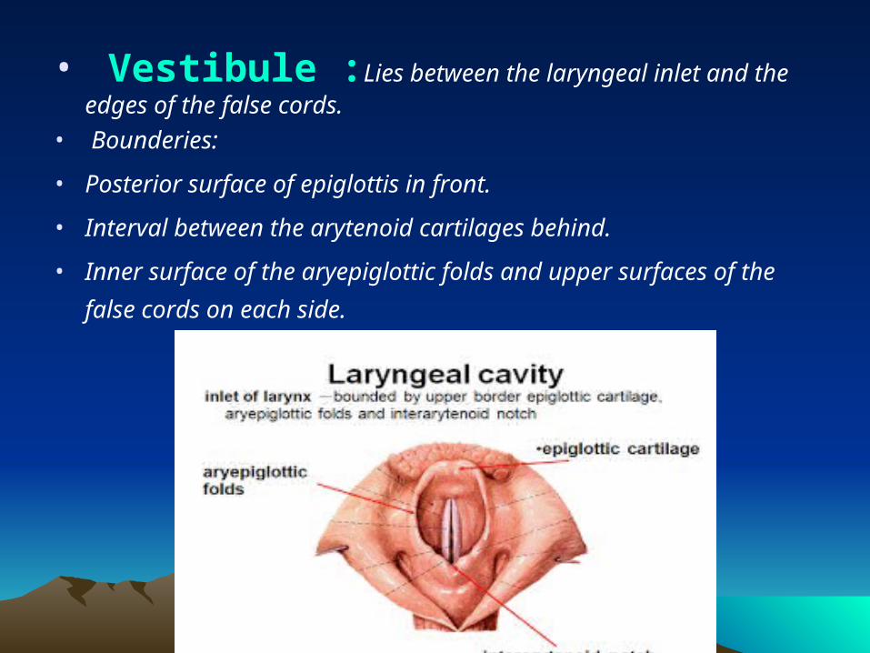
• Vestibule :Lies between the laryngeal inlet and the edges of the false cords.
• Bounderies:
• Posterior surface of epiglottis in front.
• Interval between the arytenoid cartilages behind.
• Inner surface of the aryepiglottic folds and upper surfaces of the
false cords on each side.
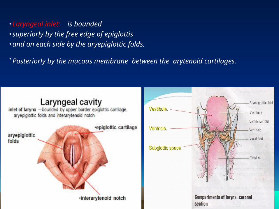
•Laryngeal inlet: is bounded
•superiorly by the free edge of epiglottis
•and on each side by the aryepiglottic folds.
•Posteriorly by the mucous membrane between the arytenoid cartilages.
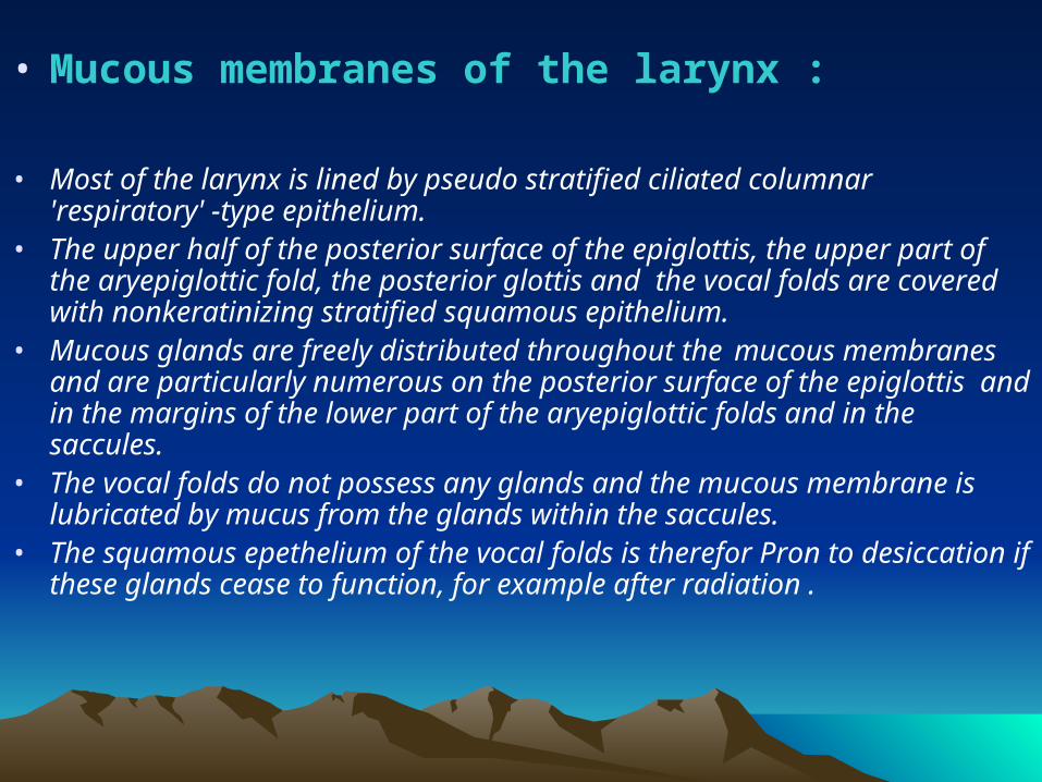
• Mucous membranes of the larynx :
• Most of the larynx is lined by pseudo stratified ciliated columnar 'respiratory' -type epithelium.
• The upper half of the posterior surface of the epiglottis, the upper part of the aryepiglottic fold, the posterior glottis and the vocal folds are covered with nonkeratinizing stratified squamous epithelium.
• Mucous glands are freely distributed throughout the mucous membranes and are particularly numerous on the posterior surface of the epiglottis and in the margins of the lower part of the aryepiglottic folds and in the saccules.
• The vocal folds do not possess any glands and the mucous membrane is lubricated by mucus from the glands within the saccules.
• The squamous epethelium of the vocal folds is therefor Pron to desiccation if these glands cease to function, for example after radiation .
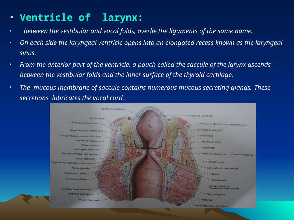
• Ventricle of larynx:• between the vestibular and vocal folds, overlie the ligaments of the same name.
• On each side the laryngeal ventricle opens into an elongated recess known as the laryngeal
sinus.
• From the anterior part of the ventricle, a pouch called the saccule of the larynx ascends
between the vestibular folds and the inner surface of the thyroid cartilage.
• The mucous membrane of saccule contains numerous mucous secreting glands. These
secretions lubricates the vocal cord.
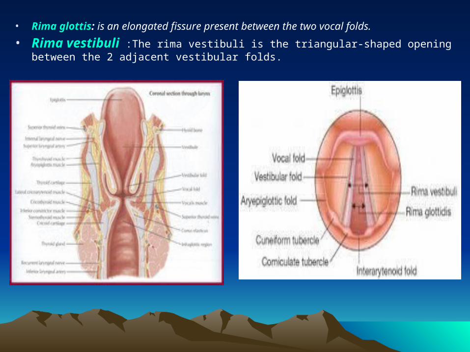
• Rima glottis: is an elongated fissure present between the two vocal folds.
• Rima vestibuli :The rima vestibuli is the triangular-shaped opening between the 2 adjacent vestibular folds.
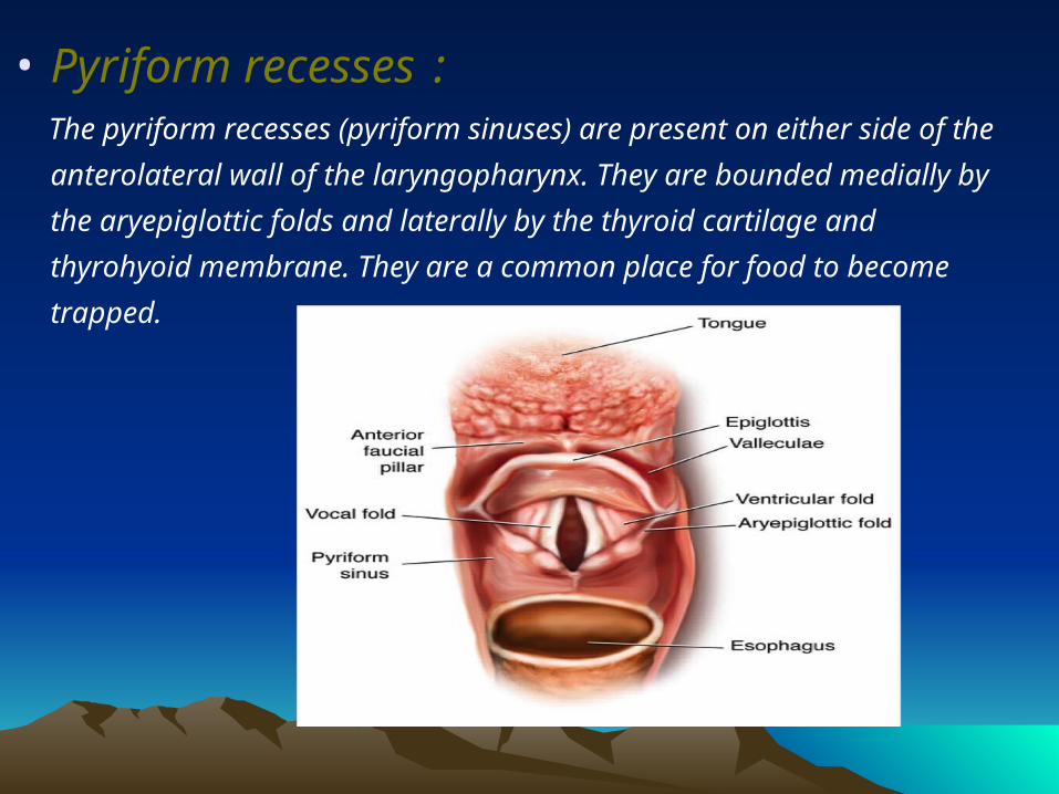
• Pyriform recesses : The pyriform recesses (pyriform sinuses) are present on either side of
the anterolateral wall of the laryngopharynx. They are bounded medially
by the aryepiglottic folds and laterally by the thyroid cartilage and
thyrohyoid membrane. They are a common place for food to become
trapped.
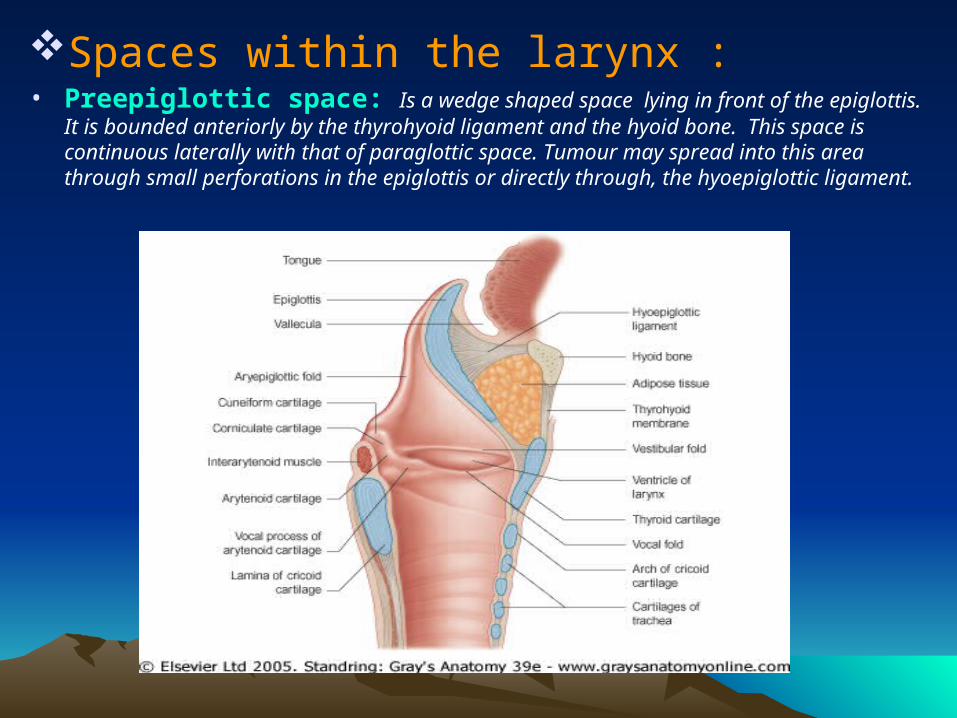
Spaces within the larynx :• Preepiglottic space: Is a wedge shaped space lying in front of the epiglottis. It is
bounded anteriorly by the thyrohyoid ligament and the hyoid bone. This space is continuous laterally with that of paraglottic space. Tumour may spread into this area through small perforations in the epiglottis or directly through, the hyoepiglottic ligament.
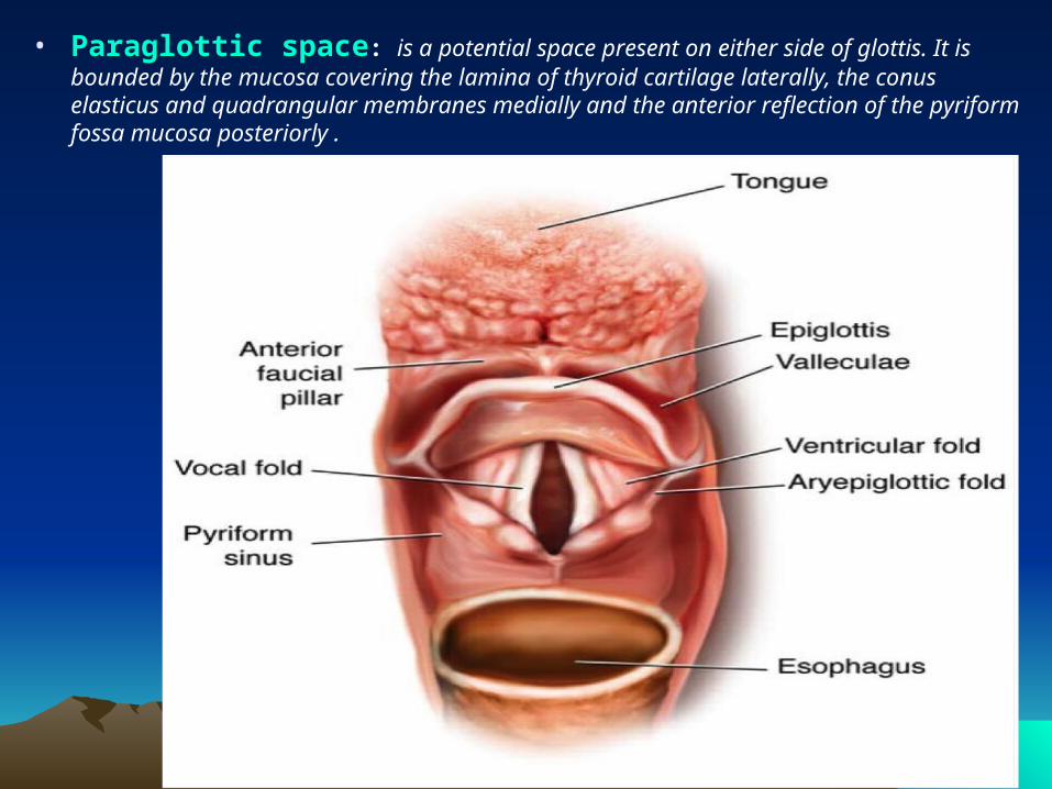
• Paraglottic space: is a potential space present on either side of glottis. It is bounded by the mucosa covering the lamina of thyroid cartilage laterally, the conus elasticus and quadrangular membranes medially and the anterior reflection of the pyriform fossa mucosa posteriorly .
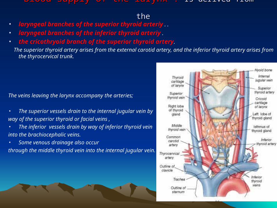
Blood supply of the larynx : Blood supply of the larynx : Is derived from theIs derived from the • laryngeal branches of the superior thyroid arteriy . .
• laryngeal branches of the inferior thyroid arteriy .
• the cricothryoid branch of the superior thyroid artery.
The superior thyroid artery arises from the external carotid artery, and the inferior thyroid artery arises from the thyrocervical trunk.
The veins leaving the larynx accompany the arteries;
• The superior vessels drain to the internal jugular vein by
way of the superior thyroid or facial veins ,
• The inferior vessels drain by way of inferior thyroid vein
into the brachiocephalic veins.
• Some venous drainage also occur
through the middle thyroid vein into the internal jugular vein.
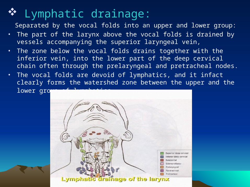
Lymphatic drainage: Separated by the vocal folds into an upper and lower group:
• The part of the larynx above the vocal folds is drained by vessels accompanying the superior laryngeal vein,
• The zone below the vocal folds drains together with the inferior vein, into the lower part of the deep cervical chain often through the prelaryngeal and pretracheal nodes.
• The vocal folds are devoid of lymphatics, and it infact clearly forms the watershed zone between the upper and the lower group of lymphatics.
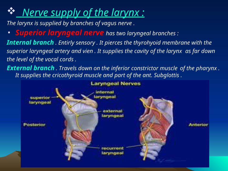
Nerve supply of the larynx :The larynx is supplied by branches of vagus nerve .
• Superior laryngeal nerve has two laryngeal branches :
Internal branch . Entirly sensory . It pierces the thyrohyoid membrane with the
superior laryngeal artery and vien . It supplies the cavity of the larynx as far down
the level of the vocal cords .
External branch . Travels down on the inferior constrictor muscle of the pharynx . It supplies the cricothyroid muscle and part of the ant. Subglottis .
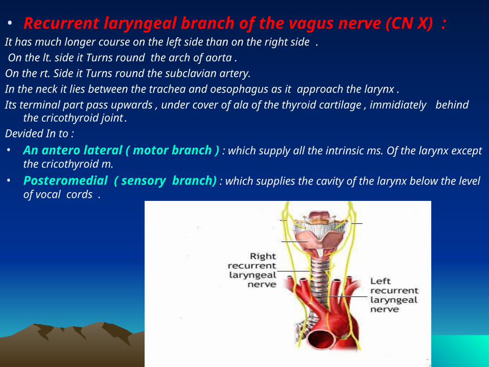
• Recurrent laryngeal branch of the vagus nerve (CN X) :It has much longer course on the left side than on the right side .
On the lt. side it Turns round the arch of aorta .
On the rt. Side it Turns round the subclavian artery.
In the neck it lies between the trachea and oesophagus as it approach the larynx .
Its terminal part pass upwards , under cover of ala of the thyroid cartilage , immidiately behind the cricothyroid joint .
Devided In to :
• An antero lateral ( motor branch ) : which supply all the intrinsic ms. Of the larynx except the cricothyroid m.
• Posteromedial ( sensory branch) : which supplies the cavity of the larynx below the level of vocal cords .
Physiology of Physiology of LarynxLarynx
Functions of larynxFunctions of larynx::
1. Airway protection2. Respiration3. Swallowing4. Coghing5. phonation
• Airway protection:
• The most important function of human larynx.
• A sphincter protecting the lower airway from secretions of oropharynx.
• Protects the airway from spillage of food during deglutition.
• Larynx has three protective mechanisms (from above downwards):
Aryepiglottic fold, ventricular band and vocal cords.
• Respiration:
• Larynx is part of the upper airway passages .
• keeps the airway open during respiration.
• Contributes to the regulation of the acid-base balance in the blood by
influencing CO 2 tension .
• Swallowing:
• During swallowing the sphincters of larynx stay contracted preventing
aspiration of food into the air passage.
• Coughing :
• Coughing is the process by which material is expelled
from the airway.
• It is preceded by rapid inspiration, followed by forceful closure of
both the vocal and vestibular folds.
• Air pressure is then built up below the adducted folds.
• The diaphragm ascends spasmodically until the folds separate
explosively and mucus or foreign material is expelled.
• Phonation:
The larynx acts as a transducer during phonation converting the
aerodynamic forces generated by the lungs, diaphragm, chest and
abdominal muscles into acoustic energy.
The requirements of normal phonation are as follows:
1. Active respiratory support
2. Adequate glottic closure
3. Normal mucosal covering of the vocal cord
4. Adequate control of vocal fold length and tension.
• The cycle of sound production involves glottic opening and
closing at set frequencies determined by the subglottic air
pressure.
• The function of vocal folds is to produce sound varying in
intensity and pitch. This sound is then modified by various
resonating chambers present above and below the larynx
and are converted into words by the articulating action of the
pharynx, tongue, palate, teeth and lips.