Embed Size (px)
Citation preview

Video Microscopy
Video Microscopy
Shinya Inoue Marine Biological Laboratory Woods Hole, Massachusetts and University of Pennsylvania Philadelphia, Pennsylvania and Universallmaging Corporation Falmouth, Massachusetts
With contributions by
Robert J. Walter, Jr. Michael W. Berns Gordon W. Ellis
and
Eric Hansen
Springer Science+Business Media, LLC
Library of Congress Cataloging in Publication Data
Inoue: Shinya. Video microscopy.
Bibliography: p. Includes index. 1. Video microscopy.
QH222.156 1986
First Printing-April 1986 Second Printing-March 1987 Third Printing-November 1987 Fourth Printing-June 1989
578'.4 85-28252
© 1986 Springer Science+Business Media New Vork Originally published by Plenum US in 1986
All rights reserved
No part of this book may be reproduced, stored in a retrieval system, or transmitted in any form or by any means, electronic, mechanical, photocopying, microfilming, recording, or otherwise, without written permission from the Publisher
Sofcover reprint of the hardcover 1st edition 1986
ISBN 978-1-4757-6927-2 ISBN 978-1-4757-6925-8 (eBook) DOI 10.1007/978-1-4757-6925-8
Preface
Ever since television became practical in the early 1950s, closed-circuit television (CCTV) in conjunction with the light microscope has provided large screen display, raised image contrast, and made the images formed by ultraviolet and infrared rays visible. With the introduction of large-scale integrated circuits in the last decade, TV equipment has improved by leaps and bounds, as has its application in microscopy.
With modem CCTV, sometimes with the help of digital computers, we can distill the image from a scene that appears to be nothing but noise; capture fluorescence too dim to be seen; visualize structures far below the limit of resolution; crispen images hidden in fog; measure, count, and sort objects; and record in time-lapsed and high-speed sequences through the light microscope without great difficulty. In fact, video is becoming indispensable for harnessing the fullest capacity of the light microscope, a capacity that itself is much greater than could have been envisioned just a few years ago.
The time seemed ripe then to review the basics of video, and of microscopy, and to examine how the two could best be combined to accomplish these tasks. The Marine Biological Laboratory short courses on Analytical and Quantitative Light Microscopy in Biology, Medicine, and the Materials Sciences, and the many inquiries I received on video microscopy, supported such an effort, and Kirk Jensen of Plenum Press persuaded me of its worth.
This book addresses the reader already somewhat experienced in light microscopy, but perhaps with less background in video or electronics. Practical matters as well as some theory needed to select and use the equipment rationally are included, but circuit diagrams, for example, are deliberately omitted.* On the other hand, I have tried to explain the basics in sufficient detail so that the reader may be encouraged to explore applications of video to microscopy even in ways not attempted before.
Chapter 1 briefly reviews the invention of TV, its early applications to microscopy, and the present state of video microscopy.
Chapter 2 provides a glossary of terms commonly used in video and microscopy, and directs the reader to sections of the book dealing with each particular subject.
Chapter 3 guides the beginner through the preliminaries of getting started, especially in very practical terms. Common problems are listed, as well as pitfalls that can be avoided by knowing their warning signs.
TV is engineered to satisfy the human observer. In Chapter 4 we review the major features of human vision that relate to the design and use of TV as well as the light microscope.
In Chapter 5 we review the essentials of microscope image formation and examine the basic adjustments and special care of the light microscope needed to obtain high-quality images using video.
*Texts such as Ennes (1979), Hansen (1969), Showalter (1969), and van Wezel (1981) provide many examples of the actual circuits involved. See the service manual provided by the manufacturer for wiring diagrams of specific pieces of equipment.
v
vi PREFACE
In Chapter 6 we study the fundamental nature of the video signals and the essentials of video signal transmission from the camera to the monitor.
In Chapter 7 we examine the hardware at the front and back ends of video (the camera, including the video _pickup device, and the video monitor, including the display device,) and compare their specific performances for various applications in light microscopy.
In Chapter 8 we examine the operation, choice, and care of video recorders, including video tape recorders and optical disk recorders, some with capabilities for time-lapse, freeze-frame, and high-speed analyses.
In Chapter 9 we survey the equipment and the methods available for video processing and analyses using analog video devices.
In Chapter 10 Walter and Berns examine the principles and applications of digital image processing and analysis, an approach that combines the power of computers with the advantages of video. Many examples of applications in microscopy are included.
In Chapter 11 I have sampled relatively recent applications of video, classified according to modes of contrast generation in microscopy (excluding those applications already discussed in the previous two chapters). Special advantages and requirements for applying video to the various contrast modes are discussed, together with suggestions for potential new applications.
In Chapter 12 we discuss the preparation of video for publication, or for tape or film presentation, including the means for stereoscopic displays.
In Appendix I Gordon Ellis discusses the removal of video scan lines by spatial filtration, and shows its striking effect on the appearance of picture details.
In Appendix II Eric Hansen discusses the fundamental relationship between resolution and contrast in the microscope and video. The relation between the optical diffraction pattern and the modulation transfer function of the system is examined.
In Appendix III I have revised an article that introduces the basics of polarized-light microscopy and its application to biology, a subject not adequately discussed in print elsewhere. This appendix contains an extensive, annotated bibliography on polarized-light microscopy and its applications to cell biology.
In Appendix IV I have attempted to provide an update of digital image processors and programs that have become available recently, and to make some recommendations on how one might get started doing digital image processing.
The organization of these chapters may appear somewhat unusual; I tried to develop the subject in a spiral fashion, interweaving the practical and the basics, hoping to help those who actually make use of video microscopy. For those using the book more as a reference source, I have included relevant section numbers for each entry in the Glossary. The glossary terms are boldfaced when they first appear in the text.
I have used footnotes liberally throughout the book. It should be emphasized that material discussed in the footnotes is by no means of secondary importance; rather, the arrangement was intended to minimize interruption in the flow of the discussion.
All references, except those for Appendix III, appear in a separate section at the end of the book, arranged alphabetically by author. Each entry is followed by bracketed notations indicating its location in the volume. Included is a separate list of pamphlets not readily available from libraries, along with addresses of their sources (p. 528).
Names and addresses of commercial firms mentioned in the text also have been compiled into a single list (pp. xxi-xxiii), except for those that appear in Tables 5-1, 7-6, and Table 1 of the Postscript.
These chapters were prepared main)y from the vantage point of a biologist, but it is hoped that they will prove useful to those in other fields as well.
As the outline shows, the discussions are heavily weighted toward video, much less being covered on modes of contrast generation and other matters of microscopy. Fortunately, several texts are in preparation at this time, and the compact paperback Fundamentals of Light Microscopy by Michael Spencer of University College, London, provides a concise overview of these subjects. I highly recommend Spencer's text as a companion to this volume.
The technology and the available equipment for video are advancing at a dizzying pace today. I
PREFACE vii
have stressed the fundamentals wherever practical, so that these chapters will not, I hope, become outdated too quickly. The advances are, however, so rapid that I have made a few suggestions in the Postscript on how to keep current on the many exciting developments. Also, while the references to the literature and the tables showing types and sources of equipment are fairly extensive, they are clearly representative and are not meant to be all-inclusive. I would, however, appreciate learning of any major gaps in the presentations. Again, sources of additional information appear in the Postscript.
Shinya Inoue Woods Hole, Massachusetts
Acknowledgments
Bob Walter and Mike Berns not only contributed an excellent Chapter 10, but waited patiently a whole year while I completed the other chapters. Gordon Ellis and E. Leitz, Inc., allowed us to use the material in Appendix I, and Eric Hansen prepared the concise discourse of Appendix II, which neatly ties together the optics and electronics scattered throughout the volume. Dave Cohen (Venus Scientific, now at Gull Airborne Instruments), Gordon Ellis, Eric Hansen, Kirk Jensen, Ernst Keller (Carl Zeiss, Inc.), Brian Laws (Crimson Camera), Doug Lutz (Marine Biological Laboratory), Ted MacNichol (Marine Biological Laboratory), Ted and Nancy Salmon (University of North Carolina), and Paul Thomas (Dage-MTI) reviewed various parts of the draft manuscript and provided much useful input.
In assembling the material for this book, many other individuals contributed original photographs, microscope slides, and other valuable information and material. In addition to those already mentioned, they include: David Agard (University of California, San Francisco), Bob Allen (Dartmouth College), Nina Allen (Wake Forest University), John Almen (Ampex), Anthony Alter (University of California, Irvine), Helene Anderson (Crimson Camera), Peter Anderson (University of Florida, St. Augustine), Peter Bartels (University of Arizona and University of Chicago), R. R. Beckman (General Electric), Gene Bell (Massachusetts Institute of Technology), Ken Braid (Sony), Sid Braginsky (Olympus), Mel Brenner (Nikon), Joe Bryan (Baylor College of Medicine), Fred Caspari (Dage-MTI), Ken Castleman (Perceptive Systems), Dick Chaison (Olympus), Jasmine Chow (University of California, Irvine), Cary Clayton (Instrumentation Marketing Corporation), Kenneth Cooper (University of California, Riverside), Michelle Crawforth (Eastman Kodak), Glenn Davis (Xybion), Jim Dvorak (National Institutes of Health), Barbara Ela (Sony), Kepie Engel (National Science Foundation), Ralph Eno (Hamamatsu), Fennell Evans (University of Minnesota), Fred Fay (University of Massachusetts, Worchester), Dennis Flanagan (Scientific American), Les Flory (RCA), Harold Fosack (Texas Instruments), Marty Prange (Panasonic), Maurice Fran<;on (Laboratoire d'Optique), Henry Fuchs (University of North Carolina), Keigi Fujiwara (Harvard Medical School), John Grace (Crimson Camera), John Harshbarger (VII), Wyndham Hannaway (Colorado Video, now at G. W. Hannaway and Assoc.), John Hayden (Dartmouth College), David Hillman (Laser Concepts for Panasonic OMDR), Jan Hinsch (E. Leitz, Inc.), Ken Hori (MP Video), Andrew Huxley (Cambridge University), Hitoshi Iida (Hamamatsu), Marcos lntaglietta (University of California, San Diego), Colin Izzard (State University of New York, Albany), Ken Jacobson (University of North Carolina), Tedd Jacoby (For-A), Ron Jafcott (Texas Instruments), Lionel Jaffe (Marine Biological Laboratory), Carl Johnson (Harvard University), Bechara Kachar (National Institutes of Health), Harold Kidd (General Electric), Jerry Kleifgen (Dage-MTI), Chuck Koester (Columbia University College of Physicians and Surgeons), Michael Koonce (University of California, Irvine), Edwin Land (Rowland Institute for Science), Peter Lemkin (National Institutes of Health), Lawrence Levine (Panasonic), Cy Levinthal (Columbia University), Lew Lipkin (National Institutes of Health), Lenny Lipton (Stereographics Corporation), Doug Lutz (Marine Biological Laboratory), Arte Machia (Panasonic), Julie Madox (RCA), Ed Martins (Amperex), Y. Mat-
ix
X ACKNOWLEDGMENTS
sushita (Sony), Bryan Mayall (Lawrence Livermore), Curtis Me Dowell (General Electric), Paul Mengers (Quantax), Dennis Neary (Spin Physics), Ken Omdorf (Dartmouth College), Barry Palewitz (University of North Carolina), Alan Penchansky (Geltzer & Co.), William Pratt (Vicom), Phil Presley (Carl Zeiss), Calvin Quate (Stanford University), George Reynolds (Princeton University), lchiji Rikukawa (Nikon), George Robinson (RCA), Fred Sachs (General Electric), Dean Sadamune (University of California, Irvine), Masao Sakai (Hitachi), Kurt Scheier (Nikon), Lee Schuett (Nikon), Martin Scott (Eastman Kodak), John Sedat (University of California, San Francisco), Walter Seidl (Olympus), Hy Shaffer (Crimson Camera), Lawrence Sher (BBN Laboratories), S. Shigemasa (Toshiba), Art Shoemaker (Reichert Scientific Instruments-A. 0. Reichert), David Shotton (University of Oxford), Ann Siemens (University of California, Irvine), Tony Silvestri (Sony), Jesse Sisken (University of Kentucky), Glen Southworth (Colorado Video), Ken Spring (National Institutes of Health), Bob Squires (WHOI), Ray Stephens (Marine Biological Laboratory), Art Sterling (Dage-MTI), Dick Taylor (Colorado Video), Lans Taylor (Carnegie-Mellon University), Ron Tomczyk (Panasonic), Ron Vale (National Institutes of Health and Marine Biological Laboratory), Fran Valenti (F. M. Valenti, Assoc.), Barbara Walter (University of California, Irvine), John Wampler (University of North Carolina), Bob Wang (Imaging Technology), Watt Webb (Cornell University), John White (Medical Research Council, Cambridge University), Bob Worbecki (Precision Echo), and Don Yansen, (Interactive Video).
lllustrations and tables without specific acknowledgment of source are originals, produced or compiled in the authors' laboratories.
Thanks to the many publishers and authors of cited sources for their permission to reproduce figures, and to those who provided original photographs and drawings for reproduction.
The rapid development of video microscopy would not have been possible without the willing and helpful participation by many commercial organizations. Although trademarks are not designated in the text, it should be pointed out that the following are registered trademarks or copyrights of the indicated companies. Chalnicon, Toshiba; lmage-1, Universal Imaging Corporation; Kimwipe, Kimberly-Clark; Kleenex, Kimberly-Clark; Newvicon, Matsushita; Plumbicon, PhillipsAmperex; Saticon, Hitachi; Scotch Cover-Up Tape, 3M Company; SIT, RCA; ST Vidicon, RCA; Ultricon, RCA; Ultricon IT, RCA; Videotherm, lSI Group.
I am grateful to these companies, and others, for permission to reproduce illustrations from their publications and product specification sheets. In this regard, extensive use has been made of the RCA Electro-Optics Handbook and the Conrac Raster Graphics Handbook.
Last but not the least of the many who helped put this volume together: NIH Grant GM-31617, NSF Grant PCM-8216301, * and Olympus Corporation of American provided generous support for the preparation of the manuscript. Nikon Instrument Company, Carl Zeiss, Colorado Video, and Dage-MTI provided especially helpful cooperation. The MBL staff and administration sul'ported my task in many ways as usual, but especially Judy Ashmore of the Library, who helped by tracking down numerous references and difficult-to-locate books. Ed Hom, of our laboratory, produced excellent photographs at the last minute in addition to designing and fabricating the many superior parts for our video, high-extinction polarizing microscope. Ted Inoue developed the Image-I digital image-processing program and opened my eyes to the world of digital computers. Linda Pellechia (North Babylon, New York) and Bob Golder of our Photo Lab prepared the many fine line drawings, and Linda Golder and Chris Inoue, the photoprints. Mary Safford at Plenum lightened the pain of editorial corrections with her patience and empathy with the author; Kenneth McCandless and Sylvia strained their eyes proofing; Bertha Woodward, Doug Lutz, and the students kept the laboratory running productively. Jane Leighton (STARS) tirelessly typed the manuscript through its many revisions. Mort Maser (Woods Hole Educational Associates) performed a heroic task in the last month of helping to bring the manuscript into fmal shape. And Sylvia kept my spirits up and the cozy home fire burning. To all my deepest thanks.
S.l.
*Any opinions, findings, and conclusions or recommendations expressed in this publication are those of the authors and do not reflect the views of the National Science Foundation or the National Institutes of Health.
Contents
List of Tables . . . . . . . . . . . . . . . . . . . . . . . . . . . . . . . . . . . . . . . . . . . . . . . . . . . . . . . xix
List of Firms Cited in the Text ....................................... xxi
Color Plates . . . . . . . . . . . . . . . . . . . . . . . . . . . . . . . . . . . . . . . . . . . . . . . . . . . . . . . . xxv
CHAPTER 1 Why Video? . . . . . . . . . . . . . . . . . . . . . . . . . . . . . . . . . . . . . . . . . . . . . . . . . . . . . . . . . . 1
1.1. Pre-1940: The Invention of TV ................................................ 1 1.2. Earlier Applications of Video to Microscopy ..................................... 4 1.3. Where We Are Today ....................................................... 6 1.4. A Few General Remarks .................................................... 11
CHAPTER 2 Glossary . . . . . . . . . . . . . . . . . . . . . . . . . . . . . . . . . . . . . . . . . . . . . . . . . . . . . . . . . . . . 13
CHAPTER 3 Getting Started: Some Practical Tips . . . . . . . . . . . . . . . . . . . . . . . . . . . . . . . . . 55
3.1. The Minimum Essential Components ......................................... 55 3.2. Initial Adjustment and Hookup of the Video ................................... 56 3.3. Coupling the Video Camera and the Microscope ................................ 60 3.4. Wait! Read before Turning the Microscope Lamp On ............................ 60 3.5. Adjustments of the Monitor ................................................. 62 3.6. Termination .............................................................. 63 3.7. Importance of Aligning the Video Camera with the Microscope ................... 64 3.8. Optical Noise, Hot Spots ................................................... 66 3.9. Other Sources of Optical Noise .............................................. 68
3.10. Electronic Reduction of Noise ............................................... 69
xi
xii CONTENTS
CHAPTER 4 Physiological Characteristics of the Eye . . . . . . . . . . . . . . . . . . . . . . . . . . . . . . . 71
4o1o Introduction 0 0 •••••••••••• 0 •••••••••• 0 •• 0 •••• 0 •••••••••• 0 • 0 •••• 0 ••••••••••• 71
4.2. Structure, Refraction, and Accommodation of the Eye ................. 0 •••••••••• 71
4.3. Visual Sensitivity and Adaptation . 0 ••••••• 0 •• 0 •••••••••••••••••••••••••••••••• 74
4.4. Resolution, Visual Acuity ........... 0 ••••••••••••••••••••••••••••••••••••••• 77
4.5. Contrast Discrimination and the Modulation Transfer Function ..................... 80
4.6. Flicker ................................................................... 83
4. 7. Color Vision ............. 0 ••••• 0 ••••••• 0 •••••••••••••••••••••••••••••••••• 86
4.8. Stereoscopy ............................................................... 90
4.9. Additional Remarks ........................ 0 ••••••••••••••••••••••••••••••• 92
CHAPTER 5 Microscope Image Formation ......................................... 93
5.1. Introduction: Essential Optical Train of the Light Microscope o •• 0 o o 0 0 o o 0 • o 0 • 0 0 0 •• 0 93
5020 Imaging Components in the Light Microscope: Magnification . 0 0 0. 0 0 0 0. 0 0 0 0 0 0. 0 0 0. 93
5030 Principles of Koehler Illumination 0 0 0 0 0 0 0 0. 0 0 0 0 0 0 0 0 0 0 0 0 0 • 0 0 0. 0 0 0 0. 0 0 0 0 0 0 0 0 0 0 101
5.40 Adjusting the Microscope for Koehler Illumination 0 0 0 0. 0 0 0 0. 0 0 0 0 0 0 0 0 0 0 0 0 0 0 0 0 0 0 0 104
Alignment and Focus of the Illuminator . 0 0 0 0. 0 0 • 0 • 0 0 0 0 0 0 0 0 0 0 0. 0 0 0 0. 0 0 0 0 0 0 0 0 0 0 105
Focusing the Condenser; Alignment of the Illuminator Continued . o 0 o o 0 0 0 o 0 0 0 o 0 0 o o 105
Centering the Condenser 0 0 0 0 0 0 0 • 0 0 0 0 0 0 0 • 0 0 0 0 • 0 0 • 0 0 0 0 • 0 0 • 0 0 0 • 0 0 o o 0 • o o • 0 0 0 •• 106
Alignment of Microscopes with a Built-in llluminator o • 0 o o 0 0 o o 0 • o o o o o o o 0 o o o 0 o o o 107
Adjustment of the Field Diaphragm 0 0 • 0 0 0 0 • 0 0 • 0 0 0 o o • 0 o 0 0 0 o o •• o 0 • o 0 o • 0 o o o 0 o o • 107
Adjustment of the Condenser Iris 0. 0 0. 0 •• 0. 0 0 o 0. 0 o o 0 0 0 o •• o o o o 0 o o o 0 o o 0 0 o o 0 0 0 o 108
5.50 Image Resolution and Wave Optics . 0 0 0 0 •• o. 0 o o 0. o o. 0 0 o o. o o •• o o 0 o 0 o o 0 o o o 0 0 o o 111
The Diffraction Pattern 0 0 0 0 • 0 0 0 0 • 0 0 • 0 0 0 •• 0 0 0 0 0 0 o •• 0 o 0 0 0 o • 0 • o 0 •• o o • o o o 0 o o o 0 111
Image Resolution and the Diffraction Pattern . 0 0 0 0 0 0 0. 0 0. 0 0 0 0. 0 0 0 0 0 0 0. 0 0. 0 0 0 0 0 0 113
The Diffraction Pattern and Out-of-Focus Image o o 0 0 o o. 0 o o 0 0 o. 0 0 o o. 0 o o 0 o o o 0 o o 0 0 115
Depth of Field 0 • 0 0 0 0 0 0 • 0 0 0 • 0 • 0 0 0 0 0 0 0 0 0 0 0 0 0 0 0 0 0 0 0 0 0 0 0 0 0 0 0 0 0 0 0 0 0 0 0 0 •• 0 0 • 0 0 118
5o6o Generation of Image Contrast 0 0 0 0. 0 0. 0. 0 0. 0 0. 0. 0 0 0 0 0. 0. 0 o. 0 0 0 0 0 0 o • 0 o o 0. o o 0. 118
Zernike's Phase Contrast . 0 0 0 0 0. 0 0 0 0 0 0 0. 0 0 0 0 0 0 0 0 0 0 0 0 0 0 0 0 0 0 0 0 0 0 0 0 0 0 0 0 0 0 0. 0 0 0 119
Other Modes of Contrast Generation 0 0. 0 0. 0 0 0 0. 0 0. 0 0 0 0. 0 0. 0 0 0. 0 0 0 o 0. 0 o. 0 0 0 0 0 122
5.7o Modulation Transfer Function 0 0. 0 0 0 0 0 0 0. 0 0 0 0 0 0 0 0 0 0 0 0 0 0 0 0 0 0 0 0 0 0 0 0. 0 0 0 0 0 0 0 0 0 0 122
508. Intensity of Light Source and Other Factors Affecting Image Brightness 0 0 • 0 0 0 0 0 0 0 0 125
Brightness of Illumination 0 0. 0 0 0 0 0 0 0 o • 0 0. 0. 0 o • 0 0 o 0. 0 o. 0 0 o 0. 0 o 0 •• o o 0 0 o • 0 0. 0 0 126
Increasing the Signal from the Specimen 0 0 0 0 0 0 0 0 0 0 0 0 0 0 0 0 0 0 0 0 0 0 0 0 0 0. 0 0 0 0 0 0 0 0 0 0 128
Light-Gathering Power of the Objective Lens 0 0. 0 0 0 0 0 0 0. 0 0 0 0 0 0 0 0 0 0 0. 0 0 0 0 0 0 0 0 0. 130
Image Brightness and Magnification 0 • o 0 • 0 o o 0 • o o • 0 o o o 0 o 0 • 0 o • 0 0 0 • 0 0 o • 0 0 0 0 •• 0 0 0 130
Light Transmission through the Microscope o o 0. o 0. 0 o. 0. o 0. 0 0 0 0 0 0 0 0 0. 0 0 0 0 •• 0 0. 0 132
509. Aberrations 0 0 • 0 0 0 0 0 0 0. 0 0 0 0 0 0 0 0 0 0 0. 0 0 0 0 0 0 0. 0. 0 0. 0 0. 0 0 0 0 0 0 0 0 0 0 0. 0 0. 0 0. 0 0 0 0 133
5ol0o Choice and Care of Lenses for Video Microscopy 0 0 0. 0 0 0 0 0 0. 0 0 0 0 0 0 0. 0 0 0 0 0 0. 0 0 0 136
Objective Lenses 0 0 o 0 0. o 0 •• o o • o o 0 0 o o. 0 o o o 0 o 0. 0 o o 0 0 o 0. 0 0. 0 0 0 0 0 0. 0 0 0 0 0 0 0 0 0 0 136
Oculars 0 0 • 0 0 0 0 • 0 0 o 0 0 • o 0 0 • o •• o o 0 0 o o 0 • o o o 0 o o o • o o • 0 o o •• o • 0 • 0 0 • 0 o • 0 0 • 0 • 0 0 0 0 136
Collector and Condenser Lenses 0 0 0 0 0 0 0 0 0 0 0 0 0 0 0 0 0 0 0 0 • 0 0 0 0 0 0 0 0 0 • 0 0 0 0 •• 0 0 • 0 0 0 0 138
Inspecting and Cleaning the Optics and Video Camera Faceplate 0 0 0 • 0 0 0 0 0 0 0 0 0 • 0 0 0 139
5o11. The Microscope Stand 0 0 0 0 0 0 • 0 0 0 0 0 0 • 0 0 • 0 0 0 0 0 0 0 0 0 • 0 0 • 0 0 0 0 • 0 0 0 0 0 • 0 0 0 0 0 0 • 0 0 • 0 145
CHAPTER 6 The Video Image and Signal . . . . . . . . . . . . . . . . . . . . . . . . . . . . . . . . . . . . . . . . 149
6.10 Introduction 0 o o 0 o o 0 0. 0 0 0 0 0 0 0 0 0 0 0 0 0 0. 0 0. 0 0. 0 0 • 0 0. 0 0 0 0 0 0 0 0 0 0 0 0 0 0 0 0 0 0. 0 0 0 0 0 0 0 149
6o2o How Video Works o o 0 o o. o o o 0 o o 0 0. 0 0 0 0 0 0 0 0 0 0 0 0 0 0 0 0 0 0 0. 0 0. 0 0 0 0 0 0 0 0 0 0 0 0 0. 0 0 0 0 151
Generation of the Video Signal 0 0 0 0 0 0 0. 0 0 0. 0 0 0 0. 0 0 0 0. 0 0 0 0 0 0 0 0 0 0 0 0 0. 0 0 0 0 0 0 •• 0 o151
CONTENTS xiii
Sequential Scanning ....................................................... 152 H and V Scans ........................................................... 152 Hand V Deflections ...................................................... 152 Blanking ................................................................ 153 Standard Scanning Rates ................................................... 153 Interlacing . . . . . . . . . . . . . . . . . . . . . . . . . . . . . . . . . . . . . . . . . . . . . . . . . . . . . . . . . . . . . . . 155 Active Picture Area ....................................................... 156 The Picture Signal ........................................................ 156
6.3. Timing Pulses and the Composite Video Signal ................................ 156 Sync Pulses and Composite Video ........................................... 156 Conventions and Standards for CCTV . . . . . . . . . . . . . . . . . . . . . . . . . . . . . . . . . . . . . . . . 158 Sync Pulses during V Blanking .............................................. 160 Other Interlace and Timing Conventions ...................................... 161 Visualizing the Waveform, Pulse-Cross Display ................................ 164
6.4. Image Resolution and the Video Signal ....................................... 169 Definitions . . . . . . . . . . . . . . . . . . . . . . . . . . . . . . . . . . . . . . . . . . . . . . . . . . . . . . . . . . . . . . 170 Vertical Resolution ........................................................ 170 Horizontal Resolution . . . . . . . . . . . . . . . . . . . . . . . . . . . . . . . . . . . . . . . . . . . . . . . . . . . . . . 172 Cascading a Number of Devices; System MTF ................................. 176
6.5. Degradation of Video Signal, Electrical Noise .................................. 178 Image Degradation and Frequency Response . . . . . . . . . . . . . . . . . . . . . . . . . . . . . . . . . . . 178 Distortion of Waveform .................................................... 178 Electrical Noise .......................................................... 180 SIN Ratio ............................................................... 181
6.6. Synchronizing the Video Equipment .......................................... 184 Compatibilities of Scan Rates and Sync Pulses . . . . . . . . . . . . . . . . . . . . . . . . . . . . . . . . . 184 Sync Stripper . . . . . . . . . . . . . . . . . . . . . . . . . . . . . . . . . . . . . . . . . . . . . . . . . . . . . . . . . . . . 185 Synchronizing an Array of Video Equipment ................................... 186
CHAPTER 7 Video Imaging Devices, Cameras, and Monitors ...................... 191
7 .1. Video Imaging Devices .................................................... 191 Video Camera Tubes ...................................................... 191 Intensifier Tubes . . . . . . . . . . . . . . . . . . . . . . . . . . . . . . . . . . . . . . . . . . . . . . . . . . . . . . . . . . 195 Solid-State Sensors ........................................................ 197
7.2. Performance of Video Imaging Devices ....................................... 203 Resolution and Contrast .................................................... 203 Sensitivity and Wavelength Dependence ....................................... 203 Light Transfer Characteristics ............................................... 209 Gamma ................................................................. 209 Dynamic Range ........................................................... 214 Dark Current and Noise .................................................... 217 Lag ..................................................................... 219 Blooming ................................................................ 221 Burn, Sticking ............................................................ 221 Shading ................................................................. 224 Geometrical Distortion ..................................................... 227 Blemishes ............................................................... 228
7.3. Choice of Video Cameras .................................................. 228 General Considerations ..................................................... 228 Monochrome or Color ..................................................... 229 Sensitivity ............................................................... 229 Resolution ............................................................... 229 Sensor Size .............................................................. 231
xiv CONTENTS
Contrast ................................................................. 231 Gray Scale .............................................................. 232 Lag, Geometrical Distortion, and Other Picture Defects .......................... 233 Noise ................................................................... 233 Dynamic Range and Safety Shutoff .......................................... 233 Signal-Processing Features .................................................. 234 Scan Rates, Interlace, and Clock Stability ..................................... 234 External Sync Capability . . . . . . . . . . . . . . . . . . . . . . . . . . . . . . . . . . . . . . . . . . . . . . . . . . . 235 Other Considerations . . . . . . . . . . . . . . . . . . . . . . . . . . . . . . . . . . . . . . . . . . . . . . . . . . . . . . 235 U.S. Sources of Instrumentation and Special-Purpose Cameras .................... 235
7.4. Color Cameras and the Encoded NTSC Signal ................................. 236 7.5. Video Monitors ........................................................... 242
The Picture Tube and Basic Monitor Circuitry ................................. 242 Picture Size . . . . . . . . . . . . . . . . . . . . . . . . . . . . . . . . . . . . . . . . . . . . . . . . . . . . . . . . . . . . . . 243 Resolution of the Monitor . . . . . . . . . . . . . . . . . . . . . . . . . . . . . . . . . . . . . . . . . . . . . . . . . . 244 Gamma, Contrast, and Picture Brightness . . . . . . . . . . . . . . . . . . . . . . . . . . . . . . . . . . . . . 246 Types of Phosphors, Hue, Persistence ........................................ 248 Geometrical Distortions . . . . . . . . . . . . . . . . . . . . . . . . . . . . . . . . . . . . . . . . . . . . . . . . . . . . 248 Shading, Streaking, Ringing, and Other Picture Defects .......................... 251 Critical Adjustment and Testing of the Monitor ................................. 255 Monitors with Special Features . . . . . . . . . . . . . . . . . . . . . . . . . . . . . . . . . . . . . . . . . . . . . . 256 Color Monitors . . . . . . . . . . . . . . . . . . . . . . . . . . . . . . . . . . . . . . . . . . . . . . . . . . . . . . . . . . . 260
CHAPTER 8 Video Recorders .................................................... 263
8 .I. Introduction .............................................................. 263 8.2. How VTRs Work ......................................................... 263
Video Tape Recording . . . . . . . . . . . . . . . . . . . . . . . . . . . . . . . . . . . . . . . . . . . . . . . . . . . . . 263 The Scanner . . . . . . . . . . . . . . . . . . . . . . . . . . . . . . . . . . . . . . . . . . . . . . . . . . . . . . . . . . . . . 267 Tape Path and Transport . . . . . . . . . . . . . . . . . . . . . . . . . . . . . . . . . . . . . . . . . . . . . . . . . . . 268 Servo Controls . . . . . . . . . . . . . . . . . . . . . . . . . . . . . . . . . . . . . . . . . . . . . . . . . . . . . . . . . . . 269 Frequency Response and Signal Processing in Magnetic Tape Recording . . . . . . . . . . . . 270
8.3. Videotape Formats ........................................................ 274 One- and Two-Inch Formats ................................................ 275 Three-quarter-Inch U format ................................................ 276 Half-Inch Formats ........................................................ 276 M-format ................................................................ 276
8.4. Choice of VTRs for Video Microscopy ....................................... 277 Introduction . . . . . . . . . . . . . . . . . . . . . . . . . . . . . . . . . . . . . . . . . . . . . . . . . . . . . . . . . . . . . . 277 Color Capability or Monochrome Only . . . . . . . . . . . . . . . . . . . . . . . . . . . . . . . . . . . . . . . 283 Resolution . . . . . . . . . . . . . . . . . . . . . . . . . . . . . . . . . . . . . . . . . . . . . . . . . . . . . . . . . . . . . . . 283 Contrast and Picture Definition . . . . . . . . . . . . . . . . . . . . . . . . . . . . . . . . . . . . . . . . . . . . . . 284 Still-Frame and Variable-Speed Display ....................................... 285 Editing Capability . . . . . . . . . . . . . . . . . . . . . . . . . . . . . . . . . . . . . . . . . . . . . . . . . . . . . . . . . 285 Portability, RF Conversion . . . . . . . . . . . . . . . . . . . . . . . . . . . . . . . . . . . . . . . . . . . . . . . . . 286 Compatibility . . . . . . . . . . . . . . . . . . . . . . . . . . . . . . . . . . . . . . . . . . . . . . . . . . . . . . . . . . . . 286 Ease of Use, Convenience Features .......................................... 286 Reliability, Availability of Service . . . . . . . . . . . . . . . . . . . . . . . . . . . . .. . .. . .. . .. . . . . 287 Cost of Equipment, Maintenance, and Supplies ................................. 287
8.5. Time-Lapse VTRs, Time-Base Error ......................................... 288 Time-Lapse VTRs ........................................................ 288 Commercially Available Time-Lapse VTRs .................................... 289 Some Practical Considerations ............................................... 290 Time-Base Error .......................................................... 291
CONTENTS XV
8.6. High-Speed Recording ..................................................... 293 8.7. Care and Maintenance of the VCR ........................................... 295
General Precautions ....................................................... 295 Playback ................................................................ 296 Recording Operation ..............•........................................ 298 Troubleshooting and Repair ................................................. 299 Head Cleaning and Replacement ............................................. 299 When Not in Use ......................................................... 300
8.8. Choice and Care of the Videotape ........................................... 300 Choice of Tape ........................................................... 300 Storage of Recorded Tape .................................................. 301
8. 9. Magnetic and Laser Disk Recording .......................................... 301 Magnetic Disk Recording ................................................... 301 Magnetic Disk versus VTR Recording ........................................ 302 Laser Disks .............................................................. 302 Optical Memory Disk Recorders ............................................. 303
CHAPTER 9 Analog Video Processing and Analysis ............................... 309
9 .1. Introduction .............................................................. 309 9.2. General Principles of Analog Processing and Analysis ........................... 310 9.3. Gray Level Manipulation ................................................... 311 9.4. Analog Filtering and Noise Reduction ........................................ 314 9.5. Image Analysis ........................................................... 319 9.6. Commercial Analog Processors and Analyzers ................................. 325
CHAPTER 10 Digital Image Processing and Analysis . . . . . . . . . . . . . . . . . . . . . . . . . . . . . . . 327
ROBERT J. WALTER, JR., AND MICHAEL W. BERNS
10.1. Introduction ............................................................ 327 10.2. Design of an Image-Processing System ..................................... 329 10.3. Optical Digitizers ....................................................... 331 10.4. Resolution of the Digital Image ........................................... 334 10.5. The Image Histogram .................................................... 338 10.6. Histogram Transformations and Contrast Enhancement ......................... 342 10.7. Nonlinear Contrast Enhancement .......................................... 348 10.8. Manipulations Using Multiple Images ...................................... 356 10.9. Geometrical Manipulations ............................................... 358
10.10. Densitometry Using the Digital Image ...................................... 363 10.11. Digital Filtering and Spatial Convolutions ................................... 365 10.12. Digital Filtering Involving Fourier Transforms ............................... 372 10.13. Feature Extraction ...................................................... 377 10.14. Commercial Image-Processing Equipment ................................... 386
CHAPTER 11 Applications of Video to Light Microscopy ........................... 393
11.1. Introduction ............................................................. 393 11.2. Low-Light-Level Microscopy .............................................. 393
Luminescence ........................................................... 393 Auorescence ............................................................ 397
xvi CONTENTS
Darkfield ............................................................... 40 I UV and IR ............................................................. 40I
II. 3. Contrast Enhancement .................................................... 403 Polarization Microscopy ................................................... 403 Phase Contrast .......................................................... 406 Interference ............................................................. 408 Differential Interference, Single-Sideband, Anaxial Illumination .................. 4IO Brightfield .............................................................. 4I3
Il.4. Image Quantitation by Video ............................................... 415 Photometry, Densitometry ................................................. 4I5 Morphometry, Sorting, Tracking ............................................ 416
11.5. Special Types and Applications of Light Microscopy ........................... 416 Scanning Light Microscopes ............................................... 416 Tomography, Three-Dimensional Reconstruction .............................. 418
CHAPTER 12 Presentation of Video Data .......................................... 423
12.1. Publication of Video Micrographs ........................................... 423 Introduction ............................................................. 423 Photographing the Monitor ................................................ 424 Scan Line Removal ...................................................... 427
12.2. Optical Copying with Video ............................................... 429 I2.3. Copying and Editing onto Videotape ........................................ 432
The Equipment .......................................................... 432 Selecting and Organizing the Scenes ........................................ 433 Title Making ............................................................ 435 The Mechanics of Editing ................................................. 436
12.4. Special Effects .......................................................... 438 12.5. Video Projection ......................................................... 442 12.6. Video-to-Film Transfer ................................................... 445 12.7. Stereo Video Microscopy .................................................. 446
Stereoscopy with the Microscope ........................................... 446 Stereo Video Display ..................................................... 451
Postscript .......................................................... 457
Appendixes
APPENDIX I Spatial Filtration for Video Line Removal . . . . . . . . . . . . . . . . . . . . . . . . . . . . 463
GORDON W. ELLIS
APPENDIX II Modulation Transfer Function Analysis in Video Microscopy .......... 467
ERIC W. HANSEN
TI.l. Introduction ............................................................. 467 TI.2. Modulation Transfer ...................................................... 467
CONTENTS xvii
11.3. The MTF and the Diffraction Pattern ......................................... 470 11.4. Sampling and Digitized Video .............................................. 471 11.5. Systems Analysis of a Video Microscope ..................................... 472
APPENDIX Ill An Introduction to Biological Polarization Microscopy . . . . . . . . . . . . . . . . 477
111.1. Introduction ............................................................ 477 111.2. Anisotropy ............................................................ 478 III.3. Some Basic Features of Light Waves ....................................... 478 111.4. Optical Anisotropy ...................................................... 480 111.5. Birefringence .......................................................... 481 111.6. Dichroism ............................................................. 484 III.7. Determination of Crystalline Axes ......................................... 485 III.8. Molecular Structure and Birefringence ...................................... 488 III.9. The Polarizing Microscope ............................................... 492 111.10. Rectification ........................................................... 493 Annotated Bibliography ........................................................ 500 Bibliography ................................................................. 501
APPENDIX IV Addendum on Digital Image Processors ............................. 511
References . . . . . . . . . . . . . . . . . . . . . . . . . . . . . . . . . . . . . . . . . . . . . . . . . . . . . . . . . 515
Cited Pamphlets .............................................................. 528
Index .............................................................. 531
List of Tables
5-1. Selected Light Sources with High Luminous Density .................... 128
5-2. Microscope Objective Lenses with High Light-Gathering Power ........... 131
5-3. Microscope Objective Lenses with Long Working Distances .............. 135
5-4. Long-Working-Distance Condensers; Objectives Suitable as LWD Condensers ...................................................... 139
6-1. TV Display-Signal Parameters Established by International Standards ....... 158
6-2. TV Parameters for High-Resolution Systems ........................... 165
6-3. Nations That Have Adopted the NTSC, PAL, or SECAM Color-Encoding Standards ........................................................ 166
6-4. Rise Time and Horizontal Resolution at Various Video Bandwidths ........ 176
6-5. Decibels versus Voltage- and Power-Ratios ............................ 182
7-1. Characteristics of Some Standard l-Inch Vidicon Camera Tubes ........... 194
7-2. Important Performance Characteristics of Typical Camera Tubes and Intensifier Camera Tubes ........................................... 199
7-3. Examples of RS-170 Compatible, Solid-State Pickup Device and Cameras 202
7-4. Sensitivity of Selected l-Inch Video Cameras .......................... 230
7-5. Standard Video Sensor Dimensions ................................... 232
7-6. Selected U.S. Sources of Monochrome Instrumentation Video Cameras ..... 236
7-7. Phosphor Characteristics: Characteristics of Phosphors Used in Cathode-Ray Tubes ............................................... 249
8-1. Partial Specifications for Some l-Inch Reel-to-Reel VTRs ................ 279
8-2. Some %-Inch Standard-Speed VCRs .................................. 281
8-3. Some Vz-Inch Standard-Speed VCRs .................................. 282
8-4. High-Resolution VTRs ............................................. 284
8-5. Time-Lapse VCRs ................................................ 290
8-6. High-Speed Recording Systems ...................................... 294
8-7. Optical Memory Disk Recorders ..................................... 304
xix
XX
10-l.
11-l.
LIST OF TABLES
Selected Image Processors and Systems ............................... 388
Microscope Contrast Modes; Luminance Levels; Typical Video Cameras .... 394
Postscript-! Digital Image Processors-Supplementary List of Suppliers ............ 458
Postscript-2 Some Magazines That Report on Developments Related to Digital Image Processing ............................................... 458
Postscript-3 Selected Trade Magazines Relevant to Digital Image Processing ..................................................... 459
List of Firms Cited in the Text*
Alan R. Liss 150 Fifth Avenue, New York, New York 10011. (Publisher of video disk supple-ments to Cell Motility)
American Optical (see Reichert) Amperex Electronics Corporation Providence Pike, Slatersville, Rhode Island 02876. (Plumb-
icon and other vidicon tubes) Ampex Corporation 401 Broadway, Redwood City, California 94063. (l-inch VTRs) Arlunya Division, The Dindima Group Pty., Ltd. P.O. Box 106, Victoria 3133, Australia.
(Digital image processor) Bausch & Lomb 820 Linden Avenue, Rochester, New York 14620. (Microscopes, Omnicon
digital image analyzer) BBN Laboratories 10 Moulton Street, Cambridge, Massachusetts 02238. (Consultants on three-
dimensional imaging, etc.) Carl Zeiss, Inc. One Zeiss Drive, Thornwood, New York 10594. (Microscopes, IBAS image
processor) Colorado Video, Inc. Box 928, Boulder, Colorado 80306. (Analog processors, digital frame
store, slow-scan digitizers) Comprehensive Video Supply Corporation 148 Veterans Drive, Northvale, New Jersey 07647.
(Porta-Pattern test charts, video supplies, test equipment) Conrac Division, Conrac Corporation 600 North Rimsdale Avenue, Covina, California 91722.
(Monitors) Crimson Camera Technical Sales 325 Vassar Street, Cambridge, Massachusetts 02139. (Dis-
tributor of industrial video equipment, including Videotek monitors) Dage-MTI 208 Wabash Street, Michigan City, Indiana 46360. (Video cameras, high-resolu-
tion monitor, image processor) DeAnza (see Gould-DeAnza) Drexon, Drexler Technology Corporation 2557 Charleston Road, Mountain View, California
94043. (Optical memory disks) Eastman Kodak Company Rochester, New York 14650. (Lenses, film, filters, photographic
emulsions, chemicals, videotape) Edmund Scientific Company 7082 Edscorp Building, Barrington, New Jersey 08007. (Ronchi
gratings) E. Leitz, Inc. Rockleigh, New Jersey 07647. (Microscopes, 35-mm cameras, TAS image
processor, DADS photometer) EMI 80 Express Street, Plain View, New York 11803. (High-gain image intensifier tube,
photomultipliers) EOIS (Electro Optical Information Systems) 710 Wilshire Boulevard, Suite 501, Santa Monica,
California 90401. (Digital image processors)
*(See Also Tables 5-I, 7-6, and Table I of the Postscript)
xxi
xxii LIST OF FIRMS
EOS Electronics AV, Ltd. Weston Square, Barry, South Glamorgan, CF6 7YF, United King-dom. (Modification of Sony 5850 for animation)
Fish-Shurman Corporation 70 Portman Road, New Rochelle, New York 10802. (Fluorescent uranium-glass blocks, filters)
For-A Corporation of America 49 Lexington Street, West Newton, Massachusetts 02165. (Analog image processors, time-date generators, time-base correctors)
General Electric Company, Medical Systems Milwaukee, Wisconsin 53201. (High-resolution l-inch VTR)
General Electric Company, Projection Display Products Operation Electronics Park, Syracuse, New York 13201. (Light valve video projector)
Gould-DeAnza, Inc. 118 Charcot Avenue, San Jose, California 95131. (Digital image processors)
Grinnell Systems Corporation 6410 Via del Oro Drive, San Jose, California 95119. (Digital image processor)
GYYR 1335 South Claudina Street, Anaheim, California 92805. (Yz-inch time-lapse VCR) Hitachi Denshi America, Ltd., Broadcast and Professional Division 175 Crossways Park West,
Woodbury, New York 11797. (l-inch VTRs, video cameras, recorders, monitors) Ikegarni 37 Brook Avenue, Maywood, New Jersey 07607. (Video equipment, high-resolution
monitors) Image Magnification Systems New Smyrna Beach, Florida 32069. (Catadioptric video
projector) Imaging Technology, Inc. 600 West Cummings Park, Woburn, Massachusetts 01801. (Image-
processing boards) International Imaging Systems 1500 Buckeye Drive, Milpitas, California 95035. (Digital image
processors) Interpretation Systems, Inc. 6322 College Boulevard, Overland Park, Kansas 66211. (Digital
image analysis systems) lSI Group, Inc. 211 Conchas, S.E., Albuquerque, New Mexico 87123. (Digital image
processor) Joyce-Loeb! Vickers Marquisway, Team Valley, Gateshead, Tyne and Wear, NEll OQ3,
United Kingdom. (Scanning recording microdensitometer) JVC, Professional Video Division 58-75 Queens Midtown Expressway, Maspeth, New York
11378. (Video cameras, VCRs, monitors, etc.) Karl Lambrecht 4204 North Lincoln Avenue, Chicago, Illinois 60618. (High-extinction coated
calcite polarizing prisms) Kontron Bildanase Distributed in the U.S. by Carl Zeiss, Inc. (IBAS image-analyzing system) Machlett Laboratories 1065 Hope Street, Stanford, Connecticut 06907. (Sniperscope) MCI!LINK, Link Systems (USA), Inc. P.O. Box 50810, Palo Alto, California 94303. (Intel-
lect 1001200 image analyzers) Merlin Engineering Works 1880 Embarcadero Road, Palo Alto, California 94303. (Refurbish-
ing and custom modification of l-inch VTRs) Motorola, Inc.,Communications Systems Division 9733 Coors Road, N.W., Albuquerque,
New Mexico 87114. (PLZT materials) NAC, Instrument Marketing Corporation 820 South Mariposa Street, Burbank, California
91506. (High-speed video cameras and recorders) NEC America, Inc., Consumer Products Division 130 Martin Lane, Elk Grove Village, Illinois
60007. (Video cameras, VCRs, monitors, etc.) Nikon, Inc., Instrument Division 623 Stewart Street, Garden City, New York 11530.
(Microscopes; CF, rectified lenses; 35-mm cameras) Olympus Corporation, Scientific Instruments Division 4 Nevada Drive, Lake Success, New
York 11042. (Microscopes, 35-mm cameras) Panasonic Electronic Components One Panasonic Way, Secaucus, New Jersey 07094. (Video
cameras, VCRs, monitors, etc.) Panasonic Industrial Company, Optical Disk Department 333 Meadowland Parkway,
Secaucus, New Jersey 07094. (Optical memory disk recorders)
LIST OF FIRMS xxiii
Perceptive Systems, Inc. 5231 Whittier Oaks, Friendswood, Texas 77546. (Course on image processing, digital image analyzers)
Photonics Microscopy, Inc. 2625 Butterfield Road, Suite 204-S, Oak Brook, Illinois 60521. (Hamamatsu Photonic image processors, low-light-level cameras)
Polaroid Corporation 575 Tech Square, Cambridge, Massachusetts 02139. (Sheet Polaroid, Polaroid cameras, film)
Precision Echo 3105 Patrick Henry Drive, Santa Clara, California 95054. (Video magnetic disk recorder/reproducer systems)
Quantex Corporation 252 North Wolfe· Road, Sunnyvale, California194086. (Digital image processors)
RCA 600 North Sherman Drive, Indianapolis, Indiana 46201. (VCRs) RCA, New Products Division New Holland Avenue, Lancaster, Pennsylvania 17604. (Video
pickup devices, intensifiers, cameras, monitors, etc.) ~ecognition Concepts, Inc. P.O. Box 8510, Incline Village, Nevada 89450. (Digital image
processors) Reichert Scientific Instruments P.O. Box 123, Buffalo, New York 14240. (Microscopes) Rolyn Optics Company 738 Arrowgrand Circle, Covina, California 91006. (Ronchi gratings,
lenses, prisms, filters) Sanyo Electric, Inc. Electronics Division, Communication Products, 1200 West Artesia Boule-
vard, P.O. Box 5177, Compton, California 90220. (Video cameras, monitors) Seiler Instrument & Mfg. Co. 170 East Kirkham Avenue, St. Louis, Missouri 63110. (U.S.
distributor for aus Jena microscopes) Sierra Scientific 2189 Leghorn Street, Mountain View, California 94043. (Video cameras,
monitors) Sony Corporation~ of America 700 West Artesia Boulevard, Compton, California 90220. (Sony
video courses for instruction in video principles, service, and repair) Sony Corporation of America, Video Communications 5 Essex Road, Paramus, New Jersey
07652. (Industrial and broadcast video equipment and supplies) Spatial Data Systems 420 South Fairview Avenue, Goleta, California 93116. (LogE digital
image processors) Spin Physics 309~ Science Park Road, San Diego, California 92121. (Very-high-speed video
cameras and recorders) Stereographics Corporation P.O. Box 2309, San Rafael, California 94912. (Three-dimensional
video, PLZT goggles) 3D Technology Corporation 4382 Lankereshim Boulevard, North Hollywood, California
91602. (Stereo-Color TV Projection System) Tektronix, Inc. Tektronix Industrial Park, P.O. Box 500, Beaverton, Oregon 97077. (2048 X
2048 pixel CCD imager) Toshiba Corporation 1101A Lake Cook Road Dearfield, Illinois 60015. (Chalnicon, consumer
video products) TriTronics, Inc. 2921 West Alameda Avenue, Burbank, California 91505. (High-speed video
cameras) Universal Data Systems, Division of Motorola Information Systems Group 5000 Bradford
Drive, Huntsville, Alabama 35805. (Modem) Vickers Instrument Company P.O. Box 99, Malden, Massachusetts 02148. (Joyce-Loeb! Mag-
iscan image analyzer) Vicom Systems 2520 Junction Road, San Jose, California 95134. (Digital image processors) Videotek 125 North York Street, Pottstown, Pennsylvania 19464. (High-resolution color
monitors) Visual Information Institute, Inc. P.O. Box 33, Xenia, Ohio 45385. (Video test equipment, test
charts) Xybion Electronic Systems Corporation 7750-A Convoy Court, San Diego, California 92111.
(High-speed video and Gen-II CCD cameras)

Color Plates
xxvi COLOR PLATES
FIGURE 1-8. Very-low-light-level fluorescence microscope image of 1.25-JJ.m-diameter fluorescent microspheres, captured with a SIT camera. By digital image processing we can: (E) display intensities in pseudocolor; and (F) display intensity contours in three dimensions. 40/0.95 Plan Apo. Digital image processing with Image-I system developed for the author's laboratory. See Fig. 1-8A (p. 10) for unprocessed, single frame image of the same field of view. (From Ellis et al., 1985.)
FIGURE 4-19 (insert). CIE chromaticity diagram. Only the smaller figure from Fig. 4-19 (p. 89) is reproduced here. This is the 1931 CIE Chromaticity Diagram shown schematically in Figs. 4-18 (p. 88) and 7-28 (p. 239). The 1976 version shown on p. 89 better reflects the response of the average human eye to light of different hues. (See Miller, 1985.) (Courtesy of Photo Research, Burbank, California.)
FIGURE 10-55. Use of pseudocolor transformations to enhance contrast in fluorescent cell images. Monochrome images similar to that of Fig. 10-54a were subjected to pseudocolor enhancement to accentuate changes in cellular fluorescence with time. Panels (a)-( d) show changes in fluorescent patterns occurring over a period of 45 sec. (Photographs courtesy of Ann Siemens, Department of Developmental and Cell Biology, University of California, Irvine.)
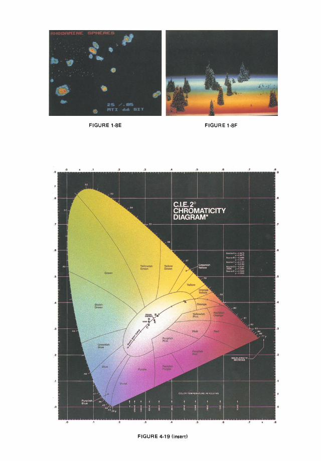
FIGURE 1-SE FIGURE 1-SF
..
..
• 1
..
..
·'
.o
.0 ·' • 2 ·' .. .. .. .1 .. FIGURE 4-19 (insert)
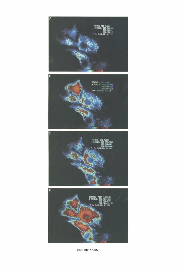
FIGURE 10-55

Video Microscopy