Embed Size (px)
Citation preview

Update on Brain Tumours
Reuben JohnsonLLB, DPhil(Oxon), FRCS (Neuro.Surg)
Senior Lecturer in NeurosurgeryUniversity of Otago

Aims & Objectives
1. Take the opportunity to review the epidemiology, classification and clinical presentation of brain tumours
2. To understand the principles of surgical management3. To be aware of some of the advances in the management of
malignant gliomas4. Review of advances in pituitary surgery
To think like a neurosurgeon, for a short while….

?

The skull is a rigid box and the
contents (CSF, brain, blood) are in
a state of volume equilbrium. An
increase in the volume of one must
be compensated by a reduction in
volume of the other in order to
maintain a constant pressure.
Alexander MonroPrimus (1720-1758)Professor of Anatomy, Edinburgh
The Monro-Kellie Doctrine
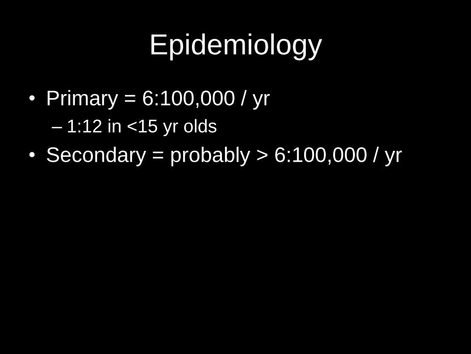
Epidemiology
• Primary = 6:100,000 / yr
– 1:12 in <15 yr olds
• Secondary = probably > 6:100,000 / yr

Site
‘Benign’ vs ‘Malignant’
Definitions have different meanings when applied to the CNS
Pathology
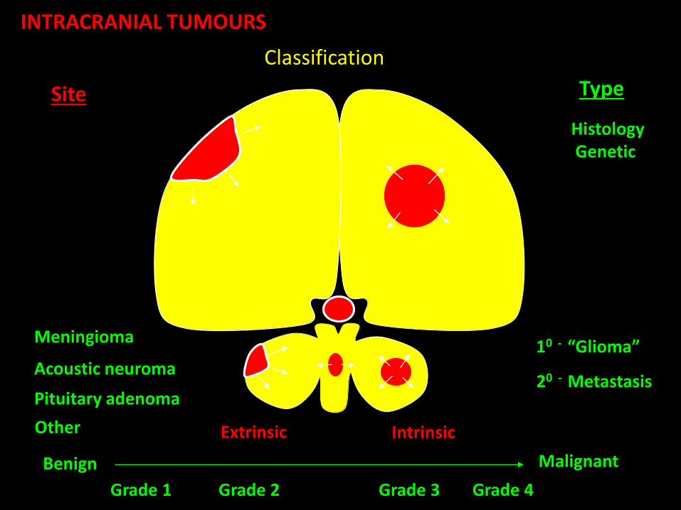
INTRACRANIAL TUMOURS
Classification
Extrinsic
Site
Meningioma
Acoustic neuroma
Pituitary adenoma
Intrinsic
10 - “Glioma”
20 - Metastasis
Benign Malignant
Type
HistologyGenetic
Other
Grade 1 Grade 2 Grade 3 Grade 4

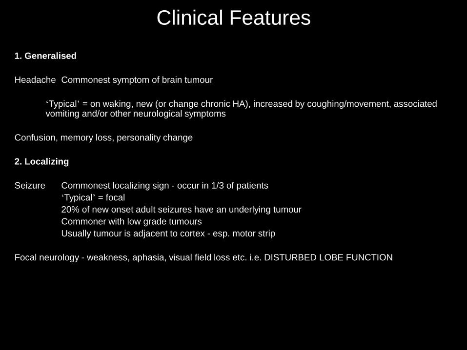
Clinical Features
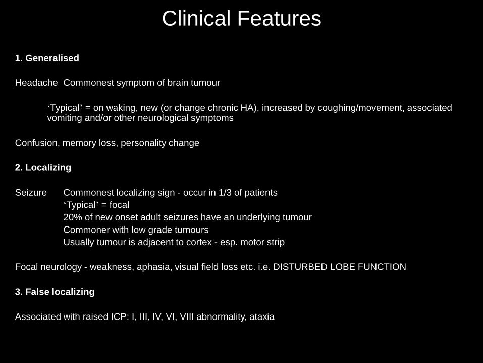
1. Generalised
Headache Commonest symptom of brain tumour
‘Typical’ = on waking, new (or change chronic HA), increased by coughing/movement, associated vomiting and/or other neurological symptoms
Confusion, memory loss, personality change
Clinical Features
1. Generalised
Headache Commonest symptom of brain tumour
‘Typical’ = on waking, new (or change chronic HA), increased by coughing/movement, associated vomiting and/or other neurological symptoms
Confusion, memory loss, personality change
2. Localizing
Seizure Commonest localizing sign - occur in 1/3 of patients
‘Typical’ = focal
20% of new onset adult seizures have an underlying tumour
Commoner with low grade tumours
Usually tumour is adjacent to cortex - esp. motor strip
Focal neurology - weakness, aphasia, visual field loss etc. i.e. DISTURBED LOBE FUNCTION
3. False localizing
Associated with raised ICP: I, III, IV, VI, VIII abnormality, ataxia

Principles of Surgical Management
Need to consider 3 factors in the risk:benefit analysis -
1. Tumour - natural history (rate of growth, effects - anatomical location)
2. Procedure - morbidity/mortality of procedure
3. Patient - life expectancy
Full resection may not be feasible
Management > cure is the aim
Palliative procedures called for in 2 circumstances -
1. Symptomatic ICP
2. Reversible local brain dysfunction
Vision - chiasmal decompression
Hemiplegia - may improve after cystic decompressiom, but can be aggravated if vascular infiltrative tumour
Seizures - may undergo temporary or permenant remission

INTRACRANIAL TUMOURS
Period of silent growth
• Epilepsy
• Focal neurol
symptoms & signs
Symptoms & signs of ICP
Brain displacement, coning & death
ICP
Neurolstatus
Time
Lesion volume
Pressure/Volume curve

Disturbed Function
• Frontal lobe
– Contralateral face,arm,leg weakness
– Expressive dysphasia
– Personality change
• Loss of inhibition
• Dementia
• Abulia

….disturbed function
• Occipital– Homonymous
Hemianopia
• Corpus Callosum– Disconnection syn.
– Apraxia
• Temporal– Receptive dysphasia
– Upper homonymous hemianopia
• Parietal
– Sensory disturbance
– Acalculia, agraphia
– Finger agnosia
– Sensory neglect

….disturbed function
• Hypothalamus/
Pituitary
– Endocrine disturbance
– Bitemporal
hemianopia
• Infratentorial
– Midbrain/Brainstem
• CN 3-12
• Long tract signs
• Tremor
• Dec. GCS
• Eye movements
• Pupillary abnormality
• Vomiting, hiccough

….function
• Cerebellum
– Ataxia
– Intention tremor
– Dysmetria
– Dysarthria
– Nystagmus
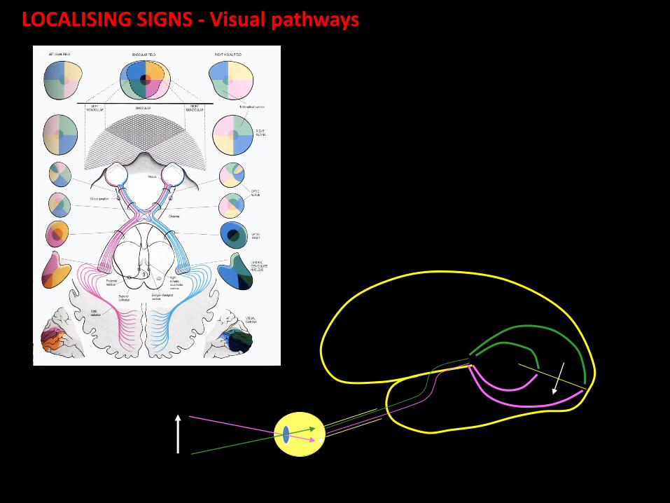
LOCALISING SIGNS - Visual pathways

ANATOMY: Visual pathways – optic radiation
(L) side
Intrinsic lesions -(L) posterior temporal glioblastoma
multiforme
(R) homonymous hemianopia - incomplete in lower quadrants, incongruous & less marked
in eye on side of lesion
Meyer’s loop

False Localising Sign - VI (Abducens)
Longest intracranial course?
• Exits the pons at its lower border just above the pyramid of the medulla.Passes in SAS of pontine basal cistern and ascends over the front of the BS before angling sharply over the petrous bone.Passes under the petroclinoidligament, or Gruber’s ligament (Dorello’s canal) to enter the back of the cavernous sinus.
• It passes freely in the cavernous sinus lateral to the ICA and enters the orbit via the SOF through the annulus of Zinn.
• Terrminatesin the medial aspect of LR.
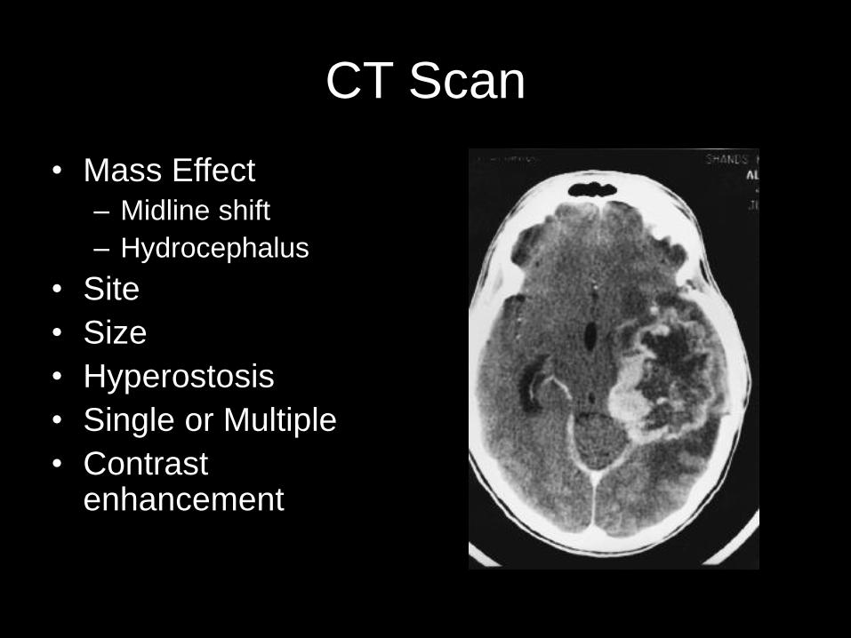
CT Scan
• Mass Effect– Midline shift
– Hydrocephalus
• Site
• Size
• Hyperostosis
• Single or Multiple
• Contrast enhancement

Principles of Surgical Management
Need to consider 3 factors in the risk:benefit analysis -
1. Tumour - natural history (rate of growth, effects - anatomical location)
2. Procedure - morbidity/mortality of procedure
3. Patient - life expectancy
Full resection may not be feasible
Management > cure is the aim
Palliative procedures called for in 2 circumstances -
1. Symptomatic ICP
2. Reversible local brain dysfunction
Vision - chiasmal decompression
Hemiplegia - may improve after cystic decompressiom, but can be aggravated if vascular infiltrative tumour
Seizures - may undergo temporary or permenant remission
o Commonest 1o brain tumours = gliomas
o Commonest brain tumour = metastases

Gliomas2/3 of all primary brain tumours
Arise from 3 basic types of glial cell -
1. Astrocytes: have endfeet on walls of capillaries, thought to be involved in chemical exchange between blood and neural tissue, probably consitute the BBB
2. Oligodendrocytes: form myelin sheath
3. Ependymal cells: involved in repair of nervous tissue, form a linining in ventricles
Gliomas share common characteristics -
Indistinct margins - complete resection almost impossible
Incurability
Genetic instability
Increased malignancy over time
Tendency for local recurrence

Gliomas
Astrocytomas*
Glioblastoma multiforme
Anaplastic astrocytoma
Astrocytoma
Pilocytic astrocytoma
Oligodendrogliomas
Anaplastic oligodendroglioma
Oligodendroglioma
* Commonest form
Mixed gliomas
Anaplastic oligoastrocytomas
Oligastrocytoma
Ependymomas
Anaplastic ependymomas
Ependymomas
Myxopapillary ependymoma
WHO Classification

Astrocytomas2 grading systems: Kernohan & WHO
Kernohan WHO Type Median Survival*
I Special tumours (e.g. pilocytic astrocytomas) 10 years
I/II II Astrocytoma (low-grade) 8 years
III III Anaplastic astrocytoma (AA) 3 years
IV IV Glioblastoma multiforme (GBM) <1 year
l Relative incidence? IV:III:II (WHO system) = 5:3:2
l Peak age incidence increases with grade
II 34 years
III 41 years
IV 53 years
III and IV also known as malignant astrocytomas

Malignant Astrocytomas
WHO III & IV
Anaplastic astrocytoma (AA) & Glioblastoma multiforme (GBM)
GBMCommonest primary brain tumour
Most aggressive brain tumour
50% of all primary brain tumours
20% of all brain tumours
M:F ratio 1.5:1
Ages 45-65
Median survival - 14 weeks (without Rx), 9-12 months (surgery & DXT)
Ring-enhance on CT due to necrosis
AA20% of all gliomas
M:F ratio 1.2:1
Median survival 24-36 months with Rx
Ages 35-55
Complex enhancement on CT

Malignant Astrocytomas
Prognostic indicators in GBMs and AAs
1) Patient age - the most significant prognostic indicator
E.g. GBMs (Wen et al. 1995)
Age 18 months survival
<40 50%
40-60 20%
>60 10%
2) Histological features - AA survive 3 x longer than GBM
3) Performance status - Karnofsky score (KPS)
E.g. GBM
KPS 18 m survival 5 yr survival
≥ 70 34% 7.6% (Wen et al. 1995)
< 60 13% 3.2% (Mahaley et al. 1989)

Low-grade Astrocytomas
WHO II
10% of all gliomas
30-50 years
Most present with seizures
Median survival 8 years
Typical MRI features
MRI - non-enhancing
- hypodense on T1 + contrast
- no/minimal oedema on T2

Treatment of AstrocytomasMalignant astrocytomas
Gold standard = cytoreductive surgery + DXT* (*DXT 40 Gy to wole brain + 20 Gy to tumour bed)
Controversy over extent of tumour resection
Reduce mass effect or get as much out as possible?
Tumours NOT cured with surgery aim for QUALITY survival
Role of chemotherapy? Not proven for all patients, but may be beneficial for some
Low-grade astrocytomas
Various options, none shown to be superior to another -
1) Observe - serial imaging
2) Surgery
3) Chemo +/- DXT
Surgery may be indicated if -
1) Large tumours, young patient, short clinical history
2) Radiological progression
3) Clinical grounds - seizures, CSF flow obstruction etc.[
Pilocytic astrocytomas
Surgery

1st Neurosurgical Operation for Intracranial Lesion
Sir William MacEwanGlasgow 1876 Frontal lobe abscess Patient survived
HughlingsJackson
David FerrierPaul Broca
Cerebral localisation by “functional neurological mapping”
Pre-XRAYLocalisation on basis of focal neurological signs

1st Operation for a Primary Brain Tumour
• Performed by Rickman Godlee (nephew of Sir Joseph Lister) in London, 1884
• Operation observed by Hughlings Jackson, David Ferrier and Victor Horsley….
• Was the surgery a success?

1st Operation for a Primary Brain Tumour
• Performed by Rickman Godlee (nephew of Sir Joseph Lister) in London, 1884
• Operation observed by Hughlings Jackson, David Ferrier and Victor Horsley….
• Was the surgery a success?
• The patient died of infection
• But for Godlee it was very successful
– Baronet 1912
– Hunterian Orator 1913
– President RCS 1914
– Surgeon to Victoria, Edward II, George V

Current Gold Standard for GBM
• Maximal safe resection
• Stupp protocol = Concomitant temozolomide during radiation treatment

Advances in Neurosurgery over Last 100 Years
• CT and MRI Scanning
• Microscope (Yasargil)
• Endoscope (Dandy, Cappabianca)
• Image Guidance (E.g. Stealth) – but brain shift
• 3D Imaging

So What is New for GBM?
Molecular characterisation of GBMs
Adjuncts for surgical resection

Molecular Classification of GBM
Weathers & Giblert. 2014
GBM one of the most molecularly characterised of all human cancersMolecular profiling has identified molecular prognostic factors and potential therapeutic targets

Antiangiogenic Therapy: BEVACIZUMAB (Avastin)
• Angiogenesis a pathologic hallmark of GBM
• Vascular Endothelial Growth Factor (VEGF) is one of the most important regulators of angiogenesis
• VEGF antagonism by humanised mono-clonal antibody was promising
• Early results with BEVACIZUMAB (Avastin) suggested prolongation of progression free survival - but radiology may have been misleading
• Effect of QoL?
Weathers & Giblert. 2014
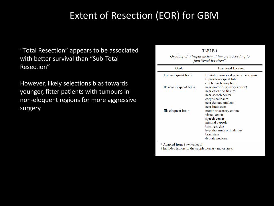
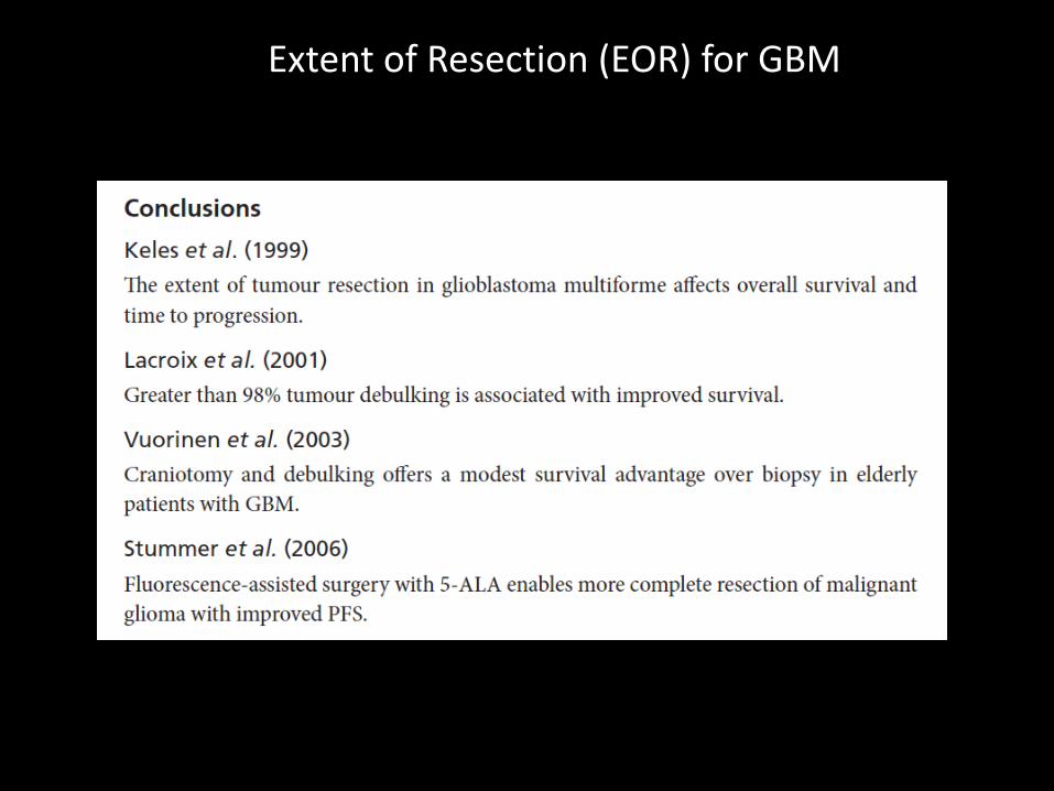
Extent of Resection (EOR) for GBM
“Total Resection” appears to be associated with better survival than “Sub-Total Resection”
However, likely selections bias towards younger, fitter patients with tumours in non-eloquent regions for more aggressive surgery
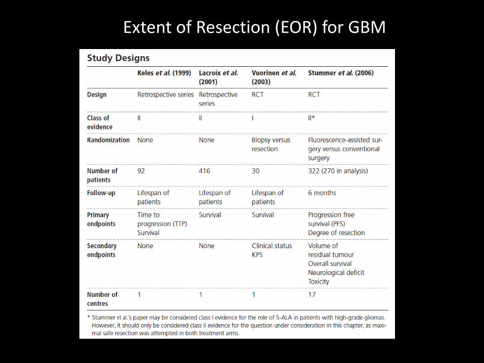
Extent of Resection (EOR) for GBM

Extent of Resection (EOR) for GBM
Extent of Resection (EOR) for GBM

Strategies to Maximise Extent of Resection
Need to augment surgical resection whilst minising risk to eloquent brain
• Cortical and subcortical mapping
• Functional neuronavigation
• Intrapoperative MRI
• Flourescence-guidance

Cortical and Subcortical Mapping
Intraoperative functional mapping of eloquent brain includes:
• Somatosensory-evoked potentials (SSEPs) to identify the central sulcus
• Awake stimulation for language function
• Subcortical and cortical stimulation mapping of motor pathways
Meta-analysis suggests that such strategies are effective
De Witt Hammer, 2012

Functional Neuronavigation
Functional MRI (fMRI) and diffusion tensor imaging (DTI) provide a means of identifying eloquent cortical areas and the course of subcortical fibretracts displaced by the lesion
One prospective randomisedtrial revealed reduced post-op motor deficits (15.3 % v 32.8%, p < 0.001).
Wu et al. 2007

Intraoperative MRI (ioMRI)
Kubben et al. 2011
One RCT suggested benefit – most likely due to correcting brain shift errors of intraoperative neuronavigation (Senft et al. 2011)
Meta-analysis shows level II evidence for effect
Likely that ioMRI will become more widespread


INTRACRANIAL TUMOURS

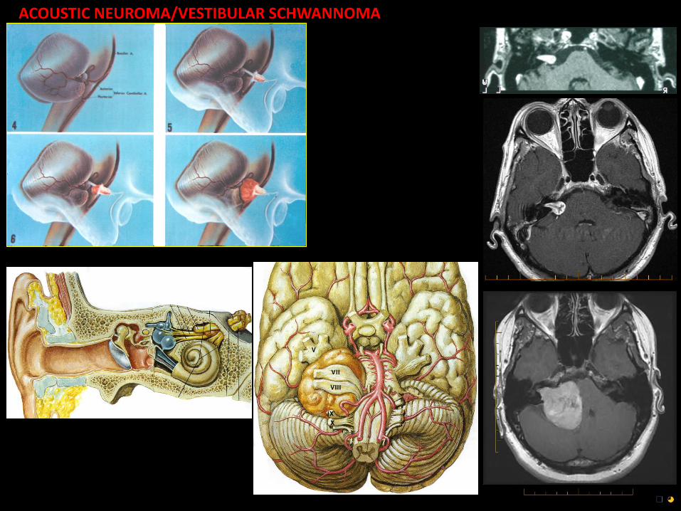
ACOUSTIC NEUROMA/VESTIBULAR SCHWANNOMA

Pituitary Surgery


1.que from Cushing abandoning the approach, talk re the cross-atlantic stories

Galen 150 AD“The Phlegm Gland”
Disposal of waste from ventricles to nose
Plate from Seventh Book of the Anatomic Fabricaof Vesalius in 1543
The oldest image in Western literature of the hypothalamic pituitary unit
A = pituitary glandB = infundibulum
C-F = ducts ‘draining the phlegm’

Professor Pierre MarieParis 19th Century
Described acromegaly and its association with pituitary tumours in
1886
Antonii De HaenVienna 18th Century
Described the amenorrhoea associated with a pituitary tumour
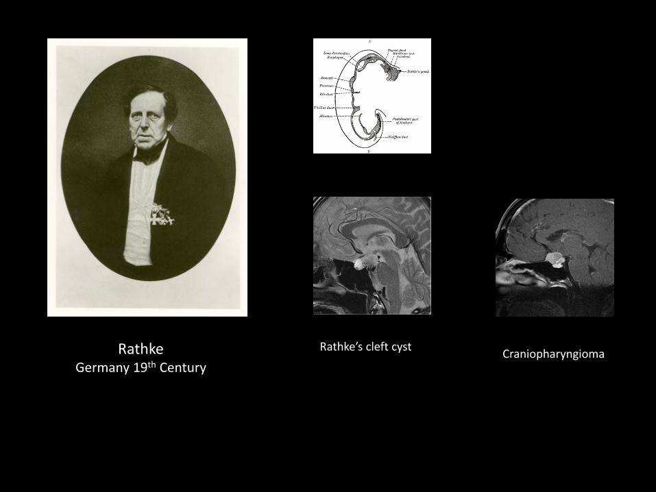
RathkeGermany 19th Century
Rathke’s cleft cystCraniopharyngioma

“The Master Gland”
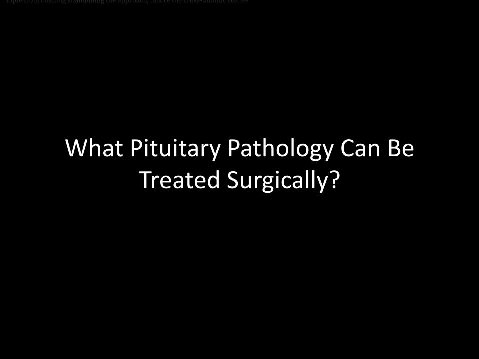
What Pituitary Pathology Can Be Treated Surgically?
1.que from Cushing abandoning the approach, talk re the cross-atlantic stories

Symptomatic SOL: Endocrine or Neurological Compromise
MeningiomaMacroadenoma
Rathke’s Craniopharyngioma

Hormone Secreting Lesions & Bleeds
Prolactinoma
Apoplexy
Acromegaly
Cushing’s
How Do We Get to the Pituitary Surgically?

Trans-Cranial Route to Sella
1st attempt at pituitary tumourresections in 1889
Subtemporal decompression – not always attempting to remove tumour
Treated 10 patients (mortality 20%)
Not reported until 1906
Sir Victor Horsley (1857-1916)London, UK

Trans-Cranial Route to Sella
F Krause (1857-1937)
Berlin, Germany
First frontal transcranial approach to sella turcica in 1904
But Mortality >50%
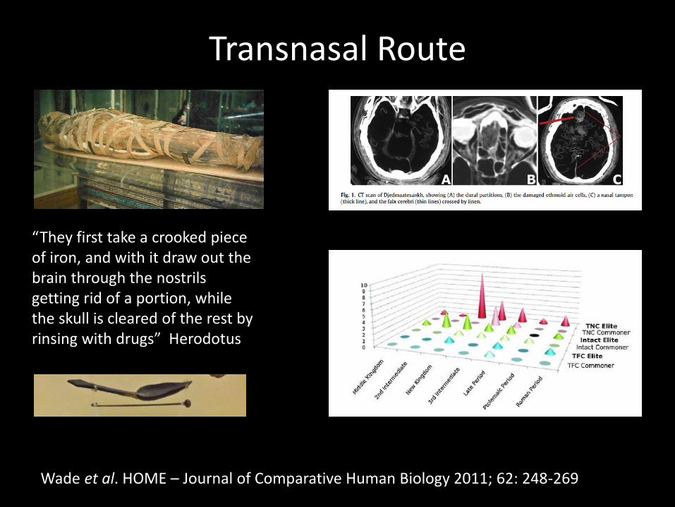
Transnasal Route
“They first take a crooked piece of iron, and with it draw out the brain through the nostrils getting rid of a portion, while the skull is cleared of the rest by rinsing with drugs” Herodotus
Wade et al. HOME – Journal of Comparative Human Biology 2011; 62: 248-269
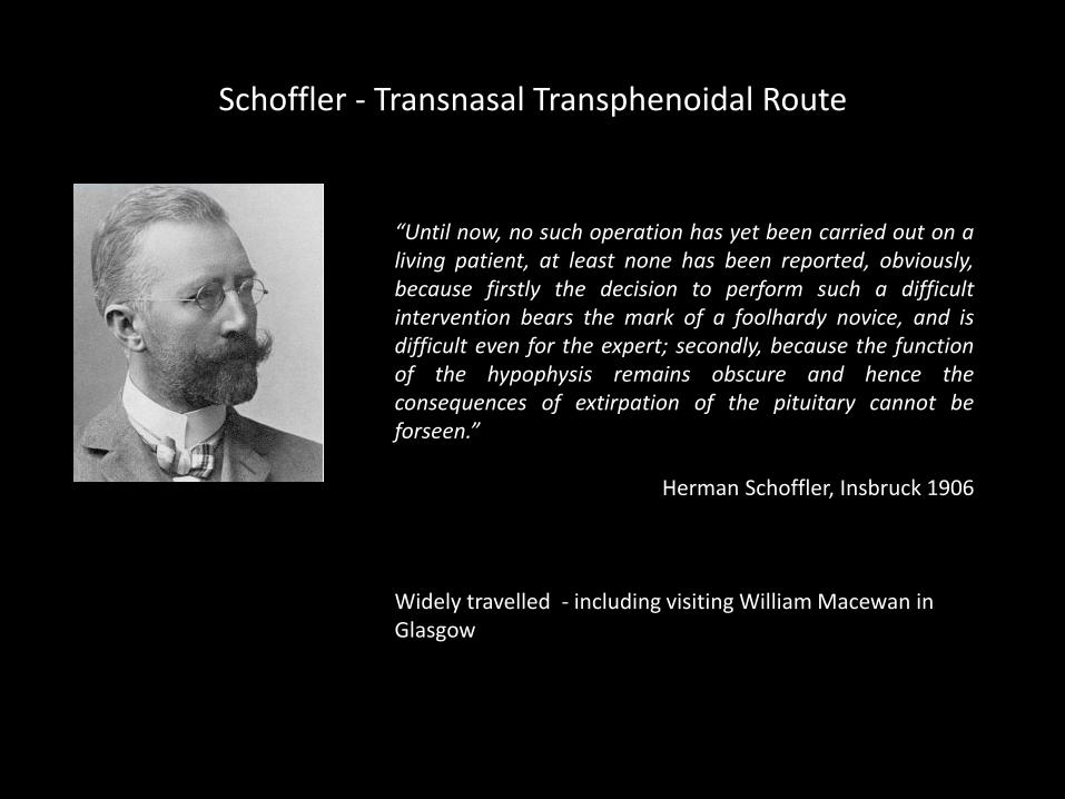
Schoffler - Transnasal Transphenoidal Route
“Until now, no such operation has yet been carried out on aliving patient, at least none has been reported, obviously,because firstly the decision to perform such a difficultintervention bears the mark of a foolhardy novice, and isdifficult even for the expert; secondly, because the functionof the hypophysis remains obscure and hence theconsequences of extirpation of the pituitary cannot beforseen.”
Herman Schoffler, Insbruck 1906
Widely travelled - including visiting William Macewan in Glasgow

Herman Schoffler - Innsbruck 1907
First transnasal trans-sphenoidal approach to sella turcica
Patient - 30 y/o maleL- 6/12 headaches and progressive visual loss- Hypogonadism- Skull radiograph showed an enlarged sella
Schoffler - Transnasal Transphenoidal Route

Attempted removal of tumour with a blunt spatula
Patient died at week 10 from hydrocephalus
Schoffler - Transnasal Transphenoidal Route
“the selection of the right cases for surgery is of primary importance for further development of the surgical technique”
Schoffler, 1907
Schoffler responsible for bringing the trans-sphenoidal route into the mainstream

Oskar Hirsch(Vienna)
Introduced endonasal procedure
4th June 1910
Cushing (Baltimore)
Modified Schoffler’sapproach
4th June 1910
Hirsch & Cushing – The Beginning and the End of the TSA?

Cushing did 231 procedures with 5.6% mortality and then abandoned it in favour of subfrontal approach (mortality 4.5% in his hands)
Cushing - Abandoned the TSA

Hirsch “The Obscure Voice in the Wilderness”
By 1937 (pre-antibiotic era) 277 patients with 5.4% mortality
By 1956 (post-antiobiotic era) 413 patients with 1.5% mortality
He emigrated to Boston in 1938 after expulsion from Austria by NazisNever recognised or allowed to operate independently

The Resurrection of the TSA
Trained under Cushing in 1920
Norman Dott (Edinburgh)
Continued TSA with >100 patients
0% mortality (antibiotics and perioperative steroids)
Low recurrence (DXT)
But never published results out of deference to Cushing

The Resurrection of the TSA
Trained under Dott
Published his results with 1000 patients
Introduced X-ray II and fluoroscopy to guide surgery
Gerard Guiot (Paris)
“In the evolutionof surgical techniques the introduction of the transcranial approach was progress, however then abandoning the transnasal approach, as a matter of fact, has been a step backwards”

The Resurrection of the TSA
Trained under Guiot
Hardy (Toronto)
Re-introduced the TSA to North America
Introduced the microscope to the TSA

Endoscopic Trans-Sphenoidal Approach (ETSA)
Kassam
(Pittsburgh, USA)
Cappabianca(Naples, Italy)
Hae-Dong Jho(Pittsburgh, USA)


Sphenoid Ostium


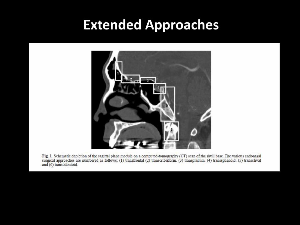
Extended Approaches

Extended Approaches
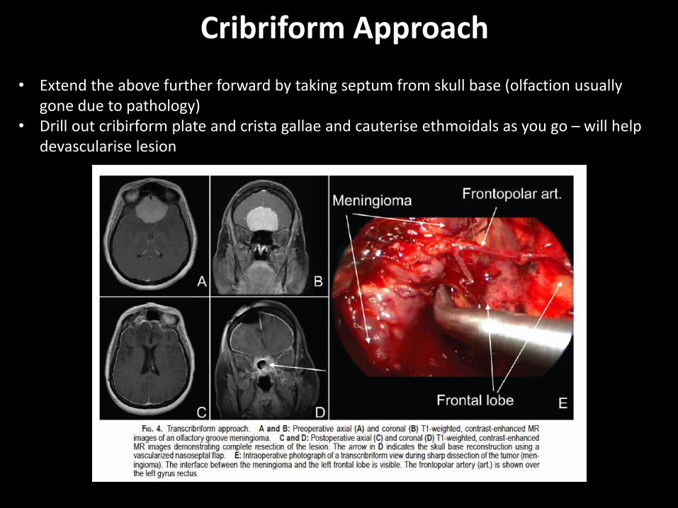
Cribriform Approach
• Extend the above further forward by taking septum from skull base (olfaction usually gone due to pathology)
• Drill out cribirform plate and crista gallae and cauterise ethmoidals as you go – will help devascularise lesion

Transclival Approach

Kassam – Endoscopic Odontoid Surgery

?

Alexander Monro Primus (1720-1758)Professor of Anatomy, Edinburgh
Alexander Monro Tertius (1798-1846)Professor of Anatomy Edinburgh
Alexander Monro Secundus (1754-1798)Professor of Anatomy Edinburgh
David Monro (1813-1877)Speaker of New Zealand House of Representatives
David Monro (1851-1933)Introduced Rugby to New Zealand in 1870